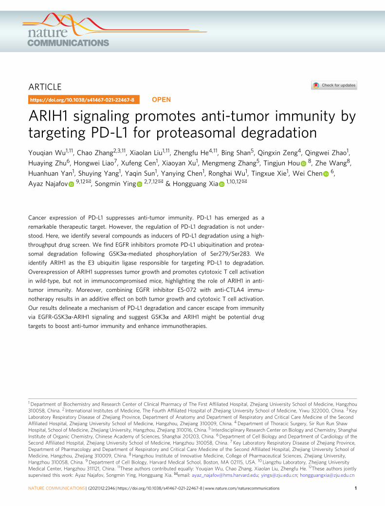
ARTICLE ARIH1 signaling promotes anti-tumor immunity by targeting PD-L1 for proteasomal degradation Youqian Wu 1,11 , Chao Zhang 2,3,11 , Xiaolan Liu 1,11 , Zhengfu He 4,11 , Bing Shan 5 , Qingxin Zeng 4 , Qingwei Zhao 1 , Huaying Zhu 6 , Hongwei Liao 7 , Xufeng Cen 1 , Xiaoyan Xu 1 , Mengmeng Zhang 5 , Tingjun Hou 8 , Zhe Wang 8 , Huanhuan Yan 1 , Shuying Yang 1 , Yaqin Sun 1 , Yanying Chen 1 , Ronghai Wu 1 , Tingxue Xie 1 , Wei Chen 6 , Ayaz Najafov 9,12 ✉ , Songmin Ying 2,7,12 ✉ & Hongguang Xia 1,10,12 ✉ Cancer expression of PD-L1 suppresses anti-tumor immunity. PD-L1 has emerged as a remarkable therapeutic target. However, the regulation of PD-L1 degradation is not under- stood. Here, we identify several compounds as inducers of PD-L1 degradation using a high- throughput drug screen. We find EGFR inhibitors promote PD-L1 ubiquitination and protea- somal degradation following GSK3α-mediated phosphorylation of Ser279/Ser283. We identify ARIH1 as the E3 ubiquitin ligase responsible for targeting PD-L1 to degradation. Overexpression of ARIH1 suppresses tumor growth and promotes cytotoxic T cell activation in wild-type, but not in immunocompromised mice, highlighting the role of ARIH1 in anti- tumor immunity. Moreover, combining EGFR inhibitor ES-072 with anti-CTLA4 immu- notherapy results in an additive effect on both tumor growth and cytotoxic T cell activation. Our results delineate a mechanism of PD-L1 degradation and cancer escape from immunity via EGFR-GSK3α-ARIH1 signaling and suggest GSK3α and ARIH1 might be potential drug targets to boost anti-tumor immunity and enhance immunotherapies. https://doi.org/10.1038/s41467-021-22467-8 OPEN 1 Department of Biochemistry and Research Center of Clinical Pharmacy of The First Affiliated Hospital, Zhejiang University School of Medicine, Hangzhou 310058, China. 2 International Institutes of Medicine, The Fourth Affiliated Hospital of Zhejiang University School of Medicine, Yiwu 322000, China. 3 Key Laboratory Respiratory Disease of Zhejiang Province, Department of Anatomy and Department of Respiratory and Critical Care Medicine of the Second Affiliated Hospital, Zhejiang University School of Medicine, Hangzhou, Zhejiang 310009, China. 4 Department of Thoracic Surgery, Sir Run Run Shaw Hospital, School of Medicine, Zhejiang University, Hangzhou, Zhejiang 310016, China. 5 Interdisciplinary Research Center on Biology and Chemistry, Shanghai Institute of Organic Chemistry, Chinese Academy of Sciences, Shanghai 201203, China. 6 Department of Cell Biology and Department of Cardiology of the Second Affiliated Hospital, Zhejiang University School of Medicine, Hangzhou 310058, China. 7 Key Laboratory Respiratory Disease of Zhejiang Province, Department of Pharmacology and Department of Respiratory and Critical Care Medicine of the Second Affiliated Hospital, Zhejiang University School of Medicine, Hangzhou, Zhejiang 310009, China. 8 Hangzhou Institute of Innovative Medicine, College of Pharmaceutical Sciences, Zhejiang University, Hangzhou 310058, China. 9 Department of Cell Biology, Harvard Medical School, Boston, MA 02115, USA. 10 Liangzhu Laboratory, Zhejiang University Medical Center, Hangzhou 311121, China. 11 These authors contributed equally: Youqian Wu, Chao Zhang, Xiaolan Liu, Zhengfu He. 12 These authors jointly supervised this work: Ayaz Najafov, Songmin Ying, Hongguang Xia. ✉ email: [email protected]; [email protected]; [email protected] NATURE COMMUNICATIONS | (2021)12:2346 | https://doi.org/10.1038/s41467-021-22467-8 | www.nature.com/naturecommunications 1 1234567890():,;

Welcome message from author
This document is posted to help you gain knowledge. Please leave a comment to let me know what you think about it! Share it to your friends and learn new things together.
Transcript
ARTICLE
ARIH1 signaling promotes anti-tumor immunity bytargeting PD-L1 for proteasomal degradationYouqian Wu1,11, Chao Zhang2,3,11, Xiaolan Liu1,11, Zhengfu He4,11, Bing Shan5, Qingxin Zeng4, Qingwei Zhao1,
Huaying Zhu6, Hongwei Liao7, Xufeng Cen1, Xiaoyan Xu1, Mengmeng Zhang5, Tingjun Hou 8, Zhe Wang8,
Huanhuan Yan1, Shuying Yang1, Yaqin Sun1, Yanying Chen1, Ronghai Wu1, Tingxue Xie1, Wei Chen 6,
Ayaz Najafov 9,12✉, Songmin Ying 2,7,12✉ & Hongguang Xia 1,10,12✉
Cancer expression of PD-L1 suppresses anti-tumor immunity. PD-L1 has emerged as a
remarkable therapeutic target. However, the regulation of PD-L1 degradation is not under-
stood. Here, we identify several compounds as inducers of PD-L1 degradation using a high-
throughput drug screen. We find EGFR inhibitors promote PD-L1 ubiquitination and protea-
somal degradation following GSK3α-mediated phosphorylation of Ser279/Ser283. We
identify ARIH1 as the E3 ubiquitin ligase responsible for targeting PD-L1 to degradation.
Overexpression of ARIH1 suppresses tumor growth and promotes cytotoxic T cell activation
in wild-type, but not in immunocompromised mice, highlighting the role of ARIH1 in anti-
tumor immunity. Moreover, combining EGFR inhibitor ES-072 with anti-CTLA4 immu-
notherapy results in an additive effect on both tumor growth and cytotoxic T cell activation.
Our results delineate a mechanism of PD-L1 degradation and cancer escape from immunity
via EGFR-GSK3α-ARIH1 signaling and suggest GSK3α and ARIH1 might be potential drug
targets to boost anti-tumor immunity and enhance immunotherapies.
https://doi.org/10.1038/s41467-021-22467-8 OPEN
1 Department of Biochemistry and Research Center of Clinical Pharmacy of The First Affiliated Hospital, Zhejiang University School of Medicine, Hangzhou310058, China. 2 International Institutes of Medicine, The Fourth Affiliated Hospital of Zhejiang University School of Medicine, Yiwu 322000, China. 3 KeyLaboratory Respiratory Disease of Zhejiang Province, Department of Anatomy and Department of Respiratory and Critical Care Medicine of the SecondAffiliated Hospital, Zhejiang University School of Medicine, Hangzhou, Zhejiang 310009, China. 4 Department of Thoracic Surgery, Sir Run Run ShawHospital, School of Medicine, Zhejiang University, Hangzhou, Zhejiang 310016, China. 5 Interdisciplinary Research Center on Biology and Chemistry, ShanghaiInstitute of Organic Chemistry, Chinese Academy of Sciences, Shanghai 201203, China. 6Department of Cell Biology and Department of Cardiology of theSecond Affiliated Hospital, Zhejiang University School of Medicine, Hangzhou 310058, China. 7 Key Laboratory Respiratory Disease of Zhejiang Province,Department of Pharmacology and Department of Respiratory and Critical Care Medicine of the Second Affiliated Hospital, Zhejiang University School ofMedicine, Hangzhou, Zhejiang 310009, China. 8 Hangzhou Institute of Innovative Medicine, College of Pharmaceutical Sciences, Zhejiang University,Hangzhou 310058, China. 9 Department of Cell Biology, Harvard Medical School, Boston, MA 02115, USA. 10 Liangzhu Laboratory, Zhejiang UniversityMedical Center, Hangzhou 311121, China. 11These authors contributed equally: Youqian Wu, Chao Zhang, Xiaolan Liu, Zhengfu He. 12These authors jointlysupervised this work: Ayaz Najafov, Songmin Ying, Hongguang Xia. ✉email: [email protected]; [email protected]; [email protected]
NATURE COMMUNICATIONS | (2021) 12:2346 | https://doi.org/10.1038/s41467-021-22467-8 |www.nature.com/naturecommunications 1
1234
5678
90():,;
Programmed death ligand-1 (PD-L1) is constitutivelyexpressed on the surface of cancer cells1–4. The interactionbetween PD-L1 and its receptor, programmed death
protein-1 (PD-1), which is mainly expressed on the surface ofT cells, results in cancer cell evasion from immune surveillance5.PD-1 and PD-L1 have become important immune checkpointblockade immunotherapy targets for different types of cancers5,6.However, resistance to such immunotherapy approaches is pre-valent and the mechanisms of resistance are not well understood.
Proteasomal degradation of PD-L1 has been reported to bepromoted by cyclin D-CDK4-mediated phosphorylation followedby Cullin 3SPOP-dependent ubiquitination7, as well as glycogensynthase kinase 3-β (GSK3β)-mediated phosphorylation followedby β-TrCP-dependent ubiquitination8. However, the dynamicubiquitin modification of PD-L1 for proteasomal degradationshould mostly occur in the intracellular segment of PD-L1, whichis not addressed before. Ariadne-1 homolog (ARIH1) is amember of the Ariadne family of E3 ubiquitin ligases with acognate E2 enzyme UBCH79–11. ARIH1 is known to play a rolein protein translation regulation in response to DNA damage andto ubiquitinate EIF4E212. The role of ARIH1 in PD-L1 degra-dation or anti-tumor immunity is not known.
Epidermal growth factor receptor (EGFR) is a major receptortyrosine kinase, frequently overactivated in cancers, with estab-lished roles in cell growth and survival13–15. Patients with EGFR-mutant-driven tumors develop resistance to EGFR tyrosinekinase inhibitor treatments, which are usually accompanied bythe acquisition of EGFR mutations16. Clinical retrospectivessuggest that EGFR mutations are associated with low responserates to immune therapies in non-small cell lung cancer17,18.Importantly, EGFR mutations are associated with increased PD-L1 expression19–21. EGFR activation promotes PD-L1 expressionvia Janus kinase/signal transducer and activator of transcription 3(JAK/STAT3) signaling pathway22 and inhibits its degradationvia phosphorylation mediated by GSK3β8 in the extracellular partof PD-L1, contributing to cancer escape from anti-tumorimmunity. These findings strongly indicate that EGFR works asa critical regulator of tumor immune surveillance via regulation ofPD-L1 expression, but the mechanism of this regulation is notfully understood, especially as the reported phosphorylation sitesare in the extracellular part of PD-L1.
In this study, we screened a panel of 2125 Food and DrugAdministration (FDA)-approved drugs or drug candidates andfound that ES-072, a third-generation EGFR inhibitor, induced apotent degradation of PD-L1. Our mechanistic findings suggestthat inhibition of EGFR activates GSK3α by suppressing AKTactivity, which subsequently promotes the phosphorylation at theintracellular Ser279 and Ser283 residues of PD-L1, leading toARIH1-mediated ubiquitination and proteasome-mediateddegradation. Cancer-associated mutation of ARIH1 compro-mises ubiquitination of PD-L1. Together, our results suggestARIH1 is the E3 ligase for PD-L1, which could lead to thedevelopment of therapeutic strategies to overcome immu-notherapy resistance in cancers and enhance checkpoint blockadetherapy efficacy.
ResultsA high-throughput screen of 2125 FDA-approved drugs ordrug candidates identifies promoters of PD-L1 degradation. Toestablish a fluorescence-based high-throughput screening proto-col for the membrane levels of endogenous PD-L1, we used aphycoerythrin-conjugated anti-PD-L1 antibody and interferon-γ(IFNγ)-treated U937 cells (histiocytic lymphosarcoma cell line).IFNγ enhances the basal expression levels of PD-L1, allowing fora wider dynamic range and, thus, a better screening system23,24.
As a positive control for drug-induced PD-L1 level decrease, weused Ruxolitinib, a JAK1/2 inhibitor25 (Supplementary Fig. 1a).As expected, PD-L1 levels were induced by IFNγ and blocked byRuxolitinib treatment (Supplementary Fig. 1b–d).
Out of 2125 FDA-approved drugs or drug candidates screened,160 were found to reduce the membranal PD-L1 levels(Supplementary Fig. 1e and Supplementary Table 1). Weclassified the positive hits according to the signaling pathwaysthey were known to be involved in, which included the JAK/STAT pathway, the phosphatidylinositol 3-kinase (PI3K)/AKT/mammalian target of the rapamycin pathway, pathways thatregulate the cell cycle, and protein tyrosine kinases (Fig. 1a).The screen confirmed the existing knowledge about PD-L1regulation, as 13 different JAK inhibitors were found todecrease plasma membrane PD-L1 levels by more than 25%; thisalso indicated that the screen setup is valid, as JAK was the topdrug target inhibition of which resulted in the PD-L1 leveldecrease.
On the other hand, EGFR has also been strongly linked to PD-L1protein level regulation. From the EGFR inhibitors that promotePD-L1 level decrease, AZD9291 and ES-072 were selected for ourscreen follow-up and mechanistic studies to investigate what drivesPD-L1 degradation downstream of EGFR inhibition. AZD9291,Osimertinib, is a potent and selective mutant EGFR (L858R/T790M) inhibitor, which is widely used in clinic26,27. ES-072 is athird-generation EGFR inhibitor designed by our team to overcomedrug-resistance-induced EGFR mutation L858R/T790M. Confirm-ing the screen findings, both ES-072 and AZD9291 dramaticallyreduced the IFNγ-induced membranal PD-L1 levels in U937 cellsfollowing treatment with the compounds, as judged by westernblotting and flow cytometry results (Fig. 1b–d). The effect of theEGFR inhibitors on PD-L1 levels was time-dependent, reaching aplateau following 12 h of treatment (Fig. 1e–g). Similar findingswere seen in H1975 cells (Supplementary Fig. 2a–d) andinterleukin-4 (IL-4)-treated peritoneal-derived macrophages(PDMs) (Supplementary Fig. 2e–g).
EGFR inhibitors are known to promote PD-L1 degradation viathe GSK3α/β-TrCP pathway8. Surprisingly, contrary to theprevious reports, we found that knockdown of β-TrCP did notfully block the reduction of PD-L1 levels induced following EGFRinhibition (Fig. 1h, i), whereas GSK3α/β inhibitor LY2090314 didblock this reduction (Fig. 1j–l). LY2090314 was found to be highlyspecific for GSK3α with an half maximal inhibitory concentra-tion (IC50) value of 0.87 ± 0.09 nM (Supplementary Table 2).These results indicated that a β-TrCP-independent, yet GSK3α/β-dependent mechanism regulating PD-L1 levels downstream ofEGFR should exist.
ES-072 promotes proteasomal PD-L1 degradation via EGFRinhibition. The rest of our studies were continued using ES-072due to its stability and specificity (Supplementary Table 3). As,upon EGFR inhibition, PD-L1 has been shown to be degraded viathe proteasome, we first determined whether ES-072 treatmentalso results in a proteasome-dependent degradation of PD-L1.Both western blotting and flow cytometry analysis of plasmamembrane PD-L1 levels confirmed that EGFR inhibition resultsin a proteasome-mediated PD-L1 degradation, as the decrease inthe PD-L1 levels induced by ES-072 was rescued by the protea-some inhibitor MG132 (Supplementary Fig. 3a–c).
In accord with previous reports28,29, EGF treatment promotedan increase of PD-L1 levels (Supplementary Fig. 3d–f), whereassmall interfering RNA (siRNA)-mediated knockdown ofEGFR decreased PD-L1 levels (Supplementary Fig. 3g). ES-072was found to be highly specific for EGFR in an in vitrokinase-profiling screen, where it was found to inhibit wild-type,
ARTICLE NATURE COMMUNICATIONS | https://doi.org/10.1038/s41467-021-22467-8
2 NATURE COMMUNICATIONS | (2021) 12:2346 | https://doi.org/10.1038/s41467-021-22467-8 | www.nature.com/naturecommunications
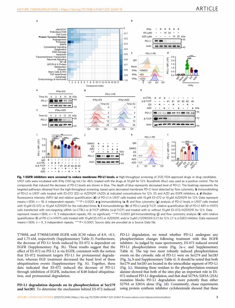
T790M, and T790M/L858R EGFR with IC50 values of 8.9, <0.5,and 1.75 nM, respectively (Supplementary Table 3). Furthermore,the decrease of PD-L1 levels induced by ES-072 is dependent onEGFR (Supplementary Fig. 3h). These results suggest that theeffect of ES-072 on PD-L1 is via EGFR, consistent with the notionthat ES-072 treatment targets PD-L1 for proteasomal degrada-tion, whereas EGF treatment decreased the basal level of theseubiquitination events (Supplementary Fig. 3i). Together, thesedata indicated that ES-072 induced the decrease of PD-L1through inhibition of EGFR, induction of K48-linked ubiquitina-tion, and proteasomal degradation.
PD-L1 degradation depends on its phosphorylation at Ser279and Ser283. To determine the mechanism behind ES-072-induced
PD-L1 degradation, we tested whether PD-L1 undergoes anyphosphorylation changes following treatment with this EGFRinhibitor. As judged by mass spectrometry, ES-072 induced severalPD-L1 phosphorylation events (Fig. 2a–c and SupplementaryTable 4). The top two most robustly induced phosphorylationevents on the cytosolic side of PD-L1 were on Ser279 and Ser283(Fig. 2a, b and Supplementary Table 4). It should be noted that bothSer279 and Ser283 are located in the intracellular segment of PD-L1(Fig. 2c). Mutating these residues to the phosphorylation-resistantalanine showed that both of the sites play an important role in ES-072-induced PD-L1 degradation, and that dual S279A; S283A (2SA)mutation blocks PD-L1 degradation more potently than eitherS279A or S283A alone (Fig. 2d). Consistently, chase experimentsusing protein synthesis inhibitor cycloheximide showed that these
Fig. 1 EGFR inhibitors were screened to reduce membrane PD-L1 levels. a High-throughput screening of 2125 FDA-approved drugs or drug candidates.U937 cells were incubated with IFNγ (100 ng/mL) for 48 h, treated with the drugs at 10 μM for 12 h. Ruxolitinib (Rux) was used as a positive control. The hitcompounds that induced the decrease of PD-L1 levels are shown in blue. The depth of blue represents decreased level of PD-L1. The heatmap represents thetargeted pathways obtained from the high-throughput screening, based upon decreased membrane PD-L1 level detected by flow cytometry. b Immunoblottingof PD-L1 in U937 cells treated with ES-072 (ES) or AZD9291 (AZD) at indicated concentrations for 12 h. ES and AZD are EGFR inhibitors. c, d Medianfluorescence intensity (MFI) (c) and relative quantification (d) of PD-L1 in U937 cells treated with 10 μM ES-072 or 10 μM AZD9291 for 12 h. Data representmeans ± SEM, n= 18, 6 independent repeats, ****P < 0.0001. e–g Immunoblotting (e, f) and flow cytometry (g) analysis of PD-L1 levels in U937 cells treatedwith 10 μM ES-072 or 10 μM AZD9291 for the indicated times. h, i Immunoblottings (h) of PD-L1 and β-TrCP, relative quantification (i) of PD-L1 MFI in H1975cells transfected with non-targeting siRNA (si-CTRL) or β-TrCP siRNAs (si-β-TrCP) and treated with or without 10 μM ES-072/AZD9291 for 12 h. Datarepresent means ± SEM, n= 9, 3 independent repeats, NS: no significant; ****P < 0.0001. j–l Immunoblotting (j) and flow cytometry analysis (k) with relativequantification (l) of PD-L1 in H1975 cells treated with 10 μM ES-072 or AZD9291, and/or 5 μM LY2090314 (LY) for 12 h. LY is a GSK3 inhibitor. Data representmeans ± SEM, n= 9, 3 independent repeats, ****P < 0.0001. Source data are provided as a Source Data file.
NATURE COMMUNICATIONS | https://doi.org/10.1038/s41467-021-22467-8 ARTICLE
NATURE COMMUNICATIONS | (2021) 12:2346 | https://doi.org/10.1038/s41467-021-22467-8 |www.nature.com/naturecommunications 3
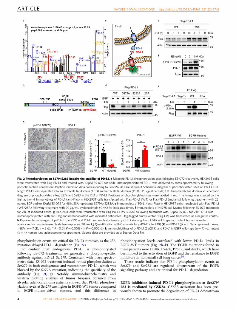
phosphorylation events are critical for PD-L1 turnover, as the 2SAmutation delayed PD-L1 degradation (Fig. 2e).
To confirm that endogenous PD-L1 is phosphorylatedfollowing ES-072 treatment, we generated a phospho-specificantibody against PD-L1 Ser279. Consistent with mass spectro-metry data, ES-072 treatment induced robust phosphorylation atSer279 in both endogenous and recombinant PD-L1, which wasblocked by the S279A mutation, indicating the specificity of theantibody (Fig. 2f, g). Notably, immunohistochemistry andwestern blotting analysis of tumor biopsies obtained fromalveolar adenocarcinoma patients showed that PD-L1 phosphor-ylation levels at Ser279 are higher in EGFR-WT tumors comparedto EGFR-mutant-driven tumors, and this difference in
phosphorylation levels correlated with lower PD-L1 levels inEGFR-WT tumors (Fig. 2h–k). The EGFR mutations found inthese patients were L858R, E542K, P753R, and Δex19, which havebeen linked to the activation of EGFR and the resistance to EGFRinhibitors in non-small cell lung cancer30.
These results indicate that PD-L1 phosphorylation events atSer279 and Ser283 are regulated downstream of the EGFRsignaling pathway and are critical for PD-L1 degradation.
EGFR inhibition-induced PD-L1 phosphorylation at Ser279/283 is mediated by GSK3α. GSK3β activation has been pre-viously shown to promote the degradation of PD-L1 downstream
Fig. 2 Phosphorylation on S279/S283 impairs the stability of PD-L1. a Mapping PD-L1 phosphorylation sites following ES-072 treatment. HEK293T cellswere transfected with Flag-PD-L1 and treated with 10 μM ES-072 for 48 h. Immunoprecipitated PD-L1 was analyzed by mass spectrometry followingphosphopeptide enrichment. Peptide ionization data corresponding to Ser279/283 are shown. b Schematic diagram of phosphorylated sites on PD-L1. Full-length PD-L1 was separated into an extracellular domain (ECD) and intracellular domain (ICD). SP: signal peptide; TM: transmembrane domain. c Schematicdiagram of phosphorylated sites, S279 and S283 in the ICD of PD-L1. Positions of phosphorylated sites were labeled in red. This image was created by thefirst author. d Immunoblots of PD-L1 (anti-Flag) in HEK293T cells transfected with Flag-PD-L1 (WT) or Flag-PD-L1 (mutants) following treatment with 25ng/mL EGF and/or 10 μM ES-072 for 48 h, 2SA represents S279A/S283A. e Immunoblots of PD-L1 (anti-Flag) in HEK293T cells transfected with Flag-PD-L1(WT/2SA) following treatment with 20 μg/mL cycloheximide (CHX) for indicated times. f Immunoblots of H1975 cell lysates following ES-072 treatmentfor 2 h, at indicated doses. g HEK293T cells were transfected with Flag-PD-L1 (WT/2SA) following treatment with 10 μM ES-072 for 2 h; PD-L1 wasimmunoprecipitated with anti-Flag and immunoblotted with indicated antibodies. Flag-tagged empty vector (Flag-EV) was transfected as a negative control.h Representative images of p-PD-L1 (Ser279) and PD-L1 immunohistochemistry (IHC) staining from EGFR wild-type vs. mutant human alveolaradenocarcinoma specimens. Scale bars represent 50 μm. i, j Quantification of IHC analysis for p-PD-L1 (Ser279) (i) and PD-L1 (j) in h. Data represent means± SEM, n= 7 (i), n= 5 (j), **P < 0.01, P= 0.0013 (i); P= 0.002 (j). k Immunoblottings of p-PD-L1 (Ser279) and PD-L1 in EGFR wild-type (n= 4) vs. mutant(n= 4) human lung adenocarcinoma specimens. Source data are provided as a Source Data file.
ARTICLE NATURE COMMUNICATIONS | https://doi.org/10.1038/s41467-021-22467-8
4 NATURE COMMUNICATIONS | (2021) 12:2346 | https://doi.org/10.1038/s41467-021-22467-8 | www.nature.com/naturecommunications
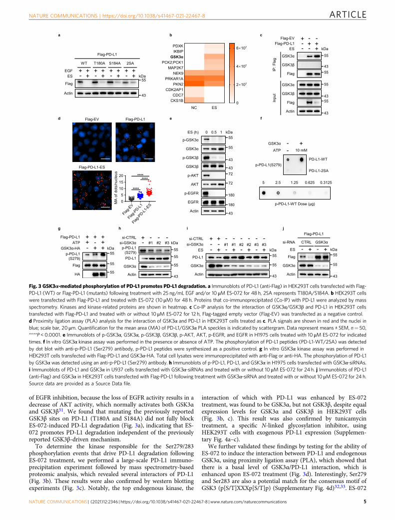
of EGFR inhibition, because the loss of EGFR activity results in adecrease of AKT activity, which normally activates both GSK3αand GSK3β31. We found that mutating the previously reportedGSK3β sites on PD-L1 (T180A and S184A) did not fully blockES-072-induced PD-L1 degradation (Fig. 3a), indicating that ES-072 promotes PD-L1 degradation independent of the previouslyreported GSK3β-driven mechanism.
To determine the kinase responsible for the Ser279/283phosphorylation events that drive PD-L1 degradation followingES-072 treatment, we performed a large-scale PD-L1 immuno-precipitation experiment followed by mass spectrometry-basedproteomic analysis, which revealed several interactors of PD-L1(Fig. 3b). These results were also confirmed by western blottingexperiments (Fig. 3c). Notably, the top endogenous kinase, the
interaction of which with PD-L1 was enhanced by ES-072treatment, was found to be GSK3α, but not GSK3β, despite equalexpression levels for GSK3α and GSK3β in HEK293T cells(Fig. 3b, c). This result was also confirmed by tunicamycintreatment, a specific N-linked glycosylation inhibitor, usingHEK293T cells with exogenous PD-L1 expression (Supplemen-tary Fig. 4a–c).
We further validated these findings by testing for the ability ofES-072 to induce the interaction between PD-L1 and endogenousGSK3α, using proximity ligation assay (PLA), which showed thatthere is a basal level of GSK3α/PD-L1 interaction, which isenhanced upon ES-072 treatment (Fig. 3d). Interestingly, Ser279and Ser283 are also a potential match for the consensus motif ofGSK3 (p[S/T]XXXp[S/T]p) (Supplementary Fig. 4d)32,33. ES-072
Fig. 3 GSK3α-mediated phosphorylation of PD-L1 promotes PD-L1 degradation. a Immunoblots of PD-L1 (anti-Flag) in HEK293T cells transfected with Flag-PD-L1 (WT) or Flag-PD-L1 (mutants) following treatment with 25 ng/mL EGF and/or 10 μM ES-072 for 48 h, 2SA represents T180A/S184A. b HEK293T cellswere transfected with Flag-PD-L1 and treated with ES-072 (10 μM) for 48 h. Proteins that co-immunoprecipitated (Co-IP) with PD-L1 were analyzed by massspectrometry. Kinases and kinase-related proteins are shown in heatmap. c Co-IP analysis for the interaction of GSK3α/GSK3β and PD-L1 in HEK293T cellstransfected with Flag-PD-L1 and treated with or without 10 μM ES-072 for 12 h, Flag-tagged empty vector (Flag-EV) was transfected as a negative control.d Proximity ligation assay (PLA) analysis for the interaction of GSK3α and PD-L1 in HEK293T cells treated as c. PLA signals are shown in red and the nuclei inblue; scale bar, 20 μm. Quantification for the mean area (MA) of PD-L1/GSK3α PLA speckles is indicated by scattergram. Data represent means ± SEM, n= 50,****P < 0.0001. e Immunoblots of p-GSK3α, GSK3α, p-GSK3β, GSK3β, p-AKT, AKT, p-EGFR, and EGFR in H1975 cells treated with 10 μM ES-072 for indicatedtimes. f In vitro GSK3α kinase assay was performed in the presence or absence of ATP. The phosphorylation of PD-L1 peptides (PD-L1-WT/2SA) was detectedby dot blot with anti-p-PD-L1 (Ser279) antibody, p-PD-L1 peptides were synthesized as a positive control. g In vitro GSK3α kinase assay was performed inHEK293T cells transfected with Flag-PD-L1 and GSK3α-HA. Total cell lysates were immunoprecipitated with anti-Flag or anti-HA. The phosphorylation of PD-L1by GSK3α was detected using an anti-p-PD-L1 (Ser279) antibody. h Immunoblots of p-PD-L1, PD-L1, and GSK3α in H1975 cells transfected with GSK3α-siRNAs.i Immunoblots of PD-L1 and GSK3α in U937 cells transfected with GSK3α-siRNAs and treated with or without 10 μM ES-072 for 24 h. j Immunoblots of PD-L1(anti-Flag) and GSK3α in HEK293T cells transfected with Flag-PD-L1 following treatment with GSK3α-siRNA and treated with or without 10 μM ES-072 for 24 h.Source data are provided as a Source Data file.
NATURE COMMUNICATIONS | https://doi.org/10.1038/s41467-021-22467-8 ARTICLE
NATURE COMMUNICATIONS | (2021) 12:2346 | https://doi.org/10.1038/s41467-021-22467-8 |www.nature.com/naturecommunications 5
treatment promoted GSK3α activation in an EGFR- and AKT-dependent manner, as judged by the decrease of phospho-GSK3αat Ser21, which is a marker of GSK3α inhibition (Fig. 3e). ES-072showed a higher activation effect on GSK3α when compared tothe other two EGFR inhibitors, as judged by loss of the inhibitoryphosphorylation of GSK3α (Supplementary Fig. 4e). Thesefindings suggest that GSK3α could be the kinase that directlyphosphorylates PD-L1 at Ser279/283 following EGFR inhibitionby ES-072.
Consistent with this hypothesis, purified GSK3α robustlyphosphorylated wild-type, but not 2SA synthetic peptidesin vitro, at Ser279, which we used to represent the intracellularregion of PD-L1 (Fig. 3f). Furthermore, purified GSK3α alsorobustly phosphorylated purified PD-L1 in vitro, at Ser279(Fig. 3g). Consistently, knockdown of GSK3α, but not GSK3β,blocked the phosphorylation of PD-L1 at Ser279 (Fig. 3h andSupplementary Fig. 4f). These results indicate that GSK3α directlyphosphorylates PD-L1 at Ser279. In agreement with the datashowing that Ser279/Ser283 phosphorylation is important for ES-072-induced PD-L1 degradation, and that these sites arephosphorylated by GSK3α, knockdown of GSK3α in U937 andH1975 cells partially rescued PD-L1 degradation induced by ES-072 (Fig. 3i and Supplementary Fig. 4g). Similar results were seenwhen GSK3α was knocked down in HEK293T cells overexpressedwith Flag-PD-L1 (Fig. 3j).
Overall, these findings showed that ES-072 treatment results ininhibition of EGFR and activation of GSK3α, which phosphor-ylates PD-L1 at its cytosolic Ser279 and Ser283 residues to targetPD-L1 for proteasomal degradation. Our results also indicate thatthis mechanism is distinct from the previously reportedphosphorylation of PD-L1 by GSK3β at T180 and S184, at theextracellular region of PD-L18.
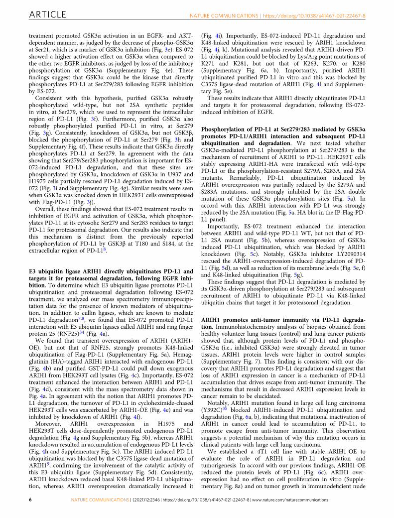
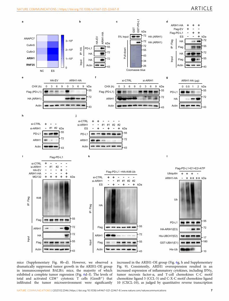
E3 ubiquitin ligase ARIH1 directly ubiquitinates PD-L1 andtargets it for proteasomal degradation, following EGFR inhi-bition. To determine which E3 ubiquitin ligase promotes PD-L1ubiquitination and proteasomal degradation following ES-072treatment, we analyzed our mass spectrometry immunoprecipi-tation data for the presence of known mediators of ubiquitina-tion. In addition to cullin ligases, which are known to mediatePD-L1 degradation7,8, we found that ES-072 promoted PD-L1interaction with E3 ubiquitin ligases called ARIH1 and ring fingerprotein 25 (RNF25)34 (Fig. 4a).
We found that transient overexpression of ARIH1 (ARIH1-OE), but not that of RNF25, strongly promotes K48-linkedubiquitination of Flag-PD-L1 (Supplementary Fig. 5a). Hemag-glutinin (HA)-tagged ARIH1 interacted with endogenous PD-L1(Fig. 4b) and purified GST-PD-L1 could pull down exogenousARIH1 from HEK293T cell lysates (Fig. 4c). Importantly, ES-072treatment enhanced the interaction between ARIH1 and PD-L1(Fig. 4d), consistent with the mass spectrometry data shown inFig. 4a. In agreement with the notion that ARIH1 promotes PD-L1 degradation, the turnover of PD-L1 in cycloheximide-chasedHEK293T cells was exacerbated by ARIH1-OE (Fig. 4e) and wasinhibited by knockdown of ARIH1 (Fig. 4f).
Moreover, ARIH1 overexpression in H1975 andHEK293T cells dose-dependently promoted endogenous PD-L1degradation (Fig. 4g and Supplementary Fig. 5b), whereas ARIH1knockdown resulted in accumulation of endogenous PD-L1 levels(Fig. 4h and Supplementary Fig. 5c). The ARIH1-induced PD-L1ubiquitination was blocked by the C357S ligase-dead mutation ofARIH19, confirming the involvement of the catalytic activity ofthis E3 ubiquitin ligase (Supplementary Fig. 5d). Consistently,ARIH1 knockdown reduced basal K48-linked PD-L1 ubiquitina-tion, whereas ARIH1 overexpression dramatically increased it
(Fig. 4i). Importantly, ES-072-induced PD-L1 degradation andK48-linked ubiquitination were rescued by ARIH1 knockdown(Fig. 4j, k). Mutational analysis revealed that ARIH1-driven PD-L1 ubiquitination could be blocked by Lys/Arg point mutations ofK271 and K281, but not that of K263, K270, or K280(Supplementary Fig. 6a, b). Importantly, purified ARIH1ubiquitinated purified PD-L1 in vitro and this was blocked byC357S ligase-dead mutation of ARIH1 (Fig. 4l and Supplemen-tary Fig. 5e).
These results indicate that ARIH1 directly ubiquitinates PD-L1and targets it for proteasomal degradation, following ES-072-induced inhibition of EGFR.
Phosphorylation of PD-L1 at Ser279/283 mediated by GSK3αpromotes PD-L1/ARIH1 interaction and subsequent PD-L1ubiquitination and degradation. We next tested whetherGSK3α-mediated PD-L1 phosphorylation at Ser279/283 is themechanism of recruitment of ARIH1 to PD-L1. HEK293T cellsstably expressing ARIH1-HA were transfected with wild-typePD-L1 or the phosphorylation-resistant S279A, S283A, and 2SAmutants. Remarkably, PD-L1 ubiquitination induced byARIH1 overexpression was partially reduced by the S279A andS283A mutations, and strongly inhibited by the 2SA doublemutation of these GSK3α phosphorylation sites (Fig. 5a). Inaccord with this, ARIH1 interaction with PD-L1 was stronglyreduced by the 2SA mutation (Fig. 5a, HA blot in the IP-Flag-PD-L1 panel).
Importantly, ES-072 treatment enhanced the interactionbetween ARIH1 and wild-type PD-L1 WT, but not that of PD-L1 2SA mutant (Fig. 5b), whereas overexpression of GSK3αinduced PD-L1 ubiquitination, which was blocked by ARIH1knockdown (Fig. 5c). Notably, GSK3α inhibitor LY2090314rescued the ARIH1-overexpression-induced degradation of PD-L1 (Fig. 5d), as well as reduction of its membrane levels (Fig. 5e, f)and K48-linked ubiquitination (Fig. 5g).
These findings suggest that PD-L1 degradation is mediated byits GSK3α-driven phosphorylation at Ser279/283 and subsequentrecruitment of ARIH1 to ubiquitinate PD-L1 via K48-linkedubiquitin chains that target it for proteasomal degradation.
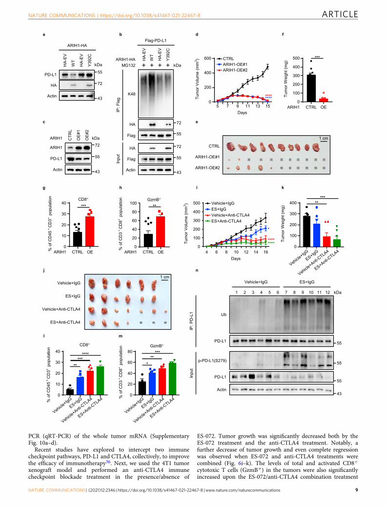
ARIH1 promotes anti-tumor immunity via PD-L1 degrada-tion. Immunohistochemistry analysis of biopsies obtained fromhealthy volunteer lung tissues (control) and lung cancer patientsshowed that, although protein levels of PD-L1 and phospho-GSK3α (i.e., inhibited GSK3α) were strongly elevated in tumortissues, ARIH1 protein levels were higher in control samples(Supplementary Fig. 7). This finding is consistent with our dis-covery that ARIH1 promotes PD-L1 degradation and suggest thatloss of ARIH1 expression in cancer is a mechanism of PD-L1accumulation that drives escape from anti-tumor immunity. Themechanisms that result in decreased ARIH1 expression levels incancer remain to be elucidated.
Notably, ARIH1 mutation found in large cell lung carcinoma(Y392C)35 blocked ARIH1-induced PD-L1 ubiquitination anddegradation (Fig. 6a, b), indicating that mutational inactivation ofARIH1 in cancer could lead to accumulation of PD-L1, topromote escape from anti-tumor immunity. This observationsuggests a potential mechanism of why this mutation occurs inclinical patients with large cell lung carcinoma.
We established a 4T1 cell line with stable ARIH1-OE toevaluate the role of ARIH1 in PD-L1 degradation andtumorigenesis. In accord with our previous findings, ARIH1-OEreduced the protein levels of PD-L1 (Fig. 6c). ARIH1 over-expression had no effect on cell proliferation in vitro (Supple-mentary Fig. 8a) and on tumor growth in immunodeficient nude
ARTICLE NATURE COMMUNICATIONS | https://doi.org/10.1038/s41467-021-22467-8
6 NATURE COMMUNICATIONS | (2021) 12:2346 | https://doi.org/10.1038/s41467-021-22467-8 | www.nature.com/naturecommunications
mice (Supplementary Fig. 8b–d). However, we observed adramatically suppressed tumor growth in the ARIH1-OE groupin immunocompetent BALB/c mice, the majority of whichexhibited a complete tumor regression (Fig. 6d–f). The levels oftotal and activated CD8+ cytotoxic T cells (GzmB+) thatinfiltrated the tumor microenvironment were significantly
increased in the ARIH1-OE group (Fig. 6g, h and SupplementaryFig. 9). Consistently, ARIH1 overexpression resulted in anincreased expression of inflammatory cytokines, including IFNγ,tumor necrosis factor-α, and T-cell chemokines C-C motifchemokine ligand 5 (CCL-5) and C-X-C motif chemokine ligand10 (CXCL-10), as judged by quantitative reverse transcription
NATURE COMMUNICATIONS | https://doi.org/10.1038/s41467-021-22467-8 ARTICLE
NATURE COMMUNICATIONS | (2021) 12:2346 | https://doi.org/10.1038/s41467-021-22467-8 |www.nature.com/naturecommunications 7
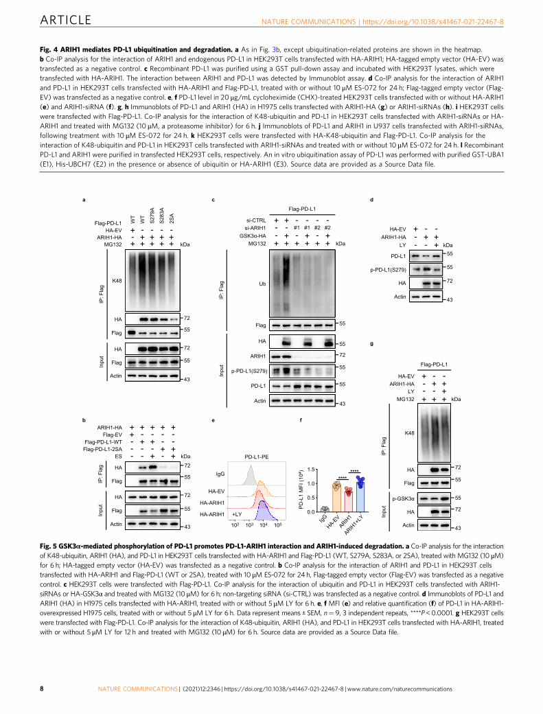
Fig. 4 ARIH1 mediates PD-L1 ubiquitination and degradation. a As in Fig. 3b, except ubiquitination-related proteins are shown in the heatmap.b Co-IP analysis for the interaction of ARIH1 and endogenous PD-L1 in HEK293T cells transfected with HA-ARIH1; HA-tagged empty vector (HA-EV) wastransfected as a negative control. c Recombinant PD-L1 was purified using a GST pull-down assay and incubated with HEK293T lysates, which weretransfected with HA-ARIH1. The interaction between ARIH1 and PD-L1 was detected by Immunoblot assay. d Co-IP analysis for the interaction of ARIH1and PD-L1 in HEK293T cells transfected with HA-ARIH1 and Flag-PD-L1, treated with or without 10 μM ES-072 for 24 h; Flag-tagged empty vector (Flag-EV) was transfected as a negative control. e, f PD-L1 level in 20 μg/mL cycloheximide (CHX)-treated HEK293T cells transfected with or without HA-ARIH1(e) and ARIH1-siRNA (f). g, h Immunoblots of PD-L1 and ARIH1 (HA) in H1975 cells transfected with ARIH1-HA (g) or ARIH1-siRNAs (h). i HEK293T cellswere transfected with Flag-PD-L1. Co-IP analysis for the interaction of K48-ubiquitin and PD-L1 in HEK293T cells transfected with ARIH1-siRNAs or HA-ARIH1 and treated with MG132 (10 μM, a proteasome inhibitor) for 6 h. j Immunoblots of PD-L1 and ARIH1 in U937 cells transfected with ARIH1-siRNAs,following treatment with 10 μM ES-072 for 24 h. k HEK293T cells were transfected with HA-K48-ubiquitin and Flag-PD-L1. Co-IP analysis for theinteraction of K48-ubiquitin and PD-L1 in HEK293T cells transfected with ARIH1-siRNAs and treated with or without 10 μM ES-072 for 24 h. l RecombinantPD-L1 and ARIH1 were purified in transfected HEK293T cells, respectively. An in vitro ubiquitination assay of PD-L1 was performed with purified GST-UBA1(E1), His-UBCH7 (E2) in the presence or absence of ubiquitin or HA-ARIH1 (E3). Source data are provided as a Source Data file.
Fig. 5 GSK3α-mediated phosphorylation of PD-L1 promotes PD-L1-ARIH1 interaction and ARIH1-induced degradation. a Co-IP analysis for the interactionof K48-ubiquitin, ARIH1 (HA), and PD-L1 in HEK293T cells transfected with HA-ARIH1 and Flag-PD-L1 (WT, S279A, S283A, or 2SA), treated with MG132 (10 μM)for 6 h; HA-tagged empty vector (HA-EV) was transfected as a negative control. b Co-IP analysis for the interaction of ARIH1 and PD-L1 in HEK293T cellstransfected with HA-ARIH1 and Flag-PD-L1 (WT or 2SA), treated with 10 μM ES-072 for 24 h, Flag-tagged empty vector (Flag-EV) was transfected as a negativecontrol. c HEK293T cells were transfected with Flag-PD-L1. Co-IP analysis for the interaction of ubiquitin and PD-L1 in HEK293T cells transfected with ARIH1-siRNAs or HA-GSK3α and treated with MG132 (10 μM) for 6 h; non-targeting siRNA (si-CTRL) was transfected as a negative control. d Immunoblots of PD-L1 andARIH1 (HA) in H1975 cells transfected with HA-ARIH1, treated with or without 5 μM LY for 6 h. e, f MFI (e) and relative quantification (f) of PD-L1 in HA-ARIH1-overexpressed H1975 cells, treated with or without 5 μM LY for 6 h. Data represent means ± SEM, n= 9, 3 independent repeats, ****P<0.0001. g HEK293T cellswere transfected with Flag-PD-L1. Co-IP analysis for the interaction of K48-ubiquitin, ARIH1 (HA), and PD-L1 in HEK293T cells transfected with HA-ARIH1, treatedwith or without 5 μM LY for 12 h and treated with MG132 (10 μM) for 6 h. Source data are provided as a Source Data file.
ARTICLE NATURE COMMUNICATIONS | https://doi.org/10.1038/s41467-021-22467-8
8 NATURE COMMUNICATIONS | (2021) 12:2346 | https://doi.org/10.1038/s41467-021-22467-8 | www.nature.com/naturecommunications
PCR (qRT-PCR) of the whole tumor mRNA (SupplementaryFig. 10a–d).
Recent studies have explored to intercept two immunecheckpoint pathways, PD-L1 and CTLA4, collectively, to improvethe efficacy of immunotherapy36. Next, we used the 4T1 tumorxenograft model and performed an anti-CTLA4 immunecheckpoint blockade treatment in the presence/absence of
ES-072. Tumor growth was significantly decreased both by theES-072 treatment and the anti-CTLA4 treatment. Notably, afurther decrease of tumor growth and even complete regressionwas observed when ES-072 and anti-CTLA4 treatments werecombined (Fig. 6i–k). The levels of total and activated CD8+
cytotoxic T cells (GzmB+) in the tumors were also significantlyincreased upon the ES-072/anti-CTLA4 combination treatment
NATURE COMMUNICATIONS | https://doi.org/10.1038/s41467-021-22467-8 ARTICLE
NATURE COMMUNICATIONS | (2021) 12:2346 | https://doi.org/10.1038/s41467-021-22467-8 |www.nature.com/naturecommunications 9
when compared to either of the single treatments (Fig. 6l, m).Furthermore, western blotting analysis of the tumor lysatesshowed that ES-072 treatment decreased PD-L1 levels, whileincreasing its phosphorylation and ubiquitylation levels (Fig. 6n).
Taken together, our data indicate that ARIH1 plays a role inpromoting anti-tumor immunity, and that ES-072 is a promisingagent to boost anti-tumor immunity and anti-CTLA4 immunecheckpoint blockade immunotherapies.
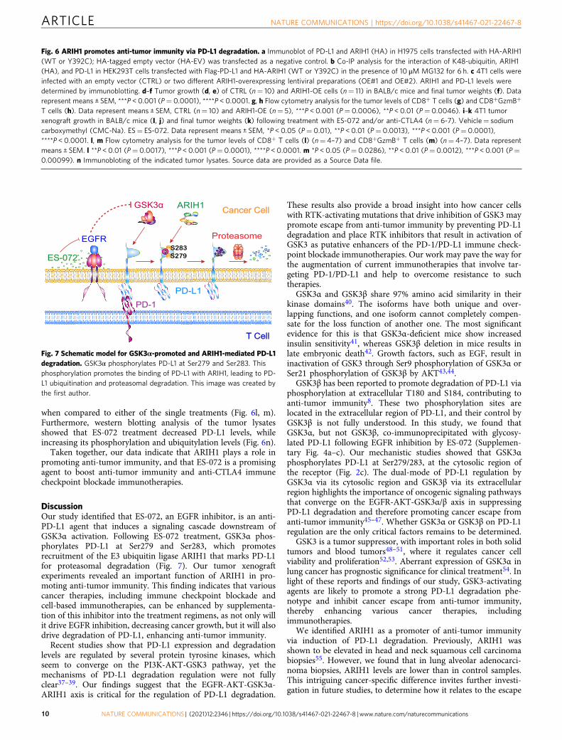
DiscussionOur study identified that ES-072, an EGFR inhibitor, is an anti-PD-L1 agent that induces a signaling cascade downstream ofGSK3α activation. Following ES-072 treatment, GSK3α phos-phorylates PD-L1 at Ser279 and Ser283, which promotesrecruitment of the E3 ubiquitin ligase ARIH1 that marks PD-L1for proteasomal degradation (Fig. 7). Our tumor xenograftexperiments revealed an important function of ARIH1 in pro-moting anti-tumor immunity. This finding indicates that variouscancer therapies, including immune checkpoint blockade andcell-based immunotherapies, can be enhanced by supplementa-tion of this inhibitor into the treatment regimens, as not only willit drive EGFR inhibition, decreasing cancer growth, but it will alsodrive degradation of PD-L1, enhancing anti-tumor immunity.
Recent studies show that PD-L1 expression and degradationlevels are regulated by several protein tyrosine kinases, whichseem to converge on the PI3K-AKT-GSK3 pathway, yet themechanisms of PD-L1 degradation regulation were not fullyclear37–39. Our findings suggest that the EGFR-AKT-GSK3α-ARIH1 axis is critical for the regulation of PD-L1 degradation.
These results also provide a broad insight into how cancer cellswith RTK-activating mutations that drive inhibition of GSK3 maypromote escape from anti-tumor immunity by preventing PD-L1degradation and place RTK inhibitors that result in activation ofGSK3 as putative enhancers of the PD-1/PD-L1 immune check-point blockade immunotherapies. Our work may pave the way forthe augmentation of current immunotherapies that involve tar-geting PD-1/PD-L1 and help to overcome resistance to suchtherapies.
GSK3α and GSK3β share 97% amino acid similarity in theirkinase domains40. The isoforms have both unique and over-lapping functions, and one isoform cannot completely compen-sate for the loss function of another one. The most significantevidence for this is that GSK3α-deficient mice show increasedinsulin sensitivity41, whereas GSK3β deletion in mice results inlate embryonic death42. Growth factors, such as EGF, result ininactivation of GSK3 through Ser9 phosphorylation of GSK3α orSer21 phosphorylation of GSK3β by AKT43,44.
GSK3β has been reported to promote degradation of PD-L1 viaphosphorylation at extracellular T180 and S184, contributing toanti-tumor immunity8. These two phosphorylation sites arelocated in the extracellular region of PD-L1, and their control byGSK3β is not fully understood. In this study, we found thatGSK3α, but not GSK3β, co-immunoprecipitated with glycosy-lated PD-L1 following EGFR inhibition by ES-072 (Supplemen-tary Fig. 4a–c). Our mechanistic studies showed that GSK3αphosphorylates PD-L1 at Ser279/283, at the cytosolic region ofthe receptor (Fig. 2c). The dual-mode of PD-L1 regulation byGSK3α via its cytosolic region and GSK3β via its extracellularregion highlights the importance of oncogenic signaling pathwaysthat converge on the EGFR-AKT-GSK3α/β axis in suppressingPD-L1 degradation and therefore promoting cancer escape fromanti-tumor immunity45–47. Whether GSK3α or GSK3β on PD-L1regulation are the only critical factors remains to be determined.
GSK3 is a tumor suppressor, with important roles in both solidtumors and blood tumors48–51, where it regulates cancer cellviability and proliferation52,53. Aberrant expression of GSK3α inlung cancer has prognostic significance for clinical treatment54. Inlight of these reports and findings of our study, GSK3-activatingagents are likely to promote a strong PD-L1 degradation phe-notype and inhibit cancer escape from anti-tumor immunity,thereby enhancing various cancer therapies, includingimmunotherapies.
We identified ARIH1 as a promoter of anti-tumor immunityvia induction of PD-L1 degradation. Previously, ARIH1 wasshown to be elevated in head and neck squamous cell carcinomabiopsies55. However, we found that in lung alveolar adenocarci-noma biopsies, ARIH1 levels are lower than in control samples.This intriguing cancer-specific difference invites further investi-gation in future studies, to determine how it relates to the escape
Fig. 6 ARIH1 promotes anti-tumor immunity via PD-L1 degradation. a Immunoblot of PD-L1 and ARIH1 (HA) in H1975 cells transfected with HA-ARIH1(WT or Y392C); HA-tagged empty vector (HA-EV) was transfected as a negative control. b Co-IP analysis for the interaction of K48-ubiquitin, ARIH1(HA), and PD-L1 in HEK293T cells transfected with Flag-PD-L1 and HA-ARIH1 (WT or Y392C) in the presence of 10 μM MG132 for 6 h. c 4T1 cells wereinfected with an empty vector (CTRL) or two different ARIH1-overexpressing lentiviral preparations (OE#1 and OE#2). ARIH1 and PD-L1 levels weredetermined by immunoblotting. d–f Tumor growth (d, e) of CTRL (n= 10) and ARIH1-OE cells (n= 11) in BALB/c mice and final tumor weights (f). Datarepresent means ± SEM, ***P < 0.001 (P= 0.0001), ****P < 0.0001. g, h Flow cytometry analysis for the tumor levels of CD8+ T cells (g) and CD8+GzmB+
T cells (h). Data represent means ± SEM, CTRL (n= 10) and ARIH1-OE (n= 5), ***P < 0.001 (P= 0.0006), **P < 0.01 (P= 0.0046). i–k 4T1 tumorxenograft growth in BALB/c mice (I, j) and final tumor weights (k) following treatment with ES-072 and/or anti-CTLA4 (n= 6-7). Vehicle= sodiumcarboxymethyl (CMC-Na). ES= ES-072. Data represent means ± SEM, *P < 0.05 (P= 0.01), **P < 0.01 (P= 0.0013), ***P < 0.001 (P= 0.0001),****P < 0.0001. l, m Flow cytometry analysis for the tumor levels of CD8+ T cells (l) (n= 4–7) and CD8+GzmB+ T cells (m) (n= 4–7). Data representmeans ± SEM. l **P < 0.01 (P= 0.0017), ***P < 0.001 (P= 0.0001), ****P < 0.0001. m *P < 0.05 (P= 0.0286), **P < 0.01 (P= 0.0012), ***P < 0.001 (P=0.00099). n Immunobloting of the indicated tumor lysates. Source data are provided as a Source Data file.
Fig. 7 Schematic model for GSK3α-promoted and ARIH1-mediated PD-L1degradation. GSK3α phosphorylates PD-L1 at Ser279 and Ser283. Thisphosphorylation promotes the binding of PD-L1 with ARIH1, leading to PD-L1 ubiquitination and proteasomal degradation. This image was created bythe first author.
ARTICLE NATURE COMMUNICATIONS | https://doi.org/10.1038/s41467-021-22467-8
10 NATURE COMMUNICATIONS | (2021) 12:2346 | https://doi.org/10.1038/s41467-021-22467-8 | www.nature.com/naturecommunications
from each cancer type from anti-tumor immunity. Interestingly,ARIH1 missense mutations are found in 4% of non-small celllung cancer patients (n= 75)56, 3.25% of prostate cancer patients(n= 154)57, and 5.13% of cutaneous squamous cell carcinomapatients (n= 39)58.
In addition to ARIH1, PD-L1 is also targeted for degradationby Cul3SPOP and β-TrCP7,8 E3 ubiquitin ligases. Together, thesefindings suggest that combination treatments that target morethan one of these E3 ubiquitin ligases may have potentiallyadditive/synergistic effects on PD-L1 degradation in cancer,allowing a more potent stimulation of anti-tumor immunity.
In summary, our study sheds light on the mechanisms ofcancer escape from anti-tumor immunity via increased PD-L1protein levels downstream of EGFR overexpression and over-activation in cancer. Our work suggests that GSK3α- and ARIH1-activating agents, as well as EGFR inhibitors, are likely to sti-mulate anti-tumor immunity and therefore enhance existingcancer therapies by triggering the PD-L1 degradation pathwaythis work delineates.
MethodsReagents and antibody generation. The compounds and their sources are as follows:S-Ruxolitinib (#INCB018424; Selleck), AZD9291, Osimertinib (#S7297; Selleck), MG132(#S2619; Selleck), LY2090314 (#S7063; Selleck), and ES-072 were synthesizedin colla-boration with Shanghai Institute of Organic Chemistry, Chinese Academy of Sciences.The recombinant cytokines and their sources are as follows: Human recombinant IFNγ(#300-02; Peprotech), Human recombinant EGF (#AF-100-15; Peprotech), and Mouserecombinant IL-4 (#214-14; Peprotech). The following antibodies were used in this study:PE anti-human CD274 (#329706; 1 : 200; Biolegend), PE anti-mouse CD274 (#124308;1 : 200; Biolegend), PE Mouse IgG2b (isotype control) (#400312; 1 : 200; Biolegend),Zombie Violet™ Fixable Viability Kit (#423114; 1 : 200; Biolegend), PerCP/Cyanine5.5anti-mouse CD45 (#103132; 1 : 200; Biolegend), PE/Cyanine7 anti-mouse CD3 (#100320;1 : 200; Biolegend), FITC anti-mouse CD8 (#100706; 1 : 200; Biolegend), APC anti-human/mouse Granzyme B (#372204; 1 : 200; Biolegend), PD-L1 (ab213524, 1 : 1000;Abcam), PD-L1 (66248-1-Ig, 1 : 1000; Proteintech), β-TrCP (D13F10) (#4394, 1 : 1000;Cell Signaling Technology), EGFR (D38B1, 1 : 1000; Cell Signaling Technology),Phospho-EGFR (Tyr1068, 1 : 1000; Cell Signaling Technology), Ubiquitin (P4D1) (#SC-8017, 1 : 200; Santa Cruz Biotechnology), K48 (05-1307, 1 : 1000; Milipore), GSK3α(#4337, 1 : 1000; Cell Signaling Technology), Phospho-GSK3α (Ser21) (#9631, 1 : 1000;Cell Signaling Technology), GSK3β (Y174) (ab32391, 1 : 5000; Abcam), Phospho-GSK3β(Ser9) (#P49841, 1 : 1000; Cell Signaling Technology), AKT (#9272, 1 : 1000; Cell Sig-naling Technology), Phospho-AKT (Ser473) (#4046, 1 : 2000; Cell Signaling Technology),ARIH1 (C-7) (#SC-514551, 1 : 200; Santa Cruz Biotechnology), ARIH1 (Goat)(#EB05812, 1 : 100; Everestbiotech), GST (B-14) (#SC-138, 1 : 200; Santa Cruz Bio-technology), Granyzme B (D6E9W) (#46890, 1 : 50; Cell Signaling Technology), His-tag(#66005-1-Ig, 1 : 1000; Proteintech), Flag-tag (0912-1, 1 : 2000; HuaAn Biotechnology),HA-tag (0906-1, 1 : 2000; HuaAn Biotechnology), and β-Actin (M1210-2, 1 : 2000;HuaAn Biotechnology). The anti-human phospho-PD-L1 Ser279 antibody was raisedagainst the region near Ser279 phosphorylation site of PD-L1. The secondary antibodiesfor western blotting were used: goat anti-mouse (1 : 20,000, #31430, Thermo Fisher Sci-entific, Ltd) and goat anti-rabbit (1 : 20,000, #31460, Thermo Fisher Scientific, Ltd). Thephosphorylated synthetic peptide [QDTNSKKQSDTHLEC] was used for immunizationin rabbits. The antibody was generated by GenScript (Nanjing, China). Anti-HAmagneticbeads (#B26202) and Anti-Flag (DYKDDDDK) Affinity Gel (#B23102) were fromBimake. Lipofectamine 2000 (#1901433) and Lipofectamine 3000 (#2067450) were fromInvitrogen. Collagenase/hyaluronidase (#17100-017, Vancouver, BC, Canada) were fromStemcell Technologies and DNase (#10104159001) were from Sigma. The peptides andtheir sources are as follows: PD-L1-WT [LRKGRMMDVKKCGIQDTNSKKQSDTH-LERT], phosphorylated PD-L1-WT [LRKGRMMDVKKCGIQDTNS (PO3H2) KKQS(PO3H2) DTHLERT], and PD-L1-2SA [LRKGRMMDVKKCGIQDTNAKKQADTH-LEET] were synthesized by Zhongtai (Hangzhou, China).
Cell culture. Human histiocytic lymphosarcoma cell line (U937), Non-small celllung adenocarcinoma cancer cell line (H1975), and Human embryonic kidney cellline (HEK293T) were a gift from J.Y. Yuan (Harvard Medical School, Boston).PDMs were obtained from male BALB/c mice. PDMs, U937, and H1975 cells werecultured in RPMI-1640 (Hyclone, with L-glutamine); HEK293T cells were culturedin Dulbecco’s modified Eagle’s medium (Hyclone, with L-glutamine, with 4.5 g/Lglucose, without pyruvate). These media were supplemented with 10% heat-inactivated fetal calf serum (Gibco) and 1% Penicillin/Streptomycin (Gibco). Allcells were cultured at 37 °C with 5% CO2. Cells were transiently transfected withDNA using Lipofectamine 2000 or Lipofectamine 3000.
Plasmids construction and RNA interference. Flag-PD-L1 (WT and mutants),GSK3α-HA, and ARIH1-HA (WT and mutants) were amplified by PCR and fused
the fragments into pCMV3 via seamless cloning. The constructs and siRNAs weretransfected into cell lines (HEK293T, H1975, and U937) with Lipofectamine 2000or Lipofectamine 3000, according to the the manufacturer’s protocol. siRNAs usedin this study are provided in Supplementary Table 5.
In vitro high-throughput drug screens. U937 cells (2 × 10⁵) were preincubatedwith 100 μg/L IFNγ for 48 h and plated in 96-well plates per well (Corning). Then10 μM FDA-approved drugs or drug candidates were added and incubated for 12 h.All compounds were commercially purchased. Treatments were performed twice;each plate contained a negative control (dimethyl sulfoxide) and a positive control(Ruxolitinib). U937 cells were centrifuged for 5 min at 1000 × g and the super-natant discarded. The collected cells were washed with phosphate-buffered saline(PBS) twice and incubated with PE anti-human CD274 at 4 °C for 30 min. Thenthe incubated U937 cells were washed and resuspended with PBS, and the proteinlevel of membrane PD-L1 reflected by PD-L1-PE median fluorescence intensity(MFI) was determined using flow cytometry analysis. The hit compounds werepicked and classified according to the PD-L1-PE-MFI and the targeted pathways.
Western blot analysis. For western blot analysis, cells were collected and washedwith PBS, then lysed in radioimmunoprecipitation assay (RIPA) buffer (1% TritonX-100, 100 mM Tris-HCl pH 8.8, 100 mM NaCl, 0.5 mM EDTA). After incubationon ice for 30 min, the lysates were centrifuged at 12,000 × g for 10 min at 4 °C. Thesupernatant was collected and the protein concentration was measured bybicinchoninic acid reaction. Protein samples were added with 2× loading bufferand heated at 100 °C for 10 min, separated with SDS-polyacrylamide gel electro-phoresis (SDS-PAGE), transferred onto polyvinylidene difluoride membranes, andblocked with 5% non-fat milk in Phosphate Buffered Saline with 0.1% Tween-20(PBST) for 1 h at room temperature. The membranes were probed with the cor-responding primary antibodies at 4 °C overnight and horseradish peroxidase-conjugated secondary antibodies at room temperature for 1 h. Signals were detectedusing chemiluminescence reagents (#4AW001-500, 4A649 Biotech, Co.).
Immunoprecipitation. For immunoprecipitation between PD-L1 and GSK3α/ARIH1/K48, cells were lysed in RIPA buffer (1% Triton X-100, 100 mM Tris-HClpH 8.8, 100 mM NaCl, 0.5 mM EDTA) supplemented with a complete proteaseinhibitor cocktail (Bimake, added fresh), and mixed with antibodies at 4 °C for 4 h;protein A/G agarose beads were added and incubated at 4 °C overnight. Beads werewashed three times with RIPA buffer and subjected to western blotting.
Mass spectrometry and data analysis. Flag-tagged PD-L1 was overexpressed inHEK293T cells and were trypsin-digested. PD-L1 was immunoprecipitated withbeads following immunoprecipitation. The resulting peptides were subjected to thephosphopeptide enrichment using TiO2 beads. The enriched phospho-peptideswere analyzed on the Q Exactive™ HF mass spectrometer (Thermo Fisher Scien-tific). The identification and quantification of phosphorylated peptides were doneby MaxQuant. The tandem mass spectra were searched against the UniProt humanprotein database together with a set of commonly observed contaminants. Theprecursor mass tolerance was set as 20 p.p.m. and the fragment mass tolerance wasset as 0.1 Da. The 33 cysteine carbamide methylation was set as a static mod-ification and the methionine oxidation, as well as serine, threonine, and tyrosinephosphorylation, were set as variable modifications. The false discovery rate (FDR)at peptide spectrum match level was controlled below 1%.
Duolink® PLA fluorescence analysis. For the interaction between PD-L1 andGSK3α/GSK3β, the samples were pre-treated with respect to fixation, retrieval,and/or permeabilization. Then the samples were incubated with Duolink® BlockingSolution in a heated humidity chamber for 60 min at 37 °C. The samples wereincubated with diluted primary antibody in a humidity chamber overnight at 4 °C.The samples were washed with 1× Wash Buffer A at room temperature for 5 mintwice after the primary antibody solution was moved then incubated with dilutedPLUS, and MINUS PLA probes (1 : 5) in a pre-heated humidity chamber for 1 h at37 °C. The samples were washed with 1× Wash Buffer A at room temperature for 5min twice after the PLA probes were moved, then incubated with ligation solutionin a pre-heated humidity chamber for 30 min at 37 °C. The samples were washedwith 1× Wash Buffer A at room temperature for 5 min twice after the ligationsolution was moved then incubated with amplification solution in a pre-heatedhumidity chamber for 100 min at 37 °C. The samples were washed with 1× WashBuffer B for 10 min twice and 0.01x Wash Buffer B for 1 min at room temperatureafter the amplification solution was moved. The slides were mounted with a cov-erslip using a minimal volume of Duolink® PLA Mounting Medium with 4′,6-diamidino-2-phenylindole analyzed with confocal microscope. The images werecollected using Cytation 3 and were analyzed using Gen5 2.0.
Protein purification and in vitro kinase assays. For purification of PD-L1, Flag-tagged PD-L1 was transfected into HEK293T cells for 24 h. Cells were lysed in 1mL of lysis buffer (TAP) (0.5% NP-40, 1 mM Na3VO4, 20 mM Tris-HCl pH 7.5, 1mM NaF, 150 mM NaCl, 1 mM EDTA) supplemented with a complete proteaseinhibitor cocktail (Bimake, added fresh), and incubated with anti-Flag magnetic
NATURE COMMUNICATIONS | https://doi.org/10.1038/s41467-021-22467-8 ARTICLE
NATURE COMMUNICATIONS | (2021) 12:2346 | https://doi.org/10.1038/s41467-021-22467-8 |www.nature.com/naturecommunications 11
beads (after washing the beads with PBS twice) for 6 h on a rotating wheel at 4 °C.The beads were washed with TAP buffer three times and treated with CIP(#M0290, Biolabs) at 37 °C for 30 min. The kinase assays were performed withrecombinant human GSK3α proteins (#Ab42597, Abcam). The purified PD-L1 orsynthetic peptides (PD-L1-WT/2SA) were incubated in 30 μL of kinase buffer (25mM Tris-HCl pH 7.5, 5 mM β-glycerophosphate, 2 mM dithiothreitol (DTT),0.1 mM Na3VO4, 10 mM MgCl2) supplemented with phosphatase inhibitor cock-tail (#b15001, Bimake), with or without 100 μM ATP for 2 h at 37 °C. The reactionswere stopped by adding SDS-PAGE 2× loading buffer (100 mM Tris-HCl pH 6.8,20% glycerol, 4% SDS, 0.1% Bromophenol blue 0.2 M DTT) and heating at 100 °Cfor 10 min. Kinase activity was evaluated by dot blot or western blotting with anti-phospho-human PD-L1 Ser279 antibody.
Flow cytometry analysis of membrane PD-L1. For flow cytometric analysis formembrane PD-L1, H1975 or HEK293T cells were collected by centrifugation at1000 × g for 5 min, incubated with PBS (0.5% bovine serum albumin) for 10 min atroom temperature. The cells were probed with PE-conjugated PD-L1 antibody(#329706, Biolegend) and a matched isotype control at 4 °C for 30 min in the dark.After washing three times with PBS, the cells were analyzed using flow cytometry(Beckman Coulter Cytoflex) and data were analyzed using CytExpert V2.3 and andFlowJo X software.
GST-pull down. GST-tagged PD-L1 was expressed in Escherichia coli BL21 andpurified. HA-tagged ARIH1 was expressed in HEK293T cells and purified usingmagnetic HA beads. For reaction, purified GST-PD-L1 was first incubated withglutathione-Sepharose 4B beads at 4 °C for 1 h. Then, the HA-tagged protein wasadded and incubated at 4 °C for 2 h. The mixture was boiling with 1× loadingbuffer for 10 min, then subjected to western blotting.
In vitro ubiquitination assays. Plasmids GST-UBA1 (E1), Flag-PD-L1, HA-ARIH1 (E3), and ARIH1 inactive mutant were transfected into HEK293T cells.Post transfection, cells were collected and lysed in RIPA Lysis Buffer at 4 °C for 1 h.Then, the lysates were incubated with indicated beads at 4 °C overnight. His-taggedprotein UBCH7 (E2) ubiquitin was purified by E. coli BL21 expression. Reactionswere performed in a 30 μL reaction mixture at 37 °C for 2 h in the presence of His-Ub, E1, E2, E3, Flag-PD-L1, ATP regeneration solution (Enzo Life Sciences) andUbiquitin Reaction Buffer (Enzo Life Sciences). All reactions were terminated byboiling 10 min with SDS sample buffer and then subjected to western blotting.
Immunohistochemistry. EGFR-WT tumors and EGFR-mutant-driven tumorsfrom primary lung adenocarcinoma tissues were obtained from 8 patients (4 caseseach group, median age: 60 years old, range from 47 to 81) at the Sir Run Run ShawHospital, Zhejiang University. The four EGFR mutations are L858R point mutationof exon 21; E542K point mutation of exon 10 and deletion of exon 19 (ex19del745–750); P753R point mutation of exon 19; L858R point mutation of exon 21. Allsamples were collected with signed informed consent according to the internalreview and ethics boards of Sir Run Run Shaw Hospital. These tissues were rapidlyexcised, fixed with 4% paraformaldehyde, and embedded in paraffin for tissuesections (5 μm thick) and immunohistochemical staining. The primary antibodiesused are anti-PD-L1 (1 : 200), anti-p-PD-L1 (Ser279) (1 : 200), anti-p-GSK3α (1 :200), and anti-ARIH1 (1 : 200). Visualization of cell nuclei was performed withhematoxylin and analysis was done using the Olympus BX61 light microscope.
Generation of ARIH1-OE stable 4T1 cell lines. Briefly, HEK293T cells weretransfected with PCDH-CTRL and PCDH-mouse ARIH1 with packaging plasmids.Medium with secreted viruses was collected at 48, 72, and 96 h, and was filteredthrough 0.45 μm filters. Twenty-four hours post infection, the medium wasreplaced with fresh medium and the infected 4T1 cells were selected with 4 μg/mLpuromycin for 3 days.
Tumor xenograft experiments. Female BALB/c mice or nude mice (aged8–10 weeks) were purchased from Shanghai SLAC Laboratory Animal, Co., Ltd(Shanghai, China). All the animal experiments were strictly conducted in accor-dance with the protocols approved by the Ethics Committee for Animal Studies atZhejiang University, China.
For xenograft model with control or ARIH1-stable 4T1 cells, 5 × 10⁵ control orARIH1-stable 4T1 cells suspended in 50 μL PBS and Matrigel (1 : 1 v/v) wereinjected into the fourth breast fat pad. On days 3–5 after injection, tumor size wasmeasured and calculated by using the formula 1/2 × length × width2. Tumor weightwas recorded on the day of killing.
For xenograft model with 4T1 cells and combination therapy with ES-072 andanti-CTLA4, 5 × 10⁵ 4T1 cells suspended in 50 μL PBS and Matrigel (1 : 1 v/v) wereinjected into the fourth breast fat pad. On days 3 after injection, tumor size wasmeasured and calculated by using the formula 1/2 × length × width2. Mice were thenrandomly divided into control group: IgG (100 μg/100 μL, intraperitoneal injection, i.p.) and 0.5% sodium carboxymethyl (200 μL, intragastric administration, i.g)treatment; ES-072 treatment group: IgG (100 μg/100 μL, i.p.) and ES-072 (60mg/kg,200 μL, i.g.); anti-CTLA4 treatment group: anti-CTLA4 (100 μg/100 μL, i.p.) and 0.5%
sodium carboxymethyl (200 μL, i.g.); ES-072 and anti-CTLA4 combination group:anti-CTLA4 (100 μg/100 μL, i.p.) and ES-072 (60mg/kg, 200 μL, i.g.). ES-072 wasgiven daily from 3 days after inoculation and anti-CTLA4 antibody was administeredon day 7, 10, and 13 after inoculation with respective control treatment.
Tumor sample preparation and flow cytometry. Excised tumors were digested incollagenase/hyaluronidase and DNase at 37 °C for 45min to make cell suspensionwith a 45 μm filter (BD Bioscience). Then, cells were stained with Percp-Cy5.5-conjugated-CD45, PE-Cy7-conjugated-CD8, FITC-conjugated-CD3 antibodies, fixedand permeabilized with a Fix/Perm kit (Biolegend), and finally stained with APC-conjugated-GzmB antibody. Data acquisition was performed using flow cytometry(Beckman Coulter Cytoflex) and data were analyzed using CytExpert V2.3 software.
Immunoprecipitation assay with mouse tumor tissues. For immunoprecipita-tion between PD-L1 and ubiquitin, fresh xenograft tissues were lysed in RIPAbuffer (1% Triton X-100, 100 mM Tris-HCl pH 8.8, 100 mM NaCl, 0.5 mM EDTA)supplemented with a complete protease inhibitor cocktail (Bimake, added fresh)and mixed with anti-PD-L1 at 4 °C for 4 h; protein A/G agarose beads were addedand incubated at 4 °C overnight. Beads were washed three times with RIPA bufferand subjected to western blotting.
qRT-PCR analysis for tumor cytokines. Fresh tumor tissues were lysed and totalRNA was extracted using TRIzol™ Plus RNA Purification Kit (Invitrogen). cDNAwas synthesized from purified RNA using the PrimeScript RT reagent Kit(TAKARA, RR047A) according to the manufacturer’s instructions. QuantitativePCR was performed using a StepOnePlus Real-Time PCR Systems (ABI). Thecomparative Ct method was used for the data analysis and mouse β-actin mRNAwas used as an internal control. The sequences of primers used for qRT-PCR areprovided in Supplementary Table 6.
Statistics and reproducibility. Numerical data are presented as means ± SEM; alldata analyses were performed using GraphPad Prism (version 7.0, GraphPadSoftware, Inc.). Unpaired Student’s t-test was used to analyze the flow cytometrydata. One-way analysis of variance was used to analyze the statistical differencesamong the groups with P-values indicated in the related graphs. The level ofstatistical significance was set at a p < 0.05. All assays were carried out at least threeindependent times with the same results.
Reporting summary. Further information on research design is available in the NatureResearch Reporting Summary linked to this article.
Data availabilityThe data that support the findings of this study are available from the correspondingauthor upon reasonable request. The mass spectrometry proteomics data have beendeposited to the ProteomeXchange Consortium (http://proteomecentral.proteomexchange.org) via the iProX partner repository with the dataset identifierPXD024452. Source data are provided with this paper.
Received: 9 June 2020; Accepted: 12 March 2021;
References1. Dong, H. et al. Tumor-associated B7-H1 promotes T-cell apoptosis: a
potential mechanism of immune evasion. Nat. Med. 8, 793–800 (2002).2. Dong, H., Zhu, G., Tamada, K. & Chen, L. B7-H1, a third member of the B7
family, co-stimulates T-cell proliferation and interleukin-10 secretion. Nat.Med. 5, 1365–1369 (1999).
3. Gibbons, J. R. & Dong, H. Functional expression of programmed death-ligand1 (B7-H1) by immune cells and tumor cells. Front. Immunol. 8, 961 (2017).
4. Schreiner, B., Bailey, S. L., Shin, T., Chen, L. & Miller, S. D. PD-1 ligandsexpressed on myeloid-derived APC in the CNS regulate T-cell responses inEAE. Eur. J. Immunol. 38, 2706–2717 (2008).
5. Liu, X., Yang, Z., Latchoumanin, O. & Qiao, L. Antagonizing programmeddeath-1 and programmed death ligand-1 as a therapeutic approach for gastriccancer. Ther. Adv. Gastroenterol. 9, 853–860 (2016).
6. Ansell, S. M. et al. PD-1 blockade with nivolumab in relapsed or refractoryHodgkin’s lymphoma. N. Engl. J. Med. 372, 311–319 (2015).
7. Zhang, J. et al. Cyclin D–CDK4 kinase destabilizes PD-L1 via cullin 3–SPOPto control cancer immune surveillance. Nature 553, 91–95 (2018).
8. Li, C. W. et al. Glycosylation and stabilization of programmed death ligand-1suppresses T-cell activity. Nat. Commun. 7, 12632 (2016).
9. Scott, D. C. et al. Two distinct types of E3 ligases work in unison to regulatesubstrate ubiquitylation. Cell 166, 1198–1214 (2016).
ARTICLE NATURE COMMUNICATIONS | https://doi.org/10.1038/s41467-021-22467-8
12 NATURE COMMUNICATIONS | (2021) 12:2346 | https://doi.org/10.1038/s41467-021-22467-8 | www.nature.com/naturecommunications
10. Kelsall, I. R. et al. TRIAD1 and HHARI bind to and are activated by distinctneddylated Cullin-RING ligase complexes. EMBO J. 32, 2848–2860 (2013).
11. Wenzel, D. M., Lissounov, A., Brzovic, P. S. & Klevit, R. E. UBCH7 reactivityprofile reveals parkin and HHARI to be RING/HECT hybrids. Nature 474,105–108 (2011).
12. Von Stechow, L. et al. The E3 ubiquitin ligase ARIH1 protects againstgenotoxic stress by initiating a 4EHP-mediated mRNA translation arrest. Mol.Cell. Biol. 35, 1254–1268 (2015).
13. Jutten, B. & Rouschop, K. EGFR signaling and autophagy dependence forgrowth, survival, and therapy resistance. Cell Cycle 13, 42–51 (2014).
14. Sigismund, S., Avanzato, D. & Lanzetti, L. Emerging functions of the EGFR incancer. Mol. Oncol. 12, 3–20 (2018).
15. Weihua, Z. et al. Survival of cancer cells is maintained by EGFR independentof its kinase activity. Cancer Cell 13, 385–393 (2008).
16. Wang, X., Goldstein, D., Crowe, P. & Yang, J. Next-generation EGFR/HERtyrosine kinase inhibitors for the treatment of patients with non-small-celllung cancer harboring EGFR mutations: a review of the evidence. OncotargetsTher. 9, 5461–5473 (2016).
17. Gainor, J. F. et al. EGFR mutations and ALK rearrangements are associatedwith low response rates to PD-1 pathway blockade in non-small cell lungcancer: a retrospective analysis. Clin. Cancer Res. 22, 4585–4593 (2016).
18. Camidge, D. R., Doebele, R. C. & Kerr, K. M. Comparing and contrastingpredictive biomarkers for immunotherapy and targeted therapy of NSCLC.Nat. Rev. Clin. Oncol. 16, 341–355 (2019).
19. Azuma, K. et al. Association of PD-L1 overexpression with activating EGFRmutations in surgically resected nonsmall-cell lung cancer. Ann. Oncol. 25,1935–1940 (2014).
20. Zhang, M. et al. PD-L1 expression in lung cancer and its correlation withdriver mutations: a meta-analysis. Sci. Rep. 7, 10255 (2017).
21. Chen, N. et al. Upregulation of PD-L1 by EGFR activation mediates theimmune escape in EGFR-driven NSCLC: implication for optional immunetargeted therapy for NSCLC patients with EGFR mutation. J. Thorac. Oncol.10, 910–923 (2015).
22. Zhang, N. et al. The EGFR pathway is involved in the regulation of PD-L1expression via the IL-6/JAK/STAT3 signaling pathway in EGFR-mutated non-small cell lung cancer. Int. J. Oncol. 49, 1360–1368 (2016).
23. Horiuchi, M. et al. Interferon-gamma induces AT(2) receptor expression infibroblasts by Jak/STAT pathway and interferon regulatory factor-1. Circ. Res.86, 233–240 (2000).
24. Lau, T. S., Chan, K. Y., Cheung, T. H., Yim, S. F. & Kwong, J. Abstract 648:interferon-gamma induces PD-L1 expression via IFNGR-JAK-STAT pathwayin ovarian cancer. Cancer Res. 77, 648 (2017).
25. Chen, M. et al. JAK2 and PD-L1 amplification enhance the dynamicexpression of PD-L1 in triple-negative breast cancer. Clin. Breast Cancer 18,e1205–e1215 (2018).
26. Cross, D. A. et al. AZD9291, an irreversible EGFR TKI, overcomes T790M-mediated resistance to EGFR inhibitors in lung cancer. Cancer Discov. 4,1046–1061 (2014).
27. Ku, B. M. et al. AZD9291 overcomes T790M-mediated resistance throughdegradation of EGFRL858R/T790M in non-small cell lung cancer cells. Invest.N. Drug. 34, 407–415 (2016).
28. Cheng, C. C. et al. Epidermal growth factor induces STAT1 expression toexacerbate the IFNr‐mediated PD‐L1 axis in epidermal growth factorreceptor‐positive cancers. Mol. Carcinog. 57, 1588–1598 (2018).
29. Chen, M. et al. Insluin and epithelial growth factor (EGF) promoteprogrammed death ligand 1(PD-L1) production and transport in colon cancerstem cells. BMC Cancer 19, 112–153 (2019).
30. Engelman, J. A. & Janne, P. A. Mechanisms of acquired resistance toepidermal growth factor receptor tyrosine kinase inhibitors in non-small celllung cancer. Clin. Cancer Res. 14, 2895–2899 (2008).
31. Kim, J. et al. The effect of Helicobacter pylori on epidermal growth factorreceptor-induced signal transduction and the preventive effect of celecoxib ingastric cancer cells. Gut Liver 7, 552–559 (2013).
32. Nicot, A. et al. Phosphorylation of NBR1 by GSK3 modulates proteinaggregation. Autophagy 10, 1036–1053 (2014).
33. Xu, C., Kim, N. G. & Gumbiner, B. M. Regulation of protein stability by GSK3mediated phosphorylation. Cell Cycle 8, 4032–4039 (2009).
34. Bouchal, P. et al. Combined proteomics and transcriptomics identifiescarboxypeptidase B1 and nuclear factor κB (NF-κB) associated proteins asputative biomarkers of metastasis in low grade breast cancer. Mol. Cell.Proteomics 14, 1814–1830 (2015).
35. Abaan, O. D. et al. The exomes of the NCI-60 panel: a genomic resource forcancer biology and systems pharmacology. Cancer Res. 73, 4372–4382 (2013).
36. Cha, J. et al. Metformin promotes antitumor immunity via endoplasmic-reticulum-associated degradation of PD-L1. Mol. Cell 71, 606–620 (2018).
37. Kim, S. et al. Alterations in PD-L1 expression associated with acquisition ofresistance to ALK inhibitors in ALK-rearranged lung cancer. Cancer Res.Treat. 51, 1231–1240 (2019).
38. Ota, K. et al. Induction of PD-L1 expression by the EML4-ALK oncoproteinand downstream signaling pathways in non-small cell lung cancer. Clin.Cancer Res. 21, 4014–4021 (2015).
39. Balan, M. et al. Novel roles of c-Met in the survival of renal cancer cellsthrough the regulation of HO-1 and PD-L1 expression. J. Biol. Chem. 290,8110–8120 (2015).
40. Woodgett, J. R. Molecular cloning and expression of glycogen synthase kinase-3/factor A. Embo J. 9, 2431–2438 (1990).
41. Lal, H. et al. Glycogen synthase kinase-3α limits ischemic injury, cardiacrupture, post-myocardial infarction remodeling and death. Circulation 125,65–75 (2012).
42. Kerkela, R. et al. Deletion of GSK-3β in mice leads to hypertrophiccardiomyopathy secondary to cardiomyoblast hyperproliferation. J. Clin.Invest. 118, 3609–3618 (2008).
43. Matsuda, T. et al. Distinct roles of GSK-3alpha and GSK-3betaphosphorylation in the heart under pressure overload. Proc. Natl Acad. Sci.USA 105, 20900–20905 (2008).
44. Ahmad, F. et al. Cardiomyocyte-specific deletion of Gsk3alpha mitigates post-myocardial infarction remodeling, contractile dysfunction, and heart failure. J.Am. Coll. Cardiol. 64, 696–706 (2014).
45. Abdelhamed, S., Ogura, K., Yokoyama, S., Saiki, I. & Hayakawa, Y. AKT-STAT3 pathway as a downstream target of EGFR signaling to regulate PD-L1expression on NSCLC cells. J. Cancer 7, 1579–1586 (2016).
46. Lastwika, K. J. et al. Control of PD-L1 expression by oncogenic activation ofthe AKT–mTOR pathway in non-small cell lung cancer. Cancer Res. 76,227–238 (2016).
47. Ahmad, F. & Woodgett, J. R. Emerging roles of GSK-3α in pathophysiology:emphasis on cardio-metabolic disorders. Biochim. Biophys. Acta Mol. Cell Res.1867, 118616 (2020).
48. Dembowy, J., Adissu, H. A., Liu, J. C., Zacksenhaus, E. & Woodgett, J. R.Effect of glycogen synthase kinase-3 inactivation on mouse mammary glanddevelopment and oncogenesis. Oncogene 34, 3514–3526 (2015).
49. Mancinelli, R. et al. Multifaceted roles of GSK-3 in cancer and autophagy-related diseases. Oxid. Med. Cell. Longev. 2017, 1–14 (2017).
50. Cervello, M. et al. Pivotal roles of glycogen synthase-3 in hepatocellularcarcinoma. Adv. Biol. Regul. 65, 59–76 (2017).
51. McCubrey, J. A. et al. GSK-3 as potential target for therapeutic intervention incancer. Oncotarget 5, 2881 (2014).
52. Banerji, V. et al. The intersection of genetic and chemical genomic screensidentifies GSK-3α as a target in human acute myeloid leukemia. J. Clin.Investig. 122, 935–947 (2012).
53. Park, S., Lee, J. W., Herbst, R. S. & Koo, J. S. GSK-3α is a novel target of CREBand CREB-GSK-3α signaling participates in cell viability in lung cancer. PLosONE 11, e153075 (2016).
54. MacAulay, K. & Doble, B. W. Glycogen synthase kinase 3a-specific regulationof murine hepatic glycogen metabolism. Cell Metab. 6, 329–337 (2007).
55. Elmehdawi, F. et al. Human Homolog of Drosophila Ariadne (HHARI) is amarker of cellular proliferation associated with nuclear bodies. Exp. Cell Res.319, 161–172 (2013).
56. Hellmann, M. D. et al. Genomic features of response to combinationimmunotherapy in patients with advanced non-small-cell lung cancer. CancerCell 33, 843–852 (2018).
57. Kumar, A. et al. Substantial interindividual and limited intraindividualgenomic diversity among tumors from men with metastatic prostate cancer.Nat. Med. 22, 369–378 (2016).
58. Pickering, C. R. et al. Mutational landscape of aggressive cutaneous squamouscell carcinoma. Clin. Cancer Res. 20, 6582–6592 (2014).
AcknowledgementsThis work was supported by the National Natural Science Foundation of China (number81773182, 91854108, 31601121, 81870007, 81920108001, and 81800024), the NationalKey R&D Program of China (2017YFA0104200), Zhejiang Provincial Program for theCultivation of High-Level Innovative Health Talents (2016-63) and Zhejiang ProvincialNatural Science Foundation (LD19H160001). We thank Professor Hui Yang from theInstitute of Neuroscience, Chinese Academy of Sciences for cDNA, Professor QimingSun from Zhejiang University for his helpful suggestions to this project.
Author contributionsH.X. conceived and coordinated the project. H.X., S.Y., and A.N. designed the experiments,interpreted the data, and wrote the manuscript. Y.W., C.Z., X.L., and Z.H. performed most ofthe experiments and interpreted the data. B.S., Q. Zeng, Q. Zhao, H.Z., H.L., X.C., X.X., M.Z.,T.H., Z.W., H.Y., S. Yang, Y.S., Y.C., R.W., T.X., and W.C. assisted with the experiments andhelped to analyze the data. H.X., A.N., S. Ying, Y.W., and C.Z. revised the manuscript.
Competing interestsThe authors declare no competing interests.
NATURE COMMUNICATIONS | https://doi.org/10.1038/s41467-021-22467-8 ARTICLE
NATURE COMMUNICATIONS | (2021) 12:2346 | https://doi.org/10.1038/s41467-021-22467-8 |www.nature.com/naturecommunications 13

Additional informationSupplementary information The online version contains supplementary materialavailable at https://doi.org/10.1038/s41467-021-22467-8.
Correspondence and requests for materials should be addressed to A.N., S.Y. or H.X.
Peer review information Nature Communications thanks Seung-Oe Lim and the other,anonymous, reviewer(s) for their contribution to the peer review of this work. Peerreviewer reports are available.
Reprints and permission information is available at http://www.nature.com/reprints
Publisher’s note Springer Nature remains neutral with regard to jurisdictional claims inpublished maps and institutional affiliations.
Open Access This article is licensed under a Creative CommonsAttribution 4.0 International License, which permits use, sharing,
adaptation, distribution and reproduction in any medium or format, as long as you giveappropriate credit to the original author(s) and the source, provide a link to the CreativeCommons license, and indicate if changes were made. The images or other third partymaterial in this article are included in the article’s Creative Commons license, unlessindicated otherwise in a credit line to the material. If material is not included in thearticle’s Creative Commons license and your intended use is not permitted by statutoryregulation or exceeds the permitted use, you will need to obtain permission directly fromthe copyright holder. To view a copy of this license, visit http://creativecommons.org/licenses/by/4.0/.
© The Author(s) 2021
ARTICLE NATURE COMMUNICATIONS | https://doi.org/10.1038/s41467-021-22467-8
14 NATURE COMMUNICATIONS | (2021) 12:2346 | https://doi.org/10.1038/s41467-021-22467-8 | www.nature.com/naturecommunications
Related Documents