Arenavirus Stable Signal Peptide Is the Keystone Subunit for Glycoprotein Complex Organization Lydia H. Bederka, a Cyrille J. Bonhomme, a * Emily L. Ling, a * Michael J. Buchmeier a,b Department of Molecular Biology & Biochemistry, University of California Irvine, Irvine, California, USA a ; Division of Infectious Disease, Department of Medicine, University of California Irvine, Irvine, California, USA b * Present address: Cyrille J. Bonhomme, PPD, Vaccine Sciences Department, Richmond, Virginia, USA; Emily L. Ling, Department of Art as Applied to Medicine, Johns Hopkins University School of Medicine, Baltimore, Maryland, USA. ABSTRACT The rodent arenavirus glycoprotein complex encodes a stable signal peptide (SSP) that is an essential structural com- ponent of mature virions. The SSP, GP1, and GP2 subunits of the trimeric glycoprotein complex noncovalently interact to stud the surface of virions and initiate arenavirus infectivity. Nascent glycoprotein production undergoes two proteolytic cleavage events: first within the endoplasmic reticulum (ER) to cleave SSP from the remaining precursor GP1/2 (glycoprotein complex [GPC]) glycoprotein and second within the Golgi stacks by the cellular SKI-1/S1P for GP1/2 processing to yield GP1 and GP2 subunits. Cleaved SSP is not degraded but retained as an essential glycoprotein subunit. Here, we defined functions of the 58- amino-acid lymphocytic choriomeningitis virus (LCMV) SSP in regard to glycoprotein complex processing and maturation. Us- ing molecular biology techniques, confocal microscopy, and flow cytometry, we detected SSP at the plasma membrane of trans- fected cells. Further, we identified a sorting signal (FLLL) near the carboxyl terminus of SSP that is required for glycoprotein maturation and trafficking. In the absence of SSP, the glycoprotein accumulated within the ER and was unable to undergo pro- cessing by SKI-1/S1P. Mutation of this highly conserved FLLL motif showed impaired glycoprotein processing and secretory pathway trafficking, as well as defective surface expression and pH-dependent membrane fusion. Immunoprecipitation of SSP confirmed an interaction between the signal peptide and the GP2 subunit; however, mutations within this FLLL motif disrupted the association of the GP1 subunit with the remaining glycoprotein complex. IMPORTANCE Several members of the Arenaviridae family are neglected human pathogens capable of causing illness ranging from a nondescript flu-like syndrome to fulminant hemorrhagic fever. Infections by arenaviruses are mediated by attachment of the virus glycoprotein to receptors on host cells and virion internalization by fusion within an acidified endosome. SSP plays a critical role in the fusion of the virus with the host cell membrane. Within infected cells, the retained glycoprotein SSP plays a neglected yet essential role in glycoprotein biosynthesis. Without this 6-kDa polypeptide, the glycoprotein precursor is retained within the endoplasmic reticulum, and trafficking to the plasma membrane where SSP, GP1, and GP2 localize for glycoprotein assembly into infectious virions is inhibited. To investigate SSP contributions to glycoprotein maturation and function, we cre- ated an SSP-tagged glycoprotein to directly detect and manipulate this subunit. This resource will aid future studies to identify host factors that mediate glycoprotein maturation. Received 30 September 2014 Accepted 2 October 2014 Published 28 October 2014 Citation Bederka LH, Bonhomme CJ, Ling EL, Buchmeier MJ. 2014. Arenavirus stable signal peptide is the keystone subunit for glycoprotein complex organization. mBio 5(6): e02063-14. doi:10.1128/mBio.02063-14. Editor Terence S. Dermody, Vanderbilt University School of Medicine Copyright © 2014 Bederka et al. This is an open-access article distributed under the terms of the Creative Commons Attribution-Noncommercial-ShareAlike 3.0 Unported license, which permits unrestricted noncommercial use, distribution, and reproduction in any medium, provided the original author and source are credited. Address correspondence to Michael J. Buchmeier, [email protected]. This article is a direct contribution from a Fellow of the American Academy of Microbiology. A renaviruses asymptomatically infect geographically specific rodent species and are capable of causing severe disease in humans (1). Human infections most frequently occur via close contact with rodent excreta and several arenaviruses, such as the Old World (OW) Lassa and Lujo viruses, as well as the New World (NW) arenaviruses Junín, Machupo, Guanarito, and Sabia, which are causative agents of hemorrhagic fever (2). Lymphocytic cho- riomeningitis virus (LCMV) is the prototype and most researched arenavirus. LCMV is capable of causing illness ranging from mild flu-like symptoms to aseptic meningitis. Additionally, LCMV has contributed to multiorgan failure and death in organ transplant recipients from undiagnosed, infected organs (3–6). The principal rodent host for LCMV is the ubiquitous Mus musculus, which allows for the near global distribution of LCMV (7, 8). Arenaviruses are simple in their design, consisting of two seg- ments of single-stranded RNA, the large (L) and small (s) seg- ment. These segments each encode two nonoverlapping open reading frames. The large genome segment encodes the RNA- dependent RNA polymerase (L protein; ~220 kDa) and the matrix protein (Z protein; 11 kDa). The small genome segment encodes the glycoprotein complex (GPC; 75 kDa) and the nucleoprotein (NP; 63 kDa). The Z protein and the GP complex are expressed in RESEARCH ARTICLE crossmark November/December 2014 Volume 5 Issue 6 e02063-14 ® mbio.asm.org 1 on July 29, 2020 by guest http://mbio.asm.org/ Downloaded from

Welcome message from author
This document is posted to help you gain knowledge. Please leave a comment to let me know what you think about it! Share it to your friends and learn new things together.
Transcript

Arenavirus Stable Signal Peptide Is the Keystone Subunit forGlycoprotein Complex Organization
Lydia H. Bederka,a Cyrille J. Bonhomme,a* Emily L. Ling,a* Michael J. Buchmeiera,b
Department of Molecular Biology & Biochemistry, University of California Irvine, Irvine, California, USAa; Division of Infectious Disease, Department of Medicine, Universityof California Irvine, Irvine, California, USAb
* Present address: Cyrille J. Bonhomme, PPD, Vaccine Sciences Department, Richmond, Virginia, USA; Emily L. Ling, Department of Art as Applied to Medicine, Johns HopkinsUniversity School of Medicine, Baltimore, Maryland, USA.
ABSTRACT The rodent arenavirus glycoprotein complex encodes a stable signal peptide (SSP) that is an essential structural com-ponent of mature virions. The SSP, GP1, and GP2 subunits of the trimeric glycoprotein complex noncovalently interact to studthe surface of virions and initiate arenavirus infectivity. Nascent glycoprotein production undergoes two proteolytic cleavageevents: first within the endoplasmic reticulum (ER) to cleave SSP from the remaining precursor GP1/2 (glycoprotein complex[GPC]) glycoprotein and second within the Golgi stacks by the cellular SKI-1/S1P for GP1/2 processing to yield GP1 and GP2subunits. Cleaved SSP is not degraded but retained as an essential glycoprotein subunit. Here, we defined functions of the 58-amino-acid lymphocytic choriomeningitis virus (LCMV) SSP in regard to glycoprotein complex processing and maturation. Us-ing molecular biology techniques, confocal microscopy, and flow cytometry, we detected SSP at the plasma membrane of trans-fected cells. Further, we identified a sorting signal (FLLL) near the carboxyl terminus of SSP that is required for glycoproteinmaturation and trafficking. In the absence of SSP, the glycoprotein accumulated within the ER and was unable to undergo pro-cessing by SKI-1/S1P. Mutation of this highly conserved FLLL motif showed impaired glycoprotein processing and secretorypathway trafficking, as well as defective surface expression and pH-dependent membrane fusion. Immunoprecipitation of SSPconfirmed an interaction between the signal peptide and the GP2 subunit; however, mutations within this FLLL motif disruptedthe association of the GP1 subunit with the remaining glycoprotein complex.
IMPORTANCE Several members of the Arenaviridae family are neglected human pathogens capable of causing illness rangingfrom a nondescript flu-like syndrome to fulminant hemorrhagic fever. Infections by arenaviruses are mediated by attachment ofthe virus glycoprotein to receptors on host cells and virion internalization by fusion within an acidified endosome. SSP plays acritical role in the fusion of the virus with the host cell membrane. Within infected cells, the retained glycoprotein SSP plays aneglected yet essential role in glycoprotein biosynthesis. Without this 6-kDa polypeptide, the glycoprotein precursor is retainedwithin the endoplasmic reticulum, and trafficking to the plasma membrane where SSP, GP1, and GP2 localize for glycoproteinassembly into infectious virions is inhibited. To investigate SSP contributions to glycoprotein maturation and function, we cre-ated an SSP-tagged glycoprotein to directly detect and manipulate this subunit. This resource will aid future studies to identifyhost factors that mediate glycoprotein maturation.
Received 30 September 2014 Accepted 2 October 2014 Published 28 October 2014
Citation Bederka LH, Bonhomme CJ, Ling EL, Buchmeier MJ. 2014. Arenavirus stable signal peptide is the keystone subunit for glycoprotein complex organization. mBio 5(6):e02063-14. doi:10.1128/mBio.02063-14.
Editor Terence S. Dermody, Vanderbilt University School of Medicine
Copyright © 2014 Bederka et al. This is an open-access article distributed under the terms of the Creative Commons Attribution-Noncommercial-ShareAlike 3.0 Unportedlicense, which permits unrestricted noncommercial use, distribution, and reproduction in any medium, provided the original author and source are credited.
Address correspondence to Michael J. Buchmeier, [email protected].
This article is a direct contribution from a Fellow of the American Academy of Microbiology.
Arenaviruses asymptomatically infect geographically specificrodent species and are capable of causing severe disease in
humans (1). Human infections most frequently occur via closecontact with rodent excreta and several arenaviruses, such as theOld World (OW) Lassa and Lujo viruses, as well as the New World(NW) arenaviruses Junín, Machupo, Guanarito, and Sabia, whichare causative agents of hemorrhagic fever (2). Lymphocytic cho-riomeningitis virus (LCMV) is the prototype and most researchedarenavirus. LCMV is capable of causing illness ranging from mildflu-like symptoms to aseptic meningitis. Additionally, LCMV hascontributed to multiorgan failure and death in organ transplant
recipients from undiagnosed, infected organs (3–6). The principalrodent host for LCMV is the ubiquitous Mus musculus, whichallows for the near global distribution of LCMV (7, 8).
Arenaviruses are simple in their design, consisting of two seg-ments of single-stranded RNA, the large (L) and small (s) seg-ment. These segments each encode two nonoverlapping openreading frames. The large genome segment encodes the RNA-dependent RNA polymerase (L protein; ~220 kDa) and the matrixprotein (Z protein; 11 kDa). The small genome segment encodesthe glycoprotein complex (GPC; 75 kDa) and the nucleoprotein(NP; 63 kDa). The Z protein and the GP complex are expressed in
RESEARCH ARTICLE crossmark
November/December 2014 Volume 5 Issue 6 e02063-14 ® mbio.asm.org 1
on July 29, 2020 by guesthttp://m
bio.asm.org/
Dow
nloaded from
a pseudopositive-sense orientation, while the L protein and theNP are expressed in a negative-sense orientation, giving the are-naviruses an ambisense gene expression strategy (9).
The arenavirus glycoprotein complex is expressed as a poly-protein precursor that undergoes two cleavage events. First, thestable signal peptide (SSP) is cleaved by cellular signal peptidasewithin the endoplasmic reticulum (ER). This 58-amino-acid pro-tein is retained as a stable subunit and is critical for downstream,mature glycoprotein complex formation (10–12). Second, SSPtraffics with immature GP1/2 precursor (amino acids 59 to 498),which undergoes cleavage by the SKI-1/S1P enzyme within theGolgi stacks to yield the GP1 and GP2 subunits (13, 14). The threeindependent subunits, consisting of the SSP, GP1, and GP2, trafficto the plasma membrane for viral assembly and egress (15–18).These three subunits interact noncovalently at the virion surfaceto bind with host cell receptors to initiate infectivity. The GP1subunit interacts with the known cellular receptors:�-dystroglycan (�-DG) for the OW arenaviruses and transferrinreceptor 1 (TfR1) for the NW viruses (19, 20). Recent studiesusing next-generation technologies provided evidence for the in-volvement of additional host factors for both Junín virus andLassa virus entry (21, 22). The acidification of endocytic vesiclesinduces irreversible dissociation of the GP1 subunit from the re-maining GP2 and SSP subunits, followed by a conformationalchange within the GP2 subunit to reveal the fusion domainswhich, in concert with SSP, induce fusion events between the vi-rion and the host membrane to deliver the viral core to the newlyinfected cell cytoplasm (23, 24).
Generally, signal peptides are short, extended amino-terminalsequences found within secreted or transmembrane proteinswhose function is to direct nascent proteins to the ER for matura-tion via the secretory pathway (25). Few viruses possess signalsequences that serve additional functions. The Ebola virus signalpeptide was shown to modulate the extent of GP glycosylation,thereby moderating the extent of glycoprotein interaction withDC-SIGN/R (26). Additionally, the HIV-1 Env protein signal pep-tide is thought to regulate glycoprotein folding and maturation(27). In a nonviral model, the prion protein signal peptide inter-acts with the transmembrane domain for correct protein expres-sion and membrane orientation (28).
Our current studies focus on the importance of SSP in terms ofnascent glycoprotein complex expression, maturation, and func-tion. Using Western blot analysis, confocal microscopy, and flowcytometry, we show that LCMV GP lacking the native SSP doesnot exit the endoplasmic reticulum or mature through the secre-tory pathway. We detect SSP at the plasma membrane, and muta-tions within SSP affect trafficking of the entire glycoprotein com-plex. Further, we define a conserved FLLL motif within the LCMVSSP that is the driving force for full-length and functional matu-ration and glycoprotein subunit association.
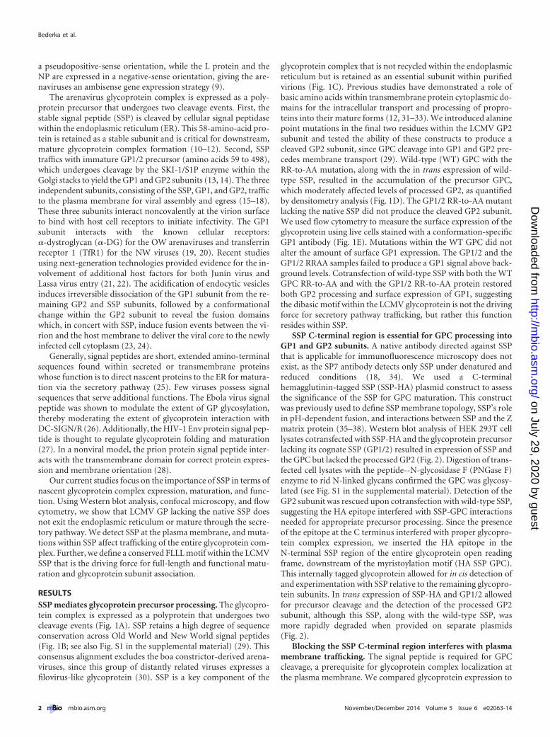
RESULTSSSP mediates glycoprotein precursor processing. The glycopro-tein complex is expressed as a polyprotein that undergoes twocleavage events (Fig. 1A). SSP retains a high degree of sequenceconservation across Old World and New World signal peptides(Fig. 1B; see also Fig. S1 in the supplemental material) (29). Thisconsensus alignment excludes the boa constrictor-derived arena-viruses, since this group of distantly related viruses expresses afilovirus-like glycoprotein (30). SSP is a key component of the
glycoprotein complex that is not recycled within the endoplasmicreticulum but is retained as an essential subunit within purifiedvirions (Fig. 1C). Previous studies have demonstrated a role ofbasic amino acids within transmembrane protein cytoplasmic do-mains for the intracellular transport and processing of propro-teins into their mature forms (12, 31–33). We introduced alaninepoint mutations in the final two residues within the LCMV GP2subunit and tested the ability of these constructs to produce acleaved GP2 subunit, since GPC cleavage into GP1 and GP2 pre-cedes membrane transport (29). Wild-type (WT) GPC with theRR-to-AA mutation, along with the in trans expression of wild-type SSP, resulted in the accumulation of the precursor GPC,which moderately affected levels of processed GP2, as quantifiedby densitometry analysis (Fig. 1D). The GP1/2 RR-to-AA mutantlacking the native SSP did not produce the cleaved GP2 subunit.We used flow cytometry to measure the surface expression of theglycoprotein using live cells stained with a conformation-specificGP1 antibody (Fig. 1E). Mutations within the WT GPC did notalter the amount of surface GP1 expression. The GP1/2 and theGP1/2 RRAA samples failed to produce a GP1 signal above back-ground levels. Cotransfection of wild-type SSP with both the WTGPC RR-to-AA and with the GP1/2 RR-to-AA protein restoredboth GP2 processing and surface expression of GP1, suggestingthe dibasic motif within the LCMV glycoprotein is not the drivingforce for secretory pathway trafficking, but rather this functionresides within SSP.
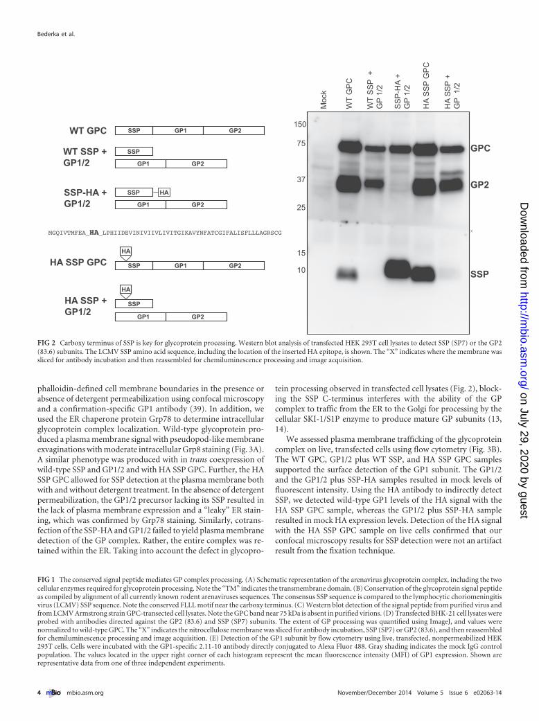
SSP C-terminal region is essential for GPC processing intoGP1 and GP2 subunits. A native antibody directed against SSPthat is applicable for immunofluorescence microscopy does notexist, as the SP7 antibody detects only SSP under denatured andreduced conditions (18, 34). We used a C-terminalhemagglutinin-tagged SSP (SSP-HA) plasmid construct to assessthe significance of the SSP for GPC maturation. This constructwas previously used to define SSP membrane topology, SSP’s rolein pH-dependent fusion, and interactions between SSP and the Zmatrix protein (35–38). Western blot analysis of HEK 293T celllysates cotransfected with SSP-HA and the glycoprotein precursorlacking its cognate SSP (GP1/2) resulted in expression of SSP andthe GPC but lacked the processed GP2 (Fig. 2). Digestion of trans-fected cell lysates with the peptide--N-glycosidase F (PNGase F)enzyme to rid N-linked glycans confirmed the GPC was glycosy-lated (see Fig. S1 in the supplemental material). Detection of theGP2 subunit was rescued upon cotransfection with wild-type SSP,suggesting the HA epitope interfered with SSP-GPC interactionsneeded for appropriate precursor processing. Since the presenceof the epitope at the C terminus interfered with proper glycopro-tein complex expression, we inserted the HA epitope in theN-terminal SSP region of the entire glycoprotein open readingframe, downstream of the myristoylation motif (HA SSP GPC).This internally tagged glycoprotein allowed for in cis detection ofand experimentation with SSP relative to the remaining glycopro-tein subunits. In trans expression of SSP-HA and GP1/2 allowedfor precursor cleavage and the detection of the processed GP2subunit, although this SSP, along with the wild-type SSP, wasmore rapidly degraded when provided on separate plasmids(Fig. 2).
Blocking the SSP C-terminal region interferes with plasmamembrane trafficking. The signal peptide is required for GPCcleavage, a prerequisite for glycoprotein complex localization atthe plasma membrane. We compared glycoprotein expression to
Bederka et al.
2 ® mbio.asm.org November/December 2014 Volume 5 Issue 6 e02063-14
on July 29, 2020 by guesthttp://m
bio.asm.org/
Dow
nloaded from

Arenavirus SSP and Glycoprotein Maturation
November/December 2014 Volume 5 Issue 6 e02063-14 ® mbio.asm.org 3
on July 29, 2020 by guesthttp://m
bio.asm.org/
Dow
nloaded from
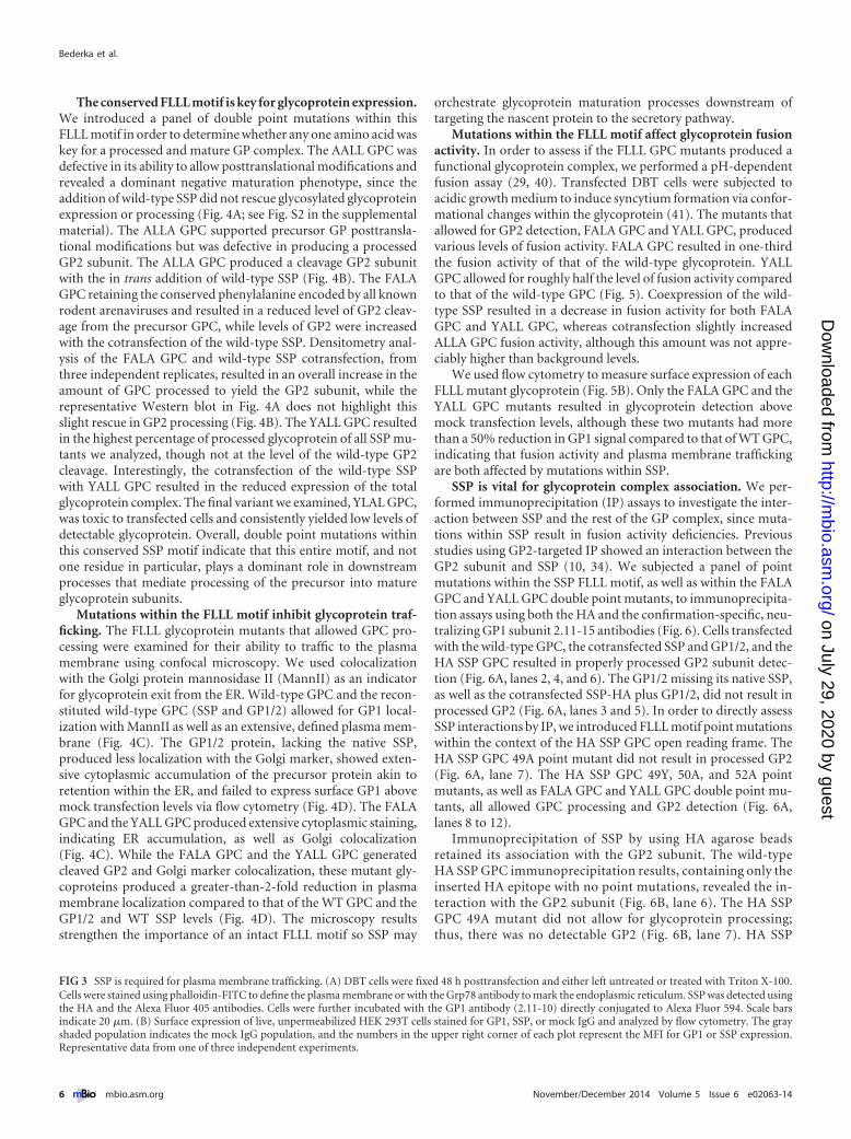
phalloidin-defined cell membrane boundaries in the presence orabsence of detergent permeabilization using confocal microscopyand a confirmation-specific GP1 antibody (39). In addition, weused the ER chaperone protein Grp78 to determine intracellularglycoprotein complex localization. Wild-type glycoprotein pro-duced a plasma membrane signal with pseudopod-like membraneexvaginations with moderate intracellular Grp8 staining (Fig. 3A).A similar phenotype was produced with in trans coexpression ofwild-type SSP and GP1/2 and with HA SSP GPC. Further, the HASSP GPC allowed for SSP detection at the plasma membrane bothwith and without detergent treatment. In the absence of detergentpermeabilization, the GP1/2 precursor lacking its SSP resulted inthe lack of plasma membrane expression and a “leaky” ER stain-ing, which was confirmed by Grp78 staining. Similarly, cotrans-fection of the SSP-HA and GP1/2 failed to yield plasma membranedetection of the GP complex. Rather, the entire complex was re-tained within the ER. Taking into account the defect in glycopro-
tein processing observed in transfected cell lysates (Fig. 2), block-ing the SSP C-terminus interferes with the ability of the GPcomplex to traffic from the ER to the Golgi for processing by thecellular SKI-1/S1P enzyme to produce mature GP subunits (13,14).
We assessed plasma membrane trafficking of the glycoproteincomplex on live, transfected cells using flow cytometry (Fig. 3B).The WT GPC, GP1/2 plus WT SSP, and HA SSP GPC samplessupported the surface detection of the GP1 subunit. The GP1/2and the GP1/2 plus SSP-HA samples resulted in mock levels offluorescent intensity. Using the HA antibody to indirectly detectSSP, we detected wild-type GP1 levels of the HA signal with theHA SSP GPC sample, whereas the GP1/2 plus SSP-HA sampleresulted in mock HA expression levels. Detection of the HA signalwith the HA SSP GPC sample on live cells confirmed that ourconfocal microscopy results for SSP detection were not an artifactresult from the fixation technique.
FIG 1 The conserved signal peptide mediates GP complex processing. (A) Schematic representation of the arenavirus glycoprotein complex, including the twocellular enzymes required for glycoprotein processing. Note the “TM” indicates the transmembrane domain. (B) Conservation of the glycoprotein signal peptideas compiled by alignment of all currently known rodent arenaviruses sequences. The consensus SSP sequence is compared to the lymphocytic choriomeningitisvirus (LCMV) SSP sequence. Note the conserved FLLL motif near the carboxy terminus. (C) Western blot detection of the signal peptide from purified virus andfrom LCMV Armstrong strain GPC-transected cell lysates. Note the GPC band near 75 kDa is absent in purified virions. (D) Transfected BHK-21 cell lysates wereprobed with antibodies directed against the GP2 (83.6) and SSP (SP7) subunits. The extent of GP processing was quantified using ImageJ, and values werenormalized to wild-type GPC. The “X” indicates the nitrocellulose membrane was sliced for antibody incubation, SSP (SP7) or GP2 (83.6), and then reassembledfor chemiluminescence processing and image acquisition. (E) Detection of the GP1 subunit by flow cytometry using live, transfected, nonpermeabilized HEK293T cells. Cells were incubated with the GP1-specific 2.11-10 antibody directly conjugated to Alexa Fluor 488. Gray shading indicates the mock IgG controlpopulation. The values located in the upper right corner of each histogram represent the mean fluorescence intensity (MFI) of GP1 expression. Shown arerepresentative data from one of three independent experiments.
10
15
25
37
75
150
Moc
k
WT
GP
C
WT
SS
P +
GP
1/2
SS
P-H
A +
G
P 1
/2
HA
SS
P G
PC
HA
SS
P +
G
P 1
/2
SSP
GP2
GPC
x
WT GPC
WT SSP +GP1/2
SSP GP2GP1
HA SSP GPC
HA SSP +GP1/2
SSP
GP2GP1
SSP-HA +GP1/2
HASSP
GP2GP1
SSP GP2GP1
HA
GP2GP1
SSP
HA
MGQIVTMFEA_HA_LPHIIDEVINIVIIVLIVITGIKAVYNFATCGIFALISFLLLAGRSCG
FIG 2 Carboxy terminus of SSP is key for glycoprotein processing. Western blot analysis of transfected HEK 293T cell lysates to detect SSP (SP7) or the GP2(83.6) subunits. The LCMV SSP amino acid sequence, including the location of the inserted HA epitope, is shown. The “X” indicates where the membrane wassliced for antibody incubation and then reassembled for chemiluminescence processing and image acquisition.
Bederka et al.
4 ® mbio.asm.org November/December 2014 Volume 5 Issue 6 e02063-14
on July 29, 2020 by guesthttp://m
bio.asm.org/
Dow
nloaded from

Arenavirus SSP and Glycoprotein Maturation
November/December 2014 Volume 5 Issue 6 e02063-14 ® mbio.asm.org 5
on July 29, 2020 by guesthttp://m
bio.asm.org/
Dow
nloaded from
The conserved FLLL motif is key for glycoprotein expression.We introduced a panel of double point mutations within thisFLLL motif in order to determine whether any one amino acid waskey for a processed and mature GP complex. The AALL GPC wasdefective in its ability to allow posttranslational modifications andrevealed a dominant negative maturation phenotype, since theaddition of wild-type SSP did not rescue glycosylated glycoproteinexpression or processing (Fig. 4A; see Fig. S2 in the supplementalmaterial). The ALLA GPC supported precursor GP posttransla-tional modifications but was defective in producing a processedGP2 subunit. The ALLA GPC produced a cleavage GP2 subunitwith the in trans addition of wild-type SSP (Fig. 4B). The FALAGPC retaining the conserved phenylalanine encoded by all knownrodent arenaviruses and resulted in a reduced level of GP2 cleav-age from the precursor GPC, while levels of GP2 were increasedwith the cotransfection of the wild-type SSP. Densitometry anal-ysis of the FALA GPC and wild-type SSP cotransfection, fromthree independent replicates, resulted in an overall increase in theamount of GPC processed to yield the GP2 subunit, while therepresentative Western blot in Fig. 4A does not highlight thisslight rescue in GP2 processing (Fig. 4B). The YALL GPC resultedin the highest percentage of processed glycoprotein of all SSP mu-tants we analyzed, though not at the level of the wild-type GP2cleavage. Interestingly, the cotransfection of the wild-type SSPwith YALL GPC resulted in the reduced expression of the totalglycoprotein complex. The final variant we examined, YLAL GPC,was toxic to transfected cells and consistently yielded low levels ofdetectable glycoprotein. Overall, double point mutations withinthis conserved SSP motif indicate that this entire motif, and notone residue in particular, plays a dominant role in downstreamprocesses that mediate processing of the precursor into matureglycoprotein subunits.
Mutations within the FLLL motif inhibit glycoprotein traf-ficking. The FLLL glycoprotein mutants that allowed GPC pro-cessing were examined for their ability to traffic to the plasmamembrane using confocal microscopy. We used colocalizationwith the Golgi protein mannosidase II (MannII) as an indicatorfor glycoprotein exit from the ER. Wild-type GPC and the recon-stituted wild-type GPC (SSP and GP1/2) allowed for GP1 local-ization with MannII as well as an extensive, defined plasma mem-brane (Fig. 4C). The GP1/2 protein, lacking the native SSP,produced less localization with the Golgi marker, showed exten-sive cytoplasmic accumulation of the precursor protein akin toretention within the ER, and failed to express surface GP1 abovemock transfection levels via flow cytometry (Fig. 4D). The FALAGPC and the YALL GPC produced extensive cytoplasmic staining,indicating ER accumulation, as well as Golgi colocalization(Fig. 4C). While the FALA GPC and the YALL GPC generatedcleaved GP2 and Golgi marker colocalization, these mutant gly-coproteins produced a greater-than-2-fold reduction in plasmamembrane localization compared to that of the WT GPC and theGP1/2 and WT SSP levels (Fig. 4D). The microscopy resultsstrengthen the importance of an intact FLLL motif so SSP may
orchestrate glycoprotein maturation processes downstream oftargeting the nascent protein to the secretory pathway.
Mutations within the FLLL motif affect glycoprotein fusionactivity. In order to assess if the FLLL GPC mutants produced afunctional glycoprotein complex, we performed a pH-dependentfusion assay (29, 40). Transfected DBT cells were subjected toacidic growth medium to induce syncytium formation via confor-mational changes within the glycoprotein (41). The mutants thatallowed for GP2 detection, FALA GPC and YALL GPC, producedvarious levels of fusion activity. FALA GPC resulted in one-thirdthe fusion activity of that of the wild-type glycoprotein. YALLGPC allowed for roughly half the level of fusion activity comparedto that of the wild-type GPC (Fig. 5). Coexpression of the wild-type SSP resulted in a decrease in fusion activity for both FALAGPC and YALL GPC, whereas cotransfection slightly increasedALLA GPC fusion activity, although this amount was not appre-ciably higher than background levels.
We used flow cytometry to measure surface expression of eachFLLL mutant glycoprotein (Fig. 5B). Only the FALA GPC and theYALL GPC mutants resulted in glycoprotein detection abovemock transfection levels, although these two mutants had morethan a 50% reduction in GP1 signal compared to that of WT GPC,indicating that fusion activity and plasma membrane traffickingare both affected by mutations within SSP.
SSP is vital for glycoprotein complex association. We per-formed immunoprecipitation (IP) assays to investigate the inter-action between SSP and the rest of the GP complex, since muta-tions within SSP result in fusion activity deficiencies. Previousstudies using GP2-targeted IP showed an interaction between theGP2 subunit and SSP (10, 34). We subjected a panel of pointmutations within the SSP FLLL motif, as well as within the FALAGPC and YALL GPC double point mutants, to immunoprecipita-tion assays using both the HA and the confirmation-specific, neu-tralizing GP1 subunit 2.11-15 antibodies (Fig. 6). Cells transfectedwith the wild-type GPC, the cotransfected SSP and GP1/2, and theHA SSP GPC resulted in properly processed GP2 subunit detec-tion (Fig. 6A, lanes 2, 4, and 6). The GP1/2 missing its native SSP,as well as the cotransfected SSP-HA plus GP1/2, did not result inprocessed GP2 (Fig. 6A, lanes 3 and 5). In order to directly assessSSP interactions by IP, we introduced FLLL motif point mutationswithin the context of the HA SSP GPC open reading frame. TheHA SSP GPC 49A point mutant did not result in processed GP2(Fig. 6A, lane 7). The HA SSP GPC 49Y, 50A, and 52A pointmutants, as well as FALA GPC and YALL GPC double point mu-tants, all allowed GPC processing and GP2 detection (Fig. 6A,lanes 8 to 12).
Immunoprecipitation of SSP by using HA agarose beadsretained its association with the GP2 subunit. The wild-typeHA SSP GPC immunoprecipitation results, containing only theinserted HA epitope with no point mutations, revealed the in-teraction with the GP2 subunit (Fig. 6B, lane 6). The HA SSPGPC 49A mutant did not allow for glycoprotein processing;thus, there was no detectable GP2 (Fig. 6B, lane 7). HA SSP
FIG 3 SSP is required for plasma membrane trafficking. (A) DBT cells were fixed 48 h posttransfection and either left untreated or treated with Triton X-100.Cells were stained using phalloidin-FITC to define the plasma membrane or with the Grp78 antibody to mark the endoplasmic reticulum. SSP was detected usingthe HA and the Alexa Fluor 405 antibodies. Cells were further incubated with the GP1 antibody (2.11-10) directly conjugated to Alexa Fluor 594. Scale barsindicate 20 �m. (B) Surface expression of live, unpermeabilized HEK 293T cells stained for GP1, SSP, or mock IgG and analyzed by flow cytometry. The grayshaded population indicates the mock IgG population, and the numbers in the upper right corner of each plot represent the MFI for GP1 or SSP expression.Representative data from one of three independent experiments.
Bederka et al.
6 ® mbio.asm.org November/December 2014 Volume 5 Issue 6 e02063-14
on July 29, 2020 by guesthttp://m
bio.asm.org/
Dow
nloaded from

Arenavirus SSP and Glycoprotein Maturation
November/December 2014 Volume 5 Issue 6 e02063-14 ® mbio.asm.org 7
on July 29, 2020 by guesthttp://m
bio.asm.org/
Dow
nloaded from

GPC 49Y and HA SSP GPC 50A point mutants also resulted invaried GP2 subunit immunoprecipitation (Fig. 6B, lanes 8 and9). The HA SSP GPC 52A sample, as well as FALA GPC andYALL GPC, did not reveal any GP2 pulldown product (Fig. 6B,lanes 10 to 12). FALA GPC and YALL GPC did not contain HAepitope expression for successful immunoprecipitation andserved as additional agarose bead controls.
The GP1 subunit was immunoprecipitated using the 2.11-15conformation-specific monoclonal antibody. The only samples toprecipitate the cleaved GP2 subunit were the WT GPC, the in transreconstituted GPC, and the HA SSP GPC (Fig. 6C, lanes 2, 4, and6). None of the SSP single or double point mutant glycoproteinconstructs allowed for glycoprotein subunit associations. In nosample did the GP1 immunoprecipitate SSP (Fig. 6C). FALA GPCand YALL GPC samples did not result in GP2 subunit detection,indicating that mutations within the SSP affect proper glycopro-tein complex association. For both the HA and the GP1 IP assays,we did not interpret GPC pulldown results as positive IP interac-tions, since the antibodies are capable of interacting with the nas-cent, full-length, uncleaved precursor glycoprotein.
DISCUSSION
Given that the arenavirus signal peptide is an essential componentof the glycoprotein complex, our goal was to dissect this subunit’srole in glycoprotein maturation. SSP was not degraded upontranslocation into the secretory pathway but rather was retainedand trafficked with the rest of the glycoprotein complex to theplasma membrane for viral packaging and egress. The require-ment for SSP packaging within virions demonstrates two criticalroles for this 58-amino-acid peptide. First, SSP acts as the leaderprotein for ER exit and GPC processing within the Golgi stacks, asdeletion of either hydrophobic domain inhibits downstream GPCprocessing (29, 34). Second, SSP is an important structural com-ponent within virions alongside the spike GP1 and transmem-brane GP2 subunits.
Using truncated glycoprotein constructs, previous authors re-ported that SSP is dispensable for GP ectodomain expression (42).Our current results contrast previous findings, as we used, or re-constituted, full-length glycoprotein expression vectors and nottruncated plasmid constructs. The presence of the signal peptidewas required for proper, full-length glycoprotein ectodomainprocessing (43). Our results further differed from previous re-search using both Old World and New World arenavirus glyco-proteins in that the LCMV Armstrong strain (Arm-4) glycopro-tein does not utilize basic residues within the GP2 cytoplasmicdomain as the driving force for ER exit (12, 33). Our mutationalanalysis of the GP1/2 subunit containing a mutated basic motif atthe ultimate portion of the GP2 cytoplasmic domain did not resultin any cleaved GP2 subunit, indicating that this glycoprotein lack-ing its SSP was not capable of exiting the ER for proteolytic pro-cessing. When wild-type SSP was coexpressed with the mutatedGP1/2, we detected the processed GP2 subunit. Additionally, our
results contrasted Lassa glycoprotein studies which indicated theGP2 cytoplasmic domain played a role in GP maturation cleavage,although the authors did not use a full-length Lassa glycoproteinin their characterizations (44).
The SSP and the GP2 cytoplasmic domains have been shown toplay critical roles in maturation. Recent studies using reverse ge-netics permitted infectious, chimeric Lassa/Junín-Candid1 virusproduction when the glycoprotein SSP and GP2 cytoplasmic do-mains were homologous and not of mismatched viral origin (45).This observation, together with our immunoprecipitation exper-iments, highlights the critical interactions between the SSP andGP2 subunits. Further, our confocal microscopy and flow cytom-etry analyses did not result in GP1/2 localization at the plasmamembrane. Since we used only LCMV glycoprotein constructs forour experiments, it is possible the differing results we observed aredue to slight differences between different arenavirus protein ex-pression mechanisms.
Signal peptides, although diverse in their amino acid compo-sitions, share a similar organization (25). Each signal peptide has avariable-length amino terminus, a hydrophobic center region,and a carboxyl terminus, including the signal peptidase cleavagesite. In order to directly analyze the signal peptide, we took advan-tage of the variable amino-terminal region by inserting an HAepitope within this SSP region while in the context of the full-length glycoprotein open reading frame (HA SSP GPC). HA SSPGPC was an expansion of a tool previously used for Junín SSPmembrane topology characterization, though in those studies SSPwas expressed in trans with the remaining portion of the glycopro-tein (37). Insertion of the HA epitope did not alter glycoproteintrafficking, as SKI-1/S1P-deficient cells were unable to cleave GPCinto mature GP1 and GP2 unless functional SKI-1/S1P was sup-plemented in trans (data not shown). This HA SSP GPC behavedat wild-type levels in terms of its expression, GP2 cleavage, andplasma membrane localization. Additionally, this HA SSP GPCallowed us to target experiments, both biochemical and with mi-croscopy, directly focusing on SSP while eliminating the techni-calities of cotransfection.
We and others have shown the importance of the first signalpeptide hydrophobic domain for precursor glycoprotein cleavageand pH-dependent membrane fusion (29, 34, 46). We were able todetect the HA epitope at the cell surface by flow cytometry usinglive, nonpermeabilized cells, indicating that the amino-terminalportion of SSP upstream of the first hydrophobic domain waspresented on the exterior surface of the plasma membrane. Thissuggests SSP may exhibit more than one membrane orientation,and the lack of fusion activity resulting from the inserted epitopemay be interfering with late-state SSP orientation to produce afully assembled glycoprotein complex (34, 35, 47). This observa-tion does not conflict with the proposed options for signal peptidehairpin-like orientation within the plasma membrane but merelyprovides for an alternative orientation during glycoprotein com-plex maturation and assembly (18, 35, 37).
FIG 4 Mutations within the conserved FLLL motif affect GPC processing. (A) HEK 293T cell lysates were transfected with the indicated glycoprotein constructsand were probed with antibodies to detect the cleaved GP2 and SSP subunits. (B) Densitometry quantification of the processed GP2 subunit levels from the GPCprecursor. All samples are normalized to the wild-type GPC, and error bars represent the standard errors of the means (SEM) from three independentexperiments. (C) Confocal microscopy of DBT cell expression GP constructs and detection of the Golgi protein mannosidase II (MannII). Colocalization isshown, with the indicated areas depicted in white. Also included in the upper right corner of each image is the Manders’ overlay coefficient, quantified usingImageJ. (D) Surface expression of the GP1 subunit using flow cytometry. Gray shading indicates the mock IgG population. Values within each histogram plotrepresent the MFI of GP1 expression. Data represent results from one of three independent experiments.
Bederka et al.
8 ® mbio.asm.org November/December 2014 Volume 5 Issue 6 e02063-14
on July 29, 2020 by guesthttp://m
bio.asm.org/
Dow
nloaded from

In order to determine whether the carboxy-terminal region ofSSP is genuinely mediating the intracellular trafficking of the gly-coprotein complex and not a function of the inserted, foreignepitope, we focused on a highly conserved hydrophobic motif(FLLL) upstream of the signal peptidase recognition signal in thecontext of the full-length glycoprotein open reading frame. Asstated previously, SSP is the most heavily conserved proteinthroughout the Arenaviridae family, although it is partnered with
the GP2 subunit for phylogenetic analyses (7). Results from theexpression of the FLLL GPC mutants indicate that each residuewithin this motif plays an equal role in glycoprotein maturationcleavage. Additionally, the lack of observed fusion activity ob-served by the HA SSP GPC indicates a defect in one of the finalstages of glycoprotein complex assembly, an observation previ-ously noted using internally tagged Junín SSP cotransfection as-says (37). This lack of fusion activity serves as a limitation for the
Mock- - - + - + WT SSP- +- +- +- +
WTGPC
YLALGPC
YALLGPC
FALAGPC
ALLAGPC
AALLGPC
GP 1/2
% W
T G
PC S
urfa
ce E
xpre
ssio
nR
elat
ive
Fusi
on In
dex
Mock WTGPC
YLALGPC
YALLGPC
FALAGPC
ALLAGPC
AALLGPC
A
B
FIG 5 FLLL motif acts in concert for acidic fusion activity. (A) Quantification of pH-dependent fusion activity using transfected DBT cells, as previouslydescribed (29, 40). (B) Flow cytometry quantification of GP1 expression using live HEK 293T cells. All values are normalized to WT GPC expression. Error barsrepresent the SEM of GP1 expression from three independent experiments.
Arenavirus SSP and Glycoprotein Maturation
November/December 2014 Volume 5 Issue 6 e02063-14 ® mbio.asm.org 9
on July 29, 2020 by guesthttp://m
bio.asm.org/
Dow
nloaded from
potential use of the HA SSP GPC in recombinant virus produc-tion.
Previous studies reported that single point mutations withinthis FLLL region, specifically the phenylalanine at position 49,
affect both pH-dependent membrane fusion and glycoprotein in-fectivity (29, 37). Additionally, SSP residues at positions 5 and 50,the latter of which resides within this conserved FLLL motif, havebeen implicated to play roles in LCMV pathogenicity and virus
75
37
15
10
X
Moc
k
WT
GP
C
GP
1/2
WT
SS
P +
G
P 1
/2
SS
P-H
A +
G
P 1
/2
HA
SS
P G
PC
HA
SS
P 4
9Y G
PC
HA
SS
P 4
9A G
PC
HA
SS
P 5
0A G
PC
HA
SS
P 5
2A G
PC
FALA
GP
C
YA
LL G
PC
GPC
GP2
SSP
kDa
75
37
15
10
X
GPC
GP2
SSP
75
37
15
10
X
GPC
GP2
SSP
1 2 3 4 5 6 7 8 9 10 11 12
1 2 3 4 5 6 7 8 9 10 11 12
1 2 3 4 5 6 7 8 9 10 11 12
A
B
C
IP: HA (SSP)
IB: GP2 and SSP
Input lysate
IP: GP1
IB: GP2 and SSP
FIG 6 Mutations within the FLLL motif affect inter-GP subunit association. (A) Western blot detection of HEK 293T cell lysate expression using the panel ofglycoprotein variants. Nitrocellulose membranes were immunoblotted (IB) with antibodies directed against the GP2 (anti-83.6) and SSP (anti-SP7) subunits.Bound proteins from SSP immunoprecipitation (IP) using anti-HA agarose beads (B) and bound protein from GP1 immunoprecipitation (C) were separatedusing 12% SDS-PAGE. All membranes were probed for the GP2 (83.6) and SSP (SP7) subunits. For each blot, the “X” denotes where the nitrocellulosemembranes were cut for antibody incubation prior to membrane reassembly for image acquisition.
Bederka et al.
10 ® mbio.asm.org November/December 2014 Volume 5 Issue 6 e02063-14
on July 29, 2020 by guesthttp://m
bio.asm.org/
Dow
nloaded from

propagation (48). Our microscopy results from the FALA GPCand YALL GPC mutants revealed defects in the progressionthrough the secretory pathway. Levels of processed GP2 from theFALA GPC increased with the addition of supplemented wild-type SSP, although the presence of additional SSP failed to en-hance the fusion activity of the glycoprotein. The FALA GPC andthe YALL GPC, though producing the cleaved GP2 subunit, bothresulted in significant ER and Golgi staining, as well as a greaterthan 50% reduction in both fusion activity and GP1 surface ex-pression, indicating a defect in post-Golgi intracellular transport.The reduction in membrane localization exhibited by these mu-tants at 48 h posttransfection indicates an impaired maturationprocess, as the intracellular staining patterns observed weremerely nascent glycoprotein at intermediate maturation stages, astransfection efficiencies were comparable under all conditions.Defects observed by mutating the FLLL motif indicate that thisSSP functions as a sorting signal for secretory pathway trafficking.
In regard to the AALL GPC and the YLAL GPC, these twomutants lacked the ability to produce a glycosylated precursorGPC. Results from the GP1/2 expression, where the glycoproteincompletely lacks its native SSP, demonstrated SSP was not re-quired for the posttranslational N-linked glycan modification.AALL GPC and YLAL GPC produced a defective, nonfunctionalprotein whereby the presence of in trans wild-type SSP was unableto rescue downstream functions for cleavage and transport. Thesetwo GPC variants, especially the YLAL GPC, were cytotoxic, likelydue to the induction of ER stress responses, as these two mutantGPCs lacked the ability to traffic to the Golgi stacks for precursorprocessing. In trans SSP expression was able to moderately rescuethe ALLA GPC processing, since this mutant GP did produce aglycosylated precursor, thus allowing SSP to provide some chap-erone function to an otherwise able (glycosylated) precursor.Based on our fusion assay results, the levels of processed GP2 fromthe cotransfection of wild-type SSP with the ALLA GPC were notenough to rescue a significant amount of functional glycoprotein.For the FALA GPC and the YALL GPC mutants, the decrease infusion activity with the in trans wild-type SSP indicated a defect inSSP-mediated transport. This fusion activity defect was likely me-diated by ER stress due to unequal ratios and overabundance ofSSP relative to the rest of the glycoprotein complex.
Functional insertion of the HA epitope within the signal pep-tide region of the LCMV glycoprotein was further confirmed bythe ability of the signal peptide to retain its interaction with theGP2 subunit, as determined by immunoprecipitation studies. Weconfirmed this SSP-GP2 interaction, as Schrempf et al. detectedthe signal peptide via GP2 pulldown experiments (34). Insertionof the FLLL double point mutations within the HA SSP GPC con-struct was detrimental to GPC expression, although single pointmutations within this motif were tolerated. These mutations,while allowing SSP to retain its association with thetransmembrane-spanning GP2 subunit, obstructed the interac-tion with the remaining extracellular spike GP1 subunit.
This study highlights the importance of this often-overlooked58-amino-acid protein in orchestrating the transport and produc-tion of functional noncovalently interacting trimeric glycoproteincomplexes. Components of all three glycoprotein subunits play arole in the proper glycosylation and proteolytic processing of theglycoprotein complex (43). Without SSP, the glycoprotein pre-cursor is retained within the endoplasmic reticulum and is notcapable of continuing its passage through the secretory pathway.
Immunoprecipitation of both the SSP and the GP1 subunits re-vealed that SSP is the subunit that stabilizes the organization of thetrimeric glycoprotein complex. Whole-genome small interferingRNA (siRNA) screens have implicated the involvement of theCOP1 subunit for LCMV infection (49). Furthermore, proteomicanalysis led to the identification of ERGIC-53 as a host factorinvolved in the production of infectious particles (50). Futureexperiments using the tools presented here may allow for the iden-tification of host proteins that interact with SSP. A deeper under-standing of the mechanism by which this family of viruses involveshost pathways would define targets for antiviral therapeutics.
MATERIALS AND METHODSCell culture and virus stocks. BHK-21, DBT, HEK 293T, and Vero E6 celllines were maintained at 37°C, 5% CO2, with 1� Dulbecco’s modifiedEagle’s medium (DMEM), 10% fetal bovine serum, penicillin-streptomycin, and L-glutamine. The LCMV Armstrong 4 (Arm-4) strainwas used in this report (51, 52). Viral stocks were propagated in BHK-21cells, and viral titers were determined via plaque assay using Vero E6 cells.Transient transfections were performed using Lipofectamine 2000 as perthe manufacturer’s protocol (Life Technologies).
Plasmids. Total RNA was isolated using Trizol from BHK-21 cellsinfected with LCMV Arm-4 at a multiplicity of infection (MOI) of 0.1 for48 h at 37°C, 5% CO2. Viral glycoprotein cDNA was synthesized using aGP-specific primer (sequence provided upon request) flanked by XhoIrecognition sequences using the GoScript reverse transcriptase as per themanufacturer’s protocol and cloned into the pTargeT vector (Promega).Point mutations were introduced using the site-directed QuikChangeLightning kit (Agilent Technologies), sequence verified using standardautomated sequencing methods, and subcloned into the pCAGGS vectorusing the XhoI sites (53). The HA epitope tag (YPYDVPDYA), as well asthe replacement of the native stable signal peptide with the signal se-quence from the influenza A hemagglutinin protein (MEKIVLLFAIVS-LVKS), was inserted into the GPC open reading frame using the overlapPCR technique (54).
For construction of the HA SSP GPC plasmid, the HA epitope wasinserted between amino acids, A10 and L11, in the SSP region of the full-length glycoprotein open reading frame. The plasmid encoding only thetagged SSP was created by introduction of two consecutive stop codons atamino acids 59 and 60, the first two residues of the GP1 subunit, andimmediately downstream of the SPase recognition site. Both these mu-tants were initially produced using the pTargeT plasmid and were shuttledinto the pCAGGS backbone as stated above.
SSP alignment. Amino acid sequences used to highlight the signalpeptide conservation were aligned using CLC Sequence Viewer 6 andusing the following arenaviruses: Allpahuayo virus (AY012687), Amaparivirus (AF512834), Bear Canyon virus (AF512833), Big Brushy Tank virus(EF619036), Catarina virus (DQ865245), Chapare virus (EU260463), Cu-pixi virus (AF512832), Flexal virus (AF512831), Guanarito virus(NC_005077), Ippy virus (DQ328877), Junín virus (D10072), Lassa virus(AY628203), Latino virus (AF512830), Lujo virus (NC_012776), Lunavirus (AB697691), LCMV (AY847350), Machupo virus (NC_005078),Mobala virus (AY342390), Mopeia virus (DQ328874), Morogoro virus(EU914103), Oliveros virus (U34248), Parana virus (AF512829),Pichinde virus (NC_006447), Pirital virus (AF277659), Sabia virus(NC_006317), Skinner Tank virus (EU123328), Tacaribe virus(NC_004293), Tamiami virus (AF512828), Tonto Creek virus(EF619033), and White water Arroyo virus (AF228063).
Western blotting. Transfected cells were incubated for 48 h, washedwith 1� phosphate-buffered saline (PBS) and 1 mM EDTA, collected(600 � g, 4°C, 15 min), and lysed on ice with 1% NP-40 lysis buffer andcomplete protease inhibitors (RPI Corp.). Clarified lysates (10,000 � g,4°C, 15 min) were separated under reduced and denatured SDS-PAGEconditions and transferred to nitrocellulose membranes. For LCMV gly-coprotein complex detection, the anti-GP2 (83.6) and anti-SSP (SP7)
Arenavirus SSP and Glycoprotein Maturation
November/December 2014 Volume 5 Issue 6 e02063-14 ® mbio.asm.org 11
on July 29, 2020 by guesthttp://m
bio.asm.org/
Dow
nloaded from

antibodies have been described previously (18, 55, 56). We also usedanti-HA (Sigma), anti-actin (Millipore), goat anti-mouse horseradishperoxidase (HRP), and goat anti-rabbit HRP (Jackson ImmunoResearch)antibodies. Blots were processed using the Amersham ECL Prime detec-tion reagents (GE Healthcare) and were developed using the Bio-RadChemiDoc XRS system. Densitometry quantification was performed us-ing ImageJ (http://imagej.nih.gov/ij/).
Immunofluorescence. DBT cells were plated onto flame-sterilizedglass coverslips and transfected using Lipofectamine 2000 (Life Technol-ogies). After 48 h of incubation, cells were fixed using 3.7% paraformal-dehyde (PFA) at room temperature and, when relevant, permeabilizedwith PBS and 0.1% Triton X-100 for 5 min. Cells were blocked using 5%bovine serum albumin (BSA) in PBS-Tween 20 for 1 h prior to overnightprimary antibody incubation at 4°C. The phalloidin-fluorescein isothio-cyanate (FITC) and Alexa Fluor 405-, 488-, and 594-conjugated second-ary antibodies (Life Technologies) were used to detect viral or cellularepitopes. For the triple labeling microscopy, cells were incubated withphalloidin-FITC overnight, followed by mouse anti-HA (Sigma) for anadditional overnight staining prior to 1 h of incubation with Alexa Fluor405. Ultimately, cells were stained with GP1 antibody (2.11-10) directlyconjugated to Alexa Fluor 594 (Life Technologies). For the SSP-HA andGP1/2 and the HA SSP GPC samples, coverslips were mounted with amounting medium lacking DAPI (4=,6-diamidino-2-phenylindole),while other samples used DAPI-containing mounting medium (SouthernBiotechnologies). The mannosidase II antibody was kindly provided by K.Moreman (University of Georgia). Confocal microscopy images were ac-quired using the Nikon Eclipse Ti microscope. All images were processedlinearly using ImageJ and cropped for assembly using Adobe Photoshop.Colocalization measurements were acquired using the ImageJ colocaliza-tion finder plugin (http://rsb.info.nih.gov/ij/plugins/colocalization-finder.html).
Flow cytometry. Transfected HEK 293T cells were collected and dis-associated using cold 5 mM EDTA in 1� PBS. Cells were resuspended in5% fetal bovine serum (FBS), passed through a 26-gauge syringe, andblocked on ice for 30 min. Cells were incubated with either the anti-HAantibody (Sigma), the GP1 antibody directly conjugated to Alexa Fluor488 (2/11/10-488), or the normal mouse IgG antibody (BioLegend) for30 min on ice. Cells were pelleted (100 � g, 3 min, 10°C) and extensivelywashed with 5% FBS. For the HA antibody or normal IgG samples, cellswere stained with the Alexa Fluor 488 secondary antibody for 30 min onice. Cells were washed twice with 5% FBS prior to one wash with 1� PBS.Cells were transferred to prechilled fluorescence-activated cell sorting(FACS) tubes prior to 7-AAD viability staining (BioLegend). Flow cytom-etry was performed using the FACSCalibur and CellQuest Pro software(BD Biosciences). All data were analyzed using FlowJo software (Ashland,OR).
Fusion assay. DBT cells were plated onto flame-sterilized glass cover-slips and transfected using Lipofectamine 2000 (Life Technologies). After48 h of incubation, cells were washed with 1� PBS and incubated with pH5-adjusted DMEM growth medium for 1 h before returning to pH 7.4DMEM maintenance medium for an additional 1 h. Cells were fixed with3.7% paraformaldehyde and extensively washed with 1� PBS, and cover-slips were mounted with DAPI-containing mounting medium. Imageswere acquired using the Nikon Eclipse Ti microscope. The fusion indexwas determined by counting the number of cells versus the total nuclei, aspreviously described (29).
Coimmunoprecipitation. Transfected HEK 293T cells were lysed us-ing 0.1% NP-40 lysis buffer (200 mM Tris [pH 6.8], 150 mM NaCl, 1 mMEDTA) with fresh protease inhibitors (RPI Corp.). Clarified lysates wereincubated with protein G beads (GE Healthcare) for 30 min at 4°C andcollected by mild centrifugation (8,200 � g, 4°C, 30 s) prior to overnightincubation with either anti-HA agarose beads (Sigma) or the GP1-specific2.11-15 antibody at 4°C with continuous end-over-end rotation (39).With the GP1 immunoprecipitation samples, after overnight incubationprotein G agarose beads were added to each sample for 1 h at 4°C. Beads
were washed thrice with 0.1% NP-40 wash buffer and once without de-tergent.
SUPPLEMENTAL MATERIALSupplemental material for this article may be found at http://mbio.asm.org/lookup/suppl/doi:10.1128/mBio.02063-14/-/DCSupplemental.
Figure S1, EPS file, 2.8 MB.Figure S2, EPS file, 1.3 MB.
ACKNOWLEDGMENTS
This work was supported by the National Institutes of Health (NIH) grantAI-065359 from the Pacific Southwest Regional Center for Excellence andby the National Science Foundation (NSF) grant DGE-0638751.
We are indebted to Bernhard Dobberstein (University of Heidelberg)for supplying the rabbit SP7 antibody. We thank Christine Sütterlin andMegan Angelini for helpful discussions and manuscript editing, respec-tively. We thank Melissa Lodoen, Norikiyo Ueno, and Andrea Tenner forflow cytometry assistance.
We declare no conflicts of interest.
REFERENCES1. Buchmeier M, de la Torre JC, Peters CF. 2013. Arenaviridae, p
1283–1302. In Knipe DM, Howley PM, Griffin DE, Lamb RA, Martin MA,Roizman B, Straus SE (ed), Fields virology, vol 2, 5th ed. Lippincott Wil-liams & Wilkins, Philadelphia, PA.
2. Buchmeier MJ, de la Torre J, Peters CJ. 2007. Arenaviridae: the virusesand their replication, p 1792–1828. In Knipe DM, Howley PM, Griffin DE,Lamb RA, Martin MA, Roizman B, Straus SE (ed), Fields virology, 5th ed.Lippincott Williams & Wilkins, Philadelphia, PA.
3. Jahrling PB, Peters CJ. 1992. Lymphocytic choriomeningitis virus. Aneglected pathogen of man. Arch. Pathol. Lab. Med. 116:486 – 488.
4. Mets MB, Barton LL, Khan AS, Ksiazek TG. 2000. Lymphocytic cho-riomeningitis virus: an underdiagnosed cause of congenital chorioretini-tis. Am. J. Ophthalmol. 130:209 –215. http://dx.doi.org/10.1016/S0002-9394(00)00570-5.
5. Centers for Disease Control and Prevention (CDC). 2005. Lymphocyticchoriomeningitis virus infection in organ transplant recipients—Massachusetts, Rhode Island. 2005. MMWR Morb. Mortal. Wkly. Rep.54:537–539.
6. Fischer SA, Graham MB, Kuehnert MJ, Kotton CN, Srinivasan A,Marty FM, Comer JA, Guarner J, Paddock CD, DeMeo DL, Shieh WJ,Erickson BR, Bandy U, DeMaria A, Jr, Davis JP, Delmonico FL, PavlinB, Likos A, Vincent MJ, Sealy TK, Goldsmith CS, Jernigan DB, RollinPE, Packard MM, Patel M, Rowland C, Helfand RF, Nichol ST, Fish-man JA, Ksiazek T, Zaki SR, LCMV in Transplant Recipients Investi-gation Team. 2006. Transmission of lymphocytic choriomeningitis virusby organ transplantation. N. Engl. J. Med. 354:2235–2249. http://dx.doi.org/10.1056/NEJMoa053240.
7. Charrel RN, de Lamballerie X, Emonet S. 2008. Phylogeny of the genusarenavirus. Curr. Opin. Microbiol. 11:362–368. http://dx.doi.org/10.1016/j.mib.2008.06.001.
8. Yama IN, Cazaux B, Britton-Davidian J, Moureau G, Thirion L, deLamballerie X, Dobigny G, Charrel RN. 2012. Isolation and character-ization of a new strain of lymphocytic choriomeningitis virus from ro-dents in southwestern France. Vector Borne Zoonotic Dis. 12:893–903.http://dx.doi.org/10.1089/vbz.2011.0892.
9. Auperin DD, Romanowski V, Galinski M, Bishop DH. 1984. Sequenc-ing studies of Pichinde arenavirus S RNA indicate a novel coding strategy,an ambisense viral S RNA. J. Virol. 52:897–904.
10. Eichler R, Lenz O, Strecker T, Eickmann M, Klenk HD, Garten W.2003. Identification of Lassa virus glycoprotein signal peptide as a trans-acting maturation factor. EMBO Rep. 4:1084 –1088. http://dx.doi.org/10.1038/sj.embor.7400002.
11. York J, Romanowski V, Lu M, Nunberg JH. 2004. The signal peptide ofthe Junín arenavirus envelope glycoprotein is myristoylated and forms anessential subunit of the mature G1-G2 complex. J. Virol. 78:10783–10792.http://dx.doi.org/10.1128/JVI.78.19.10783-10792.2004.
12. Agnihothram SS, York J, Nunberg JH. 2006. Role of the stable signalpeptide and cytoplasmic domain of G2 in regulating intracellular trans-
Bederka et al.
12 ® mbio.asm.org November/December 2014 Volume 5 Issue 6 e02063-14
on July 29, 2020 by guesthttp://m
bio.asm.org/
Dow
nloaded from

port of the Junín virus envelope glycoprotein complex. J. Virol. 80:5189 –5198. http://dx.doi.org/10.1128/JVI.00208-06.
13. Lenz O, ter Meulen J, Klenk HD, Seidah NG, Garten W. 2001. The Lassavirus glycoprotein precursor GP-C is proteolytically processed by subti-lase SKI-1/S1P. Proc. Natl. Acad. Sci. U. S. A. 98:12701–12705. http://dx.doi.org/10.1073/pnas.221447598.
14. Beyer WR, Pöpplau D, Garten W, von Laer D, Lenz O. 2003. Endo-proteolytic processing of the lymphocytic choriomeningitis virus glyco-protein by the subtilase SKI-1/S1P. J. Virol. 77:2866 –2872. http://dx.doi.org/10.1128/JVI.77.5.2866-2872.2003.
15. Cordo SM, Cesio y Acuña M, Candurra NA. 2005. Polarized entry andrelease of Junin virus, a New World arenavirus. J. Gen. Virol. 86:1475–1479. http://dx.doi.org/10.1099/vir.0.80473-0.
16. Strecker T, Eichler R, Meulen J, Weissenhorn W, Dieter Klenk H,Garten W, Lenz O. 2003. Lassa virus Z protein is a matrix protein andsufficient for the release of virus-like particles [corrected]. J. Virol. 77:10700 –10705. http://dx.doi.org/10.1128/JVI.77.19.10700-10705.2003.
17. Urata S, Noda T, Kawaoka Y, Yokosawa H, Yasuda J. 2006. Cellularfactors required for Lassa virus budding. J. Virol. 80:4191– 4195. http://dx.doi.org/10.1128/JVI.80.8.4191-4195.2006.
18. Froeschke M, Basler M, Groettrup M, Dobberstein B. 2003. Long-livedsignal peptide of lymphocytic choriomeningitis virus glycoproteinpGP-C. J. Biol. Chem. 278:41914 – 41920. http://dx.doi.org/10.1074/jbc.M302343200.
19. Cao W, Henry MD, Borrow P, Yamada H, Elder JH, Ravkov EV, NicholST, Compans RW, Campbell KP, Oldstone MB. 1998. Identification ofalpha-dystroglycan as a receptor for lymphocytic choriomeningitis virusand Lassa fever virus. Science 282:2079 –2081. http://dx.doi.org/10.1126/science.282.5396.2079.
20. Radoshitzky SR, Abraham J, Spiropoulou CF, Kuhn JH, Nguyen D, LiW, Nagel J, Schmidt PJ, Nunberg JH, Andrews NC, Farzan M, Choe H.2007. Transferrin receptor 1 is a cellular receptor for New World haem-orrhagic fever arenaviruses. Nature 446:92–96. http://dx.doi.org/10.1038/nature05539.
21. Lavanya M, Cuevas CD, Thomas M, Cherry S, Ross SR. 2013. siRNAscreen for genes that affect Junínvirus entry uncovers voltage-gated cal-cium channels as a therapeutic target. Sci. Transl. Med. 5:204ra131. http://dx.doi.org/10.1126/scitranslmed.3006827.
22. Jae LT, Raaben M, Herbert AS, Kuehne AI, Wirchnianski AS, Soh TK,Stubbs SH, Janssen H, Damme M, Saftig P, Whelan SP, Dye JM,Brummelkamp TR. 2014. Virus entry. Lassa virus entry requires a trigger-induced receptor switch. Science 344:1506 –1510. http://dx.doi.org/10.1126/science.1252480.
23. Di Simone C, Buchmeier MJ. 1995. Kinetics and pH dependence ofacid-induced structural changes in the lymphocytic choriomeningitis vi-rus glycoprotein complex. Virology 209:3–9. http://dx.doi.org/10.1006/viro.1995.1225.
24. Klewitz C, Klenk HD, ter Meulen J. 2007. Amino acids from bothN-terminal hydrophobic regions of the Lassa virus envelope glycoproteinGP-2 are critical for pH-dependent membrane fusion and infectivity. J.Gen. Virol. 88:2320 –2328. http://dx.doi.org/10.1099/vir.0.82950-0.
25. Martoglio B, Dobberstein B. 1998. Signal sequences: more than justgreasy peptides. Trends Cell Biol. 8:410 – 415. http://dx.doi.org/10.1016/S0962-8924(98)01360-9.
26. Marzi A, Akhavan A, Simmons G, Gramberg T, Hofmann H, Bates P,Lingappa VR, Pöhlmann S. 2006. The signal peptide of the ebolavirusglycoprotein influences interaction with the cellular lectins DC-SIGN andDC-SIGNR. J. Virol. 80:6305– 6317. http://dx.doi.org/10.1128/JVI.02545-05.
27. Li Y, Luo L, Thomas DY, Kang CY. 2000. The HIV-1 Env protein signalsequence retards its cleavage and down-regulates the glycoprotein folding.Virology 272:417– 428. http://dx.doi.org/10.1006/viro.2000.0357.
28. Kim SJ, Rahbar R, Hegde RS. 2001. Combinatorial control of prionprotein biogenesis by the signal sequence and transmembrane domain. J.Bio l . Chem. 276:26132–26140. ht tp : / /dx .doi .org/10 .1074/jbc.M101638200.
29. Saunders AA, Ting JP, Meisner J, Neuman BW, Perez M, de la Torre JC,Buchmeier MJ. 2007. Mapping the landscape of the lymphocytic chorio-meningitis virus stable signal peptide reveals novel functional domains. J.Virol. 81:5649 –5657. http://dx.doi.org/10.1128/JVI.02759-06.
30. Stenglein MD, Sanders C, Kistler AL, Ruby JG, Franco JY, Reavill DR,Dunker F, Derisi JL. 2012. Identification, characterization, and in vitroculture of highly divergent arenaviruses from boa constrictors and
annulated tree boas: candidate etiological agents for snake inclusionbody disease. mBio 3(4):e00180-12. http://dx.doi.org/10.1128/mBio.00180-12.
31. Feliciangeli S, Kitabgi P, Bidard JN. 2001. The role of dibasic residues inprohormone sorting to the regulated secretory pathway. A study withproneurotensin. J. Biol. Chem. 276:6140 – 6150. http://dx.doi.org/10.1074/jbc.M009613200.
32. McBride CE, Li J, Machamer CE. 2007. The cytoplasmic tail of the severeacute respiratory syndrome coronavirus spike protein contains a novelendoplasmic reticulum retrieval signal that binds COPI and promotesinteraction with membrane protein. J. Virol. 81:2418 –2428. http://dx.doi.org/10.1128/JVI.02146-06.
33. Kunz S, Edelmann KH, de la Torre JC, Gorney R, Oldstone MB. 2003.Mechanisms for lymphocytic choriomeningitis virus glycoprotein cleav-age, transport, and incorporation into virions. Virology 314:168 –178.http://dx.doi.org/10.1016/S0042-6822(03)00421-5.
34. Schrempf S, Froeschke M, Giroglou T, von Laer D, Dobberstein B.2007. Signal peptide requirements for lymphocytic choriomeningitis virusglycoprotein C maturation and virus infectivity. J. Virol. 81:12515–12524.http://dx.doi.org/10.1128/JVI.01481-07.
35. Eichler R, Lenz O, Strecker T, Eickmann M, Klenk HD, Garten W.2004. Lassa virus glycoprotein signal peptide displays a novel topologywith an extended endoplasmic reticulum luminal region. J. Biol. Chem.279:12293–12299. http://dx.doi.org/10.1074/jbc.M312975200.
36. York J, Nunberg JH. 2007. Distinct requirements for signal peptidaseprocessing and function in the stable signal peptide subunit of the Junínvirus envelope glycoprotein. Virology 359:72– 81. http://dx.doi.org/10.1016/j.virol.2006.08.048.
37. Agnihothram SS, York J, Trahey M, Nunberg JH. 2007. Bitopic mem-brane topology of the stable signal peptide in the tripartite Junín virusGP-C envelope glycoprotein complex. J. Virol. 81:4331– 4337. http://dx.doi.org/10.1128/JVI.02779-06.
38. Capul AA, Perez M, Burke E, Kunz S, Buchmeier MJ, de la Torre JC.2007. Arenavirus Z-glycoprotein association requires Z myristoylationbut not functional RING or late domains. J. Virol. 81:9451–9460. http://dx.doi.org/10.1128/JVI.00499-07.
39. Buchmeier MJ, Lewicki HA, Tomori O, Oldstone MB. 1981. Monoclo-nal antibodies to lymphocytic choriomeningitis and pichinde viruses:generation, characterization, and cross-reactivity with other arenaviruses.Virology 113:73– 85. http://dx.doi.org/10.1016/0042-6822(81)90137-9.
40. Bonhomme CJ, Capul AA, Lauron EJ, Bederka LH, Knopp KA, Buch-meier MJ. 2011. Glycosylation modulates arenavirus glycoprotein expres-sion and function. Virology 409:223–233. http://dx.doi.org/10.1016/j.virol.2010.10.011.
41. Di Simone C, Zandonatti MA, Buchmeier MJ. 1994. Acidic pH triggersLCMV membrane fusion activity and conformational change in the gly-coprotein spike. Virology 198:455– 465. http://dx.doi.org/10.1006/viro.1994.1057.
42. Burri DJ, Pasquato A, da Palma JR, Igonet S, Oldstone MB, Kunz S.2013. The role of proteolytic processing and the stable signal peptide inexpression of the Old World arenavirus envelope glycoprotein ectodo-main . Viro logy 4 3 6 :127–133 . ht tp : / /dx .doi .org/10 .1016/j.virol.2012.10.038.
43. Illick MM, Branco LM, Fair JN, Illick KA, Matschiner A, Schoepp R,Garry RF, Guttieri MC. 2008. Uncoupling GP1 and GP2 expression in theLassa virus glycoprotein complex: implications for GP1 ectodomain shed-ding. Virol. J. 5:161. http://dx.doi.org/10.1186/1743-422X-5-161.
44. Schlie K, Strecker T, Garten W. 2010. Maturation cleavage within theectodomain of Lassa virus glycoprotein relies on stabilization by the cyto-plasmic tail. FEBS Lett. 584:4379 – 4382. http://dx.doi.org/10.1016/j.febslet.2010.09.032.
45. Albariño CG, Bird BH, Chakrabarti AK, Dodd KA, White DM,Bergeron E, Shrivastava-Ranjan P, Nichol ST. 2011. Reverse geneticsgeneration of chimeric infectious Junin/Lassa virus is dependent on inter-action of homologous glycoprotein stable signal peptide and G2 cytoplas-mic domains. J. Virol. 85:112–122. http://dx.doi.org/10.1128/JVI.01837-10.
46. Messina EL, York J, Nunberg JH. 2012. Dissection of the role of the stablesignal peptide of the arenavirus envelope glycoprotein in membrane fu-sion. J. Virol. 86:6138 – 6145. http://dx.doi.org/10.1128/JVI.07241-11.
47. York J, Nunberg JH. 2009. Intersubunit interactions modulate pH-induced activation of membrane fusion by the Junin virus envelope gly-
Arenavirus SSP and Glycoprotein Maturation
November/December 2014 Volume 5 Issue 6 e02063-14 ® mbio.asm.org 13
on July 29, 2020 by guesthttp://m
bio.asm.org/
Dow
nloaded from

coprotein GPC. J. Virol. 83:4121– 4126. http://dx.doi.org/10.1128/JVI.02410-08.
48. Takagi T, Ohsawa M, Morita C, Sato H, Ohsawa K. 2012. Genomicanalysis and pathogenic characteristics of lymphocytic choriomeningitisvirus strains isolated in Japan. Comp. Med. 62:185–192.
49. Panda D, Das A, Dinh PX, Subramaniam S, Nayak D, Barrows NJ,Pearson JL, Thompson J, Kelly DL, Ladunga I, Pattnaik AK. 2011. RNAiscreening reveals requirement for host cell secretory pathway in infec-tion by diverse families of negative-strand RNA viruses. Proc. Natl.Acad. Sci. U. S. A. 108:19036 –19041. http://dx.doi.org/10.1073/pnas.1113643108.
50. Klaus JP, Eisenhauer P, Russo J, Mason AB, Do D, King B, Taatjes D,Cornillez-Ty C, Boyson JE, Thali M, Zheng C, Liao L, Yates JR, III,Zhang B, Ballif BA, Botten JW. 2013. The intracellular cargo receptorERGIC-53 is required for the production of infectious arenavirus, coro-navirus, and filovirus particles. Cell Host Microbe 14:522–534. http://dx.doi.org/10.1016/j.chom.2013.10.010.
51. Wright KE, Salvato MS, Buchmeier MJ. 1989. Neutralizing epitopes oflymphocytic choriomeningitis virus are conformational and require both
glycosylation and disulfide bonds for expression. Virology 171:417– 426.http://dx.doi.org/10.1016/0042-6822(89)90610-7.
52. Beyer WR, Miletic H, Ostertag W, von Laer D. 2001. Recombinantexpression of lymphocytic choriomeningitis virus strain WEglycoproteins: a single amino acid makes the difference. J. Virol. 75:1061–1064. http://dx.doi.org/10.1128/JVI.75.2.1061-1064.2001.
53. Niwa H, Yamamura K, Miyazaki J. 1991. Efficient selection for high-expression transfectants with a novel eukaryotic vector. Gene 108:193–199. http://dx.doi.org/10.1016/0378-1119(91)90434-D.
54. Heckman KL, Pease LR. 2007. Gene splicing and mutagenesis by PCR-driven overlap extension. Nat. Protoc. 2:924 –932. http://dx.doi.org/10.1038/nprot.2007.132.
55. Bruns M, Cihak J, Müller G, Lehmann-Grube F. 1983. Lymphocyticchoriomeningitis virus. VI. Isolation of a glycoprotein mediating neutral-ization. Virology 130:247–251. http://dx.doi.org/10.1016/0042-6822(83)90135-6.
56. Weber EL, Buchmeier MJ. 1988. Fine mapping of a peptide sequencecontaining an antigenic site conserved among arenaviruses. Virology 164:30 –38. http://dx.doi.org/10.1016/0042-6822(88)90616-2.
Bederka et al.
14 ® mbio.asm.org November/December 2014 Volume 5 Issue 6 e02063-14
on July 29, 2020 by guesthttp://m
bio.asm.org/
Dow
nloaded from
Related Documents