SUPPLEMENT ARTICLE Are We Ready for Novel Detection Methods to Treat Respiratory Pathogens in Hospital-Acquired Pneumonia? Andrea Endimiani, 1,5 Kristine M. Hujer, 1,5 Andrea M. Hujer, 1,5 Sebastian Kurz, 1,5 Michael R. Jacobs, 2 David S. Perlin, 6,7 and Robert A. Bonomo 1,3,4,5 Departments of 1 Medicine, 2 Pathology, 3 Pharmacology, and 4 Molecular Biology and Microbiology, Case Western Reserve University School of Medicine, 5 Research Service, Louis Stokes Cleveland Department of Veterans Affairs Medical Center, Cleveland, Ohio; 6 Public Health Research Institute; and 7 Department of Microbiology and Molecular Genetics, New Jersey Medical School, University of Medicine and Dentistry of New Jersey, Newark, New Jersey Hospital-acquired pneumonia represents one of the most difficult treatment challenges in infectious diseases. Many studies suggest that the timely administration of appropriate, pathogen-directed therapy can be lifesaving. Because results of culture and antimicrobial susceptibility testing can take 48 h or longer, physicians currently rely on clinical, epidemiological, and demographic factors to assist with the choice of empiric therapy for antibiotic-resistant pathogens. At present, a number of rapid molecular tests are being developed that identify pathogens and the presence of genetic determinants of antimicrobial resistance (eg, GeneXpert [Cepheid], ResPlex [Qiagen], FilmArray [Idaho Technologies], and Microarray [Check-Points]). In this review, the potential impact that molecular diagnostics has to identify and characterize pathogens that cause hospital- acquired bacterial pneumonia at an early stage is examined. In addition, a perspective on a novel technology, polymerase chain reaction followed by electrospray ionization mass spectrometry, is presented, and its prospective use in the diagnosis of pneumonia is also discussed. The complexities of the pulmonary microbiome represent a novel challenge to clinicians, but many questions still remain even as these technologies improve. THE DIFFICULTIES OF TREATING HOSPITAL-ACQUIRED PNEUMONIA Acute bacterial pneumonia in hospitalized patients re- mains one of the most serious infections that physicians treat. Hospital-acquired pneumonia (HAP) is the sec- ond most common nosocomial infection and accounts for 25% of all infections in the intensive care unit. According to the American Thoracic Society (ATS) and Infectious Diseases Society of America (IDSA), HAP occurs at a rate of 5–10 cases per 1,000 hospital ad- missions, with the incidence increasing by as much as 6– 20-fold among mechanically ventilated patients [1]. Although the incidence of HAP varies depending on how each study defines this entity, ATS estimates that HAP accounts for .50% of the antibiotics prescribed [1–4]. Despite significant advances in antimicrobial chemotherapy (ie, the introduction of very potent an- tibiotics), patient support services, and radiological imaging, HAP still carries considerable morbidity and mortality (range, 25%–50%), and approximately one- half of all HAP-related deaths are directly attributable to pneumonia [2–4]. The microbiological identification of the pathogen lies at the center of this problem. Physicians struggle to determine the true microbial etiology of HAP, especially in patients hospitalized for .7 days (ie, late onset HAP). Conventional diagnosis is based on microbial culture, a time-consuming and often times an inaccurate process. Clinicians rely on sputum samples obtained at the bedside, endotracheal aspirates, or quantitative cultures obtained by protected specimen brush or by bronchoalveolar lavage [1]. Even after Correspondence: Robert A. Bonomo, MD, Infectious Diseases Section, VISN 10 GRECC, Louis Stokes Cleveland Department of Veterans Affairs Medical Center, 10701 East Blvd, Cleveland, OH 44106 ([email protected]). Clinical Infectious Diseases 2011;52(S4):S373–S383 Published by Oxford University Press on behalf of the Infectious Diseases Society of America 2011. 1058-4838/2011/52S4-0015$14.00 DOI: 10.1093/cid/cir054 Molecular Diagnostics in Pneumonia d CID 2011:52 (Suppl 4) d S373

Welcome message from author
This document is posted to help you gain knowledge. Please leave a comment to let me know what you think about it! Share it to your friends and learn new things together.
Transcript
S U P P L E M E N T A R T I C L E
Are We Ready for Novel Detection Methodsto Treat Respiratory Pathogens inHospital-Acquired Pneumonia?
Andrea Endimiani,1,5 Kristine M. Hujer,1,5 Andrea M. Hujer,1,5 Sebastian Kurz,1,5 Michael R. Jacobs,2 David S. Perlin,6,7
and Robert A. Bonomo1,3,4,5
Departments of 1Medicine, 2Pathology, 3Pharmacology, and 4Molecular Biology and Microbiology, Case Western Reserve University School of Medicine,5Research Service, Louis Stokes Cleveland Department of Veterans Affairs Medical Center, Cleveland, Ohio; 6Public Health Research Institute; and7Department of Microbiology and Molecular Genetics, New Jersey Medical School, University of Medicine and Dentistry of New Jersey, Newark,New Jersey
Hospital-acquired pneumonia represents one of the most difficult treatment challenges in infectious diseases.
Many studies suggest that the timely administration of appropriate, pathogen-directed therapy can be
lifesaving. Because results of culture and antimicrobial susceptibility testing can take 48 h or longer, physicians
currently rely on clinical, epidemiological, and demographic factors to assist with the choice of empiric therapy
for antibiotic-resistant pathogens. At present, a number of rapid molecular tests are being developed that
identify pathogens and the presence of genetic determinants of antimicrobial resistance (eg, GeneXpert
[Cepheid], ResPlex [Qiagen], FilmArray [Idaho Technologies], and Microarray [Check-Points]). In this review,
the potential impact that molecular diagnostics has to identify and characterize pathogens that cause hospital-
acquired bacterial pneumonia at an early stage is examined. In addition, a perspective on a novel technology,
polymerase chain reaction followed by electrospray ionization mass spectrometry, is presented, and its
prospective use in the diagnosis of pneumonia is also discussed. The complexities of the pulmonary microbiome
represent a novel challenge to clinicians, but many questions still remain even as these technologies improve.
THE DIFFICULTIES OF TREATING
HOSPITAL-ACQUIRED PNEUMONIA
Acute bacterial pneumonia in hospitalized patients re-
mains one of the most serious infections that physicians
treat. Hospital-acquired pneumonia (HAP) is the sec-
ond most common nosocomial infection and accounts
for �25% of all infections in the intensive care unit.
According to the American Thoracic Society (ATS) and
Infectious Diseases Society of America (IDSA), HAP
occurs at a rate of 5–10 cases per 1,000 hospital ad-
missions, with the incidence increasing by as much as 6–
20-fold among mechanically ventilated patients [1].
Although the incidence of HAP varies depending on
how each study defines this entity, ATS estimates that
HAP accounts for .50% of the antibiotics prescribed
[1–4]. Despite significant advances in antimicrobial
chemotherapy (ie, the introduction of very potent an-
tibiotics), patient support services, and radiological
imaging, HAP still carries considerable morbidity and
mortality (range, 25%–50%), and approximately one-
half of all HAP-related deaths are directly attributable to
pneumonia [2–4]. The microbiological identification of
the pathogen lies at the center of this problem.
Physicians struggle to determine the true microbial
etiology of HAP, especially in patients hospitalized for
.7 days (ie, late onset HAP). Conventional diagnosis is
based on microbial culture, a time-consuming and often
times an inaccurate process. Clinicians rely on sputum
samples obtained at the bedside, endotracheal aspirates,
or quantitative cultures obtained by protected specimen
brush or by bronchoalveolar lavage [1]. Even after
Correspondence: Robert A. Bonomo, MD, Infectious Diseases Section, VISN 10GRECC, Louis Stokes Cleveland Department of Veterans Affairs Medical Center,10701 East Blvd, Cleveland, OH 44106 ([email protected]).
Clinical Infectious Diseases 2011;52(S4):S373–S383Published by Oxford University Press on behalf of the Infectious DiseasesSociety of America 2011.1058-4838/2011/52S4-0015$14.00DOI: 10.1093/cid/cir054
Molecular Diagnostics in Pneumonia d CID 2011:52 (Suppl 4) d S373

culture data are known, physicians are uncertain about the cause
of the disease. Are the pathogens colonizers or causing infection?
How does one best decide in the presence of fever, infiltrate, and
leukocytosis? To illustrate, Streptococcus pneumoniae can be
cultured from the upper respiratory tract in up to 68% of
children and 15% of adults in the absence of respiratory tract
infection [5]. Clinicians recognize that the outcome of pneu-
monia depends on the complex interplay of factors such as (1)
delay in antimicrobial therapy (a major risk factor in mortality);
(2) diversity of the patient population; (3) comorbidities and
immune status of the host; (4) virulence of the bacteria causing
the infection; (5) inflammatory responses in the lung; and (6)
the concomitant presence of a viral pathogen [6–11]. The
choice, timing, duration, and activity of antibiotics (ie, process
of care) also significantly impact the outcome of patients being
treated for pneumonia [12]. Correctly identifying and appro-
priately treating HAP is essential, as mortality is high and there is
an association between successful outcome and the adequacy of
therapy [4, 12]. Studies show that failing to deliver appropriate,
pathogen-directed therapy for pneumonia in a timely manner
results in high morbidity and mortality [7].
Risk factors that clinicians should consider when suspecting
antibiotic-resistant or multidrug-resistant (MDR) pathogens
that cause HAP are summarized in Table 1 [1, 4]. In the case of
HAP, MDR pathogens are defined as bacteria that are resistant to
>3 different classes of antibiotics [1]. The antibiotics that are
usually recommended for the empiric treatment of HAP when
resistant Gram-negative pathogens are suspected include ure-
idopenicillins (piperacillin), extended-spectrum cephalosporins
(eg, ceftazidime or cefepime), aminoglycosides (gentamicin,
tobramycin, or amikacin), antipseudomonal quinolones
(ciprofloxacin or levofloxacin), b-lactam/b-lactamase inhibitor
combinations (eg, piperacillin/tazobactam), and carbapenems
(imipenem, meropenem, or doripenem). If methicillin-resistant
Staphylococcus aureus (MRSA) is suspected, then linezolid or
vancomycin can be used [4]. Despite our best efforts at
understanding the mechanism of action and appropriate use of
these agents, questions still remain as to which is the best empiric
antibiotic or best empiric combination of antibiotics for treat-
ment. The reason for this uncertainty largely depends on the
identity and resistance phenotype of the pathogen. At best,
clinicians can predict the pathogens causing HAP 80%–90% of
the time [4].
Recently, the IDSA highlighted a group of drug-resistant
pathogens that impact the choice of therapy. These so-called
ESKAPE pathogens, represented by Enterococcus faecium,
S. aureus, Klebsiella pneumoniae, Acinetobacter baumannii,
Pseudomonas aeruginosa, and Enterobacter spp., are major
problems in U.S. hospitals [13–15]. In general, clinicians be-
come very concerned when faced with ESKAPE pathogens as the
cause of HAP because treatment options can be limited due to
antibiotic resistance. In the case of K. pneumoniae and Escher-
ichia coli, the fear of resistance to extended-spectrum cepha-
losporins as a result of production of extended-spectrum
b-lactamases (ESBLs) or by expression of a plasmid or chro-
mosomally encoded AmpC cephalosporinase requires that
clinicians use carbapenems if a b-lactam regimen is considered
[16, 17]. Regrettably, even the use of carbapenems is threatened
by the emergence of carbapenem resistance mediated by loss
of outer membrane proteins (porins), efflux pumps, or carba-
penemases [18]. These carbapenemases undermine even these
last-line agents [19–26]. In these extreme cases, physicians use
polymyxins (ie, polymyxin B or colistin) in desperation [27–31].
For staphylococci, resistance to all b-lactams as a result of the
MRSA phenotype limits therapy to vancomycin or linezolid
(daptomycin, tigecycline, and streptogramins are not approved
for the treatment of HAP due to MRSA). The appearance of
S. aureus with intermediate susceptibility to vancomycin [32] or
full resistance to vancomycin is manifested by the acquisition of
the same genetic elements that are responsible for the vanco-
mycin-resistant phenotype in the Enterococcus spp. (VanA or
VanB) and is an emerging threat [33–37]. Recently, resistance
even to linezolid has been reported [38–48]. Therefore, real-time
assessment of these resistant pathogens is urgently needed [49].
A RATIONALE FOR RAPID DIAGNOSIS
Given these considerations, the rapid determination of the
bacterial etiology of HAP is critical. Despite the enormous
clinical challenges that are present, the development and
Table 1. Risk Factors for Antibiotic-Resistant Pathogens inHospital-Acquired Pneumonia
Risk factors
Antimicrobial therapy in preceding 90 d
Extremes of age (,2 years old or .65 years old)
Alcohol use
Previous (,90 d) or current (.2 d) hospitalization
High frequency of antibiotic resistance in the hospital unit or com-munity
Day care or long-term care
Home antibiotic therapy
Chronic dialysis within 30 d
Home wound care
Family member infected with multidrug-resistant pathogen
Immunosuppressive disease and/or therapy
Entotracheal intubation
High gastric pH
Co-existing cardiac pulmonary or renal insufficiency
Postoperative care (and age .70 years) after abdominal or thoracicsurgery
Dependant functional status
S374 d CID 2011:52 (Suppl 4) d Endimiani et al
optimization of a quick molecular assay that can be performed
on properly obtained lower respiratory tract samples offers the
opportunity for increased sensitivity and specificity of the di-
agnosis and improved outcomes. To achieve this goal, the di-
agnostic method must employ robust technology that provides
highly accurate and reproducible pathogen detection in an assay
format that is easy to perform in a routine clinical laboratory.
Automated systems that are used to identify microorganisms
cultured from respiratory tract samples were introduced into
clinical microbiology laboratories in the 1970s [50]. Currently,
clinical laboratories use the MicroScan WalkAway (Siemens
Healthcare Diagnostics), the VITEK 1 and VITEK 2 Advanced
Expert system (bioMerieux), and the Phoenix Automated Mi-
crobiology system (BD Diagnostic Systems). Automated sus-
ceptibility testing systems can require at least 48 h to yield
a result. Unfortunately, these conventional methods can also be
inaccurate when testing susceptibility to certain antibiotics [51–
54]. This inaccuracy can have serious implications on the
interpretation of susceptibility tests [55, 56]. Select examples
of this are (1) detection of carbapenem resistance mediated
by Klebsiella pneumoniae carbapenemases (KPCs) [19]; (2) ESBL
and cephalosporinase detection [55, 57–59]; and (3) testing of
some b-lactams against P. aeruginosa [54].
In response to this problem, a number of molecular assays are
being developed to decrease the detection time of pathogens.
The basis for most molecular assays includes polymerase chain
reaction (PCR, which amplifies DNA) or reverse-transcription
PCR (RT-PCR) and nucleic-acid-sequence-based amplification.
Many molecular assays target the bacterial DNA of 16S ribo-
somal RNA (rRNA) genes or 16S–23S rRNA gene spacer regions
[60]. These DNA segments contain variable ribosomal coding
sequences that confer genus and species information and are
used to identify bacteria. Moreover, variable sequences are
flanked by highly conserved DNA that permit universal ampli-
fication of the targets, utilizing a limited primer set. The basis of
these nucleic-acid-based assays requires the genes and/or
products sought to be unique, so the probe used for detection
must be sensitive and specific and the specimen needs to possess
a sufficient number of bacteria. A list of the methods to be
discussed herein is offered in Table 2.
Thus far, most of the development has focused on detecting
S. aureus, especially MRSA. Representative assays to detect
MRSA include the GeneXpert system (Cepheid), AccuProbe
(Gen-Probe), the GeneOhm MRSA assay (Becton-Dickinson),
the StaphPlex and ResPlex systems (Qiagen), the Light Cycler
(Roche), matrix-assisted laser desorption ionization time-of-
flight (MALDI-TOF) mass spectrometry (MS), and FilmArray
systems (Idaho Technologies). The most recent molecular assay
to be introduced is the T5000 Biosensor and the next-generation
PLEX-ID Biosensor (Ibis Biosciences, a subsidiary of Abbott
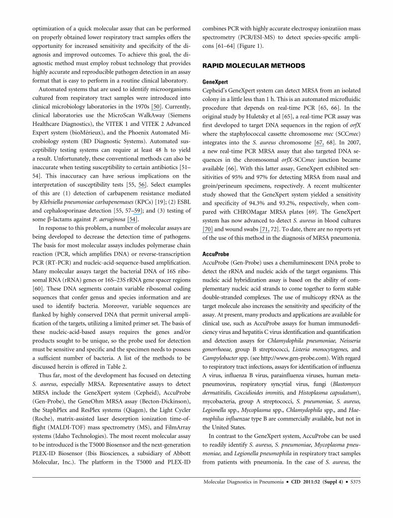
Molecular, Inc.). The platform in the T5000 and PLEX-ID
combines PCR with highly accurate electrospay ionizatiion mass
spectrometry (PCR/ESI-MS) to detect species-specific ampli-
cons [61–64] (Figure 1).
RAPID MOLECULAR METHODS
GeneXpertCepheid’s GeneXpert system can detect MRSA from an isolated
colony in a little less than 1 h. This is an automated microfluidic
procedure that depends on real-time PCR [65, 66]. In the
original study by Huletsky et al [65], a real-time PCR assay was
first developed to target DNA sequences in the region of orfX
where the staphylococcal cassette chromosome mec (SCCmec)
integrates into the S. aureus chromosome [67, 68]. In 2007,
a new real-time PCR MRSA assay that also targeted DNA se-
quences in the chromosomal orfX-SCCmec junction became
available [66]. With this latter assay, GeneXpert exhibited sen-
sitivities of 95% and 97% for detecting MRSA from nasal and
groin/perineum specimens, respectively. A recent multicenter
study showed that the GeneXpert system yielded a sensitivity
and specificity of 94.3% and 93.2%, respectively, when com-
pared with CHROMagar MRSA plates [69]. The GeneXpert
system has now advanced to detect S. aureus in blood cultures
[70] and wound swabs [71, 72]. To date, there are no reports yet
of the use of this method in the diagnosis of MRSA pneumonia.
AccuProbeAccuProbe (Gen-Probe) uses a chemiluminescent DNA probe to
detect the rRNA and nucleic acids of the target organisms. This
nucleic acid hybridization assay is based on the ability of com-
plementary nucleic acid strands to come together to form stable
double-stranded complexes. The use of multicopy rRNA as the
target molecule also increases the sensitivity and specificity of the
assay. At present, many products and applications are available for
clinical use, such as AccuProbe assays for human immunodefi-
ciency virus and hepatitis C virus identification and quantification
and detection assays for Chlamydophila pneumoniae, Neisseria
gonorrhoeae, group B streptococci, Listeria monocytogenes, and
Campylobacter spp. (see http://www.gen-probe.com). With regard
to respiratory tract infections, assays for identification of influenza
A virus, influenza B virus, parainfluenza viruses, human meta-
pneumovirus, respiratory syncytial virus, fungi (Blastomyces
dermatitidis, Coccidioides immitis, and Histoplasma capsulatum),
mycobacteria, group A streptococci, S. pneumoniae, S. aureus,
Legionella spp., Mycoplasma spp., Chlamydophila spp., and Hae-
mophilus influenzae type B are commercially available, but not in
the United States.
In contrast to the GeneXpert system, AccuProbe can be used
to readily identify S. aureus, S. pneumoniae, Mycoplasma pneu-
moniae, and Legionella pneumophila in respiratory tract samples
from patients with pneumonia. In the case of S. aureus, the
Molecular Diagnostics in Pneumonia d CID 2011:52 (Suppl 4) d S375

sensitivity and specificity of the AccuProbe system are reported
as 100% and 96%, respectively. In addition, there is good
agreement between quantitative cultures and probes in 96.3% of
cases. With the AccuProbe assay, there may be some difficulties
in diagnosing infection with atypical pneumococci with sputum
samples compared with diagnosis using PCR for the pneumo-
lysin gene, but these setbacks are uncommon [73–76].
BD GeneOhm StaphSR and BD GeneOhm MRSA AssaysThe BD GeneOhm MRSA assay (Becton-Dickinson) is a quali-
tative in vitro diagnostic test for the rapid detection of MRSA.
The assay can be performed in ,2 h (in many instances, it can
be performed in 1.5 h) and can also be performed directly from
clinical specimens [77–79]. In principle, these assays use rapid
nucleic acid tests to differentiate between coagulase-negative
Table 2. Summary of Selected Molecular Diagnostic Tests Discussed Here and Their Applications
Commercial
kit/molecular
assay (manufacturer) Advantages
Application to bacterial
pneumonia and/or
point-of-care testing
GeneXpertSystem(Cepheid)
Detects MRSA in 1 hin blood cultures andwound swabs
Undetermined
AccuProbe(Gen-Probe)
Detects Staphylococcusaureus, Streptococcuspneumoniae, Mycoplasmapneumoniae, and Legionellapneumophila
Mostly for point-of-careL. pneumophila testing
GeneOhm(Becton-Dickinson)
Detects MRSA, MSSA, andCoNS
Undetermined
ResPlex and StaphPlex(Qiagen)
Detects S. pneumoniae,Neisseria meningitidis,Haemophilus influenzae,L. pneumophila, M. pneumoniae,Chlamydophila pneumonia, andS.aureus
Yes, but large clinical trialsare needed for point-of-careS. aureus testing
Light Cycler(Roche)
Detects MRSA Undetermined
MALDI-TOF MS/Autoflex II(Bruker Daltonic)
Protein-based assays with broadmicrobiological applicability
Undetermined
FilmArray systems(Idaho Technologies)
Detects Bortedella pertussis,L. pneumophila, C. pneumoniae,and M. pneumoniae
Undetermined
Check KPC/ESBL microarray(Check-Points)
Detects b-lactamase resistance genesconferring resistance to cephalosporinsand carbapenems in 7–8 h
Undetermined
T5000 and PLEX-IDPCR/ESI-MS Biosensors(Abbott Molecular, Inc.)
Multiple species detected and typedand resistance genes mapped (gyrA, parC,mecA, and blaKPC)
Undetermined
NOTE. CoNS, coagulase-negative staphylococci; ESBL, extended-spectrum b-lactamase; KPC, Klebsiella pneumoniae carbapenemase; MALDI-TOF MS,
matrix-assisted laser desorption ionization time-of-flight mass spectrometry; MRSA, methicillin-resistant S. aureus; MSSA, methicillin-susceptible S. aureus; PCR/
ESI-MS, polymerase chain reaction followed by electrospray ionization mass spectrometry.
Figure 1. T5000 biosensor (A) and PLEX-ID biosensor and (B) (Ibis Biosciences, a subsidiary of Abbott Molecular, Inc.).
S376 d CID 2011:52 (Suppl 4) d Endimiani et al

staphylococci, methicillin-susceptible S. aureus (MSSA), and
MRSA. Similar to GeneXpert, the assay uses a multiplex real-
time PCR method to amplify a specific target sequence of S.
aureus near the SCCmec insertion site and the orfX junction gene
[65]. The assay works well in a low-prevalence setting to detect
MRSA from nasal, skin, and throat samples [80]. The ability of
this assay to detect MRSA in all situations is still under evalu-
ation [81]. In one study, the assay failed to detect the pre-
dominant Australian nosocomial clone (AUS2/3 clone; strain
type 239-MRSA-III) and a community-acquired clone prevalent
in eastern Australia (South West Pacific clone; strain type 30-
MRSA-IV) [82]. Nevertheless, the ability to differentiate
bloodstream infection caused by MSSA and MRSA from that
caused by other Gram-positive cocci is a major advantage [83].
Although reports have been published regarding the ability of
this test to detect MRSA in nasal and groin samples [71, 72],
studies are still needed to determine and validate whether this
assay is effective in the diagnosis of MRSA pneumonia.
ResPlex and StaphPlexUsing microarray technology, Qiagen developed a series of
assays—the ResPlex and StaphPlex panels. These panels in-
corporate multiplex PCR reactions that allow parallel detection
of bacterial and viral targets in a single reaction (hence, they
are called microarrays). The ResPlex assay amplifies and
detects gene-specific DNA sequences for S. pneumoniae (lytA),
Neisseria meningitidis (ctrA), encapsulated or nonencapsulated
H. influenzae (bexA and ompP2), L. pneumophila (mip), M.
pneumoniae (adenosine triphosphatase), and C. pneumoniae
(ompA) [84, 85]. The StaphPlex panel allows identification
of MRSA by amplifying and detecting 18 gene targets simulta-
neously [86]. These primers target information-rich genes in
staphylococci such as tuf for coagulase-negative staphylococci,
nuc for S. aureus, Panton-Valentine leukocidin (PVL) genes, and
antimicrobial resistance determinants of staphylococci (mecA,
SCCmecI-IV, aacA, ermA, ermB, tetM, and tetK) [86]. A similar
system was developed and used to screen for MRSA in nasal
swabs [87]. While the StaphPlex offers more robust features, the
ResPlex assay has clear potential to be used in the determination
of the etiology of HAP [88].
Roche LightCycler MRSA and SeptiFast MecA TestsThe LightCycler MRSA Advanced test (Roche) is a qualitative in
vitro diagnostic test for the direct detection of nasal colonization
by MRSA to aid in the prevention and control of MRSA in-
fections in health care settings. The test is performed on the
LightCycler 2.0 instrument with nasal swab specimens from
patients. The method uses swab extraction and mechanical lysis
for specimen preparation, followed by PCR for the amplification
of MRSA DNA and fluorogenic target-specific hybridization
probes for the detection of the amplified DNA. The LightCycler
MRSA Advanced test is designated for nasal specimens, and the
LightCycler SeptiFast MecA test is used for detection of MRSA
in bloodstream infections.
Originally, a 188-bp fragment within the mecA gene and
a 178-bp fragment within the S. aureus–specific Sa442 gene were
used for amplification. In the current version of this test, part of
the ITS region (internal transcribed spacer between 16S and 23S
gene) is targeted [89]. The LightCycler MRSA Advanced test
(nasal detection) is designed to aid in the prevention and control
of MRSA infections in health-care settings. The SeptiFast test
may be useful in determining bloodstream infections due to
MRSA. Recent evidence indicates that the latter test may prove
better than the conventional test currently performed in the
laboratory in cases of infective endocarditis in patients treated
with antibiotics before admission [90]. To date, the application
of these methods to determine whether MRSA is the causative
agent of pneumonia is still forthcoming.
MALDI-TOF MSMALDI-TOF MS is a protein/peptide-based diagnostic MS
method that can be used to assist with the rapid and accurate
identification of pathogens [91–93]. Because this method suc-
cessfully detects pathogens in blood cultures (in the best set of
analyses where 125 Gram-negative isolates were tested, there was
correct identification in 94%), there is hope that it can be ap-
plied to HAP. In a recent article in Clinical Infectious Diseases
[93], >1,600 clinical isolates were studied, and identification by
MALDI-TOF MS was compared with that by conventional cul-
ture methods (ie, Gram stain and Vitek or Analytical Profile
Index testing). MALDI-TOF MS demonstrated a sensitivity of
95% and specificity of 84.1% of the samples at the species level.
Seng and colleagues [93] found that it takes �6 min per isolate
for identification, and the cost is 22%–32% less than that of
current methods of identification. In most cases, absence of
identification or erroneous identification was due to construc-
tion of a less complete database (MALDI-TOF MS requires �10
reference samples in the database to be accurate). When the
investigators looked at the actual performance of MALDI-TOF
MS compared with that of conventional methods, MALDI-TOF
MS required less investment of time and energy, was also highly
specific, and did not increase the cost of identifying pathogens.
So far, MALDI-TOF has not been tested as a detector of
pathogens in sputum or as a point-of-care diagnostic instrument.
An interesting feature of MALDI-TOF MS is its ability to
identify PVL [91]. However, limitations to MALDI-TOF
MS exist. The large number of different staphylococci—either
S. aureus or coagulase-negative staphylococi—interferes with
the sensitivity and specificity of this assay [92, 93]. Moreover, the
identification of viridans streptococci also presents significant
problems. More relevant to the application in HAP is the clear
limitation of MALDI-TOF MS in correctly identifying a mixture
Molecular Diagnostics in Pneumonia d CID 2011:52 (Suppl 4) d S377
of species. In the study by La Scola and Raoult [92], when
a mixture of pathogens was presented, only 1 species was cor-
rectly identified, and false identification occurred. The perfor-
mance characteristics of MALDI-TOF MS will have to be
carefully monitored because sputum samples from patients with
HAP can have staphylococci, streptococci, and a mixture of
Gram-negative organisms.
Molecular BeaconsMolecular beacons are single-stranded oligonucleotide hybrid-
ization probes that form a hairpin-type stem-and-loop struc-
ture. As single-stranded probes, molecular beacons are
extraordinarily sensitive and specific and are suitable for single-
nucleotide allele discrimination. The target sequence is recog-
nized by the sequence in the loop; the stem is formed by the
annealing of complementary arm sequences that are located on
either side of the probe sequence. A quencher is covalently linked
to the end of one arm, and a fluorophore is covalently linked to
the end of the other arm. In free solution, the molecular beacon
does not emit light because the quencher is in proximity to the
fluorophore. However, when they hybridize to a nucleic acid
strand containing a target sequence, the fluorphore and
quencher are separated, resulting in bright fluorescence. Thus,
molecular beacons are considered to be molecular switches that
turn on when on their target and are off when in solution.
Molecular beacons have now been designed for the identifi-
cation of .110 different pathogens. Chakravorty et al [60] re-
cently developed mismatch-tolerant molecular beacons. These
so-called sloppy beacons enhance the diagnostic potential of the
assay by allowing less stringent detection of the molecular target
and present an important advance. The major bacterial patho-
gens (ie, S. aureus, S. pneumoniae, and P. aeruginosa) are de-
tected with this method.
In clinical specimens, S. pneumoniae (lytA gene), H. influenzae
(16S rRNA), M. pneumoniae (16S rRNA), C. pneumoniae (16S
rRNA), L. pneumophila (mip gene), and Streptococcus pyogenes
(16S rRNA) are readily detected by molecular beacons [94]. The
reported sensitivity and specificity of this real-time PCR assay
relative to conventional cultures were 96.2% and 93.2% for
S. pneumoniae, 95.8% and 95.4% for H. influenzae, and 100%
and 100% for S. pyogenes, respectively. Clinical experience with
molecular beacons to detect resistant pathogens is still required
in cases of HAP.
FilmArray SystemIdaho Technology is developing the FilmArray system to assist in
rapid molecular diagnostics. The FilmArray system is based on
microfluidics technology and promises to identify >30 pathogens
in �60 min. This method combines RT-PCR with a uniquely
designed lab-in-a-pouch system: a benchtop instrument performs
all the steps of the assay in an automated fashion, from nucleic
acid extraction to nested multiplex PCR and data analysis. By
using nested multiplex PCR, the targeting of conserved house-
keeping genes can accurately detect bacteria. The completely
automated assay takes ,60 min to run.
Primers are designed to be broad-range and are based on
alignments of housekeeping gene targets (ie, rpoB, gyrB, and
ompA). These outer primers target their domains by use of de-
generate nucleotides to provide cross-species recognition. Next,
species-specific inner primers are created and are placed in lo-
cations where the 3# end includes a characteristic nucleic acid
signature that is conserved among isolates of the same, but not
different, species. Currently, the FilmArray system detects the
following bacterial species in respiratory tract samples: Borde-
tella pertussis, C. pneumoniae, and M. pneumoniae. A wider
clinical application of this technology is still forthcoming
(hence, there have been no studies published on the use of this
technology to detect resistant Gram-negative bacteria). This
approach comprises a potential point-of-care diagnostic tool
because the support system to perform these assays is readily
mobile and inexpensive.
Microarray Technologies Detecting b-LactamasesAs shown above, microarrays possess a high multiplexing ca-
pacity and can be used for detecting an unlimited number of
genes within a reaction mixture. Recently, microarrays have
been applied to detect different b-lactamase (bla) genes that are
present in an isolate. The Check-Points Check KPC/ESBL mi-
croarray system uses a method called multiplex ligation detection
reaction. In brief, a series of specially designed DNA probes are
used that assist with PCR amplification. Next, the PCR products
are detected by hybridization to a low-density DNA microarray.
When there is hybridization, detection is accomplished using
a biotin label incorporated in one of the PCR primers. Although
this method does not identify the pathogen at the source of the
infection (ie, it cannot yet be used as a point-of-care test or for
a clinical specimen), this microarray can assist clinicians in di-
recting specific antimicrobial therapy once the resistance back-
ground of the pathogen is determined. Moreover, the assay takes
7–8 hours (1 typical working day). Endimiani and colleagues
[95] evaluated the ability of this microarray system in the de-
tection and identification of bla genes belonging to the TEM,
SHV, CTX-M, and KPC b-lactamases. This group reported
a sensitivity and specificity of 96.4%–100% when the test was
performed in a blinded fashion on previously characterized
isolates. In a complementary analysis performed by Naas et al
[96], Check KPC/ESBL microarray was also used prospectively
on clinical samples obtained directly from the microbiology
laboratory collected in a 3-month period and demonstrated
a similar sensitivity and specificity (up to 100%). Currently, this
assay is being further evaluated to detect other b-lactamase genes
such as plasmid-mediated AmpCs and NDM-1 metallo-
S378 d CID 2011:52 (Suppl 4) d Endimiani et al
b-lactamase. To date, the use of this assay in assisting with an-
tibiotic choices in cases of HAP due to antibiotic-resistant gram-
negative pathogens remains to be studied.
PCR Followed by ElectroSpray Ionization MS (PCR/ESI-MS)PCR/ESI-MS uses a rapid and highly accurate multilocus se-
quencing typing (MLST) method that allows (1) identification
of a very wide and diverse range of pathogens; (2) determination
of their genetic relatedness (clonality) compared with other
analyzed strains; (3) identification of virulence factors; and (4)
determination of resistance genotypes [62].
PCR/ESI-MS employs a robust bioinformatics infrastructure
that contains comprehensive gene sequence data [63, 64, 97].
With this database, multiple PCR amplification primers are de-
signed to amplify selected areas of the bacterial genome. These
PCR primers are broad-range and target ribosomal subunits (ri-
bosomal primers; 16S and 23S), unique housekeeping genes, or
other signature sequences from bacteria. In addition, by selecting
regions of variability, the primers yield 60–140-bp amplification
products that are information-rich. Next, the amplified double-
stranded DNA is desalted and heated to separate the strands, and
each strand is injected into a highly accurate mass spectrometer.
The mass of the single-stranded DNA is determined in 30 seconds.
An accurate sequence analysis is deduced from the mass of the
nucleotides and the DNA sequence is unambiguously determined
and compared with known DNA sequences that are present in
microbial genomes (http://www.ncbi.nlm.nih.gov/genomes/
lproks.cgi). When primers are strategically designed, �6 PCR
reactions and gene sequences (http://www.ncbi.nlm.nih.gov) can
identify almost all bacteria at a species level [98].
By means of a mathematical process called triangulation,
microorganisms with specific DNA sequences are further dis-
tinguished using precise DNA sequence information. An added
advantage to this approach is that primers can be designed to
also identify previously unknown members of a species—this
capability was demonstrated in the recent influenza pandemic
[61]. Information from several PCR/ESI-MS reactions triangu-
lates the identities of the organisms that are present. None of the
primers are designed to be specific for any one microorganism,
but instead the primers are designed to cover many pathogens
by use of a nested-coverage approach. This enables identification
of any bacterial species and even previously unknown organisms
with a single test [99–103].
Clinical Experience With PCR/ESI-MSThe first early study with the T5000 (Ibis Biosciences) involved an
outbreak of respiratory tract infection among recruits at a military
base in San Diego, California, during the years 2002–2003. Ecker
and colleagues [98] used the T5000 platform to identify the re-
sponsible pathogens and to determine the pathogen-strain ge-
notype. Hundreds of recruits became ill; 160 patients in this
outbreak were hospitalized, and 1 death was reported. By using
specific primers targeted to 23S ribosomal DNA, 3 predominant
pathogens were identified: H. influenzae, N. meningititis, and
S. pyogenes in throat swab samples (note that in this study,
isolates were examined from pure culture as well as from direct
throat swabs). Interestingly, the investigators did not detect
S. pneumoniae. This was a proof-of-concept study; PCR/ESI-MS
was able to diagnose multiple pathogens causing respiratory tract
infections.
Can one detect genes that confer resistance to antibiotics, and
can one perform epidemiological analyses with PCR/ESI-MS?
For this application, the target genes amplified by PCR/ESI-MS
need to be specific (unique), possess genetic uniformity, and be
conserved. So far, this has been applied to gyrA, parC, mecA, and
blaKPC. In a study designed to test this notion in ciprofloxacin-
resistant A. baumannii, performed by Hujer et al [104], 6 primer
pairs for conserved genes that encode amino acids in the qui-
nolone-resistance-determining regions of gyrA and parC of A.
baumannii were evaluated. The primers used were able to
identify mutations detected by PCR/ESI-MS in gyrA and parC.
This PCR/ESI-MS analysis accurately correlated with suscepti-
bility testing and sequencing results. Recently, Endimiani et al
[105] used this approach to detect the carbapenemase gene,
blaKPC, in K. pneumoniae with a high degree of sensitivity
(100%) and specificity (100%).
Wolk et al [106], identified the presence of the mecA gene and
showed very good correlation with the identification of the
MRSA phenotype. Furthermore, the identification of toxin
genes (ie, PVL and Toxic Shock Syndrome Toxin-1, TSST-1) by
PCR/ESI-MS correlated with independent PCR analyses for the
presence of these genes. Significantly, isolates were also correctly
classified into genotypic groups that correlated with genetic
clonal complexes, repetitive-element-based PCR patterns, or
pulsed-field gel electrophoresis (PFGE) types [107]. These ex-
amples show that this diagnostic approach (ie, pathogen and
resistance gene identification) could be applied to HAP.
Can this technology determine genetic relatedness? Can we
use PCR/ESI-MS to track the clonal expansion of a particular
strain type during a specific epidemic? This approach was re-
ported as successful in the analysis of S. pyogenes affecting mil-
itary recruits [98]; the identical streptococcal genotype was
found in almost all of the samples tested. In a study performed
with Acinetobacter spp., Ecker and colleagues studied 267 Aci-
netobacter spp. (216 clinical isolates and 51 reference strains)
[108]. In this collection, 47 different A. baumannii strain types
were identified. PCR/ESI-MS proved to be a significant advance
compared with Multi Locus Sequence Typing (MLST), as the
former was able to provide a real-time surveillance capability
with assay results available in ,6 h. A subsequent study by Hujer
et al [104], using Acinetobacter spp.isolates obtained from the
Walter Reed Army Medical Center, revealed that 16 different
clonal types were present in that collection (8 major clone types).
Molecular Diagnostics in Pneumonia d CID 2011:52 (Suppl 4) d S379

Many of the same strain types (eg, ST10, ST11, and ST14) were
present in the analysis by Ecker et al [108]. Interestingly, one of
these strain types (ST11) was also responsible for a case of oc-
cupational transmission of A. baumannii [109] to a nurse. So far,
the distribution of strain types between military and civilian
hospitals is different [110]. This understanding may change as
more outbreaks are analyzed. In Ohio, Perez et al [111] showed
that the T5000 biosensor was able to track an outbreak of MDR
A. baumannii infection through a health care system, identify the
main strain types (ST10 and ST12), and link the Ohio strain
types to the European clone II.
Jacobs and colleagues [112] showed that PCR/ESI-MS could
also be used to characterize S. pneumoniae isolates from serogroup
6. In this study, PCR/ESI-MS was employed to perform MLST
analysis and distinguish the distribution and the origin of serotype
6C strains. Recently, Endimiani and colleagues [39] studied clonal
complexes among linezolid-resistant isolates of S. aureus. The
linezolid-resistant isolates of S. aureus were found to be grouped
as part of clonal complex 5; USA 100 and USA 800 strain types
were detected by PFGE, and ST5 was detected by MLST.
The early clinical experience with PCR/ESI-MS in the de-
tection of S. pyogenes, S. pneumoniae, S. aureus, A. baumannii,
and P. aeruginosa is promising. PCR/ESI-MS can also assist in
the choice of targeted therapy by identifying genes that can
confer resistance to antibiotics and can help determine the
clonal relatedness of strains—an additional feature that can
enhance infection control practices.
Can PCR/ESI-MS be applied to sputum samples? In one case,
a sputum sample from a patient with cystic fibrosis was studied,
and the T5000 detected P. aeruginosa plus multiple other
potential bacterial pathogens (Chlamydophila spp., S. aureus,
S. pneumoniae, and Streptomyces rimosus) [62].
WHAT DOES THIS ALL MEAN FOR US?
These technologies offer the promise of dramatically improving
our ability to identify bacterial pathogens in respiratory tract
specimens with much needed sensitivity. These data from such
enhanced applications (ie, Check-Points or T5000 and PLEX-
ID, among others) can also be electronically integrated into
shared molecular databases. Clinicians and epidemiologists can
access such databases to ascertain local, regional, national, and
international trends. This likely will come forth as a major fea-
ture of the next-generation instruments.
Yet we must keep in mind that these new tools will not
guarantee that we will always get the best samples to analyze
or make correct antibiotic choices. Microbial recognition by
highly sensitive rapid diagnostic methods such as these will
still require good samples. Clinicians will still face difficult
questions about the meaning of these results. Most relevant
to the methods that detect nucleic acids is the question, does
finding DNA have the same impact as recovering living
pathogens? In addition, the extreme sensitivity of these as-
says (1 colony-forming unit) may result in simultaneous
detection of multiple pathogens from clinical specimens. If
this is true, what will be our new gold standard, and will this
information impact therapy? In short, for unparalleled ac-
curacy and sensitivity, are we replacing one level of ambi-
guity with new layers of uncertainty?
Right now, clinical trials are desperately needed to provide
evidence to help us decide which methods are the best and how to
apply this knowledge. Notwithstanding, we must also accept that
our comprehension of the microbial and metagenomic diversity
of the respiratory tract in health and disease is in its infancy. Will
we be able to use this information to help us reduce morbidity and
mortality and explain why patients fail to respond to antimicrobial
therapy for lung infections? Or will all the information obtained
by each of these methods serve to overwhelm the clinician? How
will we use biomarkers to help us decide what these pathogens
mean in HAP? The significance of finding bacterial DNA in the
absence of a positive culture in respiratory tract specimens will
surely reveal the complexities of the pulmonary microbiome. It
also indicates that we do not have a strong understanding of the
ecology of the airway and suffer from an inability to distinguish
between infecting and colonizing organisms. The new technolo-
gies reviewed here have opened novel vistas for detecting potential
pathogens. We now need to understand the clinical significance of
our newfound information. Information may be power, but we
should be careful what we wish for.
Acknowledgments
Financial support. This work was supported by the Veterans Affairs
Merit Review Program (R. A. B.); the National Institutes of Health (grants
R01-AI063517, R03-AI081036, and R01-AI072219 to R. A. B.); and the
Geriatric Research Education and Clinical Center VISN 10 (R. A. B.). Dr.
Perlin is supported by the National Institutes of Health.
Supplement sponsorship. This article was published as part of a sup-
plement entitled ‘‘Workshop on Molecular Diagnostics for Respiratory
Tract Infections.’’ The Food and Drug Administration and the Infectious
Diseases Society of America sponsored the workshop. AstraZeneca Phar-
maceuticals, Bio Merieux, Inc., Cepheid, Gilead Sciences, Intelligent MDX,
Inc., Inverness Medical Innovations, and Roche Molecular Systems pro-
vided financial support solely for the purpose of publishing the supplement.
Potential conflicts of interest. R. A. B. is a recipient of a research grant
from Pfizer and Steris Corporation and has collaborated with Ibis Bio-
sciences and Abbott Molecular, Inc., on publications in the screening of
bacterial isolates. D. S. P. receives grant support from Merck, Pfizer, As-
tellas, Celgene, bioMerieux, and the National Institutes of Health and serves
on advisory boards for Merck, Pfizer, Astellas, and Myconostica. All other
authors: no conflicts.
References
1. American Thoracic Society and Infectious Diseases Society of
America. Guidelines for the management of adults with hospital-ac-
quired, ventilator-associated, and healthcare-associated pneumonia.
Am J Respir Crit Care Med 2005; 171:388–416.
S380 d CID 2011:52 (Suppl 4) d Endimiani et al
2. Esperatti M, Ferrer M, Theessen A, et al. Nosocomial pneumonia
in the intensive care unit acquired by mechanically ventilated
versus nonventilated patients. Am J Respir Crit Care Med 2010;
182:1533–9.
3. Ferrer M, Liapikou A, Valencia M, et al. Validation of the American
Thoracic Society-Infectious Diseases Society of America guidelines for
hospital-acquired pneumonia in the intensive care unit. Clin Infect
Dis 2010; 50:945–52.
4. Torres A, Ferrer M, Badia JR. Treatment guidelines and outcomes of
hospital-acquired and ventilator-associated pneumonia. Clin Infect
Dis 2010; 51(suppl 1):S48–53.
5. Greenberg D, Broides A, Blancovich I, Peled N, Givon-Lavi N,
Dagan R. Relative importance of nasopharyngeal versus oropha-
ryngeal sampling for isolation of Streptococcus pneumoniae and
Haemophilus influenzae from healthy and sick individuals varies
with age. J Clin Microbiol 2004; 42:4604–9.
6. Bartlett JG, Breiman RF, Mandell LA, File TM Jr. Community-acquired
pneumonia in adults: guidelines for management. The Infectious Dis-
eases Society of America. Clin Infect Dis 1998; 26:811–38.
7. Houck PM, Bratzler DW, Nsa W, Ma A, Bartlett JG. Timing of antibiotic
administration and outcomes for Medicare patients hospitalized with
community-acquired pneumonia. Arch Intern Med 2004; 164:637–44.
8. Mandell LA, Niederman MS. Community-acquired pneumonia.
N Engl J Med 1996; 334:861.
9. Marik PE. Aspiration pneumonitis and aspiration pneumonia. N Engl
J Med 2001; 344:665–71.
10. Mizgerd JP. Acute lower respiratory tract infection. N Engl J Med
2008; 358:716–27.
11. Niederman MS. Review of treatment guidelines for community-acquired
pneumonia. Am J Med 2004; 117(suppl 3A):51S–7S.
12. Iregui M, Ward S, Sherman G, Fraser VJ, Kollef MH. Clinical im-
portance of delays in the initiation of appropriate antibiotic treatment
for ventilator-associated pneumonia. Chest 2002; 122:262–8.
13. Boucher HW, Talbot GH, Bradley JS, et al. Bad bugs, no drugs: no
ESKAPE! An update from the Infectious Diseases Society of America.
Clin Infect Dis 2009; 48:1–12.
14. Rice LB. Federal funding for the study of antimicrobial resistance in
nosocomial pathogens: no ESKAPE. J Infect Dis 2008; 197:1079–81.
15. Ritchie DJ, Alexander BT, Finnegan PM. New antimicrobial agents for
use in the intensive care unit. Infect Dis Clin North Am 2009;
23:665–81.
16. Endimiani A, Paterson DL. Optimizing therapy for infections caused
by enterobacteriaceae producing extended-spectrum beta-lactamases.
Semin Respir Crit Care Med 2007; 28:646–55.
17. Paterson DL, Bonomo RA. Extended-spectrum beta-lactamases:
a clinical update. Clin Microbiol Rev 2005; 18:657–86.
18. Queenan AM, Bush K. Carbapenemases: the versatile beta-lactamases.
Clin Microbiol Rev 2007; 20:440–58.
19. Bratu S, Mooty M, Nichani S, et al. Emergence of KPC-possessing
Klebsiella pneumoniae in Brooklyn, New York: epidemiology and
recommendations for detection. Antimicrob Agents Chemother 2005;
49:3018–20.
20. Endimiani A, Carias LL, Hujer AM, et al. Presence of plasmid-mediated
quinolone resistance in Klebsiella pneumoniae isolates possessing blaKPC
in the United States. Antimicrob Agents Chemother 2008; 52:2680–2.
21. Nordmann P, Cuzon G, Naas T. The real threat of Klebsiella pneu-
moniae carbapenemase-producing bacteria. Lancet Infect Dis 2009;
9:228–36.
22. Rice LB, Carias LL, Hutton RA, Rudin SD, Endimiani A, Bonomo RA.
The KQ element, a complex genetic region conferring transferable re-
sistance to carbapenems, aminoglycosides, and fluoroquinolones in
Klebsiella pneumoniae. Antimicrob Agents Chemother 2008; 52:3427–9.
23. Villegas MV, Lolans K, Correa A, Kattan JN, Lopez JA, Quinn JP. First
identification of Pseudomonas aeruginosa isolates producing a KPC-
type carbapenem-hydrolyzing beta-lactamase. Antimicrob Agents
Chemother 2007; 51:1553–5.
24. Woodford N, Tierno PM Jr., Young K, et al. Outbreak of Klebsiella
pneumoniae producing a new carbapenem-hydrolyzing class A beta-
lactamase, KPC-3, in a New York Medical Center. Antimicrob Agents
Chemother 2004; 48:4793–9.
25. Yigit H, Queenan AM, Anderson GJ, et al. Novel carbapenem-
hydrolyzing beta-lactamase, KPC-1, from a carbapenem-resistant
strain of Klebsiella pneumoniae. Antimicrob Agents Chemother
2001; 45:1151–61.
26. Yigit H, Queenan AM, Rasheed JK, et al. Carbapenem-resistant strain
of Klebsiella oxytoca harboring carbapenem-hydrolyzing beta-lactamase
KPC-2. Antimicrob Agents Chemother 2003; 47:3881–9.
27. Falagas ME, Grammatikos AP, Michalopoulos A. Potential of old-
generation antibiotics to address current need for new antibiotics.
Expert Rev Anti Infect Ther 2008; 6:593–600.
28. Falagas ME, Rafailidis PI, Matthaiou DK, Virtzili S, Nikita D,
Michalopoulos A. Pandrug-resistant Klebsiella pneumoniae, Pseudo-
monas aeruginosa and Acinetobacter baumannii infections: charac-
teristics and outcome in a series of 28 patients. Int J Antimicrob
Agents 2008; 32:450–4.
29. Giamarellou H, Poulakou G. Multidrug-resistant Gram-negative
infections: what are the treatment options? Drugs 2009; 69:
1879–901.
30. Michalopoulos A, Falagas ME. Colistin and polymyxin B in critical
care. Crit Care Clin 2008; 24:377–91, x.
31. Pappas G, Saplaoura K, Falagas ME. Current treatment of pseudo-
monal infections in the elderly. Drugs Aging 2009; 26:363–79.
32. Howden BP, Davies JK, Johnson PD, Stinear TP, Grayson ML.
Reduced vancomycin susceptibility in Staphylococcus aureus, including
vancomycin-intermediate and heterogeneous vancomycin-intermediate
strains: resistance mechanisms, laboratory detection, and clinical im-
plications. Clin Microbiol Rev 2010; 23:99–139.
33. Appelbaum PC. MRSA–the tip of the iceberg. Clin Microbiol Infect
2006; 12(suppl 2):3–10.
34. Cosgrove SE, Carroll KC, Perl TM. Staphylococcus aureus with
reduced susceptibility to vancomycin. Clin Infect Dis 2004;
39:539–45.
35. Sievert DM, Rudrik JT, Patel JB, McDonald LC, Wilkins MJ,
Hageman JC. Vancomycin-resistant Staphylococcus aureus in the
United States, 2002–2006. Clin Infect Dis 2008; 46:668–74.
36. Tenover FC, Weigel LM, Appelbaum PC, et al. Vancomycin-resistant
Staphylococcus aureus isolate from a patient in Pennsylvania. Anti-
microb Agents Chemother 2004; 48:275–80.
37. Whitener CJ, Park SY, Browne FA, et al. Vancomycin-resistant
Staphylococcus aureus in the absence of vancomycin exposure. Clin
Infect Dis 2004; 38:1049–55.
38. Brauers J, Kresken M, Hafner D, Shah PM. Surveillance of linezolid
resistance in Germany, 2001–2002. Clin Microbiol Infect 2005;
11:39–46.
39. Endimiani A, Blackford M, Dasenbrook EC, et al. Emergence of
linezolid-resistant clonal complex 5 Staphylococcus aureus with unique
ribosomal mutations among cystic fibrosis patients. Antimicrob
agents Chemother 2011.
40. Gales AC, Sader HS, Andrade SS, Lutz L, Machado A, Barth AL.
Emergence of linezolid-resistant Staphylococcus aureus during treat-
ment of pulmonary infection in a patient with cystic fibrosis. Int
J Antimicrob Agents 2006; 27:300–2.
41. Hill RL, Kearns AM, Nash J, et al. Linezolid-resistant ST36
methicillin-resistant Staphylococcus aureus associated with prolonged
linezolid treatment in two paediatric cystic fibrosis patients. J Anti-
microb Chemother 2010; 65:442–5.
42. Kola A, Kirschner P, Gohrbandt B, et al. An infection with linezolid-
resistant S. aureus in a patient with left ventricular assist system. Scand
J Infect Dis 2007; 39:463–5.
43. Morales G, Picazo JJ, Baos E, et al. Resistance to linezolid is mediated
by the cfr gene in the first report of an outbreak of linezolid-resistant
Staphylococcus aureus. Clin Infect Dis 2010; 50:821–5.
Molecular Diagnostics in Pneumonia d CID 2011:52 (Suppl 4) d S381

44. Peeters MJ, Sarria JC. Clinical characteristics of linezolid-resistant
Staphylococcus aureus infections. Am J Med Sci 2005; 330:
102–4.
45. Pillai SK, Sakoulas G, Wennersten C, et al. Linezolid resistance in
Staphylococcus aureus: characterization and stability of resistant phe-
notype. J Infect Dis 2002; 186:1603–7.
46. Roberts SM, Freeman AF, Harrington SM, Holland SM, Murray PR,
Zelazny AM. Linezolid-resistant Staphylococcus aureus in two pedi-
atric patients receiving low-dose linezolid therapy. Pediatr Infect Dis J
2006; 25:562–4.
47. Trevino M, Martinez-Lamas L, Romero-Jung PA, Giraldez JM,
Alvarez-Escudero J, Regueiro BJ. Endemic linezolid-resistant Staph-
ylococcus epidermidis in a critical care unit. Eur J Clin Microbiol Infect
Dis 2009; 28:527–33.
48. Yoshida K, Shoji H, Hanaki H, et al. Linezolid-resistant methicillin-
resistant Staphylococcus aureus isolated after long-term, repeated use
of linezolid. J Infect Chemother 2009; 15:417–9.
49. Kollef MH. Moving towards real-time antimicrobial management of
ventilator-associated pneumonia. Clin Infect Dis 2007; 44:388–90.
50. Stager CE, Davis JR. Automated systems for identification of micro-
organisms. Clin Microbiol Rev 1992; 5:302–27.
51. Doern GV, Brueggemann AB, Perla R, et al. Multicenter laboratory
evaluation of the bioMerieux Vitek antimicrobial susceptibility testing
system with 11 antimicrobial agents versus members of the family
Enterobacteriaceae and Pseudomonas aeruginosa. J Clin Microbiol
1997; 35:2115–9.
52. Tenover FC, Swenson JM, O’Hara CM, Stocker SA. Ability of com-
mercial and reference antimicrobial susceptibility testing methods to
detect vancomycin resistance in enterococci. J Clin Microbiol 1995;
33:1524–7.
53. Tenover FC, Williams PP, Stocker S, et al. Accuracy of six antimi-
crobial susceptibility methods for testing linezolid against staphylo-
cocci and enterococci. J Clin Microbiol 2007; 45:2917–22.
54. Torres E, Villanueva R, Bou G. Comparison of different methods of
determining beta-lactam susceptibility in clinical strains of Pseudo-
monas aeruginosa. J Med Microbiol 2009; 58:625–9.
55. Luzzaro F, Gesu G, Endimiani A, et al. Performance in detection and
reporting beta-lactam resistance phenotypes in Enterobacteriaceae:
a nationwide proficiency study in Italian laboratories. Diagn Micro-
biol Infect Dis 2006; 55:311–8.
56. Micek ST, Lloyd AE, Ritchie DJ, Reichley RM, Fraser VJ, Kollef MH.
Pseudomonas aeruginosa bloodstream infection: importance of ap-
propriate initial antimicrobial treatment. Antimicrob Agents Che-
mother 2005; 49:1306–11.
57. Steward CD, Rasheed JK, Hubert SK, et al. Characterization of clinical
isolates of Klebsiella pneumoniae from 19 laboratories using the National
Committee for Clinical Laboratory Standards extended-spectrum beta-
lactamase detection methods. J Clin Microbiol 2001; 39:2864–72.
58. Tenover FC, Emery SL, Spiegel CA, et al. Identification of plasmid-
mediated AmpC beta-lactamases in Escherichia coli, Klebsiella spp.,
and proteus species can potentially improve reporting of cephalo-
sporin susceptibility testing results. J Clin Microbiol 2009; 47:294–9.
59. Tenover FC, Mohammed MJ, Gorton TS, Dembek ZF. Detection and
reporting of organisms producing extended-spectrum beta-lactamases:
survey of laboratories in Connecticut. J Clin Microbiol 1999;
37:4065–70.
60. Chakravorty S, Aladegbami B, Burday M, et al. Rapid universal
identification of bacterial pathogens from clinical cultures by using
a novel sloppy molecular beacon melting temperature signature
technique. J Clin Microbiol 2010; 48:258–67.
61. Ecker DJ, Massire C, Blyn LB, et al. Molecular genotyping of microbes
by multilocus PCR and mass spectrometry: a new tool for hospital
infection control and public health surveillance. Methods Mol Biol
2009; 551:71–87.
62. Ecker DJ, Sampath R, Massire C, et al. Ibis T5000: a universal bio-
sensor approach for microbiology. Nat Rev Microbiol 2008; 6:553–8.
63. Ecker DJ, Sampath R, Willett P, et al. The Microbial Rosetta Stone
database: a common structure for microbial biosecurity threat agents.
J Forensic Sci 2005; 50:1380–5.
64. Ecker DJ, Sampath R, Willett P, et al. The Microbial Rosetta Stone
database: a compilation of global and emerging infectious micro-
organisms and bioterrorist threat agents. BMC Microbiol 2005; 5:19.
65. Huletsky A, Giroux R, Rossbach V, et al. New real-time PCR assay for
rapid detection of methicillin-resistant Staphylococcus aureus directly
from specimens containing a mixture of staphylococci. J Clin Mi-
crobiol 2004; 42:1875–84.
66. Rossney AS, Herra CM, Brennan GI, Morgan PM, O’Connell B.
Evaluation of the Xpert methicillin-resistant Staphylococcus aureus
(MRSA) assay using the GeneXpert real-time PCR platform for rapid
detection of MRSA from screening specimens. J Clin Microbiol 2008;
46:3285–90.
67. Hiramatsu K, Katayama Y, Yuzawa H, Ito T. Molecular genetics of
methicillin-resistant Staphylococcus aureus. Int J Med Microbiol 2002;
292:67–74.
68. Kuroda M, Ohta T, Uchiyama I, et al. Whole genome sequencing of
methicillin-resistant Staphylococcus aureus. Lancet 2001; 357:1225–40.
69. Wolk DM, Picton E, Johnson D, et al. Multicenter evaluation of the
Cepheid Xpert methicillin-resistant Staphylococcus aureus (MRSA)
test as a rapid screening method for detection of MRSA in nares. J Clin
Microbiol 2009; 47:758–64.
70. Parta M, Goebel M, Matloobi M, Stager C, Musher DM. Identification
of methicillin-resistant or methicillin-susceptible Staphylococcus au-
reus in blood cultures and wound swabs by GeneXpert. J Clin Mi-
crobiol 2009; 47:1609–10.
71. Hombach M, Pfyffer GE, Roos M, Lucke K. Detection of methicillin-
resistant Staphylococcus aureus (MRSA) in specimens from various
body sites: performance characteristics of the BD GeneOhm MRSA
assay, the Xpert MRSA assay, and broth-enriched culture in an area
with a low prevalence of MRSA infections. J Clin Microbiol 2010;
48:3882–7.
72. Malhotra-Kumar S, Van Heirstraeten L, Lee A, et al. Evaluation of
molecular assays for rapid detection of methicillin-resistant Staphy-
lococcus aureus. J Clin Microbiol 2010; 48:4598–601.
73. Allaouchiche B, Meugnier H, Freney J, Fleurette J, Motin J. Rapid
identification of Staphylococcus aureus in bronchoalveolar lavage fluid
using a DNA probe (Accuprobe). Intensive Care Med 1996; 22:683–7.
74. Finkelstein R, Brown P, Palutke WA, et al. Diagnostic efficacy of
a DNA probe in pneumonia caused by Legionella species. J Med
Microbiol 1993; 38:183–6.
75. Kaijalainen T, Rintamaki S, Herva E, Leinonen M. Evaluation of gene-
technological and conventional methods in the identification of
Streptococcus pneumoniae. J Microbiol Methods 2002; 51:111–8.
76. Kleemola M, Jokinen C. Outbreak of Mycoplasma pneumoniae in-
fection among hospital personnel studied by a nucleic acid hybrid-
ization test. J Hosp Infect 1992; 21:213–21.
77. Grobner S, Dion M, Plante M, Kempf VA. Evaluation of the BD
GeneOhm StaphSR assay for detection of methicillin-resistant and
methicillin-susceptible Staphylococcus aureus isolates from spiked
positive blood culture bottles. J Clin Microbiol 2009; 47:1689–94.
78. Paule SM, Hacek DM, Kufner B, et al. Performance of the BD Gen-
eOhm methicillin-resistant Staphylococcus aureus test before and
during high-volume clinical use. J Clin Microbiol 2007; 45:2993–8.
79. Wang XP, Ginocchio CC. Automation of the BD GeneOhm methi-
cillin-resistant Staphylococcus aureus assay for high-throughput
screening of nasal swab specimens. J Clin Microbiol 2009; 47:1546–8.
80. Svent-Kucina N, Pirs M, Mueller-Premru M, Cvitkovic-Spik V, Kofol
R, Seme K. One-year experience with modified BD GeneOhm MRSA
assay for detection of methicillin-resistant Staphylococcus aureus from
pooled nasal, skin, and throat samples. Diagn Microbiol Infect Dis
2009; 63:132–9.
81. Bartels MD, Boye K, Rohde SM, et al. A common variant of staphy-
lococcal cassette chromosome mec type IVa in isolates from
S382 d CID 2011:52 (Suppl 4) d Endimiani et al
Copenhagen, Denmark, is not detected by the BD GeneOhm methi-
cillin-resistant Staphylococcus aureus assay. J Clin Microbiol 2009;
47:1524–7.
82. Thomas L, van Hal S, O’Sullivan M, Kyme P, Iredell J. Failure of
the BD GeneOhm StaphS/R assay for identification of Australian
methicillin-resistant Staphylococcus aureus strains: duplex assays as the
‘‘gold standard’’ in settings of unknown SCCmec epidemiology. J Clin
Microbiol 2008; 46:4116–7.
83. Stamper PD, Cai M, Howard T, Speser S, Carroll KC. Clinical vali-
dation of the molecular BD GeneOhm StaphSR assay for direct de-
tection of Staphylococcus aureus and methicillin-resistant
Staphylococcus aureus in positive blood cultures. J Clin Microbiol
2007; 45:2191–6.
84. Benson R, Tondella ML, Bhatnagar J, et al. Development and evalu-
ation of a novel multiplex PCR technology for molecular differential
detection of bacterial respiratory disease pathogens. J Clin Microbiol
2008; 46:2074–7.
85. Winchell JM, Thurman KA, Mitchell SL, Thacker WL, Fields BS.
Evaluation of three real-time PCR assays for detection of Mycoplasma
pneumoniae in an outbreak investigation. J Clin Microbiol 2008;
46:3116–8.
86. Tang YW, Kilic A, Yang Q, et al. StaphPlex system for rapid and
simultaneous identification of antibiotic resistance determinants and
Panton-Valentine leukocidin detection of staphylococci from positive
blood cultures. J Clin Microbiol 2007; 45:1867–73.
87. Podzorski RP, Li H, Han J, Tang YW. MVPlex assay for direct de-
tection of methicillin-resistant Staphylococcus aureus in naris and
other swab specimens. J Clin Microbiol 2008; 46:3107–9.
88. Balada-Llasat JM, Larue H, Kelly C, Rigali L, Pancholi P. Evaluation of
commercial ResPlex II v2.0, MultiCode((R))-PLx, and xTAG((R))
respiratory viral panels for the diagnosis of respiratory viral infections
in adults. J Clin Virol 2011; 50:42–5.
89. Shrestha NK, Tuohy MJ, Padmanabhan RA, Hall GS, Procop GW.
Evaluation of the LightCycler Staphylococcus M GRADE kits on
positive blood cultures that contained gram-positive cocci in clusters.
J Clin Microbiol 2005; 43:6144–6.
90. Casalta JP, Gouriet F, Roux V, Thuny F, Habib G, Raoult D. Evalu-
ation of the LightCycler SeptiFast test in the rapid etiologic diagnostic
of infectious endocarditis. Eur J Clin Microbiol Infect Dis 2009;
28:569–73.
91. Bittar F, Ouchenane Z, Smati F, Raoult D, Rolain JM. MALDI-TOF-MS
for rapid detection of staphylococcal Panton-Valentine leukocidin. Int J
Antimicrob Agents 2009; 34:467–70.
92. La Scola B, Raoult D. Direct identification of bacteria in positive
blood culture bottles by matrix-assisted laser desorption ionisation
time-of-flight mass spectrometry. PLoS One 2009; 4:e8041.
93. Seng P, Drancourt M, Gouriet F, et al. Ongoing revolution in bac-
teriology: routine identification of bacteria by matrix-assisted laser
desorption ionization time-of-flight mass spectrometry. Clin Infect
Dis 2009; 49:543–51.
94. Morozumi M, Nakayama E, Iwata S, et al. Simultaneous detection of
pathogens in clinical samples from patients with community-acquired
pneumonia by real-time PCR with pathogen-specific molecular bea-
con probes. J Clin Microbiol 2006; 44:1440–6.
95. Endimiani A, Hujer AM, Hujer KM, et al. Evaluation of a commercial
microarray system for detection of SHV-, TEM-, CTX-M-, and KPC-
type beta-lactamase genes in Gram-negative isolates. J Clin Microbiol
2010; 48:2618–22.
96. Naas T, Cuzon G, Truong H, Bernabeu S, Nordmann P. Evaluation of
a DNA microarray, the check-points ESBL/KPC array, for rapid de-
tection of TEM, SHV, and CTX-M extended-spectrum beta-lactamases
and KPC carbapenemases. Antimicrob Agents Chemother 2010; 54:
3086–92.
97. Hari KL, Goates AT, Jain R, et al. The Microbial Rosetta Stone:
a database system for tracking infectious microorganisms. Int J Legal
Med 2009; 123:65–9.
98. Ecker DJ, Sampath R, Blyn LB, et al. Rapid identification and strain-
typing of respiratory pathogens for epidemic surveillance. Proc Natl
Acad Sci U S A 2005; 102:8012–7.
99. Deyde VM, Sampath R, Garten RJ. Genomic signature-based
identification of influenza A viruses using RT-PCR/electro-spray
ionization mass spectrometry (ESI-MS) technology. PLoS One 2010;
5:e13293.
100. Metzgar D, Baynes D, Myers CA, et al. Initial identification and
characterization of an emerging zoonotic influenza virus prior to
pandemic spread. J Clin Microbiol 2010; 48:4228–34.
101. Sampath R, Hall TA, Massire C, et al. Rapid identification of emerging
infectious agents using PCR and electrospray ionization mass spec-
trometry. Ann N Y Acad Sci 2007; 1102:109–20.
102. Sampath R, Russell KL, Massire C, et al. Global surveillance of
emerging influenza virus genotypes by mass spectrometry. PLoS One
2007; 2:e489.
103. Sauer S, Kliem M. Mass spectrometry tools for the classification and
identification of bacteria. Nat Rev Microbiol 2010; 8:74–82.
104. Hujer KM, Hujer AM, Endimiani A, et al. Rapid determination of
quinolone resistance in Acinetobacter spp. J Clin Microbiol 2009;
47:1436–2.
105. Endimiani A, Hujer KM, Hujer AM, Sampath R, Ecker DJ, Bonomo RA.
Rapid identification of blaKPC-possessing Enterobacteriaceae by PCR/
electrospray ionization-mass spectrometry. J Antimicrob Chemother
2010; 65:1833–34.
106. Wolk DM, Blyn LB, Hall TA, et al. Pathogen profiling: rapid molec-
ular characterization of Staphylococcus aureus by PCR/electrospray
ionization-mass spectrometry and correlation with phenotype. J Clin
Microbiol 2009; 47:3129–37.
107. Hall TA, Sampath R, Blyn LB, et al. Rapid molecular genotyping and
clonal complex assignment of Staphylococcus aureus isolates by PCR
coupled to electrospray ionization-mass spectrometry. J Clin Micro-
biol 2009; 47:1733–41.
108. Ecker JA, Massire C, Hall TA, et al. Identification of Acinetobacter
species and genotyping of Acinetobacter baumannii by multilocus PCR
and mass spectrometry. J Clin Microbiol 2006; 44:2921–32.
109. Whitman TJ, Qasba SS, Timpone JG, et al. Occupational transmis-
sion of Acinetobacter baumannii from a United States serviceman
wounded in Iraq to a health care worker. Clin Infect Dis 2008;
47:439–43.
110. Wortmann G, Weintrob A, Barber M, et al. Genotypic evolution of
Acinetobacter baumannii strains in an outbreak associated with war
trauma. Infect Control Hosp Epidemiol 2008; 29:553–5.
111. Perez F, Ray AJ, Endimiani A, et al. Carbapenem-resistant Klebsiella
pneumoniae across a hospital system: impact of post-acute care
facilities on dissemination. J Antimicrob Chemother 2010; 65:
1807–13.
112. Jacobs MR, Bajaksouzian S, Bonomo RA, et al. Occurrence,
distribution, and origins of Streptococcus pneumoniae Serotype 6C,
a recently recognized serotype. J Clin Microbiol 2009.; 47:64–72.
Molecular Diagnostics in Pneumonia d CID 2011:52 (Suppl 4) d S383
Related Documents