Cover Page The handle http://hdl.handle.net/1887/74362 holds various files of this Leiden University dissertation. Author: Jansen, F.A.R. Title: Are isolated heart defects really isolated? A prenatal view on submicroscopic genetics and brain development Issue Date: 2019-06-12

Welcome message from author
This document is posted to help you gain knowledge. Please leave a comment to let me know what you think about it! Share it to your friends and learn new things together.
Transcript
Cover Page
The handle http://hdl.handle.net/1887/74362 holds various files of this Leiden University dissertation. Author: Jansen, F.A.R. Title: Are isolated heart defects really isolated? A prenatal view on submicroscopic genetics and brain development Issue Date: 2019-06-12

Are isolated heart defects really isolated?A prenatal view on submicroscopic genetics
and brain development
Fenna Arina Roelien Jansen
ISBN: 978-94-6323-598-3Illustrations & cover design: Yara Francken, www.yarafrancken.comLogo Part||/HAND study: Boris Hoekmeijer, www.borishoekmeijer.nlLayout: Ilse Modder, www.ilsemodder.nlPrinting: Gildeprint Enschede, www.gildeprint.nl
Financial support by the Dutch Heart Foundation for the publication of this thesis is gratefully acknowledged.
The printing of this thesis was financially supported by the department of obstetrics and gynaecology of the Leiden University Medical Center, Leiden University Library (UB/Walaeus), Canon Medical Systems Nederland, BMA BV (Mosos).
Copyright 2019 Fenna Arina Roelien JansenAll rights reserved. No part of this publication may be reproduced or transmitted by any means without written permission from the author.
Are isolated heart defects really isolated?A prenatal view on submicroscopic genetics
and brain development
Proefschrift
ter verkrijging van de graad van Doctor aan de Universiteit Leiden,
op gezag van Rector Magnificus prof.mr. C.J.J.M. Stolker, volgens besluit van het College voor Promoties
te verdedigen op woensdag 12 juni 2019 klokke 16.15 uur
door
Fenna Arina Roelien Jansengeboren te ‘s-Gravenhage
in 1984
Promotores Prof. Dr. J.M.M. van LithProf. Dr. N.A. Blom
Co promotorDr. M.C. Haak
PromotiecommissieProf. Dr. C.M. Bilardo, Amsterdam UMCProf. Dr. J. S. Carvalho, MD, FRCPCH, Royal Brompton Hospital and St. George’s Hospital, London, United Kingdom.Prof. Dr. E. LoprioreProf. Dr. M.G. Hazekamp
Financial support by the Dutch Heart Foundation for the publication of this thesis is gratefully acknowledged.

Voor Julan,
Mickey,
Djenna en Noor,
Steven, Imran, Mila, Noemy, Haley,
Kate, Mees, Hannah, Daan, Sammy,
en de ontelbare andere kwetsbare kleintjes.

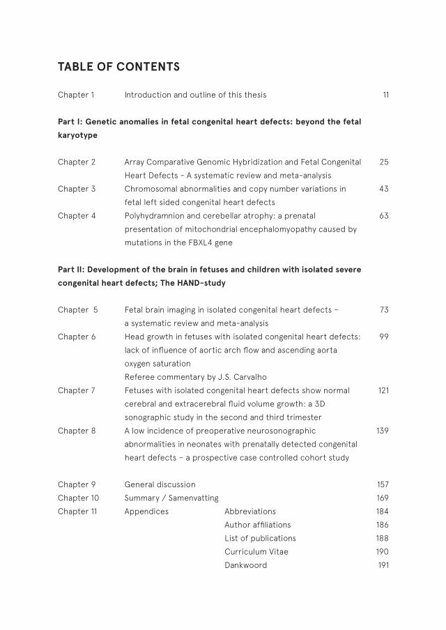
TABLE OF CONTENTS
Chapter 1 Introduction and outline of this thesis
Part I: Genetic anomalies in fetal congenital heart defects: beyond the fetal
karyotype
Chapter 2 Array Comparative Genomic Hybridization and Fetal Congenital Heart Defects - A systematic review and meta-analysisChapter 3 Chromosomal abnormalities and copy number variations in fetal left sided congenital heart defectsChapter 4 Polyhydramnion and cerebellar atrophy: a prenatal presentation of mitochondrial encephalomyopathy caused by mutations in the FBXL4 gene
Part II: Development of the brain in fetuses and children with isolated severe
congenital heart defects; The HAND-study
Chapter 5 Fetal brain imaging in isolated congenital heart defects – a systematic review and meta-analysisChapter 6 Head growth in fetuses with isolated congenital heart defects: lack of influence of aortic arch flow and ascending aorta oxygen saturation Referee commentary by J.S. CarvalhoChapter 7 Fetuses with isolated congenital heart defects show normal cerebral and extracerebral fluid volume growth: a 3D sonographic study in the second and third trimester Chapter 8 A low incidence of preoperative neurosonographic abnormalities in neonates with prenatally detected congenital heart defects – a prospective case controlled cohort study
Chapter 9 General discussion
Chapter 10 Summary / SamenvattingChapter 11 Appendices Abbreviations Author affiliations List of publications Curriculum Vitae Dankwoord
11
25
43
63
73
99
121
139
157169184186188190191


CHAPTER 1
Introduction and outline of this thesis
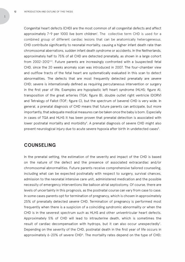
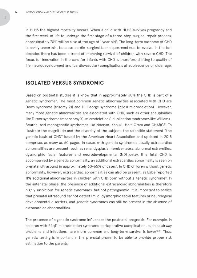
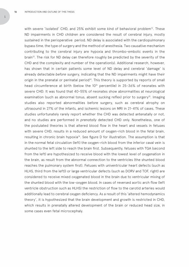
Congenital heart defects (CHD) are the most common of all congenital defects and affect approximately 7-9 per 1000 live born children1. The collective term CHD is used for a combined group of different cardiac lesions that can be anatomically heterogeneous. CHD contribute significantly to neonatal mortality, causing a higher infant death rate than chromosomal aberrations, sudden infant death syndrome or accidents. In the Netherlands, approximately half to 75% of all CHD are detected prenatally, as shown in a large cohort from 2002-20122;3. Future parents are increasingly confronted with a (suspected) fetal CHD, since the 20 weeks anomaly scan was introduced in 2007. The four-chamber view and outflow tracts of the fetal heart are systematically evaluated in this scan to detect abnormalities. The defects that are most frequently detected prenatally are severe CHD; severe is internationally defined as requiring percutaneous intervention or surgery in the first year of life. Examples are hypoplastic left heart syndrome (HLHS; figure A), transposition of the great arteries (TGA; figure B), double outlet right ventricle (DORV) and Tetralogy of Fallot (TOF; figure C), but the spectrum of (severe) CHD is very wide. In general, a prenatal diagnosis of CHD means that future parents can anticipate, but more importantly, that adequate medical measures can be taken once the baby is born. Especially in cases of TGA and HLHS it has been proven that prenatal detection is associated with lower postnatal mortality and morbidity4. A prenatal diagnosis of severe CHD might also prevent neurological injury due to acute severe hypoxia after birth in undetected cases5.
COUNSELING
In the prenatal setting, the estimation of the severity and impact of the CHD is based on the nature of the defect and the presence of associated extracardiac and/or chromosomal abnormalities. Future parents receive comprehensive tailored counseling, including what can be expected postnatally with respect to surgery, survival chances, admission to the neonatal intensive care unit, administered medication and the possible necessity of emergency interventions like balloon atrial septostomy. Of course, there are levels of uncertainty in this prognosis, as the postnatal course can vary from case to case. In some cases parents opt for termination of pregnancy, which is chosen in approximately 25% of prenatally detected severe CHD. Termination of pregnancy is performed most frequently when there is a suspicion of a coinciding syndromic abnormality or when the CHD is in the severest spectrum such as HLHS and other univentricular heart defects. Approximately 5% of CHD will lead to intrauterine death, which is sometimes the result of cardiac decompensation with hydrops, but it can also occur unexpectedly2. Depending on the severity of the CHD, postnatal death in the first year of life occurs in approximately 6-20% of severe CHD6. The mortality rates depend on the type of CHD;
12
1
INTRODUCTION AND OUTLINE OF THIS THESIS
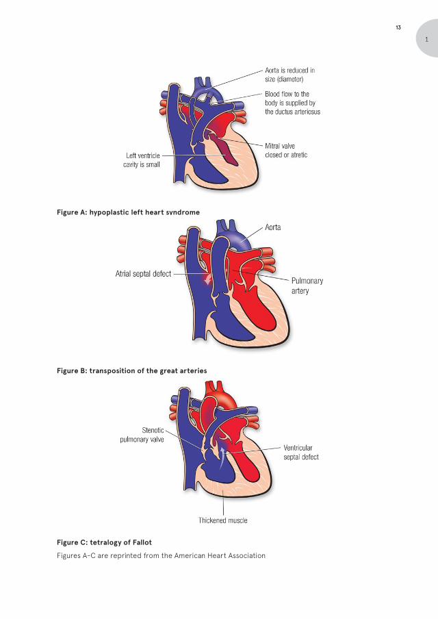
Figure A: hypoplastic left heart syndrome
Figure B: transposition of the great arteries
Figure C: tetralogy of Fallot
Figures A-C are reprinted from the American Heart Association
13
1
in HLHS the highest mortality occurs. When a child with HLHS survives pregnancy and the first week of life to undergo the first stage of a three-step surgical repair process, approximately 70% will be alive at the age of 1 year old7. The long-term outcome of CHD is partly uncertain, because cardio-surgical techniques continue to evolve. In the last decades there has been a trend of improving survival of children with severe CHD. The focus for innovation in the care for infants with CHD is therefore shifting to quality of life, neurodevelopment and (cardiovascular) complications at adolescence or older age.
ISOLATED VERSUS SYNDROMIC
Based on postnatal studies it is know that in approximately 30% the CHD is part of a genetic syndrome8. The most common genetic abnormalities associated with CHD are Down syndrome (trisomy 21) and Di George syndrome (22q11 microdeletion). However, many more genetic abnormalities are associated with CHD, such as other aneuploidies like Turner syndrome (monosomy X), microdeletion/-duplication syndromes like Williams-Beuren, and monogenetic syndromes like Noonan, Kabuki, Holt-Oram and CHARGE. To illustrate the magnitude and the diversity of the subject, the scientific statement “the genetic basis of CHD” issued by the American Heart Association and updated in 2018 comprises as many as 60 pages. In cases with genetic syndromes usually extracardiac abnormalities are present, such as renal dysplasia, hemivertebra, abnormal extremities, dysmorphic facial features and neurodevelopmental (ND) delay. If a fetal CHD is accompanied by a genetic abnormality, an additional extracardiac abnormality is seen on prenatal ultrasound in approximately 60-65% of cases2. In CHD children without genetic abnormality, however, extracardiac abnormalities can also be present, as Egbe reported 11% additional abnormalities in children with CHD born without a genetic syndrome9. In the antenatal phase, the presence of additional extracardiac abnormalities is therefore highly suspicious for genetic syndromes, but not pathognomic. It is important to realize that prenatal ultrasound cannot detect (mild) dysmorphic facial features or neurological developmental disorders, and genetic syndromes can still be present in the absence of extracardiac abnormalities.
The presence of a genetic syndrome influences the postnatal prognosis. For example, in children with 22q11 microdeletion syndrome perioperative complication, such as airway problems and infections, are more common and long-term survival is lower10;11. Thus, genetic testing is important in the prenatal phase, to be able to provide proper risk estimation to the parents.
14
1
INTRODUCTION AND OUTLINE OF THIS THESIS
There are several diagnostic genetic tests available, with varying resolutions and detection rates. The traditional karyogram is suitable to detect aneuploidies (trisomy/monosomy) and large deletions or duplications (size 5-10.000.000 base pairs). Until a few years back, karyotyping was the only option for prenatal genetic analysis. However, the molecular cytogenetic tests available nowadays enable us to study the genome in a higher resolution: array comparative genomic hybridization (array CGH, also known as micro-array) has evolved from bacterial artificial chromosome (BAC) array (resolution 1.000.000 base pairs), to oligonucleotide (resolution 30-50 base pairs) to even single nucleotide polymorphisms (SNP) array and next generation, whole exome or whole genome sequencing (NGS/WES/WGS) (resolution 1 base pair). These new tests can yield many copy number variants (CNVs) or point mutations, enabling accurate and comprehensive diagnosis of known syndromes and diseases such as Di George or Noonan syndrome. However, these more detailed genetic examinations have certain disadvantages as well. CNVs or smaller mutations also frequently occur in apparently healthy individuals, and some are considered ‘variants of unknown significance’ (VUS), indicating that its pathogenicity is unknown. This complicates the interpretation of the found genetic profile and makes counseling difficult, especially in the prenatal phase in which the phenotype can be incomplete. Counseling by a clinical geneticist is therefore of great importance in the event of an abnormal genetic profile - or any suspected syndromic disorder based on extracardiac abnormalities - because different syndromes may exhibit different penetrance, phenotypes and neurological development.
Currently, most prenatal centers in the Netherlands offer micro-array with a (reported) resolution up to 150.000 base pair. Whole exome sequencing (WES) is possible and performed incidentally, because the time window to generate and interpret the results has been reduced drastically the last few years, but it is not routinely performed prenatally yet. This is changing rapidly with the evolution of new laboratory techniques and the accelerating amount of knowledge and experience that has been acquired. Targeted testing for point mutations such as Noonan syndrome or CHARGE is possible, but rarely done because specific signs of these syndromes often lack prenatally.
NEUROLOGICAL OUTCOME
With the improving survival of children with CHD, the focus of research has shifted to the long-term and neurodevelopmental (ND) outcome. Even in the absence of chromosomal or syndromic abnormalities, ND delay may occur, especially in severe CHD cases. Large follow-up studies show that global ND delay occurs in 23% of children
15
1
with severe ‘isolated’ CHD, and 25% exhibit some kind of behavioral problem12. These ND impairments in CHD children are considered the result of cerebral injury, mostly sustained in the perioperative period. ND delay is associated with the cardiopulmonary bypass time, the type of surgery and the method of anesthesia. Two causative mechanism contributing to the cerebral injury are hypoxia and thrombo-embolic events in the brain13. The risk for ND delay can therefore roughly be predicted by the severity of the CHD and the complexity and number of the operation(s). Additional research, however, has shown that in certain patients some level of ND delay and cerebral ‘damage’ is already detectable before surgery, indicating that the ND impairments might have their origin in the prenatal or perinatal period14. This theory is supported by reports of small head circumference at birth (below the 10th percentile) in 25-36% of neonates with severe CHD. It was found that 40-55% of neonates show abnormalities at neurological examination (such as abnormal tonus, absent sucking reflex) prior to surgery15. Imaging studies also reported abnormalities before surgery, such as cerebral atrophy on ultrasound in 27% of the infants, and ischemic lesions on MRI in 21-41% of cases. These studies unfortunately rarely report whether the CHD was detected antenatally or not, and no studies are performed in prenatally detected CHD only. Nonetheless, one of the postulated theories is that altered blood flow in the heart and vessels in fetuses with severe CHD, results in a reduced amount of oxygen-rich blood in the fetal brain, resulting in chronic brain hypoxia16. See figure D for illustration. The assumption is that in the normal fetal circulation (left) the oxygen-rich blood from the inferior caval vein is shunted to the left side to reach the brain first. Subsequently, fetuses with TGA (second from the left) are hypothesized to receive blood with the lowest level of oxygenation in the brain, as result from the abnormal connection to the ventricles (the shunted blood reaches the pulmonary system first). Fetuses with univentricular heart defects (such as HLHS, third from the left)) or large ventricular defects (such as DORV and TOF, right) are considered to receive mixed oxygenated blood in the brain due to ventricular mixing of the shunted blood with the low-oxygen blood. In cases of reversed aortic arch flow (left ventricle obstruction such as HLHS) the restriction of flow to the carotid arteries would additionally lead to cerebral oxygen deficiency. As a result of this ‘altered hemodynamics theory’, it is hypothesized that the brain development and growth is restricted in CHD, which results in prenatally altered development of the brain or reduced head size, in some cases even fetal microcephaly.
16
1
INTRODUCTION AND OUTLINE OF THIS THESIS
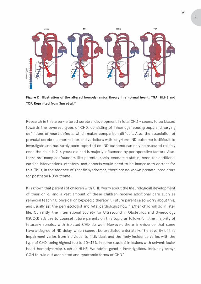
Figure D: Illustration of the altered hemodynamics theory in a normal heart, TGA, HLHS and
TOF. Reprinted from Sun et al.17
Research in this area - altered cerebral development in fetal CHD - seems to be biased towards the severest types of CHD, consisting of inhomogeneous groups and varying definitions of heart defects, which makes comparison difficult. Also, the association of prenatal cerebral abnormalities and variations with long-term ND outcome is difficult to investigate and has rarely been reported on. ND outcome can only be assessed reliably once the child is 2-4 years old and is majorly influenced by perioperative factors. Also, there are many confounders like parental socio-economic status, need for additional cardiac interventions, etcetera, and cohorts would need to be immense to correct for this. Thus, in the absence of genetic syndromes, there are no known prenatal predictors for postnatal ND outcome.
It is known that parents of children with CHD worry about the (neurological) development of their child, and a vast amount of these children receive additional care such as remedial teaching, physical or logopedic therapy12. Future parents also worry about this, and usually ask the perinatologist and fetal cardiologist how his/her child will do in later life. Currently, the International Society for Ultrasound in Obstetrics and Gynecology (ISUOG) advices to counsel future parents on this topic as follows18: ‘…the majority of fetuses/neonates with isolated CHD do well. However, there is evidence that some have a degree of ND delay, which cannot be predicted antenatally. The severity of this impairment varies from individual to individual, and the likely incidence varies with the type of CHD, being highest (up to 40–45% in some studies) in lesions with univentricular heart hemodynamics such as HLHS. We advise genetic investigations, including array‐CGH to rule out associated and syndromic forms of CHD.’
17
1
OUTLINE OF THIS THESIS
CHD are associated with chromosomal and syndromic abnormalities, as well as ND delay. In the prenatal phase however, the prevalence of genetic syndromes or risks of being affected by cerebral maldevelopment is still largely unknown. In many cases, when a CHD appears to be isolated, it is often assumed to be isolated. The aim of this thesis was to explore whether prenatally appearing isolated CHD are really isolated – without a genetic syndrome or a maldeveloped brain.
Part I explores the additional value of a array CGH and WES, two methods of genetic analysis with notable higher resolution than conventional karyotyping. In chapter 2, a systematic review and meta-analysis of the literature on array CGH in fetal CHD is presented. In chapter 3 the additional value of array CGH is assessed in a subgroup of CHD, left sided CHD, historically assumed to have a low prevalence of syndromic anomalies - when Turner syndrome is excluded. Chapter 4 describes an unusual case of a fetus with a small ventricular septal defect (VSD) and additional abnormalities. VSDs occur frequently and are usually innocent. However, in this case, additional abnormalities were found and indicated an underlying mitochondrial disease – identified with WES.
The second part of this thesis assesses prenatal and early postnatal brain development in CHD. In chapter 5, a systematic review and meta-analysis of prenatal cerebral development and cerebral variations in CHD is presented. Chapter 6 shows the results of a retrospective analysis of head circumference growth in a large cohort of fetal CHD, with a referee commentary on our study by the reviewing editor of the journal in which the paper was published. Chapters 7 and 8 describe analyses of a prospective cohort of isolated CHD cases compared to healthy controls, in which we performed extensive monthly neurosonography (the Heart And NeuroDevelopment (HAND) study). In chapter 7 the results of the volume measurements of the brain between 18 and 32 weeks of gestation are shown, exploring the prenatal cerebral growth and evolution of the extracerebral fluid compartments. Chapter 8 describes early postnatal cranial ultrasound findings and measurements, comparing prenatally detected CHD cases with healthy controls.
Chapter 9 presents a general discussion of the combined results of these studies, and chapter 10 is a general summary.
18
1
INTRODUCTION AND OUTLINE OF THIS THESIS
Additional remarks on ethical and legal aspects
In the Netherlands, there are several laws and guidelines regulating medical research; the most important being the WMO (‘law on medical research’). When fetal samples are taken - to analyze the genetic material for example - there might be left over material. This left over material can be used in medical research exempt from the WMO. Patients can object to the use in medical research when the sample is gathered, but they have to actively opt out themselves. This implicates that there is no need to request permission from the individual patients, as long as the patient has not objected, outcomes are not retraceable to individual patient data, and the additional analyses are performed on anonymized material. This was the case in chapter 3.
Observational studies with fetuses are prohibited by the ‘embryowet’ (2002). Therefore all fetal data analyzed in this thesis (chapters 6, 7, 8) were collected in the routine care for fetuses with CHD, and were therefore exempt from the WMO. The prospective inclusion of the healthy control group (chapters 7, 8) was only possible after a slight liberalization of the ‘embryowet’ in 2014, making it possible to perform observational studies on healthy fetuses after informed consent by the parents.
19
1
REFERENCES
1. Khoshnood, B., et al., Recent decrease in
the prevalence of congenital heart defects in
Europe. J Pediatr, 2013. 162(1): p. 108-13 e2.
2. van Velzen, C.L., et al., Prenatal detection of
congenital heart disease--results of a national
screening programme. BJOG, 2016. 123(3): p.
400-7.
3. Everwijn, S.M.P., et al., The effect of the
introduction of the three-vessel view on the
detection rate of transposition of the great
arteries and tetralogy of Fallot. Prenat Diagn,
2018 Nov;38(12):951-957.
4. Van Velzen, C.L., et al., Prenatal detection of
transposition of the great arteries reduces
mortality and morbidity. Ultrasound Obstet.
Gynecol, 2015 Mar;45(3):320-5
5. Peyvandi, S., et al., Association of Prenatal
Diagnosis of Critical Congenital Heart Disease
With Postnatal Brain Development and the Risk
of Brain Injury. JAMA Pediatr, 2016. 170(4): p.
e154450.
6. Oster, M.E., et al., Temporal trends in survival
among infants with critical congenital heart
defects. Pediatrics, 2013. 131(5): p. e1502-8.
7. Ohye, R.G., et al., Comparison of shunt types
in the Norwood procedure for single-ventricle
lesions. N Engl J Med, 2010. 362(21): p. 1980-92.
8. Eskedal, L., et al., A population-based study
of extra-cardiac anomalies in children with
congenital cardiac malformations. Cardiol
Young, 2004. 14(6): p. 600-7.
9. Egbe, A., et al., Prevalence of congenital
anomalies in newborns with congenital heart
disease diagnosis. Ann Pediatr Cardiol, 2014.
7(2): p. 86-91.
10. Simsic, J.M., et al., Do neonates with genetic
abnormalities have an increased morbidity and
mortality following cardiac surgery? Congenit.
Heart Dis, 2009. 4(3): p. 160-165.
11. Michielon, G., et al., Impact of DEL22q11,
trisomy 21, and other genetic syndromes on
surgical outcome of conotruncal heart defects.
J. Thorac. Cardiovasc. Surg, 2009. 138(3): p.
565-570.
12. Marino, B.S., et al., Neurodevelopmental
outcomes in children with congenital heart
disease: evaluation and management: a
scientific statement from the American Heart
Association. Circulation, 2012. 126(9): p. 1143-
1172.
13. Andropoulos, D.B., et al., Neurological
monitoring for congenital heart surgery. Anesth.
Analg, 2004. 99(5): p. 1365-1375.
14. Majnemer, A., et al., A new look at outcomes of
infants with congenital heart disease. Pediatr.
Neurol, 2009. 40(3): p. 197-204.
15. Owen, M., et al., Abnormal brain structure and
function in newborns with complex congenital
heart defects before open heart surgery:
A review of the evidence. Journal of Child
Neurology, 2011. 26(6): p. 743-755.
16. McQuillen, P.S., D.A. Goff, and D.J. Licht,
Effects of congenital heart disease on brain
development. Prog Pediatr Cardiol, 2010. 29(2):
p. 79-85.
17. Sun,L., et al., Reduced fetal cerebral oxygen
consumption is associated with smaller brain
size in fetuses with congenital heart disease.
Circulation. 2015 Apr 14. 131(15): 1313–1323.
18. Paladini, D., et al., ISUOG consensus statement
on current understanding of the association
of neurodevelopmental delay and congenital
20
1
INTRODUCTION AND OUTLINE OF THIS THESIS

heart disease: impact on prenatal counseling.
Ultrasound Obstet Gynecol, 2017. 49(2): p. 287-
288.
21
1

PART |
Genetic anomalies in fetal congenital
heart disease: beyond fetal karyotype

CHAPTER 2
Array Comparative Genomic
Hybridization and Fetal Congenital
Heart Defects - A systematic
review and meta-analysis
Ultrasound in Obstetrics and Gynecology 2015; 45: 27–35
F.A.R. Jansen Y.J. Blumenfeld A. FisherJ.M. CobbenA.O. Odibo A. Borrell M.C. Haak
ABSTRACT
ObjectiveArray comparative genomic hybridization (aCGH) is a molecular cytogenetic technique that is able to detect the presence of copy number variants (CNVs) within the genome. The detection rate of imbalances of aCGH compared to the standard karyotype and FISH 22q11 in the setting of prenatally diagnosed cardiac malformations has been reported in several studies. The objective of our study was to perform a systematic literature review and meta-analysis to document the additional diagnostic gain of aCGH in cases of congenital heart disease (CHD) diagnosed on prenatal ultrasound, in order to assist clinicians to determine whether aCGH analysis is warranted when ultrasonographic diagnosis of CHD is made, and to guide counseling in this setting.
MethodsAll articles in the PubMed, Embase and Web of Science database from January 2007 to September 2014 describing CNVs in prenatal cases of CHD were included. Search terms were: array comparative genomic hybridization, copy number variants, fetal congenital heart defects. Articles regarding karyotyping or 22q11 deletion only were excluded.
ResultsThirteen publications met the inclusion criteria for the analysis. Meta-analysis indicates an incremental yield of 7.0% (95% CI 5.3; 8.6) by aCGH, after exclusion of aneuploidy and 22q11 microdeletion. Subgroup results show 3.4% (95% CI 0.3; 6.6) incremental yield in isolated CHD and 9.3% (95% CI 6.6; 12) when extracardiac malformations are present. Overall incremental yield of 12% (95%CI 7.6; 16) was found including 22q11 deletion. There was an additional yield of 3.4% (95%CI 2.1; 4.6) of variants of unknown significance (VOUS).
DiscussionIn this review, we provide an overview of published data and discuss benefits and limitations of aCGH. If karyotyping and 22q11 microdeletion analysis by FISH are normal, aCGH has an additional value, detecting pathogenic CNVs in 7.0% of prenatally encountered CHD, with a 3.4% additional yield of detecting VOUS.
2
26 PART I: GENETIC ANOMALIES
INTRODUCTION
Congenital heart disease (CHD) is the leading cause of non-infectious neonatal mortality, affecting up to 1% of newborns. For most CHD, surgical repair or palliation is now possible with good outcome1. In some cases, however, the prognosis is dominated by the presence of chromosomal or extra-cardiac malformations2-4. In the prenatal setting the incidence of chromosomal anomalies is reported to be as high as 18-22% of all CHD, most being trisomy 21, trisomy 18, and 22q11 microdeletion5-7. Furthermore, fetuses with CHD carry a residual risk of additional genetic anomalies including microdeletion or –duplication syndromes such as Williams-Beuren and Potocki-Lupski or monogenetic anomalies such as Noonan syndrome.8;9
Providing information about the association of CHD with additional anomalies is important when counseling future parents. Assessing the presence of a pathogenic copy number variant (CNV) is crucial for prognostic purposes, given that the risk of non-iatrogenic neurological impairment is increased even in apparently isolated CHD10. Prenatal diagnosis of genetic conditions can also influence treatment plans2;4. In certain types of severe CHD, the interval between delivery and the necessary surgical procedure can be short, highlighting the importance of prenatal testing.
Cytogenetic fetal karyotyping used to be the gold standard of prenatal genetic testing. Karyotyping is able to detect aneuploidy and large chromosomal rearrangements up to 5-10 Mb. Array comparative genomic hybridization (aCGH) is a cytogenetic molecular technique that detects the presence of CNVs within the genome with increased resolution, a much higher resolution then conventional karyotyping, depending on the probe spacing and platform used.
Reports detailing the incremental yield of aCGH in the prenatal setting are rapidly emerging11-22. Most published reports include large cohorts, but describe the incremental yield for a variety of indications. Subgroup analysis of (different types of) CHD, the most common structural abnormality detected in the prenatal setting, is rarely reported. In this review, we describe the incremental yield of aCGH in prenatally diagnosed CHD. Our goal was to assist clinicians in determining whether aCGH is warranted once the diagnosis of a fetal CHD is made, and to guide them as they counsel future patients in this setting.
27
2
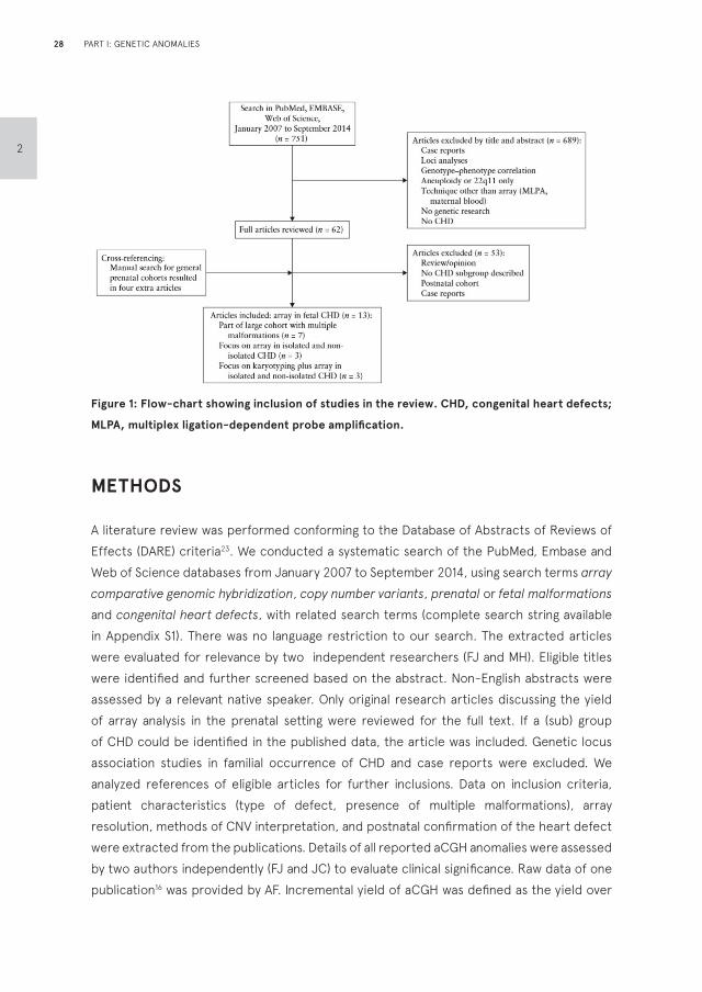
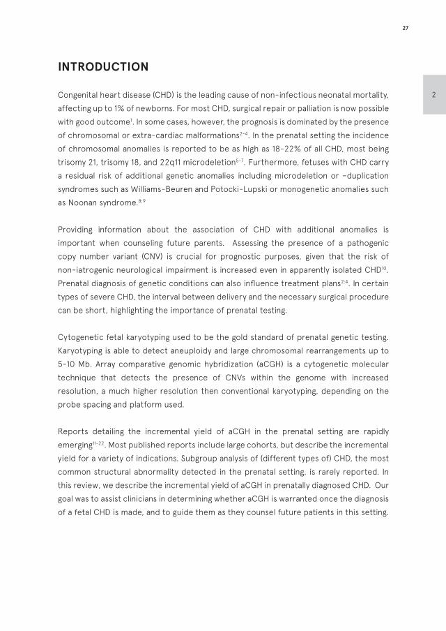
Figure 1: Flow-chart showing inclusion of studies in the review. CHD, congenital heart defects;
MLPA, multiplex ligation-dependent probe amplification.
METHODS
A literature review was performed conforming to the Database of Abstracts of Reviews of Effects (DARE) criteria23. We conducted a systematic search of the PubMed, Embase and Web of Science databases from January 2007 to September 2014, using search terms array comparative genomic hybridization, copy number variants, prenatal or fetal malformations and congenital heart defects, with related search terms (complete search string available in Appendix S1). There was no language restriction to our search. The extracted articles were evaluated for relevance by two independent researchers (FJ and MH). Eligible titles were identified and further screened based on the abstract. Non-English abstracts were assessed by a relevant native speaker. Only original research articles discussing the yield of array analysis in the prenatal setting were reviewed for the full text. If a (sub) group of CHD could be identified in the published data, the article was included. Genetic locus association studies in familial occurrence of CHD and case reports were excluded. We analyzed references of eligible articles for further inclusions. Data on inclusion criteria, patient characteristics (type of defect, presence of multiple malformations), array resolution, methods of CNV interpretation, and postnatal confirmation of the heart defect were extracted from the publications. Details of all reported aCGH anomalies were assessed by two authors independently (FJ and JC) to evaluate clinical significance. Raw data of one publication16 was provided by AF. Incremental yield of aCGH was defined as the yield over
28
2
PART I: GENETIC ANOMALIES
karyotyping only, or over karyotyping and FISH 22q11 combined. The incremental yields from each study were pooled to estimate an overall and subgroup incremental yield of aCGH using RevMan version 5.3.4 (Review Manager, The Cochrane Collaboration, Copenhagen, Denmark) and 95% CIs were computed. Studies with fewer than 20 cases were excluded from the meta-analysis. Statistical heterogeneity was examined using Higgins I2 (quantitative) test. To take into account the low statistical power of tests of heterogeneity, we considered statistically significant heterogeneity as Cochran’s Q test with a P<0.1 or I2 greater than 30%. A random effects model was used when there was significant heterogeneity. We assessed publication bias graphically using funnel plots. We assessed study quality based on the factors we considered most likely to threaten study validity (Table S1).
RESULTS
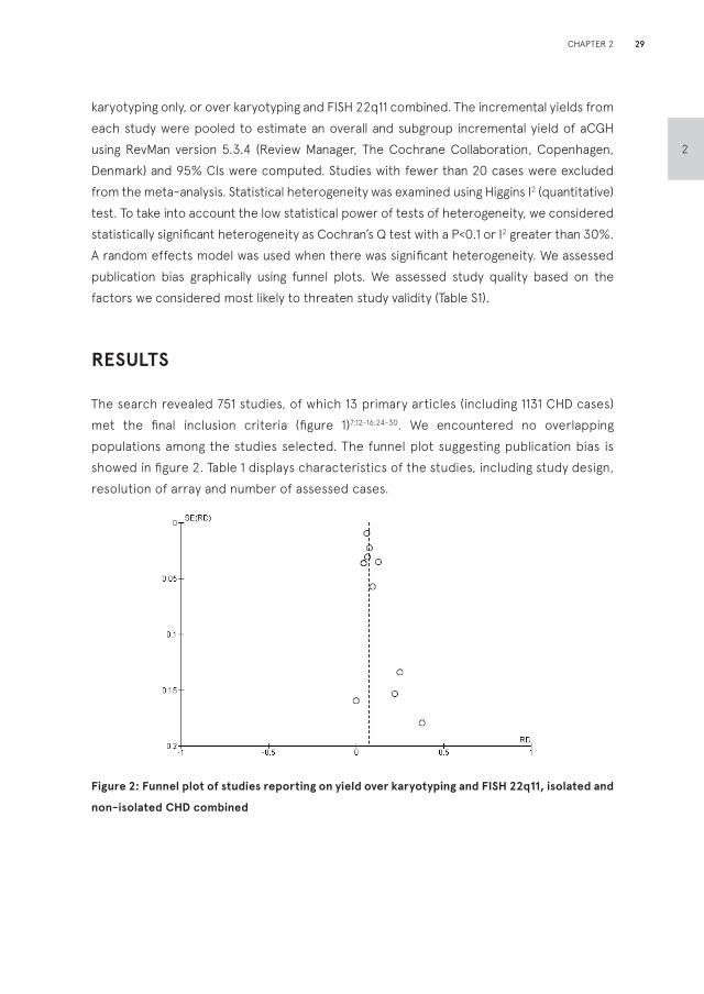
The search revealed 751 studies, of which 13 primary articles (including 1131 CHD cases) met the final inclusion criteria (figure 1)7;12-16;24-30. We encountered no overlapping populations among the studies selected. The funnel plot suggesting publication bias is showed in figure 2. Table 1 displays characteristics of the studies, including study design, resolution of array and number of assessed cases.
Figure 2: Funnel plot of studies reporting on yield over karyotyping and FISH 22q11, isolated and
non-isolated CHD combined
29
2
CHAPTER 2
Table 1: Included papers on incremental yield of aCGH in CHDA
utho
rSt
udy
desi
gnIn
clus
ion
crit
eria
of
orig
inal
stu
dy(S
ub)g
roup
of
CH
D +
aCG
HN
o non
-iso
late
d C
HD
incl
usio
n cr
iter
ia fo
r (s
ubse
quen
t) a
CG
HaC
GH
res
olut
ion
Des
crip
tion
of
CN
V in
terp
reta
tion
Post
-nat
al
confi
rm o
f C
HD
Tyre
man
200
9R
vario
us U
S ab
norm
aliti
es34
n.s.
norm
al k
aryo
type
&/o
r no
rmal
FIS
H 2
2q11
n.s.
(app
rox.
50
kb)
Yes
n.s.
Schm
id 2
012
Pfe
tal C
HD12
9 (7
1%)‡
norm
al F
ISH
22q
11 &
no
rmal
kar
yoty
pe
vario
us: 1
Mb
back
bone
/ ta
rget
ed 2
00kb
-
50kb
- 1
kb
No
n.s.
Shaf
fer 2
012
Pva
rious
US
abno
rmal
ities
580
343
(59%
)§no
rmal
kar
yoty
pe v
ario
usN
oN
o
Lee
2012
Pva
rious
indi
catio
ns
for g
enet
ic s
ampl
ing
50n.
s.no
ne (n
o an
eupl
oidy
in
CHD
gro
up)
vario
us p
lat-
form
s, 0
.5-0
.1Mb
Yes
n.s.
Faas
201
2P
vario
us U
S ab
norm
aliti
es10
0no
rmal
QF-
pcr
150
kb lo
ss /
20
0kb
gain
No
No
Bao
2013
Pfe
tal C
HD7
n.s.
“com
plex
CHD
”50
kbYe
sn.
s
Mad
emon
t-So
ler
2013
Rfe
tal C
HD o
r ca
rdia
c m
arke
rs51
*23
(45%
)‡no
rmal
FIS
H 2
2q11
&
norm
al k
aryo
type
100k
bYe
sYe
s
Hillm
an 2
013
Pva
rious
US
abno
rmal
ities
410
norm
al Q
F-pc
r>
2Mb
back
-bo
ne /
targ
eted
>2
00M
bYe
sso
me
Vest
erga
ard
2013
Pva
rious
US
abno
rmal
ities
9n.
s.no
rmal
kar
yoty
pe in
50
% o
f orig
inal
sam
ple
appr
ox. 8
0kb
Yes
n.s.
Yan
2014
Pfe
tal C
HD76
27 (3
6%) °
norm
al F
ISH
22q
11 &
no
rmal
kar
yoty
pe30
0kb
Yes
Yes
Liao
201
4P
feta
l CHD
†99
30 (3
0%)‡
norm
al k
aryo
type
100k
bYe
sn.
s.
Don
nelly
201
4P
vario
us U
S ab
norm
aliti
es15
488
(57%
)‡no
rmal
kar
yoty
pen.
s. 2
pla
tfor
ms
Yes
n.s.
Che
n 20
14R
cono
trun
cal C
HD8
n.s.
norm
al F
ISH
22q
11 &
no
rmal
kar
yoty
pe<1
kbYe
sYe
s
P pr
ospe
ctiv
e; R
retr
ospe
ctiv
e; n
.s. n
ot s
tate
d, C
HD c
onge
nita
l hea
rt d
efec
ts, C
NV
copy
num
ber v
aria
nts,
US
ultr
asou
nd ,
‡ fe
tuse
s w
ith a
dditi
onal
sof
t mar
kers
or m
inor
m
alfo
rmat
ions
incl
uded
in n
on-i
sola
ted
grou
p, §
fetu
ses
with
non
-str
uctu
ral a
bnor
mal
ities
(sof
tmar
kers
, gro
wth
ano
mal
ies,
etc
) inc
lude
d in
isol
ated
gro
up*
45 C
HD, 6
car
diac
mar
kers
, ° n
o sp
ecifi
catio
ns p
rovi
ded
on «
asso
ciat
ed a
bnor
mal
ities
», †
exc
ludi
ng m
inor
CHD
, unl
ess
addi
tiona
l mal
form
atio
ns w
ere
pres
ent
30
2
PART I: GENETIC ANOMALIES
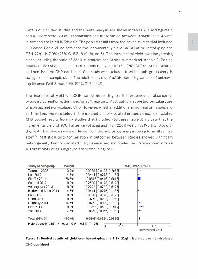
Details of included studies and the meta-analysis are shown in tables 2-4 and figures 3 and 4. There were 120 aCGH anomalies and these varied between 0.85kb29 and 14.9Mb7 in size and are listed in Table S2. The pooled results from the seven studies that included >20 cases (Table 2) indicate that the incremental yield of aCGH after karyotyping and FISH 22q11 is 7.0% (95% CI 5.3; 8.6) (figure 3). The incremental yield over karyotyping alone, including the yield of 22q11 microdeletions, is also summarized in table 2. Pooled results of five studies indicate an incremental yield of 12% (95%CI 7.6; 16) for isolated and non-isolated CHD combined. One study was excluded from this sub-group analysis owing to small sample size13. The additional yield of aCGH detecting variants of unknown significance (VOUS) was 3.4% (95% CI 2.1; 4.6).
The incremental yield of aCGH varied depending on the presence or absence of extracardiac malformations and/or soft markers. Most authors reported on subgroups of isolated and non-isolated CHD. However, whether additional minor malformations and soft markers were included in the isolated or non-isolated groups varied. For isolated CHD pooled results from six studies that included >20 cases (table 3) indicate that the incremental yield of aCGH after karyotyping and FISH 22q11 was 3.4% (95% CI 0.3; 6.6) (figure 4). Two studies were excluded from this sub-group analysis owing to small sample size14;30. Statistical tests for variation in outcomes between studies showed significant heterogeneity. For non-isolated CHD, summarized and pooled results are shown in table 4. Forest plots of all subgroups are shown in figure S1.
Figure 3: Pooled results of yield over karyotyping and FISH 22q11, isolated and non-isolated
CHD combined
31
2
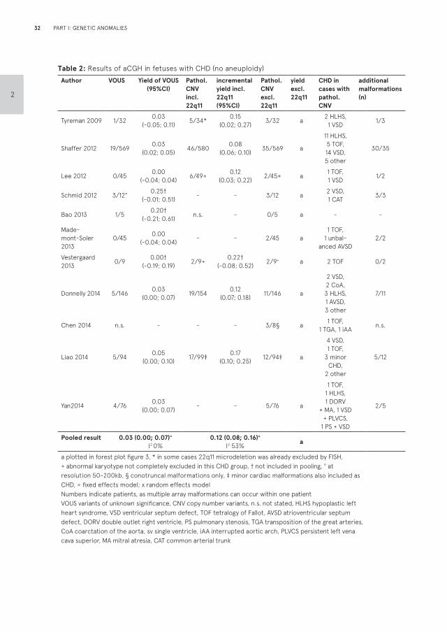
Table 2: Results of aCGH in fetuses with CHD (no aneuploidy)Author VOUS Yield of VOUS
(95%CI)Pathol. CNV incl. 22q11
incremental yield incl. 22q11 (95%CI)
Pathol. CNV excl. 22q11
yield excl. 22q11
CHD in cases with pathol. CNV
additional malformations (n)
Tyreman 2009 1/32 0.03 (-0.05; 0.11) 5/34* 0.15
(0.02; 0.27) 3/32 a 2 HLHS, 1 VSD 1/3
Shaffer 2012 19/569 0.03 (0.02; 0.05) 46/580 0.08
(0.06; 0.10) 35/569 a
11 HLHS, 5 TOF, 14 VSD, 5 other
30/35
Lee 2012 0/45 0.00 (-0.04; 0.04) 6/49+ 0.12
(0.03; 0.22) 2/45+ a 1 TOF, 1 VSD 1/2
Schmid 2012 3/12° 0.25†(-0.01; 0.51) - - 3/12 a 2 VSD,
1 CAT 3/3
Bao 2013 1/5 0.20†(-0.21; 0.61) n.s. - 0/5 a - -
Made-mont-Soler 2013
0/45 0.00(-0.04; 0.04) - - 2/45 a
1 TOF, 1 unbal-
anced AVSD2/2
Vestergaard 2013 0/9 0.00†
(-0.19; 0.19) 2/9+ 0.22†(-0.08; 0.52) 2/9+ a 2 TOF 0/2
Donnelly 2014 5/146 0.03 (0.00; 0.07) 19/154 0.12
(0.07; 0.18) 11/146 a
2 VSD, 2 CoA, 3 HLHS, 1 AVSD, 3 other
7/11
Chen 2014 n.s. - - - 3/8§ a 1 TOF, 1 TGA, 1 iAA n.s.
Liao 2014 5/94 0.05(0.00; 0.10) 17/99‡ 0.17
(0.10; 0.25) 12/94‡ a
4 VSD, 1 TOF,
3 minor CHD,
2 other
5/12
Yan2014 4/76 0.03(0.00; 0.07) - - 5/76 a
1 TOF, 1 HLHS, 1 DORV
+ MA, 1 VSD + PLVCS,
1 PS + VSD
2/5
Pooled result 0.03 (0.00; 0.07)=
I2 0%0.12 (0.08; 0.16)×
I2 53% a
a plotted in forest plot figure 3, * in some cases 22q11 microdeletion was already excluded by FISH, + abnormal karyotype not completely excluded in this CHD group, † not included in pooling, ° at resolution 50-200kb, § conotruncal malformations only, ‡ minor cardiac malformations also included as CHD, = fixed effects model; x random effects modelNumbers indicate patients, as multiple array malformations can occur within one patientVOUS variants of unknown significance, CNV copy number variants, n.s. not stated, HLHS hypoplastic left heart syndrome, VSD ventricular septum defect, TOF tetralogy of Fallot, AVSD atrioventricular septum defect, DORV double outlet right ventricle, PS pulmonary stenosis, TGA transposition of the great arteries, CoA coarctation of the aorta, sv single ventricle, iAA interrupted aortic arch, PLVCS persistent left vena cava superior, MA mitral atresia, CAT common arterial trunk
32
2
PART I: GENETIC ANOMALIES
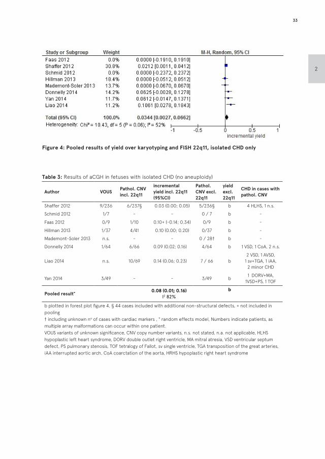
Figure 4: Pooled results of yield over karyotyping and FISH 22q11, isolated CHD only
Table 3: Results of aCGH in fetuses with isolated CHD (no aneuploidy)
Author VOUSPathol. CNV incl. 22q11
incremental yield incl. 22q11 (95%CI)
Pathol. CNV excl. 22q11
yield excl. 22q11
CHD in cases with pathol. CNV
Shaffer 2012 9/236 6/237§ 0.03 (0.00; 0.05) 5/236§ b 4 HLHS, 1 n.s.
Schmid 2012 1/7 - - 0 / 7 b -
Faas 2012 0/9 1/10 0.10+ (-0.14; 0.34) 0/9 b -
Hillman 2013 1/37 4/41 0.10 (0.00; 0.20) 0/37 b -
Mademont-Soler 2013 n.s. - - 0 / 28† b -
Donnelly 2014 1/64 6/66 0.09 (0.02; 0.16) 4/64 b 1 VSD, 1 CoA, 2 n.s.
Liao 2014 n.s. 10/69 0.14 (0.06; 0.23) 7 / 66 b2 VSD, 1 AVSD,
1 sv+TGA, 1 iAA, 2 minor CHD
Yan 2014 3/49 - - 3/49 b1 DORV+MA,
1VSD+PS, 1 TOF
Pooled result°0.08 (0.01; 0.16)
I2 82%
b
b plotted in forest plot figure 4, § 44 cases included with additional non-structural defects, + not included in pooling† including unknown no of cases with cardiac markers , ° random effects model, Numbers indicate patients, as multiple array malformations can occur within one patient.VOUS variants of unknown significance, CNV copy number variants, n.s. not stated, n.a. not applicable, HLHS hypoplastic left heart syndrome, DORV double outlet right ventricle, MA mitral atresia, VSD ventricular septum defect, PS pulmonary stenosis, TOF tetralogy of Fallot, sv single ventricle, TGA transposition of the great arteries, iAA interrupted aortic arch, CoA coarctation of the aorta, HRHS hypoplastic right heart syndrome
33
2
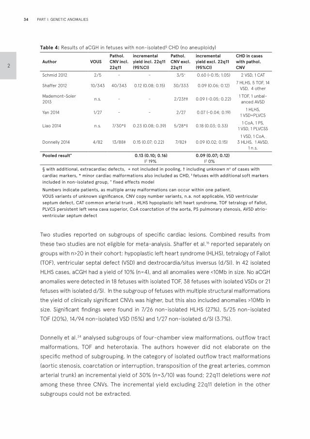
Table 4: Results of aCGH in fetuses with non-isolated§ CHD (no aneuploidy)
Author VOUSPathol. CNV incl. 22q11
incremental yield incl. 22q11 (95%CI)
Pathol. CNV excl. 22q11
incremental yield excl. 22q11 (95%CI)
CHD in cases with pathol. CNV
Schmid 2012 2/5 - - 3/5+ 0.60 (-0.15; 1.05) 2 VSD, 1 CAT
Shaffer 2012 10/343 40/343 0.12 (0.08; 0.15) 30/333 0.09 (0.06; 0.12)7 HLHS, 5 TOF, 14
VSD, 4 other
Mademont-Soler 2013
n.s. - - 2/23†‡ 0.09 (-0.05; 0.22)1 TOF, 1 unbal-
anced AVSD
Yan 2014 1/27 - - 2/27 0.07 (-0.04; 0.19)1 HLHS,
1 VSD+PLVCS
Liao 2014 n.s. 7/30*‡ 0.23 (0.08; 0.39) 5/28*‡ 0.18 (0.03; 0.33)1 CoA, 1 PS,
1 VSD, 1 PLVCSS
Donnelly 2014 4/82 13/88‡ 0.15 (0.07; 0.22) 7/82‡ 0.09 (0.02; 0.15)1 VSD, 1 CoA,
3 HLHS, 1 AVSD, 1 n.s.
Pooled result°
0.13 (0.10; 0.16)I2 19%
0.09 (0.07; 0.12) I2 0%
§ with additional, extracardiac defects, + not included in pooling, † including unknown no of cases with cardiac markers, * minor cardiac malformations also included as CHD, ‡ fetuses with additional soft markers included in non-isolated group, ° fixed effects model
Numbers indicate patients, as multiple array malformations can occur within one patient.VOUS variants of unknown significance, CNV copy number variants, n.a. not applicable, VSD ventricular septum defect, CAT common arterial trunk , HLHS hypoplastic left heart syndrome, TOF tetralogy of Fallot, PLVCS persistent left vena cava superior, CoA coarctation of the aorta, PS pulmonary stenosis, AVSD atrio-ventricular septum defect
Two studies reported on subgroups of specific cardiac lesions. Combined results from these two studies are not eligible for meta-analysis. Shaffer et al.16 reported separately on groups with n>20 in their cohort: hypoplastic left heart syndrome (HLHS), tetralogy of Fallot (TOF), ventricular septal defect (VSD) and dextrocardia/situs inversus (d/SI). In 42 isolated HLHS cases, aCGH had a yield of 10% (n=4), and all anomalies were <10Mb in size. No aCGH anomalies were detected in 18 fetuses with isolated TOF, 38 fetuses with isolated VSDs or 21 fetuses with isolated d/SI. In the subgroup of fetuses with multiple structural malformations the yield of clinically significant CNVs was higher, but this also included anomalies >10Mb in size. Significant findings were found in 7/26 non-isolated HLHS (27%), 5/25 non-isolated TOF (20%), 14/94 non-isolated VSD (15%) and 1/27 non-isolated d/SI (3.7%).
Donnelly et al.24 analysed subgroups of four-chamber view malformations, outflow tract malformations, TOF and heterotaxia. The authors however did not elaborate on the specific method of subgrouping. In the category of isolated outflow tract malformations (aortic stenosis, coarctation or interruption, transposition of the great arteries, common arterial trunk) an incremental yield of 30% (n=3/10) was found; 22q11 deletions were not among these three CNVs. The incremental yield excluding 22q11 deletion in the other subgroups could not be extracted.
34
2
PART I: GENETIC ANOMALIES
DISCUSSION
Considering the association of genetic anomalies with CHD, and the implications on both prenatal and postnatal management, obtaining the most accurate and detailed genetic information in the prenatal setting is important for both patients and providers.
In this systematic review, aCGH yielded additional clinically valuable information in 7.0% (95% CI 5.3; 8.6) of fetal CHD cases, even after karyotyping and FISH 22q11 analysis were normal. This includes both causative aCGH anomalies as well as incidental but clinical relevant findings, such as high risk for neurodevelopmental delay. The additional yield of VOUS was 3.4% (95%CI 2.1; 4.6).
In particular, there were more pathogenic CNV when extracardiac defects were present; estimated 9.3% (95% CI 6.6; 12). This yield appears lower when compared with published reports of aCGH in the postnatal setting, which describe yields of 17-53% in CHD with extracardiac malformations, neurodevelopmental delay and/or dysmorphic features32-39. This discrepancy can be attributed to non-comparable cohorts. There may be an ascertainment bias of cases in the postnatal groups that already present with neurodevelopmental delay or dysmorphic features.
When analyzing isolated CHD an incremental yield of 3.4% (95% CI 0.3; 6.6) was found. In postnatal cohorts of isolated CHD with normal karyotype and 22q11 analysis, the yield appears to be somewhat lower, 0-4%35;40-44. This small difference may be due to the limitation of prenatal ultrasound in detecting dysmorphic features and other subtle expressions of syndromic anomalies45.
It seems that VSDs (mainly perimembranous46) with extracardiac malformations, conotruncal malformations (TOF, interrupted arch) and left ventricle outflow tract malformations are common in prenatal cases which yield pathogenic aCGH results. Even transposition of the great arteries and heterotaxia, which are not considered to be associated with chromosomal anomalies by karyotyping, were found to have pathogenic aCGH results. However, the reported CHD with aCGH anomalies are very heterogeneous, and subgroups of different types of CHD are not large enough to analyse separately. Moreover, the categorisation of CHD is not consistent in the different reports, which inhibits calculation of the yield per specific CHD. Our recommendation therefore is to offer aCGH in all types of CHD.
In addition to submicroscopic anomalies <5-10 Mb in size, aCGH also yields anomalies
35
2
>10Mb. For example, karyotype failed to detect a large 14.9 Mb deletion, detected subsequently by aCGH7. Shaffer et al. reported the yield >10Mb separately. This emphasizes that karyotyping does not detect 100% of anomalies >10Mb in size, and aCGH may be a more reliable method of detecting these mutations47.
The possibility of aCGH replacing FISH 22q11 analysis in the prenatal setting of CHD merit consideration. The reported prevalence of 22q11 microdeletion in fetal CHD is as high as 7%7 ,with aortic arch and conotruncal malformations having the highest yields9;48. FISH 22q11 analysis is therefore already an important part of the diagnostic genetic work up in cases of isolated and non-isolated CHD. An important benefit of aCGH over FISH analysis for 22q11 microdeletions was noted by Chen29, reporting on 2 deletions in the 22q11 region which were not detected by FISH.
The limitation of our review is that pooled results are predominantly influenced by the report from Shaffer16, which has considerable uncertainty regarding confirmation of diagnosis. Furthermore, publications show large variability in the size of the cohort, platforms used, patient characteristics, and classification of extracardiac malformations. This results in high rates of statistical heterogeneity, especially in the isolated CHD subgroup. The process of interpreting CNV as pathogenic or benign is not always described and seems to vary significantly between the different groups. Larger prospective cohorts, focusing further on different types of CHD, are therefore warranted. Questions regarding the optimal probe spacing, platform and resolution, as well as the method of CNV categorization into benign and pathogenic, remain important.
There are some limitations of aCGH to be considered. First of all, the detection of VOUS could lead to challenges in counseling and parental anxiety. Combining data from large cohorts and linking certain aCGH anomalies with specific anatomic malformations could, however, increasingly reduce the frequency and clinical ambiguity of VOUS, for providers and their patients. Also, comparison with parental aCGH results can aid in detecting VOUS which are inherited from presumably healthy parents, therefore being less likely to be pathogenic. From our review it appears that studies that routinely performed aCGH of both parents, encountered a lower frequency of VOUS.
Secondly, clinicians should also be aware that single-gene disorders are also associated with CHD and they will not be detected by aCGH. These remain to be screened for individually on a case-by-case indication, until whole genome sequencing is available in the prenatal setting. Moreover, triploidies, chromosomal inversions and balanced translocations will not be detected by aCGH. Considerations for karyotype replacement
36
2
PART I: GENETIC ANOMALIES
by aCGH should therefore include an additional rapid method of aneuploidy/triploidy detection (RAD) such as quantitative fluorescent polymerase chain reaction17. Detection of balanced translocations and inversions does not seem a solid reason to perform complete karyotyping, as those chromosomal rearrangements will be detected if accompanied by a small deletion. Furthermore, if they are truly balanced, they are most probably not causative for CHD.
For pre-test counseling purposes results can be summarized as follows: the chance of finding an aCGH anomaly in prenatal CHD (including 22q11) is approximately 14% in total; 3% VOUS, 4% microdeletion 22q11 and 7% other pathogenic CNVs. In cases of isolated CHD with normal karyotype and 22q11 microdeletion analysis by FISH, the yield of additional aCGH has not yet been firmly established, but may be approximately 3%. In non-isolated cases, this yield is more evident, approximately 9%. In our opinion, given the available data, aCGH should be considered in cases of prenatally diagnosed fetal cardiovascular malformations, even if the lesion is apparently isolated based on prenatal imaging. As the common aneuploidies are most frequently associated with CHD, especially in cases of additional extra-cardiac malformations, aCGH can be considered if RAD results are normal, in order to reduce healthcare utilization and costs. However, local practices, the gestational age of the pregnancy, and regulations on pregnancy termination may lead providers to consider RAD and aCGH concurrently.
AcknowledgementsD. Zhao for the translation of the Chinese abstracts, our librarian J. Schoones for his help with the searches.
Supporting information on the internetThe following supporting information may be found in the online version of this article: Appendix S1 Complete search string Table S1 Quality assessment Table S2 List of encountered aCGH anomalies Figure S1 Forest plots of all subgroup analyses
37
2
REFERENCES
1. Hoffman JI, Kaplan S, Liberthson RR. Prevalence
of congenital heart disease. Am Heart J 2004
Mar;147(3):425-39.
2. Michielon G, Marino B, Oricchio G, Digilio MC,
Iorio F, Filippelli S, et al. Impact of DEL22q11,
trisomy 21, and other genetic syndromes on
surgical outcome of conotruncal heart defects. J
Thorac Cardiovasc Surg 2009 Sep;138(3):565-70.
3. Michielon G, Marino B, Formigari R, Gargiulo G,
Picchio F, Digilio MC, et al. Genetic syndromes and
outcome after surgical correction of tetralogy of
Fallot. Ann Thorac Surg 2006 Mar;81(3):968-75.
4. Simsic JM, Coleman K, Maher KO, Cuadrado
A, Kirshbom PM. Do neonates with genetic
abnormalities have an increased morbidity and
mortality following cardiac surgery? Congenit
Heart Dis 2009 May;4(3):160-5.
5. Song MS, Hu A, Dyamenahalli U, Chitayat D,
Winsor EJ, Ryan G, et al. Extracardiac lesions
and chromosomal abnormalities associated
with major fetal heart defects: comparison
of intrauterine, postnatal and postmortem
diagnoses. Ultrasound Obstet Gynecol 2009
May;33(5):552-9.
6. Chaoui R, Korner H, Bommer C, Goldner B,
Bierlich A, Bollmann R. (Prenatal diagnosis of
heart defects and associated chromosomal
aberrations). Ultraschall Med 1999 Oct;20(5):177-
84.
7. Mademont-Soler I, Morales C, Soler A, Martinez-
Crespo JM, Shen Y, Margarit E, et al. Prenatal
diagnosis of chromosomal abnormalities in
fetuses with abnormal cardiac ultrasound findings:
evaluation of chromosomal microarray-based
analysis. Ultrasound Obstet Gynecol 2013
Apr;41(4):375-82.
8. Hartman RJ, Rasmussen SA, Botto LD, Riehle-
Colarusso T, Martin CL, Cragan JD, et al. The
contribution of chromosomal abnormalities to
congenital heart defects: a population-based
study. Pediatr Cardiol 2011 Dec;32(8):1147-57.
9. Fahed AC, Gelb BD, Seidman JG, Seidman CE.
Genetics of congenital heart disease: the glass
half empty. Circ Res 2013 Feb 15;112(4):707-20.
10. Miller SP, McQuillen PS, Hamrick S, Xu D, Glidden
DV, Charlton N, et al. Abnormal brain development
in newborns with congenital heart disease. N Engl
J Med 2007 Nov 8;357(19):1928-38.
11. Hillman SC, Pretlove S, Coomarasamy A, McMullan
DJ, Davison EV, Maher ER, et al. Additional
information from array comparative genomic
hybridization technology over conventional
karyotyping in prenatal diagnosis: a systematic
review and meta-analysis. Ultrasound Obstet
Gynecol 2011 Jan;37(1):6-14.
12. Tyreman M, Abbott KM, Willatt LR, Nash R, Lees C,
Whittaker J, et al. High resolution array analysis:
diagnosing pregnancies with abnormal ultrasound
findings. J Med Genet 2009 Aug;46(8):531-41.
13. Vestergaard EM, Christensen R, Petersen OB,
Vogel I. Prenatal diagnosis: array comparative
genomic hybridization in fetuses with abnormal
sonographic findings. Acta Obstet Gynecol Scand
2013 Jul;92(7):762-8.
14. Faas BH, Feenstra I, Eggink AJ, Kooper AJ, Pfundt
R, van Vugt JM, et al. Non-targeted whole genome
250K SNP array analysis as replacement for
karyotyping in fetuses with structural ultrasound
anomalies: evaluation of a one-year experience.
Prenat Diagn 2012 Apr;32(4):362-70.
15. Lee CN, Lin SY, Lin CH, Shih JC, Lin TH, Su YN.
Clinical utility of array comparative genomic
38
2
PART I: GENETIC ANOMALIES
hybridisation for prenatal diagnosis: a
cohort study of 3171 pregnancies. BJOG 2012
Apr;119(5):614-25.
16. Shaffer LG, Rosenfeld JA, Dabell MP, Coppinger
J, Bandholz AM, Ellison JW, et al. Detection
rates of clinically significant genomic alterations
by microarray analysis for specific anomalies
detected by ultrasound. Prenat Diagn 2012
Oct;32(10):986-95.
17. Wapner RJ, Martin CL, Levy B, Ballif BC, Eng
CM, Zachary JM, et al. Chromosomal microarray
versus karyotyping for prenatal diagnosis. N Engl
J Med 2012 Dec 6;367(23):2175-84.
18. Gruchy N, Decamp M, Richard N, Jeanne-
Pasquier C, Benoist G, Mittre H, et al. Array CGH
analysis in high-risk pregnancies: comparing
DNA from cultured cells and cell-free fetal DNA.
Prenat Diagn 2012 Apr;32(4):383-8.
19. Armengol L, Nevado J, Serra-Juhe C, Plaja A,
Mediano C, Garcia-Santiago FA, et al. Clinical
utility of chromosomal microarray analysis in
invasive prenatal diagnosis. Hum Genet 2012
Mar;131(3):513-23.
20. Srebniak M, Boter M, Oudesluijs G, Joosten M,
Govaerts L, Van OD, et al. Application of SNP array
for rapid prenatal diagnosis: implementation,
genetic counselling and diagnostic flow. Eur J
Hum Genet 2011 Dec;19(12):1230-7.
21. Van den Veyver IB, Patel A, Shaw CA, Pursley AN,
Kang SH, Simovich MJ, et al. Clinical use of array
comparative genomic hybridization (aCGH) for
prenatal diagnosis in 300 cases. Prenat Diagn
2009 Jan;29(1):29-39.
22. Coppinger J, Alliman S, Lamb AN, Torchia BS,
Bejjani BA, Shaffer LG. Whole-genome microarray
analysis in prenatal specimens identifies clinically
significant chromosome alterations without
increase in results of unclear significance
compared to targeted microarray. Prenat Diagn
2009 Dec;29(12):1156-66.
23. http://www.crd.york.ac.uk/CRDWeb/AboutPage.
asp
24. Donnelly JC, Platt LD, Rebarber A, Zachary J,
Grobman WA, Wapner RJ. Association of copy
number variants with specific ultrasonographically
detected fetal anomalies. Obstet Gynecol 2014
Jul;124(1):83-90.
25. Hillman SC, Mcmullan DJ, Hall G, Togneri FS, James
N, Maher EJ, et al. Use of prenatal chromosomal
microarray: prospective cohort study and
systematic review and meta-analysis. Ultrasound
Obstet Gynecol 2013 Jun;41(6):610-20.
26. Liao C, Li R, Fu F, Xie G, Zhang Y, Pan M, et al.
Prenatal diagnosis of congenital heart defect by
genome-wide high-resolution SNP array. Prenat
Diagn 2014 Sep;34(9):858-63.
27. Bao B, WANG Y, Hu H, Yao H, Li Y, Tang S,
et al. Karyotypic and molecular genetic
changes associated with fetal cardiovascular
abnormalities: results of a retrospective
4-year ultrasonic diagnosis study. Int J Biol Sci
2013;9(5):463-71.
28. Yan Y, Wu Q, Zhang L, Wang X, Dan S, Deng D, et
al. Detection of submicroscopic chromosomal
aberrations by array-based comparative
genomic hybridization in fetuses with congenital
heart disease. Ultrasound Obstet Gynecol 2014
Apr;43(4):404-12.
29. Chen M, Yang YS, Shih JC, Lin WH, Lee DJ, Lin
YS, et al. Microdeletions/duplications involving
TBX1 gene in fetuses with conotruncal heart
defects which are negative for 22q11.2 deletion
39
2
on fluorescence in-situ hybridization. Ultrasound
Obstet Gynecol 2014 Apr;43(4):396-403.
30. Schmid M, Stary S, Blaicher W, Gollinger M,
Husslein P, Streubel B. Prenatal genetic diagnosis
using microarray analysis in fetuses with
congenital heart defects. Prenat Diagn 2012
Apr;32(4):376-82.
31. Krepischi-Santos AC, Vianna-Morgante AM, Jehee
FS, Passos-Bueno MR, Knijnenburg J, Szuhai K,
et al. Whole-genome array-CGH screening in
undiagnosed syndromic patients: old syndromes
revisited and new alterations. Cytogenet Genome
Res 2006;115(3-4):254-61.
32. Thienpont B, Mertens L, de RT, Eyskens B, Boshoff
D, Maas N, et al. Submicroscopic chromosomal
imbalances detected by array-CGH are a frequent
cause of congenital heart defects in selected
patients. Eur Heart J 2007 Nov;28(22):2778-84.
33. Breckpot J, Thienpont B, Peeters H, de RT, Singer
A, Rayyan M, et al. Array comparative genomic
hybridization as a diagnostic tool for syndromic
heart defects. J Pediatr 2010 May;156(5):810-7,
817.
34. Richards AA, Santos LJ, Nichols HA, Crider BP,
Elder FF, Hauser NS, et al. Cryptic chromosomal
abnormalities identified in children with
congenital heart disease. Pediatr Res 2008
Oct;64(4):358-63.
35. Rauch R, Hofbeck M, Zweier C, Koch A, Zink S,
Trautmann U, et al. Comprehensive genotype-
phenotype analysis in 230 patients with tetralogy
of Fallot. J Med Genet 2010 May;47(5):321-31.
36. Goldmuntz E, Paluru P, Glessner J, Hakonarson
H, Biegel JA, White PS, et al. Microdeletions and
microduplications in patients with congenital
heart disease and multiple congenital anomalies.
Congenit Heart Dis 2011 Nov;6(6):592-602.
37. Syrmou A, Tzetis M, Fryssira H, Kosma K,
Oikonomakis V, Giannikou K, et al. Array
comparative genomic hybridization as a clinical
diagnostic tool in syndromic and nonsyndromic
congenital heart disease. Pediatr Res 2013
Jun;73(6):772-6.
38. Lu XY, Phung MT, Shaw CA, Pham K, Neil SE,
Patel A, et al. Genomic imbalances in neonates
with birth defects: high detection rates by using
chromosomal microarray analysis. Pediatrics
2008 Dec;122(6):1310-8.
39. Erdogan F, Larsen LA, Zhang L, Tumer Z,
Tommerup N, Chen W, et al. High frequency of
submicroscopic genomic aberrations detected by
tiling path array comparative genome hybridisation
in patients with isolated congenital heart disease.
J Med Genet 2008 Nov;45(11):704-9.
40. Payne AR, Chang SW, Koenig SN, Zinn AR, Garg
V. Submicroscopic chromosomal copy number
variations identified in children with hypoplastic
left heart syndrome. Pediatr Cardiol 2012
Jun;33(5):757-63.
41. Iascone M, Ciccone R, Galletti L, Marchetti
D, Seddio F, Lincesso AR, et al. Identification
of de novo mutations and rare variants in
hypoplastic left heart syndrome. Clin Genet 2012
Jun;81(6):542-54.
42. Breckpot J, Thienpont B, Arens Y, Tranchevent
LC, Vermeesch JR, Moreau Y, et al. Challenges of
interpreting copy number variation in syndromic
and non-syndromic congenital heart defects.
Cytogenet Genome Res 2011;135(3-4):251-9.
43. Greenway SC, Pereira AC, Lin JC, DePalma
SR, Israel SJ, Mesquita SM, et al. De novo copy
number variants identify new genes and loci in
40
2
PART I: GENETIC ANOMALIES

isolated sporadic tetralogy of Fallot. Nat Genet
2009 Aug;41(8):931-5.
44. Shaffer LG, Coppinger J, Alliman S, Torchia
BA, Theisen A, Ballif BC, et al. Comparison of
microarray-based detection rates for cytogenetic
abnormalities in prenatal and neonatal specimens.
Prenat Diagn 2008 Sep;28(9):789-95.
45. Gomez O, Martinez JM, Olivella A, Bennasar M,
Crispi F, Masoller N, et al. Isolated ventricular
septal defects in the era of advanced fetal
echocardiography: risk of chromosomal
anomalies and spontaneous closure rate from
diagnosis to age of 1 year. Ultrasound Obstet
Gynecol 2014 Jan;43(1):65-71.
46. Yin A, Lu J, Liu C, Guo L, Wu J, Mai M, et
al. A prenatal missed diagnosed case of
submicroscopic chromosomal abnormalities by
karyotyping: the clinical utility of array-based
CGH in prenatal diagnostics. Mol Cytogenet
2014;7:26.
47. Pierpont ME, Basson CT, Benson DW, Jr., Gelb
BD, Giglia TM, Goldmuntz E, et al. Genetic basis
for congenital heart defects: current knowledge:
a scientific statement from the American
Heart Association Congenital Cardiac Defects
Committee, Council on Cardiovascular Disease in
the Young: endorsed by the American Academy of
Pediatrics. Circulation 2007 Jun 12;115(23):3015-
38.
41
2


F.A.R. Jansen M.J.V. Hoffer C.L. van Velzen S. Klingeman PlatiM.E.B. Rijlaarsdam S.A.B. Clur N.A. Blom E. Pajkrt S.L. BholaA.C. Knegt M.A. de Boer M.C. Haak
CHAPTER 3
Chromosomal abnormalities and copy
number variations in fetal left sided
congenital heart defects
Prenatal Diagnosis 2016; 36: 177–185
ABSTRACT
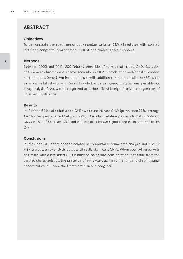
ObjectivesTo demonstrate the spectrum of copy number variants (CNVs) in fetuses with isolated left sided congenital heart defects (CHDs), and analyze genetic content.
MethodsBetween 2003 and 2012, 200 fetuses were identified with left sided CHD. Exclusion criteria were chromosomal rearrangements, 22q11.2 microdeletion and/or extra-cardiac malformations (n=64). We included cases with additional minor anomalies (n=39), such as single umbilical artery. In 54 of 136 eligible cases, stored material was available for array analysis. CNVs were categorized as either (likely) benign, (likely) pathogenic or of unknown significance.
ResultsIn 18 of the 54 isolated left sided CHDs we found 28 rare CNVs (prevalence 33%, average 1.6 CNV per person size 10.6kb – 2.2Mb). Our interpretation yielded clinically significant CNVs in two of 54 cases (4%) and variants of unknown significance in three other cases (6%).
ConclusionsIn left sided CHDs that appear isolated, with normal chromosome analysis and 22q11.2 FISH analysis, array analysis detects clinically significant CNVs. When counselling parents of a fetus with a left sided CHD it must be taken into consideration that aside from the cardiac characteristics, the presence of extra-cardiac malformations and chromosomal abnormalities influence the treatment plan and prognosis.
3
44 PART I: GENETIC ANOMALIES
INTRODUCTION
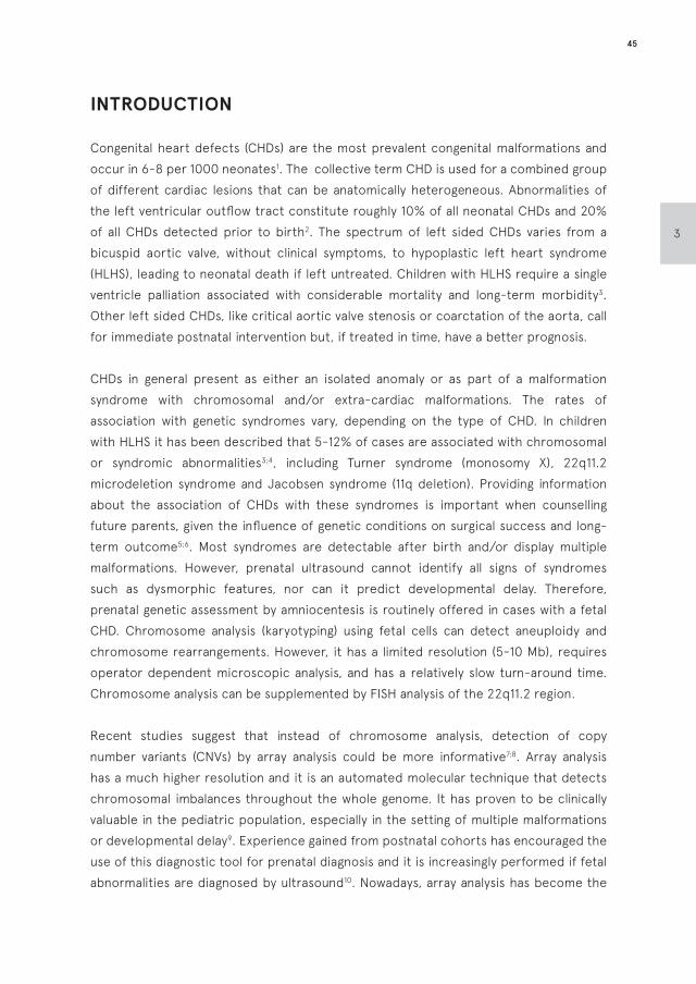
Congenital heart defects (CHDs) are the most prevalent congenital malformations and occur in 6-8 per 1000 neonates1. The collective term CHD is used for a combined group of different cardiac lesions that can be anatomically heterogeneous. Abnormalities of the left ventricular outflow tract constitute roughly 10% of all neonatal CHDs and 20% of all CHDs detected prior to birth2. The spectrum of left sided CHDs varies from a bicuspid aortic valve, without clinical symptoms, to hypoplastic left heart syndrome (HLHS), leading to neonatal death if left untreated. Children with HLHS require a single ventricle palliation associated with considerable mortality and long-term morbidity3. Other left sided CHDs, like critical aortic valve stenosis or coarctation of the aorta, call for immediate postnatal intervention but, if treated in time, have a better prognosis.
CHDs in general present as either an isolated anomaly or as part of a malformation syndrome with chromosomal and/or extra-cardiac malformations. The rates of association with genetic syndromes vary, depending on the type of CHD. In children with HLHS it has been described that 5-12% of cases are associated with chromosomal or syndromic abnormalities3;4, including Turner syndrome (monosomy X), 22q11.2 microdeletion syndrome and Jacobsen syndrome (11q deletion). Providing information about the association of CHDs with these syndromes is important when counselling future parents, given the influence of genetic conditions on surgical success and long-term outcome5;6. Most syndromes are detectable after birth and/or display multiple malformations. However, prenatal ultrasound cannot identify all signs of syndromes such as dysmorphic features, nor can it predict developmental delay. Therefore, prenatal genetic assessment by amniocentesis is routinely offered in cases with a fetal CHD. Chromosome analysis (karyotyping) using fetal cells can detect aneuploidy and chromosome rearrangements. However, it has a limited resolution (5-10 Mb), requires operator dependent microscopic analysis, and has a relatively slow turn-around time. Chromosome analysis can be supplemented by FISH analysis of the 22q11.2 region.
Recent studies suggest that instead of chromosome analysis, detection of copy number variants (CNVs) by array analysis could be more informative7;8. Array analysis has a much higher resolution and it is an automated molecular technique that detects chromosomal imbalances throughout the whole genome. It has proven to be clinically valuable in the pediatric population, especially in the setting of multiple malformations or developmental delay9. Experience gained from postnatal cohorts has encouraged the use of this diagnostic tool for prenatal diagnosis and it is increasingly performed if fetal abnormalities are diagnosed by ultrasound10. Nowadays, array analysis has become the
45
3

standard procedure for prenatal genetic analysis, and it is commonly preceded by rapid aneuploidy detection (RAD) to exclude common aneuploidies first11-13.
The prevalence of clinically significant CNVs in prenatal CHDs is described in a few cohorts14-20. As mentioned, CHD are a very heterogeneous group of lesions. The prenatal cohorts that have been published in recent years, focus on CHDs in general, but not at the level of the specific defect. These cohorts are not large enough, have significant selection bias, had no postnatal confirmation of the CHDs, or are otherwise unsuitable to extract the prevalence on the level of specific heart defects21. Thus, from a clinical point-of-view, our aim was to assess the presence and spectrum of clinically significant CNVs or variants of unknown significance (VOUS) by performing array analysis in a group of isolated fetal left sided CHDs.
MATERIALS AND METHODS
Cases with a prenatal diagnosis of a left sided CHD were selected from the CAHAL database. This is a regional cohort of fetuses with severe CHD born between 2002 and 2012 in the northwest region of the Netherlands. Methods of data collection are previously reported2. We extracted left sided CHD from this cohort, and subsequently excluded cases with additional CHD such as abnormal positioning of the great vessels. Ultrasound data were reviewed and cases were grouped as either ‘isolated’ or ‘non-isolated’ (defined as the presence of significant extra-cardiac malformations, hydrops or hygroma colli). Soft markers, minor additional findings, growth restriction, amniotic fluid pathology and/or single umbilical artery were not considered as significant extracardiac abnormalities. These cases are included in the ‘isolated’ group (see table S3). The presence and outcome of genetic analysis was assessed.
Cases with a prenatal diagnosis of an isolated left sided CHD, with a normal karyotype or rapid aneuploidy detection (RAD) result and absence of 22q11.2 microdeletion were eligible for array analysis Array was performed if frozen amniocytes, chorionic mesoderm, or isolated DNA was available in storage. Samples were anonymously processed. Affymetrix Cytoscan HD array or Agilent CGH 180K oligo array (Amadid 023363) was used as array platform and performed according to manufacturer’s instructions. Data analysis was performed using Chromosome Analysis Suite (ChAS) 2011 version CytoB-N1 2.0.232 (r4280), Nexus Copy Number versions 5.0, 6.1 and 7.0 or Genomic Workbench 6.5, and interpreted using Cartagenia BENCH 4.0 Feb-2012 (genome build hg19). Standard settings for SNPs in ChAS were adjusted: gain- size of 20 kb, marker count of 10, and a confidence of >85 and
46
3
PART I: GENETIC ANOMALIES
for loss-size of 10 kb, marker count of 10 and a confidence of >85. Standard settings for CNVs in Nexus were adjusted: threshold for probe median: gain 0.3 and loss -0.3. Minimal probes for a call: 20 per segment. Only samples meeting the quality criteria, i.e. QC >15, MapD <0.25 and a WavinessSD <0.12, were analyzed. For the oligo arrays analyzed with genomic workbench an aberration was defined as at least 3 consecutive probes with log2 ratio ≤ -0.4 or ≥0.4. The interpretation of CNVs has been done according the criteria as described by Gijsbers et al22. If parental material was available, we analyzed trios to assess whether rare CNVs were de novo or inherited. Various available online platforms were used, including the UCSC Genome Browser, Ensembl Genome Browser, the Toronto DB of Genomic Variants (DGV) and Decipher. Common polymorphic CNVs were considered as benign, with the exception of CNVs that are known as (possible) susceptibility factors, such as 15q11.2 BP1-BP2 microdeletions23;24 and Xp22.31 microduplications25;26, and maternally inherited CNVs on the X chromosome in male fetuses. The remaining variants were included for consideration for clinical significance. Inherited CNVs from parents were also considered as rare CNVs to account for CNVs with a possible reduced penetrance. To assess the function of the genes involved, we consulted PubMed and the OMIM database, as well as genecards.org (consulted between July and November 2015). Statistical analysis was performed using SPSS version 20.0.0.
RESULTS
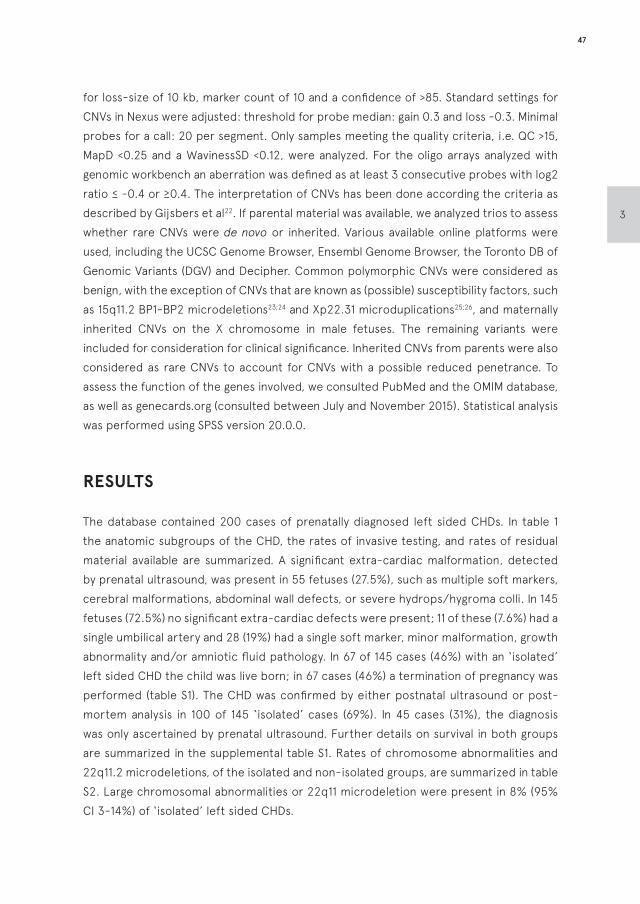
The database contained 200 cases of prenatally diagnosed left sided CHDs. In table 1 the anatomic subgroups of the CHD, the rates of invasive testing, and rates of residual material available are summarized. A significant extra-cardiac malformation, detected by prenatal ultrasound, was present in 55 fetuses (27.5%), such as multiple soft markers, cerebral malformations, abdominal wall defects, or severe hydrops/hygroma colli. In 145 fetuses (72.5%) no significant extra-cardiac defects were present; 11 of these (7.6%) had a single umbilical artery and 28 (19%) had a single soft marker, minor malformation, growth abnormality and/or amniotic fluid pathology. In 67 of 145 cases (46%) with an ‘isolated’ left sided CHD the child was live born; in 67 cases (46%) a termination of pregnancy was performed (table S1). The CHD was confirmed by either postnatal ultrasound or post-mortem analysis in 100 of 145 ‘isolated’ cases (69%). In 45 cases (31%), the diagnosis was only ascertained by prenatal ultrasound. Further details on survival in both groups are summarized in the supplemental table S1. Rates of chromosome abnormalities and 22q11.2 microdeletions, of the isolated and non-isolated groups, are summarized in table S2. Large chromosomal abnormalities or 22q11 microdeletion were present in 8% (95% CI 3-14%) of ‘isolated’ left sided CHDs.
47
3
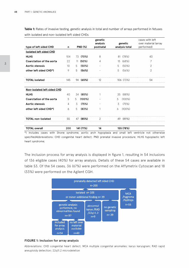
Table 1: Rates of invasive testing, genetic analysis in total and number of arrays performed in fetuses
with isolated and non-isolated left sided CHDs
type of left sided CHD n PND (%)
genetic analysis
postnatalgenetic
analysis total
cases with left over material (array performed)
Isolated left sided CHD
HLHS 104 73 (70%) 8 81 (78%) 43
Coarctation of the aorta 22 11 (50%) 4 15 (68%) 7
Aortic stenosis 10 5 (50%) - 5 (50%) 2
other left sided CHD*) 9 5 (56%) - 5 (56%) 2
TOTAL isolated 145 94 (65%) 12 106 (73%) 54
Non-isolated left sided CHD
HLHS 40 34 (85%) 1 35 (88%)
Coarctation of the aorta 5 5 (100%) - 5 (100%)
Aortic stenosis 4 3 (75%) - 3 (75%)
other left sided CHD*) 6 5 (83%) 1 6 (100%)
TOTAL non-isolated 55 47 (85%) 2 49 (89%)
TOTAL overall 200 141 (71%) 14 155 (78%)
*) includes cases with Shone syndrome, aortic arch hypoplasia and small left ventricle not otherwise specifiedAbbreviations: CHD congenital heart defect; PND prenatal invasive procedure; HLHS hypoplastic left heart syndrome;
The inclusion process for array analysis is displayed in figure 1, resulting in 54 inclusions of 136 eligible cases (40%) for array analysis. Details of these 54 cases are available in table S3. Of the 54 cases, 36 (67%) were performed on the Affymetrix Cytoscan and 18 (33%) were performed on the Agilent CGH.
FIGURE 1: Inclusion for array analysis
Abbreviations: CHD congenital heart defect; MCA multiple congenital anomalies; karyo karyogram; RAD rapid aneuploidy detection; 22q11.2 microdeletion
48
3
PART I: GENETIC ANOMALIES
Table 2 lists the encountered rare CNVs, the clinical implications, the locus on the chromosome, and the corresponding genes pertaining to that locus. We found 28 rare CNVs in 18 cases accounting for a prevalence of 33% with an average of 1.6 rare CNVs per person. The size of the CNVs ranged between 10.6 kb and 2.2 Mb. Our analysis and interpretation yielded clinically significant CNVs in 2 of 54 cases (4%; 95%CI 0 - 9%). In case 7 we found a ~10% mosaicism for trisomy 2, which remained undetected by previous chromosome analysis because at that time not enough cells (n=16) were analysed to detect the very low mosaicism. Because amniocytes were the cells used for the initial diagnosis, this result was not caused by a confined placental mosaicism. This aberration is known to be associated with cardiac defects and multiple congenital malformations27;28. Follow-up is unavailable because the pregnancy was terminated without post-mortem analysis. In case 48 we identified a 2.2Mb de novo 10q25 deletion, associated with multiple congenital malformations29;30. Genes include: DUSP5, associated with susceptibility to vascular anomalies, SMC3, associated with mild Cornelia de Lange syndrome 3, RBM20, associated with dilated cardiomyopathy, SHOC2, associated with Noonan-like disease, and ADRA2A, associated with cardiac hypertrophy and diminished contractility. Currently three years old, the child has dysmorphic features, a horseshoe kidney (missed antenatally), and appears to be developing normally compared to peers.
In the above mentioned two cases, as well as the 16 other cases, we also found 26 smaller CNVs. Most of these are unlikely to be clinically relevant or possibly causative, because the genetic involvement appears to be unrelated to critical developmental processes. Parental samples were not available for comparison in 14 of the 18 cases, therefore it is uncertain if 20 of the 26 found rare CNVs were inherited or de novo. Analysis of the involved genes demonstrated genes possibly related to abnormal cardiac development in only 1 case: In case 5 array analysis demonstrated a duplication including the 3’part of the AAK1 gene; this gene interacts with the activated form of NOTCH1a31. The clinical implications of this duplication are uncertain (VOUS). The parents were not tested, and the pregnancy was terminated without post-mortem analysis.
In case 38 we found a maternally inherited 4q21.23 deletion in a region including the WDFY3 gene. This deletion has previously been reported as a possible risk factor for autism spectrum pathology32. This child died 3 weeks after birth due to cardiovascular complications.
In case 43 we found a maternally inherited Xp22.31 duplication in a region including the STS gene in a male fetus. This gain has been reported as a possible risk factor for neurodevelopmental delay25;26. This child died after surgery due to cardiovascular complications.
49
3
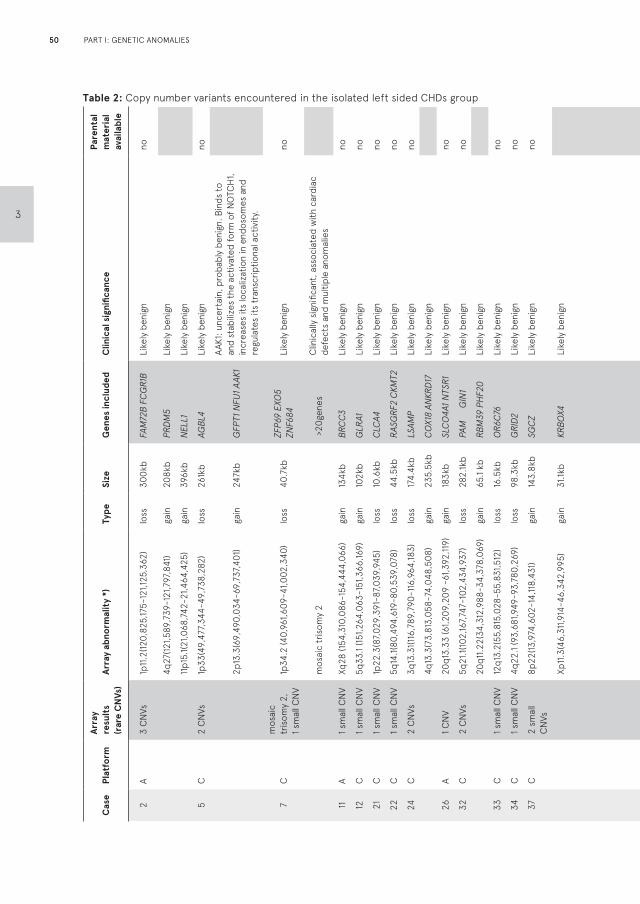
Table 2: Copy number variants encountered in the isolated left sided CHDs groupC
ase
Plat
form
Arr
ayre
sult
s (r
are
CN
Vs)
Arr
ay a
bnor
mal
ity
*)Ty
peSi
zeG
enes
incl
uded
Clin
ical
sig
nific
ance
Pare
ntal
m
ater
ial
avai
labl
e
2A
3 C
NVs
1p11
.2(12
0,82
5,17
5-12
1,125
,362
)lo
ss30
0kb
FAM
72B
FC
GR
1B
Like
ly b
enig
nno
4q
27(12
1,589
,739
-121
,797
,841
)ga
in20
8kb
PRD
M5
Like
ly b
enig
n
11
p15.
1(21,0
68,7
42-2
1,464
,425
)ga
in39
6kb
NEL
L1Li
kely
ben
ign
5C
2 C
NVs
1p33
(49,
477,
344-
49,7
38,2
82)
loss
261k
bAG
BL4
Like
ly b
enig
nno
2p
13.3
(69,
490,
034-
69,7
37,4
01)
gain
247k
bG
FPT1
NFU
1 AAK
1
AAK1
: unc
erta
in, p
roba
bly
beni
gn. B
inds
to
and
stab
ilize
s th
e ac
tivat
ed fo
rm o
f NO
TCH
1, in
crea
ses
its lo
caliz
atio
n in
end
osom
es a
nd
regu
late
s its
tran
scrip
tiona
l act
ivity
.
7C
mos
aic
tris
omy
2,
1 sm
all C
NV
1p34
.2 (4
0,96
1,609
-41,0
02,3
40)
loss
40.7
kbZF
P69
EXO
5 ZN
F684
Like
ly b
enig
nno
m
osai
c tr
isom
y 2
>
20ge
nes
Clin
ical
ly s
igni
fican
t, as
soci
ated
with
car
diac
de
fect
s an
d m
ultip
le a
nom
alie
s
11A
1 sm
all C
NV
Xq28
(154
,310
,086
-154
,444
,066
)ga
in13
4kb
BRC
C3
Like
ly b
enig
nno
12C
1 sm
all C
NV
5q33
.1 (15
1,264
,063
-151
,366
,169)
gain
102k
bG
LRA1
Like
ly b
enig
nno
21C
1 sm
all C
NV
1p22
.3(8
7,029
,391
-87,0
39,9
45)
loss
10.6
kbC
LCA4
Like
ly b
enig
nno
22C
1 sm
all C
NV
5q14
.1(80
,494
,619
-80,
539,
078)
loss
44.5
kbR
ASG
RF2
CKM
T2Li
kely
ben
ign
no
24C
2 C
NVs
3q13
.31(1
16,7
89,7
90-1
16,9
64,18
3)lo
ss17
4.4k
bLS
AMP
Like
ly b
enig
nno
4q
13.3
(73,
813,
058-
74,0
48,5
08)
gain
235.
5kb
CO
X18
ANKR
D17
Like
ly b
enig
n
26A
1 CN
V20
q13.
33 (6
1,209
,209
-61
,392
,119)
gain
183k
bSL
CO
4A1 N
TSR
1Li
kely
ben
ign
no
32C
2 C
NVs
5q21
.1(10
2,16
7,747
-102
,434
,937
)lo
ss28
2.1k
bPA
M
G
IN1
Like
ly b
enig
nno
20
q11.2
2(34
,312
,988
-34,
378,
069)
gain
65.1
kbRB
M39
PH
F20
Like
ly b
enig
n
33C
1 sm
all C
NV
12q1
3.2(
55,8
15,0
28-5
5,83
1,512
)lo
ss16
.5kb
OR
6C76
Like
ly b
enig
nno
34C
1 sm
all C
NV
4q22
.1 (9
3,68
1,949
-93,
780,
269)
loss
98.3
kbG
RID
2Li
kely
ben
ign
no
37C
2 sm
all
CN
Vs8p
22(13
,974
,602
-14,
118,
431)
gain
143.
8kb
SGC
ZLi
kely
ben
ign
no
Xp
11.3
(46,
311,9
14-4
6,34
2,99
5)ga
in31
.1kb
KRB
OX4
Like
ly b
enig
n
Cas
ePl
atfo
rmA
rray
resu
lts
(rar
e C
NVs
)A
rray
abn
orm
alit
y *)
Type
Size
Gen
es in
clud
edC
linic
al s
igni
fican
cePa
rent
al
mat
eria
l av
aila
ble
38C
2 C
NVs
4q21
.23(
85,7
84,5
54-8
5,82
5,21
8)x1
mat
loss
40.7
kbW
DFY
3Un
cert
ain,
see
n in
non
-aff
ecte
d m
othe
r, po
ssi-
ble
risk
fact
or a
utis
m s
pect
rum
pat
holo
gyye
s
4q32
.1(15
8,94
1,501
-159
,158,
531)
x1 p
atlo
ss21
7kb
FAM
198B
TMEM
144
Like
ly b
enig
n
43A
2 C
NVs
Xp2
2.31
(6,4
57,4
03-8
,131,8
10)m
atga
in1.7
Mb
STS
Unce
rtai
n, s
een
in n
on-a
ffec
ted
mot
her,
poss
i-bl
e ris
k fa
ctor
dev
elop
men
tal d
elay
ye
s
11
p11.2
(43,
858,
992-
44,0
33,2
81)
mat
gain
174.
2kb
HSD
17B
12 A
LKBH
3Li
kely
ben
ign
44A
1 CN
V21
q21.3
(29,
922,
501-
30,3
08,9
57)
gain
368.
6kb
N6A
MT1
LTN
1Li
kely
ben
ign
no
48C
2 C
NVs
10q2
5(11
1,473
,160-
113,
665,
727)
dnlo
ss2.
2 M
b
XPN
PEP1
AD
D3
MXI
1 SM
ND
C1
DU
SP5
SMC
3 RB
M20
PD
CD
4 BB
IP1 S
HO
C2
ADR
A2A
Clin
ical
sig
nific
ant;
ass
ocia
ted
with
car
diac
de-
fect
s an
d m
ultip
le a
nom
alie
s. (D
USP5
ass
ocia
ted
with
sus
cept
ibili
ty t
o va
scul
ar a
nom
alie
s S
MC
3 is
ass
ocia
ted
with
mild
Cor
nelia
de
Lang
e sy
n-dr
ome,
RBM
20 i
s as
soci
ated
with
dila
ted
car-
diom
yopa
thy,
SHO
C2
is a
ssoc
iate
d w
ith n
oona
n lik
e di
seas
e, A
DRA2
A is
ass
ocia
ted
with
car
diac
hy
pert
roph
ia a
nd d
imin
ishe
d co
ntra
ctili
ty)
yes
12
q24.
33(13
1,536
,220
- 13
1,893
,995
)pat
gain
357.
8kb
FBRS
L1 P
2RX2
P
OLE
PXM
P2
PG
AM5
ANKL
E2
GO
LGA3
Like
ly b
enig
n, s
een
in n
on-a
ffec
ted
fath
er
54C
1 CN
V1q
31.3
(198,
042,
714-
198,
267,2
73)
mat
gain
224.
6kb
NEK
7Li
kely
ben
ign,
see
n in
non
-aff
ecte
d m
othe
rye
s
50
3
PART I: GENETIC ANOMALIES
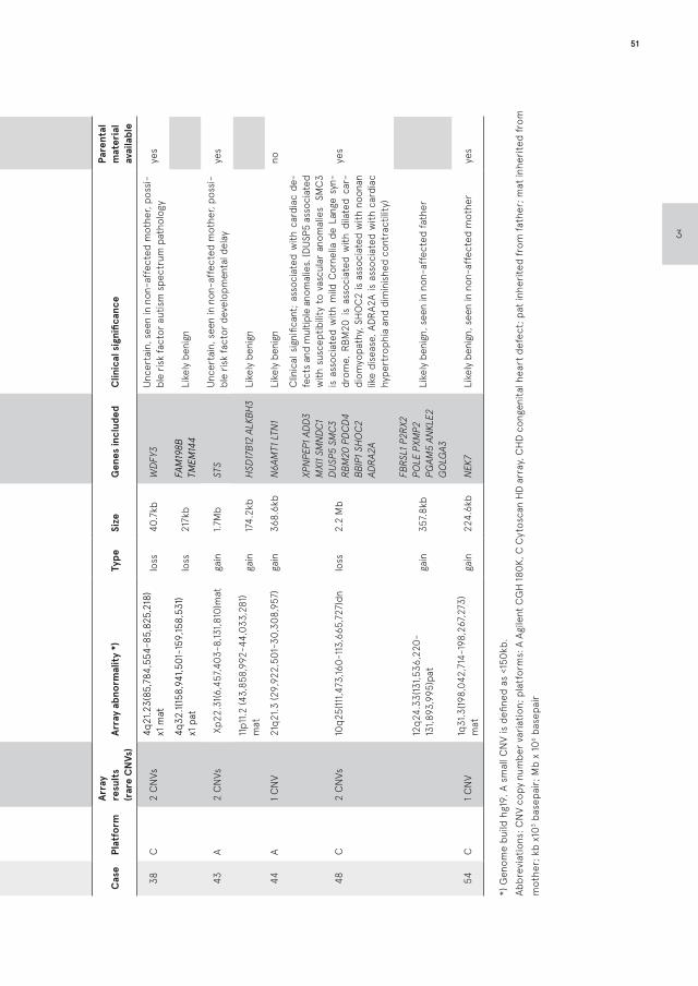
Cas
ePl
atfo
rmA
rray
resu
lts
(rar
e C
NVs
)A
rray
abn
orm
alit
y *)
Type
Size
Gen
es in
clud
edC
linic
al s
igni
fican
cePa
rent
al
mat
eria
l av
aila
ble
2A
3 C
NVs
1p11
.2(12
0,82
5,17
5-12
1,125
,362
)lo
ss30
0kb
FAM
72B
FC
GR
1B
Like
ly b
enig
nno
4q
27(12
1,589
,739
-121
,797
,841
)ga
in20
8kb
PRD
M5
Like
ly b
enig
n
11
p15.
1(21,0
68,7
42-2
1,464
,425
)ga
in39
6kb
NEL
L1Li
kely
ben
ign
5C
2 C
NVs
1p33
(49,
477,
344-
49,7
38,2
82)
loss
261k
bAG
BL4
Like
ly b
enig
nno
2p
13.3
(69,
490,
034-
69,7
37,4
01)
gain
247k
bG
FPT1
NFU
1 AAK
1
AAK1
: unc
erta
in, p
roba
bly
beni
gn. B
inds
to
and
stab
ilize
s th
e ac
tivat
ed fo
rm o
f NO
TCH
1, in
crea
ses
its lo
caliz
atio
n in
end
osom
es a
nd
regu
late
s its
tran
scrip
tiona
l act
ivity
.
7C
mos
aic
tris
omy
2,
1 sm
all C
NV
1p34
.2 (4
0,96
1,609
-41,0
02,3
40)
loss
40.7
kbZF
P69
EXO
5 ZN
F684
Like
ly b
enig
nno
m
osai
c tr
isom
y 2
>
20ge
nes
Clin
ical
ly s
igni
fican
t, as
soci
ated
with
car
diac
de
fect
s an
d m
ultip
le a
nom
alie
s
11A
1 sm
all C
NV
Xq28
(154
,310
,086
-154
,444
,066
)ga
in13
4kb
BRC
C3
Like
ly b
enig
nno
12C
1 sm
all C
NV
5q33
.1 (15
1,264
,063
-151
,366
,169)
gain
102k
bG
LRA1
Like
ly b
enig
nno
21C
1 sm
all C
NV
1p22
.3(8
7,029
,391
-87,0
39,9
45)
loss
10.6
kbC
LCA4
Like
ly b
enig
nno
22C
1 sm
all C
NV
5q14
.1(80
,494
,619
-80,
539,
078)
loss
44.5
kbR
ASG
RF2
CKM
T2Li
kely
ben
ign
no
24C
2 C
NVs
3q13
.31(1
16,7
89,7
90-1
16,9
64,18
3)lo
ss17
4.4k
bLS
AMP
Like
ly b
enig
nno
4q
13.3
(73,
813,
058-
74,0
48,5
08)
gain
235.
5kb
CO
X18
ANKR
D17
Like
ly b
enig
n
26A
1 CN
V20
q13.
33 (6
1,209
,209
-61
,392
,119)
gain
183k
bSL
CO
4A1 N
TSR
1Li
kely
ben
ign
no
32C
2 C
NVs
5q21
.1(10
2,16
7,747
-102
,434
,937
)lo
ss28
2.1k
bPA
M
G
IN1
Like
ly b
enig
nno
20
q11.2
2(34
,312
,988
-34,
378,
069)
gain
65.1
kbRB
M39
PH
F20
Like
ly b
enig
n
33C
1 sm
all C
NV
12q1
3.2(
55,8
15,0
28-5
5,83
1,512
)lo
ss16
.5kb
OR
6C76
Like
ly b
enig
nno
34C
1 sm
all C
NV
4q22
.1 (9
3,68
1,949
-93,
780,
269)
loss
98.3
kbG
RID
2Li
kely
ben
ign
no
37C
2 sm
all
CN
Vs8p
22(13
,974
,602
-14,
118,
431)
gain
143.
8kb
SGC
ZLi
kely
ben
ign
no
Xp
11.3
(46,
311,9
14-4
6,34
2,99
5)ga
in31
.1kb
KRB
OX4
Like
ly b
enig
n
Cas
ePl
atfo
rmA
rray
resu
lts
(rar
e C
NVs
)A
rray
abn
orm
alit
y *)
Type
Size
Gen
es in
clud
edC
linic
al s
igni
fican
cePa
rent
al
mat
eria
l av
aila
ble
38C
2 C
NVs
4q21
.23(
85,7
84,5
54-8
5,82
5,21
8)x1
mat
loss
40.7
kbW
DFY
3Un
cert
ain,
see
n in
non
-aff
ecte
d m
othe
r, po
ssi-
ble
risk
fact
or a
utis
m s
pect
rum
pat
holo
gyye
s
4q32
.1(15
8,94
1,501
-159
,158,
531)
x1 p
atlo
ss21
7kb
FAM
198B
TMEM
144
Like
ly b
enig
n
43A
2 C
NVs
Xp2
2.31
(6,4
57,4
03-8
,131,8
10)m
atga
in1.7
Mb
STS
Unce
rtai
n, s
een
in n
on-a
ffec
ted
mot
her,
poss
i-bl
e ris
k fa
ctor
dev
elop
men
tal d
elay
ye
s
11
p11.2
(43,
858,
992-
44,0
33,2
81)
mat
gain
174.
2kb
HSD
17B
12 A
LKBH
3Li
kely
ben
ign
44A
1 CN
V21
q21.3
(29,
922,
501-
30,3
08,9
57)
gain
368.
6kb
N6A
MT1
LTN
1Li
kely
ben
ign
no
48C
2 C
NVs
10q2
5(11
1,473
,160-
113,
665,
727)
dnlo
ss2.
2 M
b
XPN
PEP1
AD
D3
MXI
1 SM
ND
C1
DU
SP5
SMC
3 RB
M20
PD
CD
4 BB
IP1 S
HO
C2
ADR
A2A
Clin
ical
sig
nific
ant;
ass
ocia
ted
with
car
diac
de-
fect
s an
d m
ultip
le a
nom
alie
s. (D
USP5
ass
ocia
ted
with
sus
cept
ibili
ty t
o va
scul
ar a
nom
alie
s S
MC
3 is
ass
ocia
ted
with
mild
Cor
nelia
de
Lang
e sy
n-dr
ome,
RBM
20 i
s as
soci
ated
with
dila
ted
car-
diom
yopa
thy,
SHO
C2
is a
ssoc
iate
d w
ith n
oona
n lik
e di
seas
e, A
DRA2
A is
ass
ocia
ted
with
car
diac
hy
pert
roph
ia a
nd d
imin
ishe
d co
ntra
ctili
ty)
yes
12
q24.
33(13
1,536
,220
- 13
1,893
,995
)pat
gain
357.
8kb
FBRS
L1 P
2RX2
P
OLE
PXM
P2
PG
AM5
ANKL
E2
GO
LGA3
Like
ly b
enig
n, s
een
in n
on-a
ffec
ted
fath
er
54C
1 CN
V1q
31.3
(198,
042,
714-
198,
267,2
73)
mat
gain
224.
6kb
NEK
7Li
kely
ben
ign,
see
n in
non
-aff
ecte
d m
othe
rye
s
*) G
enom
e bu
ild h
g19.
A s
mal
l CN
V is
defi
ned
as <
150k
b.Ab
brev
iatio
ns: C
NV
copy
num
ber v
aria
tion;
pla
tfor
ms:
A A
gile
nt C
GH
180K
, C C
ytos
can
HD
arr
ay, C
HD
con
geni
tal h
eart
def
ect;
pat
inhe
rite
d fr
om fa
ther
; mat
inhe
rite
d fr
om
mot
her;
kb
x103 b
asep
air;
Mb
x 10
6 bas
epai
r
51
3
As deduced from table S3, minor additional findings were present in 17 of 54 ‘isolated’ cases (35%), including enlarged nuchal translucency/neck cysts (n=4), ascites/pericardial effusion (n=6), single umbilical artery (n=4), and other minor findings (n=3). Additionally, two fetuses were postnatally identified with extra-cardiac malformations (horseshoe kidney in cases 23 and 48), where one had a clinically significant CNV (case 48). These 19 fetuses with prenatally detectable (although missed in 2 cases) additional malformations did not differ in the frequency of rare CNVs from fetuses that are ‘truly’ isolated, without additional findings (both 31%). Furthermore, one child with normal array results currently displays neurodevelopmental delay (case 3). Another child with a normal array result developed hydrocephalus of an unknown cause (case 52). Both fetuses with a clinically significant CNV had an additional finding (cases 7 and 48), however in case 48 the extra-cardiac anomaly was only detected after birth. This results in 1/17 (6%) clinically significant array findings in fetuses with additional findings and 1/37 (3%) clinically significant array findings in prenatal isolated appearing cases (independent samples T test p=0.6).
DISCUSSION
Congenital heart defects (CHDs) are known to be associated with chromosomal abnormalities and 22q11.2 microdeletion4. This is confirmed by our study (table S2). Furthermore, our study shows that array analysis can yield clinically significant abnormalities in 4% of euploid fetuses without a 22q11.2 microdeletion. Thus, in the absence of ultrasonographically detected significant extra-cardiac malformations, and with a normal karyotype/FISH 22q11.2 result, array can in some cases predict if fetuses with a left sided CHD are at risk for a more severe phenotype. In our study, the risk of array abnormalities appears to be unrelated to the presence of minor additional malformations such as enlarged nuchal translucency. In two cases additional malformations (horseshoe kidney) remained undetected prior to birth.
Previous reports on the incidence of submicroscopic chromosomal abnormalities in fetal CHDs focus on CHDs in general, or analyze postnatal cohorts21;33-35, which is impractical in prenatal counselling. As the diagnostic accuracy of prenatal ultrasound increases, targeted information concerning the specific diagnosis will also need to emerge. The current study determines the specific incidence of genetic abnormalities in the subgroups of isolated and non-isolated left sided CHDs. Left sided CHD are generally considered not to be associated with genetic syndromes, if they appear isolated on prenatal ultrasound. Compared to other CHD, tetralogy of Fallot for example, which is highly associated with syndromic and chromosomal anomalies, physicians may be
52
3
PART I: GENETIC ANOMALIES
more reluctant to stress the need for fetal genetic sampling in absence of other fetal abnormalities. Thus, with our data, physicians are able to counsel parents more tailored to this specific condition. A great strength in our study is the large rate of postnatal confirmation (69% in isolated cases), thus analyzing a sharply defined phenotype of left sided CHD only.
Our array data confirms a previously reported additional yield of 6% with clinically significant submicroscopic chromosomal abnormalities in two large cohorts of euploid fetuses with isolated malformations in general36;37. When focusing on left sided CHD only, our findings are in concordance with Shaffer, who reported a subgroup with isolated HLHS in a large cohort of fetuses with various ultrasound abnormalities14. Shaffer found 4 (9.5%) significant findings (all < 10Mb) in 42 isolated HLHS fetuses. This study, however, does not provide follow-up data to validate the prenatal findings with regard to postnatal outcome, nor does it elaborate on the details of the array abnormalities and inheritance. Hitz et al. stated that in 10% of left sided CHDs, CNVs play a causative or contributing role38. Though this study included a well-focused phenotype, Hitz studied families with postnatally proven isolated left sided CHDs, excluding known syndromes and dysmorphic features. As this information is not available in the prenatal setting, the data of Hitz are not applicable for parental counselling in a fetal diagnosis.
Our study is the first to report the detection of rare CNVs, in a prenatal cohort. Our data demonstrate an average of 1.6 rare CNVs per person in 33% of fetuses with left sided CHDs. Our data coincide with findings in postnatal similar patient groups with similar array resolution: Hitz found 1.35 rare CNVs per person in 31% of children with left sided CHD (n= 54/174) with a resolution of 10kb, and Iascone found 1.32 rare CNVs per person, in 47% of postnatal HLHS cases (n= 25/53), with an average resolution of 20kb38;39. Payne reported on the frequency of small CNVs (<60kb), not likely to be disease-causing in 43 postnatal isolated and non-isolated cases of HLHS. Their found average (1.49 CNVs per person) was significantly higher when compared to 16 healthy controls39. In comparison to Hitz38 and Iascone39, the availability of parental material is somewhat lower in our dataset. Considering the fact that we found a similar number, or fewer, patients with rare CNVs, we do not expect this to have resulted in a high number of false CNVs calls.
The interpretation of CNVs remains controversial and prone to differences between centers. The identification of clinically significant CNVs is subject to variations in the used platform and the consulted genomic databases. The clinical (in)significance of variants of unknown significance (VOUS) are increasingly unveiled. Our interpretation of the CNVs yielded two array anomalies with clinical significance. Both anomalies are
53
3
known to be associated with cardiac defects and multiple congenital malformations27-30. However, these findings include some ambiguity. The degree of mosaicism trisomy 2 and affected tissues cannot be predicted (case 7). However, it would trigger suspicion of additional fetal congenital abnormalities. Interestingly, the 10q25 deletion case (case 48) did present with an additional structural abnormality, but neurodevelopment is normal.
Three VOUS were identified that were of interest. In the duplication of chromosome 2 in case 5, AAK1 appears to be an interesting gene due to its interaction with the activated form of NOTCH131. However only the 3’part of the gene is duplicated; further investigation is needed to determine whether this duplication will disrupt this gene and subsequently has an effect on the gene function . The second and third VOUS are maternally inherited variants. The Xp22.31 duplication in case 43, including the STS gene is a variant that is present at a low frequency in the population, but is still considered clinically significant because it is found at higher frequency in affected individuals. Although this variant will not explain the HLHS, it could be a risk factor for neurodevelopmental delay25. The 4q21.23 deletion in case 38, including the WDFY3 gene, has been correlated to cerebral changes in mice that could be characteristic for autism spectrum disorders and epilepsy. The implications of both variants are unclear, and both children died at very young age due to cardiac complications. As our study was done on banked samples, it is unclear how these findings would have influenced the prenatal counselling.
Previous studies have implicated several loci and genes in left sided CHDs (mainly HLHS), including NOTCH1, NKX2.5, NKX2.6, HAND1, HAND2, SNAI2, GATA6, GJA1, FGF8, FOXC1, FOXC2, FOXH1 and FOXL140-45. Identifying a new candidate gene or combination of genes responsible, however, remains difficult, mainly due to variable penetrance41. In isolated left sided CHDs, there appears to be no single genetic cause. Familial recurrence does occur, but left sided CHDs are considered to be genetically heterogeneous. Embryological blood flow alterations also seem to play an important role in the etiology46;47. The reported genes were not found in any of the CNV regions we identified. However, the platforms we used either lacked or had few probes specific for the following genes: HAND1, HAND2, SNAI2, NKX2.5 FOXC1 NKX2.6 FOXH1 and FGF8; intragenic insertions or deletions could have been missed.
Our study has some limitations. Due to the retrospective nature of our study, segmental analysis of the development of the CHDs was not available in all cases. It is complicated to provide a link between a CNV or a candidate gene and the observed phenotype39;48. In left sided CHDs it is even more difficult because the anomaly itself displays high rates of anatomic variation49. Clinical classifications of left sided CHDs are focused on
54
3
PART I: GENETIC ANOMALIES
a functional outcome. In HLHS, as an end stage development product, it is not always possible to identify the developmental cause of the observed anomaly. In our cohort, segmental developmental analysis was only possible in a small group, mainly in those that underwent postmortem dissection after termination of pregnancy. In the live born cases specific developmental details, regarding the presence of mitral or aortic valve hypo- or aplasia, as cause of HLHS were not always identifiable.
Furthermore, only 22% of our samples were analysed as trios, so information regarding the presence or absence of identified CNVs in parents is lacking in the remaining 42 cases. The importance of information regarding inheritance is evidenced by the findings of Warburton, where de novo rare CNVs occurred in 12.7% of their 71 postnatal HLHS cases versus 2% in their cohort of healthy controls50. The history of familial occurrence of cardiac defects was not always available in our cohort, and parents were generally not tested for the presence of mild left sided CHDs such as a bicuspid aortic valve. Familial segregation analysis (linkage studies) and subsequent speculation on other potentially contributing CNVs, labelled in our study as clinically not significant, is therefore not possible. Thus, we are unable to rule out a possible influence of a yet unknown, common CNV as a susceptibility factor. Known susceptibility factors, such as 15q11.2 BP1-BP2 microdeletions, were not found in our study. Furthermore, the resolution of the used array method is restricted to 10kb in deletions and 20 kb in duplications; smaller intragenic deletions or duplications could not have been detected by this test.
Also, genetic material was not available in all eligible cases. As we have demonstrated in table 1, parents typically opted for an invasive procedure when additional malformations were present. Also, in 53 cases, genetic material was unavailable due to logistic challenges, absence of stored material and failure of cell culture. Therefore a selection bias cannot be ruled out.
Despite the limitations, our data serves as guide in focused prenatal counselling when genetic analysis is offered in left sided CHDs. Considering the fact that the long-term outcome may also be dominated by non-iatrogenic neurological impairment, even in apparently isolated CHDs, attempting to identify beforehand which cases are at highest risk for a more severe phenotype is important51,52. As mentioned, our data also confirm reports that left sided CHDs are associated with chromosome abnormalities and 22q11.2 microdeletion syndrome4, detecting these aberrations in as many as 57% of fetuses with left sided CHDs in the presence of significant extra-cardiac malformations in this study. Left sided CHDs which seem to be isolated on prenatal ultrasound also carry a 7% risk of clinical significant chromosome abnormalities and 22q11.2 microdeletions in our cohort.
55
3
Together with clinically significant CNVs found in 4%, the yield of genetic analysis could be as high as 11% when using karyotyping and array analysis combined. However, all of the significant chromosome abnormalities found in our study with karyotyping (table S2) are also identifiable by array. It is advisable therefore to perform array analysis as a first tier test. Depending on local policies and costs deliberations, array analysis can be preceded by RAD to exclude common aneuploidies first. However, our study also shows that array analysis cannot predict all cases that display adverse (neurodevelopmental) outcome. Furthermore, as discussed, the significant array findings include some ambiguity. Therefore, while array analysis would have identified individual cases where the search for additional phenotypic abnormalities would be warranted, counselling may still involve some uncertainty. In the future, if whole exome or genome sequencing becomes widely available in the prenatal setting, this effect might even be stronger. To attach consequences to subtle array abnormalities, such as refusal of certain palliative interventions, has to be avoided until evidence of adverse outcome can be ascertained.
In conclusion, our data show that performing array analysis in a high resolution in cases of prenatal left sided CHD could aid parental counselling. It could identify some fetuses that are at high risk for a more severe phenotype, because of its capability to demonstrate unbalanced submicroscopic chromosome abnormalities and low mosaic aneuploidies. As the first to explore this in a prenatal setting, our research supports the use of array analysis as a first tier diagnostic test in isolated left sided CHD53. Left sided CHD are usually considered to have a low risk for genetic anomalies, if not accompanied by additional congenital anomalies, leading to lower rates of invasive procedure performed. This study however confirms that fetal ultrasound misses certain additional lesions, thus emphasizing the importance of fetal genetic analysis. Because array analysis is also able to detect 22q11.2 microdeletion, it can be performed instead of FISH analysis, preceded by RAD (or karyotyping). The relative small size of our cohort, however, attenuates our findings.
56
3
PART I: GENETIC ANOMALIES
REFERENCES
1. van der Linde D, Konings EE, Slager MA et al.
Birth prevalence of congenital heart disease
worldwide: a systematic review and meta-
analysis. J Am Coll Cardiol 2011;58:2241-2247.
2. van VC, Clur S, Rijlaarsdam M et al. Prenatal
detection of congenital heart disease-results
of a national screening programme. BJOG 2015.
3. Barron DJ, Kilby MD, Davies B, Wright JG, Jones
TJ, Brawn WJ. Hypoplastic left heart syndrome.
Lancet 2009;374:551-564.
4. Ferencz C, Neill CA, Boughman JA, Rubin JD,
Brenner JI, Perry LW. Congenital cardiovascular
malformations associated with chromosome
abnormalities: an epidemiologic study. J Pediatr
1989;114:79-86.
5. Simsic JM, Coleman K, Maher KO, Cuadrado
A, Kirshbom PM. Do neonates with genetic
abnormalities have an increased morbidity and
mortality following cardiac surgery? Congenit
Heart Dis 2009;4:160-165.
6. Cramer JW, Bartz PJ, Simpson PM, Zangwill SD.
The Spectrum of Congenital Heart Disease and
Outcomes After Surgical Repair Among Children
With Turner Syndrome: A Single-Center Review.
Pediatr Cardiol 2013.
7. Evangelidou P, Alexandrou A, Moutafi M et
al. Implementation of high resolution whole
genome array CGH in the prenatal clinical
setting: advantages, challenges, and review of
the literature. Biomed Res Int 2013;2013:346762.
8. Park SJ, Jung EH, Ryu RS et al. Clinical
implementation of whole-genome array CGH as
a first-tier test in 5080 pre and postnatal cases.
Mol Cytogenet 2011;4:12.
9. Miller DT, Adam MP, Aradhya S et al. Consensus
statement: chromosomal microarray is a
first-tier clinical diagnostic test for individuals
with developmental disabilities or congenital
anomalies. Am J Hum Genet 2010;86:749-764.
10. Van den Veyver IB, Patel A, Shaw CA et al. Clinical
use of array comparative genomic hybridization
(aCGH) for prenatal diagnosis in 300 cases.
Prenat Diagn 2009;29:29-39.
11. Lichtenbelt KD, Knoers NV, Schuring-Blom GH.
From karyotyping to array-CGH in prenatal
diagnosis. Cytogenet Genome Res 2011;135:241-
250.
12. Callaway JL, Shaffer LG, Chitty LS, Rosenfeld
JA, Crolla JA. The clinical utility of microarray
technologies applied to prenatal cytogenetics
in the presence of a normal conventional
karyotype: a review of the literature. Prenat
Diagn 2013;33:1119-1123.
13. Vanakker O, Vilain C, Janssens K et al.
Implementation of genomic arrays in prenatal
diagnosis: the Belgian approach to meet the
challenges. Eur J Med Genet 2014;57:151-156.
14. Shaffer LG, Rosenfeld JA, Dabell MP et al.
Detection rates of clinically significant genomic
alterations by microarray analysis for specific
anomalies detected by ultrasound. Prenat
Diagn 2012;32:986-995.
15. Yan Y, Wu Q, Zhang L et al. Detection of
submicroscopic chromosomal aberrations by
array-based comparative genomic hybridization
in fetuses with congenital heart disease.
Ultrasound Obstet Gynecol 2014;43:404-412.
16. Liao C, Li R, Fu F et al. Prenatal diagnosis
of congenital heart defect by genome-wide
high-resolution SNP array. Prenat Diagn
2014;34:858-863.
17. Donnelly JC, Platt LD, Rebarber A, Zachary
57
3
J, Grobman WA, Wapner RJ. Association
of copy number variants with specific
ultrasonographically detected fetal anomalies.
Obstet Gynecol 2014;124:83-90.
18. Mademont-Soler I, Morales C, Soler A et
al. Prenatal diagnosis of chromosomal
abnormalities in fetuses with abnormal cardiac
ultrasound findings: evaluation of chromosomal
microarray-based analysis. Ultrasound Obstet
Gynecol 2013;41:375-382.
19. Schmid M, Stary S, Blaicher W, Gollinger
M, Husslein P, Streubel B. Prenatal genetic
diagnosis using microarray analysis in fetuses
with congenital heart defects. Prenat Diagn
2012;32:376-382.
20. Chen M, Yang YS, Shih JC et al. Microdeletions/
duplications involving TBX1 gene in fetuses with
conotruncal heart defects which are negative
for 22q11.2 deletion on fluorescence in-situ
hybridization. Ultrasound Obstet Gynecol
2014;43:396-403.
21. Jansen FA, Blumenfeld YJ, Fisher A et al. Array
comparative genomic hybridization and fetal
congenital heart defects: a systematic review
and meta-analysis. Ultrasound Obstet Gynecol
2015;45:27-35.
22. Gijsbers AC, Schoumans J, Ruivenkamp CA.
Interpretation of array comparative genome
hybridization data: a major challenge. Cytogenet
Genome Res 2011;135:222-227.
23. Cox DM, Butler MG. The 15q11.2 BP1-BP2
microdeletion syndrome: a review. Int J Mol Sci
2015;16:4068-4082.
24. Soemedi R, Topf A, Wilson IJ et al. Phenotype-
specific effect of chromosome 1q21.1
rearrangements and GJA5 duplications in 2436
congenital heart disease patients and 6760
controls. Hum Mol Genet 2012;21:1513-1520.
25. Esplin ED, Li B, Slavotinek A et al. Nine patients
with Xp22.31 microduplication, cognitive
deficits, seizures, and talipes anomalies. Am J
Med Genet A 2014;164A:2097-2103.
26. Li F, Shen Y, Kohler U et al. Interstitial
microduplication of Xp22.31: Causative of
intellectual disability or benign copy number
variant? Eur J Med Genet 2010;53:93-99.
27. Sago H, Chen E, Conte WJ et al. True trisomy
2 mosaicism in amniocytes and newborn liver
associated with multiple system abnormalities.
Am J Med Genet 1997;72:343-346.
28. Chen CP, Chen YY, Chern SR et al. Prenatal
diagnosis of mosaic trisomy 2 associated
with abnormal maternal serum screening,
oligohydramnios, intrauterine growth
restriction, ventricular septal defect, preaxial
polydactyly, and facial dysmorphism. Taiwan J
Obstet Gynecol 2013;52:395-400.
29. Gil-Rodriguez MC, Deardorff MA, Ansari M
et al. De novo heterozygous mutations in
SMC3 cause a range of Cornelia de Lange
syndrome-overlapping phenotypes. Hum Mutat
2015;36:454-462.
30. Stark Z, Bruno DL, Mountford H, Lockhart PJ,
Amor DJ. De novo 325 kb microdeletion in
chromosome band 10q25.3 including ATRNL1
in a boy with cognitive impairment, autism
and dysmorphic features. Eur J Med Genet
2010;53:337-339.
31. Gupta-Rossi N, Ortica S, Meas-Yedid V et al. The
adaptor-associated kinase 1, AAK1, is a positive
regulator of the Notch pathway. J Biol Chem
2011;286:18720-18730.
58
3
PART I: GENETIC ANOMALIES

32. Orosco LA, Ross AP, Cates SL et al. Loss of Wdfy3
in mice alters cerebral cortical neurogenesis
reflecting aspects of the autism pathology. Nat
Commun 2014;5:4692.
33. Erdogan F, Larsen LA, Zhang L et al. High
frequency of submicroscopic genomic
aberrations detected by tiling path array
comparative genome hybridisation in patients
with isolated congenital heart disease. J Med
Genet 2008;45:704-709.
34. Greenway SC, Pereira AC, Lin JC et al. De novo
copy number variants identify new genes and
loci in isolated sporadic tetralogy of Fallot. Nat
Genet 2009;41:931-935.
35. Breckpot J, Thienpont B, Arens Y et al.
Challenges of interpreting copy number
variation in syndromic and non-syndromic
congenital heart defects. Cytogenet Genome
Res 2011;135:251-259.
36. Wapner RJ, Martin CL, Levy B et al. Chromosomal
microarray versus karyotyping for prenatal
diagnosis. N Engl J Med 2012;367:2175-2184.
37. Shaffer LG, Dabell MP, Fisher AJ et al. Experience
with microarray-based comparative genomic
hybridization for prenatal diagnosis in over
5000 pregnancies. Prenat Diagn 2012;32:976-
985.
38. Hitz MP, Lemieux-Perreault LP, Marshall C et
al. Rare copy number variants contribute to
congenital left-sided heart disease. PLoS Genet
2012;8:e1002903.
39. Iascone M, Ciccone R, Galletti L et al.
Identification of de novo mutations and rare
variants in hypoplastic left heart syndrome. Clin
Genet 2012;81:542-554.
40. Reamon-Buettner SM, Ciribilli Y, Inga A, Borlak
J. A loss-of-function mutation in the binding
domain of HAND1 predicts hypoplasia of the
human hearts. Hum Mol Genet 2008;17:1397-
1405.
41. Wessels MW, Willems PJ. Genetic factors in
non-syndromic congenital heart malformations.
Clin Genet 2010;78:103-123.
42. Maitra M, Schluterman MK, Nichols HA et al.
Interaction of Gata4 and Gata6 with Tbx5 is
critical for normal cardiac development. Dev
Biol 2009;326:368-377.
43. Stankiewicz P, Sen P, Bhatt SS et al. Genomic
and genic deletions of the FOX gene cluster
on 16q24.1 and inactivating mutations of FOXF1
cause alveolar capillary dysplasia and other
malformations. Am J Hum Genet 2009;84:780-
791.
44. Yamagishi H, Yamagishi C, Nakagawa O, Harvey
RP, Olson EN, Srivastava D. The combinatorial
activities of Nkx2.5 and dHAND are essential
for cardiac ventricle formation. Dev Biol
2001;239:190-203.
45. Elliott DA, Kirk EP, Yeoh T et al. Cardiac
homeobox gene NKX2-5 mutations and
congenital heart disease: associations with
atrial septal defect and hypoplastic left heart
syndrome. J Am Coll Cardiol 2003;41:2072-
2076.
46. Hinton RB, Martin LJ, Rame-Gowda S, Tabangin
ME, Cripe LH, Benson DW. Hypoplastic left
heart syndrome links to chromosomes 10q and
6q and is genetically related to bicuspid aortic
valve. J Am Coll Cardiol 2009;53:1065-1071.
47. Kodo K, Yamagishi H. A decade of advances
in the molecular embryology and genetics
underlying congenital heart defects. Circ J
59
3

2011;75:2296-2304.
48. Marino B, Digilio MC. Congenital heart disease
and genetic syndromes: specific correlation
between cardiac phenotype and genotype.
Cardiovasc Pathol 2000;9:303-315.
49. Tchervenkov CI, Jacobs ML, Tahta SA. Congenital
Heart Surgery Nomenclature and Database
Project: hypoplastic left heart syndrome. Ann
Thorac Surg 2000;69:S170-S179.
50. Warburton D, Ronemus M, Kline J et al. The
contribution of de novo and rare inherited copy
number changes to congenital heart disease
in an unselected sample of children with
conotruncal defects or hypoplastic left heart
disease. Hum Genet 2014;133:11-27.
51. Miller SP, McQuillen PS, Hamrick S et al. Abnormal
brain development in newborns with congenital
heart disease. N Engl J Med 2007;357:1928-
1938.
52. Sanchez-Valle A, Pierpont ME, Potocki L. The
severe end of the spectrum: Hypoplastic left
heart in Potocki-Lupski syndrome. Am J Med
Genet A 2011;155A:363-366.
53. Carey AS, Liang L, Edwards J et al. Effect of copy
number variants on outcomes for infants with
single ventricle heart defects. Circ Cardiovasc
Genet 2013;6:444-451.
60
3
PART I: GENETIC ANOMALIES
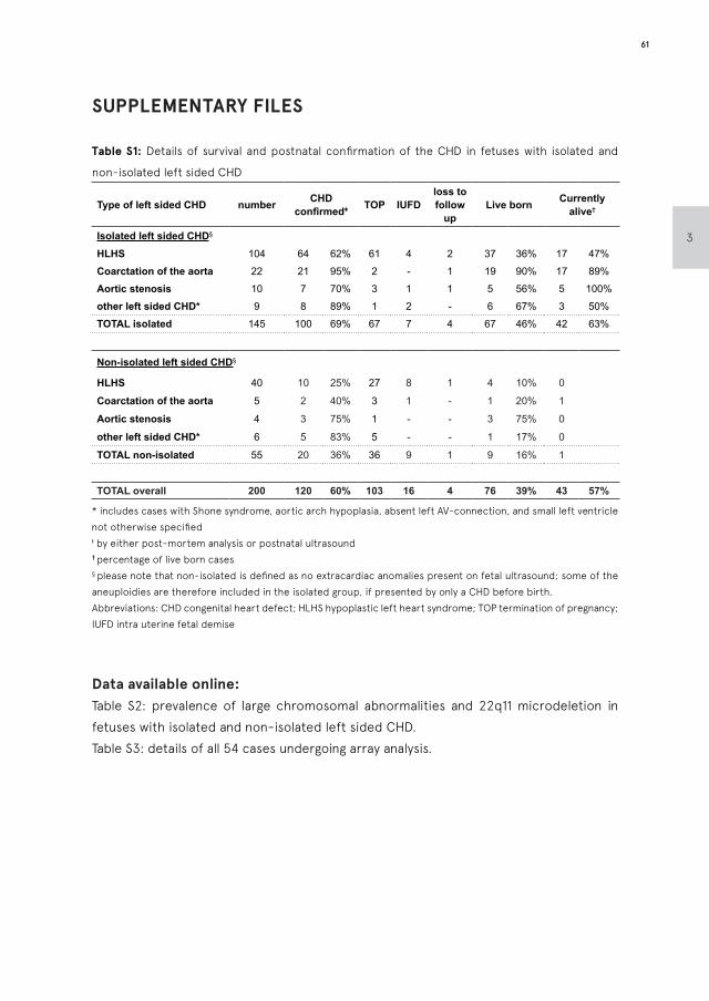
SUPPLEMENTARY FILES
Table S1: Details of survival and postnatal confirmation of the CHD in fetuses with isolated and
non-isolated left sided CHD
Type of left sided CHD number CHD confirmedǂ TOP IUFD
loss to follow
upLive born Currently
alive†
Isolated left sided CHD§
HLHS 104 64 62% 61 4 2 37 36% 17 47%
Coarctation of the aorta 22 21 95% 2 - 1 19 90% 17 89%
Aortic stenosis 10 7 70% 3 1 1 5 56% 5 100%
other left sided CHD* 9 8 89% 1 2 - 6 67% 3 50%
TOTAL isolated 145 100 69% 67 7 4 67 46% 42 63%
Non-isolated left sided CHD§
HLHS 40 10 25% 27 8 1 4 10% 0
Coarctation of the aorta 5 2 40% 3 1 - 1 20% 1
Aortic stenosis 4 3 75% 1 - - 3 75% 0
other left sided CHD* 6 5 83% 5 - - 1 17% 0
TOTAL non-isolated 55 20 36% 36 9 1 9 16% 1
TOTAL overall 200 120 60% 103 16 4 76 39% 43 57%
* includes cases with Shone syndrome, aortic arch hypoplasia, absent left AV-connection, and small left ventricle not otherwise specifiedǂ by either post-mortem analysis or postnatal ultrasound † percentage of live born cases§ please note that non-isolated is defined as no extracardiac anomalies present on fetal ultrasound; some of the aneuploidies are therefore included in the isolated group, if presented by only a CHD before birth. Abbreviations: CHD congenital heart defect; HLHS hypoplastic left heart syndrome; TOP termination of pregnancy; IUFD intra uterine fetal demise
Data available online:Table S2: prevalence of large chromosomal abnormalities and 22q11 microdeletion in fetuses with isolated and non-isolated left sided CHD.Table S3: details of all 54 cases undergoing array analysis.
61
3

F.A.R. Jansen*
M.C. van Rij*
D.M.E.I. Hellebrekers W. Onkenhout H.J.M. Smeets A.T. Hendrickx R.W.H. GottschalkS.J. SteggerdaC.M.P.C.D. Peeters-ScholteM.C. Haak Y. Hilhorst-Hofstee
* Both authors contributed equally
CHAPTER 4
Polyhydramnion and cerebellar
atrophy: a prenatal presentation
of mitochondrial encephalomyopathy
caused by mutations in the FBXL4 gene
Clinical Case Reports 2016; 4(4): 425–428
KEY CLINICAL MESSAGE
Severe recessive mitochondrial myopathy caused by FBXL4 gene mutations may present prenatally with polyhydramnios and cerebellar hypoplasia. Characteristic dysmorphic features are: high and arched eyebrows, triangular face, a slight upslant of palpebral fissures, and a prominent pointed chin. Metabolic investigations invariably show increased serum lactate and pyruvate levels.
CASE DESCRIPTION
Polyhydramnios in the second and third trimester of pregnancy is defined by (semiquantative) measurements such as a maximum vertical pocket (MVP) >8cm, or an amniotic fluid index (AFI) >24 cm. Approximately 90% of cases are idiopathic or caused by gestational diabetes (GDM)1. However, 10% of cases are associated with fetal structural abnormalities2. Most frequent causes are impaired swallowing from any cause (gastro-intestinal, facial, musculoskeletal or brain abnormalities), cardiac failure, hydrops and renal abnormalities. Metabolic diseases, such as congenital disorders of glycogen storage, are also incidentally reported to present with polyhydramnios in pregnancy3.
The usual diagnostic work-up of polyhydramnios is to exclude GDM and maternal infections, and to perform an extensive structural assessment to rule out fetal anomalies. The presence of structural anomalies involves increased risk of aneuploidy or other chromosomal and syndromic disorders. In isolated polyhydramnios, the risk of perinatal adverse outcome is, however, still increased when compared to uneventful pregnancies4. A 30-year old primigravid woman, with a so far uneventful pregnancy, was referred to our clinic with polyhydramnios. Aside from a spontaneously closed ventricular septal defect (VSD) in her own infancy, both parents were healthy. A maternal uncle of the mother had died postnatally of an unknown cause.
First-trimester combined test revealed a low risk for trisomies (NT 1.1 mm). The anomaly scan was performed at 20 weeks GA and showed no abnormalities. Transverse cerebellar diameter was normal at p50. At 25+5 weeks of gestation she presented with signs of polyhydramnios (uterine size that outpaced gestational age) and premature contractions. She received tocolytics and corticosteroids for fetal lung maturation. Ultrasound showed polyhydramnios (MVP 9.9 cm, AFI 27.5 cm) with normal fluid-filled stomach and mild dilatation of both lateral ventricles (11mm). Amniotic fluid drainage (1.8 liter) was performed in an attempt to cease the premature contractions. More detailed fetal
64
4
PART I: GENETIC ANOMALIES
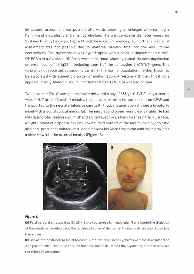
intracranial assessment was possible afterwards, showing an enlarged cisterna magna (12mm) and a dysplastic and small cerebellum. The transcerebellar diameter measured 25.4 mm (slightly below p3, Figure 1A, with head circumference p50). Further intracranial assessment was not possible due to maternal habitus, fetal position and uterine contractions. The myocardium was hypertrophic with a small perimembraneous VSD. QF PCR and a CytoScan HD Array were performed, showing a small de novo duplication on chromosome 11 (11q22.1), including exon 1 of the contactine 5 (CNTN5) gene. This variant is not reported as genomic variant in the normal population, neither known to be associated with a genetic disorder or malformation. A relation with the clinical signs appears unlikely. Maternal serum infection testing (TORCHES) was also normal.
Two days later (26+0) she spontaneously delivered a boy of 835 g (-0.5 SDS), Apgar scores were 4/4/7 after 1.5 and 10 minutes respectively. At birth he was started on CPAP and transported to the neonatal intensive care unit. Physical examination showed a hypotonic infant with a lack of subcutaneous fat. The muscles and bones were clearly visible. He had mild dysmorphic features with high and arched eyebrows, a hairy forehead, triangular face, a slight upslant of palpebral fissures, down turned corners of the mouth, mild hypoplastic alae nasi, prominent pointed chin, deep incisura between tragus and antitragus providing a clear view into the external meatus (Figure 1B).
Figure 1.
(A) Fetal cerebral ultrasound at GA 25 + 5 showed cerebellar hypoplasia (*) and borderline dilatation
of the ventricles. In retrospect, the outlines of some of the periventricular cysts are also discernible
(see arrows).
(B) shows the postmortem facial features. Note the prominent eyebrows and the triangular face
with pointed chin. The bruises around the nose and philtrum, and the asymmetry of the nostrils are
the effect of ventilation.
65
4
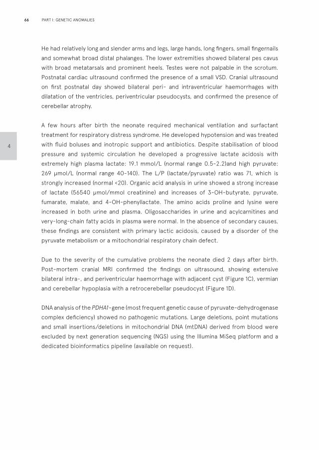
He had relatively long and slender arms and legs, large hands, long fingers, small fingernails and somewhat broad distal phalanges. The lower extremities showed bilateral pes cavus with broad metatarsals and prominent heels. Testes were not palpable in the scrotum. Postnatal cardiac ultrasound confirmed the presence of a small VSD. Cranial ultrasound on first postnatal day showed bilateral peri- and intraventricular haemorrhages with dilatation of the ventricles, periventricular pseudocysts, and confirmed the presence of cerebellar atrophy.
A few hours after birth the neonate required mechanical ventilation and surfactant treatment for respiratory distress syndrome. He developed hypotension and was treated with fluid boluses and inotropic support and antibiotics. Despite stabilisation of blood pressure and systemic circulation he developed a progressive lactate acidosis with extremely high plasma lactate: 19.1 mmol/L (normal range 0.5-2.2)and high pyruvate: 269 µmol/L (normal range 40-140). The L/P (lactate/pyruvate) ratio was 71, which is strongly increased (normal <20). Organic acid analysis in urine showed a strong increase of lactate (56540 µmol/mmol creatinine) and increases of 3-OH-butyrate, pyruvate, fumarate, malate, and 4-OH-phenyllactate. The amino acids proline and lysine were increased in both urine and plasma. Oligosaccharides in urine and acylcarnitines and very-long-chain fatty acids in plasma were normal. In the absence of secondary causes, these findings are consistent with primary lactic acidosis, caused by a disorder of the pyruvate metabolism or a mitochondrial respiratory chain defect.
Due to the severity of the cumulative problems the neonate died 2 days after birth. Post-mortem cranial MRI confirmed the findings on ultrasound, showing extensive bilateral intra-, and periventricular haemorrhage with adjacent cyst (Figure 1C), vermian and cerebellar hypoplasia with a retrocerebellar pseudocyst (Figure 1D).
DNA analysis of the PDHA1-gene (most frequent genetic cause of pyruvate-dehydrogenase complex deficiency) showed no pathogenic mutations. Large deletions, point mutations and small insertions/deletions in mitochondrial DNA (mtDNA) derived from blood were excluded by next generation sequencing (NGS) using the Illumina MiSeq platform and a dedicated bioinformatics pipeline (available on request).
66
4
PART I: GENETIC ANOMALIES
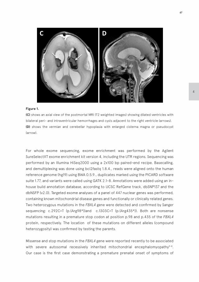
Figure 1.
(C) shows an axial view of the postmortal MRI (T2 weighted images) showing dilated ventricles with
bilateral peri- and intraventricular hemorrhages and cysts adjacent to the right ventricle (arrows).
(D) shows the vermian and cerebellar hypoplasia with enlarged cisterna magna or pseudocyst
(arrow).
For whole exome sequencing, exome enrichment was performed by the Agilent SureSelectXT exome enrichment kit version 4, including the UTR regions. Sequencing was performed by an Illumina HiSeq2000 using a 2x100 bp paired-end recipe. Basecalling, and demultiplexing was done using bcl2fastq 1.8.4., reads were aligned onto the human reference genome (hg19) using BWA 0.5.9., duplicates marked using the PICARD software suite 1.77, and variants were called using GATK 2.1-8. Annotations were added using an in-house build annotation database, according to UCSC RefGene track, dbSNP137 and the dbNSFP (v2.0). Targeted exome analyses of a panel of 447 nuclear genes was performed, containing known mitochondrial disease genes and functionally or clinically related genes. Two heterozygous mutations in the FBXL4 gene were detected and confirmed by Sanger sequencing: c.292C>T (p.(Arg98*))and c.1303C>T (p.(Arg435*)). Both are nonsense mutations resulting in a premature stop codon at position p.98 and p.435 of the FBXL4 protein, respectively. The location of these mutations on different alleles (compound heterozygosity) was confirmed by testing the parents.
Missense and stop mutations in the FBXL4 gene were reported recently to be associated with severe autosomal recessively inherited mitochondrial encephalomyopathy5-8. Our case is the first case demonstrating a premature prenatal onset of symptoms of
67
4
FBXL4-related mitochondrial encephalomyopathy. The polyhydramnios was the primary sign, leading to the detection of brain abnormalities on more detailed fetal ultrasound examination. This early presentation of polyhydramnios is most likely caused by hypotonia and diminished fetal movements.
The cases of FBXL4-related encephalopathy reported so far are characterised by increased serum lactate level, psychomotor delay, hypotonia, failure to thrive, swallowing difficulty and muscle wasting6. Onset of symptoms varied from neonatal onset after term birth, to the age of 14 months5,6,8. Other reported cases were born at term or premature due to medical intervention for intrauterine growth retardation or reduced fetal movements5,6.
The FBXL4 gene is situated on nuclear DNA on chromosome 6q16.1. The FBXL4 mitochondrial protein contains an F box in its N-terminal half, followed by 11 leucine-rich repeats9, and is expressed in heart, kidney, liver, lung, pancreas, and placenta10. Evidence of the pathogenetic effect of FBXL4 mutations was provided by skeletal muscle biopsies and fibroblasts showing defects in mitochondrial respiratory chain enzyme activities, loss of mitochondrial membrane potential, a disturbance of the dynamic mitochondrial network, and mtDNA depletion5.
The nonsense mutation p.Arg435* in the FBXL4 gene, present in our patient, was earlier reported in homozygous form in a child of consanguineous parents with early onset mitochondrial encephalopathy, severe hypotonia, cardiomyopathy, MRI abnormalities, increased serum lactate and premature death5. The second mutation detected in our patient (p.Arg98* mutation) has not been reported previously. However, like the other mutation reported, this nonsense mutation leads to nonsense-mediated decay (NMD, RNA degradation) or a truncated protein5,6. As a result our patient would have been unable to produce a normal FBXL4 gene product.
The mother was pregnant again before exome sequencing had started. Within the prenatal timeframe, exome sequencing lead to the diagnosis of the first child, consequently enabling prenatal diagnosis for the second child. Amniocentesis with sequence analysis of the FBXL4 gene confirmed that the fetus was unaffected. The pregnancy resulted in the birth of a healthy child. The VSD in our patient is considered to be a separate finding (familial trait) which is not related to the syndrome.
Our case demonstrated a prenatal onset mitochondrial encephalomyopathy presenting with polyhydramnios, causing premature delivery, and cerebellar atrophy. The neonate
68
4
PART I: GENETIC ANOMALIES
was hypotonic and in a poor condition with need for mechanical ventilation, inotropic support, and persistent lactate acidosis. Targeted exome sequencing using a mitochondrial gene panel proved its benefit by revealing compound heterozygous mutations in the FBXL4 gene. Prenatal testing was successfully carried out in the subsequent pregnancy. Direct testing for mutations in the FBXL4 gene should be considered in patients with severe encephalomyopathy with high levels of serum lactate.
REFERENCES
1. Harman C. Amniotic fluid abnormalities.
Seminars in perinatology. 2008;32(4):288-94.
2. Dashe J, McIntire D, Ramus R, et al. Hydramnios:
anomaly prevalence and sonographic detection.
Obstetrics and gynecology. 2002;100(1):134-9.
3. Raju GP, Li H-C, Bali D, et al. A case of congenital
glycogen storage disease type IV with a novel
GBE1 mutation. Journal of Child Neurology.
2008;23(3):349-52.
4. Magann E, Chauhan S, Doherty D, et al. A
review of idiopathic hydramnios and pregnancy
outcomes. Obstetrical & gynecological survey.
2007;62(12):795-802.
5. Bonnen Penelope E, Yarham John W, Besse A,
et al. Mutations in FBXL4 Cause Mitochondrial
Encephalopathy and a Disorder of Mitochondrial
DNA Maintenance. The American Journal of
Human Genetics. 2013;93(3):471-81.
6. Gai X, Ghezzi D, Johnson Mark A, et al.
Mutations in FBXL4, Encoding a Mitochondrial
Protein, Cause Early-Onset Mitochondrial
Encephalomyopathy. The American Journal of
Human Genetics. 2013;93(3):482-95.
7. Wortmann S, Koolen D, Smeitink J, et al. Whole
exome sequencing of suspected mitochondrial
patients in clinical practice. J Inherit Metab Dis.
2015:1-7.
8. Huemer M, Karall D, Schossig A, et al. Clinical,
morphological, biochemical, imaging and out-
come parameters in 21 individuals with mito-
chondrial maintenance defect related to FBXL4
mutations. J Inherit Metab Dis. 2015.
9. Jin J, Cardozo T, Lovering RC, et al. System-
atic analysis and nomenclature of mamma-
lian F-box proteins. Genes & Development.
2004;18(21):2573-80.
10. Winston JT, Koepp DM, Zhu C, et al. A fam-
ily of mammalian F-box proteins. Curr Biol.
1999;9(20):1180-S3.
69
4


PART ||
Development of the brain in fetuses
and children with isolated severe
congenital heart disease;
The Heart And NeuroDevelopment
(HAND) study


CHAPTER 5
Fetal brain imaging in isolated
congenital heart defects –
a systematic review and meta-analysis
Prenatal Diagnosis 2016; 36: 601–613
F.A.R. Jansen*S.M.P. Everwijn* R. Scheepjens T. Stijnen C.M.P.C.D. Peeters-ScholteJ.M.M. van Lith M.C. Haak
* Both authors contributed equally
ABSTRACT
Congenital heart defects (CHD) are associated with neurodevelopmental (ND) delay. This study aims to assess evidence for impaired prenatal brain development, in fetuses with CHD. A systematical search was performed and 34 studies evaluating the fetal brain (MRI or ultrasound) in isolated CHD were included (1990-2015). Data regarding cerebral abnormalities, head circumference (HC) growth and middle cerebral artery (MCA) flow were extracted. Antenatal MRI was studied in 10 articles (445 fetuses), resulting in a pooled prevalence of 18% (95%CI -6% to 42%) for combined structural and acquired cerebral abnormalities. Prenatal HC was studied in 13 articles (753 fetuses), resulting in a pooled z-score of -0.51 (95%CI -0.84;-0.18). Doppler was studied in 21 articles (1412 fetuses), resulting in a lower MCA pulsatility index (z-score -0.70, 95%CI -0.99;-0.41) in left sided CHD only. We conclude that prenatal MRI and ultrasound demonstrate brain abnormalities, delay in head growth and brainsparing in subgroups of CHD. However, large MRI studies are scarce and ultrasound data are biased towards severe and left-sided CHD. Long term follow-up studies correlating prenatal findings with postnatal ND outcome are limited and data is lacking to support counseling families regarding ND outcome based on prenatal findings suggestive of altered brain development.
5
74 PART II: BRAIN DEVELOPMENT
BACKGROUND
Congenital heart disease (CHD) is the most common congenital malformation, affecting six to eight per 1000 newborns. Although the survival rates of these children have increased over the last decades, there is a significant risk for adverse neurodevelopmental (ND) outcome, even in the absence of associated chromosomal or syndromic abnormalities1-4. ND sequelae, like developmental delay and low IQ, are mainly encountered in children with severe CHD, who require surgery in the first year of life5. Until recently, these ND sequelae were assumed to be the result of perioperative conditions resulting in cerebral hypoxia and thrombo-embolic events6.
Recent studies demonstrated signs of abnormal neurological development already present at birth, prior to surgery. These studies demonstrated abnormal results of early neurological examination and abnormal imaging findings such as periventricular leukomalacia, white matter injury and cerebral atrophy7;8. Some studies related these findings to a poor neurological development later in life9;10. The characteristics of certain pre-operative neurological abnormalities, such as cerebral atrophy and delayed maturation, suggest that these abnormalities originate in utero. A second finding indicating towards a fetal origin is a smaller head circumference (HC), found in neonates with severe isolated CHD. Smaller HC is mainly reported in neonates with transposition of the great arteries (TGA), tetralogy of Fallot (TOF) and hypoplastic left heart syndrome (HLHS)11-14 and is associated with a higher risk for ND outcome15.When a CHD is identified before birth, basic fetal ultrasound (US) can be used to identify delayed fetal head growth and abnormal cerebral flow. Dedicated fetal neurosonography or fetal brain magnetic resonance imaging (MRI) can be used to identify more subtle signs impaired fetal cerebral development16;17;18.The aim of this study was to systematically review existing evidence for impaired brain development in utero, in fetuses with isolated CHD. More specifically, we aimed to objectify the presence of fetal hemodynamic brainsparing effects, delay in fetal brain growth or fetal brain abnormalities in general, in these cases. Furthermore we attempted to stratify the findings to the type of CHD.
METHODS
Search strategyA systematic search was conducted in PubMed, Embase, Web of Science and Cochrane databases in October 2015. Publications from 1 January 1990 to 28 October 2015, containing the search terms imaging (ultrasonography or MRI), fetology, congenital heart
75
5
disease and neurodevelopment were included. The complete search string is available in Supplement 1. Studies on genetic syndromes associated with CHD, such as Trisomy 21, Noonan syndrome and 22q11.2 microdeletion syndrome were excluded, as well as functional CHD, arrhythmias and lethal abnormalities. The extracted articles were evaluated for relevance by 3 independent researchers (FJ, SE, MH). Studies were eligible for inclusion if MRI and/or US was performed before birth, assessing cerebral maturation, brain volume or growth, measuring Doppler flow patterns in the middle cerebral artery (MCA), and/or measuring head biometry, in fetuses with isolated CHD. To maximize the sample size, selected articles were cross-referenced. We assessed study quality and risk of bias by rating the articles based on the Strobe criteria19. Disagreement was resolved by consensus. Low methodological quality was not an exclusion criterion. The consensus statement on reporting in meta-analysis of observational studies in epidemiology (MOOSE-statement), was followed when possible and appropriate20.
Data extraction and processingThe number and type of identified cerebral abnormalities were extracted from the MRI studies. Reported abnormalities were assessed by a pediatric neurologist (CP) and subdivided into four categories: 1) structural malformations, such as callosal agenesis, 2) cystiform anomalies including arachnoid, subependymal and germinolytic cysts, 3) ventricular anomalies including asymmetrical appearing ventricles and intraventricular hemorrhage, and 4) lesions possibly caused by hemodynamic changes, such as cerebral atrophy, white matter injury and delay in maturation. The prevalence of the abnormalities was pooled per category, based on the available data.
Biometrical values of head circumference (HC) and MCA-pulsatility indices (MCA PIs) were extracted from the US studies, as mean or median z-scores, percentiles or absolute values. . If z-scores or percentiles were reported, the used reference population was noted. Reported percentiles, absolute values or median PI z-scores with range intervals were transformed to mean z-scores to correct for differences in gestational age (GA) at sampling (Supplement 2). To transform the HC outcome measures into z-scores, the population parameters by Hadlock were used21. To transform the MCA-PI outcome measures into z-scores, the population parameters by Arduini were used22. Furthermore, we extracted the gestational age of assessment, the type of included CHD, the used exclusion criteria and the centers and time span of data-collection. Also, if included, the used control group was noted, as well as the method of postnatal neurodevelopmental assessment, if performed. An e-mail request was sent to authors if data were not extractable from the original article.
76
5
PART II: BRAIN DEVELOPMENT
Meta-analysis modelsThe metafor package (Viechtbauer 2010, version 1.9-4) for the statistical program R (R Development Core Team 2010), was used to conduct the meta-analyses. For the HC and MCA PI measurements, meta-analysis models were constructed for all CHD combined (mixed types of CHD). For the MCA PI measurements meta-analysis models were also constructed for the following subgroups: 1) left sided obstructive lesions (LSOL) such as hypoplastic left heart syndrome and coarctation of the aorta, 2) right sided obstructive lesions (RSOL), such as tricuspid atresia, pulmonary stenosis, Ebstein’s anomaly and tetralogy of Fallot, and 3) transposition of great arteries (TGA). If combined effect sizes for a mixture of CHDs were not reported in a study, missing combined effect sizes were calculated from documented subgroup effect sizes (Supplement 2).
For each meta-analysis model the following parameters were calculated using a random effect model: the estimated overall effect size with its standard error, the statistical significance of the estimated effect and several parameters describing the heterogeneity (between studies variance (tau2), Cochran’s Q-test and I2). The variance between studies was calculated with the restricted maximum likelihood method. A forest plot and a funnel plot were created additionally for each fitted model. Sensitivity analyses were performed to check whether the meta-analysis models should be corrected for two possible origins of estimation biases: bias related to overlapping cohorts between studies (duplicate/secondary publication) and missing effect sizes of control groups.
RESULTS
The search resulted in 34 included articles (1983 fetuses, Figure 1). The study characteristics are summarized in Tables 1a (MRI studies) and 1b (US studies). Quality assessment is summarized in Figures 2a (MRI studies) and 2b (US studies). The included types of CHD per cohort are delineated in Tables 1a and 1b. CHD characteristics were not always described thoroughly. Several authors divided the cohort into LSOL, RSOL and mixed lesions. Others only included certain types of CHD (TGA, TOF, HLHS). Variation was found in the description of HLHS and LSOL: some only included those with retrograde aortic arch flow, others included cases of aortic stenosis, coarctatio of the aorta and/or interrupted arch as well in the LSOL group. In this meta-analysis, all types of LSOL were combined in one group.
77
5
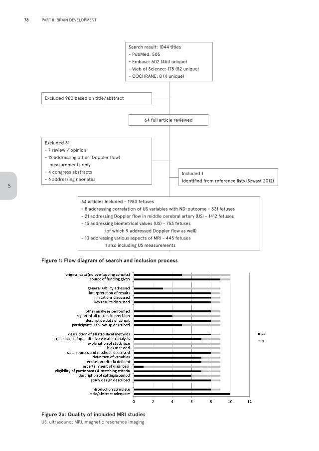
Figure 1: Flow diagram of search and inclusion process
Figure 2a: Quality of included MRI studiesUS, ultrasound; MRI, magnetic resonance imaging
Search result: 1044 titles
- PubMed: 505
- Embase: 602 (453 unique)
- Web of Science: 175 (82 unique)
- COCHRANE: 8 (4 unique)
34 articles included - 1983 fetuses
- 8 addressing correlation of US variables with ND-outcome - 331 fetuses
- 21 addressing Doppler flow in middle cerebral artery (US) - 1412 fetuses
- 13 addressing biometrical values (US) - 753 fetuses
(of which 9 addressed Doppler flow as well)
- 10 addressing various aspects of MRI - 445 fetuses
1 also including US measurements
Excluded 980 based on title/abstract
Included 1
Identified from reference lists (Szwast 2012)
Excluded 31
- 7 review / opinion
- 12 addressing other (Doppler flow)
measurements only
- 4 congress abstracts
- 6 addressing neonates
64 full article reviewed
78
5
PART II: BRAIN DEVELOPMENT
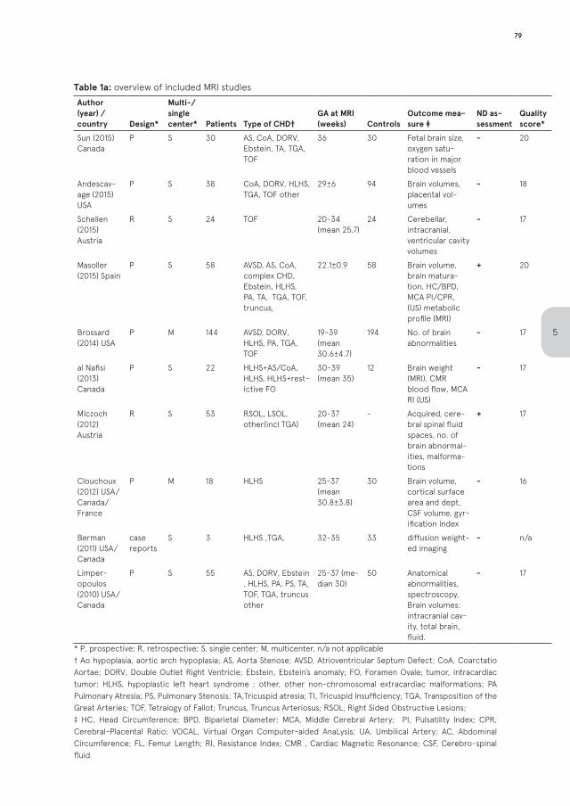
Table 1a: overview of included MRI studies
Author (year) / country Design*
Multi-/single center* Patients Type of CHD†
GA at MRI (weeks) Controls
Outcome mea-sure ‡
ND as-sessment
Quality score*
Sun (2015) Canada
P S 30 AS, CoA, DORV, Ebstein, TA, TGA, TOF
36 30 Fetal brain size, oxygen satu-ration in major blood vessels
- 20
Andescav-age (2015) USA
P S 38 CoA, DORV, HLHS, TGA, TOF other
29±6 94 Brain volumes, placental vol-umes
- 18
Schellen (2015) Austria
R S 24 TOF 20-34 (mean 25.7)
24 Cerebellar, intracranial, ventricular cavity volumes
- 17
Masoller (2015) Spain
P S 58 AVSD, AS, CoA, complex CHD, Ebstein, HLHS, PA, TA, TGA, TOF, truncus,
22.1±0.9 58 Brain volume, brain matura-tion, HC/BPD, MCA PI/CPR, (US) metabolic profile (MRI)
+ 20
Brossard (2014) USA
P M 144 AVSD, DORV, HLHS, PA, TGA, TOF
19-39 (mean 30.6±4.7)
194 No. of brain abnormalities
- 17
al Nafisi (2013) Canada
P S 22 HLHS+AS/CoA, HLHS, HLHS+rest-ictive FO
30-39 (mean 35)
12 Brain weight (MRI), CMR blood flow, MCA RI (US)
- 17
Mlczoch (2012) Austria
R S 53 RSOL, LSOL, other(incl TGA)
20-37 (mean 24)
- Acquired, cere-bral spinal fluid spaces, no. of brain abnormal-ities, malforma-tions
+ 17
Clouchoux (2012) USA/Canada/France
P M 18 HLHS 25-37 (mean 30.8±3.8)
30 Brain volume, cortical surface area and dept, CSF volume, gyr-ification index
- 16
Berman (2011) USA/Canada
case reports
S 3 HLHS ,TGA, 32-35 33 diffusion weight-ed imaging
- n/a
Limper-opoulos (2010) USA/Canada
P S 55 AS, DORV, Ebstein , HLHS, PA, PS, TA, TOF, TGA, truncus other
25-37 (me-dian 30)
50 Anatomical abnormalities, spectroscopy, Brain volumes: intracranial cav-ity, total brain, fluid.
- 17
* P, prospective; R, retrospective; S, single center; M, multicenter, n/a not applicable† Ao hypoplasia, aortic arch hypoplasia; AS, Aorta Stenose; AVSD, Atrioventricular Septum Defect; CoA, Coarctatio Aortae; DORV, Double Outlet Right Ventricle; Ebstein, Ebstein’s anomaly; FO, Foramen Ovale; tumor, intracardiac tumor; HLHS, hypoplastic left heart syndrome ; other, other non-chromosomal extracardiac malformations; PA Pulmonary Atresia; PS, Pulmonary Stenosis; TA,Tricuspid atresia; TI, Tricuspid Insufficiency; TGA, Transposition of the Great Arteries; TOF, Tetralogy of Fallot; Truncus, Truncus Arteriosus; RSOL, Right Sided Obstructive Lesions;‡ HC, Head Circumference; BPD, Biparietal Diameter; MCA, Middle Cerebral Artery; PI, Pulsatility Index; CPR, Cerebral-Placental Ratio; VOCAL, Virtual Organ Computer-aided AnaLysis; UA, Umbilical Artery; AC, Abdominal Circumference; FL, Femur Length; RI, Resistance Index; CMR , Cardiac Magnetic Resonance; CSF, Cerebro-spinal fluid
79
5

Table 1b: overview of included Ultrasound studiesAuthor (year) / country Design*
Multi-/single-center* Cases Type of CHD† GA at US(weeks) Controls Outcome measures ‡
ND assessment
Quality score
Meta-analysis** Used normal values
Williams (2015) USA/Canada
P S 66 HLHS, TGA ,TOF F1 (mean 23.6±2), F2 (mean30.6±1.6), F3 (mean 36.4±1)
41 BPD, HC + 15 HC Hadlock 1984
Hahn (2015) USA R M 133 single ventricle
20-34 (mean 27.2±5.3) - Biometry(n=133), MCA PI(n=119)
+ 18 mix; HC Arduini 1990; Hadlock 1984
Masoller (2015) Spain P S 58 AS, AVSD, CoA, complex CHD, Ebstein, HLHS, PA, TA, TGA, TOF, truncus
20-24 (mean 22.3±0.9) 58 BPD, CPR, composite score, correlation with MRI, HC, MCA PI,
+ 19 mix (n); HC (n)
Arduini 1990; Kurma-nanavicius 1999
Zeng 3d Flow (2015) China
P S 112 HLHS, LSOL, RSOL, TGA 19+6-30+3 (mean 25.7±2.74)
112 3D flow , MCA PI + 20 mix; lsol; rsol; tga
Arduini 1990
Zeng 3d Volume (2015) China
P S 73 Ao hypoplasia, AVSD, HLHS, PA, PS, TA, TGA, TOF, truncus
20+4–36+4 (mean 28.2 ± 4.4)
168 cerebral volume(VOCAL), HC, MCA PI
- 18 mix (n) n/a
Miller (2014) USA R S 43 AS, HLHS mean 33+5 - Biometry - 19 n/a Olsen 2010
Masoller (2014) Spain P S 95 Group 1: AS, CoA, HLHS, TGA Group 2: AVSD, complex CHD, DORV, TOF, truncus arteriosusGroup 3: Ebstein, PA, PS, TA
20-23+5 (mean 22+3) 95 Biometry, CPR, fractional mov-ing blood volume, MCA PI
- 19 mix; rsol; HC Arduini 1990; Kurma-nanavicius 1999
Williams (2013) USA/Canada
R M 119 single ventricle(79% HLHS) 18-38 (mean 27) - MCA PI, neonatal HC + 18 mix (n); lsol (n) Arduini 1990
Hangge (2013) USA R S 38 HLHS mean 26.6±5 - Biometry, MCA PI, neonatal HC + 19 lsol (n); HC (n) Olsen 2010
Cnota (2013) USA R S 33 HLHS 19-37 (median 27) - Biometry, UA flow, neonatal HC
- 17 n/a Olsen 2010+Hadlock 1984
Yamamoto (2013) Canada
R S 89 CoA, HLHS, HLHS + CoA, PA , TGA
closest to term (mean 32±5)
89 CPR, MCA PI, neonatal HC - 17 mix; lsol; rsol; tga
Ebbing 2007
Szwast (2012) USA R S 131 single ventricle (RSOL/LSOL) LSOL mean 28.0±5.4; RSOL mean 24.4±4.0
92 CPR, MCA PI - 15 mix (n); lsol; rsol
Arduini 1990
Williams (2012) USA pilot S 16 HLHS, TGA, TOF 18-24 (mean 22.8±2.8) - CPR, MCA PI, + 16 mix; lsol; tga Arduini 1990
Arduini (2011) Italy R S 60 AS, AVSD, CoA, HLHS, PA, PS, TGA, TOF, other
30-35 (mean 34.1±1.9) 65 Biometry, CPR , MCA PI - 15 mix; HC n/a
Itsukaichi (2011) Japan
R S 44 CoA, DORV, Ebstein, HLHS, HRHS, PA, PS, single ven-tricle, TA, TGA, TOF, tumor, truncus, VSD
28-34 140 Biometry(AC, BPD, FL), CPR, MCA PI
- 18 n/a n/a
McElhinney (2010) USA
P S 46 HLHS + valvuloplasty 20-31 (mean 24.3 ±3) - Biometry, MCA PI - 17 lsol; HC Arduini 1990; Kurma-nanavicius 1999
Guorong (2009) China
P S 45 AS, CoA, completed en-docardial cushion defect, DORV, Ebstein, HLHS, PA, PS, single ventricle, TA, TGA, TOF, truncus,
20-40 275 CPR, MCA PI - 15 mix; lsol; rsol not reported
Chen (2009) China R S 11 Ebstein 23-37 44 MCA PI - 16 rsol (n) n/a
Berg (2009) Germany R S 113 AS, HLHS, PA, TGA, TOF 19-41 ‘closest to term’ 1378 CPR, HC postnatal, MCA PI - 18 mix; lsol; rsol; tga
Ebbing 2007
Hinton (2008) USA/Canada
R S 28 HLHS/AS 17-36 - HC pre -and postnatal - 15 HC (n) Hadlock 1984
Modena (2006) USA R S 71 AS, AVSD, CoA, DORV, HLHS, HRHS, PA, TA, TGA, TOF, Truncus, tumor, VSD
‘closest to mid second trimester’
71 CPR, MCA PI - 16 mix Arduini 1990
80
5
PART II: BRAIN DEVELOPMENT
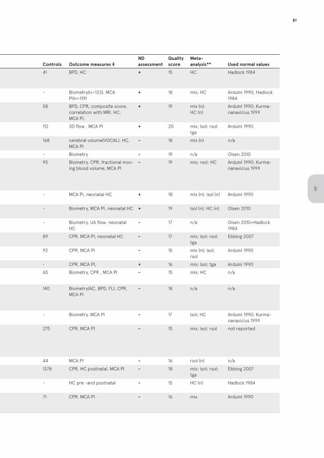
Table 1b: overview of included Ultrasound studiesAuthor (year) / country Design*
Multi-/single-center* Cases Type of CHD† GA at US(weeks) Controls Outcome measures ‡
ND assessment
Quality score
Meta-analysis** Used normal values
Williams (2015) USA/Canada
P S 66 HLHS, TGA ,TOF F1 (mean 23.6±2), F2 (mean30.6±1.6), F3 (mean 36.4±1)
41 BPD, HC + 15 HC Hadlock 1984
Hahn (2015) USA R M 133 single ventricle
20-34 (mean 27.2±5.3) - Biometry(n=133), MCA PI(n=119)
+ 18 mix; HC Arduini 1990; Hadlock 1984
Masoller (2015) Spain P S 58 AS, AVSD, CoA, complex CHD, Ebstein, HLHS, PA, TA, TGA, TOF, truncus
20-24 (mean 22.3±0.9) 58 BPD, CPR, composite score, correlation with MRI, HC, MCA PI,
+ 19 mix (n); HC (n)
Arduini 1990; Kurma-nanavicius 1999
Zeng 3d Flow (2015) China
P S 112 HLHS, LSOL, RSOL, TGA 19+6-30+3 (mean 25.7±2.74)
112 3D flow , MCA PI + 20 mix; lsol; rsol; tga
Arduini 1990
Zeng 3d Volume (2015) China
P S 73 Ao hypoplasia, AVSD, HLHS, PA, PS, TA, TGA, TOF, truncus
20+4–36+4 (mean 28.2 ± 4.4)
168 cerebral volume(VOCAL), HC, MCA PI
- 18 mix (n) n/a
Miller (2014) USA R S 43 AS, HLHS mean 33+5 - Biometry - 19 n/a Olsen 2010
Masoller (2014) Spain P S 95 Group 1: AS, CoA, HLHS, TGA Group 2: AVSD, complex CHD, DORV, TOF, truncus arteriosusGroup 3: Ebstein, PA, PS, TA
20-23+5 (mean 22+3) 95 Biometry, CPR, fractional mov-ing blood volume, MCA PI
- 19 mix; rsol; HC Arduini 1990; Kurma-nanavicius 1999
Williams (2013) USA/Canada
R M 119 single ventricle(79% HLHS) 18-38 (mean 27) - MCA PI, neonatal HC + 18 mix (n); lsol (n) Arduini 1990
Hangge (2013) USA R S 38 HLHS mean 26.6±5 - Biometry, MCA PI, neonatal HC + 19 lsol (n); HC (n) Olsen 2010
Cnota (2013) USA R S 33 HLHS 19-37 (median 27) - Biometry, UA flow, neonatal HC
- 17 n/a Olsen 2010+Hadlock 1984
Yamamoto (2013) Canada
R S 89 CoA, HLHS, HLHS + CoA, PA , TGA
closest to term (mean 32±5)
89 CPR, MCA PI, neonatal HC - 17 mix; lsol; rsol; tga
Ebbing 2007
Szwast (2012) USA R S 131 single ventricle (RSOL/LSOL) LSOL mean 28.0±5.4; RSOL mean 24.4±4.0
92 CPR, MCA PI - 15 mix (n); lsol; rsol
Arduini 1990
Williams (2012) USA pilot S 16 HLHS, TGA, TOF 18-24 (mean 22.8±2.8) - CPR, MCA PI, + 16 mix; lsol; tga Arduini 1990
Arduini (2011) Italy R S 60 AS, AVSD, CoA, HLHS, PA, PS, TGA, TOF, other
30-35 (mean 34.1±1.9) 65 Biometry, CPR , MCA PI - 15 mix; HC n/a
Itsukaichi (2011) Japan
R S 44 CoA, DORV, Ebstein, HLHS, HRHS, PA, PS, single ven-tricle, TA, TGA, TOF, tumor, truncus, VSD
28-34 140 Biometry(AC, BPD, FL), CPR, MCA PI
- 18 n/a n/a
McElhinney (2010) USA
P S 46 HLHS + valvuloplasty 20-31 (mean 24.3 ±3) - Biometry, MCA PI - 17 lsol; HC Arduini 1990; Kurma-nanavicius 1999
Guorong (2009) China
P S 45 AS, CoA, completed en-docardial cushion defect, DORV, Ebstein, HLHS, PA, PS, single ventricle, TA, TGA, TOF, truncus,
20-40 275 CPR, MCA PI - 15 mix; lsol; rsol not reported
Chen (2009) China R S 11 Ebstein 23-37 44 MCA PI - 16 rsol (n) n/a
Berg (2009) Germany R S 113 AS, HLHS, PA, TGA, TOF 19-41 ‘closest to term’ 1378 CPR, HC postnatal, MCA PI - 18 mix; lsol; rsol; tga
Ebbing 2007
Hinton (2008) USA/Canada
R S 28 HLHS/AS 17-36 - HC pre -and postnatal - 15 HC (n) Hadlock 1984
Modena (2006) USA R S 71 AS, AVSD, CoA, DORV, HLHS, HRHS, PA, TA, TGA, TOF, Truncus, tumor, VSD
‘closest to mid second trimester’
71 CPR, MCA PI - 16 mix Arduini 1990
81
5
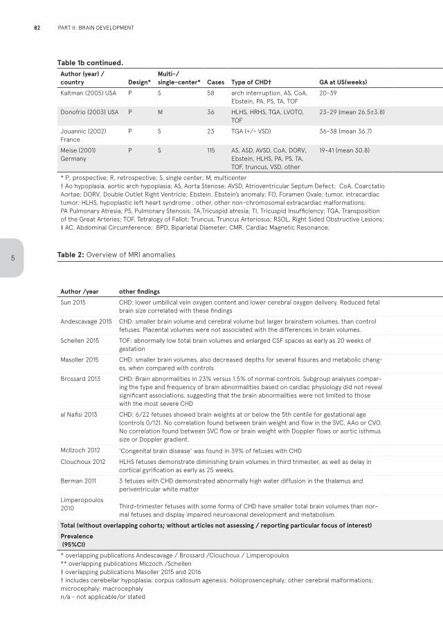
Table 1b continued.Author (year) / country Design*
Multi-/single-center* Cases Type of CHD† GA at US(weeks) Controls Outcome measures ‡
ND assessment
Quality score
Meta-analysis** Used normal values
Kaltman (2005) USA P S 58 arch interruption, AS, CoA, Ebstein, PA, PS, TA, TOF
20-39 114 CPR, MCA PI - 19 mix; lsol; rsol Arduini 1990
Donofrio (2003) USA P M 36 HLHS, HRHS, TGA, LVOTO, TOF
23-29 (mean 26.5±3.8) 21 CPR, HC, MCA RI - 20 n/a n/a
Jouannic (2002) France
P S 23 TGA (+/- VSD) 36-38 (mean 36.7) 40 MCA PI, UA PI - 15 tga n/a
Meise (2001) Germany
P S 115 AS, ASD, AVSD, CoA, DORV, Ebstein, HLHS, PA, PS, TA, TOF, truncus, VSD, other
19-41 (mean 30.8) 100 MCA PI, UA PI - 14 mix not reported
* P, prospective; R, retrospective; S, single center; M, multicenter† Ao hypoplasia, aortic arch hypoplasia; AS, Aorta Stenose; AVSD, Atrioventricular Septum Defect; CoA, Coarctatio Aortae; DORV, Double Outlet Right Ventricle; Ebstein, Ebstein’s anomaly; FO, Foramen Ovale; tumor, intracardiactumor; HLHS, hypoplastic left heart syndrome ; other, other non-chromosomal extracardiac malformations; PA Pulmonary Atresia; PS, Pulmonary Stenosis; TA,Tricuspid atresia; TI, Tricuspid Insufficiency; TGA, Transposition of the Great Arteries; TOF, Tetralogy of Fallot; Truncus, Truncus Arteriosus; RSOL, Right Sided Obstructive Lesions;‡ AC, Abdominal Circumference; BPD, Biparietal Diameter; CMR, Cardiac Magnetic Resonance;
CRP, Cerebral-Placental Ratio; CSF, Cerebro-spinal fluid; FL, Femur Length; HC, Head Circumference; MCA, Middle Cerebral Artery; PI, Pulsatility Index; RI, Resistance Index; UA, Umbilical Artery; VOCAL, Virtual Organ Computer-aided AnaLysis; n/a not applicable** in which meta-analyses are data included: mix, all/mixed types of CHD combined (MCA); lsol, left sided obstructive defects (MCA); rsol, rightsided obstructive defects (MCA); tga, transposition of the great arteries (MCA); HC, head circumference; (n) not included in the final model, overlapping publication
Table 2: Overview of MRI anomalies
Author /year other findings
total reported anomalies
(CHD)structural
anomalies†cystiform
abnormalitiesventricular
abnormalities
lesions/delay possibly caused by
hemodynamic changes
total reported anomalies (controls)
Sun 2015 CHD: lower umbilical vein oxygen content and lower cerebral oxygen delivery. Reduced fetal brain size correlated with these findings
1 in 30 1 n/a n/a n/a n/a in 30
Andescavage 2015 CHD: smaller brain volume and cerebral volume but larger brainstem volumes, than control fetuses. Placental volumes were not associated with the differences in brain volumes.
4 in 38* 0* 1* 1* 2* 0 in 94
Schellen 2015 TOF: abnormally low total brain volumes and enlarged CSF spaces as early as 20 weeks of gestation
5 in 24** 0** n/a 5** n/a 0 in 24
Masoller 2015 CHD: smaller brain volumes, also decreased depths for several fissures and metabolic chang-es, when compared with controls
0 in 58‡ 0‡ n/a n/a n/a 0 in 58
Brossard 2013 CHD: Brain abnormalities in 23% versus 1.5% of normal controls. Subgroup analyses compar-ing the type and frequency of brain abnormalities based on cardiac physiology did not reveal significant associations, suggesting that the brain abnormalities were not limited to those with the most severe CHD
33 in 144* 3* 2 13 15 3 in 194
al Nafisi 2013 CHD: 6/22 fetuses showed brain weights at or below the 5th centile for gestational age (controls 0/12). No correlation found between brain weight and flow in the SVC, AAo or CVO. No correlation found between SVC flow or brain weight with Doppler flows or aortic isthmus size or Doppler gradient.
n/a n/a n/a n/a n/a n/a in 12
Mcllzoch 2012 ‘Congenital brain disease’ was found in 39% of fetuses with CHD 21 in 53** 7** 4 9** 1 n/a
Clouchoux 2012 HLHS fetuses demonstrate diminishing brain volumes in third trimester, as well as delay in cortical gyrification as early as 25 weeks.
5 in 18* 0* n/a 5* n/a 0 in 30
Berman 2011 3 fetuses with CHD demonstrated abnormally high water diffusion in the thalamus and periventricular white matter
1 in 3 1 n/a n/a n/a n/a in 33
Limperopoulos 2010 Third-trimester fetuses with some forms of CHD have smaller total brain volumes than nor-
mal fetuses and display impaired neuroaxonal development and metabolism.
6 in 52* 2* 2* 4* n/a 0 in 55
Total (without overlapping cohorts; without articles not assessing / reporting particular focus of interest) 56 in 288 12 in 288 6 in 197 22 in 197 16 in 197 3 in 276Prevalence (95%CI)
0.18(-0.06; 0.42)
0.03(-0.01; 0.08)
0.04(-0.03; 0.10)
0.12(0.04; 0.19)
0.06(-0.03; 0.16)
0.01(-0.01; 0.03)
* overlapping publications Andescavage / Brossard /Clouchoux / Limperopoulos ** overlapping publications Mlczoch /Schellen‡ overlapping publications Masoller 2015 and 2016† includes cerebellar hypoplasia; corpus callosum agenesis; holoprosencephaly; other cerebral malformations; microcephaly; macrocephalyn/a - not applicable/or stated
82
5
PART II: BRAIN DEVELOPMENT
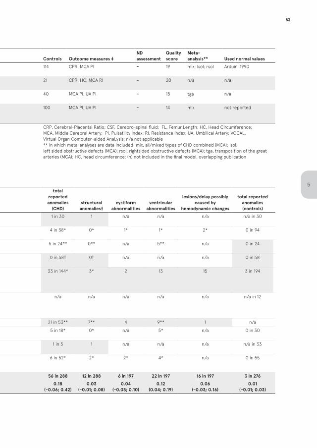
Table 1b continued.Author (year) / country Design*
Multi-/single-center* Cases Type of CHD† GA at US(weeks) Controls Outcome measures ‡
ND assessment
Quality score
Meta-analysis** Used normal values
Kaltman (2005) USA P S 58 arch interruption, AS, CoA, Ebstein, PA, PS, TA, TOF
20-39 114 CPR, MCA PI - 19 mix; lsol; rsol Arduini 1990
Donofrio (2003) USA P M 36 HLHS, HRHS, TGA, LVOTO, TOF
23-29 (mean 26.5±3.8) 21 CPR, HC, MCA RI - 20 n/a n/a
Jouannic (2002) France
P S 23 TGA (+/- VSD) 36-38 (mean 36.7) 40 MCA PI, UA PI - 15 tga n/a
Meise (2001) Germany
P S 115 AS, ASD, AVSD, CoA, DORV, Ebstein, HLHS, PA, PS, TA, TOF, truncus, VSD, other
19-41 (mean 30.8) 100 MCA PI, UA PI - 14 mix not reported
* P, prospective; R, retrospective; S, single center; M, multicenter† Ao hypoplasia, aortic arch hypoplasia; AS, Aorta Stenose; AVSD, Atrioventricular Septum Defect; CoA, Coarctatio Aortae; DORV, Double Outlet Right Ventricle; Ebstein, Ebstein’s anomaly; FO, Foramen Ovale; tumor, intracardiactumor; HLHS, hypoplastic left heart syndrome ; other, other non-chromosomal extracardiac malformations; PA Pulmonary Atresia; PS, Pulmonary Stenosis; TA,Tricuspid atresia; TI, Tricuspid Insufficiency; TGA, Transposition of the Great Arteries; TOF, Tetralogy of Fallot; Truncus, Truncus Arteriosus; RSOL, Right Sided Obstructive Lesions;‡ AC, Abdominal Circumference; BPD, Biparietal Diameter; CMR, Cardiac Magnetic Resonance;
CRP, Cerebral-Placental Ratio; CSF, Cerebro-spinal fluid; FL, Femur Length; HC, Head Circumference; MCA, Middle Cerebral Artery; PI, Pulsatility Index; RI, Resistance Index; UA, Umbilical Artery; VOCAL, Virtual Organ Computer-aided AnaLysis; n/a not applicable** in which meta-analyses are data included: mix, all/mixed types of CHD combined (MCA); lsol, left sided obstructive defects (MCA); rsol, rightsided obstructive defects (MCA); tga, transposition of the great arteries (MCA); HC, head circumference; (n) not included in the final model, overlapping publication
Table 2: Overview of MRI anomalies
Author /year other findings
total reported anomalies
(CHD)structural
anomalies†cystiform
abnormalitiesventricular
abnormalities
lesions/delay possibly caused by
hemodynamic changes
total reported anomalies (controls)
Sun 2015 CHD: lower umbilical vein oxygen content and lower cerebral oxygen delivery. Reduced fetal brain size correlated with these findings
1 in 30 1 n/a n/a n/a n/a in 30
Andescavage 2015 CHD: smaller brain volume and cerebral volume but larger brainstem volumes, than control fetuses. Placental volumes were not associated with the differences in brain volumes.
4 in 38* 0* 1* 1* 2* 0 in 94
Schellen 2015 TOF: abnormally low total brain volumes and enlarged CSF spaces as early as 20 weeks of gestation
5 in 24** 0** n/a 5** n/a 0 in 24
Masoller 2015 CHD: smaller brain volumes, also decreased depths for several fissures and metabolic chang-es, when compared with controls
0 in 58‡ 0‡ n/a n/a n/a 0 in 58
Brossard 2013 CHD: Brain abnormalities in 23% versus 1.5% of normal controls. Subgroup analyses compar-ing the type and frequency of brain abnormalities based on cardiac physiology did not reveal significant associations, suggesting that the brain abnormalities were not limited to those with the most severe CHD
33 in 144* 3* 2 13 15 3 in 194
al Nafisi 2013 CHD: 6/22 fetuses showed brain weights at or below the 5th centile for gestational age (controls 0/12). No correlation found between brain weight and flow in the SVC, AAo or CVO. No correlation found between SVC flow or brain weight with Doppler flows or aortic isthmus size or Doppler gradient.
n/a n/a n/a n/a n/a n/a in 12
Mcllzoch 2012 ‘Congenital brain disease’ was found in 39% of fetuses with CHD 21 in 53** 7** 4 9** 1 n/a
Clouchoux 2012 HLHS fetuses demonstrate diminishing brain volumes in third trimester, as well as delay in cortical gyrification as early as 25 weeks.
5 in 18* 0* n/a 5* n/a 0 in 30
Berman 2011 3 fetuses with CHD demonstrated abnormally high water diffusion in the thalamus and periventricular white matter
1 in 3 1 n/a n/a n/a n/a in 33
Limperopoulos 2010 Third-trimester fetuses with some forms of CHD have smaller total brain volumes than nor-
mal fetuses and display impaired neuroaxonal development and metabolism.
6 in 52* 2* 2* 4* n/a 0 in 55
Total (without overlapping cohorts; without articles not assessing / reporting particular focus of interest) 56 in 288 12 in 288 6 in 197 22 in 197 16 in 197 3 in 276Prevalence (95%CI)
0.18(-0.06; 0.42)
0.03(-0.01; 0.08)
0.04(-0.03; 0.10)
0.12(0.04; 0.19)
0.06(-0.03; 0.16)
0.01(-0.01; 0.03)
* overlapping publications Andescavage / Brossard /Clouchoux / Limperopoulos ** overlapping publications Mlczoch /Schellen‡ overlapping publications Masoller 2015 and 2016† includes cerebellar hypoplasia; corpus callosum agenesis; holoprosencephaly; other cerebral malformations; microcephaly; macrocephalyn/a - not applicable/or stated
83
5
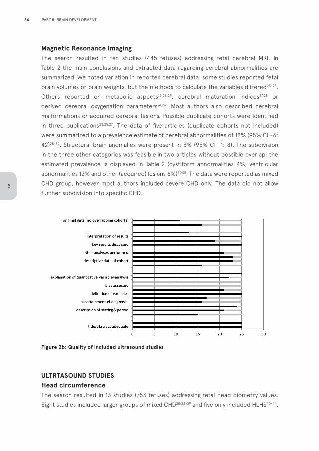
Magnetic Resonance ImagingThe search resulted in ten studies (445 fetuses) addressing fetal cerebral MRI. In Table 2 the main conclusions and extracted data regarding cerebral abnormalities are summarized. We noted variation in reported cerebral data: some studies reported fetal brain volumes or brain weights, but the methods to calculate the variables differed23-28. Others reported on metabolic aspects23;28;29, cerebral maturation indices27;28 or derived cerebral oxygenation parameters24;26. Most authors also described cerebral malformations or acquired cerebral lesions. Possible duplicate cohorts were identified in three publications23;25;27. The data of five articles (duplicate cohorts not included) were summarized to a prevalence estimate of cerebral abnormalities of 18% (95% CI -6; 42)28-32. Structural brain anomalies were present in 3% (95% CI -1; 8). The subdivision in the three other categories was feasible in two articles without possible overlap; the estimated prevalence is displayed in Table 2 (cystiform abnormalities 4%, ventricular abnormalities 12% and other (acquired) lesions 6%)30;31. The data were reported as mixed CHD group, however most authors included severe CHD only. The data did not allow further subdivision into specific CHD.
Figure 2b: Quality of included ultrasound studies
ULTRTASOUND STUDIESHead circumferenceThe search resulted in 13 studies (753 fetuses) addressing fetal head biometry values. Eight studies included larger groups of mixed CHD28;33-39 and five only included HLHS40-44.
84
5
PART II: BRAIN DEVELOPMENT
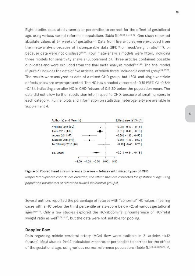
Eight studies calculated z-scores or percentiles to correct for the effect of gestational age, using various normal reference populations (Table 1b)28;33-36;40-42. One study reported absolute values at 34 weeks of gestation37. Data from five articles were excluded from the meta-analysis because of incomparable data (BPD36 or head/weight ratio38;43), or because data were not displayed39;44. Four meta-analysis models were fitted, including three models for sensitivity analysis (Supplement 3). Three articles contained possible duplicates and were excluded from the final meta-analysis model28;41;42. The final model (Figure 3) includes the data of five articles, of which three included a control group34;35;37. The results were analyzed as data of a mixed CHD group, but LSOL and single ventricle defects cases are overrepresented. The HC has a pooled z-score of -0.51 (95% CI -0.84; -0.18), indicating a smaller HC in CHD fetuses of 0.5 SD below the population mean. The data did not allow further subdivision into in specific CHD, because of small numbers in each category. Funnel plots and information on statistical heterogeneity are available in Supplement 4.
Figure 3: Pooled head circumference z-score – fetuses with mixed types of CHD
Suspected duplicate cohorts are excluded; the effect sizes are corrected for gestational age using
population parameters of reference studies (no control groups).
Several authors reported the percentage of fetuses with “abnormal” HC values, meaning cases with a HC below the third percentile or a z-score below -2, at various gestational ages28;41;42. Only a few studies explored the HC/abdominal circumference or HC/fetal weight ratio as well37;38;41;43, but the data were not suitable for pooling.
Doppler flowData regarding middle cerebral artery (MCA) flow were available in 21 articles (1412 fetuses). Most studies (n=14) calculated z-scores or percentiles to correct for the effect of the gestational age, using various normal reference populations (Table 1b)28;33;35;40;45-54.
85
5
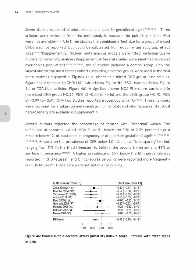
Seven studies reported absolute values at a specific gestational age36-39;42;55;56. Three articles were excluded from the meta-analysis because the pulsatility indices (PIs) were not available23;36;38. In three studies the combined effect size for a group of mixed CHDs was not reported, but could be calculated from documented subgroup effect sizes46;52;53(Supplement 2). Sixteen meta-analysis models were fitted, including twelve models for sensitivity analyses (Supplement 3). Several studies were identified to report overlapping populations28;39;42;52;54;56, and 13 studies included a control group. Only the largest and/or the most recent cohorts, including a control group, were used in the final meta-analyses displayed in Figures 4a-d; either as a mixed CHD group (nine articles, Figure 4a) or for specific CHD: LSOL (six articles, Figure 4b); RSOL (seven articles, Figure 4c) or TGA (four articles, Figure 4d). A significant lower MCA PI z-score was found in the mixed CHD group (-0.33; 95% CI -0.50 to -0.16) and the LSOL group (-0.70; 95% CI -0.99 to -0.41). Only two studies reported a subgroup with TOF49;50. These numbers were too small for a subgroup meta-analysis. Funnel plots and information on statistical heterogeneity are available in Supplement 4.
Several authors reported the percentage of fetuses with “abnormal” values. The definitions of abnormal varied (MCA PI, or RI, below the fifth or 2.3rd percentile or a z-score below -2, at least once in pregnancy or at a certain gestational age)27;34-37;40;42;46-
48;50;52;56. Reports on the prevalence of CPR below 1.0 (labeled as “brainsparing”) varied, ranging from 11% (in the third trimester) to 56% (in the second trimester) and 44% at any time in pregnancy36;38;49. A higher prevalence of CPR below the fifth percentile was reported in CHD fetuses47, and CPR z-scores below -2 were reported more frequently in HLHS fetuses50. These data were not suitable for pooling.
Figure 4a: Pooled middle cerebral artery pulsatility index z-score – fetuses with mixed types
of CHD
86
5
PART II: BRAIN DEVELOPMENT
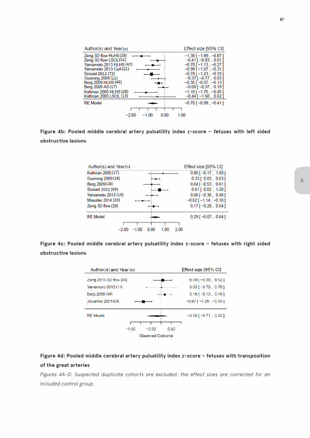
Figure 4b: Pooled middle cerebral artery pulsatility index z-score – fetuses with left sided
obstructive lesions
Figure 4c: Pooled middle cerebral artery pulsatility index z-score – fetuses with right sided
obstructive lesions
Figure 4d: Pooled middle cerebral artery pulsatility index z-score – fetuses with transposition
of the great arteries
Figures 4A-D: Suspected duplicate cohorts are excluded; the effect sizes are corrected for an
included control group.
87
5
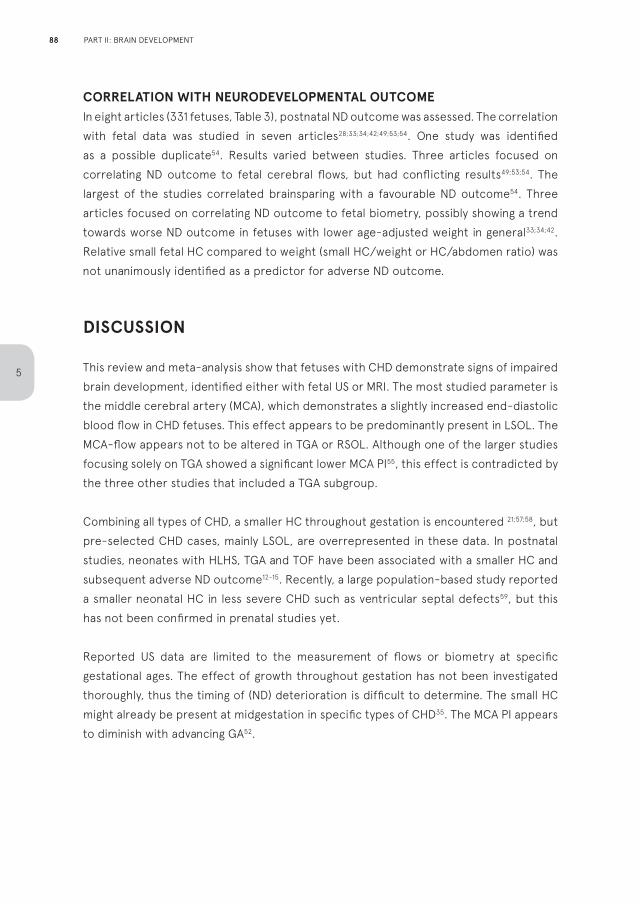
CORRELATION WITH NEURODEVELOPMENTAL OUTCOMEIn eight articles (331 fetuses, Table 3), postnatal ND outcome was assessed. The correlation with fetal data was studied in seven articles28;33;34;42;49;53;54. One study was identified as a possible duplicate54. Results varied between studies. Three articles focused on correlating ND outcome to fetal cerebral flows, but had conflicting results49;53;54. The largest of the studies correlated brainsparing with a favourable ND outcome54. Three articles focused on correlating ND outcome to fetal biometry, possibly showing a trend towards worse ND outcome in fetuses with lower age-adjusted weight in general33;34;42. Relative small fetal HC compared to weight (small HC/weight or HC/abdomen ratio) was not unanimously identified as a predictor for adverse ND outcome.
DISCUSSION
This review and meta-analysis show that fetuses with CHD demonstrate signs of impaired brain development, identified either with fetal US or MRI. The most studied parameter is the middle cerebral artery (MCA), which demonstrates a slightly increased end-diastolic blood flow in CHD fetuses. This effect appears to be predominantly present in LSOL. The MCA-flow appears not to be altered in TGA or RSOL. Although one of the larger studies focusing solely on TGA showed a significant lower MCA PI55, this effect is contradicted by the three other studies that included a TGA subgroup.
Combining all types of CHD, a smaller HC throughout gestation is encountered 21;57;58, but pre-selected CHD cases, mainly LSOL, are overrepresented in these data. In postnatal studies, neonates with HLHS, TGA and TOF have been associated with a smaller HC and subsequent adverse ND outcome12-15. Recently, a large population-based study reported a smaller neonatal HC in less severe CHD such as ventricular septal defects59, but this has not been confirmed in prenatal studies yet.
Reported US data are limited to the measurement of flows or biometry at specific gestational ages. The effect of growth throughout gestation has not been investigated thoroughly, thus the timing of (ND) deterioration is difficult to determine. The small HC might already be present at midgestation in specific types of CHD35. The MCA PI appears to diminish with advancing GA52.
88
5
PART II: BRAIN DEVELOPMENT
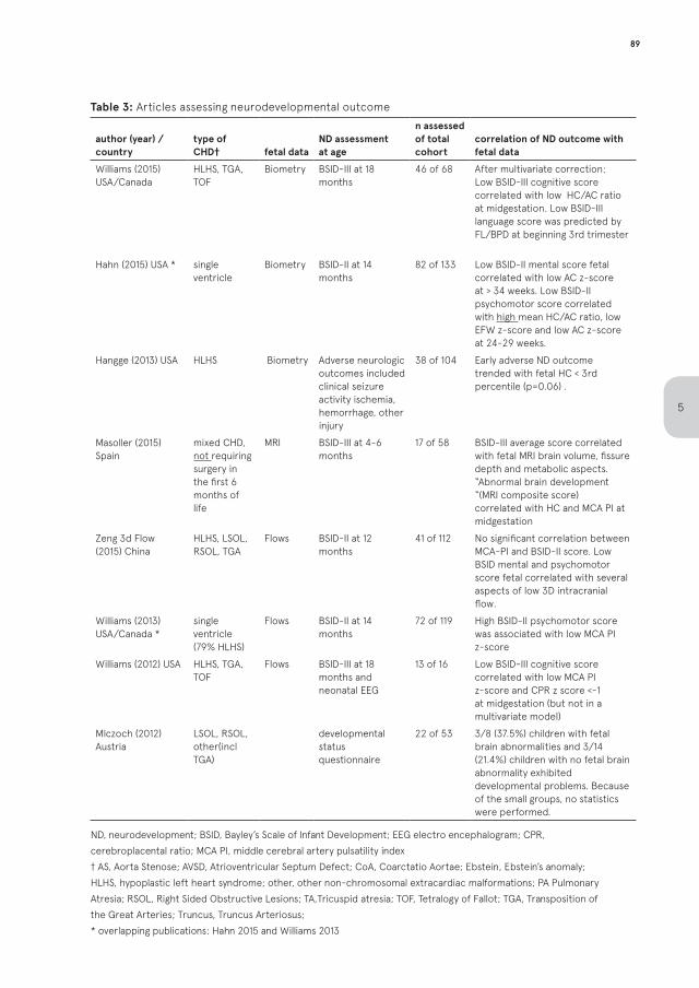
Table 3: Articles assessing neurodevelopmental outcome
author (year) / country
type of CHD† fetal data
ND assessment at age
n assessed of total cohort
correlation of ND outcome with fetal data
Williams (2015) USA/Canada
HLHS, TGA, TOF
Biometry BSID-III at 18 months
46 of 68 After multivariate correction: Low BSID-III cognitive score correlated with low HC/AC ratio at midgestation. Low BSID-III language score was predicted by FL/BPD at beginning 3rd trimester
Hahn (2015) USA * single ventricle
Biometry BSID-II at 14 months
82 of 133 Low BSID-II mental score fetal correlated with low AC z-score at > 34 weeks. Low BSID-II psychomotor score correlated with high mean HC/AC ratio, low EFW z-score and low AC z-score at 24-29 weeks.
Hangge (2013) USA HLHS Biometry Adverse neurologic outcomes included clinical seizure activity ischemia, hemorrhage, other injury
38 of 104 Early adverse ND outcome trended with fetal HC < 3rd percentile (p=0.06) .
Masoller (2015) Spain
mixed CHD, not requiring surgery in the first 6 months of life
MRI BSID-III at 4-6 months
17 of 58 BSID-III average score correlated with fetal MRI brain volume, fissure depth and metabolic aspects. “Abnormal brain development “(MRI composite score) correlated with HC and MCA PI at midgestation
Zeng 3d Flow (2015) China
HLHS, LSOL, RSOL, TGA
Flows BSID-II at 12 months
41 of 112 No significant correlation between MCA-PI and BSID-II score. Low BSID mental and psychomotor score fetal correlated with several aspects of low 3D intracranial flow.
Williams (2013) USA/Canada *
single ventricle(79% HLHS)
Flows BSID-II at 14 months
72 of 119 High BSID-II psychomotor score was associated with low MCA PI z-score
Williams (2012) USA HLHS, TGA, TOF
Flows BSID-III at 18 months and neonatal EEG
13 of 16 Low BSID-III cognitive score correlated with low MCA PI z-score and CPR z score <-1 at midgestation (but not in a multivariate model)
Mlczoch (2012) Austria
LSOL, RSOL, other(incl TGA)
developmental status questionnaire
22 of 53 3/8 (37.5%) children with fetal brain abnormalities and 3/14 (21.4%) children with no fetal brain abnormality exhibited developmental problems. Because of the small groups, no statistics were performed.
ND, neurodevelopment; BSID, Bayley’s Scale of Infant Development; EEG electro encephalogram; CPR, cerebroplacental ratio; MCA PI, middle cerebral artery pulsatility index † AS, Aorta Stenose; AVSD, Atrioventricular Septum Defect; CoA, Coarctatio Aortae; Ebstein, Ebstein’s anomaly; HLHS, hypoplastic left heart syndrome; other, other non-chromosomal extracardiac malformations; PA Pulmonary Atresia; RSOL, Right Sided Obstructive Lesions; TA,Tricuspid atresia; TOF, Tetralogy of Fallot; TGA, Transposition of the Great Arteries; Truncus, Truncus Arteriosus; * overlapping publications: Hahn 2015 and Williams 2013
89
5
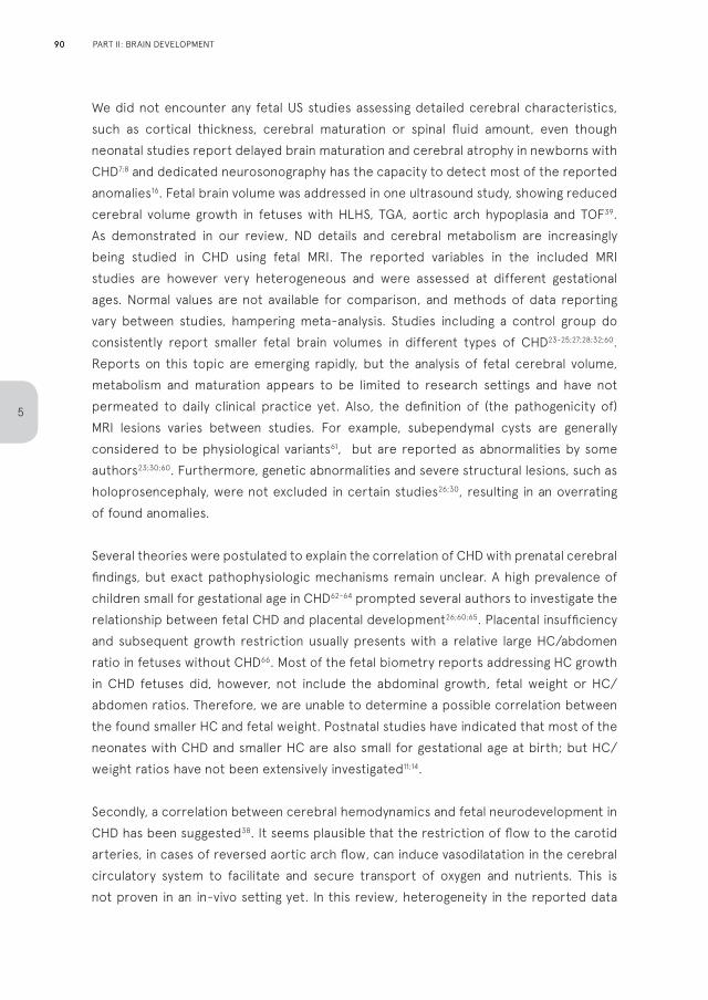
We did not encounter any fetal US studies assessing detailed cerebral characteristics, such as cortical thickness, cerebral maturation or spinal fluid amount, even though neonatal studies report delayed brain maturation and cerebral atrophy in newborns with CHD7;8 and dedicated neurosonography has the capacity to detect most of the reported anomalies16. Fetal brain volume was addressed in one ultrasound study, showing reduced cerebral volume growth in fetuses with HLHS, TGA, aortic arch hypoplasia and TOF39. As demonstrated in our review, ND details and cerebral metabolism are increasingly being studied in CHD using fetal MRI. The reported variables in the included MRI studies are however very heterogeneous and were assessed at different gestational ages. Normal values are not available for comparison, and methods of data reporting vary between studies, hampering meta-analysis. Studies including a control group do consistently report smaller fetal brain volumes in different types of CHD23-25;27;28;32;60. Reports on this topic are emerging rapidly, but the analysis of fetal cerebral volume, metabolism and maturation appears to be limited to research settings and have not permeated to daily clinical practice yet. Also, the definition of (the pathogenicity of) MRI lesions varies between studies. For example, subependymal cysts are generally considered to be physiological variants61, but are reported as abnormalities by some authors23;30;60. Furthermore, genetic abnormalities and severe structural lesions, such as holoprosencephaly, were not excluded in certain studies26;30, resulting in an overrating of found anomalies.
Several theories were postulated to explain the correlation of CHD with prenatal cerebral findings, but exact pathophysiologic mechanisms remain unclear. A high prevalence of children small for gestational age in CHD62-64 prompted several authors to investigate the relationship between fetal CHD and placental development26;60;65. Placental insufficiency and subsequent growth restriction usually presents with a relative large HC/abdomen ratio in fetuses without CHD66. Most of the fetal biometry reports addressing HC growth in CHD fetuses did, however, not include the abdominal growth, fetal weight or HC/abdomen ratios. Therefore, we are unable to determine a possible correlation between the found smaller HC and fetal weight. Postnatal studies have indicated that most of the neonates with CHD and smaller HC are also small for gestational age at birth; but HC/weight ratios have not been extensively investigated11;14.
Secondly, a correlation between cerebral hemodynamics and fetal neurodevelopment in CHD has been suggested38. It seems plausible that the restriction of flow to the carotid arteries, in cases of reversed aortic arch flow, can induce vasodilatation in the cerebral circulatory system to facilitate and secure transport of oxygen and nutrients. This is not proven in an in-vivo setting yet. In this review, heterogeneity in the reported data
90
5
PART II: BRAIN DEVELOPMENT
resulted in an impossibility to pool the data on the cerebroplacental ratio, hampering a definite conclusion regarding a possible brainsparing effect in CHD. Moreover, research concerning the brainsparing effect has mainly been performed in placental insufficiency, in which it is considered a mechanism to prevent fetal brain hypoxia rather than a sign of impending brain damage67. Because of the totally different pathophysiological and hemodynamic circumstances in CHD versus growth restriction, conclusions are not interchangeable. Brainsparing is correlated with worse ND outcome in growth restricted fetuses68. Even though available data suggests that brainsparing correlates with altered brain development in CHD27;28, a correlation with long-term outcome has not been established. On the contrary, it has even been suggested that brainsparing is associated with more favorable ND outcome in CHD54.
Another explanation for the cerebral variations in neonates with CHD could be a common genetic pathway, causing fetal CHD and ND delay69;70. The fact that fetuses and neonates with types of CHD that do not have a significant effect on fetal hemodynamics (minor CHD), also demonstrate cerebral anomalies, a smaller HC and low birth weight59 supports this theory. The development of the fetal brain in less severe CHD, such as ventricular septal defects, has however not been reported separately. In future research, the inclusion of minor CHD will be necessary to investigate this. Furthermore, a thorough genetic assessment in fetal CHD studies is necessary to exclude the effect of confounding genetic factors. Prenatal genetic testing is generally limited to array CGH with a reasonable resolution71, but it is known that smaller (point)mutations can play a role in CHD or neurodevelopment72-74.
To our knowledge, this review and meta-analysis is the first to perform a meta-analysis of fetal cerebral flow abnormalities in fetuses with (severe) CHD. A narrative review published in 2010 and a recent systematic review on MRI findings are available75;76. Certain included publications overlap, but our study includes a larger sample size with regard to cerebral flow, facilitating meta-analyses and subgroup assessment. Furthermore, exclusion of certain duplicate MRI cohorts results in a lower prevalence of MRI abnormalities in our review, which did not reach statistical significance. Two reviews focusing on postnatal findings are available for comparison, reporting high rates of cerebral damage in fetuses with CHD prior to surgery7;8. However, these reviews probably included children with CHD that were undetected prior to birth, thus possibly including postnatally developed abnormalities, possibly due to asphyxia because of a delayed diagnosis.
In our meta-analyses we compared z-scores. A z-score is a statistical tool to compare the results from cases with a standardized population, eliminating the influence of
91
5
gestational age. We have tried to maximize our sample size by transforming divergent outcome measures to mean z-scores, which also has some drawbacks. Grouped effect sizes would have been more accurate when individual measurements were corrected for individual gestational age, but these data are not available and assumptions on a higher data aggregation level are made. Another source of bias can be the various used normal values, which causes difficulty comparing the studies with each other. In the sensitivity analyses we determined that the use of population references, compared to the use of a control group, leads to a different outcome. In most studies it lead to a larger difference in effect size. To eliminate the effect of the various used standard populations we have chosen to use the studies reported control group as reference value.
ConclusionThis meta-analysis shows that fetuses with isolated, severe CHD demonstrate signs of impaired fetal cerebral development, demonstrated by fetal ultrasound or MRI. Our findings underline the importance of fetal neurological evaluation in CHD. However, reports correlating postnatal ND outcome to fetal findings remain inconclusive. Therefore, it is too early to conclude that aberrations revealed by neuroimaging involve a high risk of ND delay. To define the clinical meaning of prenatal cerebral variations in CHD, prospective large and long-term cohort studies are required, combining pre- and postnatal data. Such studies should include thorough genetic assessment and compare the findings to healthy controls. Parents should be counselled about the correlation of (specific types of) CHD with ND delay77, but until a correlation with ND outcome has been ascertained, there is insufficient data to support counseling families regarding ND outcome, or as a rationale for fetal therapy, based on prenatal findings suggestive of altered brain development.
Acknowledgement: We greatly acknowledge Theresa Costa Castro for her input.Funding: noneConflict of interest: none
Supplemental material available online: S1: complete search stringS2: transformation of extracted data to z-scoresS3: results of sensitivity analysesS4: estimation of heterogeneity and funnel plots
92
5
PART II: BRAIN DEVELOPMENT
REFERENCES
1. Bellinger DC, Jonas RA, Rappaport LA et al.
Developmental and neurologic status of children
after heart surgery with hypothermic circulatory
arrest or low-flow cardiopulmonary bypass. N
Engl J Med 1995;332:549-555.
2. Stieh J, Kramer HH, Harding P, Fischer G. Gross
and fine motor development is impaired in
children with cyanotic congenital heart disease.
Neuropediatrics 1999;30:77-82.
3. Marino B, Digilio MC. Congenital heart disease
and genetic syndromes: specific correlation
between cardiac phenotype and genotype.
Cardiovasc Pathol 2000;9:303-315.
4. Marino BS, Lipkin PH, Newburger JW et al.
Neurodevelopmental outcomes in children
with congenital heart disease: evaluation and
management: a scientific statement from
the American Heart Association. Circulation
2012;126:1143-1172.
5. McQuillen PS, Miller SP. Congenital heart disease
and brain development. Ann N Y Acad Sci
2010;1184:68-86.
6. du Plessis AJ. Mechanisms of brain injury during
infant cardiac surgery. Semin Pediatr Neurol
1999;6:32-47.
7. Owen M, Shevell M, Majnemer A, Limperopoulos
C. Abnormal brain structure and function
in newborns with complex congenital heart
defects before open heart surgery: A review
of the evidence. Journal of Child Neurology
2011;26:743-755.
8. Khalil A, Suff N, Thilaganathan B, Hurrell A,
Cooper D, Carvalho JS. Brain abnormalities and
neurodevelopmental delay in congenital heart
disease: systematic review and meta-analysis.
Ultrasound Obstet Gynecol 2014;43:14-24.
9. Majnemer A, Limperopoulos C, Shevell MI,
Rohlicek C, Rosenblatt B, Tchervenkov C. A
new look at outcomes of infants with congenital
heart disease. Pediatr Neurol 2009;40:197-204.
10. Donofrio MT, Duplessis AJ, Limperopoulos C.
Impact of congenital heart disease on fetal
brain development and injury. Current Opinion in
Pediatrics 2011;23:502-511.
11. Rosenthal GL. Patterns of prenatal growth among
infants with cardiovascular malformations:
possible fetal hemodynamic effects. Am J
Epidemiol 1996;143:505-513.
12. Manzar S, Nair AK, Pai MG, Al-Khhusaiby SM.
Head size at birth in neonates with transposition
of great arteries and hypoplastic left heart
syndrome. Saudi Medical Journal 2005;26:453-
456.
13. Shillingford AJ, Ittenbach RF, Marino BS et
al. Aortic morphometry and microcephaly in
hypoplastic left heart syndrome. Cardiol Young
2007;17:189-195.
14. Barbu D, Mert I, Kruger M, Bahado-Singh RO.
Evidence of fetal central nervous system injury in
isolated congenital heart defects: microcephaly
at birth. Am J Obstet Gynecol 2009;201:43-47.
15. Limperopoulos C, Majnemer A, Shevell MI et
al. Predictors of developmental disabilities
after open heart surgery in young children with
congenital heart defects. J Pediatr 2002;141:51-
58.
16. Pistorius LR, Stoutenbeek P, Groenendaal F et
al. Grade and symmetry of normal fetal cortical
development: a longitudinal two- and three-
dimensional ultrasound study. Ultrasound
Obstet Gynecol 2010;36:700-708.
17. Rossi AC, Prefumo F. Additional value of fetal
93
5

magnetic resonance imaging in the prenatal
diagnosis of central nervous system anomalies:
a systematic review of the literature. Ultrasound
Obstet Gynecol 2014;44:388-393.
18. Frick N, Fazelnia C, Kanzian K et al. The
reliability of fetal MRI in the assessment of brain
malformations. Fetal Diagn Ther 2015;37:93-101.
19. von EE, Altman DG, Egger M, Pocock SJ,
Gotzsche PC, Vandenbroucke JP. The
Strengthening the Reporting of Observational
Studies in Epidemiology (STROBE) statement:
guidelines for reporting observational studies.
Lancet 2007;370:1453-1457.
20. Stroup DF, Berlin JA, Morton SC et al. Meta-
analysis of observational studies in epidemiology:
a proposal for reporting. Meta-analysis Of
Observational Studies in Epidemiology (MOOSE)
group. JAMA 2000;283:2008-2012.
21. Hadlock FP. Computer-assisted, multiple-
parameter assessment of fetal age and growth.
Semin Ultrasound CT MR 1989;10:383-395.
22. Arduini D, Rizzo G. Normal values of Pulsatility
Index from fetal vessels: a cross-sectional
study on 1556 healthy fetuses. J Perinat Med
1990;18:165-172.
23. Limperopoulos C, Tworetzky W, McElhinney DB
et al. Brain volume and metabolism in fetuses
with congenital heart disease: evaluation with
quantitative magnetic resonance imaging and
spectroscopy. Circulation 2010;121:26-33.
24. Al NB, van Amerom JF, Forsey J et al. Fetal
circulation in left-sided congenital heart
disease measured by cardiovascular magnetic
resonance: a case-control study. J Cardiovasc
Magn Reson 2013;15:65.
25. Schellen C, Ernst S, Gruber GM et al. Fetal MRI
detects early alterations of brain development
in Tetralogy of Fallot. Am J Obstet Gynecol
2015;213:392-397.
26. Sun L, Macgowan CK, Sled JG et al. Reduced
fetal cerebral oxygen consumption is associated
with smaller brain size in fetuses with congenital
heart disease. Circulation 2015;131:1313-1323.
27. Clouchoux C, du Plessis AJ, Bouyssi-Kobar M et
al. Delayed cortical development in fetuses with
complex congenital heart disease. Cereb Cortex
2013;23:2932-2943.
28. Masoller N, Sanz-Cortes M, Crispi F et al. Severity
of Fetal Brain Abnormalities in Congenital Heart
Disease in Relation to the Main Expected Pattern
of in utero Brain Blood Supply. Fetal Diagn Ther
2015.
29. Berman JI, Hamrick SE, McQuillen PS et al.
Diffusion-weighted imaging in fetuses with
severe congenital heart defects. AJNR Am J
Neuroradiol 2011;32:E21-E22.
30. Mlczoch E, Brugger P, Ulm B et al. Structural
congenital brain disease in congenital heart
disease: Results from a fetal MRI program. Eur J
Paediatr Neurol 2012.
31. Brossard-Racine M, du Plessis AJ, Vezina G et al.
Prevalence and spectrum of in utero structural
brain abnormalities in fetuses with complex
congenital heart disease. AJNR Am J Neuroradiol
2014;35:1593-1599.
32. Paladini, D., et al., ISUOG consensus statement
on current understanding of the association
of neurodevelopmental delay and congenital
heart disease: impact on prenatal counseling.
Ultrasound Obstet Gynecol, 2017. 49(2): p. 287-
288.
33. Hahn E, Szwast A, Cnota J et al. The association
94
5
PART II: BRAIN DEVELOPMENT

of fetal growth, cerebral blood flow, and
neurodevelopmental outcome in single ventricle
fetuses. Ultrasound Obstet Gynecol 2016
Apr;47(4):460-5.
34. Williams IA, Fifer WP, Andrews H. Fetal Growth
and Neurodevelopmental Outcome in Congenital
Heart Disease. Pediatr Cardiol 2015;(6):1135–44.
35. Masoller N, Martinez JM, Gomez O et al.
Evidence of second-trimester changes in head
biometry and brain perfusion in fetuses with
congenital heart disease. Ultrasound Obstet
Gynecol 2014;44:182-187.
36. Itsukaichi M, Kikuchi A, Yoshihara K, Serikawa T,
Takakuwa K, Tanaka K. Changes in fetal circulation
associated with congenital heart disease and
their effects on fetal growth. Fetal Diagn Ther
2011;30:219-224.
37. Arduini M, Rosati P, Caforio L et al. Cerebral
blood flow autoregulation and congenital heart
disease: possible causes of abnormal prenatal
neurologic development. J Matern Fetal
Neonatal Med 2011;24:1208-1211.
38. Donofrio MT, Bremer YA, Schieken RM et al.
Autoregulation of cerebral blood flow in fetuses
with congenital heart disease: the brain sparing
effect. Pediatr Cardiol 2003;24:436-443.
39. Zeng S, Zhou QC, Zhou JW, Li M, Long C,
Peng QH. Volume of intracranial structures on
three-dimensional ultrasound in fetuses with
congenital heart disease. Ultrasound Obstet
Gynecol 2015;46:174-181.
40. McElhinney DB, Benson CB, Brown DW et
al. Cerebral blood flow characteristics and
biometry in fetuses undergoing prenatal
intervention for aortic stenosis with evolving
hypoplastic left heart syndrome. Ultrasound
Med Biol 2010;36:29-37.
41. Hinton RB, Andelfinger G, Sekar P et al.
Prenatal head growth and white matter injury
in hypoplastic left heart syndrome. Pediatr Res
2008;64:364-369.
42. Hangge PT, Cnota JF, Woo JG et al. Microcephaly
is associated with early adverse neurologic
outcomes in hypoplastic left heart syndrome.
Pediatr Res 2013;74:61-67.
43. Miller TA, Joss-Moore L, Menon SC, Weng
C, Puchalski MD. Umbilical artery systolic
to diastolic ratio is associated with growth
and myocardial performance in infants with
hypoplastic left heart syndrome. Prenat Diagn
2014;34:128-133.
44. Cnota JF, Hangge PT, Wang Y et al. Somatic
growth trajectory in the fetus with hypoplastic
left heart syndrome. Pediatr Res 2013;74:284-
289.
45. Meise C, Germer U, Gembruch U. Arterial
Doppler ultrasound in 115 second- and third-
trimester fetuses with congenital heart disease.
Ultrasound Obstet Gynecol 2001;17:398-402.
46. Kaltman JR, Di H, Tian Z, Rychik J. Impact of
congenital heart disease on cerebrovascular
blood flow dynamics in the fetus. Ultrasound
Obstet Gynecol 2005;25:32-36.
47. Modena A, Horan C, Visintine J, Chanthasenanont
A, Wood D, Weiner S. Fetuses with congenital
heart disease demonstrate signs of decreased
cerebral impedance. Am J Obstet Gynecol
2006;195:706-710.
48. Guorong L, Shaohui L, Peng J et al.
Cerebrovascular blood flow dynamic changes
in fetuses with congenital heart disease. Fetal
Diagn Ther 2009;25:167-172.
95
5

49. Williams IA, Tarullo AR, Grieve PG et al. Fetal
cerebrovascular resistance and neonatal
EEG predict 18-month neurodevelopmental
outcome in infants with congenital heart disease.
Ultrasound Obstet Gynecol 2012;40:304-309.
50. Berg C, Gembruch O, Gembruch U, Geipel A.
Doppler indices of the middle cerebral artery
in fetuses with cardiac defects theoretically
associated with impaired cerebral oxygen
delivery in utero: is there a brain-sparing effect?
Ultrasound Obstet Gynecol 2009;34:666-672.
51. Yamamoto Y, Khoo NS, Brooks PA, Savard
W, Hirose A, Hornberger LK. Severe left
heart obstruction with retrograde arch flow
influences fetal cerebral and placental blood
flow. Ultrasound Obstet Gynecol 2013;42:294-
299.
52. Szwast A, Tian Z, McCann M, Soffer D, Rychik
J. Comparative analysis of cerebrovascular
resistance in fetuses with single-ventricle
congenital heart disease. Ultrasound Obstet
Gynecol 2012;40:62-67.
53. Zeng S, Zhou J, Peng Q et al. Assessment by
three-dimensional power Doppler ultrasound
of cerebral blood flow perfusion in fetuses with
congenital heart disease. Ultrasound Obstet
Gynecol 2015;45:649-656.
54. Williams IA, Fifer C, Jaeggi E, Levine JC,
Michelfelder EC, Szwast AL. The association
of fetal cerebrovascular resistance with early
neurodevelopment in single ventricle congenital
heart disease. Am Heart J 2013;165:544-550.
55. Jouannic JM, Benachi A, Bonnet D et al.
Middle cerebral artery Doppler in fetuses with
transposition of the great arteries. Ultrasound
Obstet Gynecol 2002;20:122-124.
56. Chen Y, Lv G, Li B, Wang Z. Cerebral vascular
resistance and left ventricular myocardial
performance in fetuses with Ebstein’s anomaly.
Am J Perinatol 2009;26:253-258.
57. Kurmanavicius J, Wright EM, Royston P et al.
Fetal ultrasound biometry: 1. Head reference
values. Br J Obstet Gynaecol 1999;106:126-135.
58. Olsen IE, Groveman SA, Lawson ML, Clark RH,
Zemel BS. New intrauterine growth curves
based on United States data. Pediatrics
2010;125:e214-e224.
59. Matthiesen NB, Henriksen TB, Gaynor JW et al.
Congenital Heart Defects and Indices of Fetal
Cerebral Growth in a Nationwide Cohort of 924
422 Liveborn Infants. Circulation 2016;133:566-
575.
60. Andescavage N, Yarish A, Donofrio M et al. 3-D
volumetric MRI evaluation of the placenta in
fetuses with complex congenital heart disease.
Placenta 2015;36:1024-1030.
61. Pappalardo EM, Militello M, Rapisarda G et al.
Fetal intracranial cysts: prenatal diagnosis and
outcome. J Prenat Med 2009;3:28-30.
62. Kramer HH, Trampisch HJ, Rammos S, Giese A.
Birth weight of children with congenital heart
disease. Eur J Pediatr 1990;149:752-757.
63. Rosenthal GL, Wilson PD, Permutt T, Boughman
JA, Ferencz C. Birth weight and cardiovascular
malformations: a population-based study.
The Baltimore-Washington Infant Study. Am J
Epidemiol 1991;133:1273-1281.
64. Spiers PS. Does growth retardation predispose
the fetus to congenital malformation? Lancet
1982;1:312-314.
65. Huhta J, Linask KK. Environmental origins of
congenital heart disease: the heart-placenta
96
5
PART II: BRAIN DEVELOPMENT

connection. Semin Fetal Neonatal Med
2013;18:245-250.
66. Kramer MS, McLean FH, Olivier M, Willis DM,
Usher RH. Body proportionality and head and
length ‘sparing’ in growth-retarded neonates: a
critical reappraisal. Pediatrics 1989;84:717-723.
67. Scherjon SA, Smolders-DeHaas H, Kok JH,
Zondervan HA. The “brain-sparing” effect:
antenatal cerebral Doppler findings in relation to
neurologic outcome in very preterm infants. Am
J Obstet Gynecol 1993;169:169-175.
68. Hernandez-Andrade E, Serralde JA, Cruz-
Martinez R. Can anomalies of fetal brain
circulation be useful in the management of
growth restricted fetuses? Prenat Diagn
2012;32:103-112.
69. Connor JA, Hinton RB, Miller EM, Sund KL,
Ruschman JG, Ware SM. Genetic testing practices
in infants with congenital heart disease. Congenit
Heart Dis 2014;9:158-167.
70. Levenson D. Genetic testing using array
comparative genomic hybridization may benefit
newborns with congenital heart disease: study
shows aCGH more effective at detecting
potential genetic causes of CHD than other
methods. Am J Med Genet A 2014;164A:ix.
71. Wapner RJ, Martin CL, Levy B et al. Chromosomal
microarray versus karyotyping for prenatal
diagnosis. N Engl J Med 2012;367:2175-2184.
72. Fahed AC, Gelb BD, Seidman JG, Seidman CE.
Genetics of congenital heart disease: the glass
half empty. Circ Res 2013;112:707-720.
73. Hartman RJ, Rasmussen SA, Botto LD et al. The
contribution of chromosomal abnormalities to
congenital heart defects: a population-based
study. Pediatr Cardiol 2011;32:1147-1157.
74. Wessels MW, Willems PJ. Genetic factors in non-
syndromic congenital heart malformations. Clin
Genet 2010;78:103-123.
75. Donofrio MT, Massaro AN. Impact of congenital
heart disease on brain development and
neurodevelopmental outcome. Int J Pediatr
2010;2010.
76. Khalil A, Bennet S, Thilaganathan B, Paladini D,
Griffiths P, Carvalho JS. Prevalence of Prenatal
Brain Abnormalities in Fetuses with Congenital
Heart Disease: Systematic Review. Ultrasound
Obstet Gynecol 2016.
97
5
CHAPTER 6
Head growth in fetuses with isolated
congenital heart defects: lack of
influence of aortic arch flow and
ascending aorta oxygen saturation
Ultrasound in Obstetrics and Gynecology 2016; 48: 357–364.
F.A.R. Jansen E.W. van Zwet M.E.B. Rijlaarsdam E. Pajkrt C.L. van Velzen H.R. ZuurveenA. KragtC.L. BaxS.A.B. Clur J.M.M. van Lith N.A. Blom M.C. Haak
ABSTRACT
ObjectivesCongenital heart defects (CHD) are reported to be associated with smaller head circumferences (HC) and neurodevelopmental delay. Recent studies suggest that altered intra-uterine brain hemodynamics may explain these findings.
Methods
Singleton fetuses with isolated CHD were selected in three fetal medicine units (n=436). Cases with placental insufficiency or genetic syndromes were excluded. CHD were clustered based on the flow and oxygen saturation in the aorta. HC at different gestational ages was evaluated and univariate and multivariate mixed regression analysis was performed to examine the patterns of prenatal HC growth.
ResultsFetuses with severe and less severe types of CHD demonstrate HC growth restriction with advancing gestation (slope -0.017/ day), but a statistically significant effect of fetal hemodynamics clustering was absent. Even CHD with normal brain oxygenation and normal aortic flow showed a significant decrease in HC growth (slope -0.024/day). Furthermore, only Tetralogy of Fallot demonstrated a smaller HC z-score at 20 weeks of gestation (-0.67; 95% CI -1.16; -0.18).
ConclusionsThe HC growth decline in prenatally detected isolated CHD was still within the normal HC range, raising the question of its clinical significance. Furthermore, in contrast to other studies, this large cohort did not establish a significant correlation between aortic flow or oxygen saturation and HC growth. Factors other than altered cerebral hemodynamics may contribute to HC restriction with advancing gestation, such as (epi)genetic or placental factors.
6
100 PART II: BRAIN DEVELOPMENT
INTRODUCTION
Severe congenital heart defects (CHD) occur in 2-3 per 1000 live births and are responsible for significant morbidity and mortality. New methods of neonatal support and advanced surgical techniques have improved the survival rate significantly over the last decades. As a result, the focus of attention has shifted from survival to irreversible morbidity and long-term outcome1. Children with severe CHD frequently show cerebral impairments and neurodevelopmental delay (ND)2-4. Major causes of ND are hypoxia, thrombo-embolic insults and other surgical factors5-8. However, more than half of the newborns with CHD have signs of neurological abnormalities prior to surgery, raising the question whether CHD might cause prenatal cerebral damage9-11. It has been hypothesized that intrauterine hypoxic central nervous system damage or altered cerebral arterial hemodynamics induce delayed brain maturation and growth12-16. Various studies found significantly smaller head circumference (HC) in non-syndromic CHD, mainly in children with transposition of the great arteries (TGA), tetralogy of Fallot (TOF) and hypoplastic left heart syndrome (HLHS)17-20. However, the published cohorts attempting to validate the prenatal hemodynamics hypothesis are small and focus only on specifically severe CHD12-16;21;22.
If the HC is smaller at birth, then it should be detectable prior to birth. If cerebral flow and oxygen delivery play an important role, the relationship between the type of CHD and the HC should be apparent. As the presence of reduced head growth might be associated with ND after birth23, its presence may be predictive and could have an impact on parental counselling. Few studies, however, have focused on the prenatal HC growth in CHD13;16;23-28.
Our aim was to evaluate patterns of HC growth in a large cohort of fetuses with various CHD, and to analyze these patterns per type of CHD, to estimate the effect of cerebral hemodynamics (aortic arch flow and ascending aorta oxygen saturation) with advancing gestation in the second and third trimester . Our hypothesis was that, in the presence of a normal abdominal growth, HC growth is diminished in fetuses with CHD with reduced ascending aorta oxygen saturation and/or reduced aortic arch flow.
METHODS
This study was performed in the Leiden University Medical Center (LUMC), VU Medical center (VUmc) and Amsterdam Medical Center (AMC) over a 12.5 year period. These
101
6
three university medical centers collaborate in the care for children with CHD (center for congenital heart defects Amsterdam Leiden, CAHAL). All children with CHD, born between January 2002 and January 2012 were entered in the CAHAL database. The cases and methods of data collection were previously described29. The CAHAL database was supplemented with consecutive CHD cases, born in the LUMC between January 2012 and July 2014. From this combined database, prenatally diagnosed structural CHD were extracted. Functional CHD such as cardiomyopathy and primary arrhythmias were not eligible for inclusion. Cases with co-existing significant extracardiac malformations, severe hydrops, genetic anomalies and/or postnatal appearing syndromic abnormalities were excluded from further analysis. Minor additional findings, such as soft markers (e.g. echogenic focus and echogenic bowel), amniotic fluid pathology, mild pericardial effusion and/or single umbilical artery were not considered as significant extracardiac abnormalities and were not excluded. Multiple gestation, as well as cases with maternal pathology that could have an effect on fetal growth, i.e. pregnancy induced hypertension, preeclampsia, HELLP syndrome or signs of placental insufficiency by postnatal placental weight examination, were excluded. Pre- and postnatal ultrasound data were reviewed. Postnatal follow-up regarding the definitive cardiac defect, genetic defects or syndromic diagnosis and neurodevelopment was distilled from clinical files, these were available until at least one year of age. Pre-existent maternal illnesses (such as pre-existent hypertension and thyroid disease), maternal smoking, maternal body mass index (BMI) and parity were distilled from clinical files. Gestational age (GA) was based on first-trimester ultrasound. Repeated prenatal ultrasound examinations were performed routinely to re-evaluate the cardiac defect and to assess fetal wellbeing. At each examination, fetal biometrical data were measured routinely. Thus, head and abdominal circumference (HC and AC) were prospectively entered in patient files, and extracted from the fetal ultrasound databases for the purpose of this study. The measurements were performed according to the guidelines of the Dutch Society for Obstetrics and Gynecology30, which are the same as described by the International Society of Obstetrics and Gynecology.
Clustering of CHDsFirst, the CHDs were defined according to the postnatal diagnosis, made by postnatal ultrasound or postmortem examination. Second, CHDs were clustered based on two aspects: oxygen delivery and flow to the brain. The resulting clusters are depicted in table 2. As an indication for oxygen delivery to the brain, the heart defects were clustered by ascending aorta oxygen saturation as having either 1) low oxygen delivery to the brain (e.g. transposition of the great arteries; TGA), 2) intracardiac mixing of the oxygen-rich and oxygen-poor blood (e.g. atrioventricular septal defects, tetralogy of Fallot; TOF), or 3) normal oxygen delivery to the brain (e.g. congenitally corrected TGA
102
6
PART II: BRAIN DEVELOPMENT
without additional cardiac defects, Ebstein’s anomaly with normal cardiac function). As an indication for cerebral blood flow, heart defects were labeled as having either a) reversed aortic arch flow (e.g. aortic atresia, hypoplastic left heart syndrome; HLHS), b) obstructed aortic arch flow (e.g. aortic stenosis or hypoplasia), or c) normal aortic arch flow (e.g. TOF, TGA). In some cases clustering was complex, for instance severe aortic stenosis (developing HLHS) can display forward flow in the aorta prenatally. For the subtypes of CHD in which the clustering was disputable, or in which the flow direction in the aorta was not described in the files, two researchers (FJ and MR) independently re-evaluated all pre- and postnatal ultrasound and postmortem data to retrieve information about aortic arch flow and ascending aorta oxygen saturation in the prenatal period. In the supplementary material, the methods of clustering are clarified further and the types of CHD in the different clusters are listed. Our method of clustering shows similarities to the subclassification of Masoller et al28, and is based on theoretical hemodynamics. Actual in vivo measurement of cerebral oxygenation would be preferable21, but resources and expertise to do this are not widely available. Thus our approach is the most feasible and the most accessible for large groups, therewith minimizing selection bias.
Data handling and statistical analysisAll biometrical data were compared to the growth charts of Verburg et al30, because these growth charts were produced from a large comparable Dutch population (n=3760), and constructed in approximately the same period as our own cohort with modern ultrasound machines. We constructed z-scores of HC and AC. The HC/AC ratio is not available in these growth charts. To account for the dependence between repeated measurements on the same fetus, we used a linear mixed regression model to study the effect of increasing gestational age (GA) on the z-scores. First, we analyzed the z-scores in a univariate regression model, to assess the difference with the normal values and estimate clinical relevance. Growth restriction of the fetal head was defined as a significant decrease in HC z-score with advancing GA (the slope of the regression). Secondary to this model we compared the effect of increasing GA on the z-scores between the clusters cerebral blood flow and ascending aorta oxygen saturation separately. Third, we used both clusterings, as well as known confounders, as covariates in a multivariate regression analysis. Variables of interest and confounders were only used as covariates if the univariate effect size was smaller than p=0.1. Obesity was defined as a BMI > 25 kg/m2. Percentages of HC below -2 standard deviations (SD) within subgroups were compared with each other by Chi square test. Statistical analysis was performed using SPSS statistics version 20.0.0 (IBM, Armonk, NY, USA), data were displayed using Prism version 6.02 (GraphPad Software, La Jolla, CA, USA).
103
6
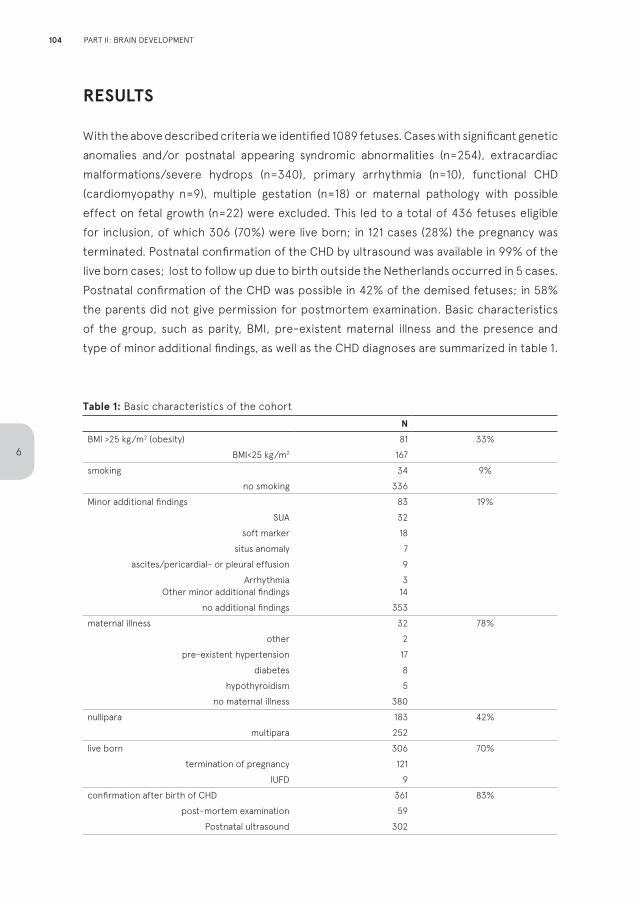
RESULTS
With the above described criteria we identified 1089 fetuses. Cases with significant genetic anomalies and/or postnatal appearing syndromic abnormalities (n=254), extracardiac malformations/severe hydrops (n=340), primary arrhythmia (n=10), functional CHD (cardiomyopathy n=9), multiple gestation (n=18) or maternal pathology with possible effect on fetal growth (n=22) were excluded. This led to a total of 436 fetuses eligible for inclusion, of which 306 (70%) were live born; in 121 cases (28%) the pregnancy was terminated. Postnatal confirmation of the CHD by ultrasound was available in 99% of the live born cases; lost to follow up due to birth outside the Netherlands occurred in 5 cases. Postnatal confirmation of the CHD was possible in 42% of the demised fetuses; in 58% the parents did not give permission for postmortem examination. Basic characteristics of the group, such as parity, BMI, pre-existent maternal illness and the presence and type of minor additional findings, as well as the CHD diagnoses are summarized in table 1.
Table 1: Basic characteristics of the cohort
NBMI >25 kg/m2 (obesity) 81 33%
BMI<25 kg/m2 167
smoking 34 9%
no smoking 336
Minor additional findings 83 19%
SUA 32
soft marker 18
situs anomaly 7
ascites/pericardial- or pleural effusion 9
ArrhythmiaOther minor additional findings
314
no additional findings 353
maternal illness 32 78%
other 2
pre-existent hypertension 17
diabetes 8
hypothyroidism 5
no maternal illness 380
nullipara 183 42%
multipara 252
live born 306 70%
termination of pregnancy 121
IUFD 9
confirmation after birth of CHD 361 83%
post-mortem examination 59
Postnatal ultrasound 302
104
6
PART II: BRAIN DEVELOPMENT
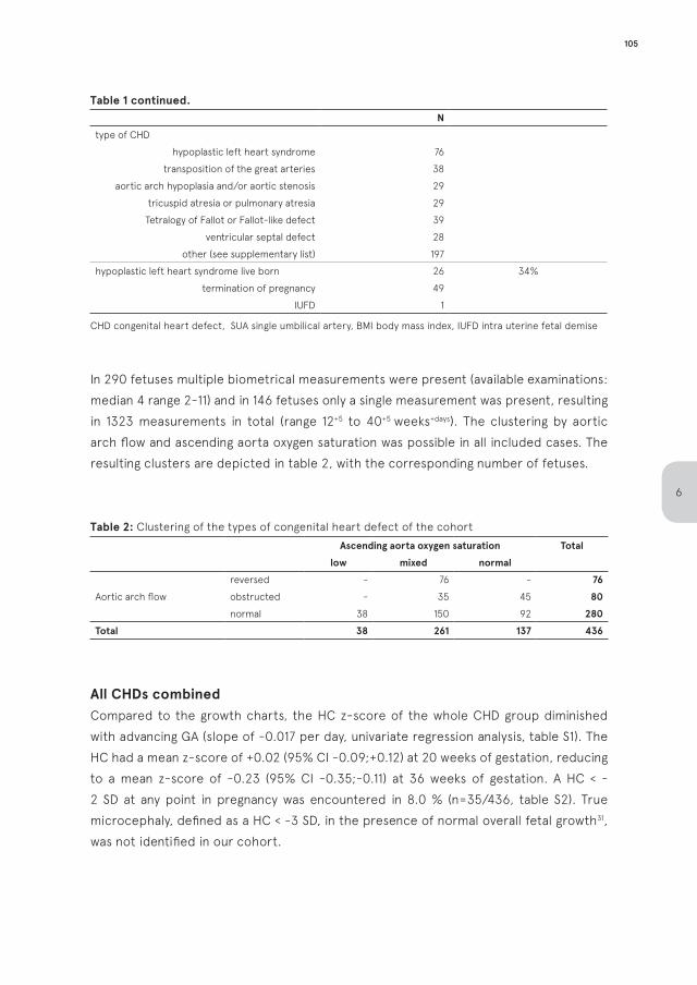
Table 1 continued. Ntype of CHD
hypoplastic left heart syndrome 76
transposition of the great arteries 38
aortic arch hypoplasia and/or aortic stenosis 29
tricuspid atresia or pulmonary atresia 29
Tetralogy of Fallot or Fallot-like defect 39
ventricular septal defect 28
other (see supplementary list) 197
hypoplastic left heart syndrome live born 26 34%
termination of pregnancy 49
IUFD 1
CHD congenital heart defect, SUA single umbilical artery, BMI body mass index, IUFD intra uterine fetal demise
In 290 fetuses multiple biometrical measurements were present (available examinations: median 4 range 2-11) and in 146 fetuses only a single measurement was present, resulting in 1323 measurements in total (range 12+5 to 40+5 weeks+days). The clustering by aortic arch flow and ascending aorta oxygen saturation was possible in all included cases. The resulting clusters are depicted in table 2, with the corresponding number of fetuses.
Table 2: Clustering of the types of congenital heart defect of the cohort
Ascending aorta oxygen saturation Totallow mixed normal
Aortic arch flow
reversed - 76 - 76obstructed - 35 45 80normal 38 150 92 280
Total 38 261 137 436
All CHDs combinedCompared to the growth charts, the HC z-score of the whole CHD group diminished with advancing GA (slope of -0.017 per day, univariate regression analysis, table S1). The HC had a mean z-score of +0.02 (95% CI -0.09;+0.12) at 20 weeks of gestation, reducing to a mean z-score of -0.23 (95% CI -0.35;-0.11) at 36 weeks of gestation. A HC < - 2 SD at any point in pregnancy was encountered in 8.0 % (n=35/436, table S2). True microcephaly, defined as a HC < -3 SD, in the presence of normal overall fetal growth31, was not identified in our cohort.
105
6
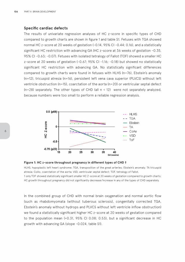
Specific cardiac defectsThe results of univariate regression analyses of HC z-score in specific types of CHD compared to growth charts are shown in figure 1 and table S1. Fetuses with TGA showed normal HC z-score at 20 weeks of gestation (-0.14, 95% CI -0.44; 0.16), and a statistically significant HC restriction with advancing GA (HC z-score at 36 weeks of gestation -0.35, 95% CI -0.63; -0.07). Fetuses with isolated tetralogy of Fallot (TOF) showed a smaller HC z-score at 20 weeks of gestation (-0.67; 95% CI -1.16; -0.18) but showed no statistically significant HC restriction with advancing GA. No statistically significant differences compared to growth charts were found in fetuses with HLHS (n=76), Ebstein’s anomaly (n=12), tricuspid atresia (n=16), persistent left vena cava superior (PLVCS) without left ventricle obstruction (n=15), coarctation of the aorta (n=20) or ventricular septal defect (n=28) separately. The other types of CHD (all n < 12) were not separately analyzed, because numbers were too small to perform a reliable regression analysis.
Figure 1: HC z-score throughout pregnancy in different types of CHD †
HLHS, hypoplastic left heart syndrome; TGA, transposition of the great arteries; Ebstein’s anomaly; TA tricuspid atresia; CoAo, coarctation of the aorta; VSD, ventricular septal defect; TOF, tetralogy of Fallot.† only TOF showed statistically significant smaller HC Z-score at 20 weeks of gestation compared to growth charts; HC growth throughout pregnancy did not significantly decrease/increase in any of the types of CHD separately.
In the combined group of CHD with normal brain oxygenation and normal aortic flow (such as rhabdomyomata (without tuberous sclerosis), congenitally corrected TGA, Ebstein’s anomaly without hydrops and PLVCS without left ventricle inflow obstruction) we found a statistically significant higher HC z-score at 20 weeks of gestation compared to the population mean (+0.31, 95% CI 0.08; 0.53), but a significant decrease in HC growth with advancing GA (slope -0.024, table S1).
106
6
PART II: BRAIN DEVELOPMENT
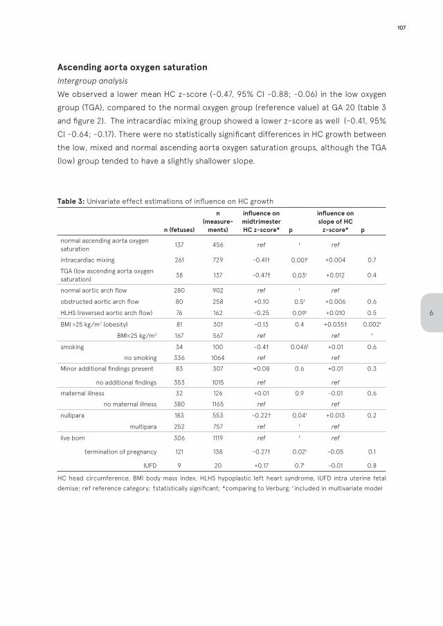
Ascending aorta oxygen saturation Intergroup analysis We observed a lower mean HC z-score (-0.47, 95% CI -0.88; -0.06) in the low oxygen group (TGA), compared to the normal oxygen group (reference value) at GA 20 (table 3 and figure 2). The intracardiac mixing group showed a lower z-score as well (-0.41, 95% CI -0.64; -0.17). There were no statistically significant differences in HC growth between the low, mixed and normal ascending aorta oxygen saturation groups, although the TGA (low) group tended to have a slightly shallower slope.
Table 3: Univariate effect estimations of influence on HC growth
n (fetuses)
n (measure-
ments)
influence onmidtrimester HC z-score* p
influence on slope of HC
z-score* p
normal ascending aorta oxygen saturation
137 456 ref ǂ ref
intracardiac mixing 261 729 -0.41† 0.001ǂ +0.004 0.7
TGA (low ascending aorta oxygen saturation)
38 137 -0.47† 0.03ǂ +0.012 0.4
normal aortic arch flow 280 902 ref ǂ ref
obstructed aortic arch flow 80 258 +0.10 0.5ǂ +0.006 0.6
HLHS (reversed aortic arch flow) 76 162 -0.25 0.09ǂ +0.010 0.5
BMI >25 kg/m2 (obesity) 81 301 -0.13 0.4 +0.035† 0.002ǂ
BMI<25 kg/m2 167 567 ref ref ǂ
smoking 34 100 -0.4† 0.046ǂ +0.01 0.6
no smoking 336 1064 ref ref
Minor additional findings present 83 307 +0.08 0.6 +0.01 0.3
no additional findings 353 1015 ref ref
maternal illness 32 126 +0.01 0.9 -0.01 0.6
no maternal illness 380 1165 ref ref
nullipara 183 553 -0.22† 0.04ǂ +0.013 0.2
multipara 252 757 ref ǂ ref
live born 306 1119 ref ǂ ref
termination of pregnancy 121 138 -0.27† 0.02ǂ -0.05 0.1
IUFD 9 20 +0.17 0.7ǂ -0.01 0.8
HC head circumference, BMI body mass index, HLHS hypoplastic left heart syndrome, IUFD intra uterine fetal demise; ref reference category; †statistically significant; *comparing to Verburg; ǂ included in multivariate model
107
6
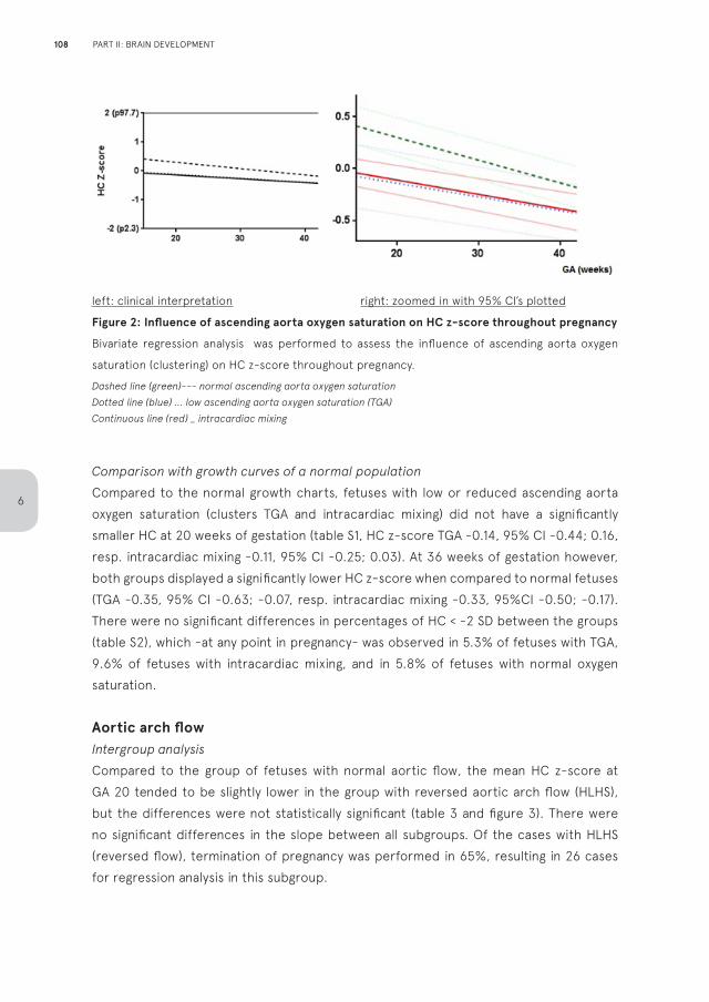
left: clinical interpretation right: zoomed in with 95% CI’s plotted
Figure 2: Influence of ascending aorta oxygen saturation on HC z-score throughout pregnancy
Bivariate regression analysis was performed to assess the influence of ascending aorta oxygen
saturation (clustering) on HC z-score throughout pregnancy.
Dashed line (green)--- normal ascending aorta oxygen saturationDotted line (blue) ... low ascending aorta oxygen saturation (TGA)Continuous line (red) _ intracardiac mixing
Comparison with growth curves of a normal populationCompared to the normal growth charts, fetuses with low or reduced ascending aorta oxygen saturation (clusters TGA and intracardiac mixing) did not have a significantly smaller HC at 20 weeks of gestation (table S1, HC z-score TGA -0.14, 95% CI -0.44; 0.16, resp. intracardiac mixing -0.11, 95% CI -0.25; 0.03). At 36 weeks of gestation however, both groups displayed a significantly lower HC z-score when compared to normal fetuses (TGA -0.35, 95% CI -0.63; -0.07, resp. intracardiac mixing -0.33, 95%CI -0.50; -0.17). There were no significant differences in percentages of HC < -2 SD between the groups (table S2), which -at any point in pregnancy- was observed in 5.3% of fetuses with TGA, 9.6% of fetuses with intracardiac mixing, and in 5.8% of fetuses with normal oxygen saturation.
Aortic arch flow Intergroup analysisCompared to the group of fetuses with normal aortic flow, the mean HC z-score at GA 20 tended to be slightly lower in the group with reversed aortic arch flow (HLHS), but the differences were not statistically significant (table 3 and figure 3). There were no significant differences in the slope between all subgroups. Of the cases with HLHS (reversed flow), termination of pregnancy was performed in 65%, resulting in 26 cases for regression analysis in this subgroup.
108
6
PART II: BRAIN DEVELOPMENT
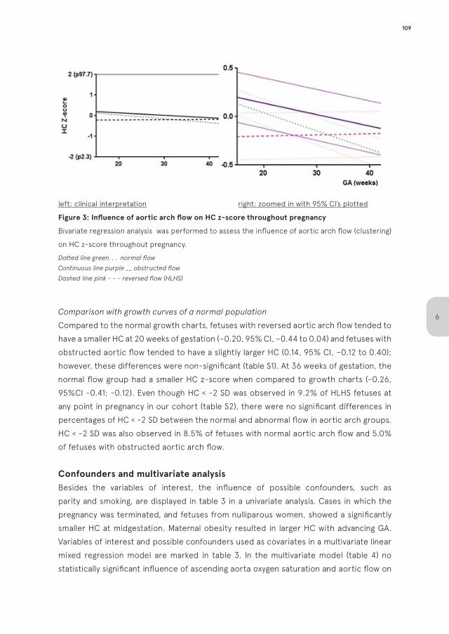
left: clinical interpretation right: zoomed in with 95% CI’s plotted
Figure 3: Influence of aortic arch flow on HC z-score throughout pregnancy
Bivariate regression analysis was performed to assess the influence of aortic arch flow (clustering)
on HC z-score throughout pregnancy.
Dotted line green. . . normal flowContinuous line purple __ obstructed flowDashed line pink - - - reversed flow (HLHS)
Comparison with growth curves of a normal populationCompared to the normal growth charts, fetuses with reversed aortic arch flow tended to have a smaller HC at 20 weeks of gestation (−0.20, 95% CI, −0.44 to 0.04) and fetuses with obstructed aortic flow tended to have a slightly larger HC (0.14, 95% CI, −0.12 to 0.40); however, these differences were non-significant (table S1). At 36 weeks of gestation, the normal flow group had a smaller HC z-score when compared to growth charts (-0.26, 95%CI -0.41; -0.12). Even though HC < -2 SD was observed in 9.2% of HLHS fetuses at any point in pregnancy in our cohort (table S2), there were no significant differences in percentages of HC < -2 SD between the normal and abnormal flow in aortic arch groups. HC < -2 SD was also observed in 8.5% of fetuses with normal aortic arch flow and 5.0% of fetuses with obstructed aortic arch flow.
Confounders and multivariate analysisBesides the variables of interest, the influence of possible confounders, such as parity and smoking, are displayed in table 3 in a univariate analysis. Cases in which the pregnancy was terminated, and fetuses from nulliparous women, showed a significantly smaller HC at midgestation. Maternal obesity resulted in larger HC with advancing GA. Variables of interest and possible confounders used as covariates in a multivariate linear mixed regression model are marked in table 3. In the multivariate model (table 4) no statistically significant influence of ascending aorta oxygen saturation and aortic flow on
109
6
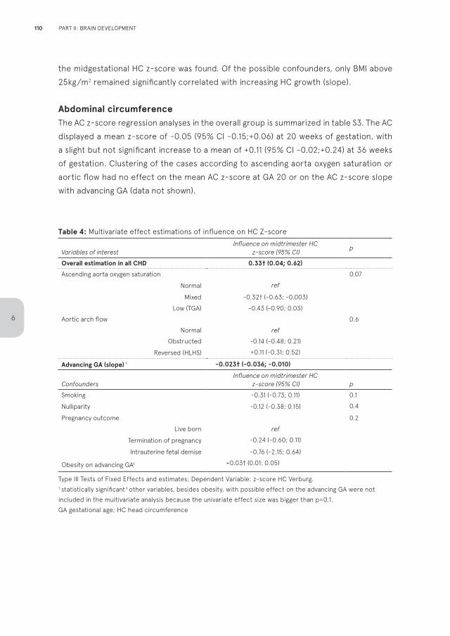
the midgestational HC z-score was found. Of the possible confounders, only BMI above 25kg/m2 remained significantly correlated with increasing HC growth (slope).
Abdominal circumferenceThe AC z-score regression analyses in the overall group is summarized in table S3. The AC displayed a mean z-score of -0.05 (95% CI -0.15;+0.06) at 20 weeks of gestation, with a slight but not significant increase to a mean of +0.11 (95% CI -0.02;+0.24) at 36 weeks of gestation. Clustering of the cases according to ascending aorta oxygen saturation or aortic flow had no effect on the mean AC z-score at GA 20 or on the AC z-score slope with advancing GA (data not shown).
Table 4: Multivariate effect estimations of influence on HC Z-score
Variables of interestInfluence on midtrimester HC
z-score (95% CI)p
Overall estimation in all CHD 0.33† (0.04; 0.62)
Ascending aorta oxygen saturation 0.07
Normal ref
Mixed -0.32† (-0.63; -0.003)
Low (TGA) -0.43 (-0.90; 0.03)
Aortic arch flow 0.6
Normal ref
Obstructed -0.14 (-0.48; 0.21)
Reversed (HLHS) +0.11 (-0.31; 0.52)
Advancing GA (slope) ǂ -0.023† (-0.036; -0.010)
ConfoundersInfluence on midtrimester HC
z-score (95% CI) p
Smoking -0.31 (-0.73; 0.11) 0.1
Nulliparity -0.12 (-0.38; 0.15) 0.4
Pregnancy outcome 0.2
Live born ref
Termination of pregnancy -0.24 (-0.60; 0.11)
Intrauterine fetal demise -0.76 (-2.15; 0.64)
Obesity on advancing GAǂ +0.03† (0.01; 0.05)
Type III Tests of Fixed Effects and estimates; Dependent Variable: z-score HC Verburg.† statistically significant ǂ other variables, besides obesity, with possible effect on the advancing GA were not included in the multivariate analysis because the univariate effect size was bigger than p=0.1. GA gestational age; HC head circumference
110
6
PART II: BRAIN DEVELOPMENT
DISCUSSION
This study shows that all fetuses with prenatally detected isolated CHD show a decline in head circumference (HC) growth with advancing GA, irrespective of aortic flow or oxygen saturation. However, the clinical relevance of this decrease is small, as the effect size compared to the HC growth charts of normal populations is small (mean HC z- score in all CHD combined -0.23, representing the 41st percentile, at GA 36 weeks). The most important finding of this study is that differences in the HC z-score growth slopes between the various CHD clusters were absent. Interestingly, fetuses with normal aortic flow and oxygen saturation (such as Ebstein’s anomaly, persistent left caval vein and ventricular septal defects(VSDs)) showed the same HC growth decline as fetuses with obstructed or reversed flow (such as HLHS) or low or intracardiac mixing (such as TGA and TOF).
Our study confirms the reported smaller HC at birth in infants with severe CHD17-20, and recently a large population based study also reported smaller neonatal HC in less severe CHD32. Our data indicate that especially fetuses with TOF present with a smaller HC, which is already present at midgestation (z-score -0.67; 25th percentile). A midgestational smaller HC was nót evidently present in fetuses with reversed flow in the aortic arch (HLHS) and TGA, nor in the other types of CHD in our cohort.
Strengths of our cohort are the consecutive, non-selective inclusion of all cases with prenatally identified CHD in 3 centers. The repeated measurements facilitated linear regression analysis of the prenatal growth, from 20 weeks of gestation until birth. The size of the cohort made it possible to analyze some specific types of CHD for the first time prenatally. Our clustering into various subgroups is very accurate due to high rates of postnatal or postmortem confirmation.
Our study demonstrates fetal growth patterns in isolated CHD, irrespective of the severity of the defect. Up to date only nine studies assessed fetal biometrics, all with different case mixes, various methods of subclassification and heterogeneous findings13;16;23-28;33. Most authors reported smaller HC at midgestation23;27;28 or in the third trimester16;24;27;33. Subtypes of CHD such as TGA and TOF are rarely reported separately, and less severe CHD have never been reported solely. The differences with our study can be explained by the fact that reports might have included pre-selected, non-consecutive, severe CHD cases and were dominated by specific CHD such as TOF. Some reports may not have excluded all IUGR cases and follow-up after birth was not always available. Three of the studies focus on cohorts of fetuses with (developing) HLHS only12;23;26. In an MRI study of 18 fetuses with HLHS, delay in cerebral development and volume growth was noted in the
111
6
third trimester, hypothesized to precede HC growth restriction34. Our study comprises the largest cohort of consecutive HLHS cases so far (n=76). We found a smaller HC at midgestation in fetuses with HLHS, but statistical significance was absent. The HC growth restriction (slope) with advancing GA appeared even less severe compared to other types of CHD.
Our findings underline the uncertainty regarding the etiology of the smaller HC in fetuses with CHD. A direct relationship between aortic flow or oxygen saturation and small HC could not be established clearly. Our results point towards an influence of type of CHD on HC growth, but sample sizes are nonetheless too small to establish a direct relation. We postulate that the smaller HC in certain types of CHD may be caused by certain genetic or embryological developmental pathways. CHDs develop through multifactorial pathways35;36 and some of these factors might also influence fetal HC growth and brain development26;30. By excluding known genetic syndromes by postnatal examination or standard antenatal genetic analysis in our study, this influence has been corrected maximally. We are however limited by the discriminating power of the used diagnostic methods used.
Placental dysfunction and smaller fetal weight could also play a role in smaller HC sizes of fetuses with isolated CHD. Suggestions for this mechanism have also been found by Sun et al., who described lower oxygen levels in the umbilical vein with MRI in fetuses with severe CHD21. Fetuses with CHD however do not appear to have smaller placental volumes37. In our study we excluded pregnancies complicated by placental dysfunction and found no evidence that AC growth is restricted in fetuses with severe CHD, nor that AC growth is influenced by the type of CHD.
An important limitation is that data on long-term neurodevelopmental outcome are not available in our cohort. With regard to implications for parental counselling, this data would be highly informative. It is known that children with severe CHD are at risk for ND, and previous studies suggest that prenatal (head) growth restriction in CHD fetuses might be associated with ND21;25. In our opinion, a clear relationship of the prenatal (neurodevelopmental) factors with long-term neurodevelopmental outcome has, however, not been firmly established so far27;33;38;39. Large case control studies with prenatal factors correlating to neurodevelopmental outcome are necessary.
In conclusion, fetuses with diminished ascending aorta oxygen saturation, but mainly fetuses with TOF, appear to have a slightly smaller HC at GA 20 weeks, and HC growth is restricted in all types of CHD with advancing GA. The cause of these findings remains
112
6
PART II: BRAIN DEVELOPMENT

elusive. In the instances where there were (statistically) significant differences in HC, measurements were still within normal range – raising the question of its clinical significance. Based on our findings, we propose to refrain from including possible prenatal indicators, such as reduced HC, in parental counselling for neurodevelopmental outcomes so far or as a rationale for fetal therapy40, until a clear correlation with ND has been ascertained.
Acknowledgments: none.Funding Sources: noneDisclosures: none
Supplemental data available onlineSupplement 1: List of primary diagnoses per group Table S1: univariate comparison of HC with population means in different CHD groupsTable S2: percentages of HC below the 2.3rd percentile (z-score <-2) in the various types of CHDTable S3: univariate comparison of AC with population means
113
6
REFERENCES
1. Marino BS, Lipkin PH, Newburger JW,
Peacock G, Gerdes M, Gaynor JW, et al.
Neurodevelopmental outcomes in children
with congenital heart disease: evaluation and
management: a scientific statement from the
American Heart Association. Circulation 2012
Aug 28;126(9):1143-72.
2. Sarrechia I, Miatton M, De WD, Francois K,
Gewillig M, Meyns B, et al. Neurocognitive
development and behaviour in school-aged
children after surgery for univentricular or
biventricular congenital heart disease. Eur J
Cardiothorac Surg 2016 Jan;49(1):167-74.
3. Jones B, Muscara F, Lloyd O, McKinlay L, Justo
R. Neurodevelopmental outcome following
open heart surgery in infancy: 6-year follow-up.
Cardiol Young 2015 Jun;25(5):903-10.
4. Snookes SH, Gunn JK, Eldridge BJ, Donath SM,
Hunt RW, Galea MP, et al. A systematic review
of motor and cognitive outcomes after early
surgery for congenital heart disease. Pediatrics
2010 Apr;125(4):e818-e827.
5. Massaro AN, El-Dib M, Glass P, Aly H. Factors
associated with adverse neurodevelopmental
outcomes in infants with congenital heart
disease. Brain Dev 2008 Aug;30(7):437-46.
6. McQuillen PS, Miller SP. Congenital heart disease
and brain development. Ann N Y Acad Sci 2010
Jan;1184:68-86.
7. Domi T, Edgell DS, McCrindle BW, Williams
WG, Chan AK, MacGregor DL, et al. Frequency,
predictors, and neurologic outcomes of
vaso-occlusive strokes associated with
cardiac surgery in children. Pediatrics 2008
Dec;122(6):1292-8.
8. Nathan M, Sadhwani A, Gauvreau K, Agus M,
Ware J, Newburger JW, et al. Association
between Technical Performance Scores and
neurodevelopmental outcomes after congenital
cardiac surgery. J Thorac Cardiovasc Surg 2014
Jul;148(1):232-7.
9. Limperopoulos C, Majnemer A, Shevell MI,
Rohlicek C, Rosenblatt B, Tchervenkov C, et
al. Predictors of developmental disabilities
after open heart surgery in young children
with congenital heart defects. J Pediatr 2002
Jul;141(1):51-8.
10. Khalil A, Suff N, Thilaganathan B, Hurrell A,
Cooper D, Carvalho JS. Brain abnormalities and
neurodevelopmental delay in congenital heart
disease: systematic review and meta-analysis.
Ultrasound Obstet Gynecol 2014 Jan;43(1):14-
24.
11. Owen M, Shevell M, Majnemer A, Limperopoulos
C. Abnormal brain structure and function
in newborns with complex congenital heart
defects before open heart surgery: A review of
the evidence. Journal of Child Neurology 2011
Jun;26(6):743-55.
12. McQuillen PS, Goff DA, Licht DJ. Effects of
congenital heart disease on brain development.
Prog Pediatr Cardiol 2010 Aug 1;29(2):79-85.
13. McElhinney DB, Benson CB, Brown DW,
Wilkins-Haug LE, Marshall AC, Zaccagnini L,
et al. Cerebral blood flow characteristics
and biometry in fetuses undergoing prenatal
intervention for aortic stenosis with evolving
hypoplastic left heart syndrome. Ultrasound
Med Biol 2010 Jan;36(1):29-37.
14. Limperopoulos C. Disorders of the fetal
circulation and the fetal brain. Clin Perinatol
2009 Sep;36(3):561-77.
114
6
PART II: BRAIN DEVELOPMENT

15. Donofrio MT. The heart-brain interaction
in the fetus: Cerebrovascular blood flow in
the developing human. Progress in Pediatric
Cardiology 2006 May;22(1):41-51.
16. Donofrio MT, Bremer YA, Schieken RM, Gennings
C, Morton LD, Eidem BW, et al. Autoregulation
of cerebral blood flow in fetuses with congenital
heart disease: the brain sparing effect. Pediatr
Cardiol 2003 Sep;24(5):436-43.
17. Barbu D, Mert I, Kruger M, Bahado-Singh
RO. Evidence of fetal central nervous system
injury in isolated congenital heart defects:
microcephaly at birth. Am J Obstet Gynecol
2009 Jul;201(1):43-7.
18. Manzar S, Nair AK, Pai MG, Al-Khhusaiby
SM. Head size at birth in neonates with
transposition of great arteries and hypoplastic
left heart syndrome. Saudi Medical Journal
2005 Mar;26(3):453-6.
19. Rosenthal GL. Patterns of prenatal growth among
infants with cardiovascular malformations:
possible fetal hemodynamic effects. Am J
Epidemiol 1996 Mar 1;143(5):505-13.
20. Shillingford AJ, Ittenbach RF, Marino BS, Rychik J,
Clancy RR, Spray TL, et al. Aortic morphometry
and microcephaly in hypoplastic left heart
syndrome. Cardiol Young 2007 Apr;17(2):189-95.
21. Sun L, Macgowan CK, Sled JG, Yoo SJ, Manlhiot
C, Porayette P, et al. Reduced fetal cerebral
oxygen consumption is associated with smaller
brain size in fetuses with congenital heart
disease. Circulation 2015 Apr 14;131(15):1313-23.
22. Al Nafisi B, van Amerom JF, Forsey J, Jaeggi
E, Grosse-Wortmann L, Yoo SJ, et al. Fetal
circulation in left-sided congenital heart
disease measured by cardiovascular magnetic
resonance: a case-control study. J Cardiovasc
Magn Reson 2013;15:65.
23. Hangge PT, Cnota JF, Woo JG, Hinton AC,
Divanovic AA, Manning PB, et al. Microcephaly
is associated with early adverse neurologic
outcomes in hypoplastic left heart syndrome.
Pediatr Res 2013 Jul;74(1):61-7.
24. Arduini M, Rosati P, Caforio L, Guariglia L, Clerici
G, Di Renzo GC, et al. Cerebral blood flow
autoregulation and congenital heart disease:
possible causes of abnormal prenatal neurologic
development. J Matern Fetal Neonatal Med 2011
Oct;24(10):1208-11.
25. Itsukaichi M, Kikuchi A, Yoshihara K, Serikawa
T, Takakuwa K, Tanaka K. Changes in fetal
circulation associated with congenital heart
disease and their effects on fetal growth. Fetal
Diagn Ther 2011;30(3):219-24.
26. Hinton RB, Andelfinger G, Sekar P, Hinton AC,
Gendron RL, Michelfelder EC, et al. Prenatal
head growth and white matter injury in
hypoplastic left heart syndrome. Pediatr Res
2008 Oct;64(4):364-9.
27. Williams IA, Fifer WP, Andrews H. Fetal
Growth and Neurodevelopmental Outcome in
Congenital Heart Disease. Pediatr Cardiol 2015
Aug;36(6):1135-44.
28. Masoller N, Martinez JM, Gomez O, Bennasar
M, Crispi F, Sanz-Cortes M, et al. Evidence of
second-trimester changes in head biometry
and brain perfusion in fetuses with congenital
heart disease. Ultrasound Obstet Gynecol 2014
Aug;44(2):182-7.
29. van Velzen C, Clur S, Rijlaarsdam M, Bax C,
Pajkrt E, Heymans M, et al. Prenatal detection
of congenital heart disease-results of a
115
6

national screening programme. BJOG 2016
Feb;123(3):400-7.
30. Verburg BO, Steegers EA, De RM, Snijders
RJ, Smith E, Hofman A, et al. New charts for
ultrasound dating of pregnancy and assessment
of fetal growth: longitudinal data from a
population-based cohort study. Ultrasound
Obstet Gynecol 2008 Apr;31(4):388-96.
31. Woods CG, Parker A. Investigating microcephaly.
Arch Dis Child 2013 Sep;98(9):707-13.
32. Matthiesen NB, Henriksen TB, Gaynor JW,
Agergaard P, Bach CC, Hjortdal V, et al.
Congenital Heart Defects and Indices of Fetal
Cerebral Growth in a Nationwide Cohort of
924,422 Liveborn Infants. Circulation 2016 Feb
9;133(6):566-75.
33. Hahn E, Szwast A, Cnota J, Levine JC, Fifer CG,
Jaeggi E, et al. The association of fetal growth,
cerebral blood flow, and neurodevelopmental
outcome in single ventricle fetuses. Ultrasound
Obstet Gynecol 2016 Apr;47(4):460-5.
34. Clouchoux C, du Plessis AJ, Bouyssi-Kobar M,
Tworetzky W, McElhinney DB, Brown DW, et
al. Delayed Cortical Development in Fetuses
with Complex Congenital Heart Disease. Cereb
Cortex 2013 Dec;23(12):2932-43.
35. Allan LD, Sharland G, Tynan MJ. The natural
history of the hypoplastic left heart syndrome.
Int J Cardiol 1989 Dec;25(3):341-3.
36. Fishman NH, Hof RB, Rudolph AM, Heymann
MA. Models of congenital heart disease in fetal
lambs. Circulation 1978 Aug;58(2):354-64.
37. Andescavage N, Yarish A, Donofrio M, Bulas D,
Evangelou I, Vezina G, et al. 3-D volumetric
MRI evaluation of the placenta in fetuses with
complex congenital heart disease. Placenta
2015 Sep;36(9):1024-30.
38. Williams IA, Fifer C, Jaeggi E, Levine JC,
Michelfelder EC, Szwast AL. The association
of fetal cerebrovascular resistance with early
neurodevelopment in single ventricle congenital
heart disease. Am Heart J 2013 Apr;165(4):544-
50.
39. Williams IA, Tarullo AR, Grieve PG, Wilpers A,
Vignola EF, Myers MM, et al. Fetal Cerebrovascular
Resistance and Neonatal EEG Predict 18-month
Neurodevelopmental Outcome in Infants with
Congenital Heart Disease. Ultrasound Obstet
Gynecol 2012 Sep;40(3):304-9.
40. http://www.chop.edu/centers-programs/
fetal-neuroprotection-and-neuroplasticity-
program; consulted in july 2015
41. Paladini D, Alfirevic Z, Carvalho JS, Khalil
A, Malinger G, Martinez JM, et al. Prenatal
counselling for neurodevelopmental delay
in congenital heart disease. The results of a
worldwide survey of experts’ attitudes advise
caution. Ultrasound Obstet Gynecol 2016 Jan 7
doi: 10.1002/uog.15852. (Epub ahead of print).
116
6
PART II: BRAIN DEVELOPMENT
Reply to: Head growth in fetuses with isolated congenital heart defects: lack of influence of aortic arch flow and ascending aorta oxygen saturation. J. S. Carvalho Ultrasound in Obstetrics and Gynecology 2016; 48: 282–284
Congenital heart disease (CHD) and long-term neurodevelopmental delay is currently one of the hottest topics in fetal cardiology, generating much interest among many specialists. A link between the two was first suggested some years ago, but its etiological basis seemed multifactorial. However, since shifting the focus of this link to fetal life, there is growing evidence that in-utero circulatory changes, caused by the heart defect itself, may be responsible for abnormal fetal brain development and perhaps long-term brain function. Accepting this causation is a tall order and caution is needed. An intuitive, yet simplistic, concept is that smaller heads contain smaller brains and perhaps this may be the starting point for some of the neurodevelopmental abnormalities reported in children and adults with CHD. Reports of a smaller head size in fetuses and neonates with CHD have often been interpreted to mean possible neurodevelopmental abnormalities. As a pediatric cardiologist, it was refreshing to read in the paper by Jansen et al. that the ‘status quo’ can, and should, be questioned. Jansen’s is a well-conducted retrospective study of a relatively large cohort of fetuses with isolated mixed type of CHD. Fetal head circumference (HC) in mid-gestation and HC growth pattern were examined and compared with those in a normal population. Cases of CHD were clustered according to perceived characteristics of aortic blood flow (normal, obstructed, reversed) and ascending aorta saturation (normal, low, mixed). Briefly, the study showed a statistically significant decrease in HC growth with advancing gestational age in all groups of CHD, resulting in reduced head size in the third trimester (and at birth). Interestingly this was irrespective of aortic flow or saturation. At 20 weeks’ gestation, however, HC in CHD fetuses was not statistically different from that of the normal population, with the exception of those with tetralogy of Fallot. This is at variance with findings of other studies. The questions remain: do statistically significantly smaller heads at birth matter if absolute measurements fall within the normal range and is a slightly smaller HC clinically significant? Statistically smaller HC should not necessarily equate to abnormal brain development.
Naturally, there are inherent limitations to any retrospective study. Two methodological strengths should, however, be highlighted. The first one relates to the likely certainty that CHDs were actually ‘isolated’. Absence of other ultrasound abnormalities at the 20-week scan and normal fetal karyotype offer no such guarantee but, in this study, which had a 70% live-birth rate, minimum follow-up time was 1 year, thus reducing the
117
6

likelihood of including syndromic CHD. Second, the growth charts used as the standard reference were derived from a similar Dutch population that was evaluated over a similar timeframe as that of the CHD cohort. However, the lack of long-term neurodevelopmental follow-up prevents firm conclusions from being drawn about clinical significance of the findings. This of course calls for larger, prospective controlled trials. But will these ever be possible? It is therefore important that we continue to explore retrospective data obtained and analysed in a sound manner. I look forward to reviewing a follow-on paper by the same group, comparing HC in this cohort with that of a cohort with non-isolated CHD. Lastly, interpreting results goes beyond their statistical significance. Whilst small differences may influence future research on mechanisms that lead to altered fetal growth in cases of CHD, the long-term implications for the individual fetus that is diagnosed antenatally with isolated CHD are far from clear.
118
6
PART II: BRAIN DEVELOPMENT

119
6

CHAPTER 7
Fetuses with isolated congenital heart
defects show normal cerebral and
extracerebral fluid volume growth:
a 3D sonographic study in the second
and third trimester
Fetal Diagnosis and Therapy 2019 Jan 17:1-9.
F.A.R. Jansen E.W. van Zwet S.M.P. Everwijn A.K.K. Teunissen L. Rozendaal J.M.M. van Lith N.A. Blom M.C. Haak
ABSTRACT
Objective
The aim of our study is to explore whether the cerebral growth is delayed in fetuses with CHD in the second and early third trimester. Methods
A prospective cohort study was conducted in 77 CHD cases, with 75 healthy controls. 3D cerebral volume acquisition was performed sequentially. The volumes of the fetal hemicerebrum and extracerebral fluid were compared by linear regression analysis, and the Sylvian fissure was measured. Results
Between 19 and 32 weeks of gestation, 158 measurements in cases and 183 measurements in controls were performed (mean 2.2/subject). The volume growth of the hemicerebrum (R2=0.95vs0.95; p=0.9) and the extracerebral fluid (R2=0.84vs0.82, p=0.9) were similar. Fetuses with abnormal oxygen delivery to the brain have a slightly smaller brain at 20 weeks of gestation (p=0.02), but this difference disappeared with advancing gestation. CHD cases demonstrated a slightly shallower Sylvian fissure (mean ratio 0.146vs0.153; p=0.004). Conclusions
Our study shows no differences in cerebral growth, studied in an unselected cohort, with successive cases of isolated CHD. Even in the severest CHD cases, cerebral size is similar in the early third trimester. The cause and meaning of a shallower Sylvian fissure is unclear, possibly it is a marker for delayed cerebral maturation or it might be an expression of decreasing amount of extracerebral fluid.
7
122 PART II: BRAIN DEVELOPMENT
INTRODUCTION
Congenital heart defects (CHD) are associated with cerebral abnormalities and neurodevelopmental delay (ND), even in the absence of chromosomal or syndromic anomalies1;2. Neonatal ultrasound imaging studies demonstrate cerebral abnormalities in 15-75% of neonates with CHD prior to surgery3-7. Most cerebral findings in CHD newborns are mild, such as asymmetrical widened ventricles and increased extracerebral fluid. Neonatal MRI studies report delayed brain maturation and signs of cerebral atrophy8. These findings led to the hypothesis that cerebral damage or altered cerebral development in fetuses with CHD already starts in the prenatal period, due to abnormal flow and oxygenation patterns9.
Imaging studies in fetuses with CHD also report brain abnormalities, such as delay in head growth and brainsparing in subgroups of CHD10-15. The prenatal studies are however dominated by series of severe CHD and left-sided CHD13,16,17, and some studies also included cases with non-isolated CHD18,19. The reported variables, such as head circumference and flow indices, are very heterogeneous and were assessed at different gestational ages15. Studies that perform multiple measurements per fetus, to assess changes over time, are very scarce. Furthermore, the methods to report the data vary between studies, hindering comparison and making general conclusion drawing difficult. Previous studies mainly focus on the third trimester, as this might be the period in which cortical folding and development accelerates. However, several studies show that at midgestation fetal head circumference might already be delayed in certain CHD, possibly even predicting developmental delay20-22.
The purpose of this study was to investigate fetal brain volume and volume growth with ultrasound, in fetuses with CHD in the second and early third trimester. Additionally, extracerebral fluid space was studied, and the total cranial volume was compared as well. We aimed to assess an unselected consecutive cohort of isolated CHD fetuses and compare them with healthy control fetuses. We hypothesized to find smaller fetal brain volume and higher extracerebral fluid levels in CHD.
METHODS
All women diagnosed with isolated fetal CHD referred to the LUMC between September 2013 and May 2016 were enrolled in this study after informed consent. All consecutive cases, detected before 32 weeks of gestation, were included. Healthy controls were
123
7
recruited after a normal 20 weeks’ standard anomaly scan in a low-risk community practice, after informed consent. The research protocols were evaluated and approved by the local ethical committee. Exclusion criteria were ultrasound anomalies other than the CHD, coinciding genetic or syndromic defects (prenatally diagnosed or postnatally apparent) and multiple gestation. Minor additional findings were not excluded, such as single umbilical artery, enlarged nuchal translucency without chromosomal defects, and small for gestational age without signs of placental insufficiency. Gestational age was based on first-trimester ultrasound. In both groups, detailed sonography was performed after inclusion and repeated sequentially, after two to five weeks. The examinations were performed by experienced sonographers (FJ/AT/SE), using a Voluson E8 or E10 (GE Healthcare ultrasound, Milwaukee, WI, USA) with a RAB 6-D probe. The examination included the measurement of the depth of the Sylvian fissure (see supplement 1). A 3D volume recording of the fetal brain was acquired at the plane of the head circumference measurement.
Offline postprocessing was performed using GE 4D viewer version 16. Ext. 1. The observer was blinded to the clinical characteristics (presence or absence of CHD and gestational age). The Virtual Organ Computer-aided AnaLysis software tool (VOCAL, GE Healthcare) was used to manually trace the outline of the cerebral hemisphere, as well as the cavity of the hemicranium, excluding the infratentorial space and cerebellum. To achieve maximum visibility, we used the hemisphere furthest from the ultrasound probe (figure 1). The detailed description of the tracing methods and the intra- and interobserver reproducibility analysis are described in supplement 1. The volumes of the cerebral hemisphere, extracerebral fluid spaces (including the lateral ventricle) and total hemicranial cavity were calculated in cm3.
The various heart defects (see supplement 3) were labelled according to the postnatal diagnosis (postnatal ultrasound). Major heart defects were defined as requiring surgery in the first year of life. Heart defects were grouped together for summary purposes as listed in table 1. For the statistical analysis CHD were clustered based on two aspects: oxygen delivery and flow to the brain. This method was previously described by our group21. For instance, the group with low or reduced oxygen delivery to the brain included transposition of the great arteries (TGA, low oxygen) and univentricular CHD (mixed oxygen), and normal oxygen group included pulmonary or aortic stenosis (AoS) and persistent left caval vein with small LV (PLVCS). The group with reversed or diminished aortic arch flow included aortic hypoplasia or coarctation (diminished flow) and hypoplastic left heart syndrome (HLHS, reversed flow), and the normal aortic arch flow group tetralogy of Fallot (TOF), TGA and double outlet right ventricle (DORV) – in the
124
7
PART II: BRAIN DEVELOPMENT
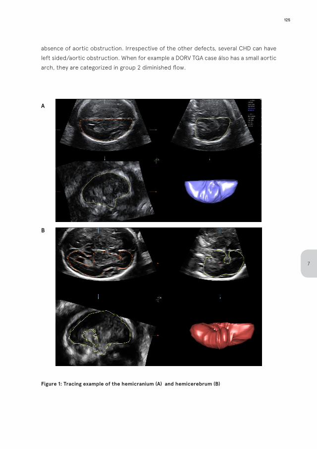
absence of aortic obstruction. Irrespective of the other defects, several CHD can have left sided/aortic obstruction. When for example a DORV TGA case álso has a small aortic arch, they are categorized in group 2 diminished flow.
A
B
Figure 1: Tracing example of the hemicranium (A) and hemicerebrum (B)
125
7
Statistical analysis and data handlingUsing the 3D measurements of the hemicerebral volume, extracerebral fluid volume and hemicranial volume, the extracerebral fluid-to-brain volume ratio was calculated, as well as the percentage of the hemicranium consisting of fluid (ventricular and extracerebral). The hemicerebral volume to estimated fetal weight ratio was calculated (brain-weight ratio). The depth of the Sylvian fissure was displayed as ratio of the inner biparietal diameter (BPD). To account for fetal growth, the relation of various variables with the advancing gestation was evaluated with scatterplots, and the linearity of the data was assessed. Volume measurements underwent a logistic transformation to facilitate linear regression (exponential growth model), and the natural logarithm (ln) of the measurements were used for further statistical analysis. Comparison of (ln of) measurements at various gestational ages (GA) was performed by comparing linear correlations (equality of regression) in different subgroups, using a linear mixed regression model. Data are presented as R2 or mean +/- SD. Group means are compared by independent-sample T-Test and percentages are compared by Chi2 analysis. P<0.05 was considered statistical significant. The sample size was reached as follows: at least 60 patients per group were required for the unpaired t-test to have 90% chance of detecting a difference in mean cerebral volume of 2 weeks (SD assumed 3 weeks) at the 1.7% level of significance. All analyses were performed in SPSS version 23.0.0.0 (IBM, Armonk, NY, USA).
RESULTS
Isolated CHD was diagnosed in 92 fetuses in the study period. Of these, 15 cases were excluded because of multiple gestation (n=3), postnatal genetic abnormalities (n=8) or a normal heart on postnatal ultrasound (these fetuses were suspected for coarctation; n=4). Thus, 77 cases were included for neurosonographic analysis. Postnatal confirmation was not possible in 12 cases (16%) in which pregnancy termination was chosen, because parents did not give consent for post-mortem examination. The type of CHD and the clustering with regard to cerebral oxygen delivery and aortic flow of the included cases are summarized in table 1, and a detailed description is available in supplement 3. Most CHD were major, and we included 8 minor CHD cases (10%). Secondly, we enrolled 75 control cases. All control infants received a neonatal cranial ultrasound in the first week of life and showed normal postnatal development until at least 1 month postpartum. In total, 158 examinations in 77 cases (mean 2.1 measurements per case) and 183 examinations in 75 controls (mean 2.4 measurements per control) were performed between GA 19+0 and GA 31+4 (mean GA at inclusion in cases 24+2 and in controls 24+1; ns). In 13 recordings (4%), the volume measurements were not possible due to poor quality.
126
7
PART II: BRAIN DEVELOPMENT
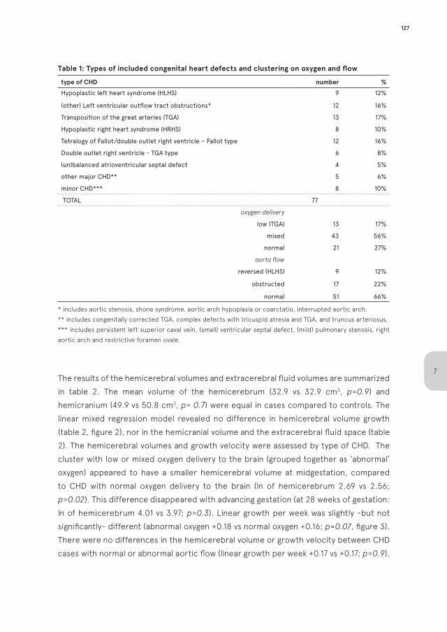
Table 1: Types of included congenital heart defects and clustering on oxygen and flow
type of CHD number %
Hypoplastic left heart syndrome (HLHS) 9 12%
(other) Left ventricular outflow tract obstructions* 12 16%
Transposition of the great arteries (TGA) 13 17%
Hypoplastic right heart syndrome (HRHS) 8 10%
Tetralogy of Fallot/double outlet right ventricle – Fallot type 12 16%
Double outlet right ventricle - TGA type 6 8%
(un)balanced atrioventricular septal defect 4 5%
other major CHD** 5 6%
minor CHD*** 8 10%
TOTAL 77
oxygen delivery
low (TGA) 13 17%
mixed 43 56%
normal 21 27%
aorta flow
reversed (HLHS) 9 12%
obstructed 17 22%
normal 51 66%
* includes aortic stenosis, shone syndrome, aortic arch hypoplasia or coarctatio, interrupted aortic arch.** includes congenitally corrected TGA, complex defects with tricuspid atresia and TGA, and truncus arteriosus.*** includes persistent left superior caval vein, (small) ventricular septal defect, (mild) pulmonary stenosis, right aortic arch and restrictive foramen ovale.
The results of the hemicerebral volumes and extracerebral fluid volumes are summarized in table 2. The mean volume of the hemicerebrum (32.9 vs 32.9 cm3, p=0.9) and hemicranium (49.9 vs 50.8 cm3, p= 0.7) were equal in cases compared to controls. The linear mixed regression model revealed no difference in hemicerebral volume growth (table 2, figure 2), nor in the hemicranial volume and the extracerebral fluid space (table 2). The hemicerebral volumes and growth velocity were assessed by type of CHD. The cluster with low or mixed oxygen delivery to the brain (grouped together as ‘abnormal’ oxygen) appeared to have a smaller hemicerebral volume at midgestation, compared to CHD with normal oxygen delivery to the brain (ln of hemicerebrum 2.69 vs 2.56; p=0.02). This difference disappeared with advancing gestation (at 28 weeks of gestation: ln of hemicerebrum 4.01 vs 3.97; p=0.3). Linear growth per week was slightly -but not significantly- different (abnormal oxygen +0.18 vs normal oxygen +0.16; p=0.07, figure 3). There were no differences in the hemicerebral volume or growth velocity between CHD cases with normal or abnormal aortic flow (linear growth per week +0.17 vs +0.17; p=0.9).
127
7
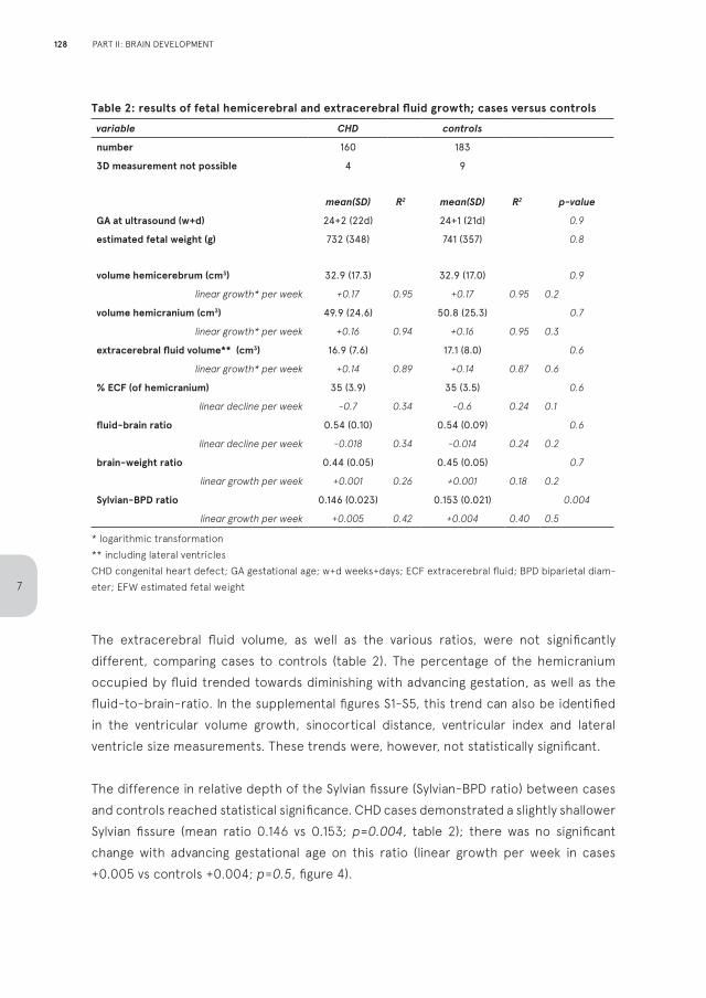
Table 2: results of fetal hemicerebral and extracerebral fluid growth; cases versus controls
variable CHD controls
number 160 183
3D measurement not possible 4 9
mean(SD) R2 mean(SD) R2 p-value
GA at ultrasound (w+d) 24+2 (22d) 24+1 (21d) 0.9
estimated fetal weight (g) 732 (348) 741 (357) 0.8
volume hemicerebrum (cm3) 32.9 (17.3) 32.9 (17.0) 0.9
linear growth* per week +0.17 0.95 +0.17 0.95 0.2
volume hemicranium (cm3) 49.9 (24.6) 50.8 (25.3) 0.7
linear growth* per week +0.16 0.94 +0.16 0.95 0.3
extracerebral fluid volume** (cm3) 16.9 (7.6) 17.1 (8.0) 0.6
linear growth* per week +0.14 0.89 +0.14 0.87 0.6
% ECF (of hemicranium) 35 (3.9) 35 (3.5) 0.6
linear decline per week -0.7 0.34 -0.6 0.24 0.1
fluid-brain ratio 0.54 (0.10) 0.54 (0.09) 0.6
linear decline per week -0.018 0.34 -0.014 0.24 0.2
brain-weight ratio 0.44 (0.05) 0.45 (0.05) 0.7
linear growth per week +0.001 0.26 +0.001 0.18 0.2
Sylvian-BPD ratio 0.146 (0.023) 0.153 (0.021) 0.004
linear growth per week +0.005 0.42 +0.004 0.40 0.5
* logarithmic transformation** including lateral ventriclesCHD congenital heart defect; GA gestational age; w+d weeks+days; ECF extracerebral fluid; BPD biparietal diam-eter; EFW estimated fetal weight
The extracerebral fluid volume, as well as the various ratios, were not significantly different, comparing cases to controls (table 2). The percentage of the hemicranium occupied by fluid trended towards diminishing with advancing gestation, as well as the fluid-to-brain-ratio. In the supplemental figures S1-S5, this trend can also be identified in the ventricular volume growth, sinocortical distance, ventricular index and lateral ventricle size measurements. These trends were, however, not statistically significant.
The difference in relative depth of the Sylvian fissure (Sylvian-BPD ratio) between cases and controls reached statistical significance. CHD cases demonstrated a slightly shallower Sylvian fissure (mean ratio 0.146 vs 0.153; p=0.004, table 2); there was no significant change with advancing gestational age on this ratio (linear growth per week in cases +0.005 vs controls +0.004; p=0.5, figure 4).
128
7
PART II: BRAIN DEVELOPMENT
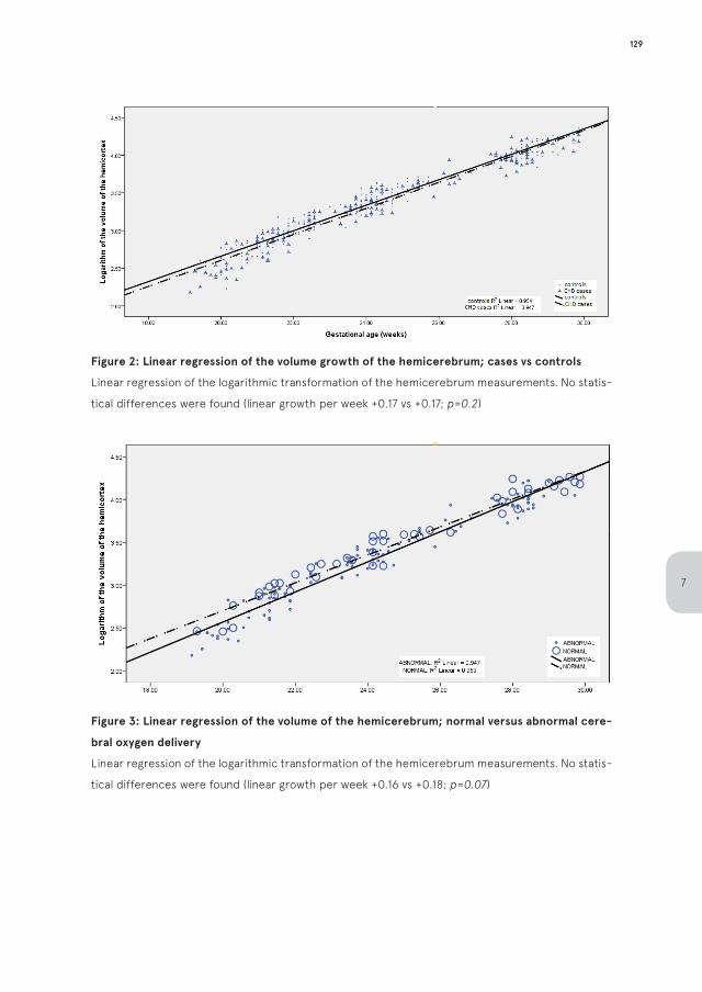
Figure 2: Linear regression of the volume growth of the hemicerebrum; cases vs controls
Linear regression of the logarithmic transformation of the hemicerebrum measurements. No statis-
tical differences were found (linear growth per week +0.17 vs +0.17; p=0.2)
Figure 3: Linear regression of the volume of the hemicerebrum; normal versus abnormal cere-
bral oxygen delivery
Linear regression of the logarithmic transformation of the hemicerebrum measurements. No statis-
tical differences were found (linear growth per week +0.16 vs +0.18; p=0.07)
129
7
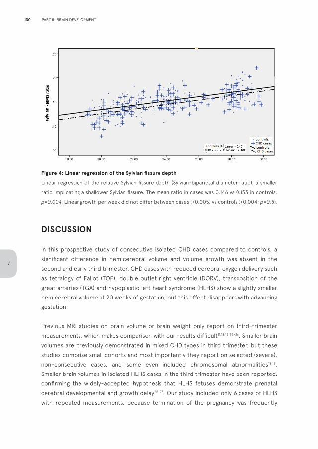
Figure 4: Linear regression of the Sylvian fissure depth
Linear regression of the relative Sylvian fissure depth (Sylvian-biparietal diameter ratio), a smaller
ratio implicating a shallower Sylvian fissure. The mean ratio in cases was 0.146 vs 0.153 in controls;
p=0.004. Linear growth per week did not differ between cases (+0.005) vs controls (+0.004; p=0.5).
DISCUSSION
In this prospective study of consecutive isolated CHD cases compared to controls, a significant difference in hemicerebral volume and volume growth was absent in the second and early third trimester. CHD cases with reduced cerebral oxygen delivery such as tetralogy of Fallot (TOF), double outlet right ventricle (DORV), transposition of the great arteries (TGA) and hypoplastic left heart syndrome (HLHS) show a slightly smaller hemicerebral volume at 20 weeks of gestation, but this effect disappears with advancing gestation.
Previous MRI studies on brain volume or brain weight only report on third-trimester measurements, which makes comparison with our results difficult11,18,19,22-26. Smaller brain volumes are previously demonstrated in mixed CHD types in third trimester, but these studies comprise small cohorts and most importantly they report on selected (severe), non-consecutive cases, and some even included chromosomal abnormalities18,19. Smaller brain volumes in isolated HLHS cases in the third trimester have been reported, confirming the widely-accepted hypothesis that HLHS fetuses demonstrate prenatal cerebral developmental and growth delay25-27. Our study included only 6 cases of HLHS with repeated measurements, because termination of the pregnancy was frequently
130
7
PART II: BRAIN DEVELOPMENT
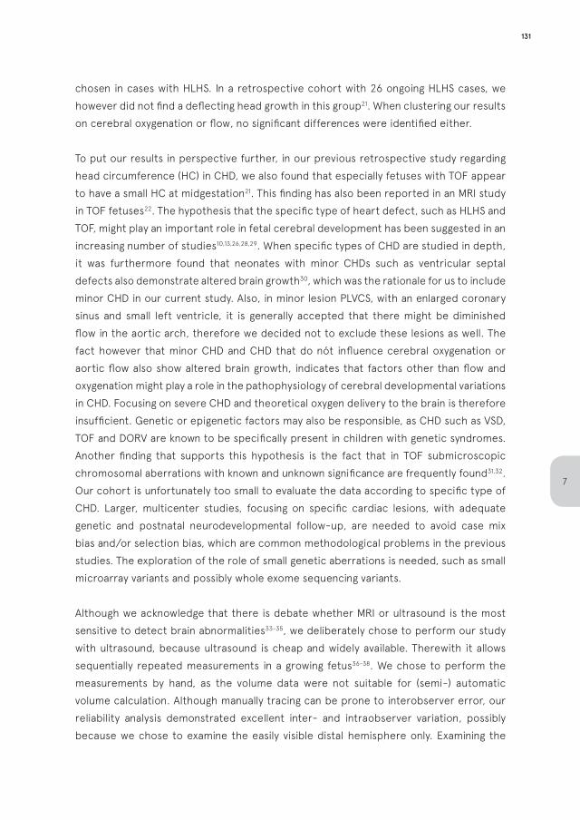
chosen in cases with HLHS. In a retrospective cohort with 26 ongoing HLHS cases, we however did not find a deflecting head growth in this group21. When clustering our results on cerebral oxygenation or flow, no significant differences were identified either.
To put our results in perspective further, in our previous retrospective study regarding head circumference (HC) in CHD, we also found that especially fetuses with TOF appear to have a small HC at midgestation21. This finding has also been reported in an MRI study in TOF fetuses22. The hypothesis that the specific type of heart defect, such as HLHS and TOF, might play an important role in fetal cerebral development has been suggested in an increasing number of studies10,13,26,28,29. When specific types of CHD are studied in depth, it was furthermore found that neonates with minor CHDs such as ventricular septal defects also demonstrate altered brain growth30, which was the rationale for us to include minor CHD in our current study. Also, in minor lesion PLVCS, with an enlarged coronary sinus and small left ventricle, it is generally accepted that there might be diminished flow in the aortic arch, therefore we decided not to exclude these lesions as well. The fact however that minor CHD and CHD that do nót influence cerebral oxygenation or aortic flow also show altered brain growth, indicates that factors other than flow and oxygenation might play a role in the pathophysiology of cerebral developmental variations in CHD. Focusing on severe CHD and theoretical oxygen delivery to the brain is therefore insufficient. Genetic or epigenetic factors may also be responsible, as CHD such as VSD, TOF and DORV are known to be specifically present in children with genetic syndromes. Another finding that supports this hypothesis is the fact that in TOF submicroscopic chromosomal aberrations with known and unknown significance are frequently found31,32. Our cohort is unfortunately too small to evaluate the data according to specific type of CHD. Larger, multicenter studies, focusing on specific cardiac lesions, with adequate genetic and postnatal neurodevelopmental follow-up, are needed to avoid case mix bias and/or selection bias, which are common methodological problems in the previous studies. The exploration of the role of small genetic aberrations is needed, such as small microarray variants and possibly whole exome sequencing variants.
Although we acknowledge that there is debate whether MRI or ultrasound is the most sensitive to detect brain abnormalities33-35, we deliberately chose to perform our study with ultrasound, because ultrasound is cheap and widely available. Therewith it allows sequentially repeated measurements in a growing fetus36-38. We chose to perform the measurements by hand, as the volume data were not suitable for (semi-) automatic volume calculation. Although manually tracing can be prone to interobserver error, our reliability analysis demonstrated excellent inter- and intraobserver variation, possibly because we chose to examine the easily visible distal hemisphere only. Examining the
131
7
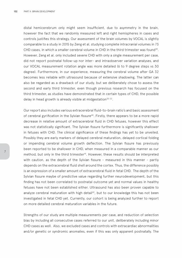
distal hemicerebrum only might seem insufficient, due to asymmetry in the brain, however the fact that we randomly measured left and right hemispheres in cases and controls justifies this strategy. Our assessment of the brain volumes by VOCAL is slightly comparable to a study in 2015 by Zeng et al, studying complete intracranial volumes in 73 CHD cases, in which a smaller cerebral volume in CHD in the third trimester was found39. However, Zeng et al. only included severe CHD with only a single measurement per case, did not report postnatal follow-up nor inter- and intraobserver variation analyses, and our VOCAL measurement rotation angle was more detailed (6 to 9 degree steps vs 30 degree). Furthermore, in our experience, measuring the cerebral volume after GA 32 becomes less reliable with ultrasound because of extensive shadowing. The latter can also be regarded as a drawback of our study, but we deliberately chose to assess the second and early third trimester, even though previous research has focused on the third trimester, as studies have demonstrated that in certain types of CHD, the possible delay in head growth is already visible at midgestation20-22.
Our report also includes various extracerebral fluid-to-brain ratio’s and basic assessment of cerebral gyrification in the Sylvian fissure40. Firstly, there appears to be a more rapid decrease in relative amount of extracerebral fluid in CHD fetuses, however this effect was not statistically significant. The Sylvian fissure furthermore is significantly shallower in fetuses with CHD. The clinical significance of these findings has yet to be unveiled. Possibly they are early markers of delayed cerebral maturation, delayed cortical folding or impending cerebral volume growth deflection. The Sylvian fissure has previously been reported to be shallower in CHD, when measured in a comparable manner as our method, but only in the third trimester41. However, these results should be interpreted with caution, as the depth of the Sylvian fissure - measured in this manner - partly depends on the extracerebral fluid shell around the cortex. Thus, the difference possibly is an expression of a smaller amount of extracerebral fluid in fetal CHD. The depth of the Sylvian fissure maybe of predictive value regarding further neurodevelopment, but this finding has not been correlated to postnatal outcome yet and normal values in healthy fetuses have not been established either. Ultrasound has also been proven capable to analyze cerebral maturation with high detail42, but to our knowledge this has not been investigated in fetal CHD yet. Currently, our cohort is being analyzed further to report on more detailed cerebral maturation variables in the future.
Strengths of our study are multiple measurements per case, and reduction of selection bias by including all consecutive cases referred to our unit, deliberately including minor CHD cases as well. Also, we excluded cases and controls with extracardiac abnormalities and/or genetic or syndromic anomalies, even if this was only apparent postnatally. The
132
7
PART II: BRAIN DEVELOPMENT
inter- and intraobserver variation analysis demonstrated high concordance. Unfortunately, long-term neurodevelopmental outcome parameters are not available in our cohort yet. Also, brain volume analysis after GA 32 was deemed unreliable due to high levels of acoustic shadowing. This might be the period in which fetal cerebral growth is mostly affected. In our opinion however, neonatal studies so far cannot exclude that hypoxia in the first days of life in prenatally undetected cases are an important cause for the pre-operative cerebral abnormalities. We state that a prenatal origin for neurodevelopmental delay in later life has not been irrefutably proven, especially in other CHD types than HLHS. Therapies like maternal hyperoxygenation should be used with caution, as it is not yet proven beneficial and might even cause less flow to the brain as it causes increased flow towards the pulmonary arteries, leading to a possible steal effect in the brain43-45.
In conclusion, our study shows no significant difference in hemicerebral volume, extracerebral fluid space and volume growth between fetuses with isolated CHD and controls. Only fetuses with specific lesions such as TOF, DORV and HLHS might have a slightly smaller brain volume at 20 weeks of gestation, but the effect-size is very small and wanes with advancing gestation. Fetuses with CHD do have a shallower Sylvian fissure. The etiology and consequences of these findings are unknown. Unfortunately, there are no known fetal ultrasound parameters available in the second trimester for prediction of neurodevelopmental outcome yet.
Supplemental data available onlineSupplement 1: Neurosonography and 3D assessment; reliability measurements.Supplement 2: assessment of additional intracranial fluid measurements.Supplement 3: Case description and categorization regarding oxygen and flow.
133
7
REFERENCES
1. Marino, B.S., et al., Neurodevelopmental
outcomes in children with congenital heart
disease: evaluation and management: a scientific
statement from the American Heart Association.
Circulation, 2012. 126(9): p. 1143-1172.
2. Sarrechia, I., et al., Neurocognitive development
and behaviour in school-aged children after
surgery for univentricular or biventricular
congenital heart disease. Eur. J. Cardiothorac.
Surg, 2016. 49(1): p. 167-174.
3. Owen, M., et al., Abnormal brain structure and
function in newborns with complex congenital
heart defects before open heart surgery: A review
of the evidence. Journal of Child Neurology, 2011.
26(6): p. 743-755.
4. Rosti, L., et al., Pattern of cerebral ultrasound in
neonatal heart surgery. Pediatr Med Chir, 2011.
33(3): p. 124-8.
5. van Houten, J.P., A. Rothman, and R. Bejar, High
incidence of cranial ultrasound abnormalities in
full-term infants with congenital heart disease.
Am J Perinatol, 1996. 13(1): p. 47-53.
6. Te Pas, A.B., et al., Preoperative cranial ultrasound
findings in infants with major congenital heart
disease. Acta Paediatr, 2005. 94(11): p. 1597-1603.
7. Rios, D.R., et al., Usefulness of routine head
ultrasound scans before surgery for congenital
heart disease. Pediatrics, 2013. 131(6): p. e1765-
70.
8. Khalil, A., et al., Brain abnormalities and
neurodevelopmental delay in congenital heart
disease: systematic review and meta-analysis.
Ultrasound Obstet. Gynecol, 2014. 43(1): p. 14-24.
9. Donofrio, M.T. and A.N. Massaro, Impact of
congenital heart disease on brain development and
neurodevelopmental outcome. Int. J. Pediatr,
2010. 2010.
10. Donofrio, M.T., et al., Autoregulation of cerebral
blood flow in fetuses with congenital heart
disease: the brain sparing effect. Pediatr Cardiol,
2003. 24(5): p. 436-443.
11. Limperopoulos, C., et al., Brain volume and
metabolism in fetuses with congenital heart
disease: evaluation with quantitative magnetic
resonance imaging and spectroscopy. Circulation,
2010. 121(1): p. 26-33.
12. Masoller, N., et al., Evidence of second-trimester
changes in head biometry and brain perfusion in
fetuses with congenital heart disease. Ultrasound
Obstet. Gynecol, 2014. 44(2): p. 182-187.
13. Hahn, E., et al., The association of fetal growth,
cerebral blood flow, and neurodevelopmental
outcome in single ventricle fetuses. Ultrasound
Obstet. Gynecol 2016 Apr;47(4):460-5.
14. Khalil, A., et al., Prevalence of prenatal brain
abnormalities in fetuses with congenital heart
disease: a systematic review. Ultrasound Obstet
Gynecol, 2016. 48(3): p. 296-307.
15. Jansen, F.A., et al., Fetal brain imaging in isolated
congenital heart defects - a systematic review
and meta-analysis. Prenat Diagn, 2016. 36(7): p.
601-13.
16. Szwast, A., et al., Comparative Analysis of
Cerebrovascular Resistance in the Fetus with
Single Ventricle Congenital Heart Disease.
Ultrasound Obstet. Gynecol, 2012.
17. Williams, I.A., et al., The association of
fetal cerebrovascular resistance with early
neurodevelopment in single ventricle congenital
heart disease. Am. Heart J, 2013. 165(4): p. 544-
550.
18. Sun, L., et al., Reduced fetal cerebral oxygen
134
7
PART II: BRAIN DEVELOPMENT

consumption is associated with smaller brain
size in fetuses with congenital heart disease.
Circulation, 2015. 131(15): p. 1313-1323.
19. Mlczoch, E., et al., Structural congenital brain
disease in congenital heart disease: Results from
a fetal MRI program. Eur. J. Paediatr. Neurol,
2012.
20. Masoller, N., et al., Mid-gestation brain Doppler
and head biometry in fetuses with congenital heart
disease predict abnormal brain development at
birth. Ultrasound Obstet. Gynecol, 2016. 47(1): p.
65-73.
21. Jansen, F.A., et al., Head growth in fetuses
with isolated congenital heart defects: lack of
influence of aortic arch flow and ascending aorta
oxygen saturation. Ultrasound Obstet Gynecol,
2016. 48(3): p. 357-64.
22. Schellen, C., et al., Fetal MRI detects early
alterations of brain development in Tetralogy of
Fallot. Am. J. Obstet. Gynecol, 2015. 213(3): p.
392-397.
23. Andescavage, N., et al., 3-D volumetric MRI
evaluation of the placenta in fetuses with complex
congenital heart disease. Placenta, 2015.
24. Masoller, N., et al., Fetal brain Doppler and
biometry at mid-gestation for the early
prediction of abnormal brain development at
birth in congenital heart disease. Ultrasound
Obstet. Gynecol, 2015.
25. Al, N.B., et al., Fetal circulation in left-
sided congenital heart disease measured by
cardiovascular magnetic resonance: a case-
control study. J. Cardiovasc. Magn Reson, 2013.
15: p. 65.
26. Clouchoux, C., et al., Delayed cortical
development in fetuses with complex congenital
heart disease. Cereb. Cortex, 2013. 23(12): p.
2932-2943.
27. Lloyd, D.F., et al., The neurodevelopmental
implications of hypoplastic left heart syndrome in
the fetus. Cardiol Young, 2017. 27(2): p. 217-223.
28. Hinton, R.B., et al., Prenatal head growth and
white matter injury in hypoplastic left heart
syndrome. Pediatr Res, 2008. 64(4): p. 364-369.
29. Chen, Y., et al., Cerebral vascular resistance
and left ventricular myocardial performance in
fetuses with Ebstein’s anomaly. Am J Perinatol,
2009. 26(4): p. 253-258.
30. Matthiesen, N.B., et al., Congenital Heart
Defects and Indices of Fetal Cerebral Growth in
a Nationwide Cohort of 924,422 Liveborn Infants.
Circulation, 2016.
31. Fahed, A.C., et al., Genetics of congenital heart
disease: the glass half empty. Circ. Res, 2013.
112(4): p. 707-720.
32. Gelb, B.D., Recent advances in understanding the
genetics of congenital heart defects. Curr Opin
Pediatr, 2013. 25(5): p. 561-6.
33. Griffiths, P.D., et al., Use of MRI in the diagnosis
of fetal brain abnormalities in utero (MERIDIAN):
a multicentre, prospective cohort study. Lancet,
2017. 389(10068): p. 538-546.
34. Malinger, G., et al., Fetal cerebral magnetic
resonance, neurosonography, the multiverse and
the brave new world of fetal medicine. Ultrasound
Obstet Gynecol, 2017.
35. Paladini, D., et al., The MERIDIAN trial: caution is
needed. Lancet, 2017. 389(10084): p. 2103.
36. Pistorius, L.R., et al., Fetal neuroimaging:
ultrasound, MRI, or both? Obstet. Gynecol. Surv,
2008. 63(11): p. 733-745.
37. Roelfsema, N.M., et al., Three-dimensional
135
7

sonographic measurement of normal fetal brain
volume during the second half of pregnancy. Am J
Obstet Gynecol, 2004. 190(1): p. 275-80.
38. Benavides-Serralde, A., et al., Three-dimensional
sonographic calculation of the volume of
intracranial structures in growth-restricted
and appropriate-for-gestational age fetuses.
Ultrasound Obstet Gynecol, 2009. 33(5): p. 530-
7.
39. Zeng, S., et al., Volume of intracranial structures
on three-dimensional ultrasound in fetuses with
congenital heart disease. Ultrasound Obstet.
Gynecol, 2015. 46(2): p. 174-181.
40. Alonso, I., et al., Depth of brain fissures in normal
fetuses by prenatal ultrasound between 19 and 30
weeks of gestation. Ultrasound Obstet Gynecol,
2010. 36(6): p. 693-9.
41. Peng, Q., et al., Reduced fetal brain fissures depth
in fetuses with congenital heart diseases. Prenat
Diagn, 2016. 36(11): p. 1047-1053.
42. Pistorius, L.R., et al., Grade and symmetry of
normal fetal cortical development: a longitudinal
two- and three-dimensional ultrasound study.
Ultrasound Obstet. Gynecol, 2010. 36(6): p. 700-
708.
43. Szwast, A., et al., Vasoreactive response to
maternal hyperoxygenation in the fetus with
hypoplastic left heart syndrome. Circ Cardiovasc
Imaging, 2010. 3(2): p. 172-8.
44. Rasanen, J., et al., Reactivity of the human
fetal pulmonary circulation to maternal
hyperoxygenation increases during the second
half of pregnancy: a randomized study.
Circulation, 1998. 97(3): p. 257-62.
45. Enzensberger, C., et al., Pulmonary Vasoreactivity
to Materno-Fetal Hyperoxygenation Testing in
Fetuses with Hypoplastic Left Heart. Ultraschall
Med, 2016. 37(2): p. 195-200.
136
7
PART II: BRAIN DEVELOPMENT

137
7

CHAPTER 8
A low incidence of preoperative
neurosonographic abnormalities in
neonates with prenatally detected
congenital heart defects –
a prospective case controlled cohort
study
Submitted
F.A.R. JansenM.C. HaakM.S. van WesemaelA.D.J. ten HarkelJ.M.M. van LithN.A. Blom S.J. Steggerda
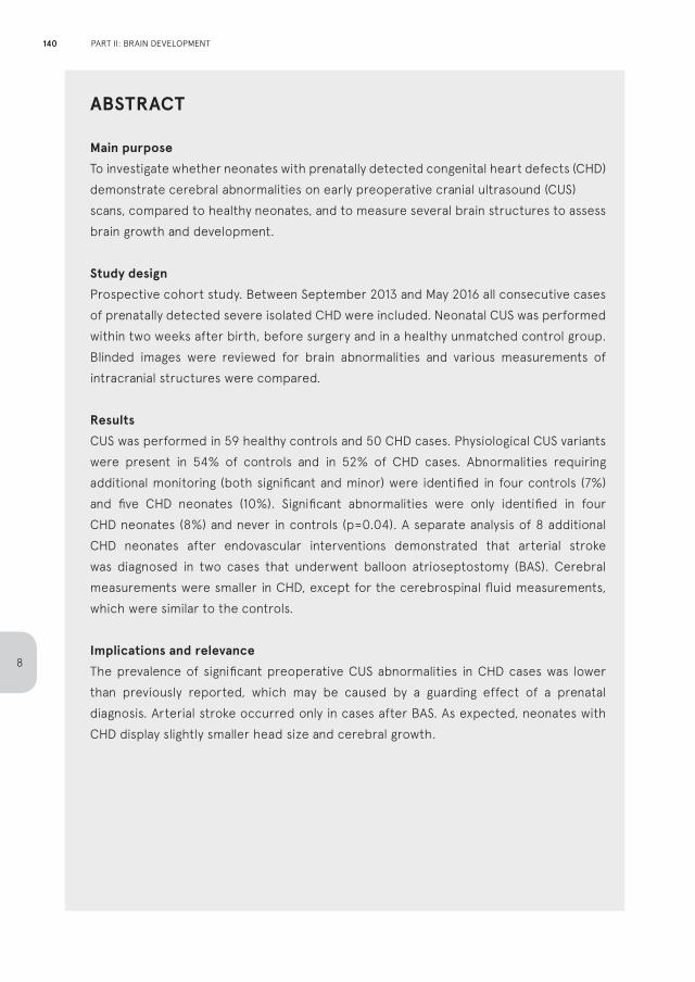
ABSTRACT
Main purpose
To investigate whether neonates with prenatally detected congenital heart defects (CHD) demonstrate cerebral abnormalities on early preoperative cranial ultrasound (CUS)scans, compared to healthy neonates, and to measure several brain structures to assess brain growth and development. Study design
Prospective cohort study. Between September 2013 and May 2016 all consecutive cases of prenatally detected severe isolated CHD were included. Neonatal CUS was performed within two weeks after birth, before surgery and in a healthy unmatched control group. Blinded images were reviewed for brain abnormalities and various measurements of intracranial structures were compared. Results CUS was performed in 59 healthy controls and 50 CHD cases. Physiological CUS variants were present in 54% of controls and in 52% of CHD cases. Abnormalities requiring additional monitoring (both significant and minor) were identified in four controls (7%) and five CHD neonates (10%). Significant abnormalities were only identified in four CHD neonates (8%) and never in controls (p=0.04). A separate analysis of 8 additional CHD neonates after endovascular interventions demonstrated that arterial stroke was diagnosed in two cases that underwent balloon atrioseptostomy (BAS). Cerebral measurements were smaller in CHD, except for the cerebrospinal fluid measurements, which were similar to the controls. Implications and relevance
The prevalence of significant preoperative CUS abnormalities in CHD cases was lower than previously reported, which may be caused by a guarding effect of a prenatal diagnosis. Arterial stroke occurred only in cases after BAS. As expected, neonates with CHD display slightly smaller head size and cerebral growth.
8
140 PART II: BRAIN DEVELOPMENT
INTRODUCTION
Congenital heart defects (CHD) occur in 7-8 per 1000 newborns and are responsible for significant morbidity and mortality1. Survival has increased over the last decades, but these infants remain at increased risk for neurodevelopmental delay (NDD)2-6. Until recently, this was assumed to result from perioperative cerebral injury, caused by hypoxia or thrombo-embolic events7;8. Abnormalities on neonatal MRI or cranial ultrasound (CUS) are also reported before surgery in 36% of CHD neonates, and include ischemic lesions, hemorrhage and delayed maturation9-20. It has been suggested that part of these abnormalities are caused by abnormal prenatal development of the brain, resulting from altered prenatal hemodynamics in CHD21. Several of the CUS studies are, however, hampered by selection bias caused by the inclusion of selected types of CHD and some included syndromic cases17-19. Furthermore, most studies assessed cases in which CHD was diagnosed after birth14,16,17,20. In severe CHD without a prenatal diagnosis, neonates may experience a period of hemodynamic collapse and hypoxia, which can cause damage to the brain, This could influence previous reported findings. Comparison with a control group was only reported in one of the seven preoperative CUS studies15 and CUS was frequently performed weeks or months after birth16-18. Therefore the results of these studies cannot be applied to fetuses with a prenatally detected CHD.
We performed a prospective CUS study in prenatally detected, isolated CHD and compared the findings to a group of healthy controls. The primary aim was to investigate whether neonates with prenatally detected CHD demonstrate cerebral abnormalities on early preoperative CUS. The secondary aim was to measure several brain structures to assess brain growth and development in both groups. Our hypothesis was that neonates with CHD more frequently display CUS abnormalities and present with smaller brain structures and wider cerebrospinal fluid (CSF) compartments.
METHODS
We performed a prospective cohort study with controls. The institutional ethical committee approved the study protocol. Consecutive cases of neonates with prenatally detected and postnatally confirmed CHD, born between September 2013 and May 2016 in the Leiden University Medical Centre (LUMC) were included. Severe CHD was defined as the need of a therapeutic catheter intervention or cardiac surgery in the first year of life. An unmatched healthy control group with a normal second trimester anomaly scan was recruited in midwifery practices between August 2014 and May 2016. Exclusion criteria
141
8
were additional structural or genetic anomalies, birth before 35 weeks gestational age (GA), small for GA (birth weight below the 2.3rd percentile), maternal illnesses influencing placental function (pre-eclampsia, hypertension) and perinatal complications (such as perinatal asphyxia, hypoglycemia and congenital/neonatal infection). Besides the postnatal CUS, both groups had monthly prenatal neurosonography (up to 36 weeks of GA) which revealed no suspected abnormalities. Additional maternal and perinatal data were collected, including mode of delivery, GA at birth, head circumference (HC) and birth weight (BW, both expressed in z-scores), type of CHD and type of surgery or endovascular intervention. Follow-up data regarding neurodevelopment were distilled from clinical charts. The sample size was a result of the prenatal inclusion of the CHD cases necessary for chapter 7.
Postnatal CUS was performed within 14 days after birth, preferably in the first postnatal week, but not on the first day of life, to allow the ventricles to open22. All scans were assessed for various ultrasound findings as listed below. All noted ultrasound findings were categorized in three categories: as a significant abnormality, a minor abnormality, or as a physiological variant (described in box 1). CUS was always performed before surgery. Because the aim was to study the intrauterine effects on the brain in CHD, cases in which CUS was performed after early endovascular interventions, such as balloon atrioseptostomy (BAS), were excluded from the primary analysis, and analyzed separately.
Box 1: Categorization of ultrasound findings
Significant abnormalities were defined as: any grade of P/IVH, persistent grade 2 PVE, cPVL,
intraparenchymal lesions suspect for stroke and structural brain abnormalities.
Minor abnormalities were defined as: any variant or combination of variants with need for further
assessment or CUS follow up, not included in the significant abnormalities group, such as a com-
bination of large/ multiple pseudocysts suspected for (congenital) infection or transient lesions
(cysts, focal echodensities) with spontaneous regression.
Physiological variants were defined as: LSV, subependymal and choroid plexus cysts, grade 1 PVE
in the first postnatal week and CSF space variations (asymmetric or plump; size within normal
limits).
142
8
PART II: BRAIN DEVELOPMENT
CUS assessment and -findingsAll scans were assessed for the presence of: periventricular echogenicity (PVE) of the white matter; cystic periventricular leucomalacia (cPVL)23; peri- and intraventricular hemorrhage (P/IVH)24;38 non-physiologic echogenicity in the deep grey matter; suspected stroke; changes in echogenicity in the cerebellum and supra- and infratentorial congenital malformations. Lenticulostriate vasculopathy (LSV) and subependymal or choroid plexus cysts (CPC) were also noted. For the classification of PVE we used grade 1 (moderately increased echogenicity, (almost) as bright as the choroid plexus) and grade 2 ((smaller areas of) increased echogenicity, being obviously brighter than the choroid plexus)38. CUS was performed with an Aloka α 10 ultrasound system (Hitachi Medical Systems Holding AG, Switzerland) or a Toshiba Aplio 400 system (Toshiba Medical Systems B.V., the Netherlands). Scanning was performed routinely by the attending neonatologist or one of the investigators (FJ/SS), according to a standardized protocol through the anterior and mastoid fontanelles39. Images were digitally stored (Clinical Assistant, RVC B.V., the Netherlands) and reviewed at least 2 months after the initial recording by two separate investigators (FJ/SS), blinded to the patients’ names and the presence or absence of CHD. Discrepancies were solved by consensus.
Cerebral measurements were performed offline (see supplement 1) and included the lateral ventricles, interhemispheric fissure, sinocortical and cavum septum pellucidum width, corpus callosum length, corpus callosum to fastigium distance, transcerebellar diameter and basal ganglia dimensions. An estimated intracranial volume was calculated according to the method of Graca25).The volume of the basal ganglia area was calculated according to the same method (see supplement 1).
Data management and statistical analysisCategorical variables were summarized with frequency counts and percentages and compared using a Chi-square or Fischer’s exact test. Continuous variables were summarized with means/SD and compared using t-test or one-way ANOVA. To check for normality, we plotted outcome measures in a histogram. If data were not distributed normally we performed a Mann-Whitney-U non-parametrical test. In case of a linear correlation of a measurement with age, regression coefficients were compared in a generalized linear model. In these models we included the postmenstrual age (PMA) at CUS and the presence of CHD as covariates, as well as the interaction between age and CHD. Using a generalized linear model, the mean+SD at the PMA of 40 weeks was calculated. The inter- and intra-observer variations of the measurements were estimated with the interclass correlation coefficient. Analyses were performed with SPSS (version 23.0, SPSS Inc, Chicago, IL, USA). A p-value <0.05 was considered significant.
143
8
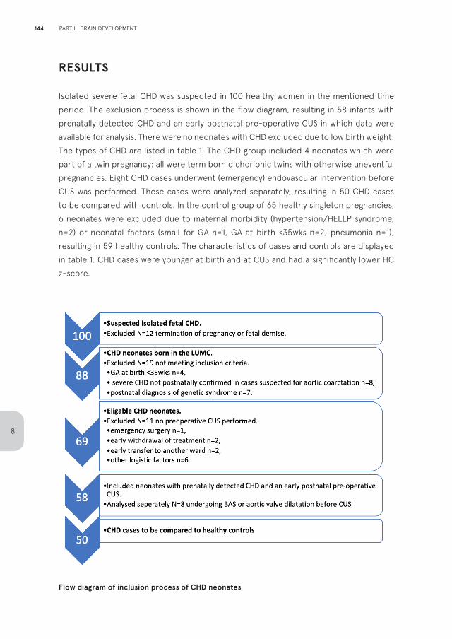
RESULTS
Isolated severe fetal CHD was suspected in 100 healthy women in the mentioned time period. The exclusion process is shown in the flow diagram, resulting in 58 infants with prenatally detected CHD and an early postnatal pre-operative CUS in which data were available for analysis. There were no neonates with CHD excluded due to low birth weight. The types of CHD are listed in table 1. The CHD group included 4 neonates which were part of a twin pregnancy: all were term born dichorionic twins with otherwise uneventful pregnancies. Eight CHD cases underwent (emergency) endovascular intervention before CUS was performed. These cases were analyzed separately, resulting in 50 CHD cases to be compared with controls. In the control group of 65 healthy singleton pregnancies, 6 neonates were excluded due to maternal morbidity (hypertension/HELLP syndrome, n=2) or neonatal factors (small for GA n=1, GA at birth <35wks n=2, pneumonia n=1), resulting in 59 healthy controls. The characteristics of cases and controls are displayed in table 1. CHD cases were younger at birth and at CUS and had a significantly lower HC z-score.
Flow diagram of inclusion process of CHD neonates
144
8
PART II: BRAIN DEVELOPMENT
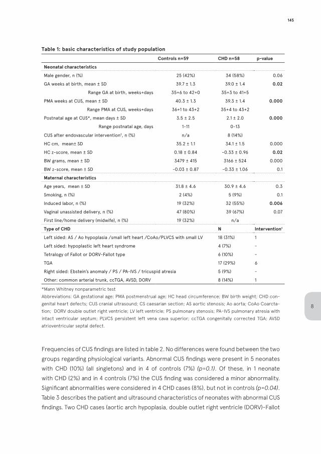
Table 1: basic characteristics of study population
Controls n=59 CHD n=58 p-value
Neonatal characteristics
Male gender, n (%) 25 (42%) 34 (58%) 0.06
GA weeks at birth, mean ± SD 39.7 ± 1.3 39.0 ± 1.4 0.02
Range GA at birth, weeks+days 35+6 to 42+0 35+3 to 41+5
PMA weeks at CUS, mean ± SD 40.3 ± 1.3 39.3 ± 1.4 0.000
Range PMA at CUS, weeks+days 36+1 to 43+2 35+4 to 43+2
Postnatal age at CUS*, mean days ± SD 3.5 ± 2.5 2.1 ± 2.0 0.000
Range postnatal age, days 1-11 0-13
CUS after endovascular intervention‡, n (%) n/a 8 (14%)
HC cm, mean± SD 35.2 ± 1.1 34.1 ± 1.5 0.000
HC z-score, mean ± SD 0.18 ± 0.84 -0.33 ± 0.96 0.02
BW grams, mean ± SD 3479 ± 415 3166 ± 524 0.000
BW z-score, mean ± SD -0.03 ± 0.87 -0.33 ± 1.06 0.1
Maternal characteristics
Age years, mean ± SD 31.8 ± 4.6 30.9 ± 4.6 0.3
Smoking, n (%) 2 (4%) 5 (9%) 0.1
Induced labor, n (%) 19 (32%) 32 (55%) 0.006
Vaginal unassisted delivery, n (%) 47 (80%) 39 (67%) 0.07
First line/home delivery (midwife), n (%) 19 (32%) n/a
Type of CHD N Intervention‡
Left sided: AS / Ao hypoplasia /small left heart /CoAo/PLVCS with small LV 18 (31%) 1
Left sided: hypoplastic left heart syndrome 4 (7%) -
Tetralogy of Fallot or DORV-Fallot type 6 (10%) -
TGA 17 (29%) 6
Right sided: Ebstein’s anomaly / PS / PA-IVS / tricuspid atresia 5 (9%) -
Other: common arterial trunk, ccTGA, AVSD, DORV 8 (14%) 1
*Mann Whitney nonparametric testAbbreviations: GA gestational age; PMA postmenstrual age; HC head circumference; BW birth weight; CHD con-genital heart defects; CUS cranial ultrasound; CS caesarian section; AS aortic stenosis; Ao aorta; CoAo Coarcta-tion; DORV double outlet right ventricle; LV left ventricle; PS pulmonary stenosis; PA-IVS pulmonary atresia with intact ventricular septum; PLVCS persistent left vena cava superior; ccTGA congenitally corrected TGA; AVSD atrioventricular septal defect.
Frequencies of CUS findings are listed in table 2. No differences were found between the two groups regarding physiological variants. Abnormal CUS findings were present in 5 neonates with CHD (10%) (all singletons) and in 4 of controls (7%) (p=0.1). Of these, in 1 neonate with CHD (2%) and in 4 controls (7%) the CUS finding was considered a minor abnormality. Significant abnormalities were considered in 4 CHD cases (8%), but not in controls (p=0.04). Table 3 describes the patient and ultrasound characteristics of neonates with abnormal CUS findings. Two CHD cases (aortic arch hypoplasia, double outlet right ventricle (DORV)-Fallot
145
8
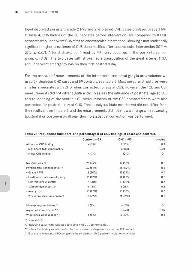
type) displayed persistent grade 2 PVE and 2 left-sided CHD cases displayed grade 1 IVH. In table 4, CUS findings of the 50 neonates before intervention, are compared to 8 CHD neonates who underwent CUS after an endovascular intervention, showing a (not statistically significant) higher prevalence of CUS abnormalities after endovascular intervention (10% vs 37%, p=0.07). Arterial stroke, confirmed by MRI, only occurred in the post-intervention group (p=0.02). The two cases with stroke had a transposition of the great arteries (TGA) and underwent emergency BAS on their first postnatal day.
For the analysis of measurements of the intracranial and basal ganglia area volumes we used 54 singleton CHD cases and 59 controls, see table 5. Most cerebral structures were smaller in neonates with CHD, when corrected for age at CUS. However, the TCD and CSF measurements did not differ significantly. To assess the influence of postnatal age at CUS and re-opening of the ventricles22, measurements of the CSF compartments were also corrected for postnatal day at CUS. These analyses (data not shown) did not differ from the results shown in table 5, and the measurements did not show a change with advancing (postnatal or postmenstrual) age, thus no statistical correction was performed.
Table 2: Frequencies (numbers and percentages) of CUS findings in cases and controls.
Controls n=59 CHD n=50 p-valueAbnormal CUS finding 4 (7%) 5 (10%) 0.4- Significant CUS abnormality - 4 (8%) 0.04- Minor CUS finding 4 (7%) 1 (2%) 0.1
No variations *) 23 (39%) 19 (38%) 0.5Physiological variants total */ 32 (54%) 26 (52%) 0.5- Grade 1 PVE 12 (20%) 12 (24%) 0.4- Lenticulostriate vasculopathy 16 (27%) 14 (28%) 0.5- Choroid plexus cyst(s) 12 (20%) 10 (20%) 0.6- Subependymal cyst(s) 8 (14%) 8 (16%) 0.5- Any cyst(s) 16 (27%) 18 (36%) 0.3- 2 or more variations present 13 (22%) 11 (22%) 0.6
Wide/plump ventricles ** 1 (2%) 4 (7%) 0.1Asymmetric ventricles ** - 3 (6%) 0.09Wide extra-axial spaces ** 3 (5%) 5 (10%) 0.3
*) normal CUS; */ including cases with variants coinciding with CUS abnormalities; ** subjective finding as interpreted by the reviewer; categorised as normal CUS variantCUS cranial ultrasound; CHD congenital heart defects; PVE periventricular echogenicity
146
8
PART II: BRAIN DEVELOPMENT
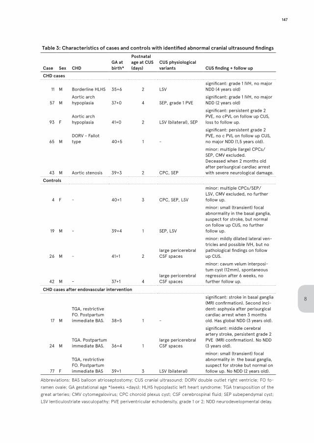
Table 3: Characteristics of cases and controls with identified abnormal cranial ultrasound findings
Case Sex CHDGA at birth*
Postnatal age at CUS (days)
CUS physiological variants CUS finding + follow up
CHD cases
11 M Borderline HLHS 35+6 2 LSVsignificant: grade 1 IVH, no major NDD (4 years old)
57 MAortic arch hypoplasia 37+0 4 SEP, grade 1 PVE
significant: grade 1 IVH, no major NDD (2 years old)
93 FAortic arch hypoplasia 41+0 2 LSV (bilateral), SEP
significant: persistent grade 2 PVE, no cPVL on follow up CUS, loss to follow up.
65 MDORV - Fallot type 40+5 1 -
significant: persistent grade 2 PVE, no c PVL on follow up CUS, no major NDD (1,5 years old).
43 M Aortic stenosis 39+3 2 CPC, SEP
minor: multiple (large) CPCs/SEP, CMV excluded. Deceased when 2 months old after perisurgical cardiac arrest with severe neurological damage.
Controls
4 F - 40+1 3 CPC, SEP, LSV
minor: multiple CPCs/SEP/LSV, CMV excluded, no further follow up.
19 M - 39+4 1 SEP, LSV
minor: small (transient) focal abnormality in the basal ganglia, suspect for stroke, but normal on follow up CUS, no further follow up.
26 M - 41+1 2large pericerebral CSF spaces
minor: mildly dilated lateral ven-tricles and possible IVH, but no pathological findings on follow up CUS.
42 M - 37+1 4large pericerebral CSF spaces
minor: cavum velum interposi-tum cyst (12mm), spontaneous regression after 6 weeks, no further follow up.
CHD cases after endovascular intervention
17 M
TGA, restrictive FO. Postpartum immediate BAS. 38+5 1 -
significant: stroke in basal ganglia (MRI confirmation). Second inci-dent: asphyxia after perisurgical cardiac arrest when 3 months old. Has global NDD (3 years old).
24 MTGA. Postpartum immediate BAS. 36+4 1
large pericerebral CSF spaces
significant: middle cerebral artery stroke, persistent grade 2 PVE (MRI confirmation). No NDD (3 years old).
77 F
TGA, restrictive FO. Postpartum immediate BAS 39+1 3 LSV (bilateral)
minor: small (transient) focal abnormality in the basal ganglia, suspect for stroke but normal on follow up. No NDD (2 years old).
Abbreviations: BAS balloon atrioseptostomy; CUS cranial ultrasound; DORV double outlet right ventricle; FO fo-ramen ovale; GA gestational age *(weeks +days); HLHS hypoplastic left heart syndrome; TGA transposition of the great arteries; CMV cytomegalovirus; CPC choroid plexus cyst; CSF cerebrospinal fluid; SEP subependymal cyst; LSV lenticulostriate vasculopathy; PVE periventricular echodensity, grade 1 or 2; NDD neurodevelopmental delay.
147
8
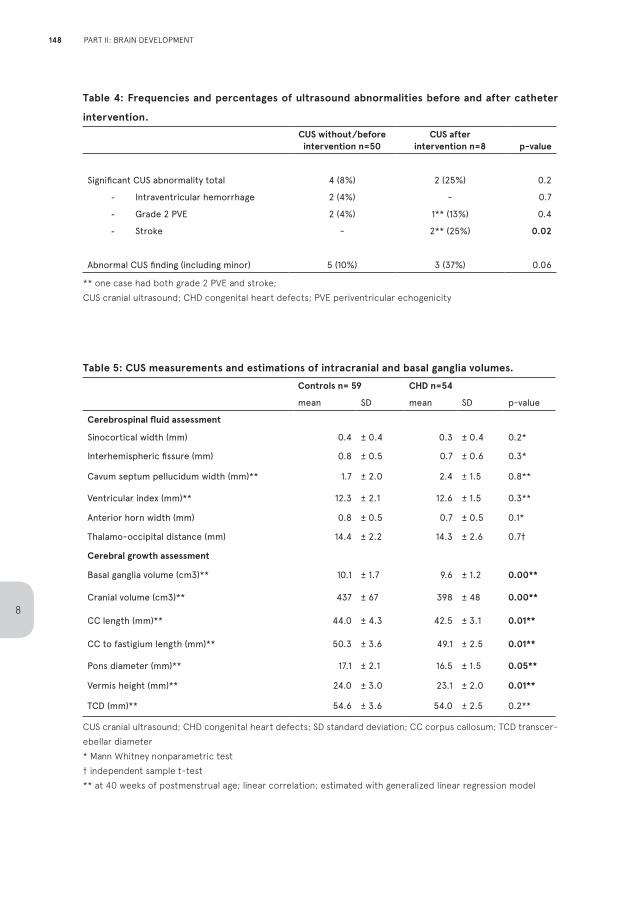
Table 4: Frequencies and percentages of ultrasound abnormalities before and after catheter
intervention.
CUS without/before intervention n=50
CUS afterintervention n=8 p-value
Significant CUS abnormality total 4 (8%) 2 (25%) 0.2
- Intraventricular hemorrhage 2 (4%) - 0.7
- Grade 2 PVE 2 (4%) 1** (13%) 0.4
- Stroke - 2** (25%) 0.02
Abnormal CUS finding (including minor) 5 (10%) 3 (37%) 0.06
** one case had both grade 2 PVE and stroke; CUS cranial ultrasound; CHD congenital heart defects; PVE periventricular echogenicity
Table 5: CUS measurements and estimations of intracranial and basal ganglia volumes. Controls n= 59 CHD n=54
mean SD mean SD p-value
Cerebrospinal fluid assessment
Sinocortical width (mm) 0.4 ± 0.4 0.3 ± 0.4 0.2*
Interhemispheric fissure (mm) 0.8 ± 0.5 0.7 ± 0.6 0.3*
Cavum septum pellucidum width (mm)** 1.7 ± 2.0 2.4 ± 1.5 0.8**
Ventricular index (mm)** 12.3 ± 2.1 12.6 ± 1.5 0.3**
Anterior horn width (mm) 0.8 ± 0.5 0.7 ± 0.5 0.1*
Thalamo-occipital distance (mm) 14.4 ± 2.2 14.3 ± 2.6 0.7†
Cerebral growth assessment
Basal ganglia volume (cm3)** 10.1 ± 1.7 9.6 ± 1.2 0.00**
Cranial volume (cm3)** 437 ± 67 398 ± 48 0.00**
CC length (mm)** 44.0 ± 4.3 42.5 ± 3.1 0.01**
CC to fastigium length (mm)** 50.3 ± 3.6 49.1 ± 2.5 0.01**
Pons diameter (mm)** 17.1 ± 2.1 16.5 ± 1.5 0.05**
Vermis height (mm)** 24.0 ± 3.0 23.1 ± 2.0 0.01**
TCD (mm)** 54.6 ± 3.6 54.0 ± 2.5 0.2**
CUS cranial ultrasound; CHD congenital heart defects; SD standard deviation; CC corpus callosum; TCD transcer-ebellar diameter* Mann Whitney nonparametric test† independent sample t-test** at 40 weeks of postmenstrual age; linear correlation; estimated with generalized linear regression model
148
8
PART II: BRAIN DEVELOPMENT
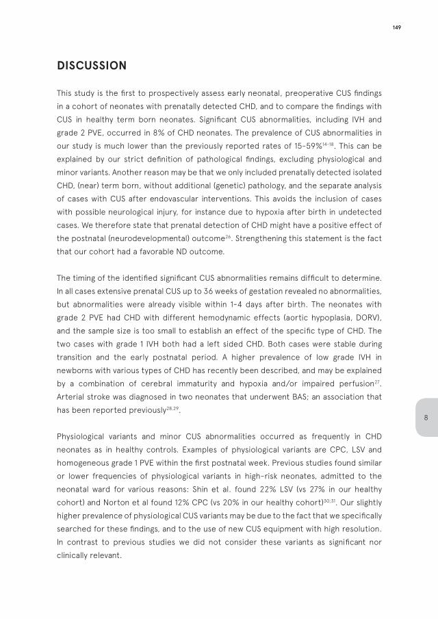
DISCUSSION
This study is the first to prospectively assess early neonatal, preoperative CUS findings in a cohort of neonates with prenatally detected CHD, and to compare the findings with CUS in healthy term born neonates. Significant CUS abnormalities, including IVH and grade 2 PVE, occurred in 8% of CHD neonates. The prevalence of CUS abnormalities in our study is much lower than the previously reported rates of 15-59%14-18. This can be explained by our strict definition of pathological findings, excluding physiological and minor variants. Another reason may be that we only included prenatally detected isolated CHD, (near) term born, without additional (genetic) pathology, and the separate analysis of cases with CUS after endovascular interventions. This avoids the inclusion of cases with possible neurological injury, for instance due to hypoxia after birth in undetected cases. We therefore state that prenatal detection of CHD might have a positive effect of the postnatal (neurodevelopmental) outcome26. Strengthening this statement is the fact that our cohort had a favorable ND outcome.
The timing of the identified significant CUS abnormalities remains difficult to determine. In all cases extensive prenatal CUS up to 36 weeks of gestation revealed no abnormalities, but abnormalities were already visible within 1-4 days after birth. The neonates with grade 2 PVE had CHD with different hemodynamic effects (aortic hypoplasia, DORV), and the sample size is too small to establish an effect of the specific type of CHD. The two cases with grade 1 IVH both had a left sided CHD. Both cases were stable during transition and the early postnatal period. A higher prevalence of low grade IVH in newborns with various types of CHD has recently been described, and may be explained by a combination of cerebral immaturity and hypoxia and/or impaired perfusion27. Arterial stroke was diagnosed in two neonates that underwent BAS; an association that has been reported previously28,29.
Physiological variants and minor CUS abnormalities occurred as frequently in CHD neonates as in healthy controls. Examples of physiological variants are CPC, LSV and homogeneous grade 1 PVE within the first postnatal week. Previous studies found similar or lower frequencies of physiological variants in high-risk neonates, admitted to the neonatal ward for various reasons: Shin et al. found 22% LSV (vs 27% in our healthy cohort) and Norton et al found 12% CPC (vs 20% in our healthy cohort)30;31. Our slightly higher prevalence of physiological CUS variants may be due to the fact that we specifically searched for these findings, and to the use of new CUS equipment with high resolution. In contrast to previous studies we did not consider these variants as significant nor clinically relevant.
149
8
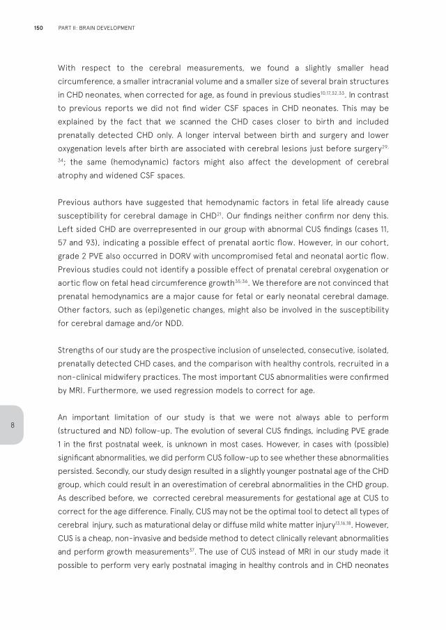
With respect to the cerebral measurements, we found a slightly smaller head circumference, a smaller intracranial volume and a smaller size of several brain structures in CHD neonates, when corrected for age, as found in previous studies10,17,32,33. In contrast to previous reports we did not find wider CSF spaces in CHD neonates. This may be explained by the fact that we scanned the CHD cases closer to birth and included prenatally detected CHD only. A longer interval between birth and surgery and lower oxygenation levels after birth are associated with cerebral lesions just before surgery29,
34; the same (hemodynamic) factors might also affect the development of cerebral atrophy and widened CSF spaces.
Previous authors have suggested that hemodynamic factors in fetal life already cause susceptibility for cerebral damage in CHD21. Our findings neither confirm nor deny this. Left sided CHD are overrepresented in our group with abnormal CUS findings (cases 11, 57 and 93), indicating a possible effect of prenatal aortic flow. However, in our cohort, grade 2 PVE also occurred in DORV with uncompromised fetal and neonatal aortic flow. Previous studies could not identify a possible effect of prenatal cerebral oxygenation or aortic flow on fetal head circumference growth35;36. We therefore are not convinced that prenatal hemodynamics are a major cause for fetal or early neonatal cerebral damage. Other factors, such as (epi)genetic changes, might also be involved in the susceptibility for cerebral damage and/or NDD.
Strengths of our study are the prospective inclusion of unselected, consecutive, isolated, prenatally detected CHD cases, and the comparison with healthy controls, recruited in a non-clinical midwifery practices. The most important CUS abnormalities were confirmed by MRI. Furthermore, we used regression models to correct for age.
An important limitation of our study is that we were not always able to perform (structured and ND) follow-up. The evolution of several CUS findings, including PVE grade 1 in the first postnatal week, is unknown in most cases. However, in cases with (possible) significant abnormalities, we did perform CUS follow-up to see whether these abnormalities persisted. Secondly, our study design resulted in a slightly younger postnatal age of the CHD group, which could result in an overestimation of cerebral abnormalities in the CHD group. As described before, we corrected cerebral measurements for gestational age at CUS to correct for the age difference. Finally, CUS may not be the optimal tool to detect all types of cerebral injury, such as maturational delay or diffuse mild white matter injury13,16,18. However, CUS is a cheap, non-invasive and bedside method to detect clinically relevant abnormalities and perform growth measurements37. The use of CUS instead of MRI in our study made it possible to perform very early postnatal imaging in healthy controls and in CHD neonates
150
8
PART II: BRAIN DEVELOPMENT
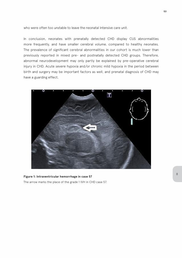
who were often too unstable to leave the neonatal intensive care unit.
In conclusion, neonates with prenatally detected CHD display CUS abnormalities more frequently, and have smaller cerebral volume, compared to healthy neonates. The prevalence of significant cerebral abnormalities in our cohort is much lower than previously reported in mixed pre- and postnatally detected CHD groups. Therefore, abnormal neurodevelopment may only partly be explained by pre-operative cerebral injury in CHD. Acute severe hypoxia and/or chronic mild hypoxia in the period between birth and surgery may be important factors as well, and prenatal diagnosis of CHD may have a guarding effect.
Figure 1: Intraventricular hemorrhage in case 57
The arrow marks the place of the grade 1 IVH in CHD case 57.
151
8
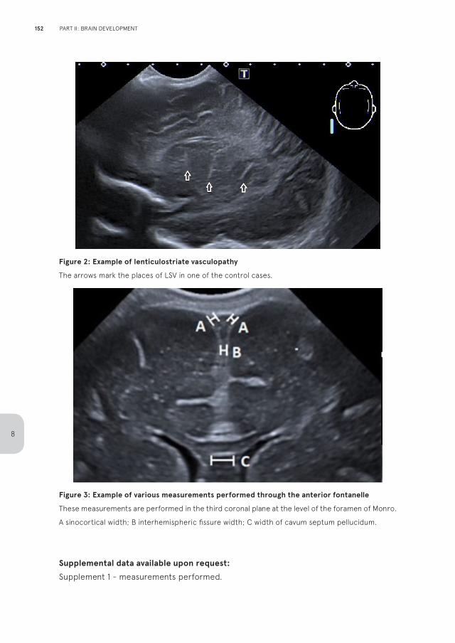
Figure 2: Example of lenticulostriate vasculopathy
The arrows mark the places of LSV in one of the control cases.
Figure 3: Example of various measurements performed through the anterior fontanelle
These measurements are performed in the third coronal plane at the level of the foramen of Monro.
A sinocortical width; B interhemispheric fissure width; C width of cavum septum pellucidum.
Supplemental data available upon request:
Supplement 1 - measurements performed.
152
8
PART II: BRAIN DEVELOPMENT
REFERENCES
1. van der Linde, D., et al., Birth prevalence of
congenital heart disease worldwide: a systematic
review and meta-analysis. J. Am. Coll. Cardiol,
2011. 58(21): p. 2241-2247.
2. Bellinger, D.C., et al., Developmental and
neurologic status of children after heart surgery
with hypothermic circulatory arrest or low-flow
cardiopulmonary bypass. N. Engl. J. Med, 1995.
332(9): p. 549-555.
3. Stieh, J., et al., Gross and fine motor development
is impaired in children with cyanotic congenital
heart disease. Neuropediatrics, 1999. 30(2): p.
77-82.
4. Marino, B. and M.C. Digilio, Congenital heart
disease and genetic syndromes: specific
correlation between cardiac phenotype and
genotype. Cardiovasc. Pathol, 2000. 9(6): p. 303-
315.
5. Marino, B.S., et al., Neurodevelopmental
outcomes in children with congenital heart
disease: evaluation and management: a scientific
statement from the American Heart Association.
Circulation, 2012. 126(9): p. 1143-1172.
6. McQuillen, P.S. and S.P. Miller, Congenital heart
disease and brain development. Ann N Y Acad Sci,
2010. 1184: p. 68-86.
7. Hovels-Gurich, H.H., Factors Influencing
Neurodevelopment after Cardiac Surgery during
Infancy. Front Pediatr, 2016. 4: p. 137.
8. du Plessis, A.J., Mechanisms of brain injury during
infant cardiac surgery. Semin. Pediatr. Neurol,
1999. 6(1): p. 32-47.
9. Miller, S.P., et al., Abnormal brain development in
newborns with congenital heart disease. N. Engl.
J. Med, 2007. 357(19): p. 1928-1938.
10. Owen, M., et al., Abnormal brain structure and
function in newborns with complex congenital
heart defects before open heart surgery: A review
of the evidence. Journal of Child Neurology, 2011.
26(6): p. 743-755.
11. Khalil, A., et al., Brain abnormalities and
neurodevelopmental delay in congenital heart
disease: systematic review and meta-analysis.
Ultrasound Obstet. Gynecol, 2014. 43(1): p. 14-24.
12. Licht, D.J., et al., Brain maturation is delayed in
infants with complex congenital heart defects. J
Thorac Cardiovasc Surg, 2009. 137(3): p. 529-
536.
13. Claessens, N.H., et al., Delayed cortical gray
matter development in neonates with severe
congenital heart disease. Pediatr Res, 2016.
80(5): p. 668-674.
14. Te Pas, A.B., et al., Preoperative cranial ultrasound
findings in infants with major congenital heart
disease. Acta Paediatr, 2005. 94(11): p. 1597-1603.
15. van Houten, J.P., A. Rothman, and R. Bejar, High
incidence of cranial ultrasound abnormalities in
full-term infants with congenital heart disease.
Am J Perinatol, 1996. 13(1): p. 47-53.
16. Rios, D.R., et al., Usefulness of routine head
ultrasound scans before surgery for congenital
heart disease. Pediatrics, 2013. 131(6): p. e1765-
70.
17. Krull, F., et al., Cerebral ultrasonography before
and after cardiac surgery in infants. Pediatr
Cardiol, 1994. 15(4): p. 159-62.
18. Latal, B., et al., Can preoperative cranial
ultrasound predict early neurodevelopmental
outcome in infants with congenital heart disease?
Dev Med Child Neurol, 2015. 57(7): p. 639-644.
19. Chock, V.Y., et al., Neurologic events in neonates
treated surgically for congenital heart disease. J
153
8
Perinatol, 2006. 26(4): p. 237-42.
20. El-Naggar, W.I., M. Keyzers, and P.J.
McNamara, Role of amplitude-integrated
electroencephalography in neonates with
cardiovascular compromise. J Crit Care, 2010.
25(2): p. 317-321.
21. Donofrio, M.T., A.J. Duplessis, and C.
Limperopoulos, Impact of congenital heart
disease on fetal brain development and injury.
Current Opinion in Pediatrics, 2011. 23(5): p. 502-
511.
22. Nelson, M.D., Jr., et al., Changes in the size of the
lateral ventricles in the normal-term newborn
following vaginal delivery. Pediatr Radiol, 2003.
33(12): p. 831-5.
23. de Vries, L.S., P. Eken, and L.M. Dubowitz,
The spectrum of leukomalacia using cranial
ultrasound. Behav Brain Res, 1992. 49(1): p. 1-6.
24. Volpe, J.J., Intraventricular hemorrhage and
brain injury in the premature infant. Diagnosis,
prognosis, and prevention. Clin Perinatol, 1989.
16(2): p. 387-411.
25. Graca, A.M., et al., Cerebral volume at term age:
comparison between preterm and term-born
infants using cranial ultrasound. Early Hum Dev,
2013. 89(9): p. 643-8.
26. Peyvandi, S., et al., Association of Prenatal
Diagnosis of Critical Congenital Heart Disease
With Postnatal Brain Development and the Risk
of Brain Injury. JAMA Pediatr, 2016. 170(4): p.
e154450.
27. Ortinau, C.M., et al., Intraventricular Hemorrhage
in Moderate to Severe Congenital Heart Disease.
Pediatr Crit Care Med, 2018. 19(1): p. 56-63.
28. McQuillen, P.S., et al., Balloon atrial septostomy
is associated with preoperative stroke in
neonates with transposition of the great arteries.
Circulation, 2006. 113(2): p. 280-285.
29. Petit, C.J., et al., Preoperative brain injury in
transposition of the great arteries is associated
with oxygenation and time to surgery, not balloon
atrial septostomy. Circulation, 2009. 119(5): p.
709-16.
30. Shin, H.J., et al., Imaging patterns of sonographic
lenticulostriate vasculopathy and correlation
with clinical and neurodevelopmental outcome. J
Clin Ultrasound, 2015. 43(6): p. 367-74.
31. Norton, K.I., et al., Prevalence of choroid
plexus cysts in term and near-term infants with
congenital heart disease. AJR Am J Roentgenol,
2011. 196(3): p. W326-9.
32. Matthiesen, N.B., et al., Congenital Heart
Defects and Indices of Fetal Cerebral Growth in
a Nationwide Cohort of 924 422 Liveborn Infants.
Circulation, 2016. 133(6): p. 566-75.
33. von Rhein, M., et al., Severe Congenital
Heart Defects Are Associated with Global
Reduction of Neonatal Brain Volumes. J Pediatr,
2015. 167(6): p. 1259-63 e1.
34. Lim, J.M., et al., Cerebral oxygen delivery is
reduced in newborns with congenital heart
disease. J Thorac Cardiovasc Surg, 2016. 152(4):
p. 1095-103.
35. Mebius, M.J., et al., Growth patterns and
cerebro-placental hemodynamics in fetuses
with congenital heart disease. Ultrasound Obstet
Gynecol, 2018. DOI 10.1002/uog.19102.
36. Jansen, F.A., et al., Head growth in fetuses
with isolated congenital heart defects: lack of
influence of aortic arch flow and ascending aorta
oxygen saturation. Ultrasound Obstet Gynecol,
2016. 48(3): p. 357-64.
154
8
PART II: BRAIN DEVELOPMENT

37. van Wezel-Meijler, G., S.J. Steggerda, and L.M.
Leijser, Cranial ultrasonography in neonates: role
and limitations. Semin Perinatol, 2010. 34(1): p.
28-38.
38. Volpe JJ. Neurology of the newborn. London:
W.B. Saunders; 2000.
39. G. Meijler and S.J. Steggerda. Neonatal cranial
ultrasound, 3rd edition. Springer; 2019.
155
8

CHAPTER 9
General discussion
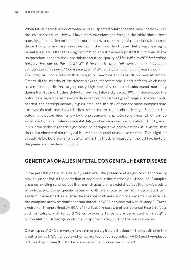
When future parents are confronted with a suspected fetal congenital heart defect within the severe spectrum, they will have many questions and fears. In the initial phase these questions focus often on the abnormal anatomy and the surgical procedures to correct those. Mortality risks are nowadays low in the majority of cases, but always leading to parental anxiety. After receiving information about the early postnatal outcome, follow up questions concern the uncertainty about the quality of life: Will our child be healthy, besides the scar on the chest? Will it be able to walk, talk, see, hear and function, comparable to its peers? Can it play sports? Will it be able to go to a normal school? The prognosis for a fetus with a congenital heart defect depends on several factors. First of all the severity of the defect plays an important role. Heart defects which need univentricular palliation surgery carry high mortality rates and subsequent morbidity during life. But most other defects have mortality risks below 10%. In these cases the outcome is largely determined by three factors: first is the type of surgical intervention(s) needed, the cardiopulmonary bypass time, and the risk of perioperative complications like hypoxia and thrombo-embolism, which can cause cerebral damage. Secondly, the outcome is determined largely by the presence of a genetic syndromes, which can be associated with neurodevelopmental delay and extracardiac malformations. Thirdly, even in children without genetic syndromes or perioperative complications, it is known that there is a chance of neurological injury and abnormal neurodevelopment. This might be already visible before or shortly after birth. This thesis is focused on the last two factors: the genes and the developing brain.
GENETIC ANOMALIES IN FETAL CONGENITAL HEART DISEASE
In the prenatal phase, on a case-by-case level, the presence of a syndromic abnormality may be suspected in the detection of additional malformations on ultrasound. Examples are a co-existing renal defect like renal dysplasia or a skeletal defect like hemivertebra or polydactyly. Some specific types of CHD are known to be highly associated with syndromic abnormalities, even in the absence of obvious additional defects. For instance, the complete atrioventricular septum defect (cAVSD) is associated with trisomy 21 (Down syndrome) in approximately 50% of the liveborn cases, and conotruncal heart defects such as tetralogy of Fallot (TOF) or truncus arteriosus are associated with 22q11.2 microdeletion (Di George syndrome) in approximately 50% of the liveborn cases.
Other types of CHD are more often seen as purely isolated lesions; in transposition of the great arteries (TGA) genetic syndromes are identified sporadically (<1%) and hypoplastic left heart syndrome (HLHS) there are genetic abnormalities in 5-12%.
158
9
GENERAL DISCUSSION
If there is a genetic syndrome, this often results in ND impairments, depending on the type and size of the genetic defect. In this thesis we investigated the frequency of submicroscopic genetic abnormalities in isolated CHD cases with a normal karyogram (in the absence large deletions/duplications and aneuploidies, such as trisomy 21), and we tried to specify which CHD are most affected by submicroscopic genetic abnormalities.
The data presented in this thesis indicate that clinically relevant genetic abnormalities can be found in all types of CHD using aCGH. New techniques, such as aCGH, offer the possibility to increase the diagnostic yield of genetic testing in CHD. The routine use of aCGH in the prenatal phase is leading to a paradigm shift, in which all types of CHD can be considered part of an associated genetic syndrome until proven otherwise. As a major benefit over karyotyping, the yield of aCGH expands beyond known genetic syndromes, and also includes susceptibility factors for ND delay or neurobehavioral disorders.
Our recommendation is to offer detailed genetic testing, like aCGH, in all types of fetal CHD, even if the lesion is apparently isolated based on prenatal imaging, and even when the type of defect is usually not associated with syndromes. The outcome of detailed genetic testing is essential when estimating the likely outcome with regard to surgical prognosis, ND functioning and co-existing morbidities. Parents should always be offered this option. Detailed genetic testing should be preceded by rapid aneuploidy detection (RAD) to rule out large chromosomal aberrations first, as the most frequent genetic syndromes associated with CHD are aneuploidies, such as trisomy 21. Performing RAD first can save time and reduce health care cost; when the results are abnormal, submicroscopic testing can be omitted. Parents should be counseled about the slight risk of amniocentesis (rupture of membranes, infection and/or miscarriage) versus the possible yield of genetic testing. With the advancing options of non-invasive prenatal testing (NIPT), in the future amniocentesis might even be unnecessary.
In the period around the publication of chapters 2,3 and 4, aCGH has indeed been increasingly used in cases of prenatal structural anomalies, and it became the new standard in many fetal medicine units. A drawback of aCGH is, however, that single-gene disorders such as CHARGE, Kabuki and Noonan – which are also associated with CHD – will not be detected. In the Heart And NeuroDevelopment study cohort, up to 2016, we found two cases with a postnatal diagnose of CHARGE syndrome (in 90 live born children with apparently isolated CHD) in the absence of any signs prenatally. This diagnosis can be made by whole exome sequencing (WES), which at this moment is still time consuming and costly. If future parents consider termination of pregnancy, the genetic work-up must be ready before 24 weeks of gestation, the legal limit to perform pregnancy
159
9
termination in the Netherlands. Turnover time of WES can be reduced to several days, yielding it possible to use in prenatal medicine. At this moment WES gradually finds its way into the daily practice of prenatal diagnosis. We recommend to reserve WES to cases in which there are suspicions there might be a single gene disorder, for instance by the combination of a CHD with other (subtle) ultrasound variations. WES is relatively new in prenatal practice and has not proven its value in large clinical trials yet.
The implementation of new techniques like aCGH and now WES in the prenatal setting has many advantages. The opportunity to identify syndromic cases prenatally, and therewith offering an estimation of ND functioning on the long term, greatly influences parental counselling and expectation management. However, implementation of these new techniques can also include some relevant downsides. New ethical dilemmas arise, for example around decisions to terminate a pregnancy or refusal for surgical repair. For some parents even a minimal genetic aberration might lead to the consideration of pregnancy termination, even when the phenotype is mild or uncertain. In other situations the refusal of genetic testing by parents, in cases that obviously appear to be syndromic with a dismal survival rate, might lead to the feeling of healthcare workers that some postnatal interventions might be disproportionate when the genetic syndrome is confirmed some weeks after birth. This can lead to conflicts between patients and healthcare workers, when patient demands collide with personal or institutional moral codes, financial constraints, cultural differences, and ideological divides. Guidance by an ethicist will increasingly be necessary when the advances of medical possibilities lead to an expansion of the morally grey areas.
Performing WES in a prenatal setting, under pressure of time, implies rapid and targeted exome analyses of panels of genes with known cardiac and/or syndromic involvement. In a short time a magnitude of data regarding genetic variations is produced which has to be categorised and valued. Comparable to the first experiences with aCGH in the prenatal setting, questions regarding the optimal WES platform and the method of categorization into (likely) benign and pathogenic remain important. It can be a challenging task for the clinical geneticist to decide which genetic variations to report. Genetic variants often display variable expressivity and genetic abnormalities might show decreased penetrance. Also, genetic syndromes have a wide spectrum of phenotypes varying from normal function to severely disabled. In the prenatal phase, the described phenotype is different and less complete than the postnatal phenotype because information about organ function, neurodevelopment and external features are missing. The clinical geneticist therefor does not have the complete picture yet to interpret the found genetic profile. The aforementioned insecurities and individual variations imply
160
9
GENERAL DISCUSSION
that prenatal detailed genetic analysis might not always render straightforward answers regarding the expected outcome, which results in challenges in subsequent parental counselling.
Another difficulty of the introduction of aCGH and WES is the additional yield of unexpected secondary findings unrelated to the initial phenotype (such as BRCA mutation or susceptibility factors for autism) and polymorphisms with unknown significance, which could lead to parental anxiety and ethical dilemmas. Regarding the first, in some cases it can be disputable whether the fetus/child will benefit on the long term from the knowledge of carrying certain genetic variations; knowing may cause emotional, psychological or social harm. With respect to the second aspect, the frequency and clinical ambiguity of polymorphisms with unknown significance is fortunately decreasing rapidly with advancing experience. In our opinion, fear of polymorphisms with unknown significance and unexpected secondary findings should not be a reason to refrain from genetic testing from a clinical perspective. Parents, however, must be informed about this possibility, as they should be able to make a well-informed choice about whether to perform genetic testing or not. Counselling by a geneticist is crucial to interpret and explain abnormal findings. Future research regarding WES findings in fetal CHD will have to lead to the development of gene panels with known cardiac and/or syndromic involvement, to avoid secondary findings.
DEVELOPMENT OF THE BRAIN IN FETUSES AND CHILDREN WITH ISOLATED SEVERE CONGENITAL HEART DISEASE
In the absence of genetic syndromes, isolated CHD also show ND delays on the long term. This is presumed to be the result of cerebral injury. Cerebral damage can be sustained in the obviously risky and possible unstable period before and around cardiothoracic surgical repair. As stated in the introduction of this thesis, results from postnatal research indicate that the brain might already be at risk in the prenatal phase in CHD. In this thesis we assessed fetuses with apparently isolated CHD for signs of impeding cerebral impairment in the prenatal phase and very shortly after birth.
We conclude that it is indisputable that fetuses with certain types of CHD display cerebral variations more frequently than healthy controls. In our systematic review (chapter 5) we found higher incidences of abnormal cerebral flow and smaller HC, but these data are biased towards left sided CHD. Our study of HC growth as a proxy for brain development in CHD (chapter 6) points towards an influence of specific types of
161
9
CHD – such as HLHS and TOF. The clinical relevance as well as a pathogenesis of these abnormalities remain to be determined, but there are several possible explanations. In this thesis we explored the possible pathogenic mechanism of altered hemodynamics, which is described hereafter. However, concomitant placental insufficiency seems to play an important role. An underlying common genetic pathway seems to be a logical explanation for the combination CHD, small fetal growth and cerebral variations.
As mentioned in the introduction of this thesis, a correlation between cerebral hemodynamics and fetal neurodevelopment in CHD has been suggested, referred to as the ‘altered hemodynamics theory’. It has been reasoned that the fetal brain in CHD suffers from a decreased flow to the carotid arteries (in cases of left ventricle obstruction with reversed aortic arch flow such as HLHS) and/or decreased oxygen delivery (in TGA, DORV and TOF). These phenomena might induce vasodilatation in the cerebral circulatory system, to facilitate transport of oxygen and nutrients. A lower middle cerebral artery (MCA) pulsatility index when compared to the umbilical artery (brainsparing) is a phenomenon observed in growth restricted fetuses. Some authors state that the same mechanism is present in CHD. This is not proven in an in-vivo setting, the cerebro-placental ratio (CPR) in blood flow has rarely been investigated in CHD, and data from the various studies as reported in chapter 5 are highly heterogeneous. There are reports of a lower MCA pulsatility index in left sided CHD, but this could also be attributed to a higher prevalence of growth restriction and placental insufficiency in CHD, which has not been clearly investigated yet. Brainsparing is correlated with worse ND outcome in growth restricted fetuses, but in CHD (irrespective of the presence or absence of growth restriction), a correlation of brainsparing with long-term outcome has not been established yet. On the contrary, it has even been suggested that it is associated with more favorable ND outcome in CHD. In a prospective cohort of 72 single ventricle CHD cases, a low MCA pulsatility index was associated with higher scores on psychomotor assessment of ND at 14 months of age1.There are limitations to this conclusion, considering the small sample size of this particular study.
To look for an effect of a theoretically decreased flow to the carotid arteries or a theoretical oxygen deficiency in the brain we investigated whether CHD clusters based on these aspects displayed variations in HC growth in the second or third trimester (chapter 6 ). We found no sign that cerebral oxygen deficiency or reduced cerebral flow results in a smaller HC or a deflecting HC growth.
Following the altered cerebral hemodynamics theory, some centers actively advocate therapies like maternal hyperoxygenation or use of erythropoietin. However, we believe
162
9
GENERAL DISCUSSION
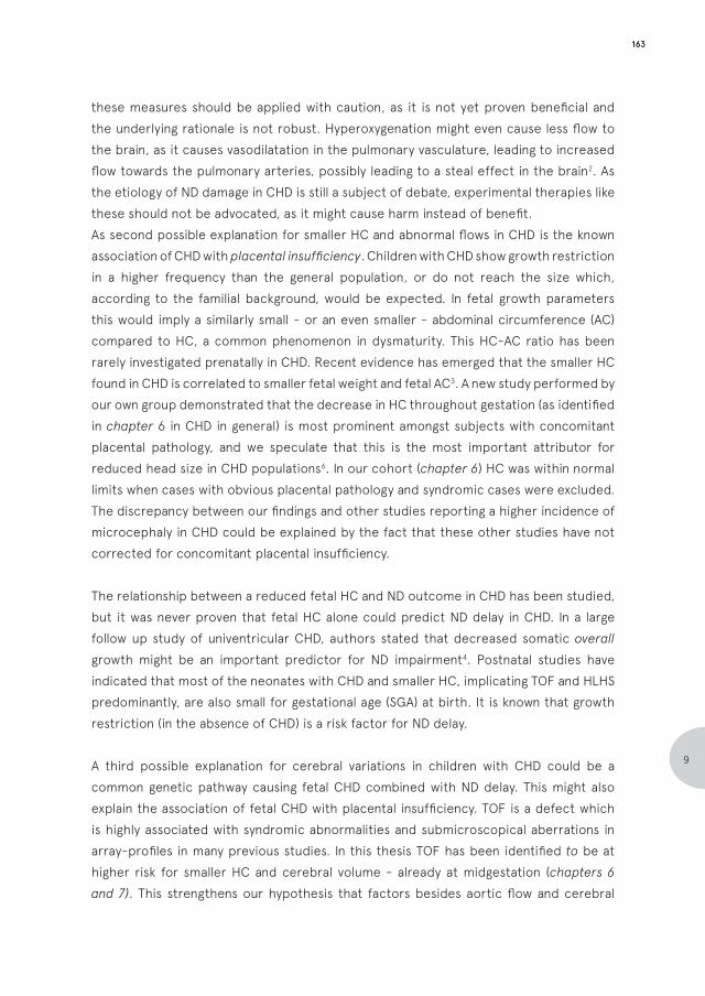
these measures should be applied with caution, as it is not yet proven beneficial and the underlying rationale is not robust. Hyperoxygenation might even cause less flow to the brain, as it causes vasodilatation in the pulmonary vasculature, leading to increased flow towards the pulmonary arteries, possibly leading to a steal effect in the brain2. As the etiology of ND damage in CHD is still a subject of debate, experimental therapies like these should not be advocated, as it might cause harm instead of benefit. As second possible explanation for smaller HC and abnormal flows in CHD is the known association of CHD with placental insufficiency. Children with CHD show growth restriction in a higher frequency than the general population, or do not reach the size which, according to the familial background, would be expected. In fetal growth parameters this would imply a similarly small - or an even smaller - abdominal circumference (AC) compared to HC, a common phenomenon in dysmaturity. This HC-AC ratio has been rarely investigated prenatally in CHD. Recent evidence has emerged that the smaller HC found in CHD is correlated to smaller fetal weight and fetal AC3. A new study performed by our own group demonstrated that the decrease in HC throughout gestation (as identified in chapter 6 in CHD in general) is most prominent amongst subjects with concomitant placental pathology, and we speculate that this is the most important attributor for reduced head size in CHD populations6. In our cohort (chapter 6) HC was within normal limits when cases with obvious placental pathology and syndromic cases were excluded. The discrepancy between our findings and other studies reporting a higher incidence of microcephaly in CHD could be explained by the fact that these other studies have not corrected for concomitant placental insufficiency.
The relationship between a reduced fetal HC and ND outcome in CHD has been studied, but it was never proven that fetal HC alone could predict ND delay in CHD. In a large follow up study of univentricular CHD, authors stated that decreased somatic overall growth might be an important predictor for ND impairment4. Postnatal studies have indicated that most of the neonates with CHD and smaller HC, implicating TOF and HLHS predominantly, are also small for gestational age (SGA) at birth. It is known that growth restriction (in the absence of CHD) is a risk factor for ND delay.
A third possible explanation for cerebral variations in children with CHD could be a common genetic pathway causing fetal CHD combined with ND delay. This might also explain the association of fetal CHD with placental insufficiency. TOF is a defect which is highly associated with syndromic abnormalities and submicroscopical aberrations in array-profiles in many previous studies. In this thesis TOF has been identified to be at higher risk for smaller HC and cerebral volume - already at midgestation (chapters 6 and 7). This strengthens our hypothesis that factors besides aortic flow and cerebral
163
9
oxygen saturation attribute to cerebral injury and ND delay. With the implementation of advanced techniques such as WES to perform a thorough genetic assessment in fetal CHD studies we will gradually gain more knowledge about the underlying genetic factors in CHD an concurrent ND delay.
The fact that fetuses and neonates with types of CHD that do not have a significant effect on fetal hemodynamics (minor CHD), also demonstrate cerebral anomalies and a smaller HC supports our theory of underlying common genetic pathways. In our cohort, we deliberately included minor CHD, such as ventricular septal defects, besides severe CHD. Our analyses support findings in a large Danish postnatal cohort5 and indicate that these minor CHD show abnormalities in HC growth as well. The meaning for long-term ND outcome is however unclear. The further development of the fetal brain in less severe CHD and the correlation to ND outcome unfortunately has not been reported separately.
Besides ultrasound data regarding HC and cerebral flows, there are surprisingly few studies performed with ultrasound to examine the developing brain in fetuses with CHD. Our cerebral volume study in chapter 7 included the assessment of the sylvian fissure. We found it to be more shallow in our CHD cohort. There are only a few comparable analyses in literature, which also implicate several gyri and sulci to be abnormal in CHD. Again, no relation to long-term ND outcome has been made yet, and the effect size of these findings (the extent of the actual delay of brain maturation) is unknown. Our HAND study group has yet to publish the results of neurosonographic assessment of cortical folding up to 36 weeks of gestation. It is important to already emphasize that we did not identify any significant cerebral lesions in the total prenatal cohort. We were therefore slightly taken aback that in our postnatal study (chapter 8) several significant cerebral lesions were identified by CUS in the CHD neonates. The timing and the origin of these lesions is debatable. Whether they were late prenatal or early postnatal cannot be deduced. The lesion types could implicate cerebral immaturity (prenatal origin) or hypoxic damage (presumably postnatal origin). Fetal ultrasound is less reliable in the late third trimester, as a result from the engaged head and acoustic shadowing. This might be the period in which fetal cerebral development, maturation and growth is affected in CHD.As reviewed in chapter 5, several prenatal MRI studies have been performed in CHD fetuses, up to the later third trimester as well, but we found no statistical proof that there is a higher prevalence of cerebral MRI abnormalities or cerebral maturation delay in CHD. Important to note is that prenatal MRI studies included in our review, as well as postnatal ultrasound studies, differ in their definition of cerebral abnormality. Mild physiological variants are incorporated in several prenatal and postnatal studies as abnormalities. In
164
9
GENERAL DISCUSSION
our postnatal ultrasound study in chapter 8 we did not consider these minor variants as clinically relevant. We therefore conclude that in a prenatally detected group of CHD of various types (severe and minor), cerebral abnormalities are much less frequent then suggested in earlier literature. Previous literature has (unintendedly) greatly exaggerated the impact of prenatal cerebral ‘damage’. This leads to much debate on how to address these issues in prenatal counseling, how to be honest and clear about the reported associations with ND delay, but avoiding disproportionate parental anxiety.
Whether MRI or ultrasound is the most sensitive modality to detect fetal brain abnormalities is disputable. Although MRI is capable of distinguishing white brain matter and abnormal gyration patterns, it has the disadvantage of high costs, less availability and large slice thickness. Just as in ultrasound, MRI is operator dependent. In experienced hands, ultrasound has also been proven capable to analyze cerebral maturation with high detail, and in our opinion it is not inferior to MRI. Reports of fetal neurosonography studies in fetal CHD are gradually emerging. Currently, the HAND-cohort is being analyzed further. Gyration patterns and the extent of the developmental delay are subjects under study. We are aiming to relate these data to the actual ND outcome on school age.
CONCLUSION
In short, in this thesis we found that clinically relevant genetic abnormalities can be detected in all types of CHD with aCGH. The yield of new genetic techniques expands beyond known genetic syndromes and can also include susceptibility factors for adverse ND outcome. With detailed prenatal genetic analyses we will increasingly be able to identify those at risk for ND delay. Second, certain types of CHD display fetal and early postnatal cerebral variations more frequently than healthy controls. We demonstrated that altered hemodynamics is unlikely to be the underlying pathogenic mechanism. Co-existing factors like genetic variations, placental insufficiency and growth restriction underly the vulnerability of the brain in CHD to be damaged, which ultimately may predispose for ND delay. Thus far there are no known ways to influence or treat these co-existing factors. We consider hypoxia after birth, in the first hours and days of life, as a far more plausible cause of the found pre-operative cerebral abnormalities. Prenatal detection of CHD will ensure adequate postnatal care, that reduces acute severe or chronic mild hypoxia after birth to a minimum, thus prenatal detection will have a positive effect of the postnatal (ND) outcome.
The data in this thesis underline the importance of fetal neurological evaluation in all
165
9

CHD cases. However, it is too early to conclude that variations revealed by prenatal neuroimaging involve a high risk of ND delay. Parental counselling regarding ND outcome should be tailored, focussed on the type of CHD that has been identified. Genetic analysis must be offered to all parents, which should be performed with the option to obtain the highest available resolution. To define the clinical implication of prenatal cerebral variations in CHD, prospective large and long-term cohort studies are required, combining pre- and postnatal data, genetic analysis, and correcting for confounding factors. This will only be possible in multicentre multinational studies, leading to large cohorts. Whether there will be a role for prenatal neuroprotective measures, such as maternal treatment with hyperoxygenation, erythropoietin, acetylsalicylic acid or allopurinol, or peripartum measures such as early term induction of labor or postponed cord clamping, remains speculation.
166
9
GENERAL DISCUSSION

REFERENCES
1. Williams, I.A., et al., The association of
fetal cerebrovascular resistance with early
neurodevelopment in single ventricle congenital
heart disease. Am. Heart J, 2013. 165(4): p. 544-
550.
2. Co-Vu, J., et al., Maternal hyperoxygenation: A
potential therapy for congenital heart disease in
the fetuses? A systematic review of the current
literature. Echocardiography, 2017. 34(12): p.
1822-1833.
3. Mebius, M.J., et al., Growth patterns and
cerebro-placental hemodynamics in fetuses
with congenital heart disease. Ultrasound
Obstet Gynecol, 2018. DOI 10.1002/uog.19102.
4. Hahn, E., et al., The association of fetal growth,
cerebral blood flow, and neurodevelopmental
outcome in single ventricle fetuses. Ultrasound
Obstet. Gynecol, 2016 Apr;47(4):460-5.
5. Matthiesen, N.B., et al., Congenital Heart
Defects and Indices of Fetal Cerebral Growth
in a Nationwide Cohort of 924,422 Liveborn
Infants. Circulation 2016;133:566-575.
6. A. van Nisselrooij, et al. OC13.07: *The impact of
additional anomalies on head growth in fetuses
with congenital heart defects. 15 October 2018.
https://doi.org/10.1002/uog.19295
167
9


CHAPTER 10
Summary
Nederlandse samenvatting
SUMMARY
Congenital heart defects (CHD) are associated with syndromal abnormalities and/or neurodevelopmental delay. The aim of this thesis was to analyze whether prenatally appearing isolated CHD are as a matter of fact really isolated, without postnatally apparent syndromes or neurological damage.
Firstly, in part I, this was done by assessing the additional value of a array comparative genomic hybridization (aCGH) and the potential value of whole exome sequencing (WES) in CHD. The main goal of these detailed genotyping modalities is to identify fetuses that are at high risk for a more severe phenotype, including long term neurological delay, additional abnormalities, other organ system involvement and dysmorphic features. This provides prognostic information for clinical outcome and could also inform the family about genetic reproductive risks.
In chapter 2, a systematic review and meta-analysis of the literature up to September 2014 on aCGH in fetal CHD is presented. aCGH yielded additional clinically valuable information in 7.0% of fetal CHD cases, even after karyotyping ànd 22q11 microdeletion analysis (with fluorescent in situ hybridization(FISH)) were normal. There were particularly more pathogenic copy number variants (9.3% CNV) when extracardiac defects were visible on prenatal ultrasound. This yield is lower when compared with published reports of aCGH in the postnatal setting of non-isolated CHD. When analyzing fetal CHD that appear isolated, an additional yield of aCGH of 3.4% CNV was found. There is no specific type of CHD associated with aCGH anomalies, as the reported types of CHD with anomalies are heterogeneous. Ventricular septal defects (VSDs) with extracardiac malformations, conotruncal malformations (tetralogy of Fallot (ToF), interrupted arch) and left ventricle outflow tract malformations are common in prenatal cases which yield pathogenic aCGH results. However, also transposition of the great arteries (TGA) and cardiac heterotaxy, which are generally not considered to be associated with genetic syndromes, were found to have pathogenic aCGH results.
We advise to perform aCGH as a replacement of FISH and karyotyping, since it has benefits over both. It should be preceded by rapid aneuploidy detection (RAD) - and only be performed if RAD is normal- to reduce healthcare utilization and costs.
In chapter 3 the additional value of prenatal aCGH was investigated in a subgroup of left sided CHD. Left sided CHD as a group - end stage developmental defects including hypoplastic left heart syndrome (HLHS), aortic stenosis and aortic arch hypoplasia -
170
10
SUMMARY
are generally considered nót to be associated with genetic syndromes, if they appear isolated on prenatal ultrasound. Therefore physicians may be reluctant to stress the need for fetal genetic sampling in prenatal counseling. Our study shows that in - apparently isolated - left sided CHD cases, in euploid fetuses (with normal karyotyping), aCGH can yield clinically significant abnormalities in 7%. 22q11 microdeletion is included in this yield. This risk of 7% appears to be unrelated to the presence of minor additional malformations such as enlarged nuchal translucency. With detail we elaborate on our abnormal findings, and the variants of unknown significance, and the way they may have affected the subsequent phenotype. Our data serve as guide in focused prenatal counselling when genetic analysis is offered in left sided CHDs.
Counselling parents whether or not to perform genetic testing in fetal CHD will always remain colored by the type of defect and the presences of additional anomalies. Our recommendation however is to offer aCGH in all types of fetal CHD, even if the lesion is apparently isolated based on prenatal imaging, and even when the type of defect is usually not associated with syndromes. The yield of array expands beyond known genetic syndromes, and also includes susceptibility factors for neurodevelopmental delay or behavioural disorders, for instance.
In chapter 4 an extraordinary case of a coinciding mitochondrial disease – identified with whole exome sequencing (WES) - in a fetus with a small VSD is described. A VSD is a frequently occurring, usually mild (minor) type of CHD. However, in this case, fetal ultrasound also revealed polyhydramnios, periventricular cysts and cerebral hypoplasia. The neonate was born in a poor condition and the child died as a result of a severe metabolic disease. Targeted WES using a mitochondrial gene panel proved its benefit by revealing mutations in the FBXL4 gene, causing recessive mitochondrial encephalomyopathy, which was the cause of death. Prenatal testing was successfully carried out in the subsequent pregnancy to rule out recurrence.
In part II, prenatal cerebral development in CHD is investigated. In chapter 5, a systematic review and meta-analysis of the literature up to November 2015 on this matter is presented, focusing on imaging of cerebral development in a very broad definition: fetal cerebral ultrasound and fetal brain magnetic resonance imaging (MRI). We conclude that the studies with abnormal cerebral findings mainly evolve around left sided CHD. The most studied ultrasound parameter is the middle cerebral artery (MCA; studied in 1412 fetuses), which demonstrates a slightly increased end-diastolic blood flow in CHD. This means that there might be cerebral dilatation of the blood vessels, increasing the volume of blood flowing to the brain. This effect appears to be predominantly present
171
10
in left sided CHD. The MCA-flow appears not to be altered in TGA or right sided CHD. In the ultrasonographic head growth studies (753 fetuses), combining all types of CHD, a smaller head circumference (HC) throughout gestation is encountered, but, again, severe CHD - and especially left sided CHD - are overrepresented in these data. In postnatal studies, small HC has also been identified in less severe CHD, such as VSDs, which was not confirmed in prenatal studies yet. We did not encounter any fetal ultrasound studies assessing detailed neurodevelopment, such as cortical thickness, cerebral maturation or spinal fluid amount, even though neonatal studies report delayed brain maturation and cerebral atrophy in newborns with CHD and dedicated neurosonography has the capacity to detect most of the reported anomalies.
Fetal brain development is increasingly being studied in CHD using fetal MRI (445 fetuses). Reports on this topic are emerging rapidly, but the analysis of fetal cerebral volume, metabolism and maturation appears to be limited to research settings and have not have not yet been implemented in daily clinical practice. A summarizing conclusion cannot be drawn, due to variations in methods of measurement and analysis. Another big issue is that the definition of (the pathogenicity of) MRI lesions varies between studies, resulting in an overestimation of the prevalence of brain anomalies. We present a table to demonstrate the variation in reported cerebral anomalies. The total prevalence of reported anomalies might be 18%, but the 95% confidence interval ranges from -6 to 42%. This means that there is high heterogeneity, much uncertainty and no statistical proof that there is a higher prevalence of cerebral MRI abnormalities in CHD. Fluid compartment (lateral ventricular) ‘anomalies’ do seem to be present in 4-19% of fetuses with CHD, but this includes asymmetrical lateral ventricles with normal sizes and mild dilation of the lateral ventricles, which can also be perceived as normal variants.
Eight studies (including 331 fetuses) correlated the fetal imaging modalities to postnatal neurodevelopmental outcome, but no solid conclusion can be drawn. Therefore, it is too early to conclude that aberrations revealed by neuroimaging, such as a higher MCA pulsatility index or a slightly smaller HC, involve a high risk of neurodevelopmental delay.In short, left sided CHD are associated with fetal cerebral abnormalities. In other CHD types, the groups are either too small or no significant difference with healthy controls is found.
Chapter 6 shows the results of a large retrospective analysis of HC growth in fetal CHD. We included the fetal biometrical data (1322 measurements) of 436 cases with a severe isolated CHD from 2002-2014. It has been hypothesized that in CHD altered hemodynamics (decreased cerebral oxygenation or flow) might be a cause of
172
10
SUMMARY
neurodevelopmental delay. Our aim was to evaluate the effect of cerebral oxygenation and aortic arch flow on HC growth. This study was the first to demonstrate fetal growth patterns in isolated CHD, showing that all fetuses with prenatally detected isolated CHD demonstrate a decline in HC growth with advancing GA, irrespective of aortic flow or oxygen saturation, irrespective of the severity of the CHD. Our study thus confirms the reported smaller HC at birth in infants with CHD, not only the severe types, but now also in minor CHD. The clinical relevance of this decrease is small, as the effect size compared to the HC growth charts of normal populations is small. Interestingly, fetuses with normal aortic flow and oxygen saturation (such as Ebstein’s anomaly, persistent left caval vein and VSDs) showed the same HC growth decline as fetuses with obstructed or reversed flow (such as HLHS) or low or intracardiac mixing (such as TGA and ToF). Our data also indicate that fetuses with ToF already demonstrate a smaller HC at 20 weeks of gestation.
We conclude that variations in cerebral oxygenation or flow, caused by the hemodynamic changes in certain CHD types, cannot be the only explanation of the deflecting HC growth in CHD. Other causes such as concomitant placental insufficiency have to considered. An underlying common genetic pathway seems to be a logical explanation.Chapters 7 and 8 describe analyses of the Heart and NeuroDevelopment (HAND) study, a prospective cohort of isolated CHD cases in which we performed extensive monthly neurosonography, compared to healthy controls. We aimed to avoid selection bias by including all cases with prenatally detected CHD when they appear isolated on fetal ultrasound. We deliberately chose to include minor, less severe types of CHD in this study as well, as previous studies have implied these to be at risk for deflecting HC and cerebral abnormalities as well. We started this HAND study cohort in 2013, and still have an ongoing inclusion up to date. In this thesis, we analyzed the data up to March 2017. We have added a group of healthy control fetuses, in which we performed the same measurements for comparison, between 2014 and 2016.
In chapter 7 the results of the volume measurements of the brain in the second and early third trimester of the HAND study are shown, exploring the prenatal cerebral growth and evolution of the extracerebral fluid compartments. 77 consecutive fetuses with CHD and 75 healthy fetuses were analyzed, with multiple measurements per subject. We performed reproduceable, detailed 3D measurements of the (hemi)cerebral volume and extracerebral fluid spaces. Our study shows no significant difference in volume, extracerebral fluid and volume growth between fetuses with isolated CHD and controls. Only fetuses with specific lesions such as ToF, DORV and HLHS might have a slightly smaller brain volume at 20 weeks of gestation, but the effect-size is very small and
173
10
wanes with advancing gestation. Again, our results point towards the direction of an influence of CHD type, and not of cerebral flow or oxygenation.
Our report also includes various extracerebral fluid-to-brain ratios and assessment of cerebral gyrification in the Sylvian fissure. Firstly, there appears to be a more rapid decrease in relative amount of extracerebral fluid in CHD fetuses, however this effect was not statistically significant. Secondly, the Sylvian fissure is significantly shallower in fetuses with CHD. Possibly these are early markers of delayed cerebral maturation, delayed cortical folding, or impending cerebral volume growth deflection, but the clinical significance of these findings has yet to be unveiled. They might also be associated to extracerebral fluid compartment differences.
In chapter 8 the results of early postnatal and preoperative cranial ultrasounds (CUS) are compared between 50 neonates of the HAND study cohort and 59 healthy controls. Significant CUS abnormalities, such as intraventricular hemorrhage (IVH) and periventricular leukomalacia (PVL), occurred only in CHD neonates (8%). The prevalence of significant CUS abnormalities in our study was much lower than previously reported, in mixed pre- and postnatally detected CHD groups (up to 59%). This is probably due to our strict definition of pathological findings, excluding minor abnormalities (such as spontaneously resolving small cysts or transient echogenic white matter) as significant. Also we avoided to include cases with possible pre-existing neurological injury, for instance due to hypoxia after birth in undetected cases, or in cases that underwent a balloon atrioseptostomy (BAS) directly after birth. We therefore state that prenatal detection of CHD might have a positive effect of the postnatal (neurodevelopmental) outcome. A separate analysis of 8 CHD neonates after endovascular interventions demonstrated that arterial stroke was indeed associated with BAS. As expected, neonates with CHD display slightly smaller head size and cerebral growth. The cerebrospinal fluid (CSF) measurements however were similar in cases and controls. This may be caused by our short interval between birth and CUS - within the first week of life -, thus preceding the effect of chronic mild hypoxia, which might cause cerebral atrophy and widened CSF spaces.
This thesis is finalized by chapter 9, which provides a general discussion with summarizing conclusions, interpretation of the results and directives for future research.
174
10
SUMMARY
NEDERLANDSE SAMENVATTING
Aangeboren hartafwijkingen als groep zijn de meest voorkomende aangeboren afwijking; ongeveer 0,8% van alle pasgeborenen heeft een hartafwijking. Ongeveer 75% van alle ernstige hartafwijkingen wordt prenataal opgespoord bij de 20 weken echo. Als toekomstige ouders worden geconfronteerd met het vermoeden op een ernstige aangeboren hartafwijking, zullen ze uiteraard veel vragen en angsten hebben.
Initieel gaan deze vragen voornamelijk over de chirurgische procedures om het hart te corrigeren en de bijbehorende kans op sterfte. Gelukkig zijn de sterftecijfers van kinderen met aangeboren hartafwijkingen de laatste jaren steeds verder dalende. Vervolgens vragen veel ouders zich derhalve af wat de kwaliteit van leven van hun kind zal zijn: zal ons kind gezond zijn, naast het litteken op de borst? Zal hij of zij kunnen lopen, praten, zien, horen en functioneren, vergelijkbaar met zijn of haar leeftijdsgenoten? Kan hij of zij sporten? Zal hij of zij naar een normale school kunnen gaan?
In de prenatale fase kan de prognose van een aangeboren hartafwijking ingeschat worden aan de hand van verschillende factoren. Allereerst speelt de ernst van het defect een belangrijke rol. Kinderen die slechts één functionele hartkamer over zullen houden (univentriculaire harten), hebben een hoog sterftecijfer en een grote kans op ernstige ziekte tijdens het leven. Maar de meeste andere aangeboren hartafwijkingen hebben een sterftecijfer van minder dan 10%. In deze gevallen wordt de lange termijn uitkomst grotendeels bepaald door drie factoren. Ten eerste speelt het type chirurgische ingreep dat nodig is een rol, inclusief de duur aan de hart-longmachine. Bij elke ingreep is er een bepaald risico op complicaties rondom de ingreep - zoals zuurstofgebrek en de vorming van bloedpropjes in de hersenen - die hersenbeschadigingen kunnen veroorzaken. Ten tweede wordt de uitkomst grotendeels bepaald door de aanwezigheid van genetische syndromen, die veelal gepaard gaan met neurologische ontwikkelingsachterstanden. Bekende syndromen die vaak samengaan met hartafwijkingen zijn het syndroom van Down (trisomie 21) en het Di-George syndroom (22q11 microdeletie). Ten derde is het, zelfs bij kinderen zonder genetische syndromen of complicaties rondom de operatie, bekend dat er een kans is op neurologische schade en afwijkende ontwikkeling. Dit kan al zichtbaar zijn vóór of kort na de geboorte.Dit proefschrift richt zich op de laatste twee factoren: de genetische afwijkingen en de zich ontwikkelende hersenen.
In het eerste deel van dit proefschrift wordt de potentiele additionele waarde van twee gedetailleerde genetische testen bij aangeboren hartafwijkingen onderzocht:
175
10
‘array comperative genomic hybridisation’ (aCGH) en ‘whole exome sequencing’ (WES). Het belangrijkste doel van deze tests is het vóór de geboorte reeds identificeren van kinderen die na de geboorte bijkomende problemen kunnen ontwikkelen, zoals ontwikkelingsachterstanden, bijkomende afwijkingen buiten het hart (andere orgaanbetrokkenheid) of een afwijkend uiterlijk. Dit biedt prognostische informatie voor zowel toekomstige ouders als zorgprofessionals om rekening mee te houden. In hoofdstuk 2 wordt een systematische review en meta-analyse over aCGH in de prenatale fase van aangeboren hartafwijkingen gepresenteerd. Uit de vergelijking van diverse artikelen kan samengevat worden dat aCGH klinisch waardevolle bijkomende informatie oplevert in 7.0% van de gevallen, zelfs nadat syndromen zoals Down en Di-George uitgesloten waren. Er waren met name meer aCGH afwijkingen (9,3%) wanneer naast het hart ook andere defecten zichtbaar waren op prenatale echografie. Ook als er naast het hart geen afwijkingen gezien werden, leverde aCGH in 3.4% van de gevallen een bijdrage. Er is geen specifiek type hartafwijking geassocieerd met afwijkende aCGH resultaten; eigenlijk bij alle typen werd wel een of meerdere keren een aCGH afwijking gevonden. Zelfs bij transpositie van de grote vaten (TGA) en hypoplastisch linkerhartsyndroom (HLHS), waar dit vaak niet bij verwacht wordt.
Wij adviseren om a CGH aan te bieden aan alle toekomstige ouders waarbij een hartafwijking bij de foetus vermoed wordt. De ‘oude’ genetische tests zoals FISH en karyotypering zouden hierdoor vervangen moeten worden. Om eerst grove chromosoomafwijkingen - zoals trisomie 21 - uit te sluiten, zou alvorens een aCGH verricht wordt ook een ‘rapid aneuploidy detection’ test gedaan kunnen worden, om tijd en kosten te besparen.In hoofdstuk 3 wordt de toegevoegde waarde van prenatale aCGH onderzocht in een subgroep - linkszijdige hartafwijkingen zoals HLHS en aortastenose. Linkszijdige hartafwijkingen worden in het algemeen niet geassocieerd met genetische syndromen. Daarom zijn artsen vaak terughoudend om prenatale genetische tests te adviseren. Onze studie laat echter zien dat in schijnbaar geïsoleerde linkszijdige hartafwijkingen, bij euploïde foetussen (met normale karyotypering), aCGH in 7% van de gevallen toch relevante afwijkingen kan opleveren (inclusief 22q11 microdeletie/Di-George syndroom). Dit risico van 7% lijkt niet gerelateerd te zijn aan de aanwezigheid van kleine extra echo-varianten, zoals een verdikte nekplooi (NT) of een navelstreng met een slagader tekort (SUA).
Overkoepelend kan gezegd worden dat informatie over de kans op genetische afwijkingen bij hartafwijkingen altijd gekleurd zal blijven door het type defect, en de eventuele aanwezigheid van bijkomende afwijkingen op een echo. Onze aanbeveling is echter om
176
10
NEDERLANDSE SAMENVATTING
aCGH aan te bieden in alle gevallen van aangeboren hartafwijkingen, zelfs als er naast de hartafwijking geen bijkomende afwijkingen gezien worden op echografie, en zelfs als het type defect meestal niet geassocieerd wordt met genetische syndromen (zoals TGA en HLHS). De klinische relevantie van aCGH gaat verder dan de ‘bekende’ genetische syndromen, en omvat ook gevoeligheidsfactoren (susceptibility loci) voor bijvoorbeeld ontwikkelingsachterstanden en gedragsproblematiek.
In hoofdstuk 4 wordt een bijzonder geval beschreven van een foetus met een hartafwijking (ventrikel septum defect; VSD), die tevens een mitochondriale ziekte bleek te hebben. Een VSD is een vaak voorkomende, mild type aangeboren hartafwijking. In dit geval was er met foetale echografie echter ook te veel vruchtwater te zien (polyhydramnion), alsmede afwijkingen in de hersenen (periventriculaire cysten en cerebellaire hypoplasie). De pasgeborene werd in een slechte conditie geboren en stierf als gevolg van een ernstige stofwisselingsziekte. Gerichte ‘whole exome sequencing’ (WES) - met behulp van een gericht mitochondrieel gen-panel - onthulde mutaties in het ‘FBXL4-gen’, die de letale recessieve mitochondriële encefalomyopathie veroorzaakten. In de volgende zwangerschap kon een gerichte genetische test worden uitgevoerd om herhaling in een vroeg stadium uit te sluiten.
In het tweede deel van dit proefschrift wordt de hersenontwikkeling van foetussen met een aangeboren hartafwijking onderzocht.Hoofdstuk 5 omhelst een systematische literatuurstudie en meta-analyse, gericht op beeldvorming van de ontwikkeling van de hersenen in een brede zin: foetale neurosonografie (echo) en magnetische resonantie (MRI). We concluderen dat met name ernstige, specifiek vooral linkszijdige, hartafwijkingen onderzocht zijn in deze studies. De meest bestudeerde echo-parameter is de doorbloeding van de arteria cerebri media (MCA; bestudeerd bij 1412 foetussen). Hieruit blijkt dat er bij hartafwijkingen vaker wordt gezien dat er een lage weerstand is in de cerebrale bloedvaten (cerebrale vasodilatatie), waardoor het volume van het bloed dat naar de hersenen stroomt mogelijk toeneemt. Dit effect lijkt voornamelijk aanwezig te zijn bij linkszijdige hartafwijkingen, en niet bij TGA of rechtszijdige hartafwijkingen zoals tetralogie van Fallot (TOF). In de echografische studies naar de hoofdomtrek (753 foetussen) wordt gezien dat bij aangeboren hartafwijkingen de prenatale hoofdomtrek gemiddeld kleiner is dan normaal. Opnieuw zijn ernstige hartafwijkingen - en vooral linkszijdige - oververtegenwoordigd in deze data. In postnatale studies werd gezien dat minder ernstige hartafwijkingen, zoals VSD’s, een kleinere hoofdomtrek bij geboorte hebben, maar dit is nog niet bevestigd in prenatale onderzoeken. Er werden geen prenatale echo-studies gevonden die gedetailleerde hersenrijping hebben bestudeerd, zoals corticale dikte, maturatie van de hersenplooien
177
10
of spinale vloeistofhoeveelheid. Foetale hersenontwikkeling wordt in toenemende mate wel nader bestudeerd met behulp van foetale MRI (445 foetussen). De analyse van foetaal cerebraal volume, metabolisme en rijping lijkt echter beperkt te zijn tot gespecialiseerde onderzoeksinstellingen en is nog niet doordrongen van de dagelijkse klinische praktijk. Een overkoepelende conclusie kan derhalve niet getrokken worden, vanwege grote variatie in onderzoeksmethoden. In de vergelijking van de MRI studies valt verder op dat ook de indeling van MRI-laesies flink verschilt tussen onderzoeken, en dat vele studies ook milde varianten meerekenen als afwijkend. Dit resulteert in een overschatting van het voorkomen van MRI afwijkingen. In totaal lijken foetale MRI afwijkingen gemiddeld bij 18% van de hartafwijkingen voor te komen, maar het 95% betrouwbaarheidsinterval van deze schatting varieert van -6 tot 42%. Dit betekent dat er een grote heterogeniteit, veel onzekerheid en geen statistisch bewijs is dat er überhaupt cerebrale MRI afwijkingen bij aangeboren hartafwijkingen voorkomen. Afwijkingen in de wijdte van de hersenvochtkamers (laterale ventrikel) lijken wel vaker voor te komen, bij 4-19% van de foetussen met hartafwijkingen, maar deze afwijkingen zouden grotendeels ook als normale varianten kunnen worden waargenomen in een gezonde groep.
Acht studies (331 foetussen) correleerden de foetale beeldvorming aan de postnatale neurologische uitkomst, maar hier kan geen solide conclusie uit worden getrokken. Er is onvoldoende bewijs om te concluderen dat variaties gezien op MRI of echografie, zoals een hogere MCA doorbloeding of een iets kleinere hoofdomtrek, een verhoogd risico op neurologische ontwikkelingsachterstand met zich meebrengen.
Hoofdstuk 6 toont de resultaten van een grote retrospectieve analyse van de groei van de hoofdomtrek bij foetale hartafwijkingen. Onze studie was de eerste die foetale groeipatronen - met herhaalde metingen - bij geïsoleerde hartafwijkingen bestudeerde. We includeerden de foetale biometrie (1322 metingen) tussen 20 en 40 weken zwangerschap, van 436 foetussen met een ernstige geïsoleerde hartafwijking. Ons doel was om het effect van cerebrale oxygenatie en doorstroming op de groei van het hoofd te evalueren door de verschillende typen hartafwijkingen te clusteren in groepen met vergelijkbare hemodynamiek. In eerdere literatuur wordt immers verondersteld dat door de veranderde anatomie (afwijkingen in de ‘loodgieterij’) er afwijkende hemodynamiek ontstaat (verminderde cerebrale oxygenatie of flow) bij hartafwijkingen. Dit zou een oorzaak zijn van neurologische afwijkingen. Wij toonden aan dat álle foetussen met prenataal gedetecteerde geïsoleerde hartafwijkingen een afbuiging van de hoofdomtrek groei vertonen naarmate de zwangerschap vordert; ongeacht de cerebrale doorbloeding of zuurstofsaturatie. Onze studie bevestigt hiermee de gerapporteerde kleinere hoofdomtrekken bij de geboorte bij kinderen met minder ernstige hartafwijkingen, zoals
178
10
NEDERLANDSE SAMENVATTING
VSDs. De afbuiging is maar minimaal in vergelijking met de normale populatie, maar wel degelijk significant aanwezig. Interessant is dat foetussen met een “normale” aorta flow en zuurstofsaturatie (zoals Ebstein’s anomalie, persisterende linker vena cava en VSD’s) dezelfde groei-afbuiging vertoonden als foetussen met een geobstrueerde aorta (zoals HLHS) of theoretisch lagere zuurstofspanning in de hersenen (zoals TGA en TOF). Onze gegevens wijzen er ook op dat elk type hartafwijking zijn eigen groeipatroon lijkt te hebben; zo hebben foetussen met TOF al een kleinere hoofdomtrek vroeg in de zwangerschap, bij 20 weken al. We concluderen dat de variaties in cerebrale oxygenatie of doorbloeding dus niet de (enige) verklaring kunnen zijn voor de afbuigende hoofdomtrek groei in deze groep foetussen.
Hoofdstukken 7 en 8 beschrijven twee analyses van de Heart and NeuroDevelopment (HAND) studie, een prospectief cohort met geïsoleerde aangeboren hartafwijkingen, ontdekt bij 20 weken zwangerschap, waarbij we uitgebreide maandelijkse neurosonografie uitvoeren. We zijn dit HAND cohort in 2013 gestart, en includeren nog steeds nieuwe gevallen. In dit proefschrift hebben we de gegevens tot maart 2017 geanalyseerd. We hebben tussen 2014 en 2016 een gezonde controlegroep toegevoegd ter vergelijking.
In hoofdstuk 7 worden hersenvolumes vergeleken, gemeten in het tweede en vroege derde trimester. De data van 77 opeenvolgende foetussen met een hartafwijking en 75 gezonde foetussen werden geanalyseerd, met meerdere metingen per persoon om de groei over tijd te evalueren. We voerden gedetailleerde 3D-metingen uit van het (hemi)cerebrale volume en de extracerebrale vloeistofruimten (liquor). Onze vergelijking toont geen significant verschil in hersenvolume, extracerebrale vloeistof ruimte of hersenvolumegroei. Alleen foetussen met specifieke typen hartafwijking (TOF, DORV en HLHS) lijken een iets kleiner hersenvolume te hebben bij 20 weken zwangerschap, maar dit trekt weer bij met vorderende zwangerschapsduur. Ook in deze studie wijzen de resultaten meer in de richting van een invloed van het specifieke type hartafwijking, en niet op veranderde cerebrale doorbloeding of zuurstofspanning, net als in hoofdstuk 6.
Dit hoofdstuk bevat ook verschillende analyses met betrekking tot de verhoudingen tussen de extracerebrale vloeistof en het hersenvolume, en beoordeling van hersenplooiing in de fissuur van Sylvius. Er lijkt een afname van de relatieve hoeveelheid liquor te ontstaan bij foetussen met een hartafwijking, maar dit effect is niet statistisch significant. Ten tweede is de fissuur van Sylvius minder diep bij foetussen met een hartafwijking. Mogelijk zijn dit beide vroege markers van vertraagde hersenrijping, vertraagde hersenplooiing (corticale gyrering) of dreigende afbuiging van volumegroei van de hersenen. De betekenis hiervan is nog niet helemaal duidelijk.
179
10
In hoofdstuk 8 worden de resultaten vergeleken van vroege postnatale en pre-operatieve craniale echografie (CUS), tussen 50 neonaten van het HAND-studiecohort en 59 gezonde controles. Ernstige CUS-afwijkingen, zoals bloedingen en witte stof schade, kwamen alleen voor bij 8% van de pasgeborenen met een hartafwijking. Dit percentage (8%) is echter veel lager dan eerder gerapporteerd (rapportages variëren van 15 tot 59%). Dit komt waarschijnlijk door onze strikte definitie van wat afwijkend genoemd wordt, waarbij we de fysiologische varianten en afwijkingen die spontaan weer verdwenen hebben geëxcludeerd. Ook hebben we kinderen niet meegenomen in de analyse als zij mogelijk reeds bestaand neurologisch letsel hadden, bijvoorbeeld als gevolg van acuut zuurstofgebrek na de geboorte (als de hartafwijking nog niet ontdekt was), of kinderen die direct na de geboorte een katheter-interventie (zoals de Rashkind procedure) ondergingen. Wij denken daarom dat prenatale detectie van hartafwijkingen een positief effect kan hebben op de postnatale (neurologische) uitkomst. Een afzonderlijke analyse van 8 kinderen die een katheter-interventie ondergingen toonde aan dat 2 gevallen een herseninfarct hadden gekregen hierna; in de andere groep kwamen herseninfarcten niet voor. Er werden nog een aantal aanvullende analyses verricht: zoals verwacht hebben pasgeborenen met een hartafwijking een iets kleinere hoofdomtrek en hersenvolume. De metingen van de cerebrospinale vloeistof (liquor ruimten) waren echter hetzelfde in beide groepen. Waarschijnlijk komt dit omdat we de CUS vroeg na de geboorte hebben gemaakt; op dat moment heeft chronisch mild zuurstofgebrek nog geen kans gekregen om hersenatrofie (en daarmee verwijde liquor ruimten) te veroorzaken. Wij denken dat dit laatste een van de belangrijkste verklaringen is dat wij een lager percentage hersenafwijkingen hebben gevonden dan eerdere literatuur.
Dit proefschrift wordt afgesloten door hoofdstuk 9, waarin de resultaten in het algemeen worden bediscussieerd, samenvattende conclusies worden getrokken, en aanbevelingen gedaan worden voor toekomstig onderzoek. Er worden adviezen gegeven hoe in de prenatale setting ‘in de spreekkamer’ om te gaan met de relatie tussen hersenafwijkingen en hartafwijkingen. Als onderliggende medeverklaring voor de neurologische variaties bij kinderen met hartafwijkingen wordt ingegaan op mogelijke placentaire afwijkingen en/of gezamenlijke genetische variaties, die zowel een hartafwijking, een groeivertraging, als neurologische schade zouden kunnen veroorzaken. Er wordt gespeculeerd over de aanvullende waarde van ‘whole exome sequencing’ in grote groepen hartafwijkingen, en de mogelijke bijkomende ethische dilemma’s alsmede de uitdagingen bij de interpretatie van de resultaten. Afgesloten wordt met de statement dat het nog te vroeg is om ‘hersenbeschermende maatregelen’ (zoals zuurstoftoediening of bepaalde medicatie aan toekomstige moeders) door te voeren in de dagelijkse praktijk.
180
10
NEDERLANDSE SAMENVATTING

181
10


CHAPTER 11
Appendices
Abbreviations
Author affiliations
List of publications
Information on the author
Acknowledgements
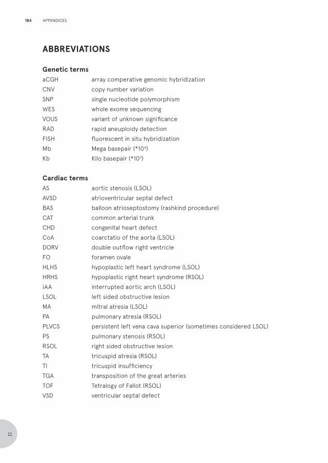
ABBREVIATIONS
Genetic termsaCGH array comperative genomic hybridizationCNV copy number variationSNP single nucleotide polymorphismWES whole exome sequencingVOUS variant of unknown significanceRAD rapid aneuploidy detectionFISH fluorescent in situ hybridizationMb Mega basepair (*106)Kb Kilo basepair (*103)
Cardiac termsAS aortic stenosis (LSOL)AVSD atrioventricular septal defectBAS balloon atrioseptostomy (rashkind procedure)CAT common arterial trunkCHD congenital heart defectCoA coarctatio of the aorta (LSOL)DORV double outflow right ventricleFO foramen ovaleHLHS hypoplastic left heart syndrome (LSOL)HRHS hypoplastic right heart syndrome (RSOL)iAA interrupted aortic arch (LSOL)LSOL left sided obstructive lesionMA mitral atresia (LSOL)PA pulmonary atresia (RSOL)PLVCS persistent left vena cava superior (sometimes considered LSOL)PS pulmonary stenosis (RSOL)RSOL right sided obstructive lesionTA tricuspid atresia (RSOL)TI tricuspid insufficiencyTGA transposition of the great arteriesTOF Tetralogy of Fallot (RSOL)VSD ventricular septal defect
184
11
APPENDICES
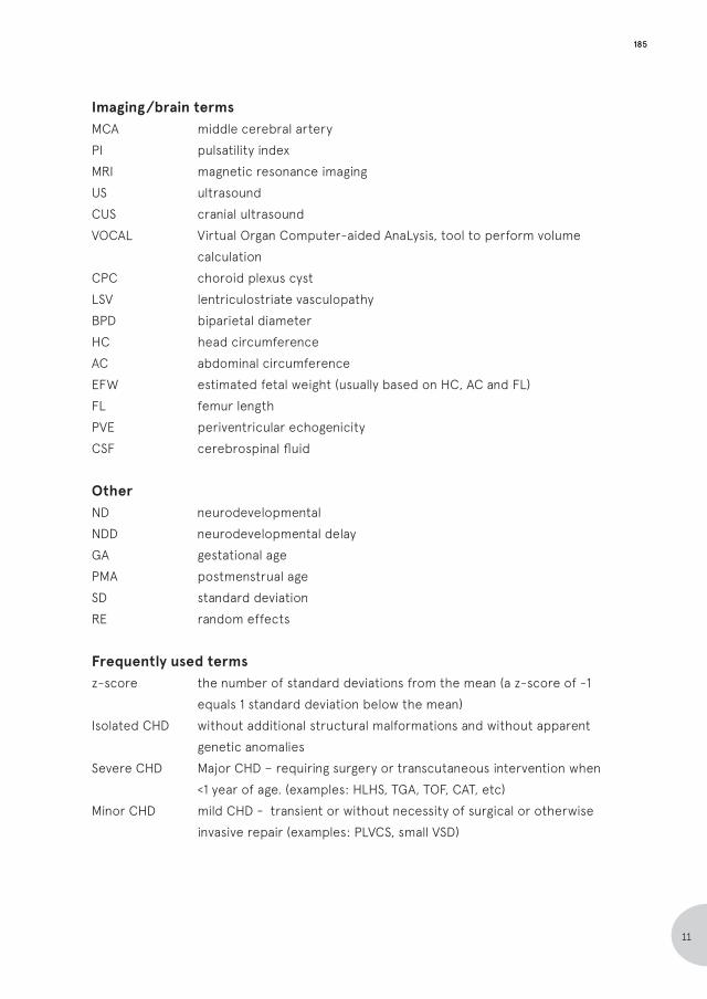
Imaging/brain termsMCA middle cerebral arteryPI pulsatility indexMRI magnetic resonance imagingUS ultrasoundCUS cranial ultrasoundVOCAL Virtual Organ Computer-aided AnaLysis, tool to perform volume calculationCPC choroid plexus cystLSV lentriculostriate vasculopathyBPD biparietal diameterHC head circumferenceAC abdominal circumferenceEFW estimated fetal weight (usually based on HC, AC and FL)FL femur lengthPVE periventricular echogenicityCSF cerebrospinal fluid
OtherND neurodevelopmentalNDD neurodevelopmental delayGA gestational agePMA postmenstrual ageSD standard deviationRE random effects
Frequently used termsz-score the number of standard deviations from the mean (a z-score of -1 equals 1 standard deviation below the mean)Isolated CHD without additional structural malformations and without apparent genetic anomaliesSevere CHD Major CHD – requiring surgery or transcutaneous intervention when <1 year of age. (examples: HLHS, TGA, TOF, CAT, etc)Minor CHD mild CHD - transient or without necessity of surgical or otherwise invasive repair (examples: PLVCS, small VSD)
185
11
AUTHOR AFFILIATIONS
Jan M.M. van Lith, Monique C. Haak, Sheila M.P. Everwijn, A.K.K. (Katinka) Teunissen - Leiden University Medical Center, Department of Obstetrics and Fetal Medicine, Leiden, the Netherlands
Sylke J. Steggerda - Leiden University Medical Center, Department of Neonatology of the Willem Alexander Children’s Hospital, Leiden, the Netherlands
Nico A. Blom, Marry E.B. Rijlaarsdam, Lieke Rozendaal, Derk-Jan Ten Harkel - Leiden University Medical Center, Department of Pediatric Cardiology of the Willem Alexander Children’s Hospital, Leiden, the Netherlands
Nico A. Blom, Sally-Ann B. Clur - Amsterdam UMC: Academic Medical Center, Department of Pediatric Cardiology of the Emma Children’s Hospital, Amsterdam, the Netherlands
Cacha M.P.C.D. Peeters-Scholte - Leiden University Medical Center, Department of Pediatric Neurology of the Willem Alexander Children’s Hospital, Leiden, the Netherlands
Eva Pajkrt - Amsterdam UMC: Academic Medical Center, Department of Obstetrics, Amsterdam, the Netherlands
Marion A. de Boer, Christine L. van Velzen, Caroline L. Bax - Amsterdam UMC: VU Medical Center, Department of Obstetrics, Amsterdam, the Netherlands
Robert Scheepjens, Theo Stijnen, Erik W. van Zwet - Leiden University Medical Center, Department of Medical Statistics, Leiden, the Netherlands
Yair J. Blumenfeld - Stanford University School of Medicine, Department of Obstetrics & Gynecology, Stanford CA, USA
Allan Fisher - Elliot Health System, Manchester, New Hampshire, USA
Anthony O. Odibo - University of South Florida, Department of Obstetrics & Gynecology, Tampa FL, USA
Antoni Borrell - University of Barcelona Medical School, Department of Maternal-Fetal Medicine, Catalonia, Spain.
186
11
APPENDICES

Mariette J.V. Hoffer, Maartje C. van Rij, Yvonne Hilhorst-Hofstee - Leiden University Medical Center, Department of Clinical Genetics, Leiden, the Netherlands
Shama L. Bhola – Amsterdam UMC: VU University Medical Center, Department of Clinical Genetics, Amsterdam, the Netherlands
Alida C. Knegt, Jan Maarten Cobben – Amsterdam UMC: Academic Medical Center, Department of Clinical Genetics, Amsterdam, the Netherlands.
187
11
LIST OF PUBLICATIONS
Van Nisselrooij AEL, Jansen FAR, van Geloven N, Linskens IH, Pajkrt E, Clur, SA, Rammelo LA, Rozendaal L, Lith JMM van, Blom NA, Haak MC. The role of additional pathology on head growth patterns in fetuses with congenital heart defects. Ultrasound Obstet Gynecol. 2019 Mar 13. Doi: 10.1002/uog.20260.
Jansen FAR, Zwet EW van, Everwijn SMP, Teunissen AKK, Rozendaal L, Lith JMM van, Blom NA, Haak MC. Fetuses with isolated congenital heart defects show normal cerebral and extracerebral fluid volume growth: a 3D sonographic study in the second and third trimester. Fetal Diagn. Ther. 2019 Jan 17:1-9.
Jansen FAR, Blom NA, Haak MC. Re: Prevalence of prenatal brain abnormalities in fetuses with congenital heart disease: a systematic review. Ultrasound Obstet Gynecol. 2016 Oct;48(4):538-539.
Jansen FAR, van Zwet EW, Rijlaarsdam ME, Pajkrt E, van Velzen CL, Zuurveen HR, Kragt A, Bax CL, Clur SA, van Lith JM, Blom NA, Haak MC. Head growth in fetuses with isolated congenital heart defects: lack of influence of aortic arch flow and ascending aorta oxygen saturation. Ultrasound Obstet Gynecol 2016; 48: 357–364.
Jansen FAR/Everwijn SMP, Scheepjens R, Stijnen T, Peeters-Scholte CM, van Lith JM, Haak MC. Fetal brain imaging in isolated congenital heart defects - a systematic review and meta-analysis. Prenatal Diagnosis 2016, 36, 601–613
Jansen FAR, Hoffer MJ, Velzen CL van, Klingeman Plati S, Rijlaarsdam ME, Clur SA, Blom NA, Pajkrt E, Bhola SL, Knegt AC, Boer MA de, Haak MC. Chromosomal abnormalities and copy number variations in fetal left sided congenital heart defect. Prenatal Diagnosis 2016, 36, 177–185
Jansen FAR/Rij MC van, Hellebrekers DMEI, Onkenhout W, Smeets HJM, Hendrickx AT, Gottschalk RWH, Steggerda SJ, Peeters-Scholte CMPCD, Haak MC, Hilhorst-Hofstee Y. Polyhydramnios and cerebellar atrophy: a prenatal presentation of mitochondrial encephalomyopathy caused by mutations in the FBXL4 gene. Clinical Case Reports 2016; 4(4): 425–428.
188
11
APPENDICES
Grande M, Jansen FAR, Blumenfeld YJ, Fisher A, Odibo AO, Haak MC, Borrell A.Genomic microarray in fetuses with increased nuchal translucency and normal karyotype - a systematic review and meta-analysis.Ultrasound Obstet Gynecol. 2015; 46(6):650-8.
Jansen FAR, Blumenfeld YJ, Fisher A, Cobben JM, Odibo AO, Borrell A, Haak MC. Array comparative genomic hybridization and fetal congenital heart defects: a systematic review and meta-analysis. Ultrasound Obstet Gynecol 2015; 45(1): 27–35.
Jansen FAR/Calkoen EE, Jongbloed MR, Bartelings MM, Haak MC. Imaging the first trimester heart: ultrasound correlation with morphology. Cardiol Young. 2014 Oct;24 Suppl 2:3-12.
van Teeffelen AS, van der Ham DP, Willekes C, Al Nasiry S, Nijhuis JG, van Kuijk S, Schuyt E, Mulder TL, Franssen MT, Oepkes D, Jansen FAR, Woiski MD, Bekker MN, Bax CJ, Porath MM, de Laat MW, Mol BW, Pajkrt E. Midtrimester preterm prelabour rupture of membranes: expectant management or amnioinfusion for improving perinatal outcomes (PPROMEXIL - III trial). BMC Pregnancy Childbirth. 2014 Apr 4;14:128.
FAR Jansen, E van der Linden, A Weir,, ThC de Winter. Veneuze malformatie in de recessus suprapatellaris bij een 14-jarige rugby-speelster. Sport en Geneeskunde 2011:may(2);22-27.
189
11
CURRICULUM VITAE
Fenna Arina Roelien Kap-Jansen is geboren op 28 juli 1984 te ‘s-Gravenhage (Bronovo ziekenhuis). Na de basis Montessorischool en het Gymnasium Haganum (1996-2002) is zij in 2003 gestart met haar Geneeskunde opleiding in het LUMC. Zij onderbrak haar studie in 2007-2008 om de functie van student-adviseur van de decaan in het LUMC te vervullen. Tijdens de coschappen (2009-2011) verrichte zij samen met de co-raad enkele onderzoeken naar de ontwikkelende keuze van vervolgspecialisatie onder co-assistenten. Tijdens haar wetenschapsstage deed zij kwalitatief onderzoek bij de afdeling Ethiek en Recht van het LUMC naar overwegingen bij starten en stoppen van nierdialyse. In 2010 startte zij als semi-arts in het HAGA ziekenhuis te ’s-Gravenhage op de afdeling verloskunde en gynaecologie, waar zij aansluitend als ANIOS werd aangenomen. Hierna werkte zij van februari 2012 tot juli 2016 in het LUMC op de afdeling prenatale diagnostiek en foetale therapie als arts-echoscopist. In deze tijd heeft zij het klinische deel van haar promotieonderzoek verricht; het verzamelen van de gegevens van de studie- en controle populatie van de Heart And NeuroDevelopment (HAND) studie.
In juli 2016 startte zij met de specialistenopleiding gynaecologie en verloskunde in het Reinier de Graaff Gasthuis te Delft (opleiders H. Bremer en K. Kapiteijn). Inmiddels is zij bezig met haar academische stage in het LUMC (opleider J. van Lith).
Fenna is in augustus 2016 op Ameland getrouwd met Geert Jan, zij wonen in Scheveningen met hun zoon Abel.
190
11
APPENDICES

DANKWOORD
Allereerst veel dank aan de vrouwen die het geduld opbrachten om de uitgebreide hersen-echo’s te ondergaan, terwijl zij een emotionele zwangerschap doormaakten en een potentieel zeer ziek kind droegen. Jullie veerkracht heeft immense indruk gemaakt.
Daarnaast wil ik alle deelneemsters van de controlegroep bedanken, die uren onder de echo-kop lagen. En met name ook hun partners, die direct postpartum met hun telg naar het ziekenhuis togen! Held(inn)en! (Dries!)
Jan en Nico, promotores, rotsen in de branding. Bakens van positiviteit en stimulans. Dank voor het geloof en de geruststellende woorden.
Monique. Scherp van tong maar zacht van hart. Jouw toewijding is een inspiratie. En al zijn er belangrijkere dingen in dit leven te verzinnen dan promoveren, ik ben blij dat je me altijd net even verder pushte dan ik zelf had bedacht.
Sylke, je geduld en je positieve, constructieve, nuancerende kritieken zijn zeer waardevol.
Alle mede-auteurs, veel dank voor jullie waardevolle bijdrage.
Kitty, dank voor je relativering, je steun, je begrip – ook voor zaken die niets met opleiding of promoveren te maken hadden, zoals het maken van het perfecte roodharige kind.
Mieke, mijn zuster, openhartige vriendin, harde werker. Dank voor je positieve inspiratie!
Sheila en Tess. Maatjes. Dank voor de gezelligheid, het schaamteloos klagen, en het delen van wel en wee.
Alle andere (oud)onderzoekers: Jolijn, Inge, Christine, Dian, Carolien, Ilona, Manon, Sanne, Amber, Vivian, Ellen en de hele Kaprun-gang. Dank voor de hulp, tips, koffietjes, Almdudler, kroketten, en luisterende oren.
Topstudenten Hannah, Amy en Miriam.
Opleidingsmaten, met name ook Marlies, Linda van Wijk, Annemijn, Sabine, en bovengenoemden.
191
11

Phebe, Katinka, Esther en alle andere toppers op de B3 van ‘mijn’ tijd; Frans, Annemieke, Dick, lieve Inge, de Mariekes en Marije.
Dana. Jouw familie leren kennen was zowel hartverwarmend als hartbrekend. Dank voor je openhartigheid.
Allerliefste Haagse homies, Mama Appelsap, Bente en Sarah.
Geert Jan. Het spijt me dat ik alleen maar Jansen heet in dit boek, en ik beloof plechtig nooit meer een boek te schrijven waar geen Kap op staat. Dank voor alles wat je doet, voor ons, voor mij, elke dag. Dank voor je liefde, je steun, je vertrouwen, je rust. Dank dat je me het mooiste gaf wat er bestaat.
Abel, onze moleculen werden de jouwe. Je zorgde voor nóg meer vertraging bij het laatste hoofdstuk, maar deze vertraging staat in het niet bij de liefde, de vreugde en het geluk dat jij aan mijn leven toevoegt.
192
11
APPENDICES

193
11

Related Documents