Tryptophan-47 in the active site of Methylophaga sp. strain SK1 flavin-monooxygenase is important for hydride transfer Andre Han a , Reeder M. Robinson a , Somayesadat Badieyan a , Jacob Ellerbrock a , Pablo Sobrado a,b,c,⇑ a Department of Biochemistry, Virginia Tech, Blacksburg, VA 24061, USA b Virginia Tech Center for Drug Discovery, Virginia Tech, Blacksburg, VA 24061, USA c Fralin Life Science Institute, Virginia Tech, Blacksburg, VA 24061, USA article info Article history: Received 31 October 2012 and in revised form 31 December 2012 Available online 25 January 2013 Keywords: Flavin monooxygenases Active site residue Flavin Hydride transfer Kinetic isotope effects abstract Flavin-dependent monooxygenase (FMO) from Methylophaga sp. strain SK1 catalyzes the NADPH- and oxygen-dependent hydroxylation of a number of xenobiotics. Reduction of the flavin cofactor by NADPH is required for activation of molecular oxygen. The role of a conserved tryptophan at position 47 was probed by site-directed mutagenesis. FMOW47A resulted in an insoluble inactive protein; in contrast, FMOW47F was soluble and active. The spectrum of the flavin in the mutant enzyme was redshifted, indi- cating a change in the flavin environment. The k cat values for NADPH, trimethylamine, and methimazole, decreased 5-8-fold. Primary kinetic isotope effect values were higher, indicating that hydride transfer is more rate-limiting in the mutant enzyme. This is supported by a decrease in the rate constant for flavin reduction and in the solvent kinetic isotope effect values. Results from molecular dynamics simulations show reduced flexibility in active site residues and, in particular, the nicotinamide moiety of NADP + in FMOW47F. This was supported by thermal denaturation experiments. Together, the data suggests that W47 plays a role in maintaining the overall protein flexibility that is required for conformational changes important in hydride transfer. Published by Elsevier Inc. Flavin-dependent monooxygenase from Methylophaga sp. strain SK1 (FMO) catalyzes the NADPH- and oxygen-dependent hydroxyl- ation of several xenobiotics (Scheme 1) [1]. The reaction mecha- nism can be divided into oxidative and reductive half-reactions. In the reductive half-reaction, FMO reacts with NADPH leading to the formation of reduced flavin and NADP + (Scheme 1a and b). In the oxidative half-reaction, the reduced flavin reacts with molecu- lar oxygen to form the C4a-hydroperoxyflavin, which is the hydroxylating species (Scheme 1c) [1,2]. In the presence of a sub- strate containing a soft-nucleophilic heteroatom, the enzyme is capable of transferring a single oxygen atom to the nucleophilic site, thereby oxygenating the substrate (Scheme 1d) [3]. Site-directed mutagenesis and structural studies have shown that the C4a-hydroperoxyflavin is stabilized by NADP + , which remains bound throughout the catalytic cycle [4–7]. If this intermediate is not stabilized it decays to hydrogen peroxide and oxidized flavin. Thus, stabilization of this intermediate is essential for preventing uncoupling of the reaction. It has been shown that interactions of several residues with NADP + are important for placing the coenzyme in a position optimal for intermediate stabilization [7]. The active site of FMO contains a conserved tryptophan residue that is in a site opposite of the NADP(H) and xenobiotic binding sites [6]. This residue is con- served in all known FMO sequences and in other related Class B flavin-monooxygenases (Fig. 1). The work presented here de- scribes the biochemical characterization and analysis of the ef- fect of mutating W47 to probe the role of this residue in catalysis. Materials and methods Materials Methimazole, glutathione, and trimethylamine hydrochloride were obtained from Sigma–Aldrich. pVP55A was obtained from the Center for Eukaryotic Structural Genomics, University of Wis- consin, Madison [8]. Escherichia coli BL21-TI R cells were from Invit- rogen. AccuPrime Pfx DNA Polymerase was from Invitrogen (Carlsbad, CA). The oligonucleotide primers used in the mutagene- sis reactions were obtained from Integrated DNA Technology. Nucleotide sequencing was performed at the Virginia Bioinformat- ics Institute DNA Sequencing facility. 0003-9861/$ - see front matter Published by Elsevier Inc. http://dx.doi.org/10.1016/j.abb.2013.01.004 ⇑ Corresponding author at: Department of Biochemistry, Virginia Tech, Blacks- burg, VA 24061, USA. Fax: +1 540 231 9070. E-mail address: [email protected] (P. Sobrado). Archives of Biochemistry and Biophysics 532 (2013) 46–53 Contents lists available at SciVerse ScienceDirect Archives of Biochemistry and Biophysics journal homepage: www.elsevier.com/locate/yabbi

Welcome message from author
This document is posted to help you gain knowledge. Please leave a comment to let me know what you think about it! Share it to your friends and learn new things together.
Transcript

Archives of Biochemistry and Biophysics 532 (2013) 46–53
Contents lists available at SciVerse ScienceDirect
Archives of Biochemistry and Biophysics
journal homepage: www.elsevier .com/ locate /yabbi
Tryptophan-47 in the active site of Methylophaga sp. strain SK1flavin-monooxygenase is important for hydride transfer
Andre Han a, Reeder M. Robinson a, Somayesadat Badieyan a, Jacob Ellerbrock a, Pablo Sobrado a,b,c,⇑a Department of Biochemistry, Virginia Tech, Blacksburg, VA 24061, USAb Virginia Tech Center for Drug Discovery, Virginia Tech, Blacksburg, VA 24061, USAc Fralin Life Science Institute, Virginia Tech, Blacksburg, VA 24061, USA
a r t i c l e i n f o
Article history:Received 31 October 2012and in revised form 31 December 2012Available online 25 January 2013
Keywords:Flavin monooxygenasesActive site residueFlavinHydride transferKinetic isotope effects
0003-9861/$ - see front matter Published by Elsevierhttp://dx.doi.org/10.1016/j.abb.2013.01.004
⇑ Corresponding author at: Department of Biochemburg, VA 24061, USA. Fax: +1 540 231 9070.
E-mail address: [email protected] (P. Sobrado).
a b s t r a c t
Flavin-dependent monooxygenase (FMO) from Methylophaga sp. strain SK1 catalyzes the NADPH- andoxygen-dependent hydroxylation of a number of xenobiotics. Reduction of the flavin cofactor by NADPHis required for activation of molecular oxygen. The role of a conserved tryptophan at position 47 wasprobed by site-directed mutagenesis. FMOW47A resulted in an insoluble inactive protein; in contrast,FMOW47F was soluble and active. The spectrum of the flavin in the mutant enzyme was redshifted, indi-cating a change in the flavin environment. The kcat values for NADPH, trimethylamine, and methimazole,decreased 5-8-fold. Primary kinetic isotope effect values were higher, indicating that hydride transfer ismore rate-limiting in the mutant enzyme. This is supported by a decrease in the rate constant for flavinreduction and in the solvent kinetic isotope effect values. Results from molecular dynamics simulationsshow reduced flexibility in active site residues and, in particular, the nicotinamide moiety of NADP+ inFMOW47F. This was supported by thermal denaturation experiments. Together, the data suggests thatW47 plays a role in maintaining the overall protein flexibility that is required for conformational changesimportant in hydride transfer.
Published by Elsevier Inc.
Flavin-dependent monooxygenase from Methylophaga sp. strainSK1 (FMO) catalyzes the NADPH- and oxygen-dependent hydroxyl-ation of several xenobiotics (Scheme 1) [1]. The reaction mecha-nism can be divided into oxidative and reductive half-reactions.In the reductive half-reaction, FMO reacts with NADPH leading tothe formation of reduced flavin and NADP+ (Scheme 1a and b). Inthe oxidative half-reaction, the reduced flavin reacts with molecu-lar oxygen to form the C4a-hydroperoxyflavin, which is thehydroxylating species (Scheme 1c) [1,2]. In the presence of a sub-strate containing a soft-nucleophilic heteroatom, the enzyme iscapable of transferring a single oxygen atom to the nucleophilicsite, thereby oxygenating the substrate (Scheme 1d) [3].
Site-directed mutagenesis and structural studies have shownthat the C4a-hydroperoxyflavin is stabilized by NADP+, whichremains bound throughout the catalytic cycle [4–7]. If thisintermediate is not stabilized it decays to hydrogen peroxideand oxidized flavin. Thus, stabilization of this intermediate isessential for preventing uncoupling of the reaction. It has beenshown that interactions of several residues with NADP+ are
Inc.
istry, Virginia Tech, Blacks-
important for placing the coenzyme in a position optimal forintermediate stabilization [7]. The active site of FMO contains aconserved tryptophan residue that is in a site opposite of theNADP(H) and xenobiotic binding sites [6]. This residue is con-served in all known FMO sequences and in other related ClassB flavin-monooxygenases (Fig. 1). The work presented here de-scribes the biochemical characterization and analysis of the ef-fect of mutating W47 to probe the role of this residue incatalysis.
Materials and methods
Materials
Methimazole, glutathione, and trimethylamine hydrochloridewere obtained from Sigma–Aldrich. pVP55A was obtained fromthe Center for Eukaryotic Structural Genomics, University of Wis-consin, Madison [8]. Escherichia coli BL21-TIR cells were from Invit-rogen. AccuPrime Pfx DNA Polymerase was from Invitrogen(Carlsbad, CA). The oligonucleotide primers used in the mutagene-sis reactions were obtained from Integrated DNA Technology.Nucleotide sequencing was performed at the Virginia Bioinformat-ics Institute DNA Sequencing facility.
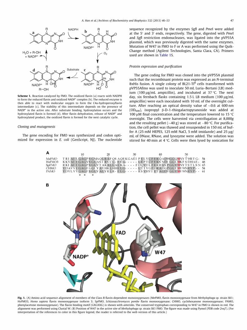
Scheme 1. Reaction catalyzed by FMO. The oxidized flavin (a) reacts with NADPHto form the reduced flavin and oxidized NADP+ complex (b). The reduced enzyme isthen able to react with molecular oxygen to form the C4a-hydroperoxyflavinintermediate (c). The stability of this intermediate depends on the presence ofNADP+ in the active site. After substrate binding, hydroxylation occurs and thehydroxylated flavin is formed (d). After flavin dehydration, release of NADP+ andhydroxylated product, the oxidized flavin is formed for the next catalytic cycle.
A. Han et al. / Archives of Biochemistry and Biophysics 532 (2013) 46–53 47
Cloning and mutagenesis
The gene encoding for FMO was synthesized and codon opti-mized for expression in E. coli (GenScript, NJ). The nucleotide
Fig. 1. (A) Amino acid sequence alignment of members of the Class B flavin-dependent mHsFMO3, Homo sapiens flavin monooxygenase isoform 3; SpFMO, Schizosacchromycephenylacetone monooxygenase). The flavin binding motif (GXGXXG) is shown with astealignment was performed using Clustal W. (B) Position of W47 in the active site of Methyinterpretation of the references to color in this figure legend, the reader is referred to th
sequence recognized by the enzymes SgfI and PmeI were addedat the 50 and 30 ends, respectively. The gene, digested with PmeIand SgfI restriction endonucleases, was ligated into the pVP55Aplasmid, which was previously digested with the same enzymes.Mutation of W47 in FMO to F or A was performed using the Quik-Change method (Agilent Technologies, Santa Clara, CA). Primersused are shown in Table 1S.
Protein expression and purification
The gene coding for FMO was cloned into the pVP55A plasmidsuch that the recombinant protein was expressed as an N-terminal8xHis fusion. A single colony of BL21-TIR cells transformed withpVP55Afmo was used to inoculate 50 mL Luria–Bertani (LB) med-ium (100 lg/mL ampicillin), and incubated at 37 �C. The nextday, six fernbach flasks containing 1.5 L LB medium (100 lg/mLampicillin) were each inoculated with 10 mL of the overnight cul-ture. After reaching an optical density value of �0.6 at 600 nm(OD600), isopropyl b-D-1-thiogalactopyranoside was added at100 lM final concentration and the temperature lowered to 15 �Covernight. The cells were harvested via centrifugation at 8,000gand the resulting pellet (�40 g) was stored at �80 �C. For purifica-tion, the cell pellet was thawed and resuspended in 150 mL of buf-fer A (25 mM HEPES, 125 mM NaCl, 5 mM imidazole) and 25 lg/mL of DNase, RNase, and lysozyme were added. The solution wasstirred for 40 min at 4 �C. Cells were then lysed by sonication for
onooxygenases (MeFMO, flavin monooxygenase from Methylophaga sp. strain SK1;s pombe flavin monooxygenase; CHMO, cyclohexanone monooxygenase; PAMO,risks. The conserved tryptophan corresponding to W47 in FMO is shown in red. Thelophaga sp. strain SK1 FMO. The figure was made using Pymol (PDB code 2vq7). (Fore web version of this article.)
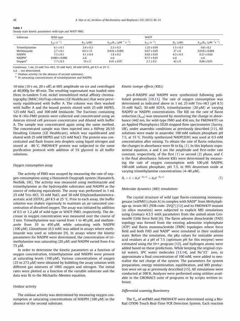
Table 1Steady-state kinetic parameters wild-type and W47F FMO.
Substrate Wild-type W47F
kcat (s�1) Km (lM) kcat/Km (lM�1 s�1) kcat (s�1) Km (lM) kcat/Km (lM�1s�1)
Trimethylamine 6.1 ± 0.1 2.6 ± 0.3 2.3 ± 0.3 1.25 ± 0.05 1.5 ± 0.3 0.8 ± 0.2Methimazole 2.7 ± 0.1 163 ± 15 0.016 ± 0.002 0.67 ± 0.05 37 ± 8 0.018 ± 0.004NADPH 7.3 ± 0.1 4.1 ± 0.4 1.8 ± 0.2 0.92 ± 0.01 4.3 ± 0.3 0.21 ± 0.02NADPHa 0.080 ± 0.006 n.d. n.d. 0.17 ± 0.01 n.d. n.d.Oxygenb 7.7 ± 0.3 19 ± 3 0.41 ± 0.07 2.7 ± 0.2 42 ± 9 0.06 ± 0.01
Conditions: 1 mL 25 mM Tris–HCl, 35 mM NaCl, 30 mM EDTA, pH 8.5 at 25 �C.n.d. – not determined.
a Oxidase activity (in the absence of second substrate).b At saturating concentrations of trimethylamine and NADPH.
48 A. Han et al. / Archives of Biochemistry and Biophysics 532 (2013) 46–53
10 min (10 s on, 20 s off) at 60% amplitude on ice and centrifugedat 40,000g for 40 min. The resulting supernatant was loaded ontothree in-tandem 5 mL nickel immobilized metal affinity chroma-tography (IMAC) HisTrap columns (GE Healthcare) that were previ-ously equilibrated with buffer A. The column was then washedwith buffer A and the bound protein eluted with 25 mM HEPES,125 mM NaCl, and 300 mM Imidazole. The fractions containingthe 8�His-FMO protein were collected and concentrated using anAmicon stirred cell pressure concentrator and diluted with bufferA. The sample was concentrated again using the same method.The concentrated sample was then injected into a HiPrep 26/10Desalting Column (GE Healthcare), which was equilibrated andeluted with 25 mM HEPES and 125 mM NaCl. The protein was con-centrated and flash frozen into droplets using liquid nitrogen andstored at �80 �C. FMOW47F protein was subjected to the samepurification protocol with addition of 5% glycerol in all buffersolutions.
Oxygen consumption assay
The activity of FMO was assayed by measuring the rate of oxy-gen consumption using a Hansatech Oxygraph system (Hansatech,Norfolk, UK). The activity was measured using methimazole andtrimethylamine as the hydroxyable substrates and NADPH as thesource of reducing equivalents. The assay was performed in 1 mL25 mM Tris–HCl, 35 mM NaCl, and 30 mM Ethylenediaminetetra-acetatic acid (EDTA), pH 8.5 at 25 �C. Prior to each assay, the buffersolution was shaken vigorously to maintain an air-saturated con-centration of dissolved oxygen. The assay was initiated by additionof 0.5 or 1.2 lM of wild-type or W47F FMO, respectively. The de-crease in oxygen concentration was measured over the course of2 min. Trimethylamine was varied from 1 to 40 lM, and methim-azole from 10 to 450 lM, while saturating with NADPH(100 lM). Glutathione (0.5 mM) was added in assays where meth-imazole was used as substrate [9]. In assays where the kineticparameters for NADPH were determined, the concentration of tri-methylamine was saturating (20 lM) and NADPH varied from 4 to75 lM.
In order to determine the kinetic parameters as a function ofoxygen concentration, trimethylamine and NADPH were presentat saturating levels (100 lM). Various concentrations of oxygen(25 to 273 lM) were obtained by bubbling the assay solution withdifferent gas mixtures of 100% oxygen and nitrogen. The initialrates were plotted as a function of the variable substrate and thedata was fit to the Michaelis–Menten equation.
Oxidase activity
The oxidase activity was determined by measuring oxygen con-sumption at saturating concentrations of NADPH (100 lM) in theabsence of the second substrate.
Kinetic isotope effects (KIEs)
pro-R-NADPD and NADPH were synthesized following pub-lished protocols [10,11]. The rate of oxygen consumption wasdetermined as indicated above in 1 mL 25 mM Tris–HCl (pH 8.5)35 mM NaCl, 30 mM EDTA, trimethylamine (20 lM) at varyingNADPD or NADPH concentrations. The KIE on the rate of flavinreduction (kred) was measured by monitoring the change in absor-bance (442 nm, for wild-type FMO and 456 nm, for FMOW47F) onan Applied Photophysics SX20 stopped-flow spectrometer (Surrey,UK), under anaerobic conditions as previously described [11]. Allsolutions were made in anaerobic 100 mM sodium phosphate pH7.5, at 15 �C. Freshly synthesized NADP(D/H) was used at 0.5 mMconcentration after mixing. To obtain the rate of flavin reduction,the changes in absorbance were fit to Eq. (1). In this biphasic expo-nential equation, a and k are the amplitude and first-order rateconstant, respectively, of the first (1) or second (2) phase, and Cis the final absorbance. Solvent KIEs were determined by measur-ing the rate of oxygen consumption with 100 lM NADPH,100 mM sodium phosphate, pH 7.5, in 99% deuterium oxide atvarying trimethylamine concentrations (4–40 lM).
At ¼ c þ a1e�ðk1�tÞ þ a2e�ðk2�tÞ ð1Þ
Molecular dynamics (MD) simulations
The crystal structure of wild type flavin-containing monooxy-genase (wtFMO) (chain A) in complex with NADP+ from Methyloph-aga sp. strain SK1 (PDB code: 2VQ7) [12] and its FMOW47F mutant(in silico mutation) were subjected to explicit MD simulationsusing Gromacs 4.5.5 with parameters from the united-atom Gro-mos96 53A6 force field [6]. The flavin adenine dinucleotide (FAD)topology was formed from the existing adenosine triphosphate(ATP) and flavin mononucleotide (FMN) topologies where forcefield and both FAD and NADP+ were simulated in their oxidizedstate. Before the simulation, the pKa values for ionizable aminoacid residues at a pH of 7.5 (optimum pH for this enzyme) wereestimated using the H++ program [12], and hydrogen atoms wereadded based on these predictions. While keeping the original crys-tal waters, SPC water molecules [13,14], and Na+/Cl� ions, toapproximate a final concentration of 100 mM, were added to neu-tralize the net charge of the system. The parameters for systempreparation, energy minimization, equilibration, and MD produc-tion were set up as previously described [15]. All simulations wereconducted at 300 K. Analyses were performed using utilities avail-able in the GROMACS suite of programs or by scripts written in-house.
Differential scanning fluorimetry
The Tm of wtFMO and FMOW47F were determined using a Bio-Rad CFX96 Touch Real-Time PCR Detection System. Each reaction
A. Han et al. / Archives of Biochemistry and Biophysics 532 (2013) 46–53 49
was performed in a final volume of 30 lL, 100 mM sodium phos-phate, pH 7.5. Each condition contained 5 lM enzyme, 5�SYPROOrange, and 0 or 100 lM NADP+. The SYPRO orange was excitedat 492 nm and emission was detected at 610 nm after incubationfor 30 s at 0.5 �C intervals from 10 �C to 80 �C. Each experimentwas performed in triplicate.
Results and discussion
Protein expression and purification
FMOW47F was expressed and purified following procedurespreviously developed for the wild-type enzyme [6]. The purifiedenzyme was stable and contained tightly bound FAD. The flavinspectra of FMOW47F appeared to have been modified by the muta-tion. A clear shift of the peak at 442 nm in the wild-type enzyme to456 nm in the W47F mutant was observed. This change in the fla-vin spectra indicates changes in the environment of the flavin ac-tive site. FMOW47A was also created and the enzymesexpressed. However, the resulting protein was found in the insolu-ble fraction after sonication and centrifugation. Therefore, we onlycharacterized the FMOW47F enzyme.
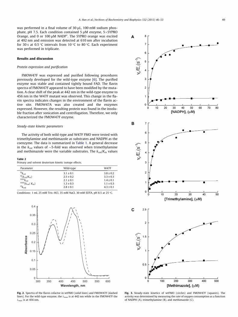
Steady-state kinetic parameters
The activity of both wild-type and W47F FMO were tested withtrimethylamine and methimazole as substrates and NADPH as thecoenzyme. The data is summarized in Table 1. A general decreasein the kcat values of �5-fold was observed when trimethylamineand methimazole were the variable substrates. The kcat/Km values
Table 2Primary and solvent deuterium kinetic isotope effects.
Parameter Wild-type W47F
Dkcat 3.1 ± 0.1 3.8 ± 0.2D(kcat/Km) 2.5 ± 0.2 3.3 ± 0.3D2Okcat 2.1 ± 0.1 1.4 ± 0.1D2O(kcat/ Km) 1.3 ± 0.3 1.1 ± 0.3Dkred 2.8 ± 0.1 4.3 ± 0.1
Conditions: 1 mL 25 mM Tris–HCl, 35 mM NaCl, 30 mM EDTA, pH 8.5 at 25 �C.
0
0.05
0.1
0.15
0.2
0.25
0.3
0.35
0.4
300 350 400 450 500 550 600
Abs
orba
nce
Wavelength, nm
Fig. 2. Spectra of the flavin cofactor in wtFMO (solid lines) and FMOW47F (dashedlines). For the wild-type enzyme, the kmax is at 442 nm while in the FMOW47F thekmax is at 456 nm.
Fig. 3. Steady-state kinetics of wtFMO (circles) and FMOW47F (squares). Theactivity was determined by measuring the rate of oxygen consumption as a functionof NADPH (A), trimethylamine (B), and methimazole (C).
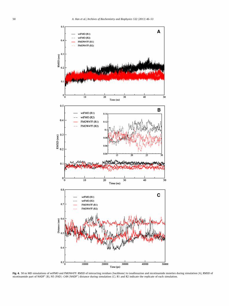
Fig. 4. 50 ns MD simulations of wtFMO and FMOW47F. RMSD of interacting residues (backbone) to isoalloxazine and nicotinamide moieties during simulation (A), RMSD ofnicotinamide part of NADP+ (B), N5 (FAD)�C4N (NADP+) distance during simulation (C). R1 and R2 indicate the replicate of each simulation.
50 A. Han et al. / Archives of Biochemistry and Biophysics 532 (2013) 46–53
A. Han et al. / Archives of Biochemistry and Biophysics 532 (2013) 46–53 51
decreased only 3-fold for trimethylamine. When NADPH was thevariable substrate and trimethylamine was kept constant, an �8-fold decrease in both kcat and kcat/Km values was measured. Theseresults show that W47 plays a role in catalysis. To test if the muta-tion had an effect in the kcat and kcat/Km values for oxygen, theactivity was measured at saturating concentrations of NADPHand trimethylamine at various concentrations of oxygen (Fig. 1S).The kinetic parameters for oxygen in the mutant enzyme were only2-fold different from the wild-type enzyme (Table 1).
Oxidase activity
We tested whether the presence of the tryptophan residue atposition 47 was necessary for the stabilization of the C4a-hydro-peroxyflavin intermediate. It has been shown that FMO is capableof reacting with NADPH to form a reduced FMO/NADP+ complex(Scheme 1b). This complex can then react with molecular oxygen
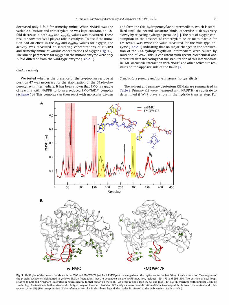
Fig. 5. RMSF plot of the protein backbone for wtFMO and FMOW47A (A). Each RMSF plotthe protein backbone (highlighted in yellow) display fluctuations that are dependent orelative to FAD and NADP are illustrated in figures nearby to that region on the plot. Twsimilar high fluctuation in both mutant and wild type enzyme. However, based on PCA antype enzymes (B). (For interpretation of the references to color in this figure legend, the
and form the C4a-hydroperoxyflavin intermediate, which is stabi-lized until the second substrate binds, otherwise it decays veryslowly by releasing hydrogen peroxide [1]. The rate of oxygen con-sumption in the absence of trimethylamine or methimazole forFMOW47F was twice the value measured for the wild-type en-zyme (Table 1) indicating that no major changes in the stabiliza-tion of the C4a-hydroperoxyflavin intermediate were caused bymutation of W47. This is consistent with recent biochemical andstructural data indicating that the stabilization of this intermediatein FMO occurs via interaction with NADP+ and other active site res-idues on the opposite side of the flavin [7].
Steady-state primary and solvent kinetic isotope effects
The solvent and primary deuterium KIE data are summarized inTable 2. Primary KIE were measured with NADP(H) as substrate todetermined if W47 plays a role in the hydride transfer step. For
is averaged over the replicates for the last 30 ns of each simulation. Two regions ofn the W47F mutation, residues 165–175 and 293–300. The position of each loopso other regions, loop 56–68 and loop 149–155 (highlighted with pink bar), exhibitalyses, movement direction of these two loops differ between the mutant and wild-reader is referred to the web version of this article.)

Table 3Unfolding temperatures of wild-type and mutant FMO.
wtFMO FMOW47F
Alone 39.9 ± 0.1 �C 40.6 ± 0.1 �C+NADP+ 41.9 ± 0.1 �C 40.8 ± 0.1 �C
Conditions: 30 lL 100 mM sodium phosphate, 5 lM enzyme, 25 lM SYPRO orange,and 100 lM NADP+.
52 A. Han et al. / Archives of Biochemistry and Biophysics 532 (2013) 46–53
wtFMO enzyme, Dkcat and D(kcat/Km) values of 3.1 and 2.5 weremeasured, respectively. These values indicate that hydride transferis partially rate-limiting. Values for the mutant enzyme were high-er. The Dkcat and D(kcat/Km) values were 3.8 and a 3.3, respectively,indicating that hydride transfer was more rate limiting in themutant enzyme. A solvent KIE value of �1 was measured for kcat/Km for wtFMO and FMOW47F. These results indicate that anexchangeable proton step is not in flight in the rate-limiting stepbefore the first irreversible step. In contrast, a normal (>1) D2Okcat
value was determined for both enzymes. Thus, a protonation ordeprotonation step is partially rate limiting for kcat, perhaps flavindehydration (Scheme 1a–d). For the FMOW47F enzyme, thesolvent KIE are lower, consistent with the results that hydridetransfer is more rate limiting in the mutant enzyme.
Rapid-reaction kinetics and primary kinetic isotope effects
The rate of flavin reduction, kred, by NADPH was directly mea-sured by monitoring the changes in flavin absorbance under anaer-obic conditions. The kred value for wild-type FMO was 24 ± 0.2 s�1,consistent with previously published results [7]. For the FMOW47Fenzyme, a kred value of 8.5 ± 0.3 s�1 was determined (Fig. 2S). Thedecrease in the kred value clearly indicates that replacement of Wat position 47 negatively affects the hydride transfer step. This isfurther supported by the increase in the Dkred value for FMOW47F(Table 2) (Fig. 3).
MD simulations of wtFMO and FMOW47F
To assess the molecular impact of mutation on hydride transfer,we performed united atom MD simulations on both the wild-typeand W47F mutant of FMO in complex to NADP+. Analyses of rootmean square deviation (RMSD) during a 50 ns simulation imply re-duced flexibility in the active site residues (interacting with isoal-loxazine and nicotinamide moieties) of FMOW47F compared to thewild-type enzyme (Fig. 4A). This difference is mainly seen in themobility of the nicotinamide part of NADP+ (Fig. 4B), which affectsN5 (FAD) and C4 (NADP+) distance. While in wild-type FMO, theN5-C4 distance appears to fluctuate during the simulation; inFMOW47F, the atoms establish an almost steady separation duringsimulations (Fig. 4C). Beside residues interacting with isoalloxa-zine and nicotinamide, analysis of root mean square fluctuation(RMSF) shows two other regions, loop 165–175 and loop 293–300, having reduced flexibility in FMOW47F (Fig. 5A, highlightedin yellow). Loop 165–175 is extended in a way that interacts withthe adenosine moiety of both FAD and NADP+. Loop 293–300 car-ries N296, which is in hydrogen bond distance from the phosphategroup of NADP+ and is highly rigid in the mutant enzyme (Fig. 5A).
The MD simulation data provided in this study may not per-fectly reflect the complete impact of the W47F mutation on hy-dride transfer because there is no available structure for theoxidized enzyme in complex with NADPH. However, the rigidifyingeffect of the W47F mutation on the residues interacting with FADand NADP+ may be extended to the NADPH binding mode of FMOstructure as well.
In addition, the Principal Component Analysis (PCA) of MD tra-jectories revealed two highly fluctuated loops (loop 56–68 andloop 149–155) moving toward each other in FMOW47F while theymoved outward in the wild-type enzyme (Fig. 5B). These two loops(both located in the FAD binding domain [7]) display the same de-gree of flexibility in both wtFMO and FMOW47F (Fig. 5A, high-lighted in pink). Thus, the difference in the loop movementmight likely affect conformational changes important for catalysis(see below).
Protein stability
The MD results suggested that by replacing W at position 47 toF, some regions of the protein became more rigid. In order to deter-mine the effect experimentally, we measured the melting temper-ature (Tm) of wtFMO and FMOW47F (Fig. 3S). In the absence ofNADP+, the mutant enzyme appeared to be more stable, but byonly 0.7 �C (Table 3). Although, this is consistent with the MD re-sults, the effect is very low. In the presence of NADP+, the Tm valuefor wtFMO increased 2 �C. In contrast, the binding of NADP+ did notresult in an increase in the Tm value for the FMOW47F enzyme (Ta-ble 3). This is not due to a change in affinity for NADP+ since the Km
value for this coenzyme was not affected by the mutation (Table 1).Instead, these results indicate that the mutant enzyme is more ri-gid, and this rigidity prevents it from acquiring the more stable en-zyme-bound conformation observed for wtFMO. Thus, it is possiblethat conformational changes that are associated with the hydridetransfer step are impaired in the mutant enzyme.
Conclusions
The active sites of flavin-dependent monooxygenases are fine-tuned to accommodate two substrates and oxygenated flavin inter-mediates during catalysis. As noted by Alfieri et al. [6], the positionof NADP+ observed in the structure is not optimal for hydridetransfer (Fig. 1). Instead, this binding mode is essential for stabil-ization of the C4-hydroperoxyflavin (Scheme 1c). Thus, it was pro-posed that protein conformational changes must occur after orduring hydride transfer [6]. These conformational changes are re-quired to position NADP+ in a conformation where it plays a rolein stabilization of the C4a-hydroperoxyflavin–the position ob-served in the X-ray structure (Fig. 1B) [6,7,16]. The data presentedhere shows that mutation of W47, which is located on the si-face ofthe flavin ring, results in only a minimal effect in the oxidativehalf-reaction. This is consistent with results that indicate that oxy-gen activation and stabilization of C4a-hydroperoxyflavin occurson the re-face of the flavin ring, where NADP+ binds [7]. Replace-ment of W47 to F, results in a decrease in the rate of hydride trans-fer from NADPH to the flavin. MD simulations show that thismutation causes changes in the flexibility of the protein, includingactive site residues and the nicotinamide moiety of NADP+. Theseminor changes translate into a more rigid FMOW47F protein. Ther-mal stability analysis shows that binding of NADP+ causes thewild-type enzyme to become more thermally stable. This is consis-tent with the protein becoming more compact upon ligand bind-ing. However, the FMOW47F enzyme does not become morethermally stable upon NADP+ binding. We propose that due tothe more rigid nature of the mutant enzyme, the conformationalchanges that occur during NADP(H) binding are prevented or min-imized. These conformational changes might be necessary to prop-erly align the NADP-C4 and FAD-N5 for hydride transfer. In fact,the simulations show that in wtFMO the NADP-C4 and FAD-N5 dis-tance fluctuates more, perhaps allowing sampling of the optimalposition for hydride transfer. In contrast, the NADP-C4 and FAD-N5 distance is less variable in the mutant protein, perhaps decreas-ing the rate of hydride transfer. Together, the data presented here

A. Han et al. / Archives of Biochemistry and Biophysics 532 (2013) 46–53 53
shows how the active site of flavin monooxygenases are exqui-sitely fine-tuned for catalysis, showing that even residues whichare on the opposite side of the ‘‘active site’’ are important for mod-ulating protein motions important for the chemical steps. Sincethis tryptophan residue is conserved in other members of the ClassB flavin-monooxygenases, it is possible that it plays a similar rolein the mechanism of action of other members of this family ofenzymes.
Acknowledgments
This work was supported in part by a grant from the NationalScience Foundation MCB-1021384 and by a grant from the VirginiaAcademy of Sciences.
References
[1] W.J. van Berkel, N.M. Kamerbeek, M.W. Fraaije, J. Biotechnol. 124 (2006) 670–689.
[2] V. Massey, J. Biol. Chem. 269 (1994) 22459–22462.
[3] P. Chaiyen, M.W. Fraaije, A. Mattevi, Trends Biochem. Sci. 371 (2012) 373–380.
[4] N.B. Beaty, D.P. Ballou, J. Biol. Chem. 256 (1981) 4619–4625.[5] D.M. Ziegler, Drug Metab. Rev. 34 (2002) 503–511.[6] A. Alfieri, E. Malito, R. Orru, M.W. Fraaije, A. Mattevi, Proc. Natl. Acad. Sci. USA
105 (2008) 6572–6577.[7] R. Orru, D.E. Pazmino, M.W. Fraaije, A. Mattevi, J. Biol. Chem. 285 (2010)
35021–35028.[8] P.G. Blommel, P.A. Martin, K.D. Seder, R.L. Wrobel, B.G. Fox, Methods Mol. Biol.
498 (2009) 55–73.[9] L.L. Poulsen, D.M. Ziegler, J. Biol. Chem. 254 (1979) 6449–6455.
[10] S.S. Jeong, J.E. Gready, Anal. Biochem. 221 (1994) 273–277.[11] E. Romero, M. Fedkenheuer, S.W. Chocklett, J. Qi, M. Oppenheimer, P. Sobrado,
Biochim. Biophys. Acta 1824 (2012) 850–857.[12] C. Oostenbrink, A. Villa, A.E. Mark, W.F. Van Gunsteren, J. Comput. Chem. 25
(2004) 1656–1676.[13] J.C. Gordon, J.B. Myers, T. Folta, V. Shoja, L.S. Heath, A. Onufriev, Nucleic Acids
Res. 33 (2005) W368–W371.[14] H.J.C. Berendsen, J.P.M. Postma, W.F. Van Gunsteren, J. Hermans,
Intermolecular Forces, in: B. Pullman (Ed.), Reidel Dordrecht, TheNetherlands, 1981, p. pp. 331.
[15] S. Badieyan, D.R. Bevan, C.M. Zhang, Protein Eng. Des. Sel. 25 (2012) 223–233.
[16] R. Orru, H.M. Dudek, C. Martinoli, D.E. Torres Pazmino, A. Royant, M. Weik,M.W. Fraaije, A. Mattevi, J. Biol. Chem. 286 (2011) 29284–29291.
Related Documents