© 2021 by the Serbian Biological Society 523 How to cite this article: Doan HV, Sritangos P, Weeranantanapan O, Chudapongse N. Aqueous extract from Chrysophyllum cainito bark exhibits embryonic toxicity in Danio rerio and negligible acute toxicity in adult Wistar rats. Arch Biol Sci. 2021;73(4):523-33. Arch Biol Sci. 2021;73(4):523-533 https://doi.org/10.2298/ABS211110046D Aqueous extract from Chrysophyllum cainito bark exhibits embryonic toxicity in Danio rerio and negligible acute toxicity in adult Wistar rats Hau Van Doan 1,2 , Pishyaporn Sritangos 1 , Oratai Weeranantanapan 1 and Nuannoi Chudapongse 1, * 1 School of Preclinical Sciences, Institute of Science, Suranaree University of Technology, Nakhon Ratchasima, Thailand 2 Department of Pharmacy, School of Medicine and Pharmacy, Tra Vinh University, Tra Vinh, Vietnam *Corresponding author: [email protected] Received: November 10, 2021; Revised: November 15, 2021; Accepted: November 17, 2021; Published online: November 29, 2021 Abstract: Chrysophyllum cainito has been used as a traditional medicine to treat a wide range of diseases, but the toxicity profile of this plant remains unknown. This study aimed to evaluate the acute toxicity of the aqueous extract of C. cainito (CE) bark based on OECD guidelines in two different in vivo experimental models: acute single-dose oral toxicity in adult Wistar rats and the zebrafish embryo acute toxicity test. All concentrations of CE (500-4000 mg/kg) tested during a 14-day period in both male and female rats showed no effect on behavior, body weight, organ weights, biochemical and hemato- logical parameters. In contrast, CE significantly delayed zebrafish embryo hatching and decreased embryo survival rates in a dose-dependent manner. Hatched larvae were notably sensitive to CE-induced toxicity compared to unhatched fish embryos. Acridine orange staining showed that CE induced apoptosis in the yolk sac region that is responsible for supplying nutrients to support larval growth and development. According to OECD guidelines, CE was identified as GHS category 5, a substance with low to no acute toxicity. However, as embryotoxicity was observed in zebrafish, CE use during pregnancy should be exercised with caution until further examination of its safety. Keywords: Chrysophyllum cainito; embryotoxicity; acute toxicity; zebrafish; Wistar rat INTRODUCTION Traditional herbal medicines offer an alternative for the treatment of common ailments such as diabetes, bacterial infections, fever. In developing countries, pa- tients with chronic diseases are the major consumers of herbal medicines due to traditional beliefs, local ac- cessibility and the low cost of these remedies, which is typically less than that of conventional modern medi- cation [1,2]. The widespread use of traditional herb- al medicine is also attributed to exaggerated claims regarding their broad preventative and therapeutic effects [3]. Unfortunately, the safety information re- garding most folk herbal medicine is based on non- scientific sources or misleading scientific evidence. Furthermore, medicinal plants, while widely used, can also exert toxic adverse effects if misused, particularly in vulnerable populations, including pregnant women and children [4]. Concerns regarding herbal medica- tion are increasing as more reports evaluate and high- light the toxicity of herbal medicinal plants [5,6]. It has been reported that changes in herbal extraction methods of the same medicinal plant can lead to in- creased toxic effects in brine shrimp [5]. The risk of herbal medicine misuse may lead to potential adverse effects, toxicity, mutagenicity and carcinogenicity [7]. The pharmacovigilance scheme employs a sci- entific approach to detect, estimate, understand and limit the adverse effect of drugs. According to the pharmacovigilance system introduced by the World Health Organization [8], safety is deemed fundamen- tal for all therapeutic products, including herb-derived medicinal and healthcare products. Recent pharma- covigilance concerns describe the use and misuse of herbals, folk and complementary medicines. Unlike modern medicines, efficacy is not a legal issue for folk remedies. Although most herbal folk remedies are generally believed to be safe, their potential adverse effects remain to be elucidated [9]. Chrysophyllum cainito is a tropical fruit tree con- sidered to exert multiple therapeutic effects, including

Welcome message from author
This document is posted to help you gain knowledge. Please leave a comment to let me know what you think about it! Share it to your friends and learn new things together.
Transcript
© 2021 by the Serbian Biological Society 523How to cite this article: Doan HV, Sritangos P, Weeranantanapan O, Chudapongse N. Aqueous extract from Chrysophyllum cainito bark exhibits embryonic toxicity in Danio rerio and negligible acute toxicity in adult Wistar rats. Arch Biol Sci. 2021;73(4):523-33.
Arch Biol Sci. 2021;73(4):523-533 https://doi.org/10.2298/ABS211110046D
Aqueous extract from Chrysophyllum cainito bark exhibits embryonic toxicity in Danio rerio and negligible acute toxicity in adult Wistar rats
Hau Van Doan1,2, Pishyaporn Sritangos1, Oratai Weeranantanapan1 and Nuannoi Chudapongse1,*
1School of Preclinical Sciences, Institute of Science, Suranaree University of Technology, Nakhon Ratchasima, Thailand2Department of Pharmacy, School of Medicine and Pharmacy, Tra Vinh University, Tra Vinh, Vietnam
*Corresponding author: [email protected]
Received: November 10, 2021; Revised: November 15, 2021; Accepted: November 17, 2021; Published online: November 29, 2021
Abstract: Chrysophyllum cainito has been used as a traditional medicine to treat a wide range of diseases, but the toxicity profile of this plant remains unknown. This study aimed to evaluate the acute toxicity of the aqueous extract of C. cainito (CE) bark based on OECD guidelines in two different in vivo experimental models: acute single-dose oral toxicity in adult Wistar rats and the zebrafish embryo acute toxicity test. All concentrations of CE (500-4000 mg/kg) tested during a 14-day period in both male and female rats showed no effect on behavior, body weight, organ weights, biochemical and hemato-logical parameters. In contrast, CE significantly delayed zebrafish embryo hatching and decreased embryo survival rates in a dose-dependent manner. Hatched larvae were notably sensitive to CE-induced toxicity compared to unhatched fish embryos. Acridine orange staining showed that CE induced apoptosis in the yolk sac region that is responsible for supplying nutrients to support larval growth and development. According to OECD guidelines, CE was identified as GHS category 5, a substance with low to no acute toxicity. However, as embryotoxicity was observed in zebrafish, CE use during pregnancy should be exercised with caution until further examination of its safety.
Keywords: Chrysophyllum cainito; embryotoxicity; acute toxicity; zebrafish; Wistar rat
INTRODUCTION
Traditional herbal medicines offer an alternative for the treatment of common ailments such as diabetes, bacterial infections, fever. In developing countries, pa-tients with chronic diseases are the major consumers of herbal medicines due to traditional beliefs, local ac-cessibility and the low cost of these remedies, which is typically less than that of conventional modern medi-cation [1,2]. The widespread use of traditional herb-al medicine is also attributed to exaggerated claims regarding their broad preventative and therapeutic effects [3]. Unfortunately, the safety information re-garding most folk herbal medicine is based on non-scientific sources or misleading scientific evidence. Furthermore, medicinal plants, while widely used, can also exert toxic adverse effects if misused, particularly in vulnerable populations, including pregnant women and children [4]. Concerns regarding herbal medica-tion are increasing as more reports evaluate and high-light the toxicity of herbal medicinal plants [5,6]. It
has been reported that changes in herbal extraction methods of the same medicinal plant can lead to in-creased toxic effects in brine shrimp [5]. The risk of herbal medicine misuse may lead to potential adverse effects, toxicity, mutagenicity and carcinogenicity [7].
The pharmacovigilance scheme employs a sci-entific approach to detect, estimate, understand and limit the adverse effect of drugs. According to the pharmacovigilance system introduced by the World Health Organization [8], safety is deemed fundamen-tal for all therapeutic products, including herb-derived medicinal and healthcare products. Recent pharma-covigilance concerns describe the use and misuse of herbals, folk and complementary medicines. Unlike modern medicines, efficacy is not a legal issue for folk remedies. Although most herbal folk remedies are generally believed to be safe, their potential adverse effects remain to be elucidated [9].
Chrysophyllum cainito is a tropical fruit tree con-sidered to exert multiple therapeutic effects, including
524 Arch Biol Sci. 2021;73(4):523-533
hypoglycemic activity, antioxidant, antihypertensive, antiinflammatory and antibacterial properties [10]. The bark of C. cainito has been traditionally used in Côte d’Ivoire and the southwest of Vietnam as a folk treatment for diabetes [10,11]. Despite the widespread use of the C. cainito extract, there is a lack of evidence regarding its safety and toxicity profile.
In vivo pharmacological evidence supporting the use of C.cainito in diabetes in our previous work showed that the aqueous extract of C. cainito bark was indeed effective in reducing blood glucose lev-els in the mouse model [12]. However, to safely rec-ommend the integration of C. cainito as part of the therapeutic remedy for diabetes, its toxicity should be considered in parallel with its benefits [13,14]. Therefore, this study aimed to evaluate the safety of the C. cainito bark extract (CE) using internationally accepted guidelines provided by the Organization for Economic Co-operation and Development (OECD). The safety profile of CE was assessed by a single-dose acute toxicity test performed in a Wistar rat model, and to examine whether CE is safe for use in pregnan-cy, fish embryo acute toxicity tests were conducted.
MATERIALS AND METHODS
Ethics statement
All experimental procedures were approved and conducted with strict adherence to the guidelines of the Institutional Animal Care and Use Committee, Suranaree University of Technology, Thailand (Approval No: 1/2559 and 10/2559). According to the current regulations of the European Union and the Institutional Animal Care and Use Committee (IACUC), zebrafish were subjected to ethical regu-lation after 5 days (120 h) postfertilization [15,16], as evidence suggests that zebrafish would be capable of nociception and distress at 5 days postfertilization [15,17-19]. No zebrafish embryos older than 72 h postfertilization were used in this study.
Chemicals
All chemicals were purchased from Sigma Aldrich (Missouri, USA) unless stated otherwise.
Plant collection and C. cainito bark extraction
C. cainito bark was collected from Mo Cay Nam dis-trict, Ben Tre Province, Vietnam. Plant verification was performed as indicated in our previous study [12]. Vouchered specimens of leaf, fruit, flowers, and stem were stored at the Suranaree University of Technology Botanical Garden (collection ID: H. DOAN-1).
C. cainito bark aqueous extraction was performed as described [12]. Briefly, shade-dried C. cainito bark was chopped and ground with a blender before extrac-tion. Fifty grams of the finely ground bark were mixed with 200 mL of deionized water. Four cycles of aque-ous extraction were performed for 2 h per extraction cycle. All extraction procedures were performed on a shaker at room temperature. The combined superna-tant was filtered with cotton gauze then centrifuged in a Sorvall Biofuge Stratos Centrifuge (Thermo Fisher Scientific, USA) at 8000 x g for 15 min, at room tem-perature to remove any remaining bark residue. The extract was concentrated by rotary evaporation and freeze-dried for 48 h using a lyophilizer. The dried C. cainito extract was stored at -20oC until further use.
Acute toxicity study in rat model
Animal husbandry
Male and female Wistar rats were obtained from the Laboratory Animal Facility, Suranaree University of Technology. The animals were housed in stainless steel cages lined with wood shavings at Laboratory Animal Facility, Suranaree University of Technology, under standard conditions of 25±2oC, 45-50% rela-tive humidity and a 12-h light/dark cycle. Rats had ad libitum access to standard pellet food and water. All animals were acclimatized for seven days before an acute toxicity test was performed.
Acute toxicity in rats
Based on OECD Guideline no. 423 [20], healthy male (120-150 g) and female Wistar rats (100-140 g) were selected for the acute toxicity test. After seven days of acclimatization, rats were fasted overnight. The animals were divided into five treatment groups (3 animals of each sex per group; N=6 per group) as
525Arch Biol Sci. 2021;73(4):523-533
follows: control group (that received deionized water) and four treatment groups that were administered a single dose of increasing concentrations of CE at 500, 1000, 2000 and 4000 mg/kg of rat body weight. Minor dose deviations from the OECD guidelines were cho-sen. The animal sample sizes were calculated based on the “resource equation” method [21].
Prior to treatment, CE was freshly prepared by dissolving the lyophilized CE in sterile distilled wa-ter. All treatments were administered by oral gavage. Post-treatment, the rats were monitored for signs of toxicity at 0.25, 0.5, 1, 4 and 24 h, then daily for 14 days. According to OECD guidelines, signs of toxicity indicative of animal suffering were considered as exper-imental endpoints, and any affected animals were im-mediately euthanized [20]. Rats were weighed on days 0, 7 and 14. At the end of the experiment, all rats fasted overnight and were killed by CO2 inhalation. Necropsy was performed, and the internal organs were examined. Rat organs (liver, heart, kidney, lung, spleen, testis and ovary) were isolated and weighed. Blood samples were collected for hematological analysis (Mindray BC-6800 Auto Hematology Analyzer) and biochemical analy-sis (Mindray BS-800 Automatic Chemistry Analyzer). The following liver and kidney function parameters were monitored: alanine transaminase (ALT), aspartate transaminase (AST), alkaline phosphatase (ALP), blood urea nitrogen, total protein and albumin.
Developmental toxicity in zebrafish embryos
Zebrafish housing and breeding
Adult zebrafish (Danio rerio) were sourced from a local fish shop that routinely supplies zebrafish for breeding purposes to Suranaree University of Technology. Adult zebrafish were housed in glass tanks, reared in reversed osmosis water, exposed to room temperature of 26±2oC and a 12-h light/dark cycle. Male and female fish were raised in separate tanks. Zebrafish were routinely fed three times per day with commercial micropellet (morning and eve-ning at 7.00 and 17.00 h) and frozen artemia (noon, 12.00 h). Zebrafish were monitored daily for signs of disease, and routine feces removal was performed to maintain water quality. To stimulate breeding and egg fertilization, active male and female fish (ratio 2:1) were placed in the same spawning tank fitted with
a transparent barrier and a spawn trap 1-2 h before the dark cycle. At the beginning of the light cycle, the transparent barrier was removed to allow male and female fish to mate. After 45-50 min, adult zebraf-ish were removed from the spawning tank and the fish eggs were collected and rinsed with E3 medium (5 mM NaCl, 0.17 mM KCl, 0.33 mM CaCl2, 0.33 mM MgSO4).
Fish embryo acute toxicity test (FET)
Acute toxicity of CE on zebrafish embryos was per-formed according to OECD guideline 236 [22] with minor amendments [23]. Healthy fertilized eggs were chosen under a stereomicroscope and maintained in E3 medium. Selected fertilized eggs were placed into a 24-well plate, one egg/well. Fifteen to twenty eggs were used per treatment and five independent experiments were performed. At about 3-4 h postfertilization (hpf), eggs were exposed to different concentrations of CE (0, 15.63, 31.25, 62.5, 125, 250 and 500 µg/mL) for 72 h. The developmental morphologies of embryos were monitored at 24, 48, and 72 h post-treatment administration (hpta). Parameters that were moni-tored included: hatching rate, death and noticeable ab-normal embryonic development. Fish embryo images were captured using AM423X/AM4023CT Dino-Eye C Mount digital cameras (AnMo Electronics Corp., Taiwan) fitted on Olympus SZX7/SZ61 stereomicro-scopes (Olympus, Japan). Images were acquired by DinoCapture 2.0 software (AnMo Electronics Corp., Taiwan).
Fish larvae acute apoptosis test
From the LC50 calculated from the FET test, an acute apoptosis screening test was performed to determine whether CE induces selective or non-specific toxicity. Healthy fertilized eggs were maintained in E3 medium until hatching at 72 hpf. Newly hatched larvae were placed into 24-well plates containing the designated treatment conditions, including 0, 31.25 and 62.5 µg CE/mL, and were exposed to the treatment for either 0.5 or 1 h. At least two replicates were performed per treatment condition.
After the designated treatment, zebrafish larvae were stained with 3.33 μg/mL of acridine orange (cat#
526 Arch Biol Sci. 2021;73(4):523-533
A8097, Sigma-Aldrich, Missouri, USA) at 37oC for 15 min. The stained larvae were then washed with E3 medium for 5 min. Washing was repeated three times. The stained larvae were then immobi-lized in an ice-water bath for at least 30 min prior to image capture, after which all larvae were euthanized in an ice-water bath overnight (>12 h) [24]. Fluorescent zebrafish larvae imaging was carried out using an Olympus DP72 fluorescent microscope (4x objective lens, FITC channel, 400 ms exposure) fitted with a Nikon Eclipse 80i camera. Images were taken using Olympus CellD software (Olympus, Japan) and were processed by Fiji ImageJ software [25].
Statistical analysis
All quantitative results are expressed as the mean±SEM. Statistical analysis was performed using Graphpad Prism version 9.2.0 (GraphPad Software, USA). Normality tests were performed to determine whether the data sets used for statistical analysis were para-metric or nonparametric. For multiple comparisons of parametric data, results were analyzed by one-way ANOVA followed by Tukey’s test. Kruskal-Wallis one-way ANOVA and Dunn’s post-hoc test were
performed for multiple comparisons of nonparametric data. For multifactor comparisons, two-way ANOVA and Tukey’s post-hoc test were performed. P<0.05 was considered as a statistically significant difference.
RESULTS
C. cainito bark extract exhibited negligible acute oral toxicity in Wistar rat
As safety data regarding oral route administration of C. cainito bark are currently unavailable, it is important to determine the safety category of CE.
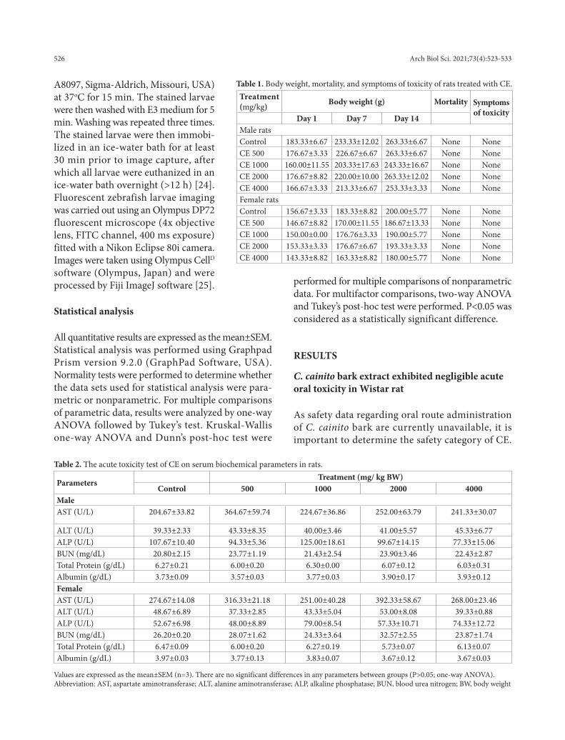
Table 1. Body weight, mortality, and symptoms of toxicity of rats treated with CE.Treatment (mg/kg) Body weight (g) Mortality Symptoms
of toxicityDay 1 Day 7 Day 14
Male ratsControl 183.33±6.67 233.33±12.02 263.33±6.67 None NoneCE 500 176.67±3.33 226.67±6.67 263.33±6.67 None NoneCE 1000 160.00±11.55 203.33±17.63 243.33±16.67 None NoneCE 2000 176.67±8.82 220.00±10.00 263.33±12.02 None NoneCE 4000 166.67±3.33 213.33±6.67 253.33±3.33 None NoneFemale ratsControl 156.67±3.33 183.33±8.82 200.00±5.77 None NoneCE 500 146.67±8.82 170.00±11.55 186.67±13.33 None NoneCE 1000 150.00±0.00 176.76±3.33 190.00±5.77 None NoneCE 2000 153.33±3.33 176.67±6.67 193.33±3.33 None NoneCE 4000 143.33±8.82 163.33±8.82 180.00±5.77 None None
Table 2. The acute toxicity test of CE on serum biochemical parameters in rats.
ParametersTreatment (mg/ kg BW)
Control 500 1000 2000 4000MaleAST (U/L) 204.67±33.82 364.67±59.74 224.67±36.86 252.00±63.79 241.33±30.07
ALT (U/L) 39.33±2.33 43.33±8.35 40.00±3.46 41.00±5.57 45.33±6.77ALP (U/L) 107.67±10.40 94.33±5.36 125.00±18.61 99.67±14.15 77.33±15.06BUN (mg/dL) 20.80±2.15 23.77±1.19 21.43±2.54 23.90±3.46 22.43±2.87Total Protein (g/dL) 6.27±0.21 6.00±0.20 6.30±0.00 6.07±0.12 6.03±0.31Albumin (g/dL) 3.73±0.09 3.57±0.03 3.77±0.03 3.90±0.17 3.93±0.12FemaleAST (U/L) 274.67±14.08 316.33±21.18 251.00±40.28 392.33±58.67 268.00±23.46ALT (U/L) 48.67±6.89 37.33±2.85 43.33±5.04 53.00±8.08 39.33±0.88ALP (U/L) 52.67±6.98 48.00±8.89 79.00±8.54 57.33±10.71 74.33±12.72BUN (mg/dL) 26.20±0.20 28.07±1.62 24.33±3.64 32.57±2.55 23.87±1.74Total Protein (g/dL) 6.47±0.09 6.00±0.20 6.27±0.19 5.73±0.07 6.13±0.07Albumin (g/dL) 3.97±0.03 3.77±0.13 3.83±0.07 3.67±0.12 3.67±0.03
Values are expressed as the mean±SEM (n=3). There are no significant differences in any parameters between groups (P>0.05; one-way ANOVA). Abbreviation: AST, aspartate aminotransferase; ALT, alanine aminotransferase; ALP, alkaline phosphatase; BUN, blood urea nitrogen; BW, body weight
527Arch Biol Sci. 2021;73(4):523-533
Preliminary toxicity studies are typically performed using in vivo rodent models. To ensure that CE toxic-ity could be classified based on the widely accepted Globally Harmonized System (GHS) of classification, our study compliantly assessed the safety of CE extract using the OECD acute oral toxicity test guidelines. A single dose of CE or vehicle control was orally ad-ministered to healthy Wistar rats. Then symptoms of toxicity (tremors, convulsions, salivation, diarrhea, lethargy and coma) were checked every day for 14 days. Compared to the untreated control, doses of CE at 500 mg/kg up to 4000 mg/kg did not produce any noticeable adverse effects or toxicity (Table 1). No mortality was observed in any of the treated groups for the duration of the study. There were no significant changes in body weight (Table 1), serum biochemical
parameters (Table 2) and hematological parameters (Table 3). Necropsy of key internal organs (liver, kid-neys, lungs, heart, or digestive and reproductive or-gans) showed no noticeable abnormalities compared to the control animals. There were no changes in the relative organ weights of liver, kidneys, lungs, heart, spleen, testis/ovaries (Table 4) when compared to the control group (P>0.05). CE did not induce mortality and signs of toxicities at 4000 mg/kg.
The embryotoxic effect of CE on zebrafish
As CE may be beneficial for the management of diseases during pregnancy, it is crucial to establish whether CE induces embryotoxicity. CE treatments at 72 hpta induced noticeable embryotoxicity in
Table 3. The acute toxicity test of CE on hematological biochemical parameters in rats
ParametersTreatment (mg/ kg BW)
Control 500 1000 2000 4000MaleRBC (× 106 /μL) 7.80 ±0.25 7.76±0.27 8.91±0.43 8.09±0.06 8.03±0.14HGB (g/dL ) 14.87±0.66 13.93±1.48 17.33±0.83 15.85±0.12 16.23±0.38HCT (%) 52.00±0.58 52.67±1.45 61.67±3.18 55.50±0.58 56.67±1.45WBC (× 103/μL) 4.82±2.29 9.97±2.25 9.69±2.38 8.53±0.50 9.92±2.57LYM (× 103/μL) 84.73±0.64 81.23±3.01 86.30±1.44 86.90±2.16 84.63±1.42MON (× 103/μL) 2.33±0.17 2.77±0.38 1.93±0.19 2.33± 0.20 2.77±0.13NEU (%) 11.53±0.60 12.10±1.31 10.40±0.95 8.97±1.48 10.87±1.52PLT (× 104/μL) 49.10±13.92 77.50±14.07 79.70±11.11 86.33±4.88 80.03±4.18MCV (fL) 67.00±1.16 67.67±0.67 69.00±0.58 68.33±0.33 70.33±0.67MCH (pg) 19.07±0.35 17.87±1.49 19.47±0.12 19.43±0.19 20.20±0.15MCHC (g/dL) 28.60±1.00 26.50±2.4 28.23±0.34 28.53±0.45 28.80±0.12RDW-CV (%) 14.33±0.33 14.57±0.29 15.00±0.38 14.63±0.48 14.73±0.27Female RBC (× 106 /μL) 8.61±0.11 9.01±0.50 8.30±0.27 8.40±0.44 8.87±0.13HGB (g/dl) 17.00±0.06 19.20±0.98 16.83±0.55 15.77±1.47 17.43±0.41HCT (%) 57.33±0.88 58.67±4.18 54.67±2.03 53.33±3.33 57.00±1.53WBC (× 103/μL) 6.52±1.92 7.45±1.27 5.03±1.67 4.08±0.80 5.29±1.63LYM (× 103/μL) 76.36±5.66 88.50±1.85 85.80±0.57 80.37±0.90 83.67±1.53MON (× 103/μL) 3.80±0.76 1.83±0.48 4.10±0.53 3.77±0.92 2.93±0.69NEU (%) 13.80±3.10 6.93±1.56 7.80±0.57 11.13±0.90 12.13±0.96PLT (× 104/μL) 103.73±2.89 57.40±34.22 52.33±27.23 43.80±15.55 84.73±9.48MCV (fL) 66.33±0.33 65.00±1.00 66.00±0.58 63.67±0.88 64.00±1.00MCH (pg) 19.70±0.23 19.83±1.36 20.30±0.20 18.67±1.13 19.67±0.18MCHC (g/dL) 29.77±0.46 30.67±2.59 30.63±0.17 29.33±1.45 30.57±0.28RDW-CV (%) 14.03±0.23 14.13±0.54 12.93±0.07 13.27±0.33 13.33±0.13
Values are expressed as mean±SEM (n=3). There are no significant differences in any parameters between groups (P>0.05; one-way ANOVA). Abbreviation: BW, body weight; RBC, red blood cell; HGB, hemoglobin; HCT, hematocrit; WBC, white blood cell; LYM, lymphocyte; MON, monocyte; NEU, neutrophil; PLT, platelet; MCV, mean corpuscular volume; MCH, mean corpuscular hemoglobin; MCHC: mean corpuscular hemoglobin concentration; RDW-CV, red cell distribution width - coefficient of variation.
528 Arch Biol Sci. 2021;73(4):523-533
zebrafish embryos. Although the overall hatch-ing rate of zebrafish embryos was not significantly affected by CE treatments at 15.63-250 µg/mL, a trend of reduced hatching rate in a concentration-dependent manner was observed. Exposure to CE at 500 µg/mL significantly decreased the hatching rate compared to the untreated control (P=0.0051; Fig. 1A).
Survival rates of embryos, including egg and larvae, were observed at 24, 48 and 72 hpta. Before hatching, between 24-48 hpta, the major lethal effect observed was coagulation (Fig. 1B). Our results showed insignificant changes in mortal-ity between 24 and 48 hpta (Fig. 1C). However, treatments of CE at 31.25-500 μg/mL significantly decreased the survival rate at 72 hpta as compared to the untreated control and similar doses at 24-48 hpta (Fig. 1C). The median lethal concentration
that caused 50% embryonic death (LC50), calculated at 72 hpta, was 22.80+1.23 μg/mL (Fig. 1D). Representative images of zebrafish embryos treated with CE were monitored at 24, 48 and 72 hpta (Fig. 2). No apparent abnormalities in the mor-phology of the embryos were observed in any of the treatment conditions. Overall,
Table 4. Relative organ weights of rats treated with CE.
OrgansTreatment (mg/ kg BW)
Control 500 1000 2000 4000MaleLiver 3.86±0.12 3.91±0.04 3.45±0.16 3.75±0.21 3.55±0.09Heart 0.43±0.03 0.39±0.02 0.43±0.04 0.42±0.02 0.44±0.02Kidneys 0.77±0.01 0.81±0.02 0.75±0.02 0.79±0.01 0.74±0.02Lungs 0.57±0.03 0.58±0.09 0.60±0.05 0.54±0.00 0.66±0.07Spleen 0.26±0.01 0.30±0.03 0.29±0.01 0.26±0.01 0.29±0.03Testis 1.23±0.01 1.25±0.03 1.26±0.04 1.27±0.04 1.27±0.06FemaleLiver 3.73±0.35 3.68±0.04 3.42±0.10 3.52±0.11 3.64±0.05Heart 0.49±0.04 0.41± 0.01 0.46±0.02 0.45±0.01 0.47±0.02Kidneys 0.79±0.05 0.75±0.01 0.77±0.02 0.69±0.05 0.73±0.04Lungs 0.67±0.09 0.73±0.09 0.81±0.09 0.79±0.12 0.74±0.05Spleen 0.34±0.04 0.31±0.05 0.26±0.00 0.29±0.03 0.27±0.02Ovaries 0.08±0.01 0.06±0.01 0.08±0.00 0.08±0.01 0.09±0.00
Values are expressed as mean±SEM (n=3). There are no significant differences in any parameters between groups (P>0.05; one-way ANOVA). Abbreviation: BW, body weight.
Fig. 1. Acute toxicity of CE on zebrafish embryos. A – Effect of CE on zebrafish hatching rate. The cumulative hatching rate is expressed as the percentage of hatched embryos at 72 h post-administration (hpta) of CE normalized to the initial unhatched embryos at 0 hpta, regardless of viability. Statistical analysis was performed using Kruskal-Wallis one-way analysis of variance with Dunn’s post-hoc test. * P<0.05 in comparison to the untreated control. B – Representative im-age of CE-induced embryo coagulation. C – Cumulative effect of CE on zebrafish embryo survival rate at 24, 48, and 72 h. Embryos exhibiting signs of coagulation, lack of heartbeat at late developmental stages or apparent decomposition were considered dead. Repeated-measure two-way ANOVA with Tukey’s post-hoc test was performed. * P<0.05 in comparison to 24 and 48 hpta at the same CE concentration. # P<0.05 in comparison to 24 hpta at 500 μg/mL CE. D – Median lethal concentration (LC50) of CE at 72 hpta. The survival data of embryos exposed to log [CE (μg/mL)] at 72 hpta were fitted to a sigmoidal non-linear regression model (red line). All data shown are expressed as mean±SEM (n=5).
Fig. 2. Effect of CE on zebrafish embryo morphol-ogy. Representative brightfield images of zebrafish embryos treated with designated concentrations of CE at 24, 48 and 72 hpta.
529Arch Biol Sci. 2021;73(4):523-533
although CE potentially lacked teratogenic effects, the extract demonstrated notable embryotoxicity when exposed to hatched larvae.
Apoptosis induced by CE
It was noted that hatched larvae rapidly died within a few hours after exposure to CE. To broadly assess how rapidly CE induced mortality in these larvae, healthy larvae were exposed to CE concentrations above the LC50 (30.25 and 60.5 μg/mL) for either 0.5 or 1 h. Acridine orange staining was performed to de-tect CE-induced apoptosis in larvae. Compared to the untreated control, signs of apoptosis became manifest in the yolk sac and yolk sac extension regions of the larvae in a dose and time-dependent manner (Fig. 3).
DISCUSSION
C. cainito bark aqueous extract has been reported to contain multiple bioactive compounds such as phenols, tannins, terpenoids, glycosides, saponin, phenolics and antioxidants [12]. We demonstrated that CE exerted antidiabetic effects in a mouse model and exhibited anticancer effects in the HepG2 hepatocellular carcino-ma cell line [10]. Despite the potential benefits of CE,
there is a lack of evidence regard-ing the safety of the extract. Due to the misconception that herbal remedies are always safe and ben-eficial for prophylaxis and multiple diseases, herbal remedy-associated toxicities are unclear and remain an understated concern. Safety re-ports regarding most herbal rem-edies, including C. cainito, are cur-rently unavailable.
Toxicological and safety eval-uation of herbal preparations are commonly performed in mouse and/or rat models [26-28]. The first recommended step to de-termine the systemic safety and toxicity of unknown compounds often involves identifying the low-est observed adverse effect level (LOEAL) and LC50 using a “single-
dose” acute toxicity test [29-31]. Subsequently, further subacute and chronic repeated dose toxicity studies may then be performed to provide an accurate picture of the typical use of C. cainito for the treatment of subchronic (inflammation and viral infections) and chronic diseases (diabetes) [10]. The current study at-tempted to evaluate the safety and classified the acute toxicity class of CE according to the GHS classifica-tion. In compliance with international standards, this study used the OECD “single-dose” acute oral toxicity and FET guidelines as the means to assess the prelimi-nary toxicities of CE.
We performed a single-dose acute oral toxicity test in a Wistar rat model to establish the LC50 range of CE, and to identify potential signs of toxicity neces-sary for toxicant classification. We showed that none of the tested concentrations of CE (500, 1000, 2000 and 4000 mg/kg) induced mortality or any apparent signs of toxicity (distress behavior, appearance, weight loss) during the 14-day duration of the experiment, suggest-ing that the LD50 of CE was higher than 4000 mg/kg, and indicating that the extract was well tolerated even at the OECD’s highest dose range (2000-5000 mg/kg).
Despite the lack of apparent external signs of tox-icity, it was critical to assess the effect of CE on the
Fig. 3. Apoptotic effects of CE in zebrafish larvae. Healthy larvae (72 hpf) were exposed to designated concentrations of CE for 0.5 or 1 h. Acridine orange dye was used to visu-alize cell apoptosis in live larvae. Apoptosis signals (green) were detected on the FITC channel using a fluorescence microscope at 40× magnification. Apoptotic signals were overlaid onto brightfield images from the same field of view and are presented as merged images. Representative images of CE-induced apoptosis in yolk-sac regions of hatched larvae are shown (n=2).
530 Arch Biol Sci. 2021;73(4):523-533
morphology of key internal organs as any manifested abnormalities could indicate morbid signs of toxic-ity. The liver and kidneys are functionally crucial for drug/toxicant metabolism and elimination, respec-tively [32,33]. Therefore, altered serum levels of liver and kidney biomarker enzymes potentially indicate hepatotoxicity and renal toxicity. Altered blood bio-chemistry and hematologic parameters are also con-sidered as signs of toxicity as the blood is deemed one of the most sensitive systems often affected by toxi-cants [26]. Our data revealed that none of the doses of CE significantly affected the morphology of any key organs, liver and kidney functional biomarkers, the hematological profile or the blood chemistry of Wistar rats. Cumulatively, these data indicated that single oral exposure to CE had no acute toxic effects in adult Wistar rats. Based on the GHS, the acute toxicity data suggested that the aqueous extract of C. cainito bark could be classified as category 5, exhibiting low to no acute toxicity [20,34].
As the CE demonstrated cytotoxicity in a highly proliferative hepatocellular carcinoma cell line but not in normal fibroblast cells [35], we speculated that CE might affect proliferative cells during embryonic de-velopment. Furthermore, C. cainito may potentially be used for treatment of gestational diabetes – the condition when diabetes is present during pregnancy. Hence, we believed that it was imperative to assess the effect of CE on embryotoxicity. Zebrafish embryos are commonly used for developmental and embryo-toxicity assessment. The transparency and rapid de-velopment of zebrafish embryos enable mutagenesis and teratogenicity screening [36]. The early stage of zebrafish embryonic development is also suscep-tible to toxicant-induced toxicities [37]. Using the FET guideline, our results showed that CE exerted noticeable embryotoxicity at 72 hpta in a relatively concentration-dependent manner.
A delayed hatching rate is considered a sign of embryotoxicity [38]. We showed that the highest dose of CE (500 μg/mL) significantly inhibited the hatch-ing rate at 72 hpta. Although the current study did not further investigate the mechanism of CE-induced delayed hatching, multiple plausible mechanisms associated with toxicant-induced delayed hatching were proposed. Zebrafish hatching is likely dependent on multiple enzymes, including zebrafish hatching
enzymes (ZHE) 1 and 2 [39] and CD63, a proteo-lytic enzyme belonging to the tetraspanin family [40]. Previous studies suggested that these proteolytic en-zymes are required for chorion-softening necessary for zebrafish hatching. We hypothesized that CE potentially modulated the activities of the hatching enzymes, resulting in hatching delay. However, fur-ther investigation is required to decipher the exact mechanism of CE on zebrafish hatching.
Unlike the adult Wistar rat model, we demonstrat-ed that zebrafish embryos were sensitive to CE-induced toxicity with an LC50 of 22.80+1.23 μg/mL. Clear signs of observed embryotoxicity were embryo death in the form of egg coagulation and larvae death. None of the tested concentrations of CE induced any significant ze-brafish embryo death at 24-48 htpa as compared to the control. At 72 hpta, except for the 500 μg/mL treatment that induced delayed hatching, CE caused significant zebrafish embryo death in a dose-dependent manner. However, it should be noted that the control zebrafish embryos were completely unhatched at 24 hpta, par-tially hatched at 48 hpta and completely hatched at 72 hpta. Therefore, the highest CE concentration (500 μg/mL) induced less toxicity as the embryo:larvae ratio was altered due to the delayed hatching. We speculated that the unhatched embryos were protected by the egg cho-rion, whereas the hatched larvae were directly exposed to CE, leading to enhanced sensitivity to CE-induced toxicity. The chorion acts as a protective barrier that prevents and reduces direct embryo exposure to toxi-cants [41]. We noted that newly hatched larvae often died within a few hours after direct CE exposure at 31.25-250 μg/mL. Thus, we postulated that the sensitiv-ity of zebrafish to CE differed at different developmen-tal stages where the hatched larvae were most sensitive to CE-induced toxicity.
In highly proliferative hepatocellular carcinoma cells, CE-induced caspase-3 cleavage subsequently resulted in apoptotic cell death [35]. Since CE caused relatively rapid zebrafish larvae death, we hypothesize that CE induced apoptotic cell death in these larvae. Hence, low concentrations of CE were chosen to assess whether CE induced organ-specific or non-specific toxicity in healthy zebrafish hatchling (72 hpf). The ac-ridine orange staining results showed that CE exhibited a time- and concentration-dependent apoptotic effect. Interestingly, CE only induced selective apoptotic cell
531Arch Biol Sci. 2021;73(4):523-533
death within the yolk sac and yolk sac extension re-gions. In zebrafish larvae, it is well recognized that the yolk and yolk sac are metabolically active. Zebrafish embryos exhibit similar anatomical traits to the early stages of the human embryo before blastocyte implan-tation and placenta formation. In terms of function, both the human and zebrafish embryonic yolks serve as the sole nutrient supply vital for the growth and development of embryos [42]. The yolk sac epithe-lium contains functionally active receptors involved in transporting nutrients, potentially playing key roles in toxicokinetics, notably the accumulation and dis-tribution of toxicants. Toxicant accumulation in the yolk can induce toxicity by either damaging the yolk or altering the rate of nutrient usage, leading to yolk utilization impairment and embryo starvation [43]. Furthermore, maternal exposure may lead to toxicant deposition in the embryonic yolk of zebrafish [44,45]. We therefore speculated that CE was rapidly absorbed and selectively accumulated in the yolk and yolk sac extension of the larvae, resulting in cytotoxicity and apoptotic cell death. Unfortunately, to the best of our knowledge, the ability of the active ingredients of CE to cross the placenta has not been investigated. Human teratogenicity of the extract remains to be elucidated.
Collectively, although no acute toxicities were observed in the adult Wistar rat model, the current study demonstrated that CE exerted acute embryo-toxicity in zebrafish embryos. Our data shows that CE markedly induced selective toxicity in the embryonic yolk region, consequently affecting the survival rate of zebrafish embryos. Therefore, caution should be exercised when using CE preparations in female pa-tients planning for pregnancy or during pregnancy. Further investigation of CE is required to understand the mechanism of toxicity and assess the safety of ex-tract use during pregnancy. As the extracts of this plant are often used for long-term management of diabetes, further thorough investigations should be performed to assess the safety of CE for subchronic and chronic applications.
CONCLUSION
The current study is the first to evaluate the safety of C. cainito bark extract, commonly used as a folk remedy for multiple ailments. Based on the OECD
single-dose acute oral toxicity guidelines, our study in the adult Wistar rat model suggests that the safety of C. cainito bark extract could be classified as GHS category 5, i.e. low to no toxicity. In contrast, the ex-tract interfered with normal embryonic hatching in the zebrafish model and reduced the survival rate of embryos. Hatched zebrafish larvae were more suscep-tible to CE than unhatched embryos likely due to the protection of the chorion. CE induced apoptosis and demonstrated selective toxicity in the zebrafish larvae yolk and yolk sac extension regions. Further study is necessary to decipher the mechanism of CE-derived embryonic toxicity and to determine the repeated subchronic and chronic safety of CE.
Funding: The authors would like to extend their appreciation to Suranaree University of Technology (SUT) for the financial support.
Author contributions: H.V.D. and N.C. designed the studies. H.V.D., P.S., O.W. and N.C. analyzed the data, performed the ex-periments, wrote and revised the manuscript. N.C supervised the project, reviewed, and approved the manuscript.
Conflict of interest disclosure: The authors declare that they have no competing interests.
Data availability: All data generated or analyzed during this study are included in this published article and its supplementary infor-mation file which can be accessed via the link:http://www.serbiosoc.org.rs/NewUploads/Uploads/Doan%20et%20al_7224_Supplementary%20Material.pdf.
REFERENCES
1. The World Health Organization. WHO Traditional Medi-cine Strategy 2014 - 2023. Geneva: WHO. 2021-[cited 2021 Jan 26]. Available from: https://www.who.int/publications/i/item/9789241506096
2. Neergheen-Bhujun VS. Underestimating the toxicologi-cal challenges associated with the use of herbal medici-nal products in developing countries. Biomed Res Int. 2013;2013:804086. https://doi.org/10.1155/2013/804086
3. Sofowora A, Ogunbodede E, Onayade A. The role and place of medicinal plants in the strategies for disease prevention. Afr J Tradit Complement Altern Med. 2013;10(5):210-29. https://doi.org/10.4314/ajtcam.v10i5.2
4. Tamilselvan N, Thirumalai T, Shyamala P, David E. A review on some poisonous plants and their medicinal values. J Acute Dis. 2014;3(2):85-9. https://doi.org/10.1016/S2221-6189(14)60022-6
5. Bussmann RW, Malca G, Glenn A, Sharon D, Nilsen B, Par-ris B, Dubose D, Ruiz D, Saleda J, Martinez M, Carillo L, Walker K, Kuhlman A, Townesmith A. Toxicity of medici-
532 Arch Biol Sci. 2021;73(4):523-533
nal plants used in traditional medicine in Northern Peru. J Ethnopharmacol. 2011;137(1):121-40. https://doi.org/10.1016/j.jep.2011.04.071
6. Woo CSJ, Lau JSH, El-Nezami H. Herbal medicine: Toxicity and recent trends in assessing their potential toxic effects. Adv. Bot. Res. 2012;62:365-84. https://doi.org/10.1016/B978-0-12-394591-4.00009-X
7. Fennell CW, Lindsey KL, McGaw LJ, Sparg SG, Stafford GI, Elgorashi EE, Grace OM, Staden JV. Assessing African medicinal plants for efficacy and safety: pharmacological screening and toxicology. J Ethnopharmacol. 2004;94(2-3):205-17. https://doi.org/10.1016/j.jep.2004.05.012
8. World Health Organization. WHO guidelines on safety monitoring of herbal medicines in pharmacovigilance sys-tems. Geneva: World Health Organization; 2004.
9. Ekor M. The growing use of herbal medicines: Issues relat-ing to adverse reactions and challenges in monitoring safety. Front Neurol. 2014;4:1-10. https://doi.org/10.3389/fphar.2013.00177
10. Doan HV, Le TP. Chrysophyllum cainito: a tropical fruit with multiple health benefits. Evid Based Complement Alternat Med. 2020;2020:7259267. https://doi.org/10.1155/2020/7259267
11. Koffi Ng, Ernest AK, Marie-Solange T, Beugré K, Noël ZG. Effect of aqueous extract of Chrysophyllum cainito leaves on the glycaemia of diabetic rabbits. Afr J Pharm Pharmacol. 2009;3(10):501-6.
12. Doan HV, Riyajan S, Iyara R, Chudapongse N. Antidia-betic activity, glucose uptake stimulation and α-glucosidase inhibitory effect of Chrysophyllum cainito L. stem bark extract. BMC Complement Altern Med. 2018;18(1):267. https://doi.org/10.1186/s12906-018-2328-0
13. Moreira DdL, Teixeira SS, Monteiro MHD, De-Oliveira ACAX, Paumgartten FJR. Traditional use and safety of herbal medicines. Rev Bras Farmacogn. 2014;24:248-57. https://doi.org/10.1016/j.bjp.2014.03.006
14. Nasri H, Shirzad H. Toxicity and safety of medicinal plants. J Herbmed Pharmacol. 2013;2:21-2.
15. Strähle U, Scholz S, Geisler R, Greiner P, Hollert H, Ras-tegar S, Schumacher A, Selderslaghs I, Weiss C, Witters H, Braunbeck T. Zebrafish embryos as an alternative to animal experiments-A commentary on the definition of the onset of protected life stages in animal welfare regulations. Reprod Toxicol. 2012;33(2):128-32. https://doi.org/10.1016/j.reprotox.2011.06.121
16. The Office of Laboratory Animal Welfare. 21st century cures act - Animal care and use in research. Maryland: National Institutes of Health. 2021-[cited 2021 Oct 26]. Available from: https://olaw.nih.gov/policies-laws/21st-century-cures-act/Zebrafish#policies
17. Lopez-Luna J, Al-Jubouri Q, Al-Nuaimy W, Sneddon LU. Impact of analgesic drugs on the behavioural responses of larval zebrafish to potentially noxious temperatures. Appl Anim Behav Sci. 2017;188:97-105. https://doi.org/10.1016/j.applanim.2017.01.002
18. Lopez-Luna J, Al-Jubouri Q, Al-Nuaimy W, Sneddon LU. Impact of stress, fear and anxiety on the nociceptive responses of larval zebrafish. PLoS One. 2017;12(8):e0181010-e. https://doi.org/10.1371/journal.pone.0181010
19. Malafoglia V, Colasanti M, Raffaeli W, Balciunas D, Gior-dano A, Bellipanni G. Extreme thermal noxious stimuli induce pain responses in zebrafish larvae. J Cell Physiol. 2014;229(3):300-8. https://doi.org/10.1002/jcp.24447
20. The Organisation for Economic Co-operation and Develop-ment. Test No. 423: Acute oral toxicity - Acute toxic class method. OECD Guidelines for the testing of chemicals. Sec-tion 4. Paris: OECD Publishing. 2002.
21. Charan J, Kantharia ND. How to calculate sample size in animal studies? J Pharmacol. Pharmacother. 2013;4:303-6. https://doi.org/10.4103/0976-500X.119726
22. The Organisation for Economic Co-operation and Develop-ment. Test No. 236: Fish embryo acute toxicity (FET) test. OECD Guidelines for the testing of chemicals, Section 2. Paris: OECD Publishing. 2013.
23. Hermsen SAB, van den Brandhof E-J, van der Ven LTM, Piersma AH. Relative embryotoxicity of two classes of chemicals in a modified zebrafish embryotoxicity test and comparison with their in vivo potencies. Toxicol In Vitro. 2011;25:745-53. https://doi.org/10.1016/j.tiv.2011.01.005
24. Wallace CK, Bright LA, Marx JO, Andersen RP, Mullins MC, Carty AJ. Effectiveness of rapid cooling as a method of euthanasia for young zebrafish (Danio rerio). J Am Assoc Lab Anim Sci. 2018;57(1):58-63.
25. Schindelin J, Arganda-Carreras I, Frise E, Kaynig V, Longair M, Pietzsch T, Preibisch S, Rueden C, Saalfeld S, Schmid B, Tinevez JY, White DJ, Hartenstein V, Eliceiri K, Tomancak P, Cardona A. Fiji: an open-source platform for biological-image analysis. Nat Methods. 2012;9(7):676-82. https://doi.org/10.1038/nmeth.2019
26. Mukinda JT, Eagles PFK. Acute and sub-chronic oral toxicity profiles of the aqueous extract of Polygala fruticosa in female mice and rats. J Ethnopharmacol. 2010;128:236-40. https://doi.org/10.1016/j.jep.2010.01.022
27. Yuet Ping K, Darah I, Chen Y, Sreeramanan S, Sasidharan S. Acute and subchronic toxicity study of Euphorbia hirta L. methanol extract in rats. Biomed Res Int. 2013;2013:182064. https://doi.org/10.1155/2013/182064
28. Silva MG, Aragão TP, Vasconcelos CF, Ferreira PA, Andrade BA, Costa IM, Costa-Silva JH, Wanderley AG, Lafayette SS. Acute and subacute toxicity of Cassia occidentalis L. stem and leaf in Wistar rats. J Ethnopharmacol. 2011;136(2):341-6. https://doi.org/10.1016/j.jep.2011.04.070
29. Parasuraman S. Toxicological screening. J Pharmacol Phar-macother. 2011;2(2):74-9. https://doi.org/10.4103/0976-500X.81895
30. Keegan TE, Simmons JE, Pegram RA. NOAEL and LOAEL determinations of acute hepatotoxicity for chloroform and bromodichloromethane delivered in an aqueous vehicle to F344 RATS. J Toxicol Environ Health, A. 1998;55(1):65-75. https://doi.org/10.1080/009841098158629
31. Denny KH, Stewart CW. Chapter 5 - Acute, sub-acute, sub-chronic and chronic general toxicity testing for preclinical drug development. In: Faqi AS, editor. A comprehensive guide to toxicology in preclinical drug development (Sec-ond edition). Cambridge: Academic Press; 2013. p. 87-105. https://doi.org/10.1016/B978-0-12-387815-1.00005-8
533Arch Biol Sci. 2021;73(4):523-533
32. Sahi J, Grepper S, Smith C. Hepatocytes as a tool in drug metabolism, transport and safety evaluations in drug dis-covery. Curr Drug Disc Technol. 2010;7:188-98. https://doi.org/10.2174/157016310793180576
33. Lohr JW, Willsky GR, Acara MA. Renal drug metabolism. Pharmacol Rev. 1998;50:107 -42.
34. National Research Council. A framework to guide selection of chemical alternatives. Washington, DC: The National Academies Press; 2014. 280 p.
35. Doan HV, Sritangos P, Iyara R, Chudapongse N. Chryso-phyllum cainito stem bark extract induces apoptosis in Human hepatocarcinoma HepG2 cells through ROS-medi-ated mitochondrial pathway. PeerJ. 2020;8:e10168. https://doi.org/10.7717/peerj.10168
36. Hill AJ, Teraoka H, Heideman W, Peterson RE. Zebrafish as a model vertebrate for investigating chemical toxicity. Toxicol Sci. 2005;86(1):6-19. https://doi.org/10.1093/toxsci/kfi110
37. Mandrell D, Truong L, Jephson C, Sarker MR, Moore A, Lang C, Simonich MT, Tanguay RL. Automated zebrafish chorion removal and single embryo placement: optimizing throughput of zebrafish developmental toxicity screens. J Lab Autom. 2012;17:66-74. https://doi.org/10.1177/2211068211432197
38. Li J, Zhang Y, Liu K, He Q, Sun C, Han J, Han L, Tian Q. Xiaoaiping induces developmental toxicity in zebrafish embryos through activation of ER stress, apoptosis and the Wnt pathway. Front Pharmacol. 2018;9:1250. https://doi.org/10.3389/fphar.2018.01250
39. Muller EB, Lin S, Nisbet RM. Quantitative adverse out-come pathway analysis of hatching in zebrafish with CuO nanoparticles. Environ Sci Technol. 2015;49:11817-24. https://doi.org/10.1021/acs.est.5b01837
40. Trikić MZ, Monk P, Roehl H, Partridge LJ. Regulation of zebrafish hatching by Tetraspanin cd63. PLoS One. 2011;6(5):e19683. https://doi.org/10.1371/journal.pone.0019683
41. Johnson A, Carew E, Sloman KA. The effects of copper on the morphological and functional development of zebrafish embryos. Aquat Toxicol. 2007;84:431-8. https://doi.org/10.1016/j.aquatox.2007.07.003
42. Fraher D, Sanigorski A, Mellett Natalie A, Meikle Peter J, Sinclair Andrew J, Gibert Y. Zebrafish embryonic lipidomic analysis reveals that the yolk cell is metabolically active in processing lipid. Cell Reports. 2016;14(6):1317-29. https://doi.org/10.1016/j.celrep.2016.01.016
43. Sant KE, Timme-Laragy AR. Zebrafish as a model for toxi-cological perturbation of yolk and nutrition in the early embryo. Curr Environ Health Rep. 2018;5(1):125-33. https://doi.org/10.1007/s40572-018-0183-2
44. Ulhaq M, Sundström M, Larsson P, Gabrielsson J, Berg-man Å, Norrgren L, Örn S. Tissue uptake, distribution and elimination of 14C-PFOA in zebrafish (Danio rerio). Aquat Toxicol. 2015;163:148-57. https://doi.org/10.1016/j.aquatox.2015.04.003
45. Choudhury S, Thomas JK, Sylvain NJ, Ponomarenko O, Gordon RA, Heald SM, Janz DM, Krone PH, Coulthard I, George GN, Pickering IJ. Selenium preferentially accumu-lates in the eye lens following embryonic exposure: a confo-cal X-ray fluorescence imaging study. Environ Sci Technol. 2015;49(4):2255-61. https://doi.org/10.1021/es503848s
Supplementary Material
The Supplementary Material is available at: http://www.ser-biosoc.org.rs/NewUploads/Uploads/Doan%20et%20al_7224_Supplementary%20Material.pdf
Related Documents


