Employing Multiple Spectroscopic Techniques Simultaneously to Observe Protein Unfolding Brennan Cull Advisor: Dr. Justin J Link Biophysics Program Xavier University Cincinnati, OH

Welcome message from author
This document is posted to help you gain knowledge. Please leave a comment to let me know what you think about it! Share it to your friends and learn new things together.
Transcript

Employing Multiple Spectroscopic Techniques Simultaneously to Observe
Protein Unfolding
Brennan CullAdvisor: Dr. Justin J Link
Biophysics ProgramXavier UniversityCincinnati, OH

• A protein is a chain of amino acids
– Twenty different amino acids
• Proteins are essential to life
– Variety of biological functions• Catalysis, Structure, Protection (Immune System),
and Regulating Cell Division
• Diseases caused by protein misfolding
– Alzheimer’s Disease
– Huntington’s Disease
– Atherosclerosis
– Type II Diabetes
– Many types of cancers
Petsko, G. A., and Ringe, D. Protein Structure and Function. New
Science Press, 2004. Print
Background InformationEmail: [email protected]
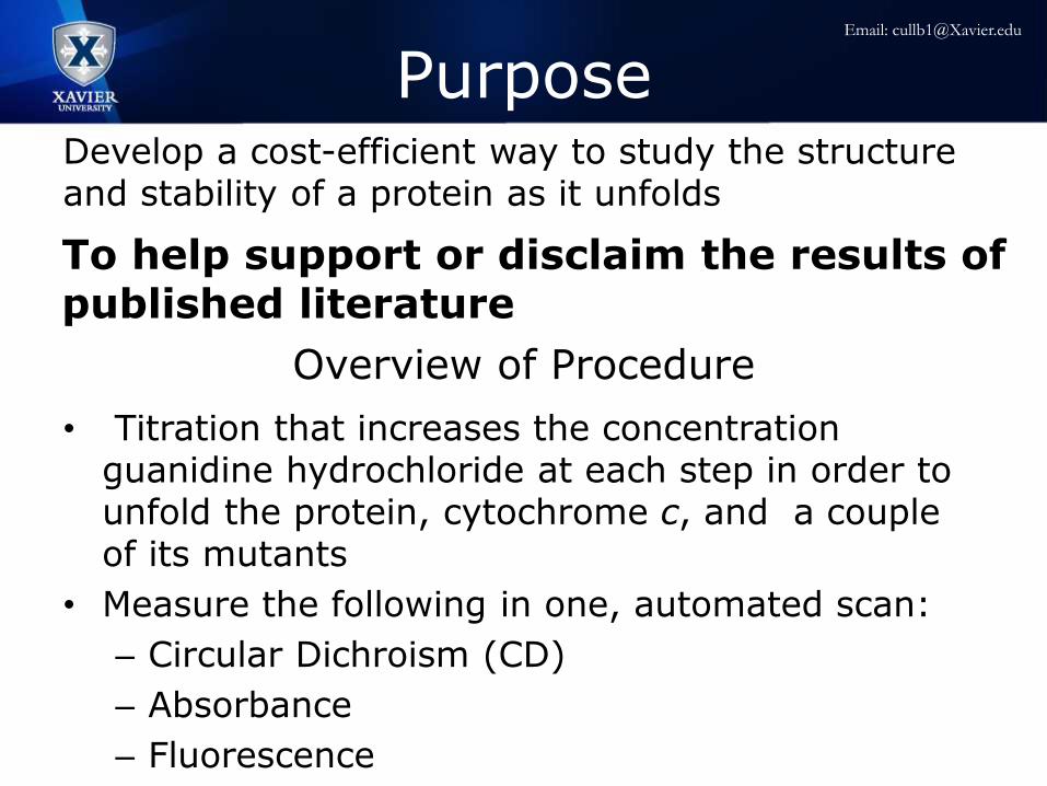
PurposeDevelop a cost-efficient way to study the structure and stability of a protein as it unfolds
Overview of Procedure
• Titration that increases the concentration guanidine hydrochloride at each step in order to unfold the protein, cytochrome c, and a couple of its mutants
• Measure the following in one, automated scan:
– Circular Dichroism (CD)
– Absorbance
– Fluorescence
To help support or disclaim the results of published literature
Email: [email protected]

Equine Cytochrome c
• Model System
• Well characterized
• Relatively small in size
• Single Tryptophan
molecule
• Cofactor: Heme
group
Zang C., et al. (2009) J Am Chem Soc 131(8):2846–2852
Email: [email protected]

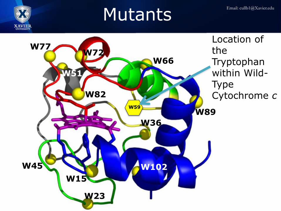
Mutants
W89
W66
W77W72
W82
W36
W23
W102W45
W15
W51
Location of the Tryptophan within Wild-Type Cytochrome c
W59
Email: [email protected]

Spectroscopic Techniques
Fluorescence monitors proximity of hemerelative to tryptophan
Zang C., et al. (2009) J Am ChemSoc 131(8):2846–2852
Absorbance monitors the local environment of the heme
Circular dichroism determines the components of secondary structure
W59
Email: [email protected]
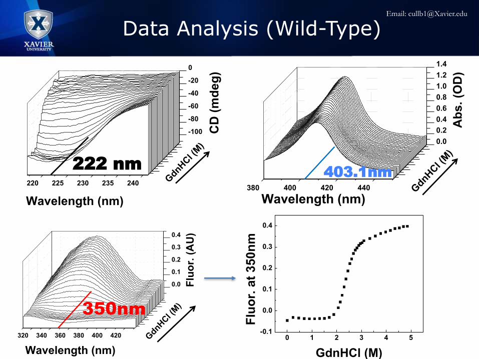
Data Analysis (Wild-Type)
320 340 360 380 400 420
0.0
0.1
0.2
0.3
0.4
GdnH
Cl (
M)
Wavelength (nm)
Flu
or.
(A
U)
220 225 230 235 240
-100
-80
-60
-40
-20
0
GdnH
Cl (
M)
Wavelength (nm) C
D (
md
eg
)
380 400 420 440
0.0
0.2
0.4
0.6
0.8
1.0
1.2
1.4
GdnH
Cl (
M)
Wavelength (nm)
Ab
s.
(OD
)
222 nm403.1nm
350nm
0 1 2 3 4 5-0.1
0.0
0.1
0.2
0.3
0.4
Flu
or.
at
35
0n
m
GdnHCl (M)
Email: [email protected]

0 1 2 3 4 5-0.1
0.0
0.1
0.2
0.3
0.4
Flu
or.
at
35
0n
m
GdnHCl (M)
Data Analysis (Wild-Type Cross Section)
0 1 2 3 4 5-0.1
0.0
0.1
0.2
0.3
0.4
Flu
or.
at
35
0n
m
GdnHCl (M)
0 1 2 3 4 5
0.85
0.90
0.95
1.00
1.05
1.10
1.15
1.20
Ab
s.
at
40
3.1
nm
GdnHCl (M)
0 1 2 3 4 5
-100
-80
-60
-40
-20
0
CD
at
22
2n
m
GdnHCl (M)
0 1 2 3 4 5
0.85
0.90
0.95
1.00
1.05
1.10
1.15
1.20
Ab
s.
at
40
3.1
nm
GdnHCl (M)0 1 2 3 4 5
-100
-80
-60
-40
-20
0
CD
at
22
2n
m
GdnHCl (M)
𝑆𝑜𝑏𝑠
=𝐶𝑓 +𝑚𝑓 𝐺𝑑𝑛𝐻𝐶𝑙 + 𝐶𝑢 +𝑚𝑢 𝐺𝑑𝑛𝐻𝐶𝑙 𝑒
−Δ𝐺+𝑚𝑔[𝐺𝑑𝑛𝐻𝐶𝑙]
𝑅𝑇
1 + 𝑒−Δ𝐺+𝑚𝑔[𝐺𝑑𝑛𝐻𝐶𝑙]
𝑅𝑇
Email: [email protected]

0 1 2 3 4 5
0.0
0.2
0.4
0.6
0.8
1.0
Abs. at 403.1nm (OD)
Fra
cti
on
Un
fold
ed
GdnHCl (M)0 1 2 3 4 5
0.0
0.2
0.4
0.6
0.8
1.0
CD at 222nm (mdeg)
Fra
cti
on
Un
fold
ed
GndHCl (M)
0 1 2 3 4 5
0.0
0.2
0.4
0.6
0.8
1.0
Fluor. at 350nm (AU)
Fra
cti
on
Un
fold
ed
GdnHCl (M)
Technique ΔG (kcal/mol) Cm (M)
CD 7.83 2.30
Absorbance 7.12 2.25
Fluorescence 7.71 2.34
Published (CD) 7.27 2.42
Published (HX) 7.40 N/A
Knapp, JA and Pace CN. Biochemistry 13, 1289-94. (1974).
Maity, H et al. J Mol Biol 343, 223-33. (2004).
Data Analysis (Wild-Type Cross Section)
Email: [email protected]

Fraction Unfolded (Wild-Type)
Technique ΔG (kcal/mol) Cm (M)
CD 7.83 2.30
Absorbance 7.12 2.25
Fluorescence 7.71 2.34
Published (CD) 7.27 2.42
Published (HX) 7.40 N/A
Global Fit 7.83 2.30
0 1 2 3 4 5-0.2
0.0
0.2
0.4
0.6
0.8
1.0
Individual Fit
CD Fit
CD Data
Abs Fit
Abs Data
Fluor Fit
Fluor Data
Fra
cti
on
Un
fold
ed
GdnHCl (M)0 1 2 3 4 5
-0.2
0.0
0.2
0.4
0.6
0.8
1.0
Global FIt
Global Fit
CD
Absorbance
Fluorescence
Fra
cti
on
Un
fold
ed
GdnHCl (M)
Knapp, JA and Pace CN. Biochemistry 13, 1289-94. (1974).
Maity, H et al. J Mol Biol 343, 223-33. (2004).
Email: [email protected]
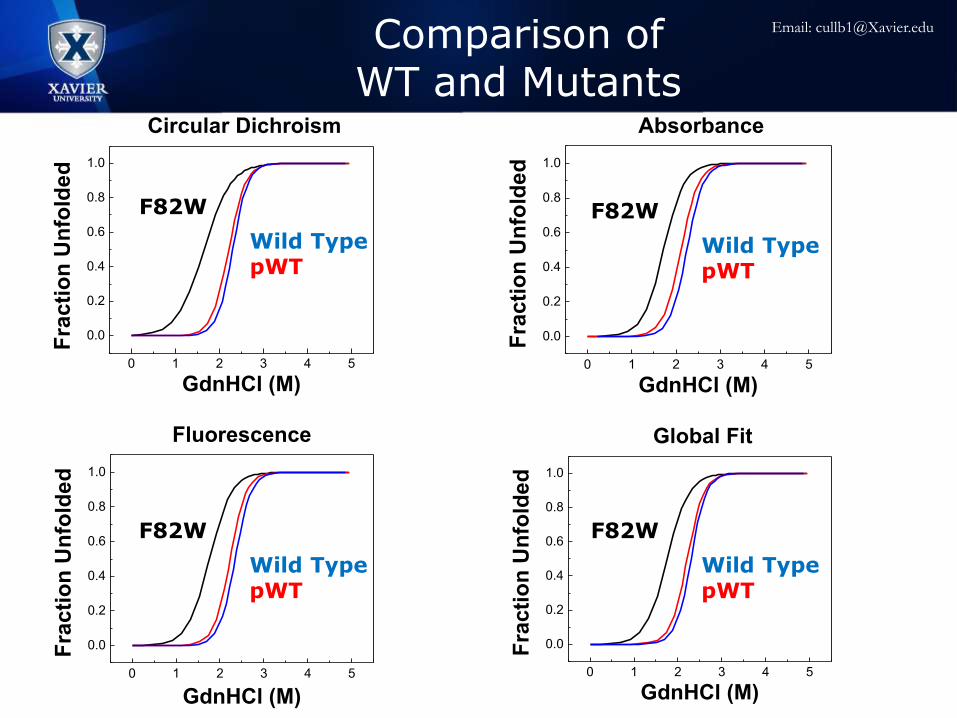
0 1 2 3 4 5
0.0
0.2
0.4
0.6
0.8
1.0
Circular Dichroism
Fra
cti
on
Un
fold
ed
GdnHCl (M)
F82W
pWT
Wild-Type
0 1 2 3 4 5
0.0
0.2
0.4
0.6
0.8
1.0
Global Fit
Fra
cti
on
Un
fold
ed
GdnHCl (M)
F82W
pWT
Wild-Type
0 1 2 3 4 5
0.0
0.2
0.4
0.6
0.8
1.0
Fluorescence
Fra
cti
on
Un
fold
ed
GdnHCl (M)
F82W
pWT
Wild-Type
0 1 2 3 4 5
0.0
0.2
0.4
0.6
0.8
1.0
Absorbance
Fra
cti
on
Un
fold
ed
GdnHCl (M)
F82W
pWT
Wild-Type
Wild TypepWT
F82W
Wild TypepWT
F82W
Wild TypepWT
F82W
Wild TypepWT
F82W
Comparison of WT and Mutants
Email: [email protected]
Acknowledgements
• Dr. Justin J Link
• Ben Kelty
• Previous Research Students
• Xavier University Physics Department
• John Hauck Foundation
Email: [email protected]

0 1 2 3 4 5
0.0
0.2
0.4
0.6
0.8
1.0
Circular Dichroism
Fra
cti
on
Un
fold
ed
GdnHCl (M)
F82W
pWT
Wild-Type
0 1 2 3 4 5
0.0
0.2
0.4
0.6
0.8
1.0
Global Fit
Fra
cti
on
Un
fold
ed
GdnHCl (M)
F82W
pWT
Wild-Type
0 1 2 3 4 5
0.0
0.2
0.4
0.6
0.8
1.0
Fluorescence
Fra
cti
on
Un
fold
ed
GdnHCl (M)
F82W
pWT
Wild-Type
0 1 2 3 4 5
0.0
0.2
0.4
0.6
0.8
1.0
Absorbance
Fra
cti
on
Un
fold
ed
GdnHCl (M)
F82W
pWT
Wild-Type
Comparison of WT and Mutants
Wild TypepWT
F82W
Wild TypepWT
F82W
Wild TypepWT
F82W
Wild TypepWT
F82W
Email: [email protected]
Related Documents