Special Issue on the 0 CAt Third Pacific Vascular Symposium 1856 Part I HAWAI I MEDICAL JO URNAL April 2000 Volume 59, No. 4 ISSN: 0017-8594 4 ——

Welcome message from author
This document is posted to help you gain knowledge. Please leave a comment to let me know what you think about it! Share it to your friends and learn new things together.
Transcript

Special Issue on the0CAt
Third Pacific Vascular Symposium1856
Part I
HAWAIIMEDICAL
JOURNALApril 2000 Volume 59, No. 4 ISSN: 0017-8594
4 ——

Some plans promise you a voice.HMSA delivers.
As a nonprofit mutual benefit society,
HMSA looks to leaders from all areas of our community
to help guide our Association. Our volunteer
board of directors is made up of people from the community,
business, labor, government, the clergy and, yes, medicine.
Of our 27 directors, seven are Hawaii practicing physicians.
As members of our board and numerous
advisory committees,
doctors have a say in almost everything we do.
HMSA. Now that’s a health plane
MSA
HAWAIIMEDICAL
JOURNAL(USPS 237-640)
Published monthly by theHawaii Medical Association
Incorporated in 1856 under the Monarchy1360 South Beretania. Second Floor
Honolulu. Hawaii 96814Phone (808) 536-7702: Fax (808) 528-2376
EditorsEditor: Norman Goldstein MD
News Editor: Henry N. Yokoyama MDContributing Editor: Russell T. Stodd MD
Editorial BoardVincent S. Aoki MD, Benjamin W. Berg MD,
John Breinich MLS, Satoru Izutsu PhD,James Lumeng MD, Douglas G. Massey MD,Myron E. Shirasu MD, Frank L. Tabrah MD.
Alfred D. Morris MD
Journal StaffManaging Editor: Becky KendroEditorial Assistant: Drake Chinen
OfficersPresident: James Lumeng MD
President-Elect: Philip Hellreich MDSecretary: Gerald McKenna MD
Treasurer: Paul DeMare MDPast President: Patricia L. Chinn MD
County PresidentsHawaii: David Camacho MD
Honolulu: Walter K.W. Young MDMaui: Michael Savona MD
West Hawaii: Ali Bairos MDKauai: Patrick Aiu MD
Advertising RepresentativeRoth Communications
2040 Alewa DriveHonolulu, Hawaii 96817
Phone (808) 595-4124Fax (808) 595-5087
The Journal cannot be held responsible for opinions expressed inpapers. discussion, communications or advertisements. The advertising policy of the HawaiiMedical Journal is governed ba therules of the Council on Drugs of the American Medical Association. The right is reservedtoreject material submitted foreditorialor adsertising columns. The Hawaii Medical Journal USPS237M0 ispuhlishedmonthlyby theHawaii Medical Association(ISSN 00)7-8594). 1360 South Beretania Street. Second Floor,Honolulu, Hawaii 96814.
Postmaster: Send address changes to the Hawaii MedicalJournal. 1360 South Beretania Street. Second Hoor. Honolulu.Hawaii 96814. Periodical postage paid at Honolulu. Hawaii.
Nonmember subscriptions are $25. Copyright 1999 by theHawaii Medical Association, Printed in the U.S.
Contents
EditorialNorman Goldstein MD 116
Guest EditorialBo EklofMD and Bob Kisrner MD 116
Medical School HotlineSteven E. Seifried PhD 117
Program of the Third Pacific Vascular Symposium
Photos from the Third Pacific Vascular Symposium
Faculty of the Third Pacific Vascular Symposium
Scientific Articles of the Third Pacific Vascular Symposium
119
121
122
124
News and NotesHenry N. Yoko3’ama MD 170
Classified Notices 171
WeathervaneRussell T. Stodd MD 174
t
I’ I
reserved by the artist.Cover art by Dietrich Varez, Volcano, Hawaii. All rights
lokini Barracks
lolani Barracks was built in 1870 for the Royal Household Guards.
HAWAII MEDICAL JOURNAL, VOL 59, APRIL 2000
115

Editorial D Guest Editorial
Norman Goldstein MDEditor, Hawaii Medical Journal
Pacific Vascular Symposium on VenousDisease
When Bo Ekiof, MD, asked if the Hawaii Medical Journal couldpublish the proceedings and discussions of a symposium held inNovember 1999, I told him that, because of the excellent speakers,this symposium would be well received by HMA members andother readers of the Journal.
We have previously published reports of scientific meetings anddevoted Special Issues to particular fields of medicine to informphysicians of the latest advancements in many fields of medicine toenhance understanding beyond our own specialties. The addedcomment to the presentations have always made the more formalpapers stimulating.
This issue on the treatment of venous disease will be of interest toall physicians. When I saw the proposed list of more than 40 of theworld’s leaders in phiebology, I did accept the Proceedings. However, because of the volume of the presentations and discussions, wecannot publish them in a single issue. More will follow.
Thanks to Bo Eklof, MD, and Bob Kistner, MD, co-chairmen ofthe Symposium, and to the presenters from around the world. TheseSpecial Issues will comprise a textbook on vascular disease.
Robert L Kistner MDBo Ekiof MD, PhD
Co-Chairmen, The Third Pacific Vascular Symposium
The Third Pacific Vascular Symposium on Venous Disease tookplace at Mauna Lani Bay Hotel on the Big Island of Hawaii onNovember 2-6, 1999. The faculty included more than 40 of theleaders in phlebology, representing five continents. The generaltheme was to contrast aggressive treatment of acute and chronicvenous disease with a more conservative approach.
The first day featured a workshop on venous interventions withpractical demonstrations of percutaneous interventions for acuteand chronic venous disease. The second day focused on treatmentand outcomes of DVT of the three segments of the leg followed bya session on air travel-related DVT. The third and fourth days weredevoted to the controversy on management of venous ulcers. Thelast day of the symposium featured a workshop on treatment ofvaricose veins and spider veins with practical demonstrations ofduplex guided sclerotherapy, lasers and new surgical developments.
We were excited when Norman Goldstein, the Editor for theHawaii Medical Journal, warmly embraced the idea to publish theproceedings of this very lively meeting. In this issue the short papersand heated discussions from the second day are presented.
We thank the staff of the Straub Foundation and the editorialoffice of the journal for their excellent work. Enjoy the informationon the latest news on management of DVT as it transpired from thePearl of Hawaiian hotels surrounded by the Five Mountains in themidst of Hawaiiana.
Bo Eklof, MD, PhDCo-Chairman
Robert L. Kistner, MDCo-Chairman
HAWAII MEDICAL JOURNAL, VOL 59, APRIL2000
116

Medical School Hotline
Genetics in the John A.. Burns School ofMedicine Curriculum:
a nationally recognized need and an opportunity for curricular Integration
Steven E. Seitried, PhDAssociate Professor of Physical BiochemistryDepartment of Genetics and Molecular Biology
The New Genetics of The Common DisorderGenetic diseases are not rare. Their impact lies far beyond cysticfibrosis, sickle cell and thallasemia. Hypertension, alcoholism,depression, diabetes, and cancer are now recognized to be, to someextent, genetic diseases. Although we do not yet know all the details,it is clear that genetic background can define predisposition to anumber of significant etiologies. Medical genetics is one of the mostrapidly advancing areas of medical practice. The knowledge ofmedical genetics that is necessary for medical practice is likely to bevery different in 5 or 10 years than it is now. Every physician whopractices in the 21st century will require a basic knowledge of theprinciples of human genetics and their application to a wide varietyof clinical problems.
The Human Genome Project is predicted to be complete by the50th anniversary of Watson and Crick’s Double Helix paper inNature. Thousands of researchers working to apply the derivedresults will yield rapid advances in genetic medicine. There has beenan explosion in the number and membership of professional organizations related to human genetics. New health care professions andcareer paths have rapidly evolved.
Because of the shear number of common gene-related diseases,and the advent of managed care, there is a general recognition thatthe General Practitioner will necessarily fill some of the roles ofgenetic councilor and diagnostician as DNA chips, family genetichistory-taking tools, and genotype-specific interventions becomecommon practice.
Recognition of a Needfor a Curriculum of The New GeneticsA recent issue of the Association of American Medical Colleges(AAMC) Newsletter’ quotes “Students must understand that if theywant to be a good internist, say, they have to understand genetics.They don’t expect to be a good physician without learning about thekidney-well, they can’t ignore genetics any more than they canignore the kidney.” The American Medical Association (AMA) hasundertaken a significant effort to develop CME courses in genetics.The latest AAMC exit poll reports 44% of new graduates wereunsatisfied with the amount of genetics they received in theircurriculum. A 1997 study found that almost a third of physicianssurveyed could not distinguish an inconclusive result from a negative one in a genetic test for colon cancer. Partly in recognition ofthese findings, genetics items have recently been added to the USMedical Licensing Examination (USMLE) Content Outline andGenetics sub scores are reported for Part I of the USMLE BoardExamination. The AAMC is concerned enough about this issue that
it hosted a series of Focus Sessions relating to the development ofgenetic curricula at its most recent national meeting.
The New Generics in the JABSOM Curriculum: Building on National Recommendations“The New Genetics” provides an opportunity to deliver an integrated curriculum, including basic science, clinical science, publichealth and epidemiology, ethics and cultural issues. This “GPEC”(Genetics, Public Health, Ethics and Cultural) curriculum would gobeyond the GEE (Genetics, Epidemiology, and Ethics) curriculumrecently rolled out by the University of Vermont with great interestand fanfare. With the recognition ofPublic Health as unfolding fromepidemiology, and the cultural complexity of our Hawaii, there areunique opportunities to develop a “whole” (not “complete”) PBLcurriculum with the new genetics as a longitudinal binding theme.An attractive attribute of this plan is a mechanism by which facultyof the School of Public Health can become involved in MD Programcurriculum design and delivery.
The American Society of Human Genetics has developed aMedical School Core Curriculum2to provide guidance to deans andcurriculum committees regarding knowledge, skills, and attitudesrelated to medical genetics that are likely to be needed by all currentmedical students during their careers as physicians. Their generalized recommendations follow:
Medical genetics is both a basic biomedical science and a clinicalspecialty; it is insufficient to teach it as either alone. Teaching inmedical genetics must span the entire undergraduate medical schoolcurriculum and must continue into the postgraduate years as well.
Medical genetics must be explicitly included in the curriculum.Although some aspects of medical genetics overlap with and may betaught by other disciplines, students are unlikely to learn what theyneed unless specific learning objectives in medical genetics areestablished for them. This is especially true of issues that lie at theheart of medical genetics, such as the importance of disease prediction and prevention, the appropriate application of novel scientificdiscoveries to clinical care, and the nondirective approach to counseling.
A person or committee should be given specific responsibility forthe curriculum in medical genetics at each medical school. Thisresponsibility should extend throughout the entire undergraduatecurriculum and should include involvement in all courses thatcontain (or that should contain) material related to medical genetics.
Medical genetics can be taught effectively by a variety ofdifferentmethods and in various formats. Problem-based learning is particularly well-suited to medical genetics, which involves theintegration of skills and knowledge from many different fields[emphasis added].
The Association of Professors of Human or Medical Geneticsreport entitled “Clinical Objectives in Medical Genetics for Undergraduate Medical Students”3 defines the knowledge, skills andattitudes in genetics that all medical students should achieve duringthe clinical phase of their education. These objectives complementthose of the ASHG Medical School Core Curriculum in Genetics,which covers both basic science and clinical aspects of medicalstudent education. The reader will find many behavioral, populational, cultural, and clinical issues listed in the objectives enumerated by this organization.
HAWAII MEDICAL JOURNAL, VOL 59, APRIL 2000117

The desired outcome of an integrated genetic curriculum at JAB SOMis to prepare the student to:
1) practice modern medicine which includes recognizing the role ofgenetic factors in health and disease. This requires knowledge of thestructure, function, and transmission of genes and understanding ofinteractions both among genes and between genes and the environment.
2) synthesize factual material related to genetic diseases and congenital anomalies and to use this information to formulate anappropriate plan for diagnostic evaluation and patient management.Students will learn to communicate information regarding geneticconditions, clearly, nondirectively, and without personal bias, topeople from greatly differing educational, socioeconomic, ethnic.and cultural backgrounds.
3) be sympathetic, nonjudgmental. and nondirective counselorswho recognize their own limitations, seek consultation whenevernecessary. and become lifelong, self-motivated learners.
References1. Not Your Fathers Genetics Curriculum. http://www.aamc.ora/newsroom/reporter/oct99/oeneticu.htm.
AAMC Reporter:9, 1999.2. Report trom the ASHG Intormation and Education Committee: Medical School Core Curriculum in
Genetics. http://www.faseb.orofaenetics/ashg/uolicv/reu-O1 .htm Am J. Hum. Gsnet. 56:535—537,1995.
3. Clinical Objectives in Medical Genetics tor Undergraduate Medmal Students. http:/!www.taseb.orglgeneticu/aphmg/aphmgt2.htm Am J Hum Genet 1995;56:535.537.
NEIGHBOR ISLANDS TOLL-FREE:1-800-362-3585
Free Hotline 24 Hours a Day.
POISON CENTER TIPS
• Keep the number of the Hawaii Poison Center onor near your telephone.
• If you suspect a poisoning, do not wait for signsand symptoms to develop. Call the Hawaii PoisonCenter immediately.
• Always keep Ipecac Syrup in your home. (This isused to make a person vomit in certain types ofpoisoning.) Do use Ipecac Syrupunless advised by the Hawaii PoisonCenter.
• Store all medicines, chemicals, and householdproducts out of reach and out of sight, preferablylocked up.
• A good rule to teach children is to “always askfirst” before eating or drinking anything—don’ttouch, don’t smell, don’t taste.
Mail checks, payable to:Hawaii Poison Center
1319 Punahou Street, Honolulu, HI 96826
OAHU: 941-4411
Air Force Healthcare.Good Pay.
Professional Respect.Why Do You
ThiiikWe Say ‘Aim High”?
Experience the best of everytlEling. Bestfacilities. Best benefits. Outstandingopportunities for travel, 30 days vacationwith pay, training and advancement.
For an information packet call
1-800-423-USAFor visit www.airforce.com.
You’ll see why we say, “Aim High’
AIM HIGH
HEALTH PROFESSIONS
Donate to help us save lives.

introducingthe only daily facial moisturizer containing Parsol® 1789.
Cetaphil Daily Facial Moisturizer with SPF 15
• Filters UVA and UVB rays for maximum sun protection
• Offers gentle moisturization for all skin types
• Can be worn under makeup — lightweight and non-greasy
• Non-comedogenic
• The OTC Skincare line most recommended by dermatologists*
The ina1 word in aerioua akin care. Cetaphilt.
www.cetphil.com
GALDERMA* Data on file, Galderma Laboratories, LP. ©1999 Galderma Laboratories, L.P. GALDERMA isa registered trademark. CET-249-1199Patent and trademark rights for Parsol’ 1789 owned by Givaudon-Roure Corp.

ACKNOWLEDGEMENTS
Straub Foundation gratefully acknowledges the following companies for their support ofthe Third Pacific Vascular Symposium on Venous Disease:
MAJOR SUPPORTERS: ESC Medical Systems Inc.Abbott Laboratories EthiconW.L. Gore & Associates, Inc. GE Medical SystemsImpra, Inc. Genentech, Inc.Rhone-Poulenc Rorer Pharmaceuticals Inc. General Surgical InnovationsWyeth-Ayerst Laboratories Guidant Corporation
Hoechst Marion Roussel, Inc.SUPPORTERS: Huntleigh Health CareAbbott Critical Care Medi USAACI Medical Inc. Medi BayreuthlMedi UKAdvanced Technology Laboratories, Inc. Novartis Pharmaceuticals CorporationAstra Pharmaceuticals Nycomed AmershamB. BraunfMcGraw Ortho-McNeil PharmaceuticalBaxter Healthcare Corporation Otsuka America Pharmaceutical, Inc.Beiersdorf - Jobst, Inc. Pharmacia & UpjohnBoston Scientific Corporation Possis Medical, Inc.Cook Sigvaris Inc.Cordis Endovascular STD PharmaceuticalCryoLife, Inc. Venosan North America Inc.Currie Medical Specialties, Inc. VenPro CorporationDuPont Pharmaceuticals VNUS Medical Technologies, Inc.
STRAUB FOUNDATIONpresents
HeartATTAC K
and BrainATTAC K
in the New MillenniumA Symposium on Prevention and Treatment
HAWAII MEDICAL JOURNAL, VOL 59, APRIL 2000120
Hilton Hawaiian Village, Tapa Ballroom
Professional MeetingMay 25 and May 26, 2000
Free Public MeetingMay 27, 2000
Supported by HMSA Foundation andThe Queen’s Medical Center
To register for both meetings, contact:STRAUB FOUNDATION1100 Ward Avenue, Suite 1045Honolulu, HI 96814Telephone: (808) 524-6755Fax: (808) 531-0123www.straub-foundation.org
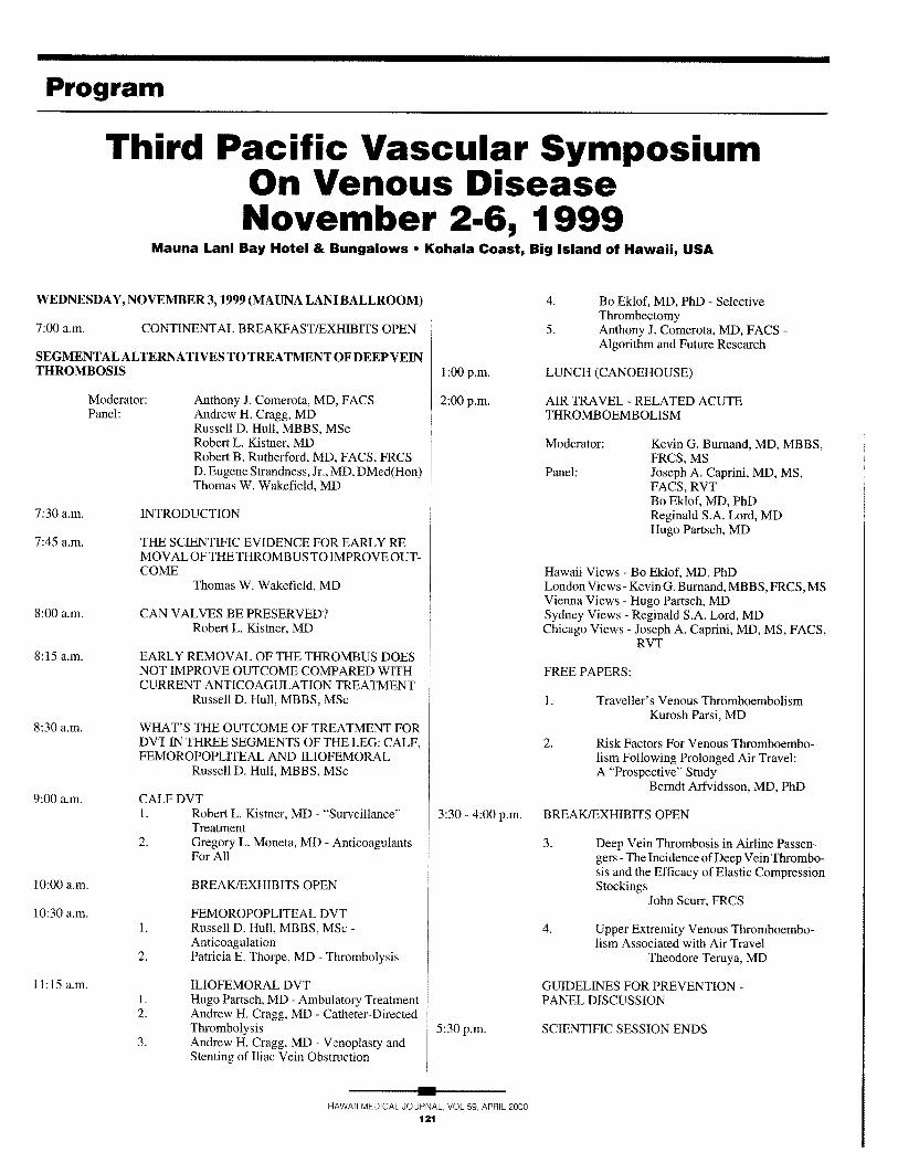
Program
Third Pacific Vascular SymposiumOn Venous DiseaseNovember 2-6, 1999
Mauna Lani Bay Hotel & Bungalows • Kohala Coast, Big Island of Hawaii, USA
WEDNESDAY, NOVEMBER 3, 1999 (MAUNA LAN! BALLROOM)
7:00 am. CONTINENTAL BREAKFAST/EXHIBITS OPEN
SEGMENTAL ALTERNATIVES TO TREATMENT OF DEEP VEINTHROMBOSIS
7:30 am. INTRODUCTION
Anthony J. Comerota, MD, FACSAndrew H. Cragg. MDRussell D. Hull, MBBS, MScRobert L. Kistner, MDRobert B. Rutherford, MD, FACS, FRCSD. Eugene Strandness, Jr., MD, DMed(Hon)Thomas W. Wakefield, MD
7:45 am. THE SCIENTIFIC EVIDENCE FOR EARLY REMOVAL OF THE THROMBUS TO IMPROVE OUTCOME
8:00 a.m. CAN VALVES BE PRESERVED?Robert L. Kistner. MD
8:15 am. EARLY REMOVAL OFTFIE THROMBUS DOESNOT IMPROVE OUTCOME COMPARED WITHCURRENT ANTICOAGULATION TREATMENT
Russell D. Hull, MBBS, MSc
8:30 a.m. WHAT’S THE OUTCOME OF TREATMENT FORDVT IN THREE SEGMENTS OF THE LEG: CALF,FEMOROPOPLITEAL AND ILIOFEMORAL
Russell D. Hull, MBBS, MSc
9:00 am. CALF DVT1. Robert L. Kistner. iD - “Surveillance”
Treatment2. Gregory L. Moneta, MD - Anticoagulants
For All
10:30 am. FEMOROPOPLITEAL DVT1. Russell D. Hull, MBBS, MSc -
Anticoagulation2. Patricia E. Thorpe, MD - Thrombolysis
ILIOFEMORAL DVT1. Hugo Partsch, MD - Ambulatory Treatment2. Andrew H. Cragg, MD - Catheter-Directed
Thrombolysis3. Andrew H. Cragg, MD - Venoplasty and
Stenting of Iliac Vein Obstruction
4. Bo Ekiof, MD, PhD - SelectiveThrombectomy
5. Anthony J. Comerota, MD, FACS -
Algorithm and Future Research
Kevin G. Burnand, MD, MBBS,FRCS, MSJoseph A. Caprini, MD, MS.FACS, RVTBo Eklof, MD, PhDReginald S.A. Lord, MDHugo Partsch, MD
Hawaii Views - Bo Ekiof. MD, PhDLondon Views - Kevin G. Bumand, MBBS, FRCS, MSVienna Views - Hugo Partsch, MDSydney Views - Reginald S.A. Lord, MDChicago Views - Joseph A. Caprini, MD, MS. FACS,
RVT
FREE PAPERS:
1. Traveller’s Venous ThromboembolismKurosh Parsi, MD
2. Risk Factors For Venous Thromboembolism Following Prolonged Air Travel:A “Prospective” Study
Berndt Arfvids son, MD, PhD
3. Deep Vein Thrombosis in Airline Passengers - The Incidence of Deep Vein Thrombosis and the Efficacy of Elastic CompressionStockings
John Scuff, FRCS
4. Upper Extremity Venous Thromboembolism Associated with Air Travel
Theodore Teruya, MD
GUIDELINES FOR PREVENTION -
PANEL DISCUSSION
HAWAII MEDICAL JOURNAL, VOL 59. APRIL 2000121
Moderator:Panel:
1:00 p.m. LUNCH (CANOEHOUSE)
2:00 p.m. AIR TRAVEL - RELATED ACUTETHROMBOEMBOLISM
Moderator:
Panel:
Thomas W. Wakefield, MD
10:00 a.m. BREAK/EXHIBITS OPEN
3:30 - 4:00 p.m. BREAK/EXHIBITS OPEN
11:15 a.m.
5:30 p.m. SCIENTIFIC SESSION ENDS

Program
MSBo Ekiof, MD, PhDErmenegildo A. Enrici, MD
“How my method of treatment changes the pathophysiology to heal theulcer and prevent recurrence?”
1. Philip D. Coleridge Smith, DM, MA, FRCS - Drugs Are the UltimateAnswer2. Gregory L. Moneta, MD - Wrapping3. Peter Gloviczki, MD - Sepsing4. John J. Bergan, MD, FACS, Hon,FRCS(Eng) - Stripping5. Michel Perrin, MD - Repairing Primary Reflux6. Seshadri Raju, MD - Repairing Secondary Incompetence
VENOUS ULCERPART TWO ANY CONTROVERSIES IN DIAGNOSIS?
Seshadri Raju, MDPhilip D. Coleridge Smith, DM,MA, FRCSKenneth A. Myers, MS, FACS,FRCSFrank Padberg, Jr., MD, FACSD. Eugene Strandness, Jr., MD,DMed(Hon)David S. Sumner. MD
LEVEL 1 - THE OFFICEI. Kenneth A. Myers. MS. FACS. FRACS -
Clinical Evaluation2. David S. Sumner. MD - The Haid-Held
Doppler3. Andre Cornu-Thenard. MD - The French C
in CEAP4. Frank Padberg, Jr., MD, FACS - Sensory
Impairment
10:30 am. BREAKJEXHIBITS OPEN
11:00 a.m. LEVEL 2- THE VASCULAR LAB1. Jan HoIm, MD - Reliability of Clinical Diag
nosis2. D. Eugene Strandness. Jr.. MD. DMed(Hon)
- Duplex in Reflux3. Paul R. Cordts. LTC. MC - Plethvsmogra
phy in Reflux4. Peter N. Neglen. MD. PhD - Evaluation of
Obstruction
12:30 p.m. LEVEL 3 - INVASIVE PROCEDURESI. Seshadri Raju, MD - Descending Venogram2. Kevin G. Burnand, MBBS, FRCS, MS - The
St. Thomas Way
1:30 p.m. SCIENTIFIC SESSION ENDS
MANAGEMENT OF THE DECOMPENSATED
Thomas F. O’Donnell, Jr., MDMichael C. Dalsing, MDRalph G. DePalma, MD, FACSPeter Gloviczki, MDRobert L. Kistner, MDMichel Perrin, MDSeshadri Raju, MD
VENOUS ULCER AND CEAPNicos Labropoulos, PhD, DIC, RVT
The management will be demonstrated by presentation of cases that willillustrate increasing severity of chronic venous disease. Each case will behandled by a group of specialists that will propose diagnostic measures andtreatment. Their suggestions will be discussed by the panel and opened upto the audience for wider participation.
8:00 a.m. CASE OF SUPERFICIAL INCOMPETENCE
A 65-year old lady from the Big Island with 5 children developed largevaricose veins during her pregnancies. She has had recurrent large ulcerations on her right leg for 30 years and within the last 6 years has developeda painful, circumferential ulceration on the right leg.
Specialists: Gianni Belcaro, MD, PhD (Save the Saphenous Vein)G. Mark Malouf, MBBS, FRACS, FRC (High Ligationand Stripping)J. Leonel Villavicencio, MD. FACS (Sclerotherapy)
9:00 a.m. CASE OF PERFORATOR INCOMPETENCE
65-year old woman with large recurrent venous ulcerations on left lowerleg. Past history is significant for left GSV ligation and stripping, 1979 andleft popliteal and superficial femoral vein DVT, 1989. Laboratory evaluation now shows the following: no significant deep venous obstruction andmild reflux (VFI 3.2 mllsec) by APG: Duplex scan shows mild recanalization changes of superficial femoral and popliteal veins, no deep reflux and3 large incompetent perforated veins in the medial aspect of the lower leg.
Specialists: Ralph G. DePalma. MD, FACS (SEPS)Ermenegildo A. Enrici, MD (Open Perforator Ligation)Jean-Jerome Guex. MD (Ultra-Sound Guided Sclerotherapy)
10:00 am. BREAKJEXHIBITS OPEN
10:30 am. CASE OF PRIMARY DEEP VENOUSREFLUX
A 36-year old gentleman from Western Samoa has had recurrent venousulcerations of both legs that first began when he was 15 years old. He hasnot been able to work for the past 4 years because of painful, largeulcerations of the right leg.
THURSDAY, NOVEMBER 4, 1999 (MAUNA LANI BALLROOM)
7:00 am. CONTINENTAL BREAKFAST/EXHIBITS OPEN
VENOUS ULCERPART ONE - THE CONTROVERSIES IN TREATMENT
7:30 a.m. Moderator:Panel:
FRIDAY, NOVEMBER 5, 1999 (MAUNA LANI BALLROOM)
Ralph G. DePalma, MD, FACSKevin G. Burnand, MBBS, FRCS,
CONTINENTAL BREAKFAST/EXHIBITS OPEN7:00 am.
VENOUS ULCERPART THREE -
LEG
7:30 am.
7:45 a.m.
Moderator:Panel:
Moderator:Panel:
9:00 a.m.
HAWAII MEDICAL JOURNAL, VOL 59, APRIL 2000
122
Program
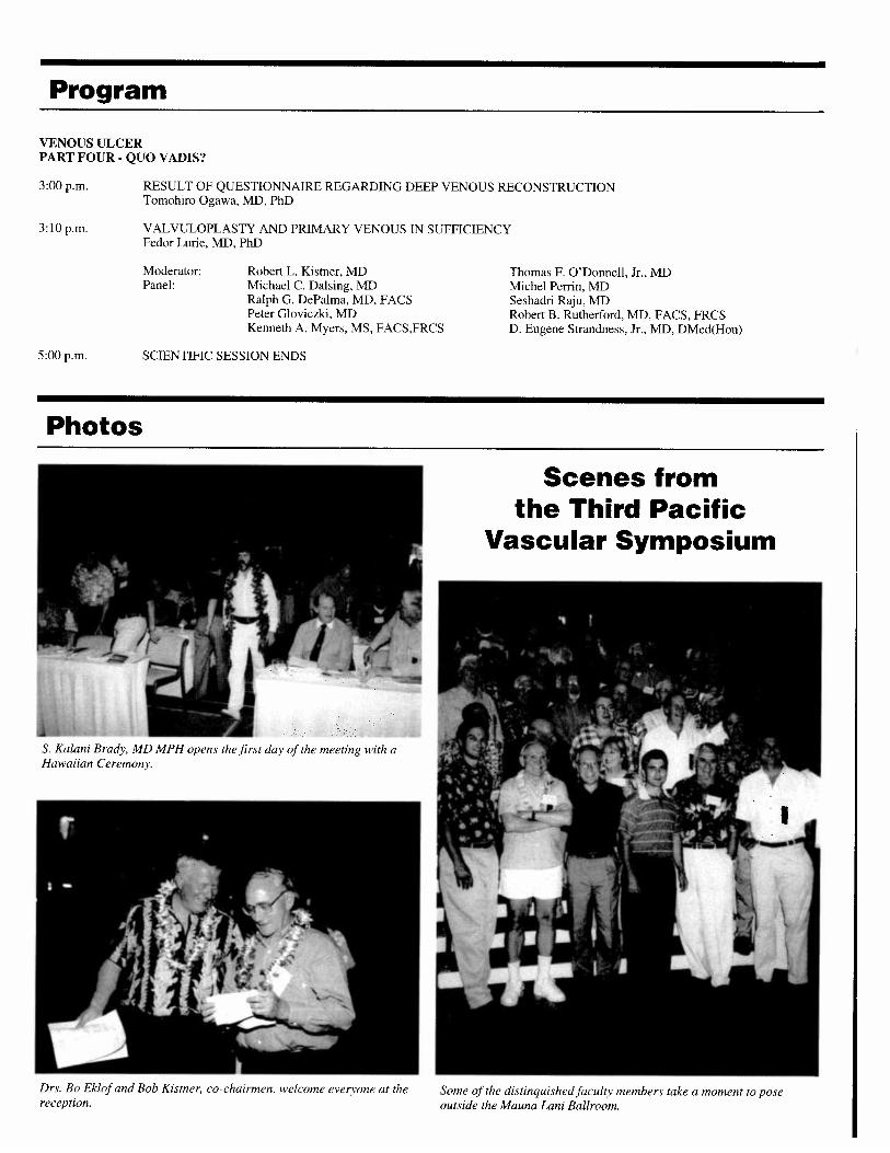
VENOUS ULCERPART FOUR - QUO VADIS?
3:00 p.m.
3:10 p.m.
RESULT OF QUESTIONNAIRE REGARDING DEEP VENOUS RECONSTRUCTIONTomohiro Ogawa, MD, PhD
VALVULOPLASTY AND PRIMARY VENOUS IN SUFFICIENCYFedor Lurie, MD, PhD
Moderator:Panel:
Robert L. Kistner, MDMichael C. Dalsing, MDRalph 0. DePalma, MD, FACSPeter Gloviczki, MDKenneth A. Myers, MS. FACS,FRCS
Thomas F. O’Donnell, Jr., MDMichel Perrin. MDSeshadri Raju, MDRobert B. Rutherford, MD, FACS, FRCSD. Eugene Strandness. Jr., D. DMed(Hon)
5:00 p.m. SCIENTIFIC SESSION ENDS
Photos
Scenes fromthe Third Pacific
Vascular Symposium
S. Kalani Brady, MD MPH opens the first day of the meeting with aHawaiian Ceremony.
Drs. Bo Ekiof and Bob Kismer. co-chairmen, welcome everyone at thereception.
Some of the distinquished faculty neinbers take a moment to pose
outside the Mauna Lani Ballroom.

FacultyMichael C. Dalsing, MDDirector of Vascular Surgery, Indiana University Medical School,Indianapolis, Indiana, USA
Peter W. Balkin, MDChief of Radiology. Straub Clinic & Hospital, Honolulu, Hawaii,USA
Gianni Belcaro, MD, PhDResearcher. Cardiovascular Institute. Chieti University. Pescara,ItalyDirector Angiology & Vascular Surgery. Pierangeli Clinic, Pescara.Italy
John J. Bergan, MD, FACS, Hon, FRCS(Eng)Professor of Surgery, University of California Medical School, SanDiego. California, USA
Kevin G. Burnand, MBBS, FRCS, MSProfessor of Vascular Surgery, Department of Surgery, St. Thomas’Hospital, London, United Kingdom
Joseph A. Caprini, MD, MS, FACS, RVTLouis W. Biegler Chair of Surgery, Northwestern University Medical School, Chicago, Illinois, USAProfessor of Bioengineering, Northwestern University, Evanston,Illinois, USADirector of Surgical Research, Evanston Northwestern Healthcare,Evanston, Illinois, USA
Philip D. Coleridge Smith, DM, MA, FRCSSenior Lecturer, Department of Surgery. UCL Medical School,London, United Kingdom
Anthony j. Comerota. ]fD, FACSProfessor of Surgery, Temple University School of Medicine,Philadelphia, Pennsylvania, USAChief of Vascular Surgery; Director. Center for Vascular Diseases,Temple University Hospital, Philadelphia, Pennsylvania, USA
Paul R. Cordts, LTC, MCClinical Assistant Professor of Surgery, USUHS, USAClinical Assistant Professor of Surgery, University of Hawaii, JohnA. Burns School of Medicine, Honolulu, Hawaii, USAVascular Surgeon, Tripler Army Medical Center, Honolulu, Hawaii, USA
Andre Cornu-Thenard, MDDirector of Phlebology Education. Saint Antoine Hospital. Paris.France
Andrew H. Cragg, MDClinical Partner. Minneapolis Vascular Institute. Minneapolis. Minnesota, USAClinical Professor of Radiology, University of Minnesota, Minneapolis. Minnesota. USAInterventional Radiologist, Fairview-University Medical Center,
Minneapolis, Minnesota, USA
Ralph G. DePalma, MD, FACSProfessor of Surgery. Vice Chair Surgery. Associate Dean. University of Nevada School of Medicine. Reno, Nevada, USAChief of Surgery, Reno VAMC, Reno. Nevada. USA
Bo Eklof, MD, PhDVascular Surgeon, Straub Clinic & Hospital. Honolulu, Hawaii.USAClinical Professor of Surgery, University of Hawaii, John A. BurnsSchool of Medicine, Honolulu. Hawaii, USAMedical Director, Straub Foundation. Honolulu, Hawaii, USA
Ermenegildo A. Enrici, MDChairman, Phleb/Lymph Department, Argentine Catholic University, Buenos Aires, ArgentinaHonorary Consultant, Central Military Hospital, Buenos Aires,ArgentinaAssociate Academic Member, Argentine Academy of Surgeons,Buenos Aires, Argentina
Peter Gloviczki, MDVice-Chair, Division of Vascular Surgery. Mayo Clinic, Rochester,Minnesota, USAProfessor of Surgery, Mayo Medical School, Rochester, Minnesota,USA
Gabriel Goren, MDDirector, Vein Disorders Center, Encino, CA, USA
J. Jerome Guex, MDVice-President, French Society of Phlebology. Nice, FranceTreasurer, International Union of Phlebology, Nice, France
Jan HoIm, MDAssociate Professor of Surgery, Department ofSurgery, SahlgrenskaUniversity Hospital, Goteborg, Sweden
Shunichi Hoshino, MD, PhDProfessor and Chainnan, Department of Cardiovascular Surgery,
Fukushima Medical University School of Medicine, Fukushima,Japan
Russell D. Hull, MBBS, MScDirector, Thrombosis Research Unit. Professor of Medicine, University of Calgary. Calgary. Alberta. Canada
Curtis B. Kamida, MDRadiologist, Straub Clinic & Hospital. Honolulu. Hawaii, USAClinical Associate Professor of Radiology, University of Hawaii,John A. Burns School of Medicine. Honolulu. Hawaii. USA
Robert L. Kistner, MDVascular Surgeon, Straub Clinic & Hospital, Honolulu. Hawaii.
HAWAII MEW CAL JOURNAL, VOL 59 APRIL 2000
124

USAClinical Professor of Surgery, University of Hawaii, John A. BurnsSchool of Medicine, Honolulu, Hawaii. USAPresident, Straub Foundation. Honolulu. Hawaii, USA
Nicos Labropoulos, PhD, DIC, RVTAssistant Professor of Surgery, Loyola University Medical Center,Maywood, Illinois, USADirector of Research, Vascular Diagnostics Ltd., Park Ridge, Illinois, USAVascular Consultant, West Suburban Hospital, Oak Park, Illinois,USA
Reginald S.A. Lord, MDProfessor, Department of Surgery, University of Sydney, Sydney,Australia
Fedor Lurie, MD, PhDDepartment of Anesthesiology, University of California DavisMedical Center, Sacramento, California, USA
G. Mark Malouf, MEBS, FRACS, FRCSSurgeon in Venous Disease, Westmead Hospital, Sydney, AustraliaPresident, Australian and New Zealand Society of Phlebology.Australia
Elna M. Masuda, MDVascular Surgeon, Straub Clinic & Hospital, Honolulu, Hawaii,USAClinical Assistant Professor of Surgery, University of Hawaii, JohnA. Bums School of Medicine, Honolulu, Hawaii. USA
Mark Mewissen, MDAssociate Professor, Department of Radiology, Medical College ofWisconsin, Milwaukee, Wisconsin, USA
Gregory L Moneta, MDProfessor of Surgery, Oregon Health Sciences University, Portland,Oregon, USA
Kenneth A. Myers, MS, FACS, FRACSProfessor, Monash Medical Centre and Epworth Hospital,Melbourne, Australia
Peter Neglen, MD, PhDVascular Surgeon, River Oaks Hospital. Jackson, Mississippi, USA
Thomas F. O’Donnell, Jr., MDPresident & CEO, New England Medical Center, Boston, Massachusetts, USAAndrews Professor of Surgery. Tufts University. Boston, Massachusetts. USA
Tomohiro Ogawa, MD, PhDCardiovascular Surgeon. Fukushima Medical University School ofMedicine, Fukushima, JapanVisiting Colleague, Department of Surgery. University of Hawaii,
John A. Bums School of Medicine, Honolulu, Hawaii, USAResearch Fellow, Straub Foundation, Honolulu, Hawaii, USA
Frank Padberg, Jr., MD, FACSProfessor of Surgery, UMDNJ — New Jersey Medical School,Newark, New Jersey, USA
Hugo Partsch, MDProfessor of Dermatology, University of Vienna, AustriaHead of Dermatological Department, Wilhelminen-Hospital, Vienna,AustriaPresident, Union Intemationale de Phiebologic. Vienna, Austria
Michel Perrin, MDAssociate Professor, University of Grenoble, Grenoble, France
John R. Pfeifer, MD, FACSProfessor of Surgery, University of Michigan School of Medicine,Ann Arbor, Michigan, USADirector, Division of Venous Disease, University of MichiganSchool of Medicine, Ann Arbor, Michigan, USA
Seshadri Raju, MDEmeritus Professor and Honorary Surgeon, University of Mississippi Medical Center, Jackson, Mississippi, USA
Robert B. Rutherford, MD, FACS, FRCSProfessor Emeritus, Department of Surgery, University of Colorado, Silverthorne, Colorado, USA
D. Eugene Strandness, Jr., MD, DMed (Hon)Professor, University of Washington, Seattle, Washington, USA
David S. Sumner, MDDistinguished Professor of Surgery, Emeritus; Southern IllinoisUniversity School of Medicine, Springfield, Illinois, USAPresident, American Venous Forum, USA
Patricia E. Thorpe, MDAssociate Professor of Radiology/Surgery, Creighton UniversityMedical Center, Omaha, Nebraska, USA
J. Leonel Villavicencio, MD, FACSProfessor of Surgery. Department of Surgery, Uniformed ServicesUniversity of the Health Sciences, Bethesda, Maryland, USASenior Consultant, Department of Surgery and Director of Venousand Lymphatic Teaching Clinics, Walter Reed Army MedicalCenter. Washington, DC, USA and Naval Medical Center, Bethesda,Maryland, USA
Thomas W. Wakefield, MDProfessor of Surgery, Section of Vascular Surgery, University ofMichigan Medical Center, Ann Arbor, Michigan, USA
Robert A. Weiss, MDAssistant Professor, Department of Dermatology, Johns HopkinsUniversity School of Medicine, Baltimore, Maryland, USA
HAWAII MEDICAL JOURNAL, VOL 59, APRIL 2000125

THE SCIENTIFIC EVIDENCE FOR EARLYREMOVAL OF THE THROMBUS TO IM
PROVE OUTCOME
Thomas W. Wakefield, MDUniversity of Michigan Medical Center
Ann Arbor, Michigan, USA
The pathophysiologic basis for early clot removal stems from theinflammatory process that accompanies venous thrombosis. Steward initially proposed four stages for this inflammatory response.1First, thrombus forms in the veins, with inflammatory cells andplatelets becoming activated within this initial thrombus. Second,further neutrophil and platelet activation occurs, generatingprocoagulant and inflammatory mediators. Third, coagulation occurs on activated platelet phospholipid surfaces, increasing thespeed of thrombin and fibrin generation. Finally, leukocytes andplatelets layer on top of existing clot, further increasing the thrombotic/inflammatory interplay. Inflammation ultimately leads to theamplification of thrombosis. driven by both tissue factor expressionon monocytes and by the exposure of vein wall collagen afterendothelial denudation, likely due to cathepsin G release fromactivated neutrophils.
The above results in leukocyte extravasation into the vein wall,migrating from both the adventitial and luminal surfaces. Using arodent model of stasis-induced venous thrombosis by JVC ligation,we have demonstrated an active vein wall pro-inflammatory response characterized by cellular trafficking. involving early neutrophil extravasation2followed by monocyte/macrophage infiltration,occurring as early as 6 hours after thrombus formation begins.3At6 hours, an increase in all leukocyte types (except for monocytes) inthe vein wall occurs, and by day 2, neutrophils are the predominantcells in the thrombosed vein wall. Although inflammatory cells canbe noted at the thrombus/vein wall interface, more leukocytes arerecognized in the media and adventitia of the vein wall than at the 6-hour time point. By day 6, neutrophil counts in the vein wall fall tonear baseline, while monocyte counts significantly rise and thethrombus/vein wall interface becomes less distinct. FACS analysisand MPO analysis support and extend these observations.
Neutrophils, initially adherent to the endothelium, lead to endothelial disruption, exposing the collagen-rich basement membrane.This leads to further thrombus propagation, followed by leukocytetransendothelial migration and further vein injury. Factors important to leukocyte extravasation include tumor necrosis factor (TNF),the earliest up-regulated selectin (P-selectin), and the adhesionreceptor intercellular adhesion molecule-i (ICAM- I In a studyevaluating P-selectin and TNF inhibition and their ability to reducevenous thrombosis-induced inflammation, the lowest vein wallneutrophil and total inflammatory count early after thrombosis, andthe lowest neutrophil and monocyte count later was found in a groupgiven antibodies to TNF plus P-selectin. Other cytokines/chemokinesthat are also up-regulated in the vein wall include epithelial neutrophil-activating protein-78 (ENA-78). KC. macrophage inflammatory protein-2 (MIP-2). JE/monocyte chemotactic protein-I (JE?MCP-l). macrophage inflammatory protein Ia (MIP-la), andinterleukin-6 (IL-6). In the rat. cytokine elevations are seen onlyunder conditions of venous thrombosis.3Levels of ENA-78, TNFa,
IL-6, and JE/MCP- i rise over a 6-day period, while MIP- 1 c peakson day 3 after thrombus formation. Additionally. rats passivelyimmunized with neutralizing antibodies to cytokines and adhesionmolecules have demonstrated a decrease in neutrophil extravasationinto the vein wall with anti-TNF, and a decrease in monocyte/macrophage extravasation with anti-ICAM- 1 and anti-TNF.
Although the above factors are pro-inflammatory, we have alsonoted that the initiation, maintenance, and eventual resolution ofphlebitis is dependent on both pro-inflammatory and anti-inflammatory cytokines. Interleukin 10 (IL-i 0). a naturally occurring anti-inflammatory cytokine produced by inflammatory cells, mast cells,and epithelial cells, has been found elevated in the vein wall aftervenous thrombosis. In the rodent stasis model of IVC thrombosis,neutralization of endogenous IL-iD increased inflammation, whilerIL- 10 supplementation decreased inflammation in a time and dose-related fashion.5Recombinant IL-b administered at 2.5.tg wasfound to significantly decrease vein wall total inflammatory cells,neutrophils, and monocytes. At a high dose of40.tg, IL-ID produceda paradoxical pro-inflammatory response. In a time-dependentfashion, rIL-lO given systemically at the most effective anti-inflammatory dose. 2.5jig at the time of thrombus induction, was mosteffective in decreasing inflammation.
Two additional measures of vein wall injury produced by inflammation have been investigated, vein wall permeability to Evans bluegiven 3 hours prior to vein removal and magnetic resonance venography (MRV) using gadolinium. Gadolinium (Gd) is a non-toxicheavy metal chelate that extravasates into areas of inflammation, asopposed to generalized edema. Gd chelates are para-magnetic heavymetals that decrease the TI relaxation times of inflammatory foci,resulting in enhancement in areas of inflammation. Enhancementwas measured and quantitated in mm2. There is essentially noenhancement without thrombosis (1.83±0.40mm2).In thrombosedIVCs associated with maximal inflammation (IL-b at 40mg), Gd-enhancement was measured at 32.7±6.2mm2.while in thrombosedIVCs in rats administered rIL-lO at the anti-inflammatory dose of2.5mg, enhancement was measured at only 14.7±1.5mm2,p<O.O5.6
Inflammation augments thrombosis. Since we had previouslynoted a correlation between inflammation and venous thrombosisand IL- 10 inhibits inflammation, we determined the ability of IL- 10to decrease clot formation. A significant (p<O.O5) 18% decrease ingross thrombus weight with rIL-lO administration compared tocontrol was found. We also noted a trend with an 8% decrease intissue factor production with rIL-lO, using flow cytometry. Tofurther examine the mechanism of IL-] 0’s anti-inflammatory andanti-thrombotic effects, the rodent IVC segment was transfectedwith an adenovims-CMV promoter-viral IL-lU (vIL-lO) constructto locally express this product. Controls included saline vehicle andthe promoter (CMV or RSV)-B galactosidase (lbgal) constructs.Successful transfection, confirmed by RT-PCR for the vIL-lOeDNA and suggested by positive X-gal staining in promoter-I3galcontrol rats, resulted in a significant decrease in both total leukocytecount and neutrophil count as well as perithrombotic Gd enhancement (p<O.O5). Interestingly, no significant difference in thrombusweight was found, perhaps a reflection of the balance between anti-IL- 10 and the pro-inflammatoiy nature of transfection. Since IL-] 0is known to decrease inflammation in part by decreasing celladhesion molecule expression,7we also investigated the expression
HAWAII MEDICAL JOURNAL, VOL 59, APRIL 2000
126

of the cell adhesion molecules P- and E-selectin and ICAM-l byimmunohistochemical techniques and cell homogenate ELISA.8Inthe vIL-lO transfected rats, a reduction in the expression of all ofthese molecules as compared with the control promoter and salinegroups was found.
Studies in the rat have been supplemented by evaluation of IVCthrombosis in a primate model. Baboons pretreated with two different antibodies to P-selectin or saline exhibited significantly lessthrombus than control animals along with a decrease in inflammation.9 Using a P- and E-selectin peptide antagonist approach, asimilar but even more striking response has been found, withthrombosis much more frequent in IVC segments from controlanimals as opposed to IVC segments from animals pretreated withthe selectin antagonist. ‘° These primate studies support the notionthat initial leukocyte adhesion is important for thrombus formationand its amplification.
Does the same inflammatory response noted in the animal studiesalso occur in clinical deep venous thrombosis (DVT)? We havedeveloped a technique to evaluate inflammation in the vein wallnoninvasively. using gadolinium (Gd) administered during magnetic resonance venography (MRV). The same enhancement notedin rats and primates has been seen clinically, and the presence orabsence of Gd-enhancement defined a DVT less than or greater than14 days old)1 Additionally, we have noted that clinical patient veinwalls demonstrate an acute increase in inflammatory cells duringsuperficial thrombophlebitis and positive immunohistochemicalstaining for IL-8 and other chemokines. This suggests that thrombotic inflammation is similar in multiple species, including man.
As inflammation augments thrombosis and is likely involved invein wall damage leading to the syndrome of chronic venousinsufficiency, removal of the thrombus or blockade of the detrimental inflammatory response appears indicated, and, thus, the scientific basis for early clot removal is established.
References1. Stewart GJ. Neutrophils and deep venous thrombosis. Haemostasis. 1993:23:127-140.2. Downing U, Strieter PM, Kadell AM. Neutrophils are the initial cell type identified in deep venous
thrombosis induced vein wall intlammation, ASAIOJ. 1996;42: M677-M682.3. Waketield TW, Strieter RM, Wilke CA. Venous thrombosis-associated inflammation and attenuation
with neutralizing antibodies to cytokines and adhesion molecules, Arterioscle Thromb, Vasc. BioI.1995;15:258-268,
4. Wakefield TW, Strieter RM, Downing U. P-selectin and TNF inhibition reduces venous thrombosisinflammation, J Surg Res. 1996:64:26-31.
5. Downing U, Strieter RM, Kadell AM. IL-b regulates thrombus-induced vein wall inflammation andthrombosis. J Immunology. 1998:161:1471-1476.
6. Londy FJ, Kadell AM. Wrobleski SK. Detection of perivenous inflammation in a rat model of venousthrombosis using MPV, J Invest Surg. In press.
7. Mulligan MS, Jones ML, Vaporciyan AA. Protective effects of IL-4 and IL-b against immune complexinduced lung injury, Jlmmunoiogy. 1993:151:5666-5674.
8. Henke PK, Debrunye L, Stdeter PM. Interleukin-lO overespression decreases inflammation and celladhesion molecule expression in venous thrombosis, Surg Forum. In press.
9. Downing U, Wakefield TW, Strieter RM. Anti-P-selectin anti body decreases inflammation andfhrombus formation in venous thrombosis, J Vasc Surg. 1997; 25:816-828.
10. Wakefield TW, Strieter RM, Schaub P. Venous thrombosis prophylmds by inflammatory inhibitionwithout anticoagulation, J Vasc Surg. (Sub mitted).
11. Froehlich JO, Prince MR, Greenfield U. “Bull’s-eye” sign on gadolinium-enhanced MRV deter minesthrombus presence and age. J Vasc Surg. 1997:26:809-816.
DISCUSSIONDR. cRAGG: That was a great talk. I had a question, perhap.c
naive, about the precursor of the thrombotic event, and I wonderedif you had any experience. Is there anybody working on possibleviral mediation for this inflammatory precursor? In other words,could someone catch a cold in their vein valve and lead to a
throinbotic event? And second, it also related to the precursor. Isdenudation a precursor there and could you just discuss the wholeevents surrounding the beginning of the thromboric process?
DR. WAKEFIELD.- Virchow ‘s Triad states that you have to havethree factors in order to have thrombosis, I think probably ifyouhave Iwo you will get a thrombotic response. For example, in ourmodel where we are producing pure stasis, with the stasis you getvein wall dilatation, and I’m sure with that you end tip losingendothelial cell con tact, with exposure ofsubendothelial co/lagen,and then you get a little bit ofthrombinforination. Once you get alittle bit of thrornbin, that thrombin can real/v rev up the wholesystem because thrombin can stun u/ate cytokine production, causeup-regulation of P-selectin and E-selectin, cause up-regulation ofthe stable adhesion molecules that then eventually lead to afurtherinflammatory cell extravasation, and can up-regulate platelets andactivate them. So I think all it takes is a little bit of something tostimulate the system to really rev it up, and probably von just needtwo ofthe three components. You don ‘t have to be hvpercoaguiable.You just have to have stasis and injury. Ithink, in order to initiate theprocess.DR. GLOVICZKI: I just had two clinical questions. First, is theinflammatory response in superficial veins different than deepveins? The second, if deep vein thrombosis is then deep veinthrombophlebitis or not?
DR. WAKEFiELD.’ 1 think it’s the same response. Ithink it’s justthat in the deep veins you don ‘t see it. In the supemficial veins you seeit. So people have recognized it for years. Obvious/v I haven’t hadthe opportunity to look at a deep vein in a patient in the same wayI have looked at supemficial veins, but I can tell you that thenoninvasive Gadolinium response with magnetic resonance venography is exactly the same that you see in animals as you see inpatients. The inflammatory response will lastfor about ten days totwo weeks, and then the Gadolinium extravasation diminishes in thesame time course that you see it decline in the primate experiments.So I think it’s the same process.
DR. COMEROTA: It appears that some patients can have thrombosis of the greater saphenous system. You can see the clotted veinwith very little inflammatory response. In this situation you can stripthat vein easily, and the process is over and done with, whereasothers hate a marked inflammatory response, which has been wellcharacterized. Those with thrombosis without inflammation represent the mninorit, and those wit/i an inflammator response themnajoril-t’. When we ‘ye operated on deep vein thrombosis, frequent/vthere is not that edematous inflammation around the deep veinswhen thern re thromnbosed. So while I think that there is an inflarnma—tory response, I don’t think it’s the same in everyone, at leastfroma clinical perspective.DR. WAKEFIELD: I think there are gradations for individualpatients because not everybody, for example, will resolve theirthrombus at the same rate, and von see some patients who at two orthree weeks by noninvasive imaging will look like they still have asubacute thrombosis and others t’ill look like they have more of achronic thrombosis.
DR. HULL: You ‘t’e identified i’emy succinct/v the importance ofthe inflammation process with leukocytes. What evidence is there,and I’m asking since I don’t know, with regards to unfractionatedheparin vemsus low molecular weight heparin ? Is one superior to the
HAWAII MEDICAL JOURNAL. VOL 59. APR:L 2000
127

other?DR. WAKEFIELD: I can answer this question with one rat study
that we have done, and we have others ongoing. When we looked atstandardunfractionatedheparin compared to Fragmin. which is thelow molecular weight heparin that we looked at, Fragmin at lowdoses, nonanticoagulant doses, was the best compared to evenhigher doses ofFragmin or compared to unfractionared heparinforeliminating neurrophil extravasation and for a functional test thatwe used to evaluate vein wall integrity. So it appeared that Fragminat nonanticoagulant doses that do not prolong the anti-factor Xalevel is markedly anti-inflammatory. I wonderfrom this, althoughit’s a big leap, whether or not some ofthe improvement in mortalityrate that has been seen with low molecular weight heparin compared to standard heparin in protocols for venous thrombosistreatment has something to do with the anti-inflammatory effect oflow molecular weight heparin compared to standard unfractionatedheparin.
DR. BURNAND: I enjoyed your presentation very much indeed.Obviously I thought the baboon model bore greater relationship tohuman thrombus than the other model. What we’ve noticed whenyou ligate everything and you don’t haveflow going out ofthe venacava is that you get a clot that’s totally different from organizedthrombus. You don ‘t get the sort ofnice streaming that you showed,and I was interested to hear that you say that. How do you explainthe differences that we ‘vefound? And a very simple question, whathappens when you deplete the animal ofleukocytes? What does thatdo to the thrombus?DR. WAKEFIELD. I can tell you that we’ve used an antibodyagainst monocytes, and we have seen that the thrombus is twice aslarge as it is when you have not inhibited monocytes. So this is verymuch in line with what your lab has been showing, that monocytesappear very importantfor the subsequent resolution ofthe thrombusovertime. We have notperformed experiments involving neutrophildepletion.DR. GONZAGA: Excellent, Dr. Wakefield. Because of the pro-inflammatory andsubsequent inflammatory reaction involved in thepathophysiology of venous thrombus formation, do oral anti-inflammatory agents e.g., aspirin, have a role in DVT prevention orrecurrence?
DR. WAKEFIELD: It’s been taught that anti-platelet agents arenot very useful for preventing thrombosis, but I think there is a roleofplatelets in deep venous thrombosis. I do not think that certainlyyou’re going to get the same anti-thromnbotic effect with ananti-platelet agentfor venous thrombosis prevention, for example,and that’s been well shown, but I do not think platelets are asinnocuous a player in D VTas people have thought in the past. As ourP-selectin work demonstrates and other work that has been performed using P-selectin receptor antagonists, you can actuallishow an improvement in various animal injury models. For example, in ischemia repeifusion injury in the liver, in animals whereinflammatory cell localization is independent of P-selectin, interventions against P-selectin are still hepatoprotective. This suggestsa platelet-mediated effect of the P-selectin inhibition. So I think theplatelets are important. As far as should we be usinganti-inflammatories in patients who have deep venous thrombosis,I think the answer is probably yes. 1 don’t have any randomizedcontrol data, though, to answer that question. Certainly, heparin or
CAN VALVES BE PRESERVED?
Robert L. Kistner, MDStraub Clinic & HospitalHonolulu, Hawaii, USA
The question of preservation of venous valves is critical in the lowerlimb below the common femoral vein (CFV) level and is of nodemonstrated importance in the veins from the common femoralthrough the iliac vein and inferior vena cava. The literature on bothmajor and minor maladies due to venous insufficiency has demonstrated the critical importance of both primary and secondary refluxin the thigh and calf, revealing this to be the dominant pathophysiologic problem. Furthermore, reflux in any of the three systems ofthe limb veins, superficial, perforator, and deep, may be sufficientto cause severe venous problems. In the proximal veins from theCFV through the iliac veins and the inferior vena cava, valves haveno recognized physiologic role, but obstruction in these veins maycause severe complications unless it is very well collateralized. Theworst physiologic combination in the lower limb veins occurs whenproximal obstruction occurs together with distal reflux.
In primary disease there is only reflux, but in post-thromboticsecondary disease both obstruction and reflux are present. Prospective studies demonstrate the great propensity for the initially obstructed veins of DVT to recanalize and to subsequently developreflux. These studies are ongoing, but it is clear that at least half ofthe thrombosed limbs develop reflux in the involved segments, andmany develop reflux in segments not originally known to have beenthrombosed. The natural history data of DVT is accumulatingthrough duplex scan studies which record reflux very well, but itremains possible that significant amounts of more subtle obstructivechanges persist in these veins undetected by the ultrasound studies.
The natural history studies also show that up to half of theoriginally thrombosed veins become recanalized and recover competence by duplex scan criteria. The question of whether thiscompetence is due to preserved normally functioning valves, orwhether another mechanism for retaining competence exists, requires an answer. Can retained intraluminal thrombotic masses, orsynechiae and strands, produce enough resistance to retrograde flowto simulate competence in these segments? If such masses werepresent. wouldn’t they be obvious on insonation of the distal veins?If the competence is truly due to recovery of normal function by thevalves in the originally thrombosed vein, the challenge is to learnwhat factors cause these particular valves to recover while valves inother segments become scarred and destroyed.
It has been shown experimentally and proven clinically that earlythrombolysis of intraluminal thrombosis can yield patent veins thathave competent valves, proven by both duplex scan and descendingvenography, and substantiated by clinical status of the patient. Thisestablished fact raises questions such as how long can a thrombus bepresent and still yield a competent valve after thrombolysis, and arethere local or other factors that influence this result other than time?
The ability of thrombectomy to result in a competent valve where
HAWAW MEDICAL JOURNAL, VOL 59, APWL 2000
low-molecular-weight heparin demonstrate some anti-inflammator effect.
128

thrombus was present prior to the operation has been studied inSweden with post-thrombectomy descending venograms whichshow valvular competence to have been preserved in the thigh veinsin the late follow-up at five years. Whether there is a critical timeduring which surgical thrombectomy can produce this result, orwhether there are other factors that are operative, remains unknown.
There are classic studies of the origin of venous thrombosis whichshow thrombus beginning deep in the valve sinus suggesting that thevalve area is highly thrombogenic, probably due to flow factors. Incontrast, other studies indicate the valve cusp itself is highly resistant to thrombosis and is prone to remain free of attached thrombuswhen the adjacent endothelium becomes involved with the thrombus. This suggests the motion of the valve cusp may resist attachment of the thrombus to its endothelium, a finding which could workto preserve valve function.
In primary disease, the valves actually shrink and graduallyatrophy overtime, perhaps to the point of disappearance. It appearsfrom longitudinal studies that primary reflux disease is a progressivephenomenon during life, but the factors that govern its progress areunknown. There is the possibility that proximal valve reflux contributes to this progression through a process of retrograde dilation ofthe vein, raising the potential that correction of proximal refluxcould forestall the progressive course of distal reflux. Some evidence for this is seen in reports of regression of saphenous dilationafter repair of the uppermost valve near the sapheno-femoral junction. In the deep veins, the persistence of good to excellent clinicalresults for 10-20 years after proximal valve repair also supports thispossibility.
The fact that the venous valve can be preserved is established. Thefact that venous reflux is the most important physiologic problem inclinical chronic venous disease is also established. The challenge forthe future is to identify the elements that cause progressive reflux inboth primary and secondary disease and to learn to modify thosefactors in a favorable direction. Basic studies that lead to betterunderstanding of the venous endothelium and the degenerativechanges in the venous wall, in addition to improvements in ourknowledge of the effect of thrombosis upon the venous surface, arethe most likely areas that will produce progress.
DISCUSSIONDR. COMEROTA: That’s a very compelling argument regarding
manyfacets of venous disease. I would like to ask Gene Strandnessto address one issue that snuck in here. I don ‘t know ifGene pickedup ornot. There was one patient that was treated, Gene, in whom theduplex showed competent valves, but the descending phiebograinshowed that they were not. I just i’onde, which one do vou believe.
DR. KISTNER: Let me interpret that for a minute. The segmentwas competent. The individual valves in the supeificial femoral veinshowed rejiux until we got to the competent popliteal valve.
DR. COMEROTA: We have oil a couple of occasions demonstrated dfferences between descending phiebograms and what wefound in the vascular laboratory. When we looked at physiologicstudies. they essentially agreed with the duplex. We now have adichotomy of what we see on the descending phlebogram and whatwe saw wit/i our physiologic studies and the duplex. Gene, do youhave any thong/its on that?
DR. STRANDNESS: I think Dr. Kistner explained it. With duplex
you’re looking at reflux by segment. You’re not looking at anyindividual valve. So you could have a valve, for example, in thesupemficialfemnoral vein that might be incompetent and one below itwould be competent. So by duplex that segment would appear to becompetent.
DR. KISTNER: Yes, I think that ‘s the finding, Gene. Let me askyou, suppose you had permanent thrombosis or blockage of thatdistal segment and von have an absence ofcompetent valves abovethat. Will that appear competent? I suggest that could appearcompetent by duplex even when the valves are actually destroyed.What do you say about that?
DR. STRANDNESS: I’m not sure about that. I’d have to thinkabout it. It’s a good question. I don’t really have an answer withoutthinking about it.
DR. HASANIYA: How do you explain that when the wholesegment is involved with deep vein thrombosis that certain valveswill continue to be competent while other valves will lose competency though they are all subject to the same pathology?
DR. KISTNER: I can’t explain it. I think that’s something we needto investigate. The thing thatiwasfascinated tofind out was whethera truly demonstrated thrombosis was lysed by treatment could resultin afu livfunctional valve? lam convincedfrom this case and afewothers--we’ve studied three cases this way-- that the valve can trulybe preserved. I look at the continued mobility ofthe valve cusp. Eventhough there ‘s thrombus around the wall, maybe thatprocess allowsseveral days to get rid oft/ic thrombus before the leaflets becomefixed.
DR. RUTHERFORD: There’s a point that you touched upon thatI think deserves emphasis. In regard to the ability of early clotremoval to preserve valves, the focus generally has been onpreserving valve function in the involved segment, which is obviously agreater challenge. The delay between onset and treatment is critical,but dearly clot removal has to be done prompt/v to preserve valves.On the other hand, we tend to overlook the valves downstream, inuninvolved segments. If the clot is not removed ear/v and you havepersistent obstruction and high distal pressure, distal valves undergo changes with dilation and ultimately, under the high ambulaton’ pressure below the obstruction, can become secondarily incompetent. 1 think there’s considerable evidence on thisfrom GeneStrandness ‘lab and their serial studies ofD VTwith duplex scanningwhich shows that valves below the involved segment progressive/vbecome incompetent with time in up to a third ofcases. Therefure,I feel that ear/v clot removal may play an important role in alsopreserving i/ic coinpetence of distal valves, which may be veryimportantfunctional/v.
DR. KISTNER: Yes. I think if we can get the duplex to the pointwhere we can study t/ie valve cusps in situ, we may be able to studya lot in a practical way. Rig/it now the standard is venographv.
DR. BURNAND: I was going to take up two points, one, i/ic pointthat you brought up about a single valve being importamit becauseone thing that perhaps you and Dr. Strandness, who’s alwaysteaching inc things, can explain tome is whatsegmental reflux belowa competent valve means, because I’ve thought that this was a sortof epiphenomnenon that wasn ‘t real because providing you ‘ye gotone comnpetent valve proximally, like you said, that’s probably allyou actually need. So is segmental incompetence real or is it justsomething that we see because ifit is true refiux, it has to presumably
HAWAII MEDICAL JOURNAL, VOL 59, APRIL 2000129

reflux through an incompetent perforator coming out of the side ofthat segment or otherwise it’s just “voyoing” up and down in theisolated segment. The second thing that you brought up was thebusiness about ligating thefemoral vein. I caution hard against that.Norman Browsefor a spell went through aperiod ofligatingfemoralveins and also Vijav Kakkar in London went through a period ofiigatingfemoral veins, and we ‘ye got some truli appalling limbs thatwere left over as a result of that.
DR. COMEROTA: Which femoral vein was it?DR. BURNAND: The superficial femoral vein just below the
profunda, exactly where Bob said he was ligating it. There issomething strange because we have people resecting the femoralvein to use as a conduit and they don’t seem to get a problem. Sothere is obvious/v something different that’s going on under dlfferent circumstances, and I wonder whetheryou could tell us what that
DR. KISTNER: I have a thought that ifall three systems, all axialsystems, have become incompetent or thrombosedandall collate ralsare incompetent, then ligation ofthe supeificialfemoral vein makesno sense. But ifthere is a coinpetentpopliteal-profunda system and!or a retained saphenous system with functional valves, then ourstudies up to 13 years showed excellent long term results whichmight have swelling, but not much further in the way ofpost-thrombotic changes, and no ulcers.
DR. BURNAND: Is the popliteal vein the most important bit ofthat segment and how can you prevent the thrombus under certaincircumstances extending back into the popliteal vein?
DR. KISTNER: Well, I think that one valve in each axial segmentis necessary and that may be the popliteal vein, hut it may be a valvehigher than the popliteal vein. On the segmental reflux, I think that’swhy it’s so key to study our patients thoroughly with the CEAPclassification that includes segmental anatomy and segmental reflux and obstruction, and then make the correlations with theclinical course. Idon ‘t think we can answer exactly which segmentalreflux is going to lead to sequelae at this point.
DR. LABROPOULOS: I did a prospective study on deep calfveinthrombosis. So far we have looked at 87 limbs and have a yearfollow-up. Less than a halfofthe patients received anticoagulation.The rest ofthe patients werefoliowed-up with duplex scanning. Only24 percent ofpatients developed reflux. Obviously valve flmctioncan be preserved in the calf veins regardless of treatment. Thefemnoropopliteal segment is very clear from Dr. Strandness’ studies,that only 69percent ofpatients that had DVTdeveioped reflux by theend of the first year. A lot of patients did not develop reflux inthrombosed segments. Therefore. valve fimction can be preserved.Looking at the saphenous vein as one axial pathway, it’s very hardto believe that one valve ii the saphenous sYstem couldpreserve thefunction ofthe vein. In our study (J Vasc Surg I 997November issue),wefound that in mostpatients reflux started at below knee segmentsof the greater saphenous vein. Therefore, if you have a competentvalve or competent valves above it, this may not preserve the wholesaphenous system. Just to comment on the segmental reflux and theperforating veins. I don ‘t believe that you need to have perforatorvein refiux to have reflux in a supemficial vein. Ifyou ‘re talking aboutthe greater saphenous vein, for example, there are many tributariesjoining the vein. If you have a saphenous segment that’s refluxing,you have inflow of blood from the tributaries.
DR. KISTNER: I think I agree with everything iou said.DR. LABROPOULOS: I don’t think that a single valve could
preserve an axialpathwav in the greater saphenous system, becausemost often reflux starts at the below knee segment.DR. COMEROTA: I think we agree with you, and you have somevery fine data to support that.
CALF VEIN DVT: SURVEILLANCE ANDTREATMENT
Elna M. Masuda, MDStraub Clinic & HospitalHonolulu, Hawaii, USA
Robert L Kistner, MDStraub Clinic & HospitalHonolulu, Hawaii, USA
The danger of clot propagation, pulmonary embolus (PE), and postthrombotic syndrome is substantial when thrombus involves theiliac or femoral veins, and treatment generally consists of anticoagulation for 3 to 6 months. Less clear are the clinical implications ofcalf vein deep venous thrombosis (DVT) involving the veins belowthe knee. Isolated calf vein DVT is estimated to be found in 5% to33% of all DVT Some have reported significant risk forpropagation up to 28% including extension into adjacent cruralveins.3 However, when this is recalculated to include only thesignificant extension into popliteal or femoral veins, the incidencedecreases to 11%. When surveillance duplex scanning alone is usedwithout anticoagulation, Solis reported a proximal propagation rateof 8%.
The direct relationship of isolated calf DVT and pulmonaryembolus is controversial. In most series where calf DVT and PEwere shown to have a high association, cases were selected byidentifying those who presented with PE. then were subsequentlyscanned and found to have DVT. The question remains in thesereports as to whether a larger, more proximal clot in the femoral orpopliteal segment could have embolized before discovery of the calfclot. Obviously, prospective collection of data is critical to accurately study the relationship between PE and calf DVT. Serialduplex scanning of calf DVT and subsequent follow-up to examinethe incidence of PE is required.
Post-thrombotic syndrome following calf DVT is believed tooccur in 3% to 37% of patients.56 In one series with follow-upextending to 13 years, there were no ulcers reported.5Others claima high incidence of severe sequelae and physiologic abnormalitiesin l9%.
Between 1990 and 1995. we examined 58 limbs in 54 patients whowere found to have isolated calf DVT by duplex scanning andreported our results.” The study consisted of two parts: the first phasewith retrospective review of calf DVT and the second phase withprospective acquisition of clinical, imaging, and physiologic data.The first phase was used to examine the rate of clot propagation andpulmonary embolism within 6 months of the DVT, and the secondphase consisted of follow-up examination, color-flow duplex scanning, and air plethysmography. Of the 58 limbs with calf DVT, 89%
HAWAII MEDICAL JOURNAL, VOL 59, APRIL 2000
130

were symptomatic and 11 % were asymptomatic. Since anticoagulation therapy was not controlled, 28 received anticoagulation and 26did not.
The most common site for isolated calf DVT was the peronealvein (76%). The second most common location involved the posterior tibial vein (36%). Interestingly, there were no cases of anterior
tibial vein involvement.Rate of clot propagation in entire group was 2 of 49 cases (4%).
These cases of propagation occurred within 2 weeks of the diagnosisof DVT. They both occurred in the group who did not receiveanticoagulation, and produced an incidence of 8% clot propagationin the untreated group. There were no cases of clinical PE during thesurveillance period of calf DVT.
Lysis of thrombi or recanalization occurred in majority by 3months. At 3 years follow-up, approximately half of all cases had
some complaint of swelling, but this was not objectively measurable, and ulceration rare. Interestingly, one third of the group had
reflux in venous segments not involved with calf DVT.In summary, isolated calf vein DVT can be safely managed by
duplex scan surveillance without anticoagulation, with a risk for clotpropagation in 8%. It is unlikely that isolated calf clot, if properlywatched, will produce significant pulmonary emboli or the post
thrombotic syndrome. Duplex scan surveillance should be extendedto at least 2 weeks, and preferably 4 weeks to adequately screen for
clot extension.
References:1 .Markel A. Manzo RA, Bergelin RO. Strandness DE. Pattern and distribution of thrombi in acute venous thrombosis. Arch Surg. 1992:127: 305-309.2.Mattos MA, Melendres G, Sumner DS, Hood DB. Barkmeier LD,Hodgson KJ, et al. Prevalence and distribution of calf vein thrombosis inpatients with symptomatic deep venous thrombosis: A color-flow duplexstudy, J Vasc Surg. 1996; 24:738-744.3.Lohr JM, James Ky, Deshmukh RM, Hasselfield KA. Calf vein thrombiare not a benign finding, Am J Surg. 1995:170:86-90.4.Solis MM, Ranval TV, Nix ML, Eidt JF, Nelson CL. Ferris EJ, et al. Isanticoagulation indicated for asymptornatic post-operative calf vein thrombosis? J Vasc Surg. 1992: 16:414-419.5.Eichlisberger R. Frauchiger B, Widmer MT. Widmer LD, Jager K.Spatfolgen der tiefen Venen thrombose: em 13-Jahres Follow-up von 223Patienten, Vasa. 1994: 23:234-243.6.Schulman 5, Granqvist S. Juhlin-Dannfelt A, Lockner D. Long-termsequelae of calf vein thrombosis treated with heparin or low-dose streptokinase, Arta Medico Scandinavica. 1 986;2 19:349-357.7.Kakkar VV, Lawrence D. Heinodynamic and clinical assessment aftertherapy for acute deep vein thrombosis, a prospective study, Am J Surg.1985; 150(4A):54-63.8.Masuda EM, Kessler DK, Kistner RL, Eklof B, Sato DT. The naturalhistory of calf vein thrombosis: Lysis of thrombi and development ofreflux.J Vasc Surg. 1998:28:67-74.
CALF VEIN THROMBOSIS: ANTICOAGULATION FOR ALL?
Gregory L Moneta, MDOregon Health Sciences University
Portland, Oregon, USA
The clinical significance of DVT limited to calf veins is controver
sial. Although acute DVT can occur anywhere in the deep venous
system of the lower extremity, it is well known the majority originate
in the infrapopliteal veins or in muscular sinuses. The thromboticprocess then subsequently extends into more proximal veins. This issupported by the observation that significant numbers of patients
with proximal DVT have coexisting calf vein thrombi. As many as32% of patients initially diagnosed with calf vein thrombosis showevidence of propagation into proximal veins on subsequent venous
duplex studies. The use of anticoagulation, therefore, in patients of
calf vein thrombosis may be indicated for any of the followingreasons:
1. An adverse natural history with regard to progression to proximal
deep vein thrombosis.
2. A risk of pulmonary embolism.
3. The possibility of preventing late abnormal venous hemodynam
ics associated with isolated calf vein thrombosis.
Pulmonary emboli in patients with DVT apparently limited to the
calf has been noted. Moreno-Cabral, et al, found a 33% incidence ofclinically “silent” pulmonary emboli by pulmonary arteriography orserial lung scans in patients with calf venous thrombosis. Similarly,Browse and Lea-Thomas also report non-lethal pulmonary emboliin patients with calf vein thrombosis only and concluded that thesource of emboli in about 25% of patients with pulmonary emboliwas DVT limited to the infrapopliteal deep venous system. Twonecropsy studies have reported fatal pulmonary emboli arising fromcalf vein thrombosis.
In 1997, we reported 105 patients with isolated calf vein thrombosis and various symptoms. Twenty-six percent of these patients
had only respiratory symptoms, nine of which (35%) had pulmonaryemboli and two died. Seventy-nine patients had only lower extremity complaints and five later developed respiratory symptoms. Allfive of these patients had pulmonary emboli and none had progression ofcalf vein thrombosis on repeat duplex scanning. Examinationof risk factors revealed that age, gender, prior DVT/PE, obesity,
pregnancy, medication, malignancy, smoking, recent surgery ortrauma all failed to predict pulmonary emboli. Therefore, it is clearthat patients with isolated calf vein thrombosis who do have associated respiratory symptoms have a high prevalence of pulmonaryemboli.
Follow-up of patients in our clinic with isolated calf vein thrombosis has revealed a significant incidence of late appearing abnormalvenous hernodynamics. Thirty-seven patients who had an isolated
calf vein thrombosis documented by duplex scanning between 1989
and 1994 were reexamined. The mean follow-up was 3.4 years after
the diagnosis of isolated calf vein thrombosis in 39 extremities. The
control group of 17 subjects was also examined with venous hemo
dynamics studies. In the patients with isolated calf vein thrombosis
at follow-up, venous recovery time was abnormal in 23% of the
extremities with the isolated calf vein thrombosis and in 9% of
extremities without isolated calf vein thrombosis. None of the
extremities in the control group had an abnormal venous recovery
time (p<O.O5). Duplex valve closure times were abnormal at one or
more venous segments in 26% of extremities diagnosed with iso
lated calf vein thrombosis and in only 6% of control extremities
HAWAII MEDICAL JOURNAL. VOL 59, APRIL 2000
131

(p<O.OS). Follow-up clinical examinations in patients with isolatedcalf vein thrombosis revealed 35% with reticular veins and 26%with varicose veins and 5.4% with edema and 2.7% with pigmentation and ulcer. It is unclear whether these abnormal hemodynamicsequelae of calf vein thrombosis could have been avoided with theuse of anticoagulations.
In summary. anticoagulations for isolated calf vein thrombosismakes sense from several perspectives. It may limit the propagationinto the deep venous system. There is a real incidence of pulmonaryemboli with calf vein thrombosis and it is possible that anticoagulation may limit abnormal venous hemodynamics that appear late inpatients with isolated calf vein thrombosis.
THE ROLE OF FLOW-DIRECTED THROMBOLYTIC THERAPY IN THE TREATMENT OFLOWER EXTREMITY THROMBOSIS: EVALU
ATING AND TREATINGTHE ENTIRE LIMB
Patricia E. Thorpe, MDCreighton University Medical Center
Omaha, Nebraska, USA
PURPOSEDeep veins in the calf, particularly those converging at the popliteallevel, provide important pathways for flow into the femoral system.Thrombosed tibio-popliteal segments, despite patent axial veins cancause acute symptoms and valvular damage related to subsequentchronic venous insufficiency. Often associated with iliofemoralocclusions, unresolved distal venous thrombus can compromiseflow, even when proximal vein patency is restored withcatheter-directed thrombolysis. Used alone, or in combination withcatheter technique, flow-directed therapy promotes purposeful delivery of thrombolytic agents into affected deep veins. When thepath of least resistance is the superficial system, intermittent tourniquet compression of the saphenous vein can optimize distal flow inpatients undergoing thrombolysis of axial vein thrombosis. Use ofthis technique can significantly improve deep venous flow to assistlarge vein patency and reduce venous hypertension.
MATERIALS AND METHODSPatients presenting with lower extremity DVT, referred for thrombolysis, were evaluated with ascending venography and duplex.Baseline clinical assessment included leg measurements. CEAPclassification and hemo-dynamic testing with PPG or APG. Upondetermination of sites of deep venous obstruction, the patient wastreated with endovascular therapy including one or more of thefollowing; catheter and/orflow-directed lysis, venoplasty and stent(s).Patients with normal iliofemoral segments received flow-directedurokinase or t-PA, whereas patients with distal and proximal thrombosis received simultaneous catheter and flow-directed therapy.Lysis was continued until complete lysis of acute thrombus wasachieved and/or acceptable improvement in venous flow was observed venographically. Systemic heparin was used during theprocedure. Post-lysis patients were maintained on warfarin andfollowed with interval duplex. and clinical evaluation
RESULTSBetween March 1989 and August 1999, 116 patients (55M, 61F)received thrombolytic therapy for lower extremity deep venousocclusion. Among the 130 treated limbs (35R, 95L), 73(56%) had anacute clinical presentation, but 54(40%) of this group had evidenceof acute thrombus superimposed upon chronic changes. Almosthalf, 44%(57/130) presented with chronic clinical history and phiebographic changes. The mean age was 49.3 yrs (range 12-90).Flow-directed lysis was given to 35%(42) whereas 88 limbs and 11IVC were treated with concomitant flow and catheter- directed lytictherapy. Adjunctive balloon dilatation and self-expanding metallicstents were used in 79%(50/63) of left limbs, and 64%(16/25) ofright limbs. Mortality <30 days was 2(1.7%). Survival, with meanfollow-up of 36 mo (range 6 mo-Il years) is 92%. Primary clinicalimprovement, at three years, in the flow-directed group is 76%(32/42) and 83%(73/88) in the combined group. Complications included: bleeding requiring transfusion (4), MI (I), pulmonary edema(4), site infection (2), hematuria (5), SDH (1). Recurrent DVToccurred in l0.3%(12/130), and one patient had a PE after abdominal surgery. Symptomatic fibrointimal hyperplasia, in stents, occurred in 8(6%) patients.
CONCLUSIONSAn important pattern of DVT, presenting to an endovascular service,is characterized by a relatively young population of productiveindividuals experiencing the signs and symptoms of acutemulti-segmental deep vein thrombosis. The phlebographic pattern,however, reveals a preponderance of patients with acute thrombussuperimposed on chronic venous changes, (left>right). Endovasculartherapy is an effective and safe approach to restoring a favorableflow pattern, in affected limbs, despite the duration of clinicalhistory. Flow-directed thrombolysis is an effective technique forincreasing flow in a limb with hernodynamically significanttibio-popliteal thrombosis.
HEPARIN AND LOW-MOLECULAR WEIGHTHEPARIN THERAPY FOR VENOUS THROMBOEMBOLISM — WILL UNFRACTIONATED
HEPARIN SURVIVE?The Impact of Improved Antithrombotic
Therapy!
Russell D. Hull, MBBS, MScUniversity of Calgary
Calgary, Alberta, Canada
BackgroundRecent methodological improvements in clinical trials and the useof accurate objective testing for the detection of venous thromboembolism have made possible a series of randomized trials to evaluatevarious treatments for venous thromboembolism. The results ofthese trials have resolved many of the uncertainties confronted by aclinician in selecting an appropriate course of anticoagulant therapy.These trials have shown that both the intensity of initial heparintreatment and long-term anticoagulant therapy must be sufficient to
HAWAII MEDICAL JOURNAL, VOL 59, APRIL 2000132

prevent unacceptable recurrence rates ofvenous thromboembolism.Patients with proximal deep vein thrombosis who receive inadequate anticoagulant therapy have a risk of clinically-evident,objectively documented recurrent venous thromboembolism approaching 20% to 25%. The need for therapy with heparin and theimportance of monitoring blood levels of the effect of heparin havebeen established. The importance of achieving adequate heparinization was suggested by a nonrandornized trial in 1972. and randomized trials in the 1980s have confirmed this finding. Furthermore,randomized trials have demonstrated the importance of achievingadequate heparinization early in the course of therapy.
Unfractionated intravenous heparin has provided an effectivetherapy for more than half a century, but the need to monitor therapyand establish therapeutic levels is a fundamental problem. It isevident that validated heparin protocols are more successful inestablishing adequate heparinization than intuitive ordering by theclinician. Even with the best of care using a heparin protocol,however, some patients treated with intravenous heparin will receive subtherapy. In this context, subtherapy reflects a practicallimitation of the use of unfractionated heparin, rather than a poorstandard of care. Furthermore, it is recognized that the practicaidifficulties associated with heparin administration are compoundedby the substantive practical difficulties of standardizing APTTtesting and the therapeutic range.
Our findings emphasize the confounding effect that initial heparintreatment can have on long-term outcome. In future long-termtherapy trials, it is imperative that the initial therapy is of adequateintensity and duration; failure to administer adequate initial treatment may lead to apoor outcome that is falsely attributed to the long-term therapy under evaluation.
A randomized, double-blind trial was conducted comparing thesafety and efficacy of intravenous heparin combined with oralanticoagulant versus oral anticoagulant alone in the initial treatmentof proximal vein thrombosis. The trial was terminated early becauseof an unacceptably high incidence of symptomatic extension orrecurrence of venous thromboembolism during follow-up in thetreatment group of patients receiving only oral anticoagulant. Therefore, it was determined that heparin in addition with oral anticoagulants is required in the initial treatment of patients with proximalvein thrombosis.
Therapy with low-molecular weight heparmn, which does notrequire monitoring and dose finding, is the likely practical solutionto these dilemmas. Based on the experience of difficulties achievingadequate therapy with subcutaneous, unfractionated heparin dosing, we administered a low-molecular weight heparin formulation ina single daily dose, rather than splitting the treatment into 2 equaldoses. The initial intensity of therapy was thereby maximized.Therapy with low-molecular weight hepari n proved to be better thantherapy with unfractionated heparin.
Specific Issues1. Unfractionated heparin has been used extensively in the treatmentof venous thromboembolism. Recently. various low-molecularweight heparins have been evaluated against unfractionated heparinfor treatment in both the hospital and outpatient setting. In a numberof countries, the low-molecular weight heparins have replacedunfractionated heparin for the treatment of venous thrombosis. This
is because of both the difficulties presented by unfractionatedheparmn treatment and the demonstrated safety and effectiveness oflow-molecular weight heparin therapy.2. The anticoagulant response to a standard dose of unfractionatedheparin varies widely among patients. Furthermore, hepann ispoorly absorbed from a subcutaneous site, especially at lower doses.Unless a prescriptive heparin nomogram is used, many patientsreceive inadequate heparin in the initial 24-48 hours of treatment.Inadequate therapy has been. shown to increase the incidence ofvenous thromboembolism during follow-up.3. The low-molecular weight heparins are different compounds withdistinct pharmacologic properties, and because different regimenshave been used in clinical trials, it is inappropriate to make clinicalrecommendations based on meta-analyses for comparing the effectsof low-molecular weight heparin with placebo, unfractionated heparm, or other active agents.4. In the treatment of established venous thromboembolism, low-molecular weight heparmn given by subcutaneous injection has anumber of advantages over continuous intravenous unfractionatedheparin. It can be given by once- or twice-daily subcutaneousinjection, and the antithrombotic response to low-molecular weightheparmn is highly correlated with body weight, permitting administration of a fixed dose without laboratory monitoring.5. A cost-effectiveness analysis indicated that low-molecularweightheparin was cost-effective when compared with continuous intravenous heparin, because monitoring was not necessary and there werefewercomplications requiring rehospitalization and treatment. Thesefindings were verified by a sensitivity analysis. It was estimated that37% of patients on low-molecular weight heparin could have beendischarged on day 2, which would have further increased the cost-effectiveness of the low-molecular weight heparmn.6. Evidence is accumulating that low-molecular weight heparin canbe used safely for the treatment of acute, sub-massive pulmonaryembolism as well. A recent study reported that initial therapy ofsubcutaneous LMWH is as safe and effective as unfractionatedintravenous hepann in acute pulmonary embolism patients. Studiesare currently underway to assess the effectiveness and safety oftreatment with low-molecular weight heparmn for three monthscompared with standard heparin followed by warfarin therapy.7. The antithrombotic treatment of segmental venous thrombosis(i.e., calf, femoropopliteal, and iliofemoral) does not differfor eitherintensity or duration of therapy using either unfractionated heparmnor low-molecular weight heparin therapy. The exception to thisgeneral therapeutic statement is the patient with calf vein thrombosiswho is at high-risk of bleeding. Serial leg testing to detect proximalextension of thrombosis (which occurs in 20-30% of these patients)is an effective strategy. In this context, serial duplex ultrasonography limits anticoagulant therapy to those patients at high risk ofpulmonary embolism due to proximal extension; the remainingpatients with isolated calf vein thrombosis are at low risk ofpulmonary embolism.8. Thrombolytic therapy for deep vein thrombosis remains highlycontroversial because of a lack of level A clinical outcome data.Initial antithrombotic therapy data with heparin may have beeninadequate given the historical time frame of the thrombolysisstudies, and concerns about the risk of bleeding with adjunctivethrombolysis.
HAWAII MEDICAL JOURNAL, VOL 59, APRIL 2000133

9. The clinical deployment of low-molecular weight heparin therapy,which may for certain nations be associated with less bleeding,mandates that clinical investigators reevaluate thrombolysis foradjunctive treatment of deep vein thrombosis immediately prior tolow-molecular weight heparin therapy in definitive level A studies.
DISCUSSIONDR. COMEROTA: I think I’d like to limit the discussion on calf
DVT to the next mini-symposium. However, Russell, can you summarize for us your conclusion as to what’s the outcome of thetreatmentforDVTin the three segments? I’m not sure I got thatfroniyour talk.
DR. HULL: You didn’t because Ididn ‘t say it. I thoughtwe ‘d drawit out in the question session.
DR. COMEROTA: Is there a difference in treatment infemoropopliteal versus iliofemoral? We will address the calfDVTnext.
DR. HULL: Segmenting thrombosis into iliac deep vein thrombosis, popliteal femoral deep vein thrombosis and calf deep veinthrombosis, is artificial and clinically unhelpful. In Canada andEurope, the majority of patients are treated with low molecularweight heparin and sent home. Before lowmolecularweight heparinthe patients were not sent home because they might deteriorate.Datafor low molecular weight heparin in the treatment ofproximaldeep vein thrombosis or calf deep vein thro,nbosis shows at leastequivalence or superiority, depending on the context versus war-fan Low molecular weight heparin has been routinely used inCanadafor about three years although patients at very high risk ofbleeding still receive intravenous heparin, which can be reversedquickly. In numerous countries, unfractiona ted heparin treatmenthas been replaced by low molecular weight heparins for the treatment ofvenous thrombosis. This is both because ofthe demonstratedsafety and effectiveness of low molecular weight heparin therapyand because ofthe difficulties presented by unfractionated heparin.Subcutaneous low molecular weight heparin has a number ofadvantages over continuous intravenous unfractionated heparin. it
can be self-administered by once or twice daily subcutaneousinjection, and the antithrombotic response to low molecular weightheparin is highly correlated with body weight, which permitsadministration ofafixed dose without needfor laboratory monitoring. With more experience, our confidence in low molecular weightheparin is gradually increasing.
DR. COMEROTA: So all DVT is the same and differentiation intovenous segments is artificial and unhelpful? Is that your conclusion?
DR. HULL: If you’re dealing with anrithro,nbotic therapy. Ifyou’re sticking a catheter infor lysis, there are obvious issues ofthemechanics of doing that.
DR. COMEROTA: 1 was attempting to getyour opinion regardingthe segmental distribution ofacute DVT.DR. HULL: And I said there was no Level A data for lvsis yet, but Isure hope we get it.
DR. STRANDNESS: I have a couple questions for you. One thatrealty concerns me most ofall is the European attitude towards thedetection 0fDVT. The use ofultrasound only involves looking at twoveins, the common femoral and the popliteal. Interestingly, theprevalence of calf vein DVTis in the range of 12 percent. What is
your view about the “stop and pop” technique versus doing acomplete limb examination. This is very important if we ‘re evergoing to deal with the issue ofcalfvein thrombosis? Ifyou don’t look,you won ‘tfind it.
DR. HULL.’ There’s good LevelA data that’s being ignored. So Ithink yourpoint isparticularlvpoignant because the Europeansjustdecided they didn ‘t want to look in the calf That was a decision thatthey made, and most North American centers that are routinediagnostic imaging centers use the European approachfor reasonsthat it’s very cost effective. The technician doesn’t have to fussaround in the calf That’s a tragedy because the Oklahoma datashows that if you fuss around in the calf-- and I’m being a littlefacetious but that’s the European view -- you do something veryimportant. You improve the initial detection rate and you decreasethe requirementsfor serial testing to one ultrasound. Now, that’s avery important observation. This is LevelA. It’s in annals. It’s recentbecause the reality is almost nobody does serial testing at all. So oneofthe tragedies ofultrasound at the moment is we ‘re doing one testand we know we ‘re having patients on lots of aggregate data, yetmost diagnostic imaging departments don’t realize that one test isnot enough. If we went to the Oklahoma approach and just did theone test, we ‘d be catching more patients. As you pointed out, Gene,and we’d be doing less harm, but the beauty of the Oklahomaapproach is you only need one extra test which people probablywould do. So it’s not two or three. So the serial noninvasive testingissue is a very thorny one, and most people don’t comply and we areharming patients.
DR. COMEROTA: To follow up on that comment by Gene, wehave over 3,000 noninvasive studies comparing the common femoral vein and popliteal vein compression versus a complete examination. This will be published in the Annals of Vascular Surgery in thefirst two months of 2000. We found approximately four percent fproximal DVT would have been missed because they were isolatedto the superficial femoral vein, obviously all calfDVT would havebeen missed. Overall, we would have missed 37 percent of allpatients with DVTfwe used only the commonfemoral andpoplitealvein compression.
DR. RUTHERFORD: I have a comment and a question. Threeyears ago at this meeting you presented similar data based on thesame outcome criteria of pulmonary embolism, death, recurrentDVT and bleeding. At that time I and others pointed out theimportance of the other outcome evaluations, specifically thosereflecting the post-phlebitic syndrome. You admitted their importance and said this aspect was to be included infuture and ongoingstudies. So I’m a little disappointed not to hear anything on postthrombotic sequelae today, although maybe you ‘re going to give ussome Level I evidence on that later in the day. Hopefully you willpresent that because that’s one of the things I expected to get out ofthis meeting. Mv question is: are there cost effectiveness studies thatshowed that the added benefits, which we’ll concede existfor a lowmolecular weight heparin, justifies the additional drug costs? You
didn’t mention them vet, but I’m sure you must have some data onthis. Do time savings from ambulatory care outweigh drug costs?
DR. PARTSCH: Also coining to post-thromnbotic syndrome, youhave mentioned several times that the key issue would be recurrentDVI. As far as I know, there is one randomized controlled studyshowing that compression stockings are able to reduce the fre
HAWAII MEDICAL JOURNAL. VOL 59, APRIL 2000134

quencv ofpost-thrombotic syndrome after proximal DVT, but thereare no such data concerning the effect oflong-term anticoagulation.I wonder how it is possible that in all large series on long-termanticoagulation the authors just looked at recurrent DVT butnobody looked at post-throinbotic syndrome?
DR. COMEROTA: Hugo, would it be incorrect to assume that fyou avoided recurrent DVT that you would lessen the risk of apost-thrombotic event in that patient?
DR. PARTSCIL No, it would not. We know from studies, forinstance, from Prandoni that recurrent DVT is really the key issue,but we do not have studies showing that long-term anticoagulationis able to decrease thepost-thrombotic syndrome. There is one studyfrom Brandjes and co-workers which was able to show this forcompression stockings alone (Lancet 1997).
DR. HULL. Graduated pressure support stockings with an anklepressure of 30 to 40 mmHg are issued routinely in Canada to allpatients with acute deep vein thrombosis. There is good evidencethat graduated pressure support both controls and prevents theevolution of the post-phlebitic syndrome.
DR. LORD: Several years ago, 1 asked a similar question. Youstarted your talk then saving that unfractionated heparin wasinferior to low molecular weight heparin, but in discussion youconceded then there was one category, namely the high risk surgicalpatient, for example, aortic surgery or something like that, in whomyou wished to give anticoagulafion but in whom you may need toreverse this quickly and need a little control. I was just wondering
what the current position is. Is that still the casefor IVheparin withprotocol for the high-risk postoperative surgical patient?
DR. HULL: Yes. We’ve been using low molecular weight heparinroutinely in Canada for about three years, and we’re graduallyeroding that principle because of the potentialfor a decreased riskofbleeding, but that caveat still remainspretty globally true because
ofthe reversal issuesfor the otherpart ofthe consultant team that’sinvolved in the patient. So we’re still honoring the very high-riskpatient who we may reverse quickly in giving them IV heparin. Yetthe paradox is I’ve showed you some proved safety data, and thatsafety data may actually be quite real. So our confidence in usinglow molecular weight heparin is gradually increasing as we getmore experience. The experience in ‘anada now is broad but
basically we haven’t hit the wall with an unreversal of all patientswho diedfrom bleeding with low molecular weight heparin. So thatfear is progressively diminishing.
DR. LORD: In the patient I’m referring to, then, what should theordinary doctor like ourselves --
DR. HULL: We’ve staved with JV heparin, but we’re close nowwith a high comfort level with the otherpart ofthe team, the surgeon.
to taking a gamble if you like. And I’m putting it deliberately as agamble because we haven ‘tvet done it in a high number ofpatients,
but because of the increased protection against bleeding, we ‘reincreasingly prepared to take the gamble in the next yearor two andwe probably will.
DR. L4BROPOULOS: Russell whardo you think ofusing D-Dimer
as a screen test to exclude DVT or FE and what do you thiuk aboutthe use of spiral CT in detecting pulmonary embolism?
DR. HULL: The D-Dimer test in thefuture is going to cut down theneed for sequential or serial testing by allowing such rapid andsensitive tests as the Eliza assay, to eliminate the diagnosis ofdeep
vein thrombosis and pulmonary embolism in patients in whom the
diagnosis is suspected. Spiral CT testing is experimental. PIOPED
II, which is an NIHproposal, is undergoing review again, and if the
NIHapprovesfunding, there will be a definitive large trial testing thevalue ofspiral CT. If the NIH doesn ‘Iflmnd it, then we’ll be left with
a chaotic mess because the small trials that have been published
show results all over the map.DR. TR1PATH1: I have a comment and I have a question. The
comment is that the role of low molecular weight heparin is fairly
well established by meta-analysis presented by Leizorovicz in 1997which shows that there is a decrease in the extension of thrombosis
in the iliofemoropopliteal system by 5.8 percent. There is a decrease
in the pulmonary embolism rate by 7.8 percent, death in thesepatients by 11 percent, and bleeding complications by over 50percent. These are data which includes over 3,000 patients, and it
includes the Canadian and the Tasman trial. My question is: Tom
Wakefield presented the inflammatory changes in DVT, and heshowed that ifyou can give anti-inflammatory drugs, you can reduce
the thrombus load. The model of DVT which he presented was a
stasis model where you ligate the vein and produce thrombosis. A
vastmajoritvofpatients don ‘thave this stasis type 0fDVT. They have
inflammatory changes which cause endothelial damage and DVT
resulting secondary to that. I believe that there may be two different
categories ofpatients, one group that has stasis related DVT and theothergrup that has endothelial damage relatedDVT. Inpatients whohave got stasis type of DVT, low molecular weight heparin wouldsuffice by reducing thrombus extension and thrombus load reducing
the inflammatoryprocess which is basically thefibrinogen-comple
ment mediated inflammatory change. In the other, endothelial
damage related DVT group, anti-inflammatory drugs may have arole. What do you think about that?
DR. HULL: Low molecular weight heparin has both an
antithromnbotic and anti-inflammatory effect. Aspirin, both anantithrombotic and anti-inflammnatory agent has some effect in theprevention of deep vein thrombosis after hip surgery: the riskreduction using aspirin is about 20%. The risk reductions withheparin and low molecular weight heparin are higher and may be ashigh as 60-70percent in certain high risk groups. Drugs like aspirindo not have a potent enough effect to be used in isolation and we donot know ifthey are additive enough in terms ofbenefit to overcome
the increased risk of bleeding.DR. RUTHERFORD: I thought the evidence presented was pretty
convincing for the treatment with low molecular weight heparin ofmost, ifnot all calfDVT. There was a time in the past, when we usedindirect noninvasive tests ofDVT, that were not goodfor detectingcalfDVT. that we excused that bvsaving, that there’s a low incidence
ofpulmonary embolus with calfDVT, so it didn ‘t matter. There wasa lot of rationalization there. Now, I think once you ‘ye detected and
visualized a vein clot, even though it propagates in only 10 to 20percent and has a very smailpulmonary embolism rate, because lowmolecular weight heparin ambulatory treatment has so little risk, it’syen’ hard to argue against treating all but those with a high risk ofbleeding complications. So I stand convinced by that argument.
DR. COMEROTA: When you refer to treatment with low molecular weight heparin, i’m assuming that means conversion to oralanticoagulation with Coumadin. How long should anticoagulationcontinue?
HAWAII MEDICAL JOURNAL, VOL 59, APRIL 2000
135

DR. RUTHERFORD: Yes, in a standardfashion, usualivfor threemonths. I would also agree with the point that was made that ifyouhave someone with a very high risk of bleeding, let’s say someonewith a brain tumor or recent operation, that those patients with calfDVT could be monitored closely without anticoagulation.
DR. COMEROTA: Therefore, treatment of most begin ii’ith lowmolecular weight heparin converted to oral anticoagulation forthree months. Those patients at risk ofa complication from anticoagulation arefollowed with surveillance duplex examinations. Veryappropriate. Andy, any comments?
DR. CRAGG: Well, I had a lot of thoughts. I may have to confineit to one or two here. Let me talkfrom a radiologic standpoint andjust make a comment about some ofthe things I heard both now andearlier this morning, that is, ultrasound detection oftibia! thrombusand how accurate t is. I think one ofthe things when you order testsin radiology or imaging ‘you think that you send it down and you geta consistent result. Dealing with ultrasoundtech.s who do, ofcourse,almost all of these exams all over the world, there ‘s a big variationin, I think, how accurate it is in terms of looking at tibial veins. It’snot an easy thing to do. Most ofthem are required in our departmentto check offwhether the think the tibia/s are normal, but I’ve got totell you, I bet a lot of times they’re guessing.
DR. COMEROTA: That’s a very goodpoint, Andy. Assume you’rethe radiologist on for the day in your hospital. I’m worried that mypatient who has tenderness in the calfmight have calfDVT. You andI would agree that if your venous duplex was negative from thepopliteal and above, we’d be comfortable that there’s no blood clotin the proximal venous system. I’m now sending this patient to youwith calf tenderness, otherwise healthy, and you don’t see any clotin the tibial veins. What’s your recommendation to me? Does thismean that there’s no DVT, or there is DVT which you missed?Should we bring the patient back in three,four, orfive days to repeatthe duplex? Should we do a venogram?
DR. CRAGG: If there is a specific request about the tibia! veins,ordinarily we sort of gloss over them in a report in using genera!terms “none seen.” That’s a little different than saving there isn’tany. So ifyou were to come to me and hopefully visit inc and say thisis aparticularpatient in whom I ‘in worried about the calfveins, thenyou’re going to have to go back and either scan ityourselforgo backwith a technologist and say, “I need you to tell me -- we need to godown each of these tibia! veins. “ Most of the time, though, when anultrasound tech tells you “I didn’t see any,” what they’re saying isthey looked in the central tibial veins at the confluence with thepopliteal vein and they looked at accessible veins around the ankle- the posterior tibia!. I don’t think it’s necessary that they go downeach peroneal vein, for instance, a!! the way to the end.
DR. COMEROTA: Ifyou have seen the tibia! veins, identifeing theposterior tibia! and the peroneal and even the gastroc and soleus,and there ‘s no c!ot visua!ized, can we accept that as trite, Gene.
DR. STRANDNESS: Dr. Cragg I can give you a reference to theAmerican Journal ofRoentgenology that comparedpower Dopp!er/co!or Dopp!er against venographyfor calf vein thrombosis. It wasfound to be very accurate if it’s properly done. With regard tovenography, a conference sponsored by the NIH several years ago,concluded that venography was generath’ poorly done in the UnitedStates. The other thing which is very important is that the technologyand methodo!ogy is still evolving. Five years ago there were serious
problems particularly for calf veins but now with the addition ofco/or and power Doppler, you can look at not only the tibia! veins,and the peronea!, but the so/cal sinuses and, as Dr. Moneta pointedout, the gastrocnemius veins as we!!. This should no longer be amystery. We do look at these routinely. Ifyour techno!ogists are notdoing the examinations properly, then they ought to be taught howto do it proper/v.
DR. C’RAGG: And I couldii ‘t agree more with you, Gene. I justthink that there’s a difference between that idea! and reality aroundthe United States.
DR. STRANDNESS: I don ‘t disagree with that. I think we have tounderstand that we can detect calf vein thrombi.
DR. COMEROTA: Let’s assume that the diagnosis ofca!fDVT isre!iable. What do you do with that information, Gene.
DR. STRANDNESS: Dr. Brenda Zierler has been looking at ourclinical practice at the University Hospital in Seattle and it’s reallyappa!ling. When we surveyed not only the attending physicians butthe residents as well, up to 25 percent ofthe residents think that thecalf veins are superficia! veins. A cardiac surgeon thought that thesuperficial femora! imi was a superficial vein! Wit/i this level ofunderstanding, the treatment will by definition be verypoor. We alsofound in our survey that one-halfofthe physicians think that calfveinthrombosis should not be treated. From an educational standpointwe’ve got a long way to go.
DR. WAKEFIELD: Ten to twelve years ago, I actually tried inthree patients who had arthroscopy, were young, healthy maleswho had calf vein thrombosis to treat with surveillance when thattreatment was on the upswing. A!l threefailed, ending uppropagating. So I stopped at that point, and I use low mo!ecu!ar weightheparin for treatment of calf vein thrombosis.
DR. COMEROTA: And three months oforal anticoagulation afterthat?
DR. WAKEFIELD: We!!, yes. We were talking yesterday. Therewas a recent study in the New England Journal that ta!ked about thelength of time for idiopathic DVTfor Coumadin therapy, and itprobably is for more proximal thrombosis longer than the three tosix-month period that we have traditionally been taught. Unfortunately, that study wasn’t segregated into the leve! of the thrombusand how long the oral anticoagulant treatment should be given. Soit may be that depending on the level ofthrombosis, the length oftheC’oumadin therapy will be found to need to vary.
DR. THORPE: Just a comment to what Andy brought up and Dr.Strandness addressed, it’s even more alarming to me that when yousend a patient downfor rule out DVT with ultrasound, the technologists normally don’t look above the inguinal ligament unless it’sspecifica!ly requested. That’s appal/ing to me because thrombus isprobably missed at that !eve!, especially if you have common iliacthromnbus where ou haveflow into a hpogastric. You still haveflowin the common femnoral.
DR. COMEROTA: That’s a technologist in i/ic vascu!ar laboratori or the radiology department?
DR. THORPE: No. We have a very nice accredited vascular !abwhere it’s shared with surgemy and radiology and i/icy ‘re great. ButI have to specifically request that they look in the inguinal area orabove the inguinal !igament because the reality around the UnitedStates is that now, because of a number ofartic!es that have comeout, they do a compression test at the common femoral and the
HAWAB MEDiCAL JOURNAL. VOL 59. APRiL 2000136

popliteal level. They don’t look below the popliteal very much, and vein, can iou make a logical case for a one-week course of lowthey don ‘t look above the inguinal ligament unless requested. Wemay be missing thrombus that way. But the question Greg broughtup on a very nice slide showed the incidence of reflux in segmentsthat were above the calfand an incidence of some valvular incompetence in both extremities in these patients that you were studying.That brings the question do iou think thatpre existing reflux may bea precursorfor an element of,ctasis in patients? That max’ be a setupfor calfvein D VTdepending on lifestyle, or on other elements? Andone other question that maybe iou all could address is that no oneever sais what numbers we ‘re talking about because we don’t knowthe epidemiology very well. Ten percent of a huge number like tenmillion people per year that might have this condition, diagnosed orundiagnosed, may be a huge number, but we ‘re always minimizingit by saying it’s only 2 out of] 00 or 10 out oflOO. What do you think?
DR. MONETA: I have three comments. One, vascular technologists usually workforvou, and thei ‘Il do whatever you ask. We askours to do complete examinations, pelvic veins down all the way tothe ankle. They’re reasonable people. You tell them what iou wouldlike them to do and they ‘Il do it, whenever possible. The secondcomment concerns why there is reflux in venous segments, thatdidn ‘t seem to have had thrombus. I don ‘t know if the reflux waspre-existing or developed later. This wasn ‘t a Level I study. Primaryreflux may have predisposed to the development ofcalfvein throm -
bosis. The patients may also have had some ongoing hypercoagulable state causing small areas of thrombosis. I think you have tokeep in mind thatfrom a public health perspective, one-tenth ofonepercent ofa large number can be very important. It ‘s just that whenyou’re dealing with small numbers in terms ofpercent the importance to an individual practitioner may be lost, butfrom an overallperspective and a public health perspective, it can still be veryimportant.
DR. LORD: I ni thinking ofambulatory surge and the big switchtoward ambulatory procedures. Is there any evidence that a singledose of low molecular weight heparin is effective in reducing theincidence of calf venous thrombosis?
DR. COMEROTA: For prophylaxis during ambulatory venoussurgery?
DR. LORD: Well, a survey ofBritish vascular surgeons showedthat some of them thought it was a good idea. About half of themthought it was a good idea for varicose vein operations, but onlyabout a third actually practiced it.
DR. COMEROTA: Well, do you have any information as to whatthe incidence ofD VTeven at the calflevel is after ambulatory venoussurgery?
DR. LORD: No, I don ‘t.
DR. COMEROTA: I think that might be an important denominator to know to get to the heart ofyour question. Any panelists havean’ thoughts or information on that? No. Does anybody on the panelor anybody in the audience for that matter give prophvlaxis ftrambulatory venous surgery? Raise vourhand. ForD VTprophvlaxisduring ambulatory venous surçery, for varicose veins, not SEPS,varicose veins. Okay. Very interesting.
DR. LORD: About a third of British vascular surgeons gaveanticoagulant prophvlaxis and half thought it was a good idea.
DR. CHARLESWORTH: Can I ask Russell Hull, in the case ofasymptomatic calf DVT with just an isolated peroneal vein or tibial
molecular weight heparin, which we know will be very good inrelieving the symptoms, but p.j proceeding to wamfarin?
DR. HULL: An early study with intravenous heparin followed byno long term treatment in symptomatic calfdeep vein thrombosisversus intravenous heparinfollowed by oral anticoagulants showedthat a lack offollow up treatment resulted in a 20 percent recurrentthrombosis rate. Logicall’, patients will have to befollowed up withlong term anticoagulants. A randomized, double-blind trial wasconducted comparing the safeix and efficacy ofintravenous heparincombined wit/i oral anticoagulant versus oral anticoagulant alonein the initial treatment ofproximnal-vein thrombosis. The trial wasterminated early because of an unacceptably high incidence ofsymptomatic extension or recurrence of venous thromboembolismduring follow-up in the treatment group ofpatients receiving onlyoral anticoagulant. Therefore, it was determined that heparin inaddition with oral anticoagulants is required in the initial treatmentofpatients wit/i proximnal- vein thrombosis.
DR. COMEROTA: I ‘in not sure Ifollow you, Russell. The absenceofdata doesn’t mneai segmental distribution and disease severity isunimportant. It means we don ‘t have the data from the Canadiantrials or the low molecular weight heparin trials,’ is that correct?
DR. HULL: I’ll rephrase it another way. We have compelling datashowing you don’t need to, except for in the calf:
DR. COMEROTA: If you’re going to anticoagulate everybody?DR. HULL: Yes.DR. COMEROTA: I think this slide speaks tot/ic question that was
just asked. These data were reported by Lagerstedtfrom a prospective randomized trial, -- These are Level I data,’ correct?
DR. HULL.’ That’s what I was referring to.DR. COMEROTA: This trial used standard unfractionated hep
arm in patients who had venographically proven isolated calf veinthrombosis randomized for five days to anticoagulation withunfractionated heparin IV alone vs. heparin converted to 3 mnonthsoforal anticoagulation. 29perceni ofthose treated with on/v 5 daisofHeparin returned within a year with recurrent venous thromboembolic complications versus those that were treated with oral anticoagulation for three months, who had no recurrent venous thromboembolism. I think that at least addresses the issue of short-termanticoagulation with unfractionated heparin. Whether these datawould be altered with low molecular weight heparin is unknown.
DR. STRANDNESS: I wanted to ask Dr. Hull a question. One ofyour former colleagues, Jack Hirsh. wrote an editorial in the NewEngland Journal of Medicine which stated that if you had DVT witha reversible risk factor, say an operation, it was not necessary to treatfor three to six months. You might get by with six weeks of therapy.It seems to me in the calf vein situation this might be exactly whatyou’re talking about. Dr. Hull, what is your view on the six-weekparadigm that Jack suggested in that editorial?
DR. HULL: There is hope that treatment could last for less thanthree months in somebody with a reversible risk factor, but data arelimited. Genetic mutations must also be considered. Twenty percentof patients presenting with deep vein thrombosis in the U.S. andCanada have identifiable genetic mutations. These patients may dopoorly even two years after stopping treatment depending on thegenetic mutation. A randomized trial looking at home treatmentwith low molecular weight heparin for three months or low molecu
HAWAII MEDICAL JOURNAL VOL 59, APRIL 2000137
lar weight heparin for six days fllowed by Coumadin is underway.coumadin has the samefrailties as heparin. You have to monitor it.
If ou get outside the therapeutic range. ‘ou ‘ii have recurrences andyou will have bleeding if you go above it. In addition to the safetydata, quality of life is the key measure. We will determine exactlyhow many people have the post-phlebitic syndrome. European dataare now pointing out that if you ambulate the person early, thethromnbotic mass as demonstrated in repeat imaging data, is less andtherefore, the post-ph lebitic syndrome may be less. With low molecular weight heparin programs, the patients can more readilyambulate even if they are in the hospital. Further, 37% ofpatientson low molecular weight heparin in our treatment trial could havebeen discharged on the second day of treatment. The cost-effectiveness of the low molecular weight heparin is demonstratedfor bothin-hospital and out-of-hospital treatment. In many centers in Canada,up to 70% of patients with deep vein thrombosis are now beingtreated at home.
DR. ABU-BAKER: Why DVT is localized to one leg, and not inboth of themn, especially after surgical operation,for example, afterappendicitis, cesarean section, total hysterectomy, and abortion?The second question, can the superficial thrombophlebitis produceDVT, and ifyes, how?
DR. MONETA: I can take a crack at the second question. Can youhave a deep venous thrombosis associated with superficial thrombophlebitis?My understanding ofthe data is that superficial venousthrombosis can be followed by deep venous thrombosis. Probably10 to 15 percent, ofpatients presenting with superficial phlebitis,have a co-existing deep venous thrombosis.DR. COMEROTA: Aside from the co-existing DVT, have any of thepanelists observed a patient with initial supemficial, that is greatersaphenous or lesser saphenous phlebitis, extend through the perforators and involve segments ofthe deep system? The secondpart, tothe question, have any observed extension through thesaphenofemoral junction and involve the conunon femoral vein?I’ve seen the latter, but I have not seen the former.DR. STRANDNESS: Anyway, I’ve worried about this, and I’ve
watched greater saphenous thromnbophlebitis progress right up tothe fossa ovalis, but I’ve never seen it extend into the commonfemoral vein. I’ve been worried about this because what do you dowith these patients? I ‘in sure it happens, but I’ve never seen it.
DR. KISTNER: I’m sure i/tat we have seen itprogress through ritesaphenofemnoral junction and give a highly inflamed very badcommonfemoral andfemnorotliac thrombosis, but I’ve never seen it
go through peiforators.DR. COMEROTA: The other question that was raised, ifpeople
are going to gerD VT, especiallypost-op, why don ‘tthev get it in bothlegs? Why do we see it in only one most of the time?
DR. STRANDNESS: 17 percent of them do.DR. COMEROTA: But rite majoritx oft/tent do not.DR. STRANDNESS: I know, but if you look at both legs when
you’re looking at a patient with DVT, it’s not terribly uncommnon in
the opposite leg, but 1 agree with you. it ‘s not universal.DR. COMEROTA: T1ierefire, the answer is we don’t know. Then
one other question with all oft/ic experts on the panel. All ofyou haveobserved that the anterior tibial vein very rarely is involved with
DVT. What is so special about the anterior tibial vein that protects
it from deep venous thrombosis?
DR. MONETA: I don ‘t know. The anterior compartment is also
the one that’s most susceptible to compartment syndrome. Maybe
the pressure in that compartment is slightly higher.DR. COMEROTA: Sovou think it’s something external to the vein
rather than intrinsic to the vein?DR. MONETA: Yes. I can ‘t imagine win’ it would be intrinsic to
the vein.DR. COMEROTA: In patients wit/i calf DVT should we do
hvpercoagulable workups? What is the role ofcompression, Greg?DR. MONETA: In mv practice I do a hypercoagulable workup
with the first DVT if there is no identified reasonfor the DVT. If the
patient develops a second venous thrombosis, then a hypercoagulab/c workup is always done. We treat anyone with anyform oflower
extremity venous thrombosis with 30 to 40 millimeter gradientcompression stockings.
DR. KISTNER: We’re very similar. I think we are doing more
hypercoagulable panels on patients andjust using that as a marker
for being more aggressive about following the patient, perhapstreating them at the time, but calf vein elastic stockings for every
body.DR. HULL.’ There is hypercoagulable screening in all irrespec
tive of the site for a very practical reason. The proband ifyou find
genetic defects -- and we are in 20 percent of patients and that
includes having hip surgery. So idiopathic, obviously you get a
better yield, but people who go to surgery still have genetic muta
tions. Think about public health. I’ll give you 15 seconds as a very
good reason to screen. I think about Europe, andfor those ofyouEuropean based, there’s been a lot ofdebate about screening before
you use a particular drug. Okay’. So I’ll answer those two questions.
Thefemale blood line in embers ofthe family are going to be exposed
to the oral contraceptive pill and hormonal replacement therapy. If
you do that in somebody with a genetic defect like Leiden, you ‘/1
knock the risk up 30 to 40-fold.DR. COMEROTA: Only ifyou use third generationprogesterones
though, not if you use some of the others.DR. HULL.’ No. That’s a separate issue. So let’s come back to that
in a moment. If you’re a female and you wish to take the oralcontraceptive pill, in Europe they’re now talking about screening
because if you have the Leiden gene, which is one of the common
ones, your risk on any OCP or HRTis 30 to 40-fold increased ifyouhave the genetic defect and no history ofDVT. Thefirst is published.
The second is available from our colleagues in the United Kingdomn,
and that’s irrespective ofthe pill. The thirdgenerationprogesteroneissue is an issue separate to that, and that is it’s nastier titan some
of the other pills. So that’s a degree of risk within the use of OCPs.
So basically if you have a youmig woman who wants to use the OCP
and has the Leiden gene, it’s absolutely con traindicated to do so,
and i you’re older, in our range, so to speak, and you are afemnale
and you wish to use HRT, the answer is ito. It’s contraindicated. So
we are actual/v screening imi very large scale and intervening in thepublic health of many of t/ie family members of the probands.
DR. WAKEFIELD: Russell, would it be reasonable to do a mini
screemi, to do pro-thromnbimi 2021 OA and Factor VLeiden and not do
Protein C, Protein S, amid all the other tests?DR. HULL: If x’ou re talking about yield, you’re making an
excellent poimtt because the yield out of that 20 percent will be 16
percentfor those two, but the otherfourpercent are there. So our lab
HAWAII MEDICAL JOURNAL, VOL 59, APRIL 2DDD
138

has bitten the bullet and does everything that we can current/v do togive us (lie 20 percent. which is the remaining four percent thatyou’d miss.
DR. STRANDNESS: Did I understand you, Dr. Hull, that youscreen everybody with DVT? Did you also say that you screeneveryone who has a total hip? Are you going to screen our wholepopulation?
DR. HULL: No. I just said we screen all patients who have DVTorPE.
DR. STRANDNESS: Even ifa patient has a reversible riskfactorlike a surgical operation or a total hip, something like that, youwould still screen them?
DR. HULL: Yes, because we wish to know the answer to yourquestion as to whether it’s worthwhile. So idiopathic I think you ‘dagree we screen.
DR. STRANDiVESS: But you didn’t say that. You saidyou screenedeverybody essentially.
DR. HULL: We do.DR. COMEROTA: Everybody with DVT or PE?DR. STRANDNESS: Where is the Level I data?DR. HULL: We’re rr’ing to generate the Level I data, but
obvious/v we’ll withdraw itfromn hip patients if we find the yield islow, but the point is it’s not. I suppose the other key point is eightpercent of the people in this room, looking at the faces that’s afairstatement, and ifyou camefrom the Middle East it’s 20percent, havethe Leiden gene. Staggering, isn’t it? Now, most of us don’t getthrombosis right off the hat or we’d have an army of thrombosis todeal with it, but the Leiden gene is a very common single genemutation, and it’s mnade thrombosis the commonest genetic diseasewhich is a real twist in ten years from acquired disease to genetic.So eight percent in this room, iwiggle your toes when you ‘reflyingback, whether you go first class or coach.
ILIOFEMORAL DVT - AMBULATORYTREATMENT
Hugo Partsch, MDWilhelminen Hospital
Vienna, Austria
INTRODUCTIONThe important question of mobilization and physical activity in themanagement of DVT is not mentioned in most therapeutic reportsincluding the large randomized trials advocating home-therapy)’For us the term “ambulatory” means that walking should be encouraged and bed-rest should be avoided. This may be done in thehospital or at home.
Three main questions are to be discussed:
Can patients with iliofemoral DVT be treated at home?2. Do patients with iliofemoral DVT need bed rest?3. What are the benefits from aggressive ambulation with compression?
1. Can Patients With Iliofemoral DVT Be Treated at Home?The most important undesirable events which may occur during themanagement of DVT and which are better to be handled in thehospital than at home are:• death.• new pulmonary emboli.• bleeding complications.• HIT II reaction.Based on a series of 1,111 consecutive patients with acute DVT, theinfluence of the following variables on these events was analyzed:• age.• localization of DVT.• previous DVT.• presence of primary PE.• presence of malignancy.
The cohort of 1,111 patients which was admitted to the hospitalbetween May 1992 and December 1998 was treated by subcutaneous injections oflow-molecular weight heparin (preferably dalteparin,200 lU/kg body-weight bid.), compression bandages and walkingexercises. Major parts of our experience with this regime have beenpublished.33
As it is demonstrated in Tab. I an increased risk concerning fiievents, all investigated by autopsy, could be shown for higher age,iliofemoral localization of DVT, and for the presence of malignancy. Three patients died from pulmonary embolism: a 98-year-old man immediately after admission, an 87-year-old-lady on day 3presenting additionally myocardial infarction and a 76-year-old-man with additional metastases in the liver. In another 14 cases othercauses for a fatal outcome, predominantly malignant disorders, werefound.
1,062 patients were investigated by a baseline-V/Q-lung scanwhich was repeated after 10 days on average. The results are shownin Table II. Most of the pulmonary emboli were clinically completely silent. Very careful interviewing revealed some dyspnoeaduring walking in about one-third of the patients. Only 6/1,062patients presented a worsening of pre-existing symptoms or newdyspnoea.
Concerning the occurrence of new pulmonary emboli. althoughclinically silent in most cases, only the presence or absence ofmalignancy could be shown to have significant influence (Tab. III).
Bleeding complications (4 major bleedings/l.1 11 patients) and aHIT II -reaction (2 cases) were rare and did not show any correlation with the calculated risks.
From this analysis it may be concluded that DVT-patients withhigher age, with iliofemoral thrombosis and with concomitantmalignant disorders are not good candidates for home-therapy.
2. Do patients with iliofemoral DVT need bed rest?In most centers patients with thrombi extending into the pelvic veinsare immobilized at least for some days because of the fear ofpulmonary embolism.
As can be seen in Tab. H pulmonary embolism is already presentat admission in about 50% of the patients with proximal DVTwithout causing severe symptoms in the majority of cases. The rateof new pulmonary emboli detected by repeated lung-scanning is7.3% in our patients with iliofemoral DVT and 5.8% in the total
HAWAII MEDICAL JOURNAL, VOL 59, APRIL2000
139
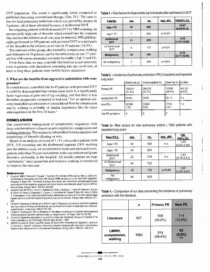
DVT population. This event is significantly lower compared to
published data using conventional therapy (Tab. TV).3 The same is
true for fatal pulmonary embolism which was proved by autopsy in
3 patients, all of them admitted because of iliofemoral DVT,
Investigating patients with iliofemoral DVT by MRI scanning, an
unexpectedly high rate of thrombi which extend into the common
iliac and into the inferior caval vein, may be detected. MRI phlebog
raphy performed on 189 patients with proximal DVT revealed parts
of the thrombi in the inferior caval vein in 35 patients (18.5%).
The outcome of this group, also treated by compression, walking
and dalteparin in 34 patients and by thrombectomy in one 17-year-
old boy with venous anomalies was quite favorable: (Tab. V and VI).
From these data we may conclude that bed rest is not necessary
even in patients with thrombosis extending into the caval vein, at
least as long these patients were mobile before admission.
3. What are the benefits from aggressive ambulation with com
pression?In a randomized, controlled trial in 45 patients with proximal DVT
it could be demonstrated that compression leads to a significantly
faster regression of pain and of leg-swelling, and that there is less
thrombus progression compared to bed-rest.5 For an optimal out
come immediate acceleration of venous blood-flow by compression
and by walking is probably of similar importance like the exact
anticoagulation in the first 24 hours.6
CONCLUSIONOur conservative management of symptomatic outpatients with
deep vein thrombosis is based on anticoagulation, compression and
walking exercises. This regime is independent from localization and
morphology of thrombi (floating or not).
Based on an analysis of a total of 1,111 consecutive patients with
DVT, 318 extending into the iliofemoral segment (18% reaching
into the inferior cava), we recommend to treat such proximal cases,
patients older than 70 years and patients with concomitant malignant
disorders, preferably in the hospital. All mobile patients are kept
“ambulatory” since immediate and intensive walking is considered
to improve the outcome.
Fatality y. stat. duff. OR(95% CI)
19,67Age >70 16 492
(4,24-91,4)
Age<70 1 605 p<0001
..476IlIof0e?raI 11 278
(1,76.12,88)
no iliofernoral 6 722 p<0.OlDVT
6,78Malignancy 10 191 (2,71-16,96)
No malignancy 7 906 p<0001
Table 2.— Incidence of pulmonary embolism (PE) in baseline and repeated
lung scan.femoropopliteal (b)
Primail, PE 159/310 299/578 74/209 a/c, b/c(51.3%) (517%) (35.4%) p<0.01
symptomatic PE 29.6°/b 29.8°/b 35.1% n.s.
new PE’s 22/300 33/563 7/199 n.s.7.3% 5.9% 3.5%
New PE’s no_ stat. duff.(951)
Age >70 34 455 n.s.(083256)
Age< 70 28 545
iliofemoral 1,43DVT
22 278 fl.S. (0,78-2,61)
no tiofemoral40 722
DVT
Malignancy 18 172 p<005(1
No 44 828malignancy
Table 4.— Comparison of our data concerning the incidence of pulmonary
embolism with the literature.
506Literature 907
(55,8%)
LMWH, 514compression, 1062 (48,4%)
walking
New PE
11412,6%
62(5,8%)
HAWAII MEDICAL JOURNAL, VOL 59, APRIL 2000
Table 1.—Risk factors forfatal events (up to 6 weeks after admission) in DVT
Iliofemoral (a) lower leg (c) p-value
new PE symptoms 4 2 0
Table 3.— Risk factors for new pulmonary emboli (1062 patients with
repeated lung-scan)
References1. Koopman MMW, Prandoni P, Piovella F, Ockelford PA, Brandles DPM, van der Meer J, Gallus AS,
Simmoneau G, Chesterman CH, Prins MH, Bossuyt PMM, de Haes H, van den Belt AGM, Sagnard K,d’Azemar P. Büller HR . Treatment of venous thrombosis with intravenous unfractionated heparinadministered in the hospital as compared with subcutaneous low-molecular-weight heparin adminis
tered at home, N Engi J Med. 1996:334: 682-687.2. Levine M. Gent M. Hirsh J. LeclercJ, Anderson D, Weitz J. Ginsberg J. Turpie AG. Demers C. Kovace
M, Geerts W, Kassis J. Desjardins L, Cusson J, Cruickshank M, Powers P, Brien W, Haley 5, WillanA. A comparison of low-molecular-weight heparin administered primarily at home with untractionated
heparin administered in the hospital for proximal deep-vein thrombosis, N EngIJ Med. 1996:334:677-
681.3. Partsch H. Kechavarz B. Mostheck A. Kdhn H. Lipp C. Frequency of pulmonary embolism in pat.ients
who have iliofemoral deep vein thrombosis and are treated with once- or twice-daily low-molecular-
weight heparin. J Vasc Surg. 1996:24:774-782.4. Partsch H, Kechavarz B, Kdhn H, Mostbeck A. The effect of mobilization of patients during treatment
of thromboembolic disorders with low-molecular-weight heparin, tnt Angiol. 1997:16:189-192.
5. Partsch H. Aggressive ambulation in symptomatic deep vein thrombosis, European Congress of theUnion Internationale de Phlebologie, Bremen Sept. 26-Oct 1. 1999
6. Hull RD. Raskob GE. Hirsh J. Jay RM. Leclerc JR. Geerts WH. Rouenbloom D. Sackett DL. Anderson
C. Harrison L. Gent M. Continuous intravenous heparin compared with intermittent subcutaneous
heparin in the initial treatment of proximal-vein thrombosis. N Eng J Med. 1986:315:1109-1114.
n Primary PE
140

Table 5.— Follow-up of 35 patients (1 months -6 years) with thrombi in theinferior caval vein
Follow-up only 3month
Deaths 6 64 f 3 months at home
89 f 4 months at home
54 m 1 year Hypernephroma
51 m 2.5 years Colon-carcinoma
70m 3 years MCI
88 f 5 years Hypernephroma
Table 6.— Re-examination of 24 patients with cava thrombosis between 4months and 6 years
Pain 3 Swelling 8
CATHETER-DIRECTED THROMBOLYSIS FORSYMPTOMATIC LOWER EXTREMITY DEEP
VEIN THROMBOSIS
Mark W. Mewissen, MDWisconsin Heart and Vascular Clinics
Milwaukee, Wisconsin, USA
ABSTRACTA national Venous Registry was organized to study catheter-directed thrombolysis (CDT) for the treatment of symptomatic lowerextremity deep vein thrombosis (DVT). Between January 1995 andDecember 1996, 287 patients with lower limb DVI were prospectively treated with CDT. There were 137 (48%) males and 150(52%) females averaging 47.5 years of age (range: 1-98). Based onsymptoms duration. 66% presented with acute DVT (10 days).
16% with chronic DVT (>10 days) and 19% with both acute andchronic DVT. From the venograms, Grade 1 (<50% lysis), Grade II(>50% lysis) or Grade III (complete lysis) were calculated. Based onfollow-up duplex scan evaluations, I-year cumulative primarypatencies were derived for different subgroups and compared usinglife-table methods. Iliofemoral DVI (IFDVT) were present in 221(7 1%) and femoral-popliteal DVI (FPDVT) in 79 (25%). Following 7.8 million iu urokinase (range: 0.5-44.0) administered over amean of 53.4 hours (range: 2.0-147.3), metallic stents were deployed to treat lesions uncovered in 99 iliac veins and 5 femoralveins. Grade III lysis was achieved in 3 1%, Grade II in 52% andGrade I in 17%. For acute limbs, Grade III lysis occurred in 34%compared to 19% for chronic limbs (p<O.Ol). Major bleedingcomplications were reported in 11.5% patients, most often occurring at the puncture site. Two patients died: one fatal PE and oneintracranial hemorrhage for a mortality rate of 0.04%. For all limbs,the 1-year primary patency was 60%. Treated IFDVT was moredurable than FPDVT (64% versus 47%; p<O.Ol). Lysis grades weremajor predictors ofpatency: for Grade III, Grade II and Grade I lyticoutcomes, the 1-year patencies were 79%, 58% and 32% respectively (p<O.OO1). CDT is safe and effective. Data from the VenousRegistry can serve as a guide to judicious patient selection for thispotentially effective form of therapy.
SUMMARYGOALS OF TREATMENTElimination of the embolic potential of existing thrombus, restoration of unobstructed flow, prevention of further thrombosis andpreservation of venous valve function, are the ideal goals of therapyfor acute DVI. Meeting these goals will not only prevent PE but willalso minimize the long-term sequelae of venous hypertension andthe development of the post-thrombotic syndrome. Multiple treatment options including anticoagulation, surgical venous thrombectomy, and thrombolytic therapy achieve these goals to a variabledegree. For instance, while anticoagulation with heparin followedby coumadin therapy is definitely effective in minimizing PE andreculTent thrombotic episodes, it does not prevent the post-thrornbotic syndrome.1In fact, with anticoagulation alone, restoration ofvenous patency relies solely upon individual endolytic correctiveprocesses. Furthermore, even with therapeutic heparin anticoagulation, 30-40% of patients will show extension of their DVI and only20-25% will have venographic evidence of partial thrombolysis.2’3Treatment strategies aimed at eliminating or reducing the risk ofPTS should focus on preserving valvular function and eliminatingthe risk of continued venous obstruction following acute DVI.Surgical removal by means of thrombectomy techniques combinedwith creation of arteriovenous fistulas have been employed successfully in Europe and the United States.45 but overall such procedureshave not been commonly performed. Thrombolytic agents are anattractive form of early therapy because they have the ability toeliminate obstructive thrombus in the deep veins and should therefore help provide protection against the PIS. The perceived benefitsof early and rapid recanalization in preserving valve function havebeen the basis for the use of lytic therapy to treat acute DVT.6
THROMBOLYTIC THERAPY: RATIONALEThere is good documentation that systemic thrombolytic therapy is
Drop-outs 2
Re-examination 24
Readmittedbefore reinvestigation:
contralateral DVT
other causes 5
Compression 16 Anticoagulation 16(11 Marcoumar,3 LMWH, 2 ASS)
HAWAII MEDICAL JOURNAL, VOL 59, APRIL 2000141

superior to heparin anticoagulation alone in achieving early lysis of
the deep veins. In a pooled analysis of 13 randomized studies,
Comerota and Aidridge found that only 4% of patients treated with
heparin had significant or complete lysis, compared to 45% of
patients randomized to systemic streptokinase therapy.8 Further
more, in the heparin group, as many as 82% had no venographic
evidence of lysis or clearly demonstrated thrombus extension,
compared to 37% in the streptokinase group. Similarly, in reviewing
pooled data from 6 trials judged to have proper randomization,
systemic thrombolysis was 3.7 times more effective in producing
some degree of lysis than was heparin.9From these data, there is
objective evidence that systemic thrombolysis is more effective than
anticoagulation in promoting early lysis.
Two studies reported late clinical outcome following randomiza
tion to either anticoagulation or systemic streptokinase for acute
lower limb DVT.’°” Although the follow-up periods in both studies
were different, 1.6 years’° versus 6.5 years,’ ‘the majority of patients
with severe symptoms of the PTS received anticoagulation alone. In
both reports. the majority of patients who received streptokinase
were free of the PTS. Although both studies suffer from small
sample sizes and lack of objective measures to grade the PTS, it can.
nonetheless, be suggested that systemic thrombolysis improves
anatomic and clinical outcomes when compared to anticoagulation
alone. Long-term improved venous hemodynamics and preserva
tion of valvular function have not been well documented following
systemic lytic therapy. ma long-term randomized follow-up study.’2
popliteal venous valve incompetence, measured by direct Doppler
examination, was more common in patients randomized to antico
agulation versus systemic streptokinase. Nine percent of patients
successfully lysed had an incompetent valve compared with 77% of
those who did not lyse. This study, however, has not been published
in peer-reviewed journals and still resides in the format of an
abstract.
CA THETER-DIRECTED THROMBOLYSIS
Over the past 10 years, catheter-directed thrombolysis techniques
have been proved effective in the endovascular treatment of acute
limb ischemia secondary to acute native arterial or bypass graft
thrombotic occlusions.’3Delivering thrombolytic agents by means
of low-profile infusing catheters and guide wires placed directly into
the occlusion under fluoroscopic guidance offers several advan
tages over systemic lytic therapy. Because highly concentrated
plasminogen activators can be delivered directly into the thrombus,
treatment duration can be reduced, complete lysis rates improved.
and lower incidence of bleeding and other complications associated
with systemic therapy can be expected. Second, the technique
allows for supplemental endovascular treatment of uncovered he
modynamic lesions by means of balloon angioplasty or stent tech
nique.’4Similar regional catheter-based lytic treatments have been
recently applied towards ilio-femoral DVT.8’5 In a series of 25
patients treated with catheter-directed thrombolysis for ilio-femoral
DVT by Semba and Dake.’° complete lysis was achieved in 18
(72%) patients, swelling was successfully reduced in all but one.
Only one patient suffered a bleeding complication of heme-positive
stools. After the drug was discontinued, there were no significant
adverse sequelae. This initial report suggests that catheter-directed
lytic therapy for patients with lower extremity DVT can be effective
in achieving significant lysis of clot and may be associated with low
complication rates.This experience stimulated the development of a multicenter
venous registry, which was recently closed after enrollment of
nearly 500 patients.’6Completed data with follow-up of at least 6
months were available on nearly 300 patients, 70% of which were
IFDVT, as determined by venography. Nearly 40% required a stent
in the iliac vein, to relieve an obstruction or a stenosis uncovered
after catheter-directed thrombolysis treatment. Significant lysis (>50%) was achieved in 80% of the limbs and approximately 30%
achieved complete lysis. A complete lyric event was more com
monly achieved for acute DVT (<10 days) which was present in
about 60% of the patients. The lysis grade obtained after lysis and
stenting was a major predictor of continued patency. At 6 months,
approximately 70% of limbs with significant lysis remained patent.
and for acute limbs, when complete lysis was achieved, 90%
remained patent. Reflux at follow-up at 6 months was found in less
than 30% of those with complete lysis, 45% of those with >50% lysis
and over 60% with <50% lysis. There have been only 2 deaths in the
entire study (0.4%), one from an intracranial hemorrhage and one of
six patients who suffered a PE. The use of the popliteal vein with
ultrasound guidance appears to be favored and will likely become
the standard access site for most patients.
References:1. Meissner MH, Manzo RA, Bergelin nO, et al. Deep venous insufficiency: The relationship between lysis
and subsequent retlux, J Vasc Surg. 1993:18:596-608.2. Strandness DE. Thrombus propagation and level ot anticoagulation, J Vasc Surg. 1990: 12:497-498
3. Krupski WC, Bass A, Dilley RB, et al. Propagation ot deep venous thrombosis by duplex ultrasonography, J Vasc Surg. 1990: 12:467-475.
4. Plate 0. Einarsson E. Ohlin P. et al. Thrombectomy and temporary arterio-venous tistula in acuteiliofemoral venous thrombosis, J Vasc Surg. 1984:1:867.
5. Comerota A, Aldridge SC, Cohen G. et al. A strategy of aggressive regional therapy tor acute itofemoral venous thrombosis with contemporary venous thrombectomy or catheter-directed thromboly
six, J Vasc Surg. 1990:20:244-254.6. Truebstein 0. Can thrombolysis prevent post phlebitic syndrome and thromboembolic disease?
Haemostasis, 1 986;3:38-50,7. KakkarVV, Howe CT. Laws JW, et al. Late results ottreatment of deep ve,n thrombosis, BrMedJ. 1969:
1810-811.8. Comerota A, Aldhdge SC. Thrombolytic therapy tor deep venous thrombosis: A clinical review, CJS.
1993:36:359-364.9. Goldhsber SZ, Buring JE, Lipnick RJ, et al. Pooled analysis of randomized trials of streptokinase and
heparin in phlebographically documented acute deep venous thrombosis, Am J Med. 1984; 76:393-397.
10. Elliot MS, Immelman EJ. Jeff rey P, et al. Acomparative randomized thai of heparin versus streptokinasein the treatment of acute proximal venous thrombosis: An interim report of a prospective trial, BrJSurg.1979; 66:838,
11. Arnesen H, Heilo A, Jakobsen E, et al, A prospective study of streptokinase and heparin in the treatmentof deep vein thromsosis, Acta Med Scand. 1978; 203:457,
12. Jeffrey P, Immelman E, Amoore J. Treatment of deep vein thrombosis with heparin or streptokinase:
Long-term venous function assessment (abstract No. S20.3). In: Proceedings ott/re Second International Vascular Symposium, 1989.
13. Kandarpa K. Technical determinant of success in catheter-directed thrombolysis for peripheral arterial
occlusions, J Vasc ?nterv Radio?. 1995,18:367-372.14. Ouriel K, Shortell CK, De Weese JA, et al. A comparison of thrombolytic therapy with operative
revascualarization in the initial treatment of acute peripheral arterial ischemia, J Vasc Surg. 1994;19:1021-1030
15. Semba CP, Dake MD. Hiofemoral deep venous thrombosis: aggressive therapy with catheter-directed
thrombolysis, Radiology. 1994: 191:487.16. Mewissen, MW. Seabrook. OR, Meissner. MH, Cynamon. J, Labropoulos. N. Haughton, SH. Catheter-
directed thrombolysis for lower evtremity deep vein thrombosis: report of a national multi-center
registry, Radiology. April 1999.
HAWAII MEDICAL JOURNAL. VOL 59, APRIL 2000
142

PHARMACO-MECHANICAL VENOUSTHROMBOLYSIS
Andrew H. Cragg, MDFairview University Medical Center
Minneapolis, Minnesota, USA
INTRODUCTIONPharmacologic dissolution of intravascular thrombi using lyticagents has established itself as a useful adjunct to many forms ofrevascularization. both surgical and interventional. The most widespread use of thrombolytic therapy has been in the treatment ofarterial thrombosis in the lower extremities. Thrombosed nativearteries and bypassed grafts can be effectively revascularized withcatheter directed infusion therapy using one of several lyric agents.For most interventionalists, urokinase is the lytic agent of choicebecause of its predictable action and acceptable side effect profile.
We recently published a randomized study reviewing two different protocols for infusion lytic therapy of thrombosed peripheralbypass grafts and native arteries. Two dosing protocols from 50,000units/hour of urokinase to 250,000 units! hour were found to beequally effective in lysing both acute and chronic thrombi, however,the time to lysis was 20-30 hours requiring overnight infusion andobservation in an intensive care setting.’
We and others have begun examining a variety ofnew devices andtechniques to speed up the lytic process when revascularization isrequired in a variety of vascular beds. These techniques allow us torapidly open thrombosed vascular conduits in a single procedure andin some situations, lytic therapy can be used on an outpatient basis.
LYTIC TECHNIQUESInfusion TherapyIntroduction of a lytic agent through a catheter imbedded in athrombosed vessel or a bypass graft is an effective means ofachieving complete thrombolysis, but one significant limitation isthe time it takes for completion of the procedure. This is due in partto the time it takes for the lytic agent to penetrate the thrombus alongthe length of the occluded segment. To speed up this process we havedeveloped a series of catheters for more effective infusion of lytictherapy. One catheter is a multi side-hole catheter which deliverslytic agent in an even fashion along the length of the infusionsegment. Infusion segments vary between 5-20 cm. The catheter hasa valve at the tip which allows passage of the catheter over a guidewire but which seals after the removal of the guide wire to allow onlyside-hole infusion. Another variation of this catheter uses a coaxialdesign to tailor the length of the infusion segment to the length of theoccluded vascular segment. Another device which enhances delivery of the lytic agent to the periphery of the thrombosed vessel is theshape memory, infusion wire which we have developed. This devicegradually reforms into a coiled configuration to approximate thevessel wall as lytic therapy progresses. With these devices, coaxialinfusions can be performed so that long vascular segments can betreated with infusion therapy.
Pulse Lytic TherapyMany interventionalists favor pulsed delivery of the lytic agent into
the thrombus in order to accelerate the lytic process. With thistechnique, small boluses of urokinase are delivered in a forcefulmanner to form jets out of the side holes of an infusion catheter. Theside-hole valve tipped infusion catheter we’ve developed allows formore convenient application of this technique since the catheter canbe placed over a wire and then used for pulsed lytic therapy withoutadditional tip occluding wires or coaxial devices.
Pharmaco-Mechanical Lytic TherapyAgitation of the thrombus during delivery of the lytic agent mayfurther accelerate the lytic process. We are currently testing the useof the Thrombolytic Brush to effect a combination of pharmacologicand mechanical lysis. The device consists of a soft nylon brushwhich is rotated at relatively low rpms during the delivery of a lyticagent. The brush agitates thrombus and enhances delivery of thelytic agent to the thrombus. The device is presently approved for usein thrombosed dialysis fistulas and we are currently investigating inpre-clinical studies the use of the device in peripheral arteries andveins. Further development of devices such as these may allow themajority of thrombosed peripheral vessels to be revascularized withlytic therapy in a single sitting. Other devices use a venturi effectfrom high-pressure saline injection to aspirate thrombus. Differentdevices may be more beneficial in certain situations than in others.
VENOUS DISEASEDeep venous thrombosis is a common medical condition with majormorbidity for those affected. Acutely, patients may develop painfulphlegmasia and chronically a large percentage of patients developvenous stasis problems secondary to valvular dysfunction. Heparinis generally ineffective in preventing the long-term complications ofdeep venous thrombosis which occur in 50-80% of patients.
Recently, investigators have begun to advocate catheter directedlysis of deep venous thrombosis in order to decrease the acutesymptoms and preserve vein patency and valve function. While thelong-term clinical efficacy of this technique is not yet established, ithas been demonstrated that venous thrombi can be readily lysedusing catheter-directed therapy. Disadvantages of this techniquehowever, include relatively cumbersome catheterization from aretrograde approach and the long times associated with lysingvenous clots.
Mechanical and pharmaco-mechanical thrombolysis may makethis procedure more rapid and efficacious. We have recently developed a technique and protocol for antegrade catheterization of thedeep venous system in the lower extremity which allows simplifiedaccess, better patient tolerance and placement of long coaxialinfusion systems.2 In experimental models, we have demonstratedthat venous thrombi can be rapidly dissolved with the ThrombolyticBrush and we anticipate clinical trials with this device in the future.Our goal with lytic therapy and deep venous thrombosis is to providecomplete lysis and vein patency in several hours rather than severaldays.
DIALYSIS ACCESSThe rapid flow through a surgically placed arteriovenous fistulainvariably leads to turbulence and pannus formation in the outflowvein near the venous anastomosis. In patients with forearm dialysisaccess grafts. this often results in narrowing of the outflow vein,
HAWAII MEDICAL JOURNAL, VOL 59. APRIL 2000143

restriction of flow through the fistula and subsequent thrombosis. To
preserve patency of the dialysis access after it is thrombosed, it is
essential to correct the underlying venous outflow restriction. Re
cently, many practitioners have found it useful to combine the
advantages of thrombolytic therapy and balloon angioplasty in a
fluoroscopic suite where the anatomic status of the arterial and
venous segments of the fistulas can be accurately determined.34In
general. access to the radiographic interventional room is more
easily obtained than to the standard operating room. In some centers,
this has facilitated a shift of patients from traditional operative
thrombectorny to fistula revascularization using lytic therapy and
angioplasty of the venous outflow. Early results using streptokinase
for fistula thrombolysis were disappointing.4However, newer de
velopments including the use of urokinase and multiple catheters for
more complete fistula lysis have improved the success of the
procedure. In general, the acute success in revascularizing throm
bosed fistulas exceeds 90% with lytic therapy and angioplasty.6
While lytic therapy can be less invasive than surgical thrombec
tomy, one disadvantage is the frequent need for overnight hospital
admission to complete the lytic procedure. This adds to the cost of
the procedure and can inconvenience the physician and the patient
because dialysis schedules are often performed on an every-other-
day basis. A method for nonsurgical, outpatient revascularization of
the thrombosed fistula should provide an attractive alternative to
existing methods both from a clinical and a financial standpoint.
Recently, techniques for revascularization have been re-ported
using maceration of clot,7 pulsed spray administration of lytic
agent,0 or embolization of thrombus without use of lytic agents.9
These techniques high-light a trend toward outpatient management
of this clinical problem. With these techniques, successful
revascularization has been achieved in 90-95% of patients. Second
ary patency for these fistulas averages about 6 months in most series
and points out the fact that dialysis fistulas require routine mainte
nance with repeat procedures in order to maintain their viability. The
large number of procedures and the need for repeat procedures
suggests that a method for rapidly revascularizing fistulas on an
outpatient basis would be very useful.
All revascularization procedures, including surgical thrombec
tomy, thrombolysis and iatrogenic embolization as described by
Trerotola,9result in pulmonary embolism to some degree. There is
a long clinical history with surgical thrombectomy to suggest that
this is relatively well tolerated. With lytic therapy, emboli tend to be
small and are generally laced with a lytic agent which should
facilitate their dissolution in the pulmonary vascular bed. There is
now a large experience with lytic treatment of dialysis access
thrombosis and symptomatic pulmonary embolism has not been a
significant clinical problem. The technique of Trerotola in which the
thrombus occluding the fistula is deliberately embolized to the lungs
without lytic therapy suggests that even larger volumes of thrombus
can be well tolerated in most patients. The comments of Dolmatch
et al. regarding this technique, however, point out the fact that many
dialysis patients have poor cardiopulmonary reserve and care should
be exercised in limiting the degree of pulmonary embolism associ
ated with the procedure.’°One method for accelerating the lytic procedure involves macera
tion of the thrombus during delivery of the lytic agent. Mechanical
agitation of the thrombus which facilitates mixing of the lytic agent
with the thrombus has been shown to rapidly accelerate lysis. This
type of mixing can be accomplished in a reproducible and controlled
manner with the Thrombolytic Brush. Animal studies suggest that
fistula thrombolysis can be accomplished in a matter of minutes
without significant pulmonary embolization.’°
With this technique. short vascular access sheaths are placed into
the arterial and venous limbs of the thrombosed fistula. The venous
outflow is then studied by diagnostic venography and stenoses in the
venous outflow are dilated by balloon angioplasty. 250,000 units of
urokinase mixed with 2,000 units of heparin are then delivered in a
divided dose into the arterial and venous limbs of the fistula. The
Thrombolytic Brush is then introduced and used to gently mix the
lytic agent and the thrombus. This agitation usually results in
complete lysis of the thrombus in the fistula within several minutes.
Follow-up angiography is then performed with additional angioplasty
as necessary of either the arterial or venous limbs. The vascular
access sheaths are compatible with dialysis tubing and can be left in
place for immediate dialysis. The lytic procedure can be completed
with-in one hour. In this fashion, the interventionalist can complete
the procedure in a single sitting without having to move the patient
either into another area for short-term lytic infusion or admit the
patient to the hospital for overnight lytic infusion. Both of these are
impediments to more widespread use of lytic therapy for
revascularization of thrombosed fistulas. The simplification of the
revascularization procedure fits with the trend toward surveillance
and maintenance of dialysis fistulas by repeat outpatient procedures
such as lytic therapy or angioplasty. Renal medicine physicians who
are largely responsible for the management of dialysis patients find
it convenient to schedule patients for this procedure since they can,
in general, be treated on a same day outpatient basis without
inconveniencing the existing dialysis schedule.
In our practice, this has become the routine standard of care for
management of thrombosed dialysis fistulas. Patients can be treated
between dialysis sessions without scheduling disruption. In our
practice, virtually all dialysis patients with thrombosed access are
managed by outpatient accelerated thrombolysis and angioplasty.
References1 Cragg AH, Smith TP. Corson J. Nakagawa N. Castaneda F, Kresowik T, Sharp J, Shanima A, Berbaum
KS. Two urokinase dose regimens in native arterial and graft occlusions: Initial results of a prospective,randomized clinical trial, Radiology. 1991178:681.686.
2. Cragg AH. Lower extremity deep venous thrombolysis: A new approach to obtaining access, JVIR.1996:7:283-288.
3. Gaylord GM, Taber TE. Long-term hemodialysis access salvage: Problems and challenges for
nephrologists and interventional radiologists, JVIR. 1993:4:103.107.4. Brunner MC, Matalon TAS, Patel 5K, McDonald V. Jensik SC. Ultrarapid Urokinase in hemodialysis
accesa occlusion, JVIR. 1991 2:503-506.5. Young AT, Hunter DW, Castenada-Zuniga WR. Thrombosed synthetic hemodialysis access fist alas:
Failure of tibrinolytic therapy, Radiology. 1985; 154:639-642.6. Ehrman KO, Gaylord GM. Thrombolytic therapy for occluded dialysis access grafts. Semin lntervenf
Radiol.1992;9:190-194.7. Davis GB, Dowd CF. Bookstein JJ. Maroney TP. Lang EV. hulsaz N. Thrombosed dialysis grafts:
Efticacyof intrathrombicdeposition of concentrated Urokinase, clot maceration, and angioplasty. AJR.
1987:149:177-18 1.8. Valji K, Bookstein JJ, Roberts AC, Davis GB. Pharmaco-mechanical thrombolysis and angloplasty in
the managementof clofted hemodialysis grafts: Eady and late clinical results. Radiology. 1991:178:243-
247.9. Trerotola SO, Lund GB, Scheel PJ Jr., SavaderSJ, Venbrux AC. Osterman, JrAC. Thrombosed dialysis
access grafts: Percutaneous mechanical declotting without Urokinase. Radiology. 1994:191:721 -726.
tO Dolmatch OL, Gray RJ. Horton KM. Will iatrogenic pulmonary embolization be our pulmonary
embarrassment? Radiology. 1994; 191:615-617.it. Custenada F, et al. (personal communication(.
HAWAII MEDICAL JOURNAL. VOL 59, APRIL 2000
144

VENOUS ANGIOPLASTY AND STENTING
Andrew H Cragg, MDFairview University Medical Center
Minneapolis, Minnesota, USA
Intraluminal stents have demonstrated their utility as adjuncts toangioplasty for the treatment of chronic arterial occlusive disease.Their application to the venous circulation has been more limited.We and others have experience with the use of intraluminal stents ina variety of venous applications. These include management offailed angioplasty of the venous outflow in dialysis access fistulas,treatment of superior vena cava syndrome and treatment of chronicpelvic venous occlusion.
Stenting in Conjunction with Dialysis Access Fistula SalvageInterventional revascularization of the thrombosed dialysis fistula isnow a common procedure. Thrombosis or dysfunction is usually dueto obstruction of the venous outflow near the venous anastamosis.Regardless of whether a surgical revision or interventional salvageof the thrombosed fistula is undertaken, both techniques exhibit ahigh recurrence rate in large part related to restenosis of the treatedvenous outflow segment. In our experience, intraluminal stents havea limited role to play in the salvage of access fistulas. S tents placedin the venous outflow after failed angioplasty can act as a bridge torevision if this cannot be accomplished immediately or if the patientrequires immediate dialysis. Stents do not, however, appear to limitrestenosis associated with angioplasty’ and have demonstrated thatpatency is similar to standard angioplasty.”2At this time, therefore,there drpes not appear to be a role for primary stenting as a means toprevent restenosis. Failure of stents in the forearm appears to be dueto a combination of the relatively hostile turbulent flow environmentand the smaller caliber of stents in this location.
Superior Vena Cava SyndromeWe have anecdotal experience with the use of intraluminal stents fortreatment of superior vena cava syndrome in patients with malignantSVC obstruction. SVC syndrome is a disturbing symptom complexwhich often occurs in patients with terminal malignancy. In thissituation, placement of even relatively smaller caliber stents in thediameter range 8-12 mm23usually results in rapid defervescence ofthe clinical manifestations of SVC syndrome. Patients are quitegratified by this relatively simple technique and stent patency hasnot generally been an issue because of the limited life expectancy ofthese patients. For patients with benign obstruction of the superiorvena cava, stents may also have a role, however, in this situationadequate caliber is necessary to limit the effects of restenosis.Careful attention to technique is necessary to minimize the important complication of stent misplacement or migration.
Pelvic Venous StentingIn conjunction with our developing experience with lysis of deepvenous thrombosis we have identified a subgroup of patients whoappear to benefit from iliac stenting. These are the patients withchronic obstruction of the left iliac vein (May-Thurner Syndrome)who develop acute deep venous thrombosis. Often these are youngpatients who are active and otherwise healthy. After lytic therapy is
accomplished to treat the thrombosis, an underlying stenosis of thecommon iliac vein is usually uncovered. This can be either a focalstenosis at the confluence of the iliac vein and vena cava or a chronicobstruction of the entire common iliac vein with collateral outflowthrough the internal iliac vein. These patients do not respond tosimple balloon angioplasty even though the initial result may appearfavorable. In our experience these patients inevitably rethromboseunless a stent is placed. In this situation we try to place as large a stentas possible (12-16 mm diameter). Patients are maintained on anticoagulation for six months after stent placement.
Other causes of chronic venous occlusion including radiationstricture, post-operative stricture or external compression may alsorespond to intraluminal stenting.4In general, the tenants of successful venous stenting are constant regardless of the location. Bestresults can be expected when the following conditions are met: 1)larger stents, 2) shorter lesions, 3) better inflow, 4) proper anticoagulation.
References1. Gray RJ, Horton KM, Dolmatch BL, Rundback JH, Anaise D, Aquino AC, Currier GB, Light JA, Sasaki
TM. Use of Walistents for hemodialysis access-related venous stenoses and occlusions untreatablewith balloon angioplasty, Radiology. 1995;195(2):479-484,
2, Ferro C, Perona F, Barile A, Borrelli M, Cianni ft Ambrogi C. Metallic stents for treatment of superiorvena cava syndrome: Results in 26 patients, CVIR. 1996; 19(2):S43,
3. Oudkerk M, Kuijpers TJA, Borel Rinkes IHM, Brutel de Ia Riviere G, Wiggers T. Animal study fortreatment of surgical induced canal stenosis with extra large Palmaz stents, CVIR. 1996;19(2):S43.
4. Nazarian GK, Bjarnason H, Dietz CA Jr., Anderson Bernadas C, Hunter OW, Iliofemoral venousstenoses: Effectiveness of treatment with metallic endovascular stents, Radiology. 1 996;200:1 93-199.
SELECTIVE THROMBECTOMY FOR TREATMENT OF ILIOFEMORAL DVT
Bo Eklof MD, PhDStraub Clinic & HospitalHonolulu, Hawaii, USA
There are data that indicate that early and quick removal of an acutevenous thrombus may prevent the destruction of the venous wall andvalve, i.e., stop the progress into the post-thrombotic syndrome(PTS).
We, therefore, recommend an aggressive treatment with rapidremoval of the occluding thrombus in active patients with acuteiliofemoral deep venous thrombosis (DVT).
The first line of treatment should be catheter-directed intrathrombus thrombolysis with or without adjunct procedures such asangioplasty and stenting. When there are contraindications or failure of thrombolysis, thrombectomy (TE) with a temporary arteriovenous fistula (AVF) is the alternative. Both interventions will befollowed by anticoagulation. These aggressive interventions are notjustified in chronically ill, bedridden, high-risk, or aged patients, orthose with serious intercurrent disease and/or limited life expectancy. In this group of patients these interventions can only bejustified for limb salvage in phlegmasia cerulea dolens whereconservative treatment does not prevent the development ofan acutecompartment syndrome with venous gangrene.
THROMBECTOMYThe first TE for iliofemoral venous thrombosis (IFVT) was per-
HAWAII MEDICAL JOURNAL, VOL 59, APRIL 2000145

formed by Lawen in Germany in 1937. Surgery today is performedunder intubation anesthesia. Ten cm water PEEP is added duringmanipulation of the thrombus to prevent perioperative PE. Theinvolved leg and abdomen are prepared. A longitudinal incision ismade in the groin to expose the long saphenous vein (LSV), whichis followed to its confluence with the common femoral vein (CFV)which is dissected up to the inguinal ligament. The superficialfemoral artery 3-4 cm below the femoral bifurcation is prepared forconstruction of the AVF. Further dissection depends upon theetiology of the IFVT. In primary IFVT with subsequent distalprogression of the thrombus, a longitudinal venotomy is made in theCFV and a venous Fogarty TE catheter is passed through thethrombus into the IVC. The balloon is inflated and repeated exercises with the Fogarty catheter are performed until no more thrombotic material is extracted. With the balloon inflated in the commoniliac vein a suction catheter is introduced to the level of the internaliliac vein to evacuate thrombi from this vein. Backflow is not areliable sign of clearance since a proximal valve in the external iliacvein may be present in 25% of cases preventing retrograde flow ina cleared vein. On the other hand, backflow can be excellent from theinternal iliac vein and its tributaries despite a remaining occlusion ofthe common iliac vein. Therefore, an intraoperative completionvenogram is mandatory. An alternative is the use of an angioscopewhich enables removal of residual thrombus material under directvision. The distal thrombus in the leg is removed by manual massageof the leg starting at the foot. The Fogarty catheter can sometimes begently advanced in retrograde fashion. The aim is to remove all freshthrombi from the leg. In IFVT secondary to ascending thrombosisfrom the calf, the thrombus in the superficial femoral vein (SFV) isoften old and adherent to the venous wall and we have already lostthe battle of the valves. The objective is to restore patency andpreserve valvular function. If iliac patency is established but thethrombus in the femoral vein is too old to remove, then it ispreferable to ligate the superficial femoral vein. Recanalization willotherwise lead to valvular incompetence and subsequent reflux. Ifnormal flow in the SFV cannot be re-established, we recommendextending the incision distally and exploring the orifices of the deepfemoral branches. These are isolated and venous flow is restoredwith a small Fogarty catheter. The SFV is ligated. The venotomy isclosed with continuous suture and an AVF created using the LSV,anastomosing it end-to-side to the superficial femoral artery. Anintraoperative venogram is performed through a catheter inserted ina branch of the AVF. After a satisfactory completion venogram thewound is closed in layers without drainage. If there are signs of iliacvein compression which can occur in about 50% of left-sided IFVT,we recommend intraoperative angioplasty and stenting.
Heparin is continued at least 5 days postoperatively, and warfarinis started the first postop day and continued routinely for 6 months.The patient is ambulant the day after the operation wearing acompression stocking. The patient is usually discharged on the tenthpostop day to return after 6 weeks for closure of the fistula. Theobjectives of a temporary AVF are to increase blood flow in thethrombectomized segment to prevent immediate rethrombosis, toallow time for healing of the endothelium, and to promote development of collaterals in case of incomplete clearance or immediaterethrombosis of the iliac segment. A new percutaneous techniquefor fistula closure was developed by Endrys in Kuwait. Through a
puncture of the femoral artery on the opposite surgically untouchedside, a catheter is inserted and positioned at the fistula level. Prior toinflation and release of the balloon or coil, an arteriovenogram canbe performed to evaluate the patency of the iliac and caval veins,which is of prognostic value. More than 10% of patients have beenshown to have remaining significant stenosis of the iliac vein despiteinitial successful surgery. A transvenous percutaneous angioplastywith stenting can be performed under the protection of the AVF,which is closed 4 weeks later after repeat arterio-venogram.
“LATE” RESULTSThere are few studies on “long-term” results after TE with AVF.There are 8 studies of clinical results in 521 patients with more than2 years follow-up where “clinical success” is claimed in 62%.’ Thereare 5 studies on iliac vein patency in 247 patients with more than 2years follow-up showing 82% patency (range 77-88%).’ There are5 studies on femoro-popliteal valvular competence in 259 patientswith more than 2 years follow-up showing 60% competency (range36-84%).’ In the prospective, randomized study from Sweden wefound a highly significant difference in the number of asymptomaticpatients after 6 months, with 42% in the surgical group versus 7% inthe conservatively treated group.2At 5 years, 37% of the operatedpatients were asymptomatic corn-pared with 18% in the conservative group.3At 10 years, 54% in the surgical group were basicallyasyrnptomatic (class 0-2 new CEAP classification) compared with23% in the conservative group, however, not a significant difference.4Iliac vein patency at 6 months was 76% in the surgical groupcompared with 35% in the conservative group, demonstrated byvenography.2This significant difference was upheld after 5 and 10years with 77% and 77% patency in the surgical group, respectively,versus 30% and 47% in the conservative group, respectively.3’4Femoro-popliteal valvular competence at 6 months was 52% in thesurgical group compared with 26% in the conservatively treatedgroup, using descending venography with Valsalva, a significantdifference.2 After 5 years, the patients who underwent TE hadsignificantly lower ambulatory venous pressures, improved venousemptying as shown by plethysmography, and a better calf pumpfunction with less reflux as measured by foot volumetry. Combiningthe results of all functional tests, 36% of the surgical patients hadnormal venous function compared with 11% of the conservativelytreated group. These differences were not statistically significantdue to loss ofpatients.3At 10 years, using duplex scanning, poplitealreflux was found in 32% of the surgical group compared with 67%of the conservative group. Six patients who had a successful TE 10years before without obstruction of the iliac vein at the time ofsurgery, were all asymptomatic with patent iliac veins, and 50% hadcompetent popliteal veins.4Successful TE seems to be beneficial inthe long term.
The treatment of acute iliofemoral DVT should aim at:• prevention of fatal PE;• decrease ofpain and swelling of the involved leg, trying to stop the
development ofphlegmasia cerulea dolens, and venous gangrene;• prevention of the disabling post-thrombotic syndrome by early
removal of the blood clot avoiding proximal venous obstructionand preserving normal, functioning valves preventing reflux.
HAWAII MEDICAL JOURNAL, VOL 59, APRIL 2000146

The first choice to accomplish early and quick removal of thethrombus is catheter-directed intra-thrombus thrombolysis. Whenthere are contraindications or failure of thrombolysis, thrombectomy with a temporary AVF is a valid alternative.
References1. Eklof B, Kistner RL. Is there a role forthrombectomy in iliofemoral venous thrombosis? Sam Vasc Surg.
1 996;9:34-45.2. Plate C, Einarsson E, Ohlin P. at al, Thrombectomy with temporary arteriovenous fistuls: The treatment
of choice in acute iliotemoral venous thrombows. J Vasc Surg. 1984;1:867-876.3. Plate G. Akesson H, Einarsson E, at al. Long-term results of venous thrombectomy combined wrth a
temporary arteriovenous fistula, Eur J Vasc Surg. 1 990;4:483-489.4. Plate C, Eklof B, Norgren L, et al. Venous thrombectomy for iliofemoral vein thrombosis. Ten-year
results of a prospective randomized study. Fur J Vasc Endovasc Surg. 1997; 14:367-374.
ALGORITHMS FOR THE TREATMENT OFACUTE DVT
Anthony 3. Comerota, MD, FACSTemple University School of Medicine
Philadelphia, Pennsylvania, USA
Figure 3
INFRAINGUINAL DVT
oms Controlled with
/ Elevation/Compression \k And Aatient Toterant of
entSymptoms
AnticoagulateCatheter Directed Thrombolysis* ILMWH SQ. x 5-7 days• Post-Tibial Vein
Oral Anticoagulation x 1 year• Popliteal VeinINR2-3 I
* All patients require elastic stockings to control swelling30mm — 50mm Hg Ankle Gradient
Figure 1
Algorithm for the Management of Calf Vein ThrombosisFigure 4
Figure 2
Management of PrimaryAxiIIo-Subclavian Vein Thrombosis
Suspected Axillo-Subclavian DVT+
Venous Duplex
__-Phlebography
Thrombosis Compression or StrictureWithout Thrombosis
Catheter DirectedThrombolysis
Repeat Phlebographywith Hy erabductiori Maneuvers
Chronically OccludedVein at Thoracic Inlet,
Patient 1v Rib Collaterals
Anticoagulation 4—Normal Vein
Balloon Angioplasty.,.__ Stricture Atone(Possible Stent) (Distal to 1e Rib)
Compression/Stricture
VBalloon Arigioplasty
Temporize)
1t Rib Resection Anticoagulate
/ 6-10 weeks
Phlebogram
Strictu Normal Vein
Iliofemoral DVT
Contrainclications toicTherapy
I Attempt Catheter-Directed Thrombolysis
HAWAII MEDICAL JOURNAL. VOL 59, APRIL 2000147
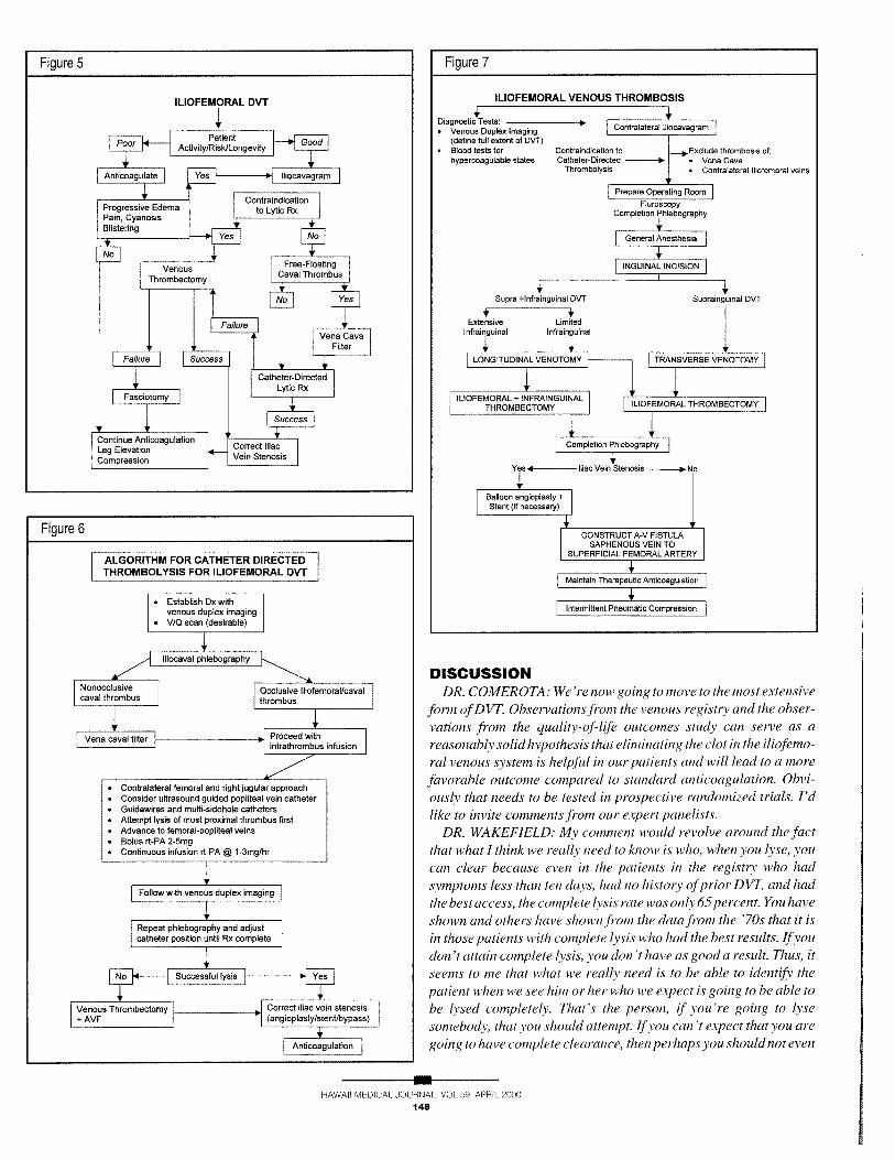
DISCUSSIONDR. COMEROTA: We’re now going to iiiove to the most extensive
form ofDVT. Observations from the venous registry and the observations from the qua 1 itv-of-life outcomes stud’ can serve as areasonably solid hypothesis that eliminating the clot in the iliofemo
ml venous system is helpful in our patients and will lead to a more
favorable outcome compared to standard anticoagulation. Obvi
ouslv that needs to he tested in prospective randonuzed trials. I’d
like to invite comments from our expert panelists.
DR. WAKEFIELD: Mv comment would revolve around the factthat what I think we really need to know is who, when you lvse, youcan clear because even in the patients in the regist who hadsymptoms less than tell days, 11(1(1 no instorv ofjirior DVT, and had
the best access, the complete lvsis rate was on/v 65percent. You haveshown and others have shown f,vm tile data from the ‘70s that it isin those patients wit/i complete lvsis who had the best results. if vonc/ott ‘t attain complete /vsis, you don ‘t have as good a result. Thus, itseems to me that what we real/v need is to be able to identify thepatient when we see him or her who we expect is going to be able to
be lysed completely. That’s the person, if you’re going to /vse
somebody, that von should attempt. Ifyou can ‘t expect that you are
going to have complete clearance, then perhaps you should not even
HAWAW MEDICAL JOURNAL, VOL 59, APRIL 20CC
148
Figure 5 Figure 7
ILIOFEMORAL VENOUS THROMBOSIS
Diagnostic Tests:• Venous Duplex Imaging Contralateral Iltocavagram
(define full extent of DVT)• Blood tests for Contraindication to Exclude thrombosis of:
hypercoagulabte states Catheter-Directed • Vena CavaThrombolysis • Contralateral Iliofemoral veins
[ Prepare Operating Room
FluroscopyCompletion Phlebography
General Anesthesia
[j4GUINAL INCISION
Supra vlnfrainguinal DVT
Extensive LimitedInfrainguinal Infrainuinal
Suprainjinal DVT
LONGITUDINAL VENOTOMY TRANSVERSE VENOTOMY
_____________________
1’ 1,ILIOFEMORAL + INFRAINGUINAL I
THROMBECTOMY
Figure 6
ILIOFEMORAL THROMBECTOMY
ALGORITHM FOR CATHETER DIRECTED -
THROMBOLYSIS FOR ILIOFEMORAL DVT
Completion Phlebography
Yes I Iliac Vein Stenosis No
nangiopIasty±Stunt (if necessary)
CONSTRUCT A-V FISTULASAPHENDUS VEIN TO
SUPERFICIAL FEMORAL ARTERY
Maintain Therapeutic Anticoagulation
Intermittent Pneumatic Compression

begin lysis.DR. COMEROTA: What are your thoughts, Andy? Do you agree?
If you can’t get rid of all the clot, should von not even try? Isn’tgetting rid of 50 or 75 percent of the clot better than leaving 100percent of it?
DR. CRAGG: That’s not a yes/no answer. I think some of thesepatients mciv have had complete lvsis, for instance, of their iliac,svsteni and incomplete /vsis oftheirfemoral system, and they wouldautomatically go into that Grade II category. I think in terms of
swelling, the key item is getting their iliac system open. It sure wouldbe nice to predict who’s not going to do well. I sure can ‘t do it aheadof time. I ‘ic got some gut feel after I ‘ic started, and that’s if von ‘renot progressing in 12 hours, von ‘re probably not going to besuccessful. We tend to slog on on these things for two to three daysirving to make a little progress, but I think von ‘i.e right in that sense,
that what we might learn is when to throw in the towel, not who to
pick.DR. THORPE: Sometimes when the patient comes to you and
they ‘ye had seven days ofsymptoms or even two days ofsymptoms,
when von do a careful history you find out that actually theirsymptoms started two or three weeks before. So there is layering ofthrombus, the oldest thrombus, then some more recent thronzbuswhere they get that critical mass that makes them swell and hurt andbrings theni into the doctor’s office. It sort ofdeludes us to think it’sa relative/v acute presentation. By the time thevfinallyfigure it out,the thrombus isn ‘t all that brand new. So when we start the lytictherapy, even wit/i catheter directed technique, we get rid of thebrand new clot ver quick/v. The older clot might take a little bitlonger, and in the venous registry there was a learning curve for alot of people. They weren’t all experienced with catheters, andsometimes the rapv could have gone a little longer as opposed towhat Andy is saving, and they might have gotten a better result.Won id that make a difference in the long run ? I don ‘t think we know.
DR. COMEROTA: Patricia, what do von tell xour patients regarding intracranial hemorrhage? Many ofthese patients are quiteyoung and it’s an identified risk. What do von address this issue?
DR. THORPE: Well, I think it’s a relatively small risk in theyounger patients because when von look at the data and thecomplications in the literature the risk frictor that predicts --
DR. COMEROTA: Do you even mention it to them?DR. THORPE: Oh, yes, I do, because the major risk factor is
diastolic hypertension, and this is a young group ofpeople who don ‘t
have hypertension problems yet. I also screen for any seizuredisorder because I’ve looked at a number ofcases that have gone tocourt for intracranial bleeding. Seizure disorder, even remoteseizure thsorder, seems to be related in a number ofcases that havehad bad outcomes in younger patients. and we don’t know enough.There are not that many patients --
DR. COMEROTA: So von wont treat anyone who ‘s had a priorseicure?
DR. THORPE: To tell von the truth. I shy away from anybodywho ‘s had a seizure disorder.
DR. CRAGG: It is worth discussing i/ic use oflvtics and intracranial hemorrhage. I alwav.s tell theni it’s a very remote possibility andit is.
DR. COMEROTA: Do von not treat patients iiith piior seizuredisorders?
DR. CRAGG: Yes, I would treat those patients.DR. RUTHERFORD: I have a comment that relates to something
earlier in the discussion. I wish to voice a concern over the impact
of the data Russell has presented and their recommendations madefroni it. You state that 80 percent ofpatients can be appropriate/vtreated on an ambulatory basis wit/i low molecular weight heparinregardless oft/ic anatomic level 0fDVT, although the data was on/vequivalentfor the upper levels, and also that treatment brings withit major cost savings. Based on this there are sweeping reconimen—dations that all patients, or all but certain selected patients, betreated wit/i low molecular weight heparin on an outpatient basis,but that also means that those patients will not be exposed to adecision of whether there should be an intervention that is thromn—bolvsis orthrombectomv to decrease the eventual risk ofpost—phlebiticsyndrome. That recommendation has been niade with a lack ofany
data on post-thrombotic sequelae. So I would like some information
on the study that you said you re going to have the data on byDecember 30th, on outcome in regard to post-thrombotic sequelae.You mentioned 400 patients, and I’d like to know whether thosepatients are divided into control and treatment groups of200 each,,which would yield quite small numbers, considering all the bignumbers you ‘ye been showing usfor other outcome criteria. Secotidwhat are your outcome criteria? Are they clinical, for if they ‘i-cclinical, you are going to have a long enough termfollow-up to showa difference in criteria such as incidence of ulcers and stasisdermatitis? We know that requires more i/ian two years to begin tobe valid, or are you going to use surrogates such as duplex studiesshowing patency and lack of reflux. So I’m very interested in i/icprotocol and outcome criteria ofthis study that we ‘re going to hearabout around the time oft/ic millennium. In addition, are you goingto project your cost analysis to include the cost ofthe post-phlebiticsyndrome because I think that while short—term cost may show asavings, if you’re not preventing the post-phlebitic syndrome, allthose cost savings will go down the drain. How much can vonpreview i/us study for us, Russell.
DR. HULL: It’s a randomized trial looking at home treatment wit/ilow molecular weight heparin for three months or low molecularweight heparin for three days versus Coumadin. Waifarin sodiumhas the same frailties as heparin. You have to monitor it. If von getoutside the therapeutic range you ‘Il have recurrences, and you 71have bleeding if you go above it. The key measure apart from theeffect of safety is the quality of life measure which is a general oneand a specific questionna ire for the post-phiebitic syndrome. Sowe’ll be able to tell you exactly how many of these people have thesyndrome. Now, recently a couple of papers have come up, andprofessor Partsch may wish to comment on this because nowEuropean data are pointing out that if von ambulate the personearly, i/ic tlirombotic mass is less and therefore the post-phlebiticsyndrome may be less. So one oft/ic little intriguing things is thatwith the low molecular weight heparin programs. the pditient.c canmore readily anibulate even i/they ‘i.e in the hospital. Another thingIc! throw into the post—phlebitic mix is ambulation, and l’ni sittinghere feeling e.vtremelv uneasy about cat/icier direct thrombolysisbecause ofintracranial bleeding and trying to reconcile who sic seecoiuie i/i rough (‘a/gary. We have a capturedpopulation. Because we
have one board that mtmmiç the health care svstemmi in the hall ofCalgary, we can acquire everybody Who has DVT. The number of
HAWAII MEDICAL JOURNAL. VOl 59. APRIL 2000149

phiegmasias we ‘re seeing is very, very s,’nall. Just to offer a cautionary comment, because I’m on the front line half the year, I see a lotof these patients in follow-up, and one of the embarrassing thingswalking into a patient’s room into the clinic is I can ‘t tell which legis involved in these programs. I’m not going to offer which group,but I have to look at the records. So the unease I have is that we’vegotten much better at giving aggressive antithrombotic the rapv. Isaid antith roinbotic, not anticoagulant, because anticoagulant wewould like to remove and with low molecular weight heparin wehave in a sense that we don ‘t have to anticoagulate monitor. Theintracranial bleeding rate is real. I’m a ssteins specialist ha if thetime, the oilier haifof the time a hematologist, and so I think a trialhas to be done. Is Abbott going to sponsor the trial?
DR. COMEROTA: Idoubt itat thepresent time because urokinasehas been taken off the market in the United States. Gene has acomment for you.
DR. STRANDNESS: We have the data on valvefunctionfrom theregistry. Mark Meissner is preparing the manuscript for publication. I can ‘t tell you thefinal details, but it appears that ifyou achievecomplete patency, (removal of the thrombus), it is possible topreserve competence of the venous valve. I think that’s extreme/vimportant. Getting back to the issue, ifvon remove obstruction vonpreserve valvefunction. clinical/v you will do better. It is importantto have open veins and goodflinctioning valves. So I thinkfrom thatstandpoint, ifnonlytic therapv doesn ‘(assist in some way topromnotethrombus lysis, it willfall short. The randomized trial wit/i thromnbolysis was about to be started, Abbott pulled out oft/ic field. Tony,do you know if there’s a randomized trial?
DR. COMEROTA: I’m not aware of any, no.DR. STRANDNESS: So this question may not be answered unfor
tunately.DR. HULL: I think it will for two reasons if We go through the
history of medicine, we now recognize that you need to giveaggressive antithrombotic therapy, and that used to be heparinprotocol. It’s now some of the low molecular weight heparinregimens. That allows early ambulation, and sonic of the lowmolecular weight heparin regimes may also be safer intrinsicallythan unfractionated heparin. I’m the glass that’s halffull sort ofpersonality. So I think with the improvement in antithrombotictherapy that we ‘ye achieved and the increased safety margin, thatone ofthe highestpriorities now is the lysis trial to resolve the issuebecause there’s no question that there are patients with very severethrombosis who may do well for a couple ofyears who need limbsalvage. On the other hand, for the average patient the intracranialbleed is very real, and so if industry won ‘t sponsor it, it needs to bein an NIH trial, but it needs to be done. All you hear me say is we needthe trial to find out who the patients are who require it.
DR. COMEROTA: Russell, what you’re suggesting is similar towhat Hugo Partsch has shown us regarding the benefits ofambulationand compression in patients with D VT. Are you using compression?Should we be adding pneumatic compression to the treatment ofacute DVT? What do you think about that, Hugo? Will that furtherhelp to resolve the clot?
DR. PARTSCH: I’m hesitating to reconiniend pmieumnatic coinprec.sion because this would be done in i/ic supine position. and voncan ‘t conipare this wit/i active ambulation in patients.
DR. COMEROTA: Well, they could sit up. They could sit and have
the legs hanging down in a pendent position. That may actuallyincrease venous velocity fyou did that.
DR. PARTSCH: It’s a contraindication ifyou read the text of themanufacturers.
DR. COMEROTA: But I’m asking you what von ‘re thinking.DR. PARTSC’H: In principle if you want to improve flow velocity
in patients who are not completely mobile, who are not able to walktwo kilometers per day, in very old patients, for instance, suchdevices could be considered to be used as an adjunctive the rapv.One point which has not been mentioned, is that there are someindirect data concerning an anti-inflammatory effect of conipression. We heard about the infiaminator’ changes in deep veinthrombosis and that low molecular weight /meparin would be beneficial due to its anti-inflammatory effect. There are new data presentw/iic/i show that compression and also pneumatic compression, is
able to exert an anti—inflam,natory effect wInch also would beiniportant, especial/v in the first days of treatment, additionally tothe immediate effect of low-molecular-weight heparin.
DR. ABU-BAKER: I’d like to ask two questions: The first, inselective thrombectomnyfor the treatment ofacute iliofemoral DVT,do von know a lion—aggressive method which is ambulatory, aesthetic, fimctional, and without risk of a pulmonary embolism? Thesecond question is, is there any recommendation of the position of(lie patient who had acute iliofèmoral D VTon the operative table inorder to prevent pulmonary embohi?
DR. EKLOF: I can answer the second part of your question. Wehave them supine. I don ‘t think it matters too much as we put PEEPpressure on the ventilator during the procedure. Yourfirst questionagain, Mohammad, was?
DR. ABU-BAKER: Thefirst question is in selective thrombectomyfor the treatment ofacute iliofemnoral D VTdo you know, ofany non-aggressive method which is ambulatory, aesthetic, functional, andwithout risk of a pulmonary embolism?
DR. EKLOF: You areforcing me into the battle here with RussellHull about low molecular weight heparin which is aesthetic. lamvery worried about this discussion about low molecular weightheparin. I think we are depriving and excluding patients from anefficient treatment that probably at least will prevent thepost-thrombotic syndrome. If you’re going to start to treat everypatient with iliofemoral DVT with low molecular weight heparinand send them home, I think this is a big step backwards. I think ifyou believe in the concept of early removal of the thrombus, wewon’t do that with heparin. We know that. So I think we need moreinformation. We need a prospective study conipa ring catheter-direcied thrombolysis and thrombectomy with anticoagulation.
DR. COMEROTA: I also believe that a major problem leading tothe confusion and many of the differences of opinion is that thepatients who are at risk of the more severe post-t/irombotic svndivine are likely to have more extensive DVT’s, with multiplesegments involved. The majority of the patients Isee with DVT aretreated as an outpatient wit/i low molecular weight /meparin con
verted to Coumnadin. They are predominant/v infrainguinal. they refocal, and much different than the patiemit wit/i the phlegmasia
cern/eu dolemis who will be incapacitated. Active patientsface majordisability ifi/me venous system is not reopened. Unfortunatelv,forthe
mostpart that stratification is not available in i/ic current literature.That speaks to issues that are being addressed by Dr. Rutherford.
HAWAII MEDICAL JOURNAL. VOL 59. APRIL 2000150
Dr. Kistner, and others in terms of appropriate classification ofpatients and stratifying them up front, so that we can all begin tocompare sini i/arpatients and risks. I think that I have to agree quiteenthusiastically with Bo ‘s answer. Albeit, that those patients thatare going to benefit the most are a relatively small group ofpatients.You may see more of them here in Hawaii than we would see inPhiladelphia just by virtue of what it takes to get here.
DR. STRANDNESS: I’m real/v concerned about one thing, Dr.Hull. At least in the United States, and I can ‘t comment on C’anada,I don ‘t know ofan internist or hematologist who has everfolloweda patient with deep venous thrombosis in the long term. They neverend up in an internist’s office. They never end up in the hematologist’soffice. They end up in the hands of a surgeon to look at thecomplications oft/us disease. Furthennore, I think in all the wondeiful work that you ‘ye talked about with regard to all these trials.you ‘ye on/v focused really on one issue, and that’s pulmonaryembolism and you know that. All those randomized trials havelooked at comparable forms of therapy without looking at the leg.You really haven’t done that, and I don i think a quality of life issuehere is going to answer that question unless attention is directed towhere the disease starts and what happens to it in the long term.
DR. HULL: We actually have used surveillance and we dofollowlong term. We have IPG data that we haven’t published which Iguess we should and we shall given that prompt, but I’m not ahematologist. I’m actually a systems specialist. So I do followpatients, and quite a few of our colleagues do. So I’ll draw thatboundary south of Canada and say that in Canada we do. Theproblem with the post—thrombotic syndrome is the long-term natureofthefollow-up, and it’s not a lack of interest or prospective. It’s alack offunding. So ifyou look at the studies that you saw today, thatinvolves an enormous amount of resource, human, kind, wise,people. I don ‘t think I mentioned the dollars, but the dollars are waybeyond $200 million justfor some of those trials that were done toget there. So the reason we’re sitting here having -- increasingly my
seat is getting hot, but the reason my seat is getting hot is less lackofdesire or less lack ofwillingness to look at the issues. It’s a lackofdata. So I’ve taken the position historically for the last 26 yearsthat we need the best trials to show the evidence, and we as a group,including the people here, have been successful doing that. It’s timeto do that for thrombolysis. It’s that simple.
DR. RAJU: I hope I’m notpiling on. Ijust wanted to piggyback onDr. Strandness and the question directed towards Dr. Hull. Yourpatient population --you set a very high standard and it’s a sourceofadmirationfor me, but at the same time I do want to ask you, youhave all these wonderful trials in a well-defined population inCalgan’, and the low molecular weight heparin trials in yourinstitution has been going onforat ieastfive or six years. Retrospeclively what is the incidence of post-thro,nbotic syndrome in thatpopulation? You have the records.
DR. HULL: We need the funding to do the work. So historically thefunding agencies have not been enthusiastic about doing thatbecause they ‘re actually quite expensive trials. You need a teani ofpeople to do it. There are actually people in this room who arediscussing the possibility ofdoing such trials, and indeed there marbe interest, but the reason that we ‘re talking philosophy rather thanevidence-based medicine is not a lack of enthusiasm. It reflects thedifficulty with the long-term nature of the trial. To give you some
idea ofthe problems, the average Canadian peer review grant goesat the most for three years. So we run into terrible problems whenwe want the ten-yearfoilow-up. So there are three problems: One isthe long-term nature ofthe disease: the other is the acquisition ofthepatients and the cost that’s involved is huge: and the third is totallack 0/interest by the funding agencies to do this up until now, butit might change. What we’ve done is we’ve cut away at the frontier.The frontier is now closing in on thrombolvsis and thepost-thromboticsyndrome. I think that’s very clear. So that’s wIn I ‘iii haIfglassfull.I think it will happen.
DR. RAJU: The second question is that I heard you to say ear/vambulation is like/v to prevent post—thrombotic syndrome. Is thatbased on evidence-based medicine or is it just a hope?
DR. HULL: Well, there are a number of reasons to say that, andi’ll pass the ball to Professor Partsch who direct/v has observedthat, but f you go to low molecular weight heparin. you get areduction in thrombotic mass which I didn ‘t mention strongly, butit’s there in all of the repeat venogram data. I alluded to it. So thereis a biological or imaging reason why a low molecular weightheparin might be better than unfractionated heparin for thepost-thrombotic syndrome. So yes, there is going to early ambulation.
DR. PARTSCH: Coming back to your first question, there aredata in the literature showing that after conservative treatment therate ofpost-thrombotic syndrome and of ulceration is much lowerthan we saw in some slides today. There was one slide from TonyComnerota showing 50 percent of venous ulceration ten years afterdeep vein thrombosis. There is for instance one study published inCirculation, by Franzeck from Zurich, Switzerland, looking atcohorts ofproximal DVT patients 13 years later. With consequentcompression the rate ofpost-thrombotic syndrome was extreme/vlow. In average thefrequency ofulceration afterfive to ten years isfive percent, as it was shown in a large studvfroni Leo Widmer. forinstance. So I think there is some overestimation ofthe complicationof severe post-thrombotic syndrome. When there is deep veinthrombosis and you treat it conservatively then the danger ofpulmonan’ embolism is behind you, and the late outcomne is obviously much more favorable than it was described until now. Concerning your second question if early ambulation, preventspost-thrombotic syndrome I must say that this is speculation. Whatwe know is that effective early treatment is obviously very important.Russell Hull showed us very convincingly, that the rate of recurrentepisodes of DVT can be decreased if in the first phase of earlytreatment with unfractionated heparin the range of a aPTI’ isprolonged to more than 1.5. If this could not be achieved therecurrence rate in thefirst months was much higher. From this it ismy extrapolation that we should, not on/v rely on the anticoagulatom-veffect of heparin, but should also to trr to improve the venous flowvelocitx as soon aspossible. ifyou have a patient coming in walking.the soonest Joint oftime to do that would be just to let him walk withcompression and not to put him into bedfor several days.
DR. RUTHERFORD: I think we need to concede that, givenenough numbers, that anticoagulant therapv will show an improvemnent in the post-phlebitic syndrome. It is a matter ofto what degree.That is it can be expected to the degree that anticoagulant the rap’.prevents pm-opagation and to the degree it prevents recurrent DVT.We would expect that degree of imnprouemnent. Therefore, to mnake aproper comparison, we cl need a trial comparing anticoagulant
HAWAII MEDICAL JOURNAL, VOL 59. APRIL 2000151

therapy with thrombolytic therapy, orat least aprotocol ofearly clotremoval, because I think we need to keep thrombectomy in ourarmamentarium because in patients with iliofemoral venous thrombosis, there are a lot ofperi-partum cases, post operative cases,post-traumatic cases, and cases with other contraindications. So, ifthe patient deserves early clot removal and is reasonab[v healthy,thrombectomy should be included in the armamentarium. Now, ifwedo a trial I think we need to stratify the cases and include onlyiliofemoral venous thrombosis in relatively healthy, active people inwhom the development ofa post-phiebitic syndrome is a valid issueand not include all the patients including those who are elderly, withsignificant comorbidities or otherfactors in whom we wouldn ‘t evenconsider as candidates for iliofeinoral venous thrombectomy orth roinbolysis.
DR. RAJU. Just a quick comment. Regarding the incidence ofvenous stasis ulceration two tofourpercent with LMWH, that hasbeen true even for unfractionated heparin over the years; the realquestion is the incidence ofpain and swelling, and that forms thebulk ofdisabling post-thrombotic syndrome.
DR. PARTSC’H: There is one randomized controlled study fromAmsterdam which has been mentioned today, and this group showedconvincingly that after proximal deep vein thrombosis the rate ofswelling andpost-thrombotic syndrome, also ofminor stages couldbe reduced to 50percent, compared with a group ofpatients withoutcompression.
DR. KISTNER: I direct my question to Gene Strandness and toBob Ruthemford. As we progress to prospective studies and werealize the importance ofdefining segmental distribution ofdiseasein the chronic phase, do we also have or can we develop an equalclassification ofthe acute disease? We should classify’ acute diseasein a universalfashionjust as we classify the chronic in order to makecorrect correlations later.
DR. COMEROTA: Is that possible, Gene?DR. STRANDNESS: It should be. I don’t see why it can’t be.DR. KISTNER: It would require standards for diagnosis.DR. STRANDNESS: Sure, I think so, and I think the other point is
-- Russell talked about it briefly. One ofhis points is that with lowmolecular weight heparin it may be possible to get a more rapidresolution ofthe thrombus load than you do with standard therapy.The other issue I wish you’d just briefly comment on relates toCoumadin therapy which is also very poorly done. The recurrencerate in our study looking at these patients in the long term onCoumadin is very high. So how are you going to get around thatproblem in the long term? That is another issue with regard to thelong-term outcome after any form of therapy.
DR. COMEROTA: But that will be universal, Gene, no matterwhat therapy thosepatients have upfront, whether it’s [vsis, whetherit’s thrombectomy, or whether it’s anticoagulation. All of thosepatients deserve appropriately dosed and duration of oral anticoagulation.
DR. STRANDNESS: I don’t disagree with that, but we’ve beentalking about good versus bad with low molecular weight as compared to unfractionated heparin. In our experience long termCoumadin therapv is not well carried out.
DR. RUTHERFORD: To address another aspect of that, we needto have a scoring system based on thefactors that are known to affectoutcome 0fDVT in condi.icting these trials. As I will point out later,
in my talk on Friday, Mark Meissner on the A VF adhoc committeeon venous outcomes assessment has been working on this aspect,and he ‘s developing a scoring system including identifiablefactorsfor which there’s evidence that they do affect DVT outcome. Withsuch a scoring system, we can produce an even playing field, orequivalent groups, when we do trials or compare the results ofdifferent trials.
DR. TRIPATHI: I have two questions, one ftr Dr. Partsch. Welearned during our residency teaching programs that the throinbusin the lumen of the vein gets stuck to the vein wall within aboutfiveto ten days, and compression of calf may extend the throinbus orfragment them. Ifyourpatients are i ‘alking into your clinic with calfvein DVT or popliteal femnoral DVT, is compression necessary?can ‘t you leave them just on ambulation?
DR. COMEROTA: Where is that shown? That’s a concern thatpermeates medicine and that’s one reason why manufacturers saythat we should not compress legs in patients with DVT. Where is itdocumented that compressing the leg with DVT causes P.E.? I’vesearched for it but I’ve not been able to find evidence for thatconcern.
DR. TRIPATHI: I’m sorry, but I haven’t read it, but this is whatwas taught during our residency.
DR. COMEROTA: Of course. Then one needs to take it the nextstep. Why does that occur?
DR. TRIPATHI: That’s exactly why I’m asking this question isbecause there needs to be a study to show --
DR. COMEROTA: No, you stated it asfact, and I wanted to makesure that the statement did not stand as fact.DR. TRIPATHI: Sure. This is information given to us during ourresidency program, and that’s what I’m stating.
DR. PARTSCH: I have presented initial data from a randomizedcontrolled study comparing three groups ofpatients, one with bedrest without compression versus two groups of ambulation withcompression. All these patients, hadpulmonary lung scan studies inthe initial stage and after nine days. There was no significantdifference concerning new pulmonary embolism in these groups. Itwas neverproved that compression increases the risk ofpulmonaryembolism.
DR. TRIPATHI: The other question isforDr. Eklof Isuppose thatthe rationalefor ligating the superficialfemoral vein after iliofemoral thrombectomy is to prevent recurrence in that segment. In howmany patients do you get recurrence of thrombosis in the femoropopliteal venous segment, after ligation ofsuperficial femoral vein?
DR. EKLOF: I can’t answer that question, hut the rationalebehind doing it, I learnedfrom Bob Kistner with his long-term studyof ligation of the superficial femoral vein. As long as you have theprofunda vein patent and competent, the patients will have nosequela, but lfyou leave the thrombus in the supemficialfemoral veinit will recanalize with destruction of the valves and subsequentreflux which will lead to the post-thrombotic syndrome with all itssevere sequelae.
DR. TRIPA THI: Why I asked you this question is that we are doingfemoral venous thrombectomv ‘ithout ligating the supeificialfemoral vein, and we get recurremit thrombosis in about 18 to 20 percentof our patients in the femnoro-popliteal venous segment belowthrombectomnv site and that needs to be looked at.
HAWAW MEWCAL JOURNAL, VOL 59, APRL 2000152

AIR TRAVEL-RELATED VENOUSTHROMBOEMBOLISM
HAWAII VIEWS
Bo Ekiof, MD, PhDStraub Clinic & HospitalHonolulu, Hawaii, USAB. Arfvidsson, MD, PhD
Venous thromboembolism associated with extended quiet sittinghas been known since World War II. Cramped sitting in air raidshelters for prolonged periods during the London Blitz was oftenfollowed by cases of PE, not infrequently fatal.
Homans in 1954, was the first who related deep venous thrombosis (DVT) to prolonged air travel. Later when intercontinentalflights became more common during the 1960s the incidenceprobably increased and the problem drew more concern.
The most prominent case of air travel-related DVT was the lateformer President Richard M. Nixon. In 1974, the last year of hispresidential term, he made a long trip to Europe, the Middle East,and the Soviet Union. Early during this trip he developed a DVT ofhis left leg. He was treated with anticoagulation but continued thetrip with recurrent symptoms while in Egypt and Russia. After hisresignation in August 1974, he had several serious recurrences andepisodes of PE. Hospitalization for treatment prevented him fromtestifying in the Watergate trial. Despite anticoagulation the thrombus extended into the iliac vein. Surgical ligation of the iliac veinresulted in bleeding complication and shock. The President was inserious condition for several days before he was able to make a fullrecovery.
Because of the methodological difficulties to study the, occurrence, very little is known about the incidence of air flight associatedDVT and PE. A 1986 study from London Heathrow airport foundthat 18% of6l sudden deaths among long-distance passengers weredue to PE. This made PE the most common cause of such death, nextto ischemic heart disease. A vast majority of patients developsymptoms during the first 24 hours after the start of the flight. Insome cases the first symptoms occur in-flight.
“Virchow’s Triad”: (1) endothelial lesion, (2) venous stasis, and(3) hypercoagulability describes the basic pathophysiology ofvenousthrombosis. In the pressurized flight cabin a number of factorsinfluence Virchow’s variables. Immobilization, cramped coachposition, low air pressure, relative hypoxia, low humidity, anddehydration constitute the most important cabin-related risk factors.
Among the patient-related risk factors, obesity is one of the mostcommon. Chronic heart disease, hormone medication, diabetesmellitus. rheumatoid arthritis, malignancies, chronic renal failure,previous DVT, smoking, and recent trauma or surgery also increasethe risk for DVT.
To be able to reduce the risk for flight-related DVT and PE. thetraveler must be aware of the possible danger, and the airlines shouldbe responsible for this information. Already with the flight ticket,basic information could be provided giving the travelers an opportunity to discuss with their physician what prophylactic measuresshould be taken, such as compression stockings. In flight, thegeneral safety information could include advice on appropriateexercise and beverage intake during the flight. Also the airlinemagazines and in-flight videos could give such information. Further
research is needed on the incidence of air travel-related thromboembolism but now, certain precautions that might reduce the risksshould be taken.
LONDON VIEWS
Kevin Burnand, MDSt. Thomas’ Hospital
London, United Kingdom
There are many anecdotal reports of thrombosis after prolonged airtravel, but there have not been any prospective studies. The beststudy to date comes from the host institution, who showed that 17%of DVTs admitted to Hawaiian hospitals over a six-year period,followed a prolonged airline flight. Dehydration, prolonged immobility combined with flexed dependent limbs, all increased the riskof thrombosis in flight. It is not known if thrombophilia is animportant independent risk factor. Airlines should be encouraged toallow prospective studies to be performed on different groups ofpassengers. We have proposed such a study looking at fibrindegradation products (d-dimer) on departure and arrival, carried outon British Airways customers on long haul flights. Duplex scanningand a thrombophilia screen would be offered to any passengerdeveloping a significant rise in fibrin degradation products. BritishAirways is considering this proposal at present, but is nervousbecause of sensitivity that by allowing such a study, they would berecognizing that there is an increased risk of thrombosis duringflight. The potential study will be discussed and other evidencereviewed.
VIENNA VIEWS
Hugo Partsch, MDWilhelminen Hospital
Vienna, Austria
INTRODUCTIONLong sitting, dehydration, and lowered oxygen pressure in the cabinare the most important triggering factors for air-travel thrombosis.However, many patients with DVT are seen in continental Europealso after longjourneys in cars and buses, in which prolonged sittingis the decisive trigger for thrombosis.
The “Vienna views” will concentrate on two points:1. Report of a consensus conference on travel thrombosis,2. Traveling compared to other risk factors based on a cohort of 543DVT patients.
REPORT OF A CONSENSUS CONFERENCE ONTRAVEL THROMBOSISIn May 1995 an Austrian consensus meeting on the subject of travelrelated thrombosis was organizd in Vienna.t
HAWAU MEOCAL JOURNAL VOL 59, APRtL 2000153

Three main subjects had been discussed: was administered for at least 3 months.
l.Definition of travel thrombosisTravel thrombosis is defined as venous thromboembolism in connection with a long journey (5 hours or longer) in the sitting position(airplane. car, bus, train) in a person without symptoms when thetravel was started. The symptoms may occur immediately during orafter the journey, up to two weeks later.
2 Risk groups for travel-thrombosisIn order to be able to give reasonable recommendations three riskgroups were defined in analogy to the risk groups in general medicalthromboprophylaxis.2
Recommendations to prevent travel thrombosisThe recommended measures (Tab. II) depend on the risk-groupsdefined in Tab. I.
The results of the Vienna consensus meeting have been worked outafter long and intense discussions and reflect a sort of minimalagreement between the discussants. However, they have been shownto give useful guidelines for the consultation of patients in everydaypractice.
TRAVELING COMPARED TO OTHER RISK FACTORS BASED ON A COHORT OF 543 DVTPATIENTSAll consecutive patients being admitted because of DVT betweenJanuary 1996 and December 1998 have beii analyzed prospectivelyregarding their individual risk factors as listed in the update of the“Reporting standards in venous disease” document of the Societyfor Vascular Surgery.3
Table III shows the frequency of patients older than 70 years, witha history of VTE and with associated malignant diseases in the totalgroup of 1,111 consecutive patients with DVT between 1992 and1998.
In a subgroup of patients who were admitted because of DVTbetween 1996 and 1998 additional risk factors3were analyzed (Tab.IV).
7.18 % of our patients presented a history of traveling in accordance with the definitions of the above-mentioned Vienna consensus.
V.Some characteristics of this group of 39 patients are given in Table
In our own experience the majority of patients give a history ofground traveling. Dyspnoea as a symptom of pulmonary embolismis seen more frequently than in the whole group of 1.111 DVTpatients, in which 30.5 % of the patients with scintigraphic PE hadsymptoms (n.s.). The fact that malignancy was found in 2 cases byactive screening underlines the importance also to consider additional risk factors besides the history of traveling.
All mobile patients including two patients who were brought byemergency directly from the airport got firm compression bandagesand were encouraged to walk. Anticoagulation was initiated bysubcutaneous injections of dalteparin (200 lU/kg body weight)given for a mean period of 11.4 days. Overlapping oral anticoagulation with Marcoumar adapted to reach an INR between 2.0 and 3.0
CONCLUSIONSA history of recent travel is a risk factor for venous thromboembolism1 In contrast to the report from Hawaii,5 in continental Europetravel thromboses are more frequent after car and bus journeys(70%) than after flights (30%). Proposals concerning definition ofrisk groups and prevention of travel-thromboses have been made bya Viennese consensus group in 1995. Recommendations of aninternational task force would be desirable.
References1 Konsensus Reisethrombose. Expert-meeting May 6, 1 995, Vienna. Arztewoche spezial. 1995; 3-152, Nicolaides AN, Bergqvist D, Hull R. Prevention of venous thromboembolism, International Consensus
Statement (Guidelines according to scientitic evidence(, mt Angiol. 1997;16:3-38.3. Porter JM, Moneta GL and an International Consensus Committee on Chronic Venous Disease.
Reporting standards in venous disease: An update, J Vasc Surg. 1g95;21 :635-645.4. Ferrari E, Chevallier T. Chapelier A, Baudouy M. Travel as a hsk factor for venous thromboembolic
disease: A case-control study, Chest. 1999;1 15:440-444.5, Eklöf B, Kistner RL, Masuda EM, Sonntag By, Wong HP. Venous thromboembolism in association with
prolonged air travel, Derrnafol Surg. 1996:22:637-641.
Table 1.— Risk groups for travel-thrombosis (increased risk if two or morefactors are present)
Everybody on a travel age >40 previous VTEin sitting position>5 hours
heart failure malignant disease
severe varicose Thrombophiliaveins,chronic venousinsufficiency
hormone-intake family-history of VTE
Obesity plaster onlower extremity
pregnancy! recent surgerypostpartum with increased risk
Table 2.— Prevention of travel-thrombosis
Low risk Medium risk High risk
Ankle-movement, like in low-risk like in low- and medium-isometric exercises, group plus: risk group plus:repeated walking,step-toes
sufficient fluid intake, lower leg — low-molecular-weight
no alcoholic drinks compression heparin (e.g. 5000 IU
(they promote dehydration) stockings class I-Il dalteparin, 40 mgenoxoparin), 2hoursbefore traveling starts
HAWAL MEDICAL JOURNAL, VOL 59, APRIL 2000
Low risk Medium risk High risk
restricted use of narcotics (daily in round trips)and tranquilizers
154

Table 3.— Age, prior history of DVT and malignancy as risk factors in 1111patients with DVT
N = 1111(5192-1 2198)
Table 5.— Characteristics of 39 patients with travel thrombosis (7,18% ofconsecutive DVT patients admitted between 1996 and 1998).
Mean age 63,1
Sext:m 24: 15
Localization:right:left 41:59iliotemoral 6 (154%)temoropopliteal 28 (72%)lower leg 5 (13%>
Malignancy 6 known, 2 newly detected (27.5%)
Primary PS (baseline lung scan> 17/38 (447%)
symptomatic (mild dyspnoea) 7/17 (41%)
Repeated lung scan:new PS 2 (asymptomatic)improved or normalized 1 1unchanged 4
Treatment:
compression bandages + 200 IU /kg/day s.c., mean duration 11.4 dayswalking + dalteparin (Fragmin”)
Secondary prophylaxis:oral anticoagulants )INR 2-3> 32dalteparin 5000 IU /day 7
Air-travel 11 (28%)
Car, bus, train 28 (72%>
SYDNEY VIEWS
Reginald S.A. Lord, MD, FRCS, FRACSSt. Vincent’s Hospital
Darlinghurst NSW, Australia
Australia is a large island continent with Sydney the most commonentry port for air travellers. Non stop flights between Sydney andLos Angeles take up to fourteen hours and between Sydney andHonolulu nearly nine hours. Singapore, Bangkok, Bombay andTokyo are similarly remote.
In the twelve monthperiod fromJuly 1997 toJune 1998, 14,144,758Domestic passengers arrived at Sydney Airport and 7,103,471International passengers landed for a total number of 21,552,661passengers. About forty passengers with air travel-related venousthrombosis attended St. Vincent’s Hospital Sydney each year. Mostof these are international travellers since the majority of Australiandomestic flights are relatively short. St. Vincent’s Hospital is aninner city hospital which, I estimate, might treat about one tenth ofthose affected. By extrapolation, about 400 individuals per yearpresent with a clinical syndrome of venous thromboembolism afterarriving in Sydney. The risk of travel related clinically presentingvenous thrombosis can thus be derived at about 0.00 14% pertraveller per international flight. This estimate is based on thenumber of all passengers including children, infants and youngeradults. A more realistic assessment of the risk for adults over 40might perhaps be three times the global estimate or 0.004% clinically significant venous thromboses per international flight. Nonclinical or latent venous thrombosis could be expected to be 25 timesthis or 0.01% per international flight. Frequent flyers or those withmultiple long flights per journey would correspondingly increasethe risk.
In 1993 we presented our initial findings in 45 patients (24females, 21 males) with venous thrombosis related to travel.’ In 37cases travel had been by air but in the other 8 travel was restrictedto road or train, in each case longer than 8 hours. Thirty five of the37 air travellers had flown for longer than 8 hours with only 2 flying4 hours or less. The patients ages ranged from 30 through to 79 with2 peaks in the age incidence, namely around 30 and at 50-60 years.In 18 patients pulmonary embolism developed or was the initialpresentation. Risk factors identified in this group of patients werea previous history of venous thromboembolism (31%), pre-existingvaricose veins (20%), systemic lupus erythematosus or other autoimmune disease (11%), pregnancy (3%). and lower limb ischaemia (2%).
Our ability to test for hypercoaguable states then was not as welldeveloped as atpresent but nevertheless, one ormore hypercoaguablefactors were detected in 21 patients (47%). These included increased plasma fibrinogen (9%). phospholipid antibody syndrome(5%), antithrombin III deficiency (3%) and polycythemia (2%). Weconcluded that long distance travel by air or other means doespredispose to venous thromboembolism but the risk of venousthromboembolism was small in comparison to the total number oftravellers. However, in travellers who are particularly susceptible tovenous thrombosis by virtue of pre-existing vascular risk factors orthe presence of a procoagulant blood abnormality, the risk is muchgreater.
Age>70y 508
Previous VTE 318
Malignancy 201
Table 4.— Frequency of additional risk factors in 543 patients with DVT
N= 543(1996.98) n
Immobilization 72
(Travel thrombosis) 39 7.18
Postoperative 23
Limb trauma 24
Abnormal clotting 52
Hormonal therapy 39
Pregnancy and postpart. 8
Obesity 89
HAWAi: MEDCAL JOURNAL, VOL 59, APRIL 2000
155

Our more recent experience of over 200 patients is summarisedelsewhere2 but procoagulant blood abnormalities were found inabout half of those with clinically significant venous thromboembolism related to flight. Protein C activating enzyme deficiency wasthe most common abnormality.
We recommend that all travellers over 40 should wear elasticsupport stockings, abstain from alcohol or restrict alcohol consumption, move around the cabin freely, drink plenty of non alcoholicbeverages, try to avoid hip and knee angulation for prolongedperiods during the flight and avoid heavy sedation. Those who areknown to be at high risk should have low molecular weight heparinbefore, during and after the flight continuing for 48 hours afterresuming normal activities.
References1. Lord RSA, McGrath M. Travellers Venous Thrombosis. Presented at the International Society of
Cardiovascular Surgery Meeting, Lisbon, Portugal, September 1993,2. Parsi K, McGrath M, Lord RSA, Travellers VenousThrombosis, Presented atthe Third Pacific Vascular
Symposium on Venous Disease, Kohala Coast, Hawaii November 1999.
CHICAGO VIEWS
Joseph A. Caprini, MD, MS, FACS, RVTNorthwestern University Medical School
Chicago, Illinois, USAJ. Dubow, BA
For the most part, modern air transportation is safe. Each year, morethan one billion people travel by air world-wide,’ and the overwhelming majority of them complete thou flights safely. The trueincidence of in-flight emergencies is unknown, as many airlineseither do not keep records of in-flight emergencies or do not maketheir records public. A recent study at Seattle-Tacoma InternationalAirport reported the incidence of in-flight emergencies to be 1/39,600 passengers.3 The death rate during flight for the yearsbetween 1977 and 1984 was reported as one death per 3.25 millionpassengers.2 Of the reported in-flight and post-flight deaths atLondon-Heathrow airport between 1979 and 1983, pulmonaryembolism was the second leading cause of death (1 8%). Based onthe number of reported cases, it may be concluded that venousthromboembolism in air travelers is very rare; however, certainpassengers are more at risk depending upon the presence of predisposing risk factors for venous thromboembolism. Venous thromboembolism may develop during or immediately after a flight, oreven days later. With the increased volume of air travel during thelast decade, the problem has become more important and has beenreferred to as the “Economy Class Syndrome.”45’7
Numerous studies report that the important extrinsic risk factorsfor air travel-induced venous thromboembolism are: advanced age,obesity, immobility, recent surgery, malignancy, heart failure, useof estrogen containing hormone preparations, and dehydration. In1996 Eklof, et al5 reported 44 cases of thromboembolism during orafter air flights of 5-17 hours at Honolulu International Airport. Ofthe 44 patients studied, the identified risk factors were: a history ofDVT (34%), presence of a related chronic disease or malignancy(25%), hormone therapy (16%), recent lower limb surgery (11%),and recent surgery or femoral catheterization (9%). Five percent ofthe total group did not have any apparent predisposing risk factors.
It is important to note that the predisposing risk factors found inpatients with confirmed venous thromboembolism following airtravel were very similar to those risk factors found to be significantlymore prevalent in non-air travel patients with confirmed venousthromboembolism. A study by Caprini, et a17 determined that therisk factors associated with confirmed DVT were: a history ofvenous thromboembolism, malignancy, age over 70, prior immobilization, hypercoagulability, history ofmajor surgery, and leg edema.In both studies, it was determined that a history of venous thromboembolism was the most significant factor related to developmentof thromboembolism. It appears that people with a history that putsthem at greater risk for developing venous thromboembolism undernormal circumstances are more at risk for developing a thromboembolism during or after air travel.
Additionally, certain intrinsic risk factors put people at a greaterrisk for venous thromboembolism. A few genetic abnormalitiesleading to hypercoagulability and thrombophilia have been associated with thromboembolic disease. The most common inheritedpredisposing cause of venous thromboembolism is the factor VLeiden mutation, which is present in four to six percent of thegeneral population, and is associated with activated protein Cresistance.8Heterozygotes for this mutation have a five to tenfoldincreased risk for venous thromboembolism, while the risk forhomozygotes increases by eighty fold.9Another mutation, a guanineto adenine nucleotide substitution at nucleotide position 20210 inthe prothrombin gene, is associated with higher prothrombin levelsand a greater risk for venous thromboembolism. In one study,carriers of this allele had a 2.8-fold increased risk for venousthromboembolism compared with persons homozygous for theallele.’° Also, hyperhomocysteinemia, caused by a mutated 5-methyltetrahydrofolate reductase (MTHFR), is linked to venousthromboembolism. A mutated MTHFR allele, when it is associatedwith elevated plasma homocysteine levels, is a con-firmed riskfactor for a thromboembolic To date, there have been noreported studies indicating the percent of air travelers with a mutatedallele who experience a venous thromboembolism after air travel,but such studies are needed to assess the effects of this risk factor.
The occurrence of venous thromboembolism from air travel mayarise from certain air cabin-related risk factors which, when superimposed on patient-related risk factors, increase the risk for airtravel-related venous thromboembolism. These variables influenceall of Virchow’s variables: hypercoagulability, stasis, and vascularendothelial damage, and cause dehydration and decreased venousflow velocity. Low cabin humidity of 8-12% increases fluid loss,which leads to dehydration. Inadequate fluid intake, the diureticeffect of alcohol consumption, and the dependency distribution offluids with swollen feet and legs further exacerbate this condition.At the physiological level, dehydration results in hemoconcentration and increased viscosity.
Furthermore, air travel puts people at risk for DVT becausepassengers experience mild hypoxia as the airplane ascends. Amodem airliner flies at cruising altitudes from about 28,000 ft. to43,000 ft. As altitude increases, the atmospheric pressure decreasesfrom 760 mmHg at sea level (14.7 psi) to 176 mmHg (3.40 psi) at thetypical operational level of 35,000 ft. Aircraft are pressurized withatmospheric air by use of compression to avoid problems such asdecreased partial pressure of oxygen and expansion of gases within
HAWAII MEDICAL JOURNAL, VOL 59, APRIL 2000156

the passenger’s body. However, aircraft are not pressurized to sealevel but to a differential of approximately 8.60 psi. Assuming aflight at 35,000 ft. where the atmospheric pressure is only 3.40 psi,the cabin compressor adds another 8.60 psi. The ambient pressure is3.40 psi plus 8.60 psi, or 12.0 psi, which is the atmospheric pressureat 5,500 ft. above sea level. Similarly, the cabin pressure at 40,000ft. is 2.72 psi plus 8.60 psi. or a psi of 11.32, which is equivalent toan altitude of 7,500 ft. Therefore, the partial pressure of oxygen inthe cabin is always decreased above a flight level of 22,500 ft.
The alveolar pO,of a person with normal lungs is 107 mmHg atsea level, where the atmospheric p02 is 159 mmHg. At the 5,000-ft.level simulated by the pressurized aircraft actually flying at 35,000ft.. the atmospheric p02drops to 130 mmHg and the alveolar p02 to76 mmHg. At a simulated cabin pressure of 8,000 ft., the atmospheric p02 is 116 mmHg, and the alveolar p02 drops to only 59mmHg. Thus, the decrease in p0. as the plane ascends can lead to ahypoxic state for the passengers. However, the vulnerability of apatient to hypoxemia at altitude depends on the alveolar or arterialp02 of the patient at sea level and his/her physiological ability tocompensate for a decrease in p02as the plane ascends.’2
Collected data suggest that hypoxia decreases endothelial cellfibrinolytic activity and may lead to the production of oxygen-derived free radicals, which can release endothelium-derived relaxing factors.’3As a result, decreased fibrinolysis enhances hypercoagulability, relaxation of the venous wall leads to decreased flowvelocity and stasis, thus putting passengers at greater risk of developing venous thromboembolism.
Moreover, being immobilized in the coach position itself causesdepressed venous flow velocity. Venous thromboembolism arisingfrom sitting in cramped aircraft seats parallels the experienceencountered by individuals who tried to escape the bombing ofLondon in 1940 by going into the Underground and sleeping overnight on deck chairs. Simpson reported an association betweensitting overnight on deck chairs in air-raid shelters and death frompulmonary embolism. In September/October of 1940 he diagnosed24 cases, 21 of which had occurred in, or soon after leaving air-raidshelters.14 Wright and Osborn5 showed that venous flow velocitywas about halved when a subject sits as opposed to being supine.Although it has not been reported, it can be hypothesized thatdecreased venous flow velocity while sitting results in venousdistension. Coleridge-Smith, et al demonstrated that administrationof muscle relax-ants to facilitate intubation during an operativeprocedure under general anesthesia results in venous distension, inaddition to the slowing of venous blood flow.’5They proposed thatthe increased diameter in the deep veins of the legs may lead toendothelial damage. a factor suggested by Virchow that predisposespeople to venous thromboembolism. Further evidence to supportthis theory has been suggested by Comerota who demonstratedelectron microscopic cracks in the endothelium following venousdistention.’6Similar physiological changes may be occurring inpatients after prolonged sitting due to the decrease in venous flowvelocity, thus putting passengers at a greater risk for venous thromboembolism. Also, pressure on the calves from the seat will impairvenous outflow and exacerbate venous stasis. Decreased circulationdue to immobilization and pressure from the seat forces capillarypressure to exceed colloid osmotic pressure, thus causing filtrationof fluid from the circulation and swelling of the legs and feet.5
To reduce the risk of the “Economy Class Syndrome”, simpleprecautions should be taken. First of all, cabin-related risk factorsthat cause hypercoagulability and stasis can be corrected by frequentleg and body exercises, and regular walks. However, passengersneed to be cautious about ambulation on the aircraft due to occasional reports of passengers experiencing injury or death from clearair turbulence. Booking reservations on the aisle seat is suggested,as there is more leg stretch room and it is easier to walk around.Individuals should definitely change their positions frequently andengage in numerous stretching exercises. Smoking (now forbiddenon virtually all flights), which causes hypoxia and increases bloodviscosity, and excessive alcohol consumption should be avoided. Toavoid dehydration, regular nonalcoholic beverages should be consumed (at least 1 liter per 5 hours of flight).5
Patients with intrinsic risk factors should use therapeutic compression stockings to reduce swelling of the legs and increasevenous flow velocity. Patients with a history of DVT, chronicdisease, malignancy, recent surgery or vascular interventions shouldconsider prophylaxis with low-molecular weight heparin to preventDVT. Some authors have also advised high-risk passengers tominimize platelet adhesion by taking an aspirin each day for a fewdays prior to a long flight, unless otherwise contraindincated.’7However, the use of aspirin as a preventative measure is controversial according to the latest consensus guidelines.’8
Another way that high-risk patients can reduce the risk of venousthromboembolism during air travel is through the use of a foot pump.An arteriovenous (A-V) impulse system has been designed tosimulate a physiological pumping mechanism in the sole of the foot,which is activated by the flattening of the plantar arch on weight-bearing, discovered by Gardner and Fox in 1983. This device hasbeen shown to maintain venous circulation as effectively as doesnormal walking and its use reduces post-traumatic pain, swelling,and compartment pressures.’9The A-V impulse system exhibits itsantithrombotic effect by generating sudden, intermittent increasesin venous flow and by producing turbulence in the valve pocketswhere thrombosis commonly begins. Studies have shown the footpump to be effective in reducing the incidence of proximal DVTfrom 3 2.5% to 5% after total hip replacement; from 23% to 0% afterhemiarthroplasty; and from 18.7% to 0% in total knee replacement.20 Unfortunately, these devices have not been adopted for useon aircraft to date.
In summary, modern air transportation is safe, but venous thrornboembolism is an identified problem associated with long flightsand deserves greater attention. Even though a few reported cases ofDVT/PE following air travel have occurred in passengers with noprevious medical history, certain risk factors are associated with thedevelopment of air travel-related venous thromboembolism. Forindividuals with pre-existing risk factors, long trips in crampedconditions by air do carry a risk of DVT/PE. In the future, physiciansshould have their patients take an individual risk assessment beforelong distance travel. Using a risk assessment scale, like that developed by Caprini, et al.7 physicians can classify passengers as low,moderate, or high in their risk for venous thromboembolism. Thosepassengers who are low-risk (1 factor) should not use prophylaxis,but should be told to drink enough fluids and occasionally stretchtheir legs. Passengers who are classified as moderate-risk (2 factors)should use calf-length antiembolism graduated hose at 18-20 mm.
HAWAII MEDICAL JOURNAL, VOL 59, APRIL 2000157
Calf-length hose are sufficient to improve the efficiency of the calfmuscle pump. Furthermore, compliance with this length of hose isexcellent compared to the long-leg variety. High-risk patients (3-4factors) should use calf-length antiembolism graduated hose at 30-40 mmHg, in addition to the other measures to decrease stasis andmaintain adequate hydration. Patients who are in the highest riskcategory (5 or more factors) should consider low-molecular weightheparin during the flight. The use of foot pumps in these passengersmay be beneficial if this method eventually becomes available.
Further study is needed to elucidate the role of cabin-related riskfactors that cause dehydration and decreased venous flow velocity.and how they have a synergistic effect on patient-related factors toincrease the risk of venous thromboembolism. To decrease theprevalence of this problem, reasonable and simple measures couldbe implemented by the airlines. For example, they could makepassengers aware of the dangers of dehydration during flight.encourage the consumption of non-alcoholic beverages, and discourage the use of alcoholic beverages. Airlines could also increasethe space between passengers to give them more room to stretchtheir legs during flight. It would also be helpful if they developedbrochures on medical conditions which may be exacerbated by airtravel in order to educate patients and reduce thrombosis-relatedproblems.
References1. Renee KS. Sate air travel: Preventng in-flight medical problems. Nurse Practitioner. 1994:1 9:39-46.2. Cummins RD. Chapman PJC. Chamberlain, DA. Schubach JA. Litwin PE. ln-tlight deaths dunng
commercial air travel. JAMA. 1988:259:1983-1988.3. Cummins P0, Schubach JA. Frequency and types of medical emergencies among commercial air
travelers, JAMA. 1989:261:1295-1299.4. Landgrat, H, Vanselow B, Schultz-Huermann D, Mulmann, MV, Bergau L. Economy Class Syndrome:
Rheology, fluid balance, and lower leg edema daring a simulated 12-hour long distance flight, AviatSpace Envir Med. 1994:930-935.
5. Eklot BO, Kistner RL, Masuda EM, Sonntag BV. Wong HP. Venous thromboembolism in associationwith prolonged air travel, Dermatol Surg. 1996:22:637-641.
6. Forbes CD, Johnston RV. Venous and arterial thrombosis in airline passengers, Journal of the RoyalSociety of Medicine. 1998:565-566.
7. Caprini JA, Arcelus Jl, Motkie GD, Zebala LP, Lee, CE, Fiske NM, Tamhane A, Reyna JJ. Risk tactorassessment in the management of patients with suspected deep vein thrombosis, InternationalAngiology, in press.
8. Ridker PM, Miletich JP, Stampfer MJ, Goldhaber SZ, Lindpainter K, Hennekens, CH. Factor V Leidenand risks of recurrent idiopathic venous thromboembolism, Circulation. 1 995;92:2800-2802.
9. Rosendaal FR, Koster T, Vandenbroucke JP, Reitsma PH. High risk of thrombosis in patientshomozygous for Factor V Leiden (activated protein C resistance(, Blood. 1995;85:1504-1508.
10. Poort SR, Rosendaal FR, Reitsma PH, Bertina RM. A common genetic variation in the 3-untranslatedregion of the prothrombin gene is associated with elevated plasma prothrombin levels and an increasein venous thrombosis, Blood. 1996:88:3698-3703.
11. Hessner MJ, Luhm RA, Pearson SL, Endean DJ, Friedman KD, Montgomery RR. Prevalence ofprothrombin G2021 OA, FactorV Gi 691 A (Leiden), and methyl-enetetrahydrofolste reductase (MTHFR)C677T in seven different populations determined by multiplex allele-specific PCR, Thromb Haemost.1999:81:733-738.
12. AMA Commision on Emergency Medical Services/Medical Aspects of Transportation Aboard Commercial Aircraft, JAMA. 1982:247: 1007-1001.
13. GertlerJP, Leland P. L’ltalien G, Chung-Welch N, Cambria RP, Orkin R, Abbott WM. Ambient oxygentension modulates endothelial fibrinolysis, J Vasc Surg. 1993;18:939-946.
14. Mime R. Venous thromboembolism and travel: Is there an association, Journal of the Royal College ofPhysicians of London. 1992:26:47-49,
15. Coleridge-Smith PD, Hasty JH, ScurrJH. Venous stasis and vein lumen changes during surgery, BrJSurgery. 1990:77:1055-1059.
16. Comerota AJ, Stewart GJ, White JV. Combined dihydroergotamine and heparin prophylasis 01postoperative deep vein thrombosis: proposed mechanism of action, Am J Surg. 1985:1 50(4A(:39-44.
17. Cruickshank JM, Gorlin R, Jennett B. Air travel and thrombotic episodes: The Economy ClassSyndrome, Lance f. 1 988:/:497-498.
18. Claggett, GP. Anderson FA Jr., Geerts W. Heit JA. Knudson M, Lieberman JR. Merli GJ. Wheeler HB.Prevention of venous thromboembolism. Chest. 1998;t14: 531S-560S.
19. Fordyce MJ. Ling RS. Avenousfoot pump reduces thrombosis after total hip replacement, J Bone JointSurg. 1 992;74:45-49.
20. Santori FS, VitulloA. Stoponi M. Santod N. Ghera S. Prophylaxis against deep-vein thrombosis intotalhip replacement: comparison, of heparin and foot impulse pump. J Bone Joint Surg. 1994:76:579-583.
DISCUSSIONDR. THORPE: That was a wonderful session. I guess it’spersonal
to all ofus whoflew here, but I wanted to compliment Dr. Eklofandeve ryoneforthis. Ido haveaquestionforyou, Dr. Burnand. You saidin laying out this elegant proposal for a studs’, that you want to
include the frequent fliers, and I’m thinking fi-oin listening to thisthat those are the low risk people, actually, because frequentfliei-sare people who are business people, who usually work out, who arevery busy, who try to stay in shape so they can travel. They take allkinds of vitamins, et cetera, and the people who might be moresusceptible to DVT are people who are not frequentfliers. What doyou think?
DR. BURNAND: There are two i’iews on this. The first is you’vegot to work nn’ithin the art ofthe possible, and the art of the possibleis that those are the people who are likeh’ to volunteer to take partin the studies. We’ve been reassu,-ed that of the frequentflier clubsof the groups that we’ve approached -- and even British Airwaysamazingly seid a lot of their staff economy class rather than at the“pointy” bit of the plane. So the concept that the frequent fliersalwaysfly business orfiist class is not the case. The otherpossibilittwith the frequent fliers, which our hematologist colleague who ispart of this study has suggested, is that the whole thing may becumulative. Namely, if you get a small thronibus occurring withinyour calf veins, that when you next fl’, f you have a previousthrombosis that you haven’t known about, that this makes extensionmore likely. Jr’s like saying if you’re up in the air a lot more rime.you ‘se got more chances ofhaving one ofthose horrific crashes that
you ‘vejust seen. So while I accept that this may not be the case, theyare the only group we have available to us, and two, there may justbe some possibilities that these people are people who have higherrisk. That’s something else obviousl’ a prospective study would geta chance to look at, but I think your points are good points and weaccept them.
DR. OSMAN: I’d like to ask the panel a simple question. In apatient who has undergone uncomplicated varicose vein surgeryand has no other riskfactors, how soon would you consider it safefor them to fly?
DR. BURNAND: Very good question. Simple varicose vein surgery, how long after would you recommend that they fly or wouldyou recommend they take prophylaxis based on the lack ofevidencewhich is around as we’ve shown today? Bo, do you want to start thatone going?
DR. EKLOF: I had one of those patients a couple of weeks ago.What I recommendedfor her was to keep the compression stockingson, and one shot of low molecular weight heparin just before herflight. This was about a week after surgery.
DR. BURNAND: So how long would you not be worried aftersurgery? I mean, if it was a month post VV surgen’ would ‘ou thinkthat was okay?DR. EKLOF: I would still recommend the stockings. maybe nothepa rin.
DR. PARTSC’H: The question was after varicose vein surgery?DR. BLJRNAND: Well, he said how long afterwards would you
,-ecommend them to fly, and if the’ had to fly what prophylacticmeasum-e.s stculd you take?
DR. PAR TScH.- We opem-ated on patients coming from Berlin,
let’s say, who went home by plane the next day, but the flight is just
HAWAII MEDICAL JOURNAL, VOL 59, APRIL 2000
158

one hour. So if this would really be a long distance flight, I wouldrecommend the same what Bo has said, but he could do it on the nextday with good compression plus prophylaxis with low molecularweight heparin.
DR. LORD: I was going to make the same point. The answerdepends on how long the flight is. If it was just a short flight I’dsuggest it’s okay andput on the stocking, butfor a longflight withina nonth ofoperation, I’d probably give our normal low molecularweight prophylaxis therapy beforehand and keep it going far 48hours after the flight has concluded.
DR. cAPRINI: I think that’s very important, the length of theflight, and 1 general/v like to see those patients at least a week or tendays postoperative/v. So oftentimes I won’t let them fly for thatperiod of time. I would have them all wear the stockings up to amonth, and ifit’s aflight where they go above 30,000feet -- ifyou’reonly going to fly back to Berlin, they ‘re not going to get up past20,000 23,000feet, but if it’s a longerfiight, then I think that youhave to take the precautions. I like the idea of giving the lowmolecular weight heparin, and I like the idea ofgiving it before theystart and continuing itfor 48 hours after they ‘re at their destination.
DR. BURNAND: We have quite afew Arabs who come to Londonfor treatment, and we usuaTh’ recommend at least Iwo weeks afteranyform ofsurgery before they even think ofgoing back. That’s ourrecommendation, but it’s based on nofactual evidence whatsoever.It is a recommendation.
DR. HASANIYA: A ver stimulating session. I worked with Dr.Ekioffor a year in research, and at one time we thought ofarranginga stud)’ where you get the consent to scan allpassengers in a Boeing747 before the flight, and then scan everybody after landing. Wecouldn’t do it, but we thought the best group would be militarypeople where people would be cooperative and compliant. Thereason I’m saying that is because I think most people that you canhave access to scan would be the pilots or co-pilots because thesepeople, are never seen ambulating in the airplane.
DR. BURNAND: We’ve got those in British Airways. They’vegiven us all theirpilots as a potential source ofguineapigs, and theydon’t ambulate evidently which is surprising.
DR. HASANIYA: But how many pilots have you found in yourstudies?
DR. EKLOF: We have discussed this, and our next discussant ismyfriendfrom the Hawaiian Airlines, Paul Casey. I was in touchwith United Airlines’ office in Los Angeles, and they had hadfourpilots who hadDVT, but Ithink it’s very rare because they are young,strong, healthy guys.
DR. BURNAND. That’s not what the Pilots Association tells usforwhat it’s worth. Your figure offour is not a surprise, and I gatherthere are quite afew more pilots with thrombosis. Perhaps we justhave unhealthy pilots in British Airways.
DR. c’APRlNI. I hate to muddy the waters, but as he began to talk,I was thinking ofsomething that was going to be a difficult issue toaddress, and that is the problems associated with duplex scanningin the asvmptomaticpatient and how we’re going to tn to get aroundthat in order to answer this question.
DR. BURNAND: I think that’s the real anxiety. That’s why we’relooking at fibrin degradation products and other markers a.s our
main form of assessment, but what we may he looking at is aprothrombotic state without everproving that passengers definitely
have a D VT because I suspect that even ifa passenger gets a big riseinfibrin degradation products, we will be able to show very few ofthem have an actual DVT.
DR. LORD: Just to say that we’ve had one pilot in the 122 patientsgoing to our lab in a three-year period.
MR. CASEY: As Dr. Eklofpointed out, my name is Paul Casey,and I’m the president and CEO of Hawaiian Airlines. And if youthink that Ifeel like Daniel walking into the lion’s den, von would becorrect. Very interesting session. You ma’ wonder why the airlineshave not done very much about this. Pure and simple, ignorance. I’mthe CEO ofa major U.S. airline. Wefly all over the U.S., West Coast,inter-island, and South Pacific, and tins is the first time I have everbeen invited to participate in any session like this. The longest flightwe have is five and a halfhours, but Qantas and British Airways, ofcourse, fly 13, 14, 15-hourflights. Some of the recommendations Isaw in the early presentations are simply not practical. You know,improving seatpitch, i.e., the room between seats is economics. It’sdriven by shear economics. The last gentleman, forgive me, madesome very practical suggestions, and I guess when you look at thosewho are at high risk, we would look to the medical fraternity torecommend heparin or whatever else it is that you ‘re talking about,with non-consumption of alcohol and increased consumption offluids. Moving around the airline is a big risk these days. We leavethe seat belt sign on all day, all night. Why? Because in clear airturbulence, people hit the roof They get their necks broken. They getkilled or whatever. So moving around the cabin is not a good ideato recommend to people, but I think there are a number ofthings thatwe as airlines can do to help prevent this problem, and we need towork with the medical community to find out what those things are.Some airlines, I believe, a number of years ago, maybe SAS, Dr.Eklof was one used to publish a brochure and had a thing on theirin-flight video which talked about squeezing your calfmuscles andmoving yourfeet around and drinking lots offluid, all ofwhich I dobecause I have worn compression stockings for ten years sufferingfrom varicose veins, which Dr. Eklofis attempting to convince me toget removed. Quite right too, he says. So ifind this interesting, andI think although lots of it I don’t understand because it’s designedfordoctors and the medicalfraternity, it is very common sense stuffIfind that I would like to pursue some kind of study for HawaiianAirlines with Dr. Eklof and Straub Foundation.
DR. BURNAND: I think that’s very good news, and I think theproblem has been to date that the airlines have had a very defensiveattitude to looking atDVT. We have examplesfrom Reg who alreadyhas his hand in the air. I know Bo has told me a wondeiful anecdotalstory about an airline that I couldn ‘tpossibly talk about. Bo, do youwant to give us your anecdote, and then, Reg, we ‘11 have yours whilehe’s still standing at the microphone.
DR. EKLOF: Aboutfive years ago we sent a questionnaire to 56airlines that all landed in Honolulu, and we asked them very simplequestions about collaboration and ifthey were interested to participate in research. That’s when we had our great ideas ofthis 747 andduplex scanning of the passengers before the flight and within oneweek after arrival in Honolulu. Iliad one positive response, and thatwasfrom the medical director ofPhilippines Airlines. He happenedto be a doctor at Straub. Then I had one letterfrom this company thatwas elegantly described by our moderator where the)’ basically toldme not to put your nose into this.
HAWAII MEDICAL JOURNAL, VOL 59, APRIL 2000159

MR. CASEY: If I could add one thing, I think the health of ourcustomers is our business. I would imagine the lawyers get involvedin this and cause all sorts ofbarriers.
DR. LORD: I don’t want to name any particular airline, but I cantell you that we’ve been making representations to themfora jointstudy between Kevin Burnand and myself There is also trafficbetween Australia and Hawaii and Bo and 1 have had discussionsabout Honolulu and Sydney. Idon ‘tknow ifRusseli Hull is still in theaudience, but Calgary might be involved also. We’ve actuallyspoken with a lot ofairlines. In the case ofthe particularAustralianairline I’m talking about I have been told that this matter has beento board level, and their lawyers have advised them not to getinvolved, believing the incidence is low. Don ‘t rock the boat.
DR. BURNAND: That was also the British Airways line which isfrom their marketing people who don ‘t want to see it as a problem.Ifit was less than one percent, then it was something thatthey wouldregard as an act ofGod and not something they should worry about.So I think before we go anywhere, we’ve got to have some hardinformation as to how big the problem is. At the moment you’veheard the answers to date, and although the studies look similar inthe ones that have been published, they’re not by any means harddata. They may be grossly overestimating the problem or underestimating it, but we ‘re very, very glad that you came and we ‘re very,very glad that you’re going to finance this big study that Bo is nowgoing to organize.
DR. LORD: I have been told that one airline has lost a suitfor $2million, settled out of court because they didn ‘t want adversepublicity. A lot ofairline antagonism to conducting a study is basedon the idea that it’s going to put people offflying. I don’t thinkrecognition of risk will put anyone offflying. I think the airlinesought to print a little disclaimer on their ticket saying flying is ahealth hazard or similar and can cause this, that, and the othermedical problem, but this won’t stop anyone flying. This approachwould protect the airlines and help the passengers.
MR. CASEY: I happen to agree with you. It will not stop an oneflying. The world population isflying more and more than ever andwill continue to do so.
DR. BERNHARD: I have truly enjoyed this discussion. It seems tome that one ofmy questions has already been answered by Reg Lord.A couple ofgood, stiflawsuits might get the attention ofthe airlines.Some of the things that may add to the hard science we would liketo have, would support the enthusiasm to look a little deeper at afewer practical questions and define simple preventive measures.Though I presume that no one has been on anticoagulation at thetime he got travelDVT, and that’s a question that should be clarified.What about the use of elastic stockings? Has anybody developedsupposed airline DVT while wearing them? What’s the minimumnumber or do we have an idea of the average number ofhours ofa flight in travelers who developed DVT. We all assume that thelonger the flight, the more likely that DVT will appear. A one-hourflight wouldn’t be a problem, whereas a 12-hour flight would. Asimple approach might be to question travelers who are consideredfrequent fliers to find out from that group the incidence offactorssuggesting venous insufficiency among these individuals.
DR. C’APRINI: Ijust wanted to say one thing about that. As Victordescribed, he described people and lives and incidence in essence,
and I think ifyou take a very global look at this, people get DVT’s
while they’re on Coumadin, ftheir INR isn’t right. People get DVTwith compression stockings. Let ‘s say they develop a cancer. Thereare plenty ofpeople. There are 15 million people in the United Statesat least that develop a venous ulcer, and there’s another 10 or 15million with various forms of chronic venous insufficiency. That’swhy I think that an individual risk assessment ofevery single patientis important. You certainly would do that ifou were going to do anoperative procedure. Well, why don ‘t you just do the same thingforthis? In the vast majority ofpatients, and! think that the airline CEOpicked up on that very quickly, putting on some stockings, drinkingsome fluids, there’s some relatively simnple measures for most of uswho are in the moderate risk group that would really help to takecare ofthisproblem and dramatically lower the incidence. Onefinalthing, ifyou take 1,000 general surgical patients and you don’t useprophyiaxis, 30 percent ofthem will get a clot. You’ll only see threeamid a halfpercent. Ifyou do something as simple as usingpneumaticcompression on that whole group, you will cut the incidencefrom 20or 25 percent down to less than two or three percent. Just simplemeasures like that could vastly lower this problem, but ofcourse, wedo have to document, that’s a problem, but I think it is about humanbeings andflying isjust another thing that they do thatputs them at risk.
DR. STRANDNESS: Ijust have one briefcomment. I served in thestrategic air command, and I can tell you that bomber pilots in thestrategic air command stay upfor very long periods oftime becausethey often refuel several times. I wonder ifyou’ve ever asked the AirForce surgeon general about any evidence or any information at allabout this problem in bomber pilots who may be up for 18 or 20hours.
DR. BURNAND: One thing I learned from our visit to BritishAirways is that the medical director who was a Jaguarpilot tells methat air force pilots are now fitted with pee packs which is a littledevice thatfits over the penis which makes it preferable fyou ‘re apilot to be a male asfar as I can see. It has a little sponge inside toabsorb the urine. So nowadays that ismi ta problem, but Idon ‘t knowwhether anyone else wants to comment specifically on the bomberpilots. Service personnel? Any experience from that? Joe, you’vedone your time in the forces.
DR. CAPRINI: Yes. The one thing I would say is now the B-2fliesout of Wittman Air Force base in Missouri anywhere in the worldand they always come back and land there.
DR. BURNAND: Is there a high incidence or is it actually lowerbecause you’re dealing again with the fit young men type problemrather than --
DR. C’APRINI: Unless they have ulcerative colitis, like I mentioned earlier, they’re probably very low risk.
TRAVELLER’S VENOUSTHROMBOEMBOLISM
Kurosh A. Parsi, MDSt Vincent’s Hospital
Sydney, AustraliaMA. McGrath, R.S.A. Lord
OBJECTIVETo examine the characteristics of patients who developed venous
H’ VAII MEDICAL JOURNAL. VOL 59, APRIL 2000160

thromboembolism after long-distance travel and to look forthrombophilic and other risk factors in this group.
METHODSBetween 1996 to June 1999, 122 patients presented to the vascularlaboratory with venous thromboembolism following travel (plane,car, train, coach) within the previous 4 weeks. Sixty four patients(32 females, 32 males) who were treated by us suffered from 71thromboembolic events including deep venous thrombosis (DVT,57 events in 51 patients, 80%), pulmonary embolism (PE, 23 eventsin 22 patients, 33%), and superficial thrombophlebitis (STP, 19events in 18 patients, 27%). The average flight time was 11.2 hours.
RESULTSForty-seven patients were tested for thrombophilic blood abnormalities with 34 (72%) demonstrating an abnormality. Seventeenpatients (36%) had one thrombophilic abnormality, 12 patients(25%) had two, 3 patients (6%) had three, and one patient (2%) had4 thrombophilic abnormalities. The procoagulant abnormalitiesfound are summarised in the following table:
Five patients (8%) had a family history of thrombophilia. Twenty-four of4l patients (58%) were considered overweight (Body MassIndex >25). Twenty-seven of 47 patients (57%) drank more than 20grams of alcohol during the fight. Twenty of 32 female patients(60%) were taking hormonal supplements either the oral contraceptive pill (11 patients) or oral hormone replacement therapy (9patients). Ten patients (16%) gave a history of previous DVT and4 (7%) a history of previous PE. Other contributing factors included: concurrent soft tissue injury (7 patients, 11 %), concurrentinfection (5 patients, 8%), recent operation (4 patients, 6%), historyof lower limb fracture (8 patients, 12%), impaired ankle jointmobility (2 patients, 3%), malignancy, inflammatory bowel diseaseand peripheral vascular disease (3 patients, 4% each) and 14 of 59patients (24%) had a family history of DVT.
CONCLUSIONVenous thromboembolism is a potential complication of long-distance travel especially when procoagulant blood abnormalitiesor other risk factors are present.
DISCUSSIONDR. BURNAND: Essentially the message seems to be that 72
percent of the patients have thrombophilia, and that now needs tobe proven in prospective studies. Does anyone want to make anycomments from the floor orfrom the panel?
DR. PARTSCH: You said 55 percent were traveling in economyclass, and I’m missing the figure. 15 percent in business class?
DR. PARSI: 15 percent, yes.DR. PARTSCH: How does this compare to the usual distribution
between economy class and business class in order to assess thecorrectness of the term “economy class-syndrome “. One couldextrapolate that there were more thrombosis in business-class;right?
DR. PARSI: That’s right. I don’t have the exact figures. Weshould look it up.
DR. BURNAND: I think that’s a important question and the onethat I also wanted to ask, which is what’s your denominator. Again,
we keep coming back to what the denominator is. Ifit’s different thannormal distribution, then it shows that business class is almost atincreased risk which seems surprising.
DR. CAPRINI: I would like to turn that around a little bit. Patientsthat fly business class and/or first class often are older, moreaffluent, have had more operations, may have had more chance todevelop more of those things on our riskfactor list that would makethem even higher risk. So although I think it was very cute and it wasa great way to put our foot in the door what Bo did with us withcoining the term economy class, I think we now have to move to riskassessment of all the patients, and that is the overriding factor. Ifyou’re Leiden positive and you ‘reflying in first class, that’s goingto be a much more important determinant ofwhether or not you’regoing to have a clot versus whetheryou’re back there in the crampedsection.
DR. BURNAND: Just one thing Ipicked up in your data. You saidthat half were women and half men; is that right?
DR. PARSI: Yes.DR. BURNAND: Yet 60percent ofyour whole population were on
the contraceptive pill.DR. PARSI: No. That was 60 percent offemale patients.DR. BURNAND: That’s a relief I knew you had some funny
fellows in Australia!
Procoagulant Patients tested Patients positive Percentageabnormalities
Activated Protein C 47 22 47%Resistance
Factor V Leiden 47 16 34%mutation
- Heterozygous 14 30%
- Homozygous 2 4%
MTHR’ gene mutation 34 15 44%
Heterozygous’’ 9 26%
- Homozygous 6 18%
Prothrombin gene 37 9 24%mutation
Protein S Deficiency 43 3 7%
Protein C Deficiency 42 2 5%Antibodies
Ig G Ant icardiolipin 44 3 6%
Lupus anticoagulants 45 1 2%
Antithrombin- III 46 0 0%
Any abnormality J 47 I J 72%
Methylene tetrahydrofolate reductase“Possible risk factor
HAWAII MEDICAL JOURNAL. VOL 59, APRIL 2000161

RISK FACTORS FOR VENOUS THROMBOEMBOLISM FOLLOWING PROLONGED
AIR TRAVEL: A PROSPECTIVE STUDY
Berndt Arfvidsson, MD, PhDVisiting Colleague, Department of Surgery,
University of Hawaii,John A. Burns School of Medicine
Honolulu, Hawaii, USA
The aim of this study was to aiialyze patients with air travel-relatedvenous thromboembolism (VTE) concerning the occurrence ofpatient-related and cabin-related risk factors. Twenty-five patientswith deep vein thrombosis (DVT) and/or pulmonary embolism (PE)with onset of symptoms during or after prolonged air travel werequestioned according to a study protocol while they were still inhospital for treatment and had the details fresh in memory. Therewere 14 women and 11 men with an age range of 36-79 years. Theflight time ranged from 5 to 18 hours. All patients had DVT, and nine(36%) had PE as well. The proximal extension of the thrombus werein tibial vein, five patients: popliteal vein, two patients: superficialfemoral vein, three patients: common femoral vein, four patients:greater saphenous vein, four patients: and iliac vein, seven patients.All but two patients (92%) had one or more patient-related riskfactors, the mean was three. Obesity was present in 76% of thepatients: chronic heart disease in 44%: hormone medication in 40%:other chronic diseases such as diabetes mellitus. rheumatoid arthritis, chronic renal failure, hypercholesterolemia in 32%: history ofprevious VTE in 28%: malignancy in 28%: smoking in 20%: recentlower limb injury in 16%: and recent surgery in 12%. Only twopatients had no known patient-related risk factor. The flight travelitself does not seem to be an important risk factor in healthyindividuals. However, when patient-related risk factors are superimposed, there is increasing evidence that cabin-related risk factorssuch as immobilization, cramped “coach” position, insufficientfluid intake, low humidity, and relative hypoxia contribute todevelopment of VTE. Quiet cramped sitting in the special ambiencethe pressurized flight cabin implies, results in a large number ofrheological and biochemical alterations affecting all three of thevariables in Virchow’ s triad: hypercoagulability, venous stasis, andvein wall lesion, which constitute the etiologic basis for development of venous thrombosis. To decrease the risks, the informationhas to be improved so that the passengers well in advance canprepare for prophylaxis if needed. Also, in-flight information aboutexercises and fluid intake should be improved. Active precautionsare described.
DISCUSSIONDR. PADBERG: I raise the question of multiple long distance
flights as opposed to only the last leg. This was just renewed in thepresentation from Sydney. Many of these people, much as RichardNixon did, continued to travel despite onset of symptoms and hadtraveled on multiple long flights. As Mr. Burnand and ProfessorPartsch also know. man of us have also traveled on at least twoplanes to get to J-Iawaii. So an’ question is whether or not you lookedat that. We recognize that it’s hard to get at: but the last reportfromStraub did not have this information. I was wondering if von wereable to add this information in the update. The second question has
to do with something a bit more local which! think is perhaps withinthe power ofStraub and its organization here in Hawaii. As is quiteobvious (since we have all come to the Big Island) not eve ryone stopsat Honolulu and goes to Straub. So idea llv one could surveyphysicians hospitals of the Hawaiian Medical Society to identifythose that had onset oftreatmentfor DVT, PE, or various coinbinations thereof on the other islands. Did you look at that is the otherquestion?
DR. ARFVIDSSON: To begin with the second question, no, we didnot. And the first question, we only added different travelers. Wedid,; ‘t look into even single trip.
DR. BERNHARD: Did you stratify those patients by class oftravel?
DR. ARFVIDSSON: No. we didn ‘t.DR. BERNHARD: We have been knocking economy travel and I
do love to fly in business and first class. On the other hand, itoccurred to me that if you’re uncomfortable you’ll move and ifyou ‘re comfortable von won ‘t. So stasis may theoretically be greaterin patients who are in business and first class because they ‘,e inmuch more comfortable seats and less inclined to move their legs.
DR. ARFVIDSSON: I think the suggestions from Vienna andChicago looked OK. We could well agree with them.
DEEP VEIN THROMBOSIS IN AIRLINEPASSENGERS - THE INCIDENCE
OF DEEP VEIN THROMBOSIS AND THEEFFICACY OF
ELASTIC COMPRESSION STOCKINGS
John H. Scurr, FRCSMiddlesex and University College Hospital
London, United KingdomP.D. Coleridge Smith, FRCS, S. Machin, MRC Path.
A prospective study to determine the incidence of deep vein thrombosis in passengers on long haul flights, and to determine theefficacy of elastic compression stockings in reducing this incidence.Two hundred patients over the age of 50, with no past history ofvenous thromboembolic disease, no co-existing medical problems,and no specific risk factors, undergoing flights of eight hours ormore, have been accessed. All patients have undergone Duplexultra-sound imaging two weeks prior to departure. Immediatelyprior to departure, a repeat Duplex ultrasound scan was carried out,and blood taken for D-dimer and thrombin antithrombin complex(TAT) as markers of a hypercoagulable state and soluble vascularcell adhesions molecule (VCAM-l). All patients completed a pretravel questionnaire. The patients were randomly allocated to receive or not receive elastic compression stockings. On return, eachpatient was again scanned, and a further blood sample taken foranalysis.
Those patients with high levels of D-dimer, TAT or VCAM- I,underwent a full thrombophilia screen. Those patients with a positive Duplex scan on visit one or two were excluded from the study.Those patients with a positive scan on return were considered tohave developed thromboembolic disease, and underwent furtherinvestigations.
The results are being analysed on an intention to treat basis. The
HAWAII MEDICAL JOURNAL, VOL 59, APRIL 2000162

study will conclude when 100 patients have been recruited to eachgroup.
DISCUSSIONDR. B URNAND: Can I say we havejust done a study with a whole
series of custom-built stockings on individual volunteers trying tolook at venous velocity and changes. and it didit ‘t seem to us therewas a huge amount ofdfference between dfferentprofiles. So whilewe ‘ye been handed down a profilefor yearsfrom the work ofSiegel,I’m not sure that we’ve got any evidence that’s the rightprofile, butit seems to work. Has anyone got any other thoughts on the stockingpressure profile?
DR. CAPRINI: This isn ‘t based on Level I data, but any time in ourvein clinics that we tnt to take care ofpeople with true symptoms ofvenous insufficiency and go below a 30 to 40 product they oftenfiuil.
DR. BURNAND: I think there’s a difference between DVTprophylaxis and venous insufficiency. I wouldn’t like the two to getmuddled.
DR. HASANIYA: Imight have missedapoint, but ifyou lookat allthe studies, we are listing all these risk factors which we havelearnedfrom other conditions ofDVT, but yet I don’t think we canblame these riskfactors unless we have controlpatients. Somethingto consider in future studies.
DR. BURNAND: That’s what we haven’t got. We’re looking atpeople that have a DVT, and that is the argument in favor ofprospective studies which is the argument you ‘re going to make. Dr.Scurr, I didn’t know whether it was Freudian slip or not, but youwent between passengers, patients, and subjects. In the end I wasn ‘t
quite clear which of these groups you have studied. You trawledforpeople who were interested with an advert in the local papers?
DR. SCURR: Yes. We actually did look for passengers really,some became patients. You re correct. It’s a Freudian slip. The waywe got the passengers was to advertise in the local newspapers. Wehadafair amount ofnational advertising as you probably recall, butof course, the patients are so far away that’s impractical. Whatwe’ve done, in fact, is advertised in the West London area. We’vealso advertised in some large travel agents, and that has been quiteeffective in getting volunteers into the study, but you’re correct,essentially passengers, not patients.
DR. BURNAND: Two other questions before we move on toquestions from the floor. One is the obvious ‘gobsmacking” percentage ofpassengers who developed a DVT. Up until now we’vebeen working on the fact that the incidence is going to be extremelylow, and here you are reporting a 25 percent incidence out of 20patients that you ‘ye seen already. I thought it was out of8O patients,but it’s in fact rather less than that. How can you explain this? Werethese very sick people who volunteeredfor the study?
DR. SCURR: No. They’re not particularly sick. They’re overtheage of5O. We will eventually be able to analyze this and seepreciselywhether there are specific riskfactors. What we ‘ic triedto do is justtake the absolute/v standard airline passenger, and I’ve made thepoint, I think, that when we had our original results from DVTstudies, nobody believed the incidence. Of course, it may be thatreally all we re interested in is the symptomatic patients, but Isuspect that we re able to pick up very small clots with a duplex, andit will be very interesting to see what the thrombophilic studies willshow. I think in your own study you willfind, and I think you alluded
to this, that there were going to be quite a large number ofpatientswho had prothrombotic changes which you weren ‘table to demonstrate a DVT, probably because you weren’t looking for it.
DR. BURMAND: But these were duplex positive DVT’s? Theseweren ‘t raises in FDP ‘s or anything else?
DR. SCURR: No. These are purely on duplex. There are fourpatients that have got positive deep vein thromboses. Admitted/vthey’re calf veins, and only one of the patients is symptomatic, andthat was, in fact, probably due to the supemficial thrombophlebiticcomponent.
DR. LORD: Echoing what you said, it’s a staggeringpercentage.I mean, there are over 300 folks on a 747. That’s about 25 percentof the older passengers.
DR. BURNAND: You have to reduce that by the number over 50,but I would still guess on some of the granny flights too --
DR. LORD: But those with positive scans must be 20 or 30. 1suppose the next question is have you confirmed the accuracy oftheduplex in your study?
DR. SCURR: Well, it’s the same people who are doing our duplexscanningft)r routine clinicalpractice. We know they’re good. Whenthey get a positive scan it is checked by one ofthe others, and I accepta degree ofobserver variation. Imean, Ifully appreciate that a studylike this is open to some questions, but what the purpose of this waswas to try and do a prospective study to try and give us some idea ofthe incidence, which is high. You’re obviously surprised by that. I’mnot sure lam surprised by it.
DR. B URNAND: I think we’re surprised by it because ofthe otherfigures that we ‘ic heard bandied about this afternoon. If it was 25percent I would think you ‘d have the airlines absolutely hopping upand down and being very anxious about the results.
DR. C’APRINI: Ca,, I look at this another way? There were 2]million passengers that went to Australia in a calendar year toSydney. Now, 4 out of3O, Idon ‘t know what that is percentage-wise,but that would mean 12 percent of2l million did not show up in thehocj,itals. So, I mean, if you look at this incidence ofDVT the otherway around, it’s very, very high.DR. BURNAND: I ‘in sure the incidence is much higher than wethink. Have you got the FDP data to back the Duplex Scans up?
DR. SCURR: No, and of course, until we finish this study, this isvery premature, and we may not get another D VTfor the next sixpatients and the incidence may come down, but I’m presenting thisas the results are today. I suspect the true incidence is quite high.They are asvmptomatic. The question is does it matter?
DR. BURNAND. Sure it matters I think is the answer to that.DR. SOBEH: Obviously from pm’zLsly what we heard is the
incidence ofsymptomatic patients attended the hospital while mostof John Scurr ‘s patients are asvmptomatic. So obvious/v the trueincidence lies between the two figures.
DR. SCURR: I think that ‘.s a fair comment. My experience with
symptomatic D VT’s is we see on average two patients a week whoget off an airplane with a DVT, and they are symptomatic, swollenleg, sometimes embolism.
DR. PARSI: Just out of curiosity, how does VC’AM-l predicts thethromnbophilic status?
DR. SCURR: Basically the hematologist who is on this projectwas given the opportunity of looking specifically at anything thativasprothrombotic or may be ofa useful indicator, etcetera, and the
HAWAW MEDICALJOURNAL, VOL 59, APFHL 2000163

three things he came up with were those. I ‘in not sure how that’sgoing to help. I think the D-Dimer is probably the most usefulindicator.
DR. BURNAND: It’s interesting that there’s a paper coming outfrom our department at St. Thomas next month in the Brochure onThrombosis and Haemostasis which shows the VCAM is a very goodpredictorfor DVT. So we ‘d go along with that.
DR. GUEX.’ May I suggest since we have three duplex machinesin next room that everybody goes to the next room to be checked.
DR. OSMAN: In the rather hard-pressed NHS, I’d be interestedto know where you got the funding to do this study.
DR. SCURR: Well, not in the hard-pressed NHS, is the simpleanswer to that. I’ve got a research grant which covered the cost ofthe duplex ultrasound scanning. We got another educational grantfor the blood analysis, and the final thing is that one of the localprivate hospitals has agreed to do the actual screening. So a varietyof sources, but not the NHS.
DR. KISTNER: Taking offon Dr. Guex, why don’t we conduct anexperiment right here. Let’s all get a scan here at the meeting, andget one when you get home, and we will have a little experiment.We’ll call it the Hawaiian PVS experiment, and invite you to comeback to discuss the results. We’re offering everyone a scan righthere.
COULD PROLONGED AIR TRAVEL BE CAUSALLY ASSOCIATED WITH SUBCLAVIAN
VEIN THROMBOEMBOLISM?
Theodore H. Teruya, MDUniversity of Hawaii,
John A. Burns School of MedicineHonolulu, Hawaii, USA
Deep venous thrombosis (DVT) is much less common in the upperextremity than in the lower extremity. Axillosubclavian thrombosishas been reported to represent 1% to 2% of all venous thrombosis.The reported incidence of pulmonary embolism (PE) associatedwith axillosubclavian DVT has increased over time and ranges from10% to 30% depending on the diagnostic tests used. “Effort thrombosis” of the subclavian vein (Paget-Schroetter Syndrome, PSS) isa condition commonly seen in young and otherwise healthy individuals, mostly men. This disorder has multiple etiologies. The mostcommon cause is compression of the axillosubclavian vein where itcrosses the first rib. This compression is caused by adjacent bones,tendons, and muscles at the thoracic inlet, facilitating venous stasis,thereby contributing to thrombotic events. Thoracic inlet syndromeis present in 80% of patients with PSS. Strenuous, repetitive shoulder-arm exercise is frequently reported prior to PSS. Injury is alsoan important cause of thoracic inlet syndrome and subsequentaxillosubclavian thrombosis.
A number of patient-related risk factors for development of airtravel-related DVT have been identified in previous reports. Veryfew air travelers suffering from DVT seem to be devoid of suchfactors. Increasing indications suggest that there is a relationshipbetween prolonged air flight and lower limb DVT and subsequentPE. However, if this relationship is causal or not, is yet to be
determined. When it comes to a possible causal association betweenair flight and upper extremity DVT, the issue is probably even moreobscure. However, a number of the cabin-related risk factors mayalso affect the rheological and biochemical circumstances in theupper extremity. The pressurized air flight cabin represents a specialambience where passengers often are immobilized in crampedpositions for a long time subjected to low air pressure with relativehypoxia and low humidity, which may cause dehydration, hemoconcentration, and subsequently a hypercoaguable state. This may,super-imposed to patient-related risk factors, play a role in precipitating venous thrombotic events.
Normally, hypoxia leads to increased pulmonary ventilation.However, immobility, drowsiness, or even sleeping might hinderproper respiration. This results in decreased oxygen saturation. Thelow air pressure and the relative hypoxia also reduce the endothelialfibrinolytic activity.
During a three-year-period, five patients with subclavian veinthrombosis were found having performed a prolonged flight inconnection with or just before the onset of symptoms. All patientswere examined with duplex scan and venography. One patient withPE as well was also examined with ventilation perfusion scan andpulmonary angiography. All patients had risk factors for DVT. Twopatients had suffered clavicular fracture and a third had had ashoulder injury and DVT. One patient had atrial fibrillation and lungcancer. The last patient had a subclavian stenosis of unknown origin.Two of the patients suffered from PSS and in both cases, swimmingwas the most likely triggering factor.
It is not possible to draw any firm conclusions from this small andretrospective case study but the aim of this study was to drawattention to the possible causal relationship between prolonged airflight and subclavian thrombosis. However, further research isneeded to settle this issue.
DISCUSSIONDR. BURNAND: I think one of the important things in the
cra,nped seats you talk about is thefact that iou lay on your arm forprolonged periods, particularly when you go to sleep. So ifyou ‘yegot an abnormality, this probably adds to it. This is obviously a muchrarer problem than lower extremnity DVI
DR. LORD: I only wanted to sm that on the longflight over I wasobserving my fellow passengers and no doubt they were observingme too, because I was thinking about the papei and what Idid noticeis that almost all their movements are less than you would think. Idon ‘t know if it’s a cultural thing or what, but they ‘re not movingtheir arms much. They don’t breathe very deeply. So fIve patientsdoesn’t make a swallow in the summer or whatever, but nevertheless, it could he a genuine link.
DR. PAR TSCH: As somebody has mentioned, a study comingfrom Innsbruck, Austria showed an increased frequency ofcerebralsinus thrombosis after air-flights too. So this wouldJit very nicely toyour study concerning atypical localization of thrombosis.
DR. ABU-BAKER: Why is the incidence of DVT higher in thelower limb compared to the upper limb? Is there a relationshipbetween the valves and the length ofthe inferior cava system that canbe the cause of this DVT?
DR. BURNAND: Do you want to make a hypothesis as to whyupper extremn it’, DVT is less common than lower? He says it’s due
HAWAII MEDICAL JOURNAL. VOL 59, APRIL 2000164

to the valves.DR. TERUYA: Yes. 1 think that’s probably true, and I think also
there is venous pooling in your lower extremities is probably muchmore significant during airflight than in the upper extremity.
DR. B URNAND: I think we’re going to try to pull this together forthe last ten minutes or so. I’ve got some things written down that I’dlike to sort of discuss with the audience and the panel. First of all,I suppose the simplest thing is not to fly. That’s an option that youall have, but I can’t see many people given the offer ofa love/v weekin Hawaii choosing this unless you ‘regoing to come by boat andthat
is going to take a rather long time. We’ve had very little discussionon the seats that are available. Now, one ofthe big things that BritishAirways said was that they’re changing the style of their seats, yetthe CEO ofHawaiian Airlines says tough luck, chaps. We’re goingto have to put up with these rotten seats forever. Can 1 have somecomments from the panel about the airline seats? Do you not thinkwe could come up with a better design for airline seats? Mv originalthoughts were that we ought to have 747s turn into those Japanesehotels where you go to sleep inside a box, but I’m told that whenpassengers were approached by Virgin, they said it was too claustrophobic. Bo, what about the seats? Do you think we’ve got theanswer altogether in the seats?
DR. EKLOF: I usually sleep when Ifly. So Ido all the wrong thingsyou shouldn’t do. The Swiss study about the changes in the poplitealvein enhances the importance ofpressurefrom the seat against thepopliteal space.
DR. BURNAND: Hugo, do you think we should be doing things toimprove seat design and do you think seats are important? Is it theonly way to fly in seats?
DR. FARTSC’H.’ In these business class seats they are quitecomfortable now.
DR. BURNAND: They’re even better in first class, I can tell you.DR. PARTSCH: But what v still have is the upright position
with the stasis.DR. B URNAND: Not infirst class. You get to lie right down now
That’s one ofthe advantages offirst class. Couldn’t they think aboutdesigning the aircraft differently? Is the CEO still herefrom Hawaiian? Can we get you back to the microphone for just a minutebecause I’m really keen on discussing airline seats in greater detail.
DR. LORD: While he ‘s coming, 1 try not toflv economy, but whenI do, I realize there’s a giant conspiracy against the big man, justanother manifestation of the midgets taking over the world. I thinkthe passengers should be stratified by height. I see little peoplesitting in great big seats, and I can barely get into my seat.
DR. BURNAND: Upgradesfor all the big guys. Is that what it is?DR. CAPRINI: I think it’s really important. I know United has
done Iwo things. They’ve now actually installed in their 757s aroomier forward section of economy class. There ‘s more room forfrequent fliers, and in first class now in the 777s they’ve got beds. Ithink that’s a great move.
DR. BURNAND: Would you like to respond to that because ‘,vu
said commercially there was no hope for us in getting more room inseating. You ‘ye got to keep bottoms on seats in order to get thedollars into the airline.
MR. c’ASEY. I need to correct your comment. You were referring,I believe, to the design oft/ic seat. I believe a lot can be done to designthe seat.
DR. BURNAND: I’d like you to address specifically the design ofthe seat, ifyou could, with us.
MR. CASEY: Right. First ofall, seat manufacturers design seatsand airlines buy them, and they buy them based on the best deal theycan get. So we don ‘t design the seats, nor actually do we design theinterior of the airplane.
DR. BURNAND: That’s a cop out, if I’m allowed to say that.MR. CASEY: Well, you can say that. The space between seats is
an economic issue. The design ofa seat -- I mean, the price for thisseat versus this seat versus an other seat is about the caine. So iftherecan be some work done with seat manufacturers to design a seat thatwill not cause this problem, we ‘dbe happy to buy them. Most airlineswould, I believe.
DR. BURNAND.’ So your point was the room between the seatsand the number that you need to get in, not the design of the seats,and yj ‘re prepared to pay a bit more ifyou could come up with abetter designed seat.
MR. CASEY: Correct.DR. BURNAND: Are there any otherpoints thatyou want to bring
to our attention having listened to the talks today that commerciallymake sense ?Are you amazed at the 25percent incidence ofDVTthatthey’re getting in the Middlesex?
MR. CASEY.’ Jam amazed, but most ofthe previous presentationstalked aboutpeople that had a predisposition to some kind ofillnessor they ‘re overweight or whatever because everybody in this roomis nice and slim so they don ‘t have a problem. What I am struck byis there are many things that airlines can do. They can use theirin-flight video. They can use their in-flight magazine. You can put abrochure together to address some basic things and not scare thehell out ofpeople because as soon as you scare the hell out ofpeople,the lawyers step in and will stop you from doing it, That’s a bigproblem, the legal aspect.
DR. C’APRINI: Before he leaves the microphone, Ijust wanted toask a question because one oft/ic things that’s been bantered about,and we ‘ic been one ofthose that’s been bantering about it, is the useof a compression pump ofsome sort, afoot pump or a calfpump. Isthatfeasible in the airlines, let’s say, as a pilotprogram in businessand first class or are we talking about something that just is notfeasible?
MR. CASEY: You know, I have no idea what a calfpump wouldlook like, what it would weigh or how you would use it, but we’rewilling to look at anything that’s commerciallyfeasible. Making abuck in the airline business is tough.
DR. CAPRINI.’ Because there are a lot ofpeople in this room whohave shown quite elegantly in some cases that the dependent limb,if it’s compressed either with a stocking or with a pneumatic deviceor both, you can get quite aflow out ofthat leg. So it seems tome thatwe’re going in the direction offoot compression instead ofprovidingbeds to everyone on the airplane.
MR. CASEY.’ Yes. I think you really need to separate it into twocategories, those that have some sort ofpredisposition and thosepeople need to be gotten to way in advance. And by and large it’sgoing to have to come from the medical community.
DR. BURNAND.’ Could we come back to talk about that nowspecifically because that’s the next thing I wanted to dwell on,particularly with the panel members andaudience, Ifyou ‘ye now gotsomeone who’s going to go on a long airline flight and they were
HAWAII MEDICAL JOURNAL. VOL 59, APRiL 2000
165

over 50, and I’ll include John Scurr in on this as well, would yourecommend that they went out and got a thrombophilia screenbefore they went? If they could spend 30 pounds ($50) to get athrombophilia screen, which is about what it costs at St. Thomas,would you recommend it?
DR. PAR TSCH: We shouldn ‘1 lose the basis of common sense.Flying is an everyday situation today.
DR. BURNAND: 25 percent DVT.DR. PARTSCH: 80 percent ofour travel thrombosis occurs after
cardriving. We have a Vienna opera where a petformance may takefive hours or more. Do you have to screen all these people beforethey go to the opera or before they do long car driving? I mean, thiswould be unrealistic.
DR. BURNAND: I’m not sure whether that’s important or not.DR. CAPRINI. I think one thing is very important, and I’m biased
about this, but this risk factor assessment, either the one that wedevelop or someone else develops. 1 think that everybody that’s overthe age of50 should sit down and we ought to check offthe boxes asto how many riskfactors they have. And if they don ‘t have many riskfactors, then we don ‘t worry about them, but if they have a bunch ofrisk factors, then we have to individualize how those patients arehandled.
DR. BURNAND: But what do you say to the airlines when they saythis is not a major riskfor afit healthy person. It’s only ifyou ‘ye gota thrombophilia that you’re really at risk. That is what seems to becoming out of the presentations that we’ve heard today.
DR. C’APRJNJ: 1 wouldn ‘tagree with that. You’ve got other thingslike obesity, previous history ofsurgery, hormones, varicose veins.He still has his varicose veins. I mean, these numbers add up verquickly. You ‘d be surprised how often you can get into the high riskcategory if you just take a look at all these factors.
DR. LORD: Kevin, I don ‘t think we should recommend anythingdrastic until we clarify if that study can be repeated. The 25 percenthas really rocked most ofus on the panel. If that’s true, then all theother things follow.
DR. BURNAND: No, I agree. We need some better bigger prospective studies which I think has definitely come out of today’swork.
DR. ABU-BAKER: We have heard about all the riskfactors withflying. What are your recommendations to avoid coach-class DVTtoday?
DR. BURNAND: I think that’s a point that Joe has been makingrepeatedly over the course of today.
DR. CAPRINI: Take low molecular weight heparin. Make sureyou get into 30 to 40 millimeter stocking. Make sure you take deepbreaths, drink lots offluids, get a bulkhead seat, move your legsaround. I mean, all of these things, until we get hard data, they justmake sense.
DR. MARCUSON. Would it be so terrible if you actually put theprice up in coach class and had a bit more room?
MR. CASEY: I would be delighted to charge more and give peoplemore room. The problem is people won ‘t pay more.
DR. BURNAND: I thought that was the commercial answer.MR. c’ASEY. It’s not a commercial answer. It’s reality.DR. SCURR: One point, when yoi go abroad, ifvon go to Africa
you take malaria prophvlaxis. You have your injections. So why notjust have an injection of subcutaneou.c heparin and wear elastic
stockings? I was just going to direct one question at Hugo Partschbecause I seem to recall that the Austrian physicians at one stagewere all taking subcutaneous heparin. Do you remember that,Hugo, or can you comment on that?
DR. PARTSH: We recommend it onlyjr the higher risk groupas we define itfor people with high risk.
DR. BURNAND. It seems to me that we recognize the fact thatthrombophilia is a major riskfactor, vet no one on the panel wantsto provide screening for thrombophilia. I think personally they’rewrong, and I think that in years to come we all will get a thrombophilia screen done. I certainly would be vet-v happy to get mythrombophilia screen done at some point because I think it’s goingto be important and I think it will be popular. We now go on to thefact of prophvlaxis and whether we can suggest any additionalmeasures outside ofthe straightforward ones, about wearing stockings. Should we be taking sonie form ofpharmacological prophvlaxis, and it seems to me that you ‘re only recommending that fromthe panel for high-risk patients. Is that correct? Pharmacologicaltreatment only for high-risk patients? Would you recommend heparm in any shape orformfor anyone other than high-riskpassengerstravelling on airlineflights? No. What about aspirin as an alternative ? Do people take an aspirin before they go on aflight? How manypeople wear compression stockings on an airline flight? (manyhands raised.) Well, that’s very interesting, isn ‘tit? That’sfascinating. That’s the answer maybe. I mean, the real message seems to bethat perhaps at least we should educate the airlines to give outanti-embolism stockings which has the advantage that it is a straightfonvard thing that would cost very little and we should perhapspublicize this. Hugo wants to make one other point.
DR. PARTSCH: I would like to discuss that Class 1 stockingswould not be enough because these stockings are designedfor therecumbent patient. In the sitting position these stockitgs are tooweak to decrease the blood-volume of the lower leg to a considerable extent. This can easily be shown byAPG,for instance. We havepublished this recently in Dermarologic Surgery that yot needhigher pressure and inelastic material ifyou want to get significantdecrease of volume.
DR. SCURR: That’s wrong, Hugo. Infact, nobody in the stockinggroup got a DVT, zero percent.
DR. BURNAND: Butyou haven ‘t got big enough numbers to makeany sensible comments on that.
DR. CAPRINI: The one point I was trying to make about thatbefore and I didn’t get it out right was that it doesn’t make sense toput something on the leg which is less than the ambulatory venouspressure, especially in the patient at higher risk the way I categorized it. And, Hugo, ifyou get a 40 millimeter stocking, that’s higherthan the normal ambulatory pressure, and there you have a chanceto increase flow out of the leg.
Continues on the ii ext page
H VAIl MEDICAL JOURNAL, VOL 59, APRIL 2000166

A Prospective Evaluation of the Risk forVenous Leg Thrombosis Associated with
Prolonged Air Travel: A Pilot Study
Berndt Arfvidsson, MD, PhDVisiting Colleague, Department of Surgery,
University of Hawaii,John A. Burns School of Medicine
Honolulu, Hawaii, USAB. Eklof, MD, PhD, R.L. Kistner, MD, T. Ogawa, MD,
PhD, K. Parsi, MD
IntroductionProlonged air travel has been associated with increased risk forvenous thromboembolism (VTE) for many years but it has not yetbeen determined if there is a causal relationship. Numerous reportshave appeared on this issue but most are very small series or casereports. Since all published reports are retrospective, very little isknown about the incidence of this potentially life-threatening complication.
Every year, more than 6 million travelers arrive at the HonoluluInternational Airport. Air travel to Hawaii takes at least 5 hours andmost passengers have been traveling much longer. Thus, Hawaii isan appropriate place to perform a study on VTE associated withprolonged air travel. Approximately one third of all published casesof air-travel related VTE have been diagnosed in Honolulu, Hawaii.
To learn more about the incidence of this condition, prospectivestudies are needed. These studies would ideally start with duplexscan screening of air flight passengers before the long flight, and theduplex scan should then be repeated soon after the flight. It wouldbe difficult to accomplish such a study on general passengers, as theywill quickly disperse in various directions after the flight. However,in a confined group of travelers this could possibly be accomplished.
Material and MethodsDuring five days in November 1999, the Third Pacific VascularSymposium On Venous Disease was held on the island of Hawaii.The meeting contained a group of phlebology minded physicians,some of them with spouses, and a number of representatives from themedical industry, altogether around 250 people. Three companieswere demonstrating their latest color flow duplex scanners to theparticipating physicians. There were also experienced sonographersperforming duplex scans on those who wished to be examined.
Stimulated by a presentation by John Scurr of London on travelrelated VTE on the second day of the meeting it was proposed thatas many participants as possible be prospectively examined with legduplex scan before the return flight. The study could be continuedwith a repeat duplex scan at the place of residency when theparticipant returned home. The same evening the idea was hatcheda study protocol was adopted, mainly focusing on individual-relatedrisk factors, and the next morning the study was launched. Duringthe following three days, 83 participants were examined withvenous duplex scan of both legs. to search for venous thrombosis.The sapheno-femoral junction, the confluence of the deep andcommon femoral veins, the superficial femoral vein, the poplitealvein as well as the sapheno-popliteal junction. and the calf veinswere examined. No pulmonary diagnostics were done. The participants filled out the study protocol.
ResultsIn the first round of duplex scans, i.e. during the symposium, 83participants representing 166 legs were scanned with duplex. Therewere 71% men and 29% women. The mean age was 52 years (range25-80). Eighty-nine percent were 40 years or older. The age distribution is shown in Table 1. The occurrence of individual-related riskfactors associated with deep vein thrombosis (DVT) are shown inTable 2. Sixteen participants were found to have a relevant riskfactor, but no participant in this series had more than one risk factor.Thirty-one percent of the travelers reported that they had used in-flight compression stockings. No case of leg DVT was recorded inthis duplex scan round.
Repeat duplex scan after the return flight home was planned to beperformed within two weeks but because of the low number of scanreports returned after two weeks, the follow up period was extendedto four weeks. Within this period, 49 repeat duplex scans werereported. One of these scans revealed a small mural thrombosis inthe superficial femoral vein. This thrombosis was not seen on theprimary scan. The thrombosis is shown in Figure 1. The participantsuffering from this event reported that the thrombosis had given noclinical symptoms. This passenger had no known risk factors forDVT. The repeat scan was performed on the II” day after the flighthome. This was the only evidence of thrombosis seen in the 132scans, and it was limited to a single focal non-occlusive thrombus.Another duplex scan was performed on the 29:th day after the flighthome showing that the thrombus had almost vanished. The meanflight time prior to these examinations was nine hours in eachdirection (range 5-21).
DiscussionThis pilot study was based on the fact that a relatively great numberof physicians interested in venous disease were gathered for a shortperiod of time in a venue temporarily equipped with three color flowduplex scanners and a number of experienced sonographers. Mostof the physicians were expected to arrange a follow up duplex scanfor themselves and their spouses after homecoming. Relatively fewparticipants had specific risk factors for thrombotic disease, only 16of 83, and no one had more than one risk factor. Age is an importantrisk factor for DVT. The age distribution is detailed in Table 1. Themean age in the present series was only 52 years. In three otherstudies with patients suffering from air-travel related VTE, the meanage varied from 56 to 63 yearsi3
The first duplex scan round, i.e. the scans performed during thesymposium, within one week after arrival by air, served both as aninitial post-flight examination and also as a primary screeningbefore the return flight. The average flight time experienced prior tothis examination was nine hours.
Forty-nine of 83 participants, (59%) reported the result of a repeatscan upon return home. This second round of scans revealed onemural deep venous lower extremity thrombosis, after two prolongedair flights within one week. This DVT was asymptomatic. Thisseries might be regarded as 132 post-flight duplex scans resulting inone single case of DVT. This result may indicate that the air flightitself may have little impact on the risk for DVT, at least in healthyindividuals, and that the thrombosis development associated withprolonged air travel requires other precipitating individual-relatedrisk factors.
HAWAII MEDICAL JOURNAL, VOL 59. APRIL 2000167

LegendIt is possible that small asymptomatic DVTs are common. Theymight be spontaneously lysed at the place or embolized into thelungs and lysed there without clinical symptoms. A thorough duplexscan is needed to detect such small thrombi.
Since the present study was not designed in advance there were nofacilities available for blood tests at the venue. A majority. 69% didnot use in-flight compression stockings.
An abstract from Scuff et a14 described a prospective study thatwill include two hundred passengers 50 years or older withoutknown thromboembolic disease and without specific risk factors forDVT. The passengers will be examined by duplex scan before andafter flights of eight hours or more. Blood will also be taken formarkers of a hypercoagulable state. The passengers will be randomized to use or not use in-flight compression stockings.
The single conclusion on the incidence of air travel-relatedvenous thrombosis that might be drawn from the present pilot studyis that future prospective studies will have to include large samplesto establish the magnitude of air travel related VTE. With the reportsof a possible relationship between air travel and VTE, passengersneed to be properly informed about the risks and advised how toavoid unwanted complications.
AcknowledgementWe acknowledge J. Jerome Guex, Nicos Labropoulos, Darcy Kessler,and Steve Mun for their skillful duplex scannings.
References1. Eklof B, Kistner RL, Masuda EM, et al. Venous thromboembolism in association with prolonged air
travel, Dermatol Surg. 1996;22:637-41,2. Ribier G, Zizka V, Cysique J, et a!. Venous thromboembolic events following air travel. A study of 40
cases diagnosed in Martinique, Rev Med lrrterne. 1 997;1 8:601-4.3. Arfvidsson B, Eklof B, Kistner RL, et al. Risk factors for venous thromboembolism following prolonged
air travel: a prospective study, Vasc Surg. 1 999;33:537-44.4. Scurr JH, Coleridge Smith PD, and Machin S. Deep vein thrombosis in airline passengers . the
incidence of deep vein thrombosis and the efficacy of elastic compression stockings (abstract).Presented at the Third Pacific Vascular Symposium on Venous Disease, Hawaii, 1999.
Table 1.— Age distribution in 83 long haul flight passengers
Yr No. of passengers20—29 330—39 640—49 2250—59 2860—69 1770—79 680—89 1
Table 2.— Risk factors associated with development of venousthrombo-embolism in 83 long haul flight passengers
Risk factor No. of passengers
Peroral estrogen medication 6Malignancy 3
Previous DVT 2Previous superficial fhrombophlebitis 2Atrial fibrillation 2Recent surgery 1
16
Figure 1.— Duplex scan showing an asymptomatic mural thrombusin the right superficial femoral vein of a 57 year old male flightpassenger. The duplex scan was performed 1 1 days after the 21hour return flight.
HAWAII MEDICAL JOURNAL, VOL 59, APRIL 2000168

Join us inthe quest
for continuedmedical
excellence.
Join your Straub colleagues as we strive forcontinuing medical excellence.
Straub Clinic & Hospital, Inc. is accredited bythe Hawaii Medical Association to sponsorcontinuing medical education for physicians.
Straub designates this educational activity for amaximum of one credit hour in Category 1 of thePhysician’s Recognition Award of the AmericanMedical Association. Each physician should claimonly those hours of credit that he/she actuallyspent in the educational activity.
StraubPartners in health
You are invited to attend...
— Friday Noon Conference —
Herbal AntiinflammatoriesTheresa C. Danao, MD
April 28, 2000, 12:30 - 1:30 p.m.Doctors Dining Room
Learning ObjectivesAt the conclusion, participants will be able to:
• Identify the major phytopharmaceuticalsmarketed as OTC products for the control andalleviation of inflammation.
• Summarize the double blind, placebo controlledstudies involving theses agents.
• Intelligently advise patients on the phytoproducts, their indications, their side effects,and their potential interactions with prescriptiondrugs.
Friday Noon Conference — LuncheonPitfalls in the Diagnosis of Headache
Mark W. Green, MDMay 12, 2000, 12:30 — 1:30 p.m.
Doctors Dining RoomLearning Objectives
At the conclusion, participants will be able to:• Gain knowledge to distinguish those headaches
which are symptomatic or serious underlyingmedical conditions from the primary headachesyndromes.
• Recognize the importance of making an accuratediagnosis and to initiate effective treatment.
• Understand the different treatment optionsavailable.
We would like to acknowledge the Educational Grantfrom Cerenex Pharmaceuticals/Glaxo Weilcome
Friday Noon Conference — LuncheonUsing Beta Blockers to Treat Heart
FailureRoger White, MD
May 19, 2000, 12:30 - 1:30 p.m.Doctors Dining Room
Learning ObjectivesAt the conclusion, participants will be able to:
• Describe the pathophysiology of congestiveheart failure.
• Summarize clinical studies on heart failure.• Understand the rationale for using beta blockers
early to reduce long term mortality of congestiveheart failure.
We would like to acknowledge theEducational Grant from Smithkline Beecham
straubhealth.com Please call Fran SmIth at 522-4471. for more Information.
HAWAII PROVIDERS NETWORK, INC.An Organization Established to Bring Justice
to the Health-Care Industry
2101 Nuuanu Avenue, Suite 1201Honolulu, HI 96817
Phone and Fax 808-566-6665
This letter has been received from the Hawaii Medical Association
HAWAII MEDICAL ASSOCIATION1360 SOUTH BERETANIA STREET, HONOLULU, HAWAII 96814TELEPHONE (808) 536-7702 • FAX (808) 528-2376
March 9, 2000
Dear Hawaii Health Care Provider,
As most of you know, Hawaii Medical Association is a voluntary organization consisting ofapproximately 1650 Hawaii physicians. Hawaii Medical Association was founded in 1856 andits goals include maintenance of high quality health care services to members of the Hawaiicommunity and expansion of such care to everyone.
In the past few years, the HMA as well as most Hawaii health care providers have experiencedincreased concern regarding various developments that seem to interfere with our efforts toachieve our goals. When appropriate, it is HMA’s intention and desire to bring such developments to the attention of the medical community.
HMA has recently reviewed the No-Fault Project initiated by Hawaii Provider’s Network. Theproject is designed to correct certain inequities that have encroached upon the ability of healthcare providers to provide quality services to No-Fault patients. HMA supports Hawaii ProvidersNetwork’s No-Fault project and urges all Hawaii health care providers to do likewise.
Very truly yours,
James Lumeng, M.D.PresidentHawaii Medical Association

The Hawaii Skin CancerCoalition will be sponsoring aSkin Cancer Prevention Fairon May 28, 2000 from 11:00am - 3:00 pm at WindwardMall. There will be free skincancer screening by the Hawaii Dermatological Society,educational exhibits and gamesand prizes for the keikis. Localsport figures will be present totalk about the importance ofsun protection in the prevention of skin cancer. For moreinformation, please call theAmerican Cancer Society at487-8420.
Janssen Pharmaceutica
Is proud to support theHawaii Medical Journal
World leader in aritimycotic research
JANSSENTitusville, NJ 08560-0200
®1 998 Janssen Pharmaceutica
AlohaLaboratories
Arleen D. Jouxson, Esq.
when results counts
Attorney at Law
A CAP accredited laboratoryspecializing in Anatomic
PathologyQuality and Service
• Surgical Pathology• Dermatopathology• Cytopathology• Gynecologic Pathology• Frozen Sections
A.K.A. Arlene Meyers,MD, MPH
• Wahiawa pediatrician since 1979• Associate Clinical Professor, JABSOM• 1998 Roscoc Pound award winner in health care law• President & founder of the Hawaii Coalition For Health
David M. Amberger, M.D.Laboratory Director
NEED HELP?
2036 Hau Street, Honolulu, HI96819
Phone (808)842-6600Fax (808)848-0663
• with Par. Agreements?• Understanding health care laws?• Resolving disputes with health
insurers?
CALL 621-8806 (Oahu) for more information or for a freeinitial consultation at your place of business.
302 California Ave, #209, Wahiawa, HI 96786, Ph: 621-8806; Fax: 622-5599

News and Notes Henry N. Yokoyama MD
Life in These Parts...Roseline K, our proud, happy, adorable 82 yr oldChinese-Hawaiian grandmother says, “Whenever I dream about a mango or guava tree fruiting,I call up my children to see who is pregnant”...The fruit dreams have accounted for 57 grandchildren, 86 great grandchildren and 15 greatgreat grandchildren all living and well...
Potpourri IQuotable Quotes: “Mobile phones are the onlysubject on which men boast who’s got the smallest..
Neil Kinnok
Limericks:A girl with acute ShigellosisSaid. “I’ve got an obscure diagnosisI’ve sat and I’ve shatTill I’m blue as my hatAnd they still won’t give me a prognosis...
Dr. Valerie Gilbert
male pattern baldness and in kids premature puberty...
Wort-Less: The popular herbal anti-depressantSt. John’s Wort interacts with two medications:the anti-rejection drug CYCLOSPORIN (Usedin organ transplantation) and the protease inhibitor INDINAVIE (used to treat AIDS)
Potpourri II“When I press my forehead with my finger, it
really hurts,” a patient complained to his doctor.“And when I do the same to my cheek, it’s alsopainful. Even if I press my stomach, I suffer.What can it be?”
Stumped, the physician sent the patient to aspecialist.
The man returned to his doctor the followingweek. “What did the specialist say?” asked thedoctor...
“I have a broken finger.”George Russell
Silent & Deadly: Feb meeting of the AmericanStroke Association’s 25th International Conference in New Orleans reported on a new study:The incidence of strokes which declined in the1960’s and 70’s is rising again... In 1999, therewere 750,000 full fledged strokes and 500,000TIA’s... (TIA’s or mini strokes last from a fewseconds to 24 hours and rarely cause permanentdamage, but are precursors of major strokes.)
A study by the National Stroke Associationindicates that 2.5% of all adults age 18 and over(4.9 million people) have experienced TIA’s.Another 1 .2 million over age 45 have suffered aTIA without realizing the subtle symptoms:
a). Numbness or weakness of the face, arm orleg esp on one sideb). Trouble seeing in one or both eyesc). Confusion and difficulty speaking or understandingd). Difficulty walking, dizziness, or loss ofcoordinatione). Severe HA with no known cause...
A Miracle Cure (Condensed Version)John Cocker MD Editor “Stitches”(John recalls his medical training days...)
The first overt surgical act that the studentperformed at St. Bartholomews (Free hospital inLondon) was the draining of hydroceles... Therewas a cadre of aged, infirm men who came to theoutpatient department every few months to havetheir hydroceles drained..,
One day I was on duty when a little old man,bent forward at the waist came limping in on thearms of a nurse,
We sat him on the table and carefully laid himback. As a veteran of the procedure, he waitedwith misplaced confidence, unaware ofthe miraclethat was about to be performed.
His pants were pulled down, exposing a grapefruit sized scrotum. The skin of the scrotum wassomewhat excoriated.. .The skin prep those dayswas CETAVLON. but this particular day, thebottle was empty. So we looked around and foundanother skin prep — ETHER.
Pouring a liberal amount of the ether in a dish,I dipped a swab and placed the dripping swab onthe excoriated scrotal skin.
The effect was remarkable... The little oldman, formerly bent and crippled, straightened up,hit a high C and cried, “Oo-oo-oo-oo” He leaptoff the table, pulled up his pants and ran out of theroom, clutching his genitals... He disappearedwith the speed of an athlete still wailing “Oo-oooo-oo” a sound that faded into the distance like apolice car driving away...
He was never seen again, and I have seen suchan effective or rapid cure for arthritis...
Medical Tib Bits IAndro Strikes Out: Good thing Mark McGuirestopped taking 300mg of Andro daily... The overthe counter supplement used by the slugger forhis 70 home run season raises testosterone andestrogen levels. In men, elevated testosteronelowers HDL, in women, causes beard growth.
One Sunday, the minister played hooky fromchurch so he could play golf, leaving his assistantto conduct the service. He drove to a far away golfcourse to avoid bumping into any parishioners.
Looking down, St. Peter said to God, “You’renot going to let him get away with this, are you?”
The lord shook his head.The minister took his first shot and scored a 420
yard hole in one. St. Peter was outraged. “Ithought you were going to punish him!” he said tothe Lord.
The Lord looked at St. Peter and replied, “Sowho’s he going to tell?”
Claire Parker
Medical Tid Bits IIAlzheimer Research: In Nov, Amgen (a biotechcompany in Thousand Oaks, CA) declared thatits researchers have identified the enzyme bcisecretase which causes toxic protein fragments toaccumulate in the brain...
Scientists have assumed that beta-amyloid protein fragments are carved out of a bigger, benignprotein by two enzymes, beta-secretase andgamma-secretase which act like chemical scissors.
Amgen’ s team looked for the gene that promptsproduction of the beta-secretase. This discoverywill spur efforts to identify inhibitors which canrender the enzme impotent...
Fertile Findings: A six year study shows thatsurvival rates of early stage ovarian cancer in thepremenopausal women are the same with or without early, immediate surgery. Researchers say itmay be possible to preserve the young woman’sfertility without routine early surgery...
Optimists Live Longer: A 30 year study showsthat folks with a positive perspective live 19%longer than pessimists. The optimistic attitudestrengthens the immune system or inspires peopleto take better care of themselves...
Researchers have listed the following key mdicators:a). Age over 60b). Symptoms lasting more than 10 minutesc). Weaknessd). History of diabetes...
Recommendations are:a). Quit smokingb). Limit alcohol to I to 2 drinks/dayc). Increase physical activity...
Potpourri IIIAutopsy?: I do coronary angiograms which arepopularly known to the patients as “dye” tests.One of my patients who was hospitalized withchest pain asked me when I was going to do this“test” and presented a sheet of paper with “dietest” written...
Dr. S. U. Mecci
Just the Pits: One evening while I was the internin ER, a young girl was brought in by her motherwho was concerned the little girl might have aforeign body in her nose. With some difficulty,we managed to find and extract a foreign body.On close examination, it was determined to be anolive pit.
I couldn’t resist asking the obvious question:“Why did you shove an olive pit up your nose?”
She looked at me squarely and nonchalantlyreplied, “Because the olive wouldn’t fit.”
Dr. Torn Kerlow
Medical Tid Bits IllColon Cancer Survival: Postop colon cancer patients with liver mets survive better with cancerdrugs delivered directly to the liver with implanted pumps. A study of chemotherapy withpump showed a 85% 2 year survival compared to72% on standard chemotherapy...
Cancer Conundrum: Nearly 65% of U.S. cancer
HAWAII MEDICAL JOURNAL, VOL 59, APRIL 2000172

patients are 65 or older, but only 25% of theseolder patients are enrolled in trials of new cancertreatments.Reasons:
a). Medicare doesn’t reimburse the cost ofexperimental procedures
b). Older patients have heart disease, diabetes.etc. making them too frail for aggressivenew therapies.
Gone to Pot: A new report suggests that smokingmarijuana once a day for several years may doublethe risk for developing throat and neck cancers.Add cigarette smoking and the odds shoot up 36 fold.Air on the Safe Side: Researchers found thatupping oxygen during surgery cuts the incidenceof wound infection. The added oxygen helpsWBC’ s fight off bacteria. The same researchersfound that upping the oxygen halves the rate ofpostop vomiting and nausea...
Potpourri IVBasic Anatomy: While making rounds at a nursing home, I was asked to see a Mrs. Jones... Thenurses explained to her that the doctor was here tolook at her bum and see if anything can be done.
I assumed that Mrs. Jones was perhaps anAlzheimer patient and that I should keep myexplanation simple. After asking her to roll over,I examined her sacral area and saw a good sizedulcer.
“Mrs. Jones, you have a hole back there.” I said.She promply looked up at me and replied “So
do you!”Arsenem MD, (Stitches)
Ah, Sigmund!: During my busy office hours, adruggist called me to discuss one of my prescriptions.
“Doctor, would you please tell me exactly whatyou prescribed for Miss Thompson?”
I looked at my file and declared. “She receivedCLINORIL for her lumbago.”
“What’s written on the prescription?” I asked.In a low embarrassed voice, he said, “You
prescribed clitoris twice a day.”In medical life, sometimes the subconscious
plays a role in our prescribing...Dr. Real Major
Medical Tid Bits IVSeize This: FDA has cleared TRICEPTOL. a newdrug for kids and adults who suffer from partialseizures (the most common type of epilepsy.)Trileptol can be combined with other drugs ortaken alone...
E-Gad!: Vit E may not help the heart after all. Adouble blind study of 10.000 high risk patients(who already had an MI or stroke) concluded that400 I.U. of Vit E didn’t work any better than asugarpill in preventing subsequentheart trouble...
Digital Mammocram: The FDA approved a newGE manufactured digital mammogram. The digital mammogram is easier to manipulate, andsuspicious areas can be enlarged or reduced toenhance contrast...
Lyme Legacy: A 10 year follow up study of Lymedisease indicates that the tick-borne illness can
sometimes cause debilitating pain, but the majority of Lyme sufferers don’t wind up with anymore numbness, fatigue or neck pain than thosenot touched with the disease...
Oral Hygiene: HIV can be transmitted thoughoral sex. A San Francisco study of men who onlyengage in oral sex showed a 8% incidence of newHIV infections...
Wee Ones: Infants born less than 5.5 lbs at 40weeks are less likely to hold managerial or professional jobs and on average earn 10% less thanthose weighing more at birth...
Potpourri VMy father-in-law had prostate surgery. We tookhim to the hospital at 7:30 am and he wasoperated on at 8:00 am. We were amazed whenthe hospital called at noon to tell us he could gohome.
Two months later, our beagle Bo also hadprostate surgery. When I took him in, I asked thevet what time I should pick him up. “My father-in-law came home the same day.”
The vet looked at me and said, “Bo’s not onMedicare.”
When I was single, I played tennis at an islandresort, riding the ferry almost everyday. Onesummer, a handsome second mate started working on the boat and we would sometimes talkduring my trip.
I was traveling with my coach and two playerswhen the captain handed me a note from the newmate. He was asking me to call him and wrotedown a phone number. I then looked up to seethree intensely interested spectators. A bit flustered, I blurted out, “He wants to learn how toplay tennis and in return he says he’ll teach mehow to mate.”
Medical Tid Bits VGulf War Syndrome: Pyridostigmine Bromide(PB), an anti-nerve gas antidote is being investigated as a source for Gulf War Syndrome. TheDefense Dept reported that 250,000 troops wereadministered and PB “cannot be ruled out” as thecontributor to the dizziness and weakness reported by 100,000 men and women who servedduring the 1991 war...
PB has been used since 1955 to treat myasthenia gravis; it inhibits the nerve regulating enzymeacetylcholinestrase and causes muscle spasms,weakness and other symptoms. but the effects areconsidered reversible according to the study author Dr. Beatrice Golomb of the San Diego VAMedical Center...
A new book, “Second Opinions” by JeromeGroopman of Harvard Medical School endorsessecond opinions: “Dealing with uncertainty isdifficult for everyone, including doctors. .““Weall want to believe in the ‘perfect solution’.”
“A second opinion should include a secondphysical exam and a review of the original pathology slides and other tests...”
But Groopman also says, “Don’t overdo yoursearch... Sometimes if you find yourself looking
for a 3rd, 4th, 5th or 6th opinion. What you’rereally doing is running away from the truth.”
Stay Fit: A 16 year study of 85,000 nurses published in Archives of Internal Medicine (Nov)shows that women who spend 7 hours/week ormore in moderate exercise lower their risk ofbreast Ca 20%. Researchers feel that exerciselowers the body estrogen which stimulates breastcell growth...
Classified Notices
Office Space
ALA MOANA BLDG.— PHYSICIANS WANTED toshare space and support services. Interest in physicalrehab. preferred. We have flexible rental arrangementsstarting at one half-day per week. Run your practicewith no fixed overhead. Contact Dr. Speers, REHABILITATION ASSOCIATES, 955-7244.
Wanted
PSYCHIATRIST,— Kaneohe residential Program foradolescents seeks consultant two half-days per month.Schedule, duties, compensation negotiable. ContactAdministrator: RAINBOW HOUSE 239-2399.
Seeking Employment
INTERNIST.— Internist seeks opportunity in INTERNAL MEDICINE / PRIMARY CARE. Any Island. Excellent clinical skills and references. Extensive tropicalmedicine, critical care and geriatric experience. E-mail:brvcemd © hotmail.com
BOARD CERTIFIED Family Practitioner with workingexperience in Hana and Hilo available for summerpractice coverage. Please contact: VADIMBRASLAVSKY, MD Telephone: (913) 685-7494
BOARD CERTIFIED Family Practitioner, 1995 UHgraduate available for short term practice coverage.Please contact: CATHERINE ARROYO, MD Telephone: (316) 424-1668
To place a classified notice:HMA members—Please send a signed and type
written ad to the HMA office. As a benefit of membership, HMA members may place a complimentary onetime classified ad in HMJ as space is available.
Nonmembers.—Please call 536-7702 for a non-member form. Rates are $1.50 a word with a minimumof 20 words or $30. Not commissionable. PaymentClyde Thak must accompany written order.
Felicit Barrington
HAWAII MEDICAL JOURNAL VOL 59, APRIL 2000173

The Weathervane Russell T. Stodd MD
WAY TO GO, SUSAN AND JORGE!Telemedicine is an almost magical way of real time sharing of difficultmedical problems. By communicating with computer videoconferencing itis possible for physicians hundreds of miles apart to share in handling rareand challenging disorders. Use of this modality is expanding almostexponentially. Recently, two of our esteemed HOS colleagues. Susan Senft.M.D. in Kamuela and Jorge Camara, M.D. in Honolulu. combined theirtalents to remove an orbital tumor from a patient in north Hawaii. Susan’ssurgical skill in excising the mass was enhanced by Jorge 200 miles awayin Honolulu, observing and assisting with computer generated images seenvia PictureTel’s Concord 4500 video conferencing package. Of course,legal issues must be confronted, such as licensure across state lines (noproblem in this case), medical liability, and insurance reimbursement, butthe science will surely expand as the technology improves. Congratulationsto two of our own on the cutting edge.
LANGUAGE WAS DEVELOPED TO SATISFY OUR DEEP NEEDTO COMPLAIN.We are all familiar with the old joke about a student’s handwriting being sobad, he/she is headed for a medical career. Very funny, except when it isn’t.A jury recently ordered a doctor to pay $225,000 in damages to the familyof a man who died after receiving the wrong medication. The patient neededa drug for angina (Isordil), and instead the pharmacist supplied a bloodpressure drug (Plendil). Not only was it the wrong medication, but the dailydose was higher than that recommended. The patient had a heart attack theday after starting the drug and died two weeks later. State law requires thepharmacist to call the doctor if any part of the prescription is not clear, butthat was not done in this case. The pharmacy was on the hook with thedoctor, and decided to settle out of court.
WAITING FOR AN INHERITANCE? TYPE IN WWW.KILLGRANDMA. PBSTV.ORG.Oregon continues to lead the nation in helping suicidal patients. Now avideo entitled “Final Exit” has been produced as a guide to committingsuicide. Author Derek Humphry claims his goal is to help desperately illpeople and their loved ones, especially those not accustomed to getting helpfrom books or libraries. The video was aired twice on public television.Gregory Hamilton, M.D., a Portland psychiatrist who opposes assistedsuicide, stated that the television show serves as a covert suggestion thatsuicide is okay, and people who are on the edge could be pushed over.
THIS SONG IS FOR ALL YOU DOCTORS OUT THERE, “I’MSORRY I STABBED YOU.” CathyThe new editor of the Journal of the American Medical Association(JAMA), Catherine D. DeAngelis, MD, generated some heat when shepublished a study allegedly indicating that nurses did just as well asphysicians when treating certain conditions. The editor claimed that thearticle was newsworthy, and did not say that nurse practitioners are as goodas doctors, however that is the tone of the piece irrespective of herprotestations. She claimed that good studies stimulate debate, especiallythose that relate to trends in current practice. Thanks a lot. doc. Since many,many patients improve without medical intervention, it follows that anykind of care will often prove beneficial. A useful study? I don’t think so. Ata time when legislatures are beset with bills allowing pretenders to practicemedicine, it is hard to see how this report serves to enhance the definitionof physicians.
WHATEVER PLAN YOU MAKE, THERE IS A HIDDEN DIFFI.CULTY SOMEWHERE.The sudden death of a teenager at the University of Pennsylvania wasdirectly linked to a gene-therapy trial. Gene therapy is alleged to be the wave
of the therapeutic future, but that future is getting more cloudy as researchcontinues. Congressional inquiries turned up the fact that a letter waswritten by an NIH official in 1996, telling researchers they didn’t have tosubmit certain data about their work to the NIH, even though the rulesrequired them to do so. That official later left the NIH to work at theUniversity of Pennsylvania. Experts estimate that hundreds of patients in
gene-therapy trials have died, but supposedly those deaths are largelyrelated to the patient’s underlying illness. Now, not only has it been foundthat researchers have not reported deaths and other adverse events inviolation of NIH rules, but some researchers have been criticized for ties tofor-profit companies, and may be rushing clinical trials. The NIH is lookingat financial ties that could influence the proper design of a gene-therapyprogram.
IF IT DOESN’T WORK, EXPAND IT!The National Practitioner Data Bank (NPDB) continues to generate controversy. Theoretically, the data bank was created to have an ongoing confidentialrecord of physicians who lose or settle malpractice suits. Some doctors havefound a way to avoid the bank by negotiating settlements in the name of theirhospital or practice instead. Now the Health Resources and ServicesAdministration (HRSA) wants to close that loophole by requiring insurersto report the name of any doctor involved in the care of a patient that laterbecame the basis for a lawsuit, even if the doctor hadn’t been named as adefendant!! Also. Rep. Tom Bliley (R-VA) wants to make the data bankaccessible to the public, because he is angry at the AMA for supporting theDemocrat-sponsored patient protection act. He wrote to Secretary Shalala,“I believe that the availability of such data is vital for consumers to makeinformed decisions in choosing their health care providers.” And manydoctors think Republicans are their friends!
A HOUSE IS JUST A PILE OF STUFF WITH A COVER ON IT.There is an alarming increase in asthma and other allergy rates in thiscountry, and the dust mite is believed to be a common culprit. The problemis that dust mites thrive in carpets, overstuffed chairs, and cushy bedcomforters, and blown-up pictures of these bugs send even the mostskeptical consumer running for help. Tapping into consumer fears, marketers have come up with a collection of devices in the war against dust mites.Hoover vacuum claims to make an expensive cleaner that sucks up mites;Proctor and Gamble sells an “electrostatic” wipe to remove dust. Alldetergent claims it will rinse the mites out of clothes. Gazoontite.com offersthousands of products aimed at the breathing market, and researchers areworking on pesticide sprays. Allergists recommend removing sources andcontrolling the environment as the most effective way to combat dust mites,but meanwhile the marketers are busy moving allergy into the mainstreamwith ads, medications and promises.
YOU HAVE TO STUDY A GREAT DEAL TO KNOW A LITTLE.In a 1976 decision the National Labor Relations Board ruled that housestaffers are “students” not employees, and therefore cannot organize. Now,the NLRB, responding to a petition filed in 1997 by residents at BostonMedical Center, said that house doctors are employees, thus grantingcollective bargaining rights to more than 90,000 interns, residents andfellows working in America’s hospitals. The Association of AmericanMedical Colleges has taken issue with the decision, stating that the decisionerodes the principle of professionalism in medicine. AAMC PresidentJordan Cohen claimed that giving students the right to strike is incompatiblewith the medical education process which should not be driven by themarketplace. Sorry about that, Doctor Cohen, but the singular attitude ofmedicine as a cherished profession that we grew up with, is fast disappear-ing.
ADDENDA+ Most humans blink about 15 times a minute. Blink frequency decreaseswith concentration, and ceases completely when auto drivers pass othervehicles at high speed or when aircraft pilots are taking off.+ I 9 century Scottish law required brides to be pregnant on their weddingday.+ Sport stars make great role models, especially if you are thinking abouta career in crime.+ “Menage a trois” is a French phrase. It means Kodak moment.Aloha and keep the faith ---rtsl
HAWAII MEDICAL JOURNAL VOL 59 APRIL 2000
174

At MIEC, we know what it means to stand out in the crowd. Twenty-five years ago, we startedthe West’s first doctor-owned, not-for-profit malpractice insurance company. The idea quicklycaught on, and now there are dozens of “doctor-owned” companies throughout the country.
But there is only one MIEC. Our rate and dividend history is unparalleled. Guess that’s whywe don’t need a large sales or marketing staff. Our track record speaks for itself.
The original is still the best. Call us.
Medical Insurance Exchange of California6250 Claremont Avenue, Oakland, California 94618Telephone 800-227-4527 www.miec.comHAWAII CLAIMS OFFICE: 1360 South Beretania Street, Honolulu, Hawaii 96814
Our 25th Anniversary, 1975—2000Sponsored by Hawaii Medical Association

m
C)
C Co
-4
-4
Co
C C CD “a C-n
C)
—I
C.) 3 0) = C CD
C.)
Cr
C.)
-4
0 0 C)
0 03
C.)
r-3
-03
-03
0 Co
3 C)
02
02
or
C’)
Cb
= 0 Co
C.)
Cr
Co.),
CO
Co C
-
Co
-4
C “a
C)
0)
.03
.03
CO
C.)
C.)
Related Documents


![]urnal Sains Teknologi - Unsada](https://static.cupdf.com/doc/110x72/616be4adeb6c8a443447c549/urnal-sains-teknologi-unsada.jpg)