Approach to mammogram

Welcome message from author
This document is posted to help you gain knowledge. Please leave a comment to let me know what you think about it! Share it to your friends and learn new things together.
Transcript

Approach to mammogram

General breast anatomy• Conical, round or hemispherical shape
• Comprised of 15-20 lobes, each encased in fascial sheath defined by AMF & PMF
• Extends from 2nd or 3rd intercostal space to 6th or 7th intercostal space• Extends laterally to anterior axillary fold and medially to lateral sternum
• Relationship to chest wall• Superior two-thirds overlies pectoralis major muscle• Lateral portions overly serratus anterior muscle• Inferior-most margin overlies upper abdominaloblique muscles
• Axillary tail of Spence: Extension of normal breast• tissue toward axilla

ZONAL ANATOMY
• Premammary (Subcutaneous) Zone• Most superficial zone• Anterior margin defined by skin, posterior margin defined
by AMF• Contains subcutaneous fat, blood vessels,
anteriorsuspensory (Cooper) ligaments• May contain ectopic ducts and TDLUs ASLs(Cooper
ligaments)• Formed from two leaflets of AMF inserting into dermis• Provide support for breast• Usually visible on mammograms and sonograms

• Mammary Zone• Defined anteriorly by AMF and posteriorly by PMF• Contains majority of ducts/TDLUs, stromal fat and
stromal connective tissue• Subdivided haphazardly by interspersed ASLs.
• Retromammary Zone• Most posterior of three zones• Defined anteriorly by PMF and posteriorly by chest wall• Contains fat and PSLs which attach PMF to chest wall


BI-RADS BREAST COMPOSITION
• The American College of Radiology Breast Imaging and Reporting Database System (BI-RADS)divides breast composition into four categories:
• 1) almost entirely fat,• 2) scattered fibroglandular densities
(approximately 25-50% glandular),• 3) heterogeneously dense (51-75% glandular),• 4) extremely dense (greater than 75% glandular).


BIRADS CATEGORIES

BIRADS INFERENCE RISK OF MALIG.
TYPICAL EXAMPLES
0 Needs additional imaging evaluation
1 Negative/ Normal2 Benign Findings.
No further evaluation needed
•Fat containing.•Benign Intramammary LN•Benign Calcifications
3 Probably Benign.
Short term Follow up is suggested
=< 2 % Round, oval or lobulated lesion with circumscribed margins.

4 Suspicious Abnormality.
Biopsy should be considered
3 – 94 %
5 Highly suggestive of malignancy
Appropriate intervention to be taken
> 95% Irregular shaped, spiculated margins.
6 Biopsy proven Malignancy

BIRADS MAMMOGRAPHY
LEXICON

MASS CALCIFICATION ASYMMETRIC BREAST FINDINGS
INTRAMAMMARYLYMPHNODE
TUBULAR DENSITY
ARCHITECTURAL DISTORTION
OTHER ASSOCIATED
FINDINGS
MASS CALCIFICATION ASYMMETRIC BREAST FINDINGS
INTRAMAMMARYLYMPHNODE
TUBULAR DENSITY
ARCHITECTURAL DISTORTION
OTHER ASSOCIATED
FINDINGS

SOL seen in two different projections and have convex borders.
1. SIZE
2. SHAPE
3. MARGINS
5. CALCIFICATION
4. DENSITY

SHAPE


MARGINS


High Iso Low ( not fat) Fat containing
Oil cystsLipomaGalactoceleHamartomasFibroadenolipomas
DENSITY


MASS CALCIFICATION ASYMMETRIC BREAST FINDINGS
INTRAMAMMARYLYMPH NODE
TUBULAR DENSITY
ARCHITECTURAL DISTORTION
OTHER ASSOCIATED
FINDINGS

Morphology
Distribution
Number
Size

MORPHOLOGY
BENIGN
INTERMEDIATE CONCERN OR SUSPICIOUS
CALCIFICATION
HIGH PROBABILITY OF MALIGNANCY

Skin CalcificationVascular CalcificationPopcorn CalcificationRod like CalcificationLucent Centered DepositsEggshell/ Rim CalcificationPrecipitated Calcification in milk of calcium.Large Dystrophic Calcification
MORPHOLOGY: Benign

Skin CalcificationTattoo SignUsually located along
inframammary fold parasternally, axilla and areola.
Can be seen in the skin which is enface

Vascular Calcification
Linear or parallel tracks that are usually clearly associated with blood vessels.

Popcorn Calcification
Involuting Fibroadenoma

Rod like calcificationWithin ectatic ducts due
to secretory deposits and follow ductal distribution radiating towards nipple.
May be continuous or discontinuous and may show branching.
Differentiate from malignant fine branching calcifications.

Lucent centered deposits
Fat NecrosisCalcified Debris in
ductsOccasionally in
Fibroadenoms

Eggshell or Rim Calcification
Wall of the Cyst.Fat Necrosis.Periphery of Fibroadenoma

Milk of CalciumAre benign sedimented
calcification in macro or micro cysts.
Typical feature is apparent change in shape on different projections.

• Whenever there is possibility of milk of calcium consider magnification medio-lateral spot film

Dystrophic Calcification
Coarse irregular lava shaped calcification.
In irradiated breast or following trauma

Round calcification
>0.5 mm.In fibrocystic changes
or adenosis or skin calcification.

MALIGNANCYOF RISK
MORPHOLOGY: Intermediate Concern

Amorphous or indistict calcification Calcification without a clearly
defined shape or form. They are usually so small or hazy in appearance, that a more specific morphologic classification can not be determined.
Present in many benign and malignant breast diseases. About 20% of amorphous calcifications turns out to be malignant.

Coarse Heterogenous
Irregular calcification that are usually larger than 0.5 mm but not the size of large heterogenous dystrophic calcifications.

Fine Pleomorphic:< 0.5 mmVariable in size,
density or form25 – 40% risk of
malignancy
MORPHOLOGY: High Probability of Malignancy

Fine Linear or Branching
< 0.5mm in width.
Linear or branching distribution

• As compared to Malignant Calcification, Benign Calcifications are: – Larger– Coarser– Round and smooth– Easily seen.

MASS CALCIFICATION ASYMMETRIC BREAST FINDINGS
INTRAMAMMARYLYMPHNODE
TUBULAR DENSITY
ARCHITECTURAL DISTORTION
OTHER ASSOCIATED
FINDINGS

• In contrast to a mass, which is a 3-D structure demonstrating convex outward borders and which is usually evident on two orthogonal views, asymmetric findings lack the convex outward borders and the conspicuity typical of a mass.


ASYMMETRIC BREAST
FINDINGS
ASYMMETRY GLOBAL ASYMMETRY
FOCAL ASYMMETRY

• If a potential mass is seen in only a single view at standard mammography, it should be called an “asymmetry” until its three-dimensionality is confirmed.
• Approximately 80% of cases are due to summation shadow, of normal fibroglandular breast.
• True lesions may sometimes appear on only one view because on other views they are either obscured by overlapping dense parenchyma or are located outside the field of view.
ASYMMETRY


• Is seen in both the views.• Involves a greater volume of breast tissue (at least a
quadrant)• Without any associated mass, suspicious
calcifications, or architectural distortions.• It is usually due to normal variations or hormonal
influence and only significant when it corresponds to a palpable abnormality.
GLOBAL ASYMMETRY

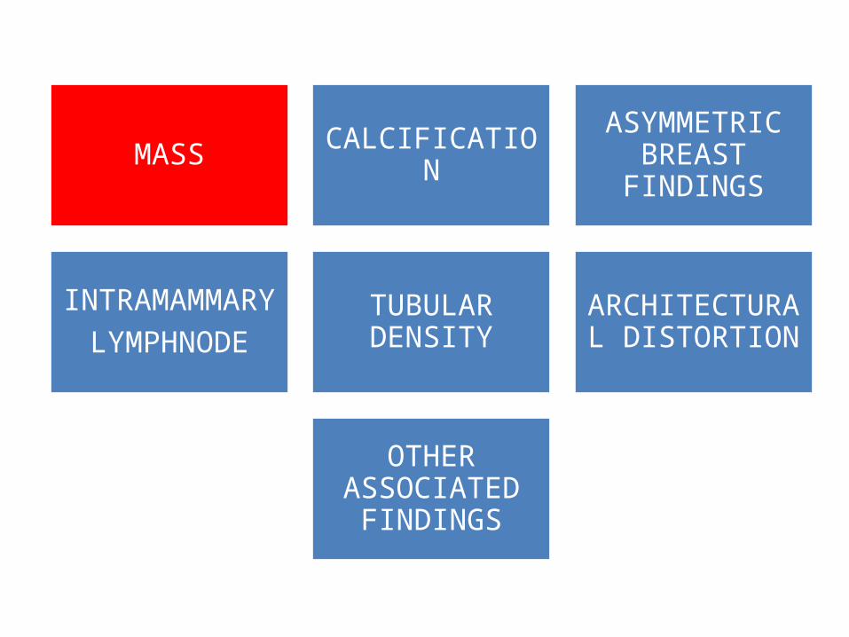
• Is seen in both the views.• Involves a less than one quadrant of breast.• It can be due to normal variations or some lesion.
FOCAL ASYMMETRY


DEVELOPING ASYMMETRY
• This is a focal asymmetry that is new, larger, or denser at current examination than at previous examinations.

ASYMMETRY BIRADS I
DEVELOPING ASYMMETRY BIRADS IV
NON- PALPABLE
GLOBAL ASYMMETRY BIRADS II
NON-PALPABLE
FOCAL ASYMMETRY BIRADS III
PALPABLE GLOBAL ASYMMETRY BIRADS IV
PALPABLE FOCAL ASYMMETRY BIRADS IV

MASS CALCIFICATION
ASYMMETRIC BREAST
FINDINGS
INTRAMAMMARY
LYMPHNODE
TUBULAR DENSITY
ARCHITECTURAL DISTORTION
OTHER ASSOCIATED
FINDINGS

• Well circumscribed.• < 1cm• UPPER AND OUTER
QUADRANT• Lucent and invaginated
fatty hilum• May appear as 3 or more
round densities in horse shoe arrangement.
BENIGN INTRAMAMMARY LYMPH NODE

• If a mass is seen in a section other than upper and outer quadrant, unless it has a clearly defined hilum.
• Lesion in upper outer quadrant does not have other characteristics, it should be considered suspicious as malignant node or primary mass.
When not to consider Benign Intramammary node

MASS CALCIFICATION ASYMMETRIC BREAST FINDINGS
INTRAMAMMARYLYMPHNODE
TUBULAR DENSITY
ARCHITECTURAL DISTORTION
OTHER ASSOCIATED
FINDINGS

• Tubular or branching structure representing dilated duct.
• Usually of minor significance.
• BIRADS III

MASS CALCIFICATION ASYMMETRIC BREAST FINDINGS
INTRAMAMMARYLYMPHNODE
TUBULAR DENSITY
ARCHITECTURAL DISTORTION
OTHER ASSOCIATED
FINDINGS

• Spiculations radiating from a point without any identifiable mass.
• The only architectural distortion that does not require further evaluation is that caused by prior surgery or trauma.
• BIRADS IV


SKIN RETRACTION
NIPPLE RETRACTION
SKIN THICKENING
TRABECULAR THICKENING
AXILLARY LYMPHADENOPATHY

• FINALLY WE HAVE to decide on the significance of the mammographic findings.
• FINALISE THE REPORT IN 7 SPECIFIC CATEGORIES.
Related Documents