Approach to azotemic patients Case presentation Shiva Seyrafian

Approach to azotemic patients Case presentation Shiva Seyrafian.
Dec 25, 2015
Welcome message from author
This document is posted to help you gain knowledge. Please leave a comment to let me know what you think about it! Share it to your friends and learn new things together.
Transcript

Approach to azotemic patients Case presentation
Shiva Seyrafian

Approach to azotemic patients
At the end of this class, you should be able to
Diagnose acute kidney injury (AKI)
Define the etiologies of AKI
Describe the evaluation of AKI
Diagnose chronic kidney disease (CKD)
Define the stages of CKD
Describe the evaluation of CKD
Discuss CKD risk factors and management of them.

Evaluation of Renal FailureIs the renal failure acute or chronic?
laboratory values do not discriminate between acute vs. chronic
Oliguria supports a diagnosis of acute renal failure
History of renal disease and azotemia helps to diagnose CKD.
Clues to chronic disease
Pre-existing illness – DM, HTN, age, vascular disease.
Uremic symptoms – fatigue, nausea, anorexia, pruritus, altered taste sensation, hiccups.
Small, echogenic kidneys by ultrasound.

5 Key Steps in Evaluating Acute kidney Injury
1. Obtain a thorough history and physical; review the chart in detail
2. Do everything you can to accurately assess volume status
3. Always order a renal ultrasound
4. Look at the urine
5. Review urinary indices

Case 1
A 42 year old male is admitted to the SICU after sustaining multiple trauma. His course is complicated by Enterobacter sepsis with profound hypotension requiring support with intravenous dopamine. The urine output has gradually decreased to only 300 ml per day. The urine sodium is 78.


Ischemic Acute Renal Failure
A form of ATN often following a prerenal insult
Late proximal tubule and medullary thick ascending limb most susceptible
Severity of renal failure correlates with duration of insult
Treatment is to optimize renal perfusion, avoid additional nephrotoxic insults and other supportive measures

Conditions that Lead to Pre-renal Acute Renal Failure
Generalizedor Localized Reduction in
Renal Blood Flow
IschemicAcute Renal Failure
Intravascular Volume Depletion
Decreased Effective Circulating VolumeCHF Cirrhosis Nephrosis
MedicationsCYA, TacrolimusACE inhibitors NSAIDSRadiocontrast Amphotericin BAminoglycosides
HepatorenalSyndrome
Sepsis
Large-vessel Renal Vascular DiseaseRenal Artery ThrombosisRenal Artery EmbolismRenal Artery Stenosis or Crossclamping
Small-vessel Renal Vascular DiseaseVasculitis AtheroemboliThrombotic MicroangiopathiesTransplant Rejection

Phases of Ischemic Epithelial Tubular Injury
Time
GFR
Pre-renal
Initiation
Extension
MaintenanceRecovery

Risk Factors for Ischemic Tubular Injury
Volume depletion
Aminoglycosides
Radiocontrast
NSAIDs, Cox-2 inhibitors
Sepsis
Rhabdomyolysis
Pre-existing renal disease
HTN
Diabetes mellitus
Age > 50
Cirrhosis

Case 2
A 56 y.o. male presents with complaints of persistent fever, chills, sore throat, and myalgias for the past 14 days. Ten days ago he started taking amoxicillin.
His physical exam is remarkable for fever to 38.6oC, an exudative pharyngitis and a diffuse maculopapular rash.

Laboratory Data Result Normal Range
Serum Na 134 mEq/L 135-145
K 5.7 mEq/L 3.5-5
Cl 106 mEq/L 100-111
Total CO2 14 mEq/L 24
BUN 46 mg/dL 4-15
Creatinine 3.8 mg/dL 0.6-1.0
Glucose 96 mg/dL 60-100
Whole blood
WBC 12 x109/L 4.5-11.0
Hgb 11 gm/dL 13.5-17.5
Hct 33 % 41.0-53.0
Platelets 216 x109/L150-440
Urine
Specific gravity 1.010 1.002-1.036
Protein 2+ Negative
Blood TraceNegative
Glucose Negative Negative
The urine sediment shows 3-5 RBC’s/h.p.f., 20-25 WBC’s/h.p.f., coarse granular and white cell casts, and rare red cell casts.

Case 2…

Acute Interstitial Nephritis-Etiology
• Allergic/Drug induced• Autoimmune
– Sarcoid -SLE– Sjogren’s
• Toxins– Chinese herb nephropathy -Heavy metals– Light chain cast nephropathy
• Infiltrative– Leukemia– Lymphoma
• Infections (Legionella, CMV, HIV, Toxoplasma)

Acute Interstitial NephritisClinical Presentation
• Non-oliguric ARF• Fever in allergic and infectious types (except NSAID type)• Rash in allergic type (except NSAID induced)• Eosinophilia• UA: WBC casts
Eosinophiluria (allergic)Hematuria (NSAID related)

Common Causes of Drug Induced AIN
• NSAIDS• Antibiotics
– Penicillins• methicillin• Ampicillin, amoxacillin, carbenacillin, oxacillin• Cephalosporins
– Quinolones (ciprofloxacin)– Anti-tuberculous medications (rifampin, INH, ethambutol)– Sulfonamides (TMP-SMX, furosemide, thiazides)
• Miscellaneous– Allopurinol, cimetidine, dilantin

Acute Interstitial NephritisTreatment
• Withdrawal of offending agent
• Treatment of underlying process if infectious/autoimmune etiology
• Trial of corticosteroids, especially in allergic presentations1 mg/kg/day or 2 mg/kg every other day– No randomized trials proving efficacy– Reversal of renal failure usually seen in < 6 weeks

Case 3
A 58 y/o woman referred to clinic due to abdominal pain, anorexia, fever and icterus. She had hx of HTN and DM. BP= 100/70, BW= 68 kg.T= 38.8 In US there was a common bile duct stone.
Lab: WBC= 20,000, Hb= 14.5, plt= 260000, BilT=5, Bill D= 4, AST= 70 ALT= 75, AlPh= 289, BUN= 25, Cr= 1.4
The physician admitted her and ordered gentamycin 80 mg tid for 7 days.
Patient recovered from fever and pain but 10 days later she developed nausea, vomiting, and edema. BP=160/100

Lab values
WBC 14500 Alb 3.9 U/A
Hb 11.5 pH 7.30 SG 1010
Plt 285000 HCO3 16 WBC 8-10
BUN 58 pCO2 38 RBC 6-8
Cr 5.4 AST 42 Ren epith 5-7
Na 139 ALT 57 Cast gran 5-6
K 5.2 AlPh 230
Ca 8.8 Bill T 1.8
P 5.6 Bill D 1
BS 186

What is the cause of vomiting?
How much is her eGFR? (First and last)

Calculations
Cockcroft-Gault Men: CrCl (mL/min) = (140 - age) x wt (kg)
SCr x72
Women: multiply by 0.85
MDRD GFR (mL/min per 1.73 m2) = 186 x (SCr x
0.0113)-1.154 x (age)-0.203 x (0.742 if female) x (1.12 if African-American

eGFR: (F, age= 58, BW= 68)
Cr= 1.4 Cr=5.4
CG 1= 47 ml/min CG 2= 12 ml/min
MDRD 1= 41 ml/min MDRD 2= 8.6 ml/min
What is the cause of azotemia?
Which kind of renal failure has she developed?
What is your prescription?
What would be her prognosis?

Aminoglycoside Nephrotoxicity
Generally presents 1 week after exposure
Non-oliguric
Low trough levels do not guard against nephrotoxicity
Incidence of ATN
10% after 1 week
40% after 2 weeks
Risk factors for ATN
Advanced age - Superimposed sepsis
Liver disease - Hypotension
CKD (DM-HTN)

Case
A 60 Y/O male patient with Hx of HTN and IHD, his cardiologist recommended coronary angiography due to recent chest pain.
Before angio: BUN= 23, Cr= 1.3, Na=138, K=4.5, Hb= 14.
3 days after angiography he developed edema, nausea and BP= 170/95, BUN=55 Cr=4.3 K=5.2 Na=132 U/A: SG= 1.011, Blood 1+, Pr 1+, WBC=5-7, RBC= 4-6, R epithelial cell= 3-4, cast granular+
What’s your diagnosis?
And what is his prognosis?

Case
Radiocontrast-induced acute renal failure
10 days later: his sCr= 1.8, BUN= 25, K= 4.5

Radiocontrast-Induced Acute Renal Failure
Induces renal vasoconstriction and direct cytotoxicity via oxygen free radical formation
Risk factors:
Renal insufficiency - Diabetes
Advanced age - > 125 ml contrast
Hypotension
Usually non-oliguric ARF; irreversible ARF rare

Case 4
A 24 y/o renal transplant woman admitted to hospital for fever, tender vesicular rash on left side of abdomen and back in the spinal nerve root, from 4 days ago. T= 38.5, BP= 135/85 Her Cr= 1.8 mg/dl, BUN=28 mg/dl, WBC= 15000, Hb= 12g/dl. U/A: WBC= 3-4 RBC= 4-5
She received acyclovir 500 mg IV tid. After 5 days became anorectic and developed vomiting, but fever recovered.
Now Cr= 6 mg/dl, BUN= 67, Na= 135, K= 5.3, Ca=8.2, P= 4.7, Alb=3.4, U/A: RBC= 8-10, WBC= 10-14, Cast granular=3-4.


Acute Renal Failure due toIntratubular Obstruction
CrystalluriaEthylene glycol: Calcium oxalate
Tumor lysis: Urate and Calcium phosphate
Medications Acyclovir
Methotrexate
Sulfonamides
Anti-retroviral agents
Myeloma cast nephropathy

Case 5
A 35-year-old female presents with a one month history of periorbital and lower extremity edema. Over two days prior to presentation she has experienced arthralgias in her wrists and elbows. On physical examination she is in no acute distress. Blood pressure is 162/94, temperature 37.4 . Her skin exam is significant for a malar erythematous rash. The heart and lungs are normal. There is 3+ edema to the thighs bilaterally.

Laboratory Data Result Normal Range
SerumNa 138 mEq/L135-145
K 4.2 mEq/L 3.5-5
Cl 108 mEq/L 100-111
Total CO2 17 mEq/L 24
BUN 75 mg/dL 4-15
Creatinine 3.5 mg/dL 0.6-1.0
Glucose 83 mg/dL 60-100
Anti-nuclear antibody1:160 Negative
Whole blood
WBC 5.9 x10⁹/L 4.5-11.0
Hgb 11.9 gm/dL 13.5-17.5
Hct 34 % 41.0-53.0
Platelets 153 x109/L 150-440
Urine
Specific gravity 1.015 1.002-1.036
Protein 3+ Negative
Blood 3+ Negative
RBC >50/h.p.f . 0-4
Sodium 10 mEq/L Variable


Acute Glomerulopathies
RPGN most commonly seen with:
Lupus nephritis (DPGN, class IV)
Pauci-immune GN (ANCA associated)
Anti-GBM disease
less commonly: IgA, post-infectious
Nephrotic presentations of ARF
Collapsing FSGS (HIV nephropathy)
Minimal change disease with ATN
Thrombotic microangiopathies (HUS, TTP, malignant hypertension, scleroderma kidney, pre-eclampsia)

Atheroembolic Renal Disease
ARF in patient with erosive atherosclerosis. Often follows aortic manipulation (angiography, surgery, trauma) or anticoagulation.
Pattern is often an acute worsening of renal function due to showering of emboli, followed by more insidious progression over several weeks to months due to ongoing embolization of atheromatous plaques
. Livedo reticularis
. Nephritic sediment, eosinophilia, eosinophiluria, low C3. Poor prognosis

Livedo Reticularis

Case
47 y/o female presents for routine dental care
PMH: chronic renal failure, kidney transplant 4 yrs ago and doing well
Meds: prednisolone, cyclosporine, mycophenolate mofetil
VS: BP: 145/87, PR: 70

What are the potential problems to consider in this patient?
Susceptibility to infection
Management recommendation: Consultation with patient’s transplant
doctor
Antibiotic prophylaxis
Daily antibacterial mouth rinses(chlorhexidine)

Case
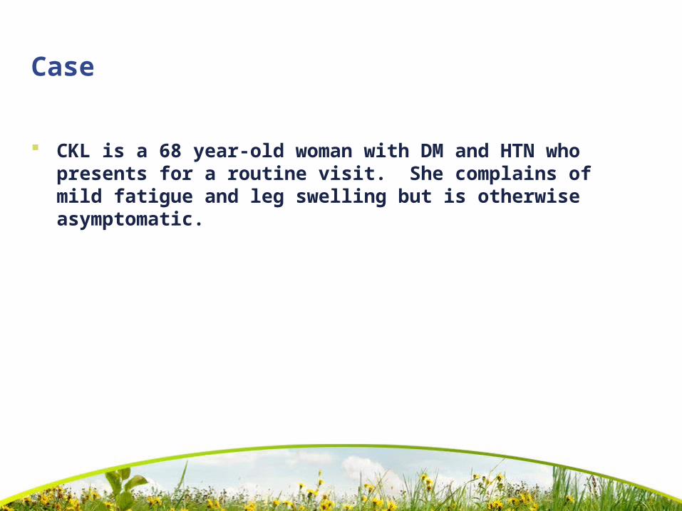
CKL is a 68 year-old woman with DM and HTN who presents for a routine visit. She complains of mild fatigue and leg swelling but is otherwise asymptomatic.
Case…
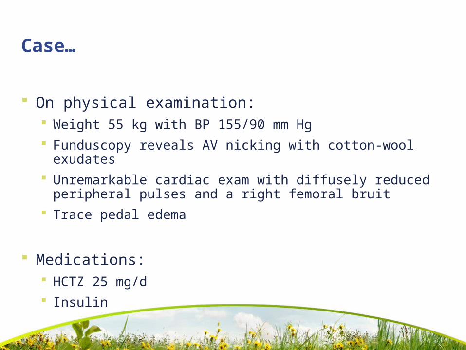
On physical examination: Weight 55 kg with BP 155/90 mm Hg Funduscopy reveals AV nicking with cotton-wool
exudates Unremarkable cardiac exam with diffusely reduced
peripheral pulses and a right femoral bruit Trace pedal edema
Medications: HCTZ 25 mg/d Insulin

Labs
18 months ago, her serum Cr: 1.5 mg/dL
One year ago, sCr: 1.6 mg/dL
How can we assess her degree of kidney dysfunction?

Calculations
Cockcroft-Gault Men: CrCl (mL/min) = (140 - age) x wt (kg)
SCr x72
Women: multiply by 0.85
MDRD GFR (mL/min per 1.73 m2) = 186 x (SCr x
0.0113)-1.154 x (age)-0.203 x (0.742 if female) x (1.12 if African-American

What is CKD?
Presence of markers of kidney damage for three months, as defined by structural or functional abnormalities of the kidney with or without decreased GFR, manifest by either pathological abnormalities or other markers of kidney damage, including abnormalities in the composition of blood or urine, or abnormalities in imaging tests.
The presence of GFR <60 mL/min/1.73 m2 for three months, with or without other signs of kidney damage as described above.

Stages of CKD
Stage 1*: GFR >= 90 mL/min/1.73 m2 Normal or elevated GFR
Stage 2*: GFR 60-89 (mild)
Stage 3: GFR 30-59 (moderate)
Stage 4: GFR 15-29 (severe; pre-HD)
Stage 5: GFR < 15 (kidney failure)Am J Kidney Dis 2002; 39 (S2): S1-246

Case cont.
Recheck her sCr: 1.7 mg/dL
CrCl (age 68 yrs; wt 55 kg): 27 mL/min
MDRD: 32 mL/min/1.73 m2
How can we quantify CKD?
What next doc?

Identify reversible causes
Think about volume contraction, urinary obstruction, or toxic effects of medications
Rx
ACEs/ARBs
NSAIDs
Aminoglycosides and amphotericin B
IV radiocontrast agents
Other etiologies
Renovascular disease
Glomerulonephritis
Nephrotic syndrome
Hypercalcemia
Multiple myeloma
Chronic UTI

Management
Identify and treat factors associated with progression of CKD
HTN
Proteinuria
Glucose control
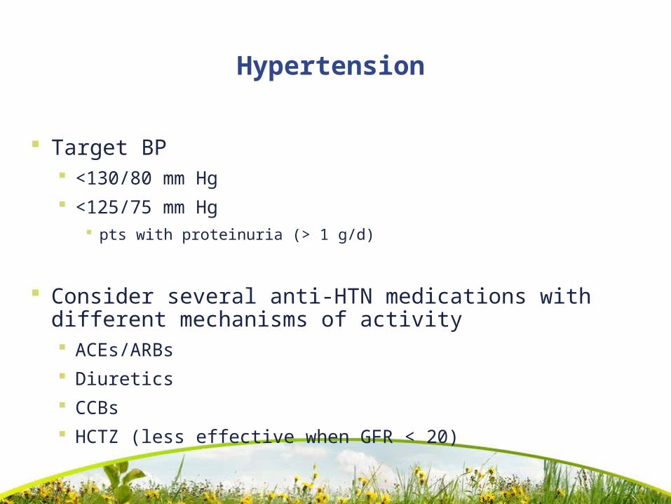
Hypertension
Target BP <130/80 mm Hg <125/75 mm Hg
pts with proteinuria (> 1 g/d)
Consider several anti-HTN medications with different mechanisms of activity ACEs/ARBs Diuretics CCBs HCTZ (less effective when GFR < 20)

Proteinuria
Single best predictor of disease progression
Normal albumin excretion <30 mg/24 hours
Microalbuminuria 20-200 g/min or 30-300 mg/24 hours
Macroalbuminuria >300 mg/24 hours
Nephrotic range proteinuria >3 g/24 hours

Continue HCTZ; add ACE and consider CCB to maintain BP <125/75 mm Hg
What biochemical abnormalities are characteristic of CKD? Or which laboratory tests and radiographic studies would you order?

Evaluation for CKD
Blood
CBC with diff
SMA-7 with Ca2+ and phosphorous
PTH
HBA1c
LFTs and FLP
Uric acid and Fe2+ studies
Urine
Urinalysis with microscopy
Spot urine for microalbumin
24-urine collection for protein and creatinine
Ultrasound

Metabolic changes with CKD
Hemoglobin/hematocrit
Bicarbonate
Calcium
Phosphate
PTH
Triglycerides

Metabolic changes…
Monitor and treat biochemical abnormalities
Anemia
Metabolic acidosis
Mineral metabolism
Dyslipidemia
Nutrition
Metabolic changes…
Monitor and treat biochemical abnormalities
Anemia
Metabolic acidosis
Mineral metabolism
Dyslipidemia
Nutrition

Treating Anemia
Epoetin alfa (rHuEPO; Epogen/Procrit)
HD: 50-100 U/kg IV/SC 3x/wk
Non-HD: 10,000 U qwk
Darbepoetin alfa (Aranesp)
HD: 0.45 g/kg IV/SC qwk
Non-HD: 60 g SC q2wks

Metabolic acidosis
Muscle catabolism
Metabolic bone disease
Sodium bicarbonate Maintain serum bicarbonate > 22 meq/L 0.5-1.0 meq/kg per day Watch for sodium loading
Volume expansion
HTN

NEJM 2000; 342(20): 1478-83
Mineral metabolism
Calcium and phosphate metabolism abnormalities associated with:
Renal osteodystrophy
Calciphylaxis and vascular calcification
14 of 16 ESRD/HD pts (20-30 yrs) had calcification on CT scan
3 of 60 in the control group

JAMA 1993; 269(23): 3015-23
Dyslipidemia
Abnormalities in the lipid profile
Triglycerides
Total cholesterol
NCEP recommends reducing lipid levels in high-risk populations
Targets for lipid-lowering therapy considered the same as those for the secondary prevention of CV disease

Kidney Int 1995; 47(1): 186-92
CV disease
70% of HD patients have concomitant CV disease
Heart disease leading cause of death in HD patients
LVH can be a risk factor
Am J Kidney Dis 2001; 37(6): 1191-200
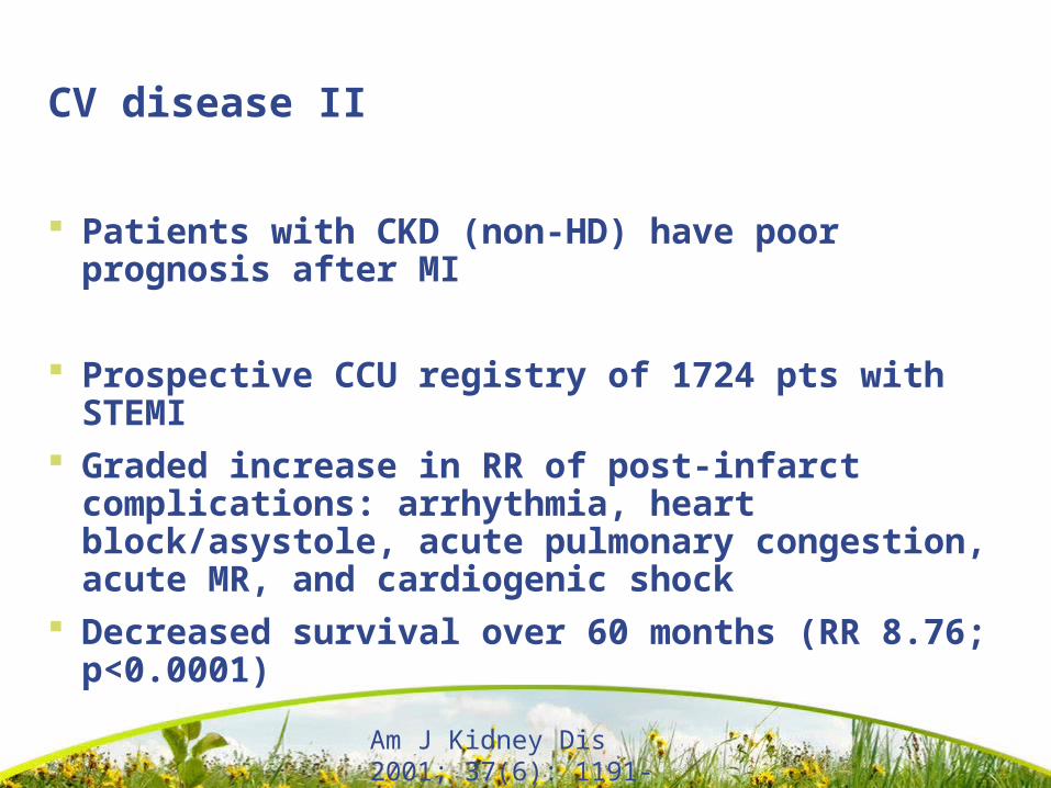
CV disease II
Patients with CKD (non-HD) have poor prognosis after MI
Prospective CCU registry of 1724 pts with STEMI
Graded increase in RR of post-infarct complications: arrhythmia, heart block/asystole, acute pulmonary congestion, acute MR, and cardiogenic shock
Decreased survival over 60 months (RR 8.76; p<0.0001)
Key points
The serum creatinine level is not enough!
Target BP for CKD
<130/80 mm Hg
<125/75 mm Hg in proteinuria
HTN and proteinuria are the two most important modifiable risk factors for progressive CKD

Related Documents