Approach to Adult Patients with Acute Dyspnea Elizabeth DeVos, MD, MPH, Lisa Jacobson, MD* INTRODUCTION Emergency Medical Services (EMS) calls en route with a 45-year-old woman who has a history of congestive heart failure, chronic bronchitis, morbid obesity, and diabetes. She is breathing 40 times per minute, maintaining oxygen saturations of 94%. She ap- pears mildly confused. You have 5 minutes to consider algorithms before the patient arrives. Perhaps respiratory therapy is paged to supply a ventilator or a biphasic pos- itive airway pressure (BIPAP) machine. Maybe you prepare airway equipment or ask your nursing staff to access medication in advance, or you might use the time to expand your differential and determine what brief information regarding the patient’s history and initial physical examination will help you treat her. Acutely dyspneic patients present in various ways. Are the lungs full of fluid or pus? Did the throat swell shut or is the patient just anxious? Did the patient aspirate a foreign body or have a slow or rapid hemorrhage? Is the patient compensating for a severe metabolic acidosis or did the patient run out of beta agonists at home? This article provides helpful guidelines in the assessment and management of these diverse patients. Disclosures: None. Department of Emergency Medicine, University of Florida College of Medicine - Jacksonville, 655 West 8th Street, Jacksonville, FL 32209, USA * Corresponding author. E-mail address: [email protected] KEYWORDS Dyspnea Shortness of breath Asthma COPD Respiratory compensation Pneumonia Pulmonary embolism Angina KEY POINTS The cause of dyspnea is often evident from a complete history and physical examination. Rapid determination of the cause of dyspnea saves lives. Shortness of breath is not always primarily a pulmonary problem. Understanding the pathophysiology of each disease allows clinicians to make rational de- cisions about testing. Emerg Med Clin N Am 34 (2016) 129–149 http://dx.doi.org/10.1016/j.emc.2015.08.008 emed.theclinics.com 0733-8627/16/$ – see front matter Ó 2016 Elsevier Inc. All rights reserved.

Welcome message from author
This document is posted to help you gain knowledge. Please leave a comment to let me know what you think about it! Share it to your friends and learn new things together.
Transcript

Approach to Adult Patientswith Acute Dyspnea
Elizabeth DeVos, MD, MPH, Lisa Jacobson, MD*
KEYWORDS
� Dyspnea � Shortness of breath � Asthma � COPD � Respiratory compensation� Pneumonia � Pulmonary embolism � Angina
KEY POINTS
� The cause of dyspnea is often evident from a complete history and physical examination.
� Rapid determination of the cause of dyspnea saves lives.
� Shortness of breath is not always primarily a pulmonary problem.
� Understanding the pathophysiology of each disease allows clinicians to make rational de-cisions about testing.
INTRODUCTION
Emergency Medical Services (EMS) calls en route with a 45-year-old woman who hasa history of congestive heart failure, chronic bronchitis, morbid obesity, and diabetes.She is breathing 40 times per minute, maintaining oxygen saturations of 94%. She ap-pears mildly confused. You have 5 minutes to consider algorithms before the patientarrives. Perhaps respiratory therapy is paged to supply a ventilator or a biphasic pos-itive airway pressure (BIPAP) machine. Maybe you prepare airway equipment or askyour nursing staff to access medication in advance, or you might use the time toexpand your differential and determine what brief information regarding the patient’shistory and initial physical examination will help you treat her.Acutely dyspneic patients present in various ways. Are the lungs full of fluid or pus?
Did the throat swell shut or is the patient just anxious? Did the patient aspirate aforeign body or have a slow or rapid hemorrhage? Is the patient compensating for asevere metabolic acidosis or did the patient run out of beta agonists at home? Thisarticle provides helpful guidelines in the assessment and management of thesediverse patients.
Disclosures: None.Department of Emergency Medicine, University of Florida College of Medicine - Jacksonville,655 West 8th Street, Jacksonville, FL 32209, USA* Corresponding author.E-mail address: [email protected]
Emerg Med Clin N Am 34 (2016) 129–149http://dx.doi.org/10.1016/j.emc.2015.08.008 emed.theclinics.com0733-8627/16/$ – see front matter � 2016 Elsevier Inc. All rights reserved.
DeVos & Jacobson130
Respiratory distress is responsible for nearly 4 million emergency department (ED)visits each year and is one of the most common presenting complaints in the elderly.1
When a patient presents with dyspnea, the primary task of the emergency physician isto assess for and ensure stability of the patient’s airway, breathing, and circulation. Inthe case of dyspnea, presentations may range from minor symptoms to extremis.Rapid assessment may necessitate the use of intubation, BIPAP, nebulizations,decompression, or other therapies in the immediate period following the patient’sarrival, to treat dyspnea.
PATIENT EVALUATION
The American Thoracic Society suggests that “dyspnea results from a . mismatchbetween central respiratory motor activity and incoming afferent information from re-ceptors in the airways, lungs and chest wall structures.”2 This dissociation can resultfrom increased metabolic demand, decreased compliance, increased dead-spacevolume, or many other disorders that are discussed later. Each patient presentingshort of breath uses a different set of phrases to describe the symptoms and exami-nation reveals a different combination of disorders. The clinician’s ability to interpretthese varying constellations is necessary to provide appropriate treatment to thesepatients, who are often in serious distress.
History
Acute dyspnea, or shortness of breath, is one of the most common chief complaints inthe ED. The differential diagnosis includes many disorders that can be divided basedon obstructive, parenchymal, cardiac, and compensatory features. A careful historycan begin to narrow this wide differential. In addition to common symptoms, considerrisk factors such as past medical and family history, trauma, travel, medications, andexposures.Schwartzstein and Lewis3 use the analogy of a machine to identify different causes
of dyspnea based on pathophysiologic data. Dysfunctions of the respiratory systemmay be caused by faulty controllers, ventilatory pumps, or gas exchangers(Table 1). This table makes it easier to understand the causes of shortness of breathrelated to respiratory causes.Cardiovascular disease manifests as dyspnea by causing disruptions of the system
that pumps oxygenated blood to tissues and then transports the carbon dioxide backto the lung. Decreases in cardiac output or increases in resistance limit oxygen deliv-ery. Similarly, decreased oxygen carrying capacity in anemia plays a role in its presen-tation with dyspnea.
Physical Examination
A detailed physical examination also provides important guidance (Table 2). Respira-tory rate and oxygen saturation are obtained with vital signs. The clinician shouldassess the patient’s work of breathing, looking for any tripoding or retractions. Crep-itance in the chest may indicate subcutaneous air and pneumothorax. Lung soundssuch as wheezing, rales, and rhonchi further guide the differential. Decreased sounds,hyperresonance, or egophony may also provide additional clues.Jugular venous distension, S3 gallop, and peripheral edema indicate that a patient
has fluid overload. Conjunctival pallor, capillary refill, and temperature of extremitiescan provide clues about blood volume and general circulation. Pulses must also beassessed.
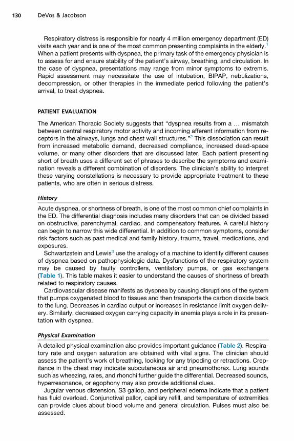
Table 1A systemic approach to dyspnea by assessing the components of the respiratory process
Part Description Manifestations Examples
Controller Malfunction presents as abnormalrespiratory rate or depth. Oftenrelated to abnormal feedback tobrain from other parts of thesystem
Air hunger, needto breathe
Abnormal feedback tobrain from othersystems. Metabolicacidosis, anxiety
Ventilatorypump
Composed of muscles, nerves thatsignal the controller, chest wall,and pleura that create negativethoracic pressure, airways andalveoli allowing flow fromatmosphere and gas exchange
Increased workof breathing,low tidalvolumes
Neuromuscular problems(eg, Guillain-Barre),decreased chest wallcompliance,pneumothorax,pneumonia,bronchospasm(COPD, asthma)
Gasexchanger
Oxygen and carbon dioxide crossthe pulmonary capillaries in thealveoli. Membrane destructionor interruption of the interfacebetween the gas and capillariesby fluid or inflammatory cellslimit gas exchange
Increasedrespiratorydrive,hypoxemia,chronichypercapnia
Emphysema, pneumonia,pulmonary edema,pleural effusion,hemothorax
Adapted from Schwartzstein RM, Lewis A. Dyspnea. In: Broaddus V, Mason RC, Ernst JD, et al, ed-itors. Murray & Nadel’s textbook of respiratory medicine. Elsevier health sciences. 6th edition. Phil-adelphia: Saunders/Elsevier; 2015.
Adult Patients with Acute Dyspnea 131
Testing
Multiple tests are available to narrow the differential diagnosis of acute dyspnea. Whenusing tests to augment clinical decision making, be sure to weigh the information theymay provide with any risks involved in performing the tests (Table 3).Ultrasonography provides valuable information about the origin of symptoms,
and, often, diagnosis in the initial assessment of an acutely dyspneic patient. Theseimages may be obtained during or shortly after initial assessment, potentially
Table 2Physical examination findings and correlating diagnoses
Symptom Differential Diagnosis
Wheeze COPD/emphysema, asthma, allergic reaction, CHF (cardiac wheeze)
Cough Pneumonia, asthma, COPD/emphysema
Pleuritic chest pain Pneumonia, pulmonary embolism, pneumothorax, COPD, asthma
Orthopnea Acute heart failure
Fever Pneumonia, bronchitis, TB, malignancy
Hemoptysis Pneumonia, TB, pulmonary embolism, malignancy
Edema Acute heart failure, pulmonary embolism (unilateral)
pulmonary edema Acute and chronic heart failure, end stage renal and liver diseases, ARDS(sepsis)
Tachypnea alone pulmonary embolism, acidosis (including aspirin toxicity), anxiety
Abbreviations: ARDS, acute respiratory distress syndrome; CHF, congestive heart failure; COPD,chronic obstructive pulmonary disease; TB, tuberculosis.
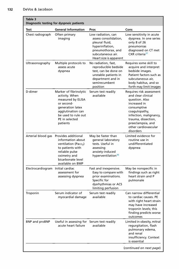
Table 3Diagnostic testing for dyspneic patients
Test General Information Pros Cons
Chest radiograph Often primaryimaging
Low radiation, canassess consolidation,pleural fluid,hyperinflation,pneumothorax, andsubcutaneous air.Heart size is apparent
Low sensitivity in acutedyspnea. In one seriesonly 8 of 26pneumoniasdiagnosed on CT metCXR criteria37
Ultrasonography Multiple protocols toassess acutedyspnea
No radiation, fast,reproducible bedsidetest, can be done onunstable patients indepartment and insemirecumbentposition
Requires some skill toacquire and interpretbedside images.Patient factors such assubcutaneous air,body habitus, and soforth may limit images
D-dimer Marker of fibrinolyticactivity. Whenmeasured by ELISAor second-generation latexagglutination canbe used to rule outPE in selectedpatients
Serum test readilyavailable
Requires risk assessmentand clear clinicalquestion. Alsoincreased inconsumptivecoagulopathy,infection, malignancy,trauma, dissection,preeclampsia, andother cardiovasculardisorders
Arterial blood gas Provides additionalinformation aboutventilation (PaCO2)to patients withreliable pulseoximetry andbicarbonate levelavailable on BMP
May be faster thangeneral laboratorytests. Useful inassessinganxiety-inducedhyperventilation36
Limited evidence forroutine use inundifferentiateddyspnea
Electrocardiogram Initial cardiacassessment forassessing dyspnea
Fast and inexpensive.Easy to compare withprior examinations.Specific fordysrhythmias or ACSlimiting perfusion
May be nonspecific infindings such as rightheart strain and Ppulmonale
Troponin Serum indicator ofmyocardial damage
Serum test readilyavailable
Can narrow differentialto cardiac causes. PEwith right heart strainmay have increasedtroponin levels; thisfinding predicts worseoutcomes
BNP and proBNP Useful in assessing foracute heart failure
Serum test readilyavailable
Limited in obesity, mitralregurgitation, flashpulmonary edema,and renalinsufficiency. Contextis essential
(continued on next page)
DeVos & Jacobson132
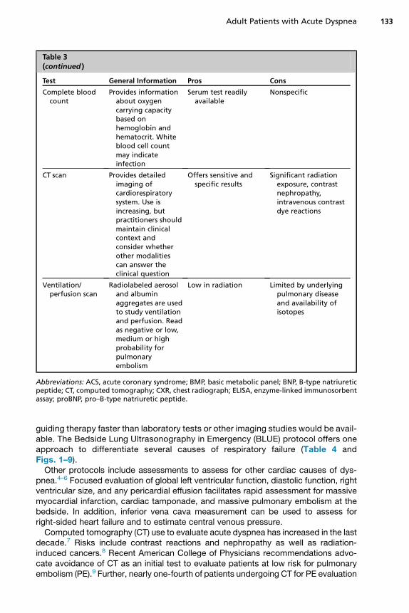
Table 3(continued )
Test General Information Pros Cons
Complete bloodcount
Provides informationabout oxygencarrying capacitybased onhemoglobin andhematocrit. Whiteblood cell countmay indicateinfection
Serum test readilyavailable
Nonspecific
CT scan Provides detailedimaging ofcardiorespiratorysystem. Use isincreasing, butpractitioners shouldmaintain clinicalcontext andconsider whetherother modalitiescan answer theclinical question
Offers sensitive andspecific results
Significant radiationexposure, contrastnephropathy,intravenous contrastdye reactions
Ventilation/perfusion scan
Radiolabeled aerosoland albuminaggregates are usedto study ventilationand perfusion. Readas negative or low,medium or highprobability forpulmonaryembolism
Low in radiation Limited by underlyingpulmonary diseaseand availability ofisotopes
Abbreviations: ACS, acute coronary syndrome; BMP, basic metabolic panel; BNP, B-type natriureticpeptide; CT, computed tomography; CXR, chest radiograph; ELISA, enzyme-linked immunosorbentassay; proBNP, pro–B-type natriuretic peptide.
Adult Patients with Acute Dyspnea 133
guiding therapy faster than laboratory tests or other imaging studies would be avail-able. The Bedside Lung Ultrasonography in Emergency (BLUE) protocol offers oneapproach to differentiate several causes of respiratory failure (Table 4 andFigs. 1–9).Other protocols include assessments to assess for other cardiac causes of dys-
pnea.4–6 Focused evaluation of global left ventricular function, diastolic function, rightventricular size, and any pericardial effusion facilitates rapid assessment for massivemyocardial infarction, cardiac tamponade, and massive pulmonary embolism at thebedside. In addition, inferior vena cava measurement can be used to assess forright-sided heart failure and to estimate central venous pressure.Computed tomography (CT) use to evaluate acute dyspnea has increased in the last
decade.7 Risks include contrast reactions and nephropathy as well as radiation-induced cancers.8 Recent American College of Physicians recommendations advo-cate avoidance of CT as an initial test to evaluate patients at low risk for pulmonaryembolism (PE).9 Further, nearly one-fourth of patients undergoing CT for PE evaluation

Table 4BLUE protocol
UltrasonographyFinding
UltrasonographyApproach Description Clinical Meaning Image
Assess forartifacts: Alines
Anterior Subpleural air causes repeated linear artifactsparallel to the pleural line (horizontal)
Air in lung: either normal or pneumothorax Fig. 1
Assess forartifacts: Blines
Anterior Seven features: hyperechoic, well-defined,hydroaeric comet-tail artifacts arising from thepleural line. They spread upwards indefinitelyand obscure A lines. When lung sliding ispresent, they move with the lung
Represents an interface of 2 widely differenttransmissions of ultrasound waves: in this case,air and fluid. When 3 or more B lines are in asingle interspace, they are B1 lines (orpulmonary rockets), indicating interstitialsyndrome
Fig. 2
Assess for lungsliding
Anterior Absence of lung sliding occurs with a disruption ofthe normal sliding of viscera on parietal pleuraor separation of the two. In Mmode, absence oflung slide is seen as the stratosphere sign (alsoknown as bar-code sign)
Absence of lung sliding in the presence of A linesnecessitates search for pneumothorax. Lungpoint is the ultrasonography finding in whichlung slide is seen in the same view with theabolished lung slide and A lines in the samelocation, indicating the tip of the lung
Fig. 3(stratospheresign)
Fig. 4 (normallung)
Fig. 5 (lungpoint)
Assess foralveolarconsolidationor pleuraleffusion(posterolateralalveolar and/orpleuralsyndrome)
Lateralsubposterior
The classic anechoic, dependent pattern may beinconsistent. Other findings include (1) quadsign: pleural effusion on expiration notedbetween the pleural and regular, lower lunglines (viscera). (2) Shred sign: tissuelike patternseenwith alveolar consolidation, with the upperborder of lung line (or pleural line when there isno effusion) with an irregular lower border. (3)Sinusoid sign: movement of the lung linetoward the pleural line in inspiration
Pleural effusion: sinusoid, plus may have quadsign. Alveolar consolidation: tissuelikeappearance or shred sign, absent lung line,absent sinusoidal sign
Figs. 6 and 7(pleuraleffusion)
Fig. 8(tissuelikelung)
Fig. 9(sinusoidal)
Deep venousthrombosis
Femoral veins Visualization of thrombus in the lumen or lack ofcompressibility is positive test
Consider pulmonary embolus if positive —
Adapted from Lichtenstein DA,Meziere GA. Relevance of lungultrasound in the diagnosis of acute respiratory failure: the BLUEprotocol. Chest 2008;134(1):117–25;and Courtesy of Petra Duran, MD, RDMS, University of Florida College of Medicine-Jacksonville (FL).
DeVos&
Jaco
bson
134
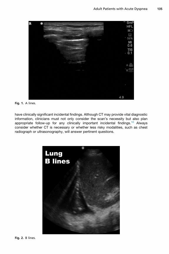
Fig. 1. A lines.
Adult Patients with Acute Dyspnea 135
have clinically significant incidental findings. Although CT may provide vital diagnosticinformation, clinicians must not only consider the scan’s necessity but also planappropriate follow-up for any clinically important incidental findings.10 Alwaysconsider whether CT is necessary or whether less risky modalities, such as chestradiograph or ultrasonography, will answer pertinent questions.
Fig. 2. B lines.

Fig. 3. Stratosphere sign.
DeVos & Jacobson136
DIFFERENTIAL DIAGNOSIS FOR ACUTELY DYSPNEIC PATIENTSObstructive Dyspnea
Consider the 35-year-old woman discussed earlier. Medics report tachypnea withvery poor air movement during transport. As she rolls through the ambulance baydoors, you are already assessing her. Adept clinicians can spot respiratory distressfrom across the room. She is diaphoretic, her shoulders are held adjacent to herears, and she is breathing extremely rapidly with minimal air movement. You decide
Fig. 4. Normal lung.

Fig. 5. Lung point.
Adult Patients with Acute Dyspnea 137
to aggressively treat her for a severe asthma exacerbation, starting BIPAP ventilationwith continuous nebulized albuterol and order adjunct therapies including intravenoussteroids, intravenous magnesium, and intramuscular epinephrine. After 20 minutes ather bedside, she begins to breathe more comfortably with the BIPAP machine andrepeat auscultation reveals diffuse wheezing and improved air movement.
Fig. 6. Pleural effusion.

Fig. 7. Pleural effusion.
DeVos & Jacobson138
As she begins to improve, EMS returns with another patient. His breath sounds areaudible to everyone in the resuscitation bay. He appears diaphoretic and panicked.Examination reveals stridor, periorbital edema, tachycardia, and hypotension. Imme-diate intervention for anaphylactic shock begins and, after 2 rounds of epinephrine,fluid boluses, antihistamines, and steroids, he too begins to look better.Wheezing, ormusical respiratory sounds, typically result frompartial airway obstruc-
tion.11 Because this obstruction can result from inflammation, secretions, or even aforeign body, patients with noisy or whistling breathing need close evaluation to
Fig. 8. Tissuelike lung.
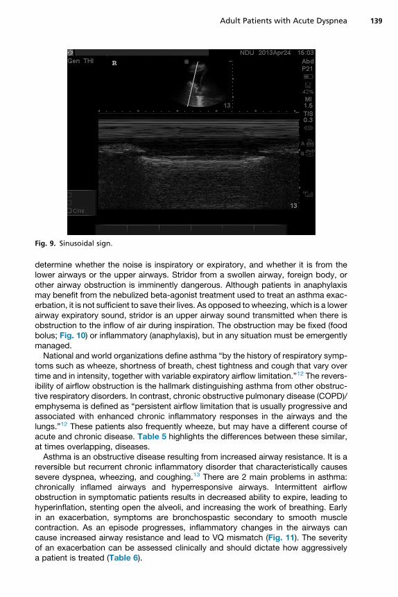
Fig. 9. Sinusoidal sign.
Adult Patients with Acute Dyspnea 139
determine whether the noise is inspiratory or expiratory, and whether it is from thelower airways or the upper airways. Stridor from a swollen airway, foreign body, orother airway obstruction is imminently dangerous. Although patients in anaphylaxismay benefit from the nebulized beta-agonist treatment used to treat an asthma exac-erbation, it is not sufficient to save their lives. As opposed to wheezing, which is a lowerairway expiratory sound, stridor is an upper airway sound transmitted when there isobstruction to the inflow of air during inspiration. The obstruction may be fixed (foodbolus; Fig. 10) or inflammatory (anaphylaxis), but in any situation must be emergentlymanaged.National and world organizations define asthma “by the history of respiratory symp-
toms such as wheeze, shortness of breath, chest tightness and cough that vary overtime and in intensity, together with variable expiratory airflow limitation.”12 The revers-ibility of airflow obstruction is the hallmark distinguishing asthma from other obstruc-tive respiratory disorders. In contrast, chronic obstructive pulmonary disease (COPD)/emphysema is defined as “persistent airflow limitation that is usually progressive andassociated with enhanced chronic inflammatory responses in the airways and thelungs.”12 These patients also frequently wheeze, but may have a different course ofacute and chronic disease. Table 5 highlights the differences between these similar,at times overlapping, diseases.Asthma is an obstructive disease resulting from increased airway resistance. It is a
reversible but recurrent chronic inflammatory disorder that characteristically causessevere dyspnea, wheezing, and coughing.13 There are 2 main problems in asthma:chronically inflamed airways and hyperresponsive airways. Intermittent airflowobstruction in symptomatic patients results in decreased ability to expire, leading tohyperinflation, stenting open the alveoli, and increasing the work of breathing. Earlyin an exacerbation, symptoms are bronchospastic secondary to smooth musclecontraction. As an episode progresses, inflammatory changes in the airways cancause increased airway resistance and lead to VQ mismatch (Fig. 11). The severityof an exacerbation can be assessed clinically and should dictate how aggressivelya patient is treated (Table 6).
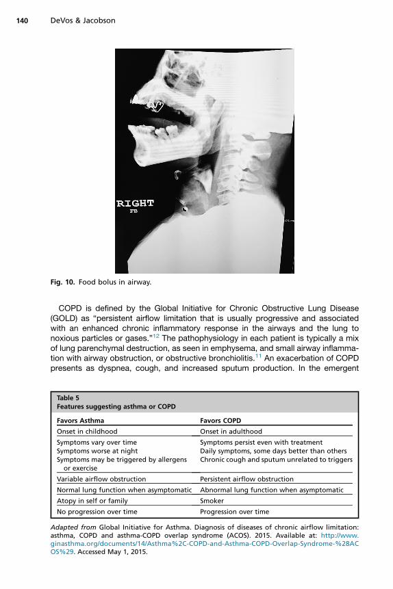
Fig. 10. Food bolus in airway.
DeVos & Jacobson140
COPD is defined by the Global Initiative for Chronic Obstructive Lung Disease(GOLD) as “persistent airflow limitation that is usually progressive and associatedwith an enhanced chronic inflammatory response in the airways and the lung tonoxious particles or gases.”12 The pathophysiology in each patient is typically a mixof lung parenchymal destruction, as seen in emphysema, and small airway inflamma-tion with airway obstruction, or obstructive bronchiolitis.11 An exacerbation of COPDpresents as dyspnea, cough, and increased sputum production. In the emergent
Table 5Features suggesting asthma or COPD
Favors Asthma Favors COPD
Onset in childhood Onset in adulthood
Symptoms vary over timeSymptoms worse at nightSymptoms may be triggered by allergens
or exercise
Symptoms persist even with treatmentDaily symptoms, some days better than othersChronic cough and sputum unrelated to triggers
Variable airflow obstruction Persistent airflow obstruction
Normal lung function when asymptomatic Abnormal lung function when asymptomatic
Atopy in self or family Smoker
No progression over time Progression over time
Adapted from Global Initiative for Asthma. Diagnosis of diseases of chronic airflow limitation:asthma, COPD and asthma-COPD overlap syndrome (ACOS). 2015. Available at: http://www.ginasthma.org/documents/14/Asthma%2C-COPD-and-Asthma-COPD-Overlap-Syndrome-%28ACOS%29. Accessed May 1, 2015.

Fig. 11. Pathophysiology of symptom development in asthma.
Adult Patients with Acute Dyspnea 141
setting, clinicians must treat the airflow limitation. As with asthma, monitoring of pulseoximetry, degree of respiratory distress, and hemodynamic stability can help cliniciansanticipate the degree of severity of a particular exacerbation. More specific testingmay also have a role, because radiographs and electrocardiograms may help differen-tiate other causes of shortness of breath from a COPD exacerbation. In addition, anincrease in sputum production or the presence of purulent sputum should be treatedwith antibiotics, regardless of other infectious symptoms.14
Anaphylaxis is a sudden, potentially fatal, allergic reaction involving multiple organsystems.15,16 The Second Symposium on the Definition and Management of Anaphy-laxis lists the following clinical criteria for diagnosis of anaphylaxis:
1. Acute onset of an illness (minutes to several hours) with involvement of the skin,mucosal tissue, or both (eg, generalized hives; pruritus or flushing; swollen lips,tongue, uvula)And at least 1 of the following:a. Respiratory compromise (eg, dyspnea, wheeze-bronchospasm, stridor,
reduced peak expiratory flow, hypoxemia)
Table 6Severity of asthma exacerbation assessment
Symptoms Mild Moderate Severe Near Death
Breathless While walking While talking At rest Decreased effort
Speaking In sentences In phrases In words Unable
Alertness May be agitated Usually agitated Usually agitated Confused
Respiratory Rate(breaths/min)
Increased Increased >30 >30, imminentfailure
Accessory Muscle Use Usually not Commonly Usually Usually
Wheeze Moderate Loud Loud or silent Silent
Heart Rate (beats/min) <100 100–120 >120 �Saturation (%) >95 92–94 <90 <90

DeVos & Jacobson142
b. Reduced blood pressure (BP) or associated symptoms of end-organ dysfunc-tion (eg, hypotonia [collapse], syncope, incontinence)
2. Two or more of the following that occur rapidly after exposure to a likely allergen forthat patient:a. Involvement of the skin-mucosal tissueb. Respiratory compromisec. Reduced BP or associated symptomsd. Persistent gastrointestinal symptoms (eg, crampy abdominal pain, vomiting)
3. Reduced BP after exposure to known allergen for that patient:a. Infants and children: low systolic BP (age specific) or greater than 30%
decrease in systolic BPb. Adults: systolic BP of less than 90 mm Hg or greater than 30% decrease from
that person’s baseline
It is the respiratory compromise that is relevant to this article, and it is important torecognize that treating the allergic component of these symptoms is necessary tosave the patients.
Parenchymal Dyspnea
Now EMS is at the back door with a 60-year-old patient with a history of COPD andcongestive heart failure (CHF). He is in respiratory distress with audible wheezingand tripoding. He is diaphoretic, hypertensive, and has pitting edema to his knees.They have given albuterol with no improvement of his symptoms.Acute dyspnea is the most common symptom of patients presenting with heart fail-
ure.17 Eighty percent of patients with acutely decompensated heart failure presentthrough the ED with a chief complaint of dyspnea.18 This symptom is related toboth pulmonary and systemic fluid overload and also low cardiac output. AmericanCollege of Emergency Physicians clinical policy makes level B recommendationsthat standard clinical judgment can be improved with the use of a single B-type natri-uretic peptide (BNP) or N-terminal pro–B-type natriuretic peptide measurement to rulein or out the diagnosis of CHF.19 However, the true utility, may be in patients with dys-pnea not expected to have a CHF exacerbation, when finding a positive BNP wouldchange management and allow a faster initiation of treatment.20
Carpenter and colleagues20 found that the classic constellation of symptoms (ju-gular venous distension, peripheral edema, rales, and S3) were no more predictive ofpatients with both pulmonary edema on chest radiograph and an increased BNPlevel greater than 500 pg/dL than any individual finding alone. Although rales werethe most sensitive finding tested for either outcome, they had specificity of onlyabout 50% each. Jugular venous distention and S3 gallop were the individual find-ings most predictive for pulmonary edema on radiograph or increased BNP level. Ul-trasonography measurements of the inferior vena cava also improve diagnosticaccuracy versus BNP and chest radiograph alone.21
The medics are back in your department, this time with a 75-year-old man withcough and fever. His family is worried that he has been eating less and is sleepierthan at hospital discharge last week. In pneumonia, the diffusion of oxygen is limitedby alveolar infiltrates, leading to shortness of breath. Common complaints and find-ings in community-acquired pneumonia include fever, cough, pleuritic chest pain,and sputum production, along with dyspnea. However, these clinical criteria mayhave a sensitivity as low as 50% compared with a chest radiograph.22 On examina-tion, many patients have crackles or evidence of consolidation. Guidelines from theInfectious Diseases Society of America and the American Thoracic Society,

Adult Patients with Acute Dyspnea 143
recommend chest radiograph in patients with suspected pneumonia, which may showlobular consolidation, interstitial infiltrate, or cavitation.23 Although infiltrate with sug-gestive symptomsmakes the diagnosis, infiltrate may not be visible initially on patientswith volume depletion. It is appropriate to treat empirically for 24 to 48 hours in thesecases and to reimage when hydration is restored.The management of pneumonia requires history to allow classification based on the
setting in which the illness was acquired. The Infectious Disease Society of Americaand American Thoracic Society define the types of pneumonia as follows:24Hospital-acquired pneumonia (HAP) is “pneumonia that occurs 48 hours or more after admis-sion which was not incubating at the time of admission.” Ventilator-associatedpneumonia (VAP) arises “more than 48-72 hours after endotracheal intubation.” Inaddition, health care–associated pneumonia (HCAP) is diagnosed in any patient whois “hospitalized in an acute care hospital for two or more days within 90 days of theinfection, resided in a nursing home or long-term care facility, received recent IV anti-biotic therapy, chemotherapy or wound care within the past 30 days of the currentinfection or attended a hospital or hemodialysis clinic.” Community-acquired pneu-monia is not acquired in any of these situations. These classifications identify typicalpathogens and guide appropriate initial management. Important historical exposuresand risk factors to refine treatment are summarized in Table 7.23
The American Thoracic Society along with the Infectious Disease Society of Amer-ica’s consensus statement offers 4 important principles in the initial management andevaluation of adult patients with bacterial HAP, VAP, or HCAP; the most important tobe accomplished in the ED is to promptly treat with “appropriate and adequate ther-apy” to decrease mortality.24
Circulatory Dyspnea
After a brief delay, you see a 28-year-old woman with shortness of breath and chestpain. She smokes, uses hormonal birth control, and reports that her symptomsstarted when she came back from a business trip. Pulmonary embolism (PE) inter-feres with both ventilation and perfusion. It ultimately causes circulatory collapsebecause of obstruction of right ventricular outflow eventually causing increased pul-monary artery pressure and failure of the right then left ventricles. Before circulatorycollapse, echocardiography can show signs of right ventricular (RV) strain, includingdilatation of the right ventricle, RV hypokinesis, paradoxic septal wall motion,McConnell sign (hypokinesis of the free RV wall with sparing of the apex), andtricuspid regurgitation.25
Dresden and colleagues26 supported the use of ultrasonography in moderate-risk tohigh-risk patients to determine whether the patients were appropriate for anticoagu-lation while awaiting definitive imaging. Early anticoagulation is recommended toimprove mortality and there is evidence to support anticoagulation before diagnosisin patients with a Wells score greater than 4 who will have a delay to diagnosis ofmore than 1 hour and 40 minutes.27,28
The assessment of patients with dyspnea and concern for PE requires a series ofrisk stratification. One common method is to use Wells criteria29 (Box 1) in patientswith suspicion for PE; although other stratification tools exist, none has been shownto be clearly superior. When there is low clinical suspicion for PE, PERC (pulmonaryembolism rule-out criteria)30 rules or D-dimer testing may be applied.27 If PERC(Box 2) is negative, or there is intermediate pretest probability for PE with negativehigh-sensitivity D-dimer, no further testing for PE is required.27 When further testingis needed (positive D-dimer or high-sensitivity D-dimer not available), negative CTangiogram or low-probability VQ scan may be used to rule out PE.

Table 7Common pathogens in community-acquired pneumonia
Condition Commonly Encountered Pathogens
Alcoholism Streptococcus pneumoniae, oral anaerobes, Klebsiellapneumoniae, Acinetobacter species, Mycobacteriumtuberculosis
COPD and/or smoking Haemophilus influenzae, Pseudomonas aeruginosa,Legionella species, S pneumonia, Moraxellacatarrhalis, Chlamydophila pneumoniae
Aspiration Gram-negative enteric pathogens, oral anaerobes
Lung abscess CA-MRSA, oral anaerobes, endemic fungal pneumonia,M tuberculosis, atypical mycobacteria
Exposure to bat or bird droppings Histoplasma capsulatum
Exposure to birds Chlamydophila psittaci (if poultry: avian influenza)
Exposure to rabbits Francisella tularensis
Exposure to farm animals orparturient cats
Coxiella burnetii (Q fever)
HIV infection (early) S pneumoniae, H influenzae, M tuberculosis
HIV infection (late) The pathogens listed for early infection plusPneumocystis jirovecii, Cryptococcus, Histoplasma,Aspergillus, atypical mycobacteria (especiallyMycobacterium kansasii), P aeruginosa, H influenzae
Hotel or cruise ship stay in previous2 wk
Legionella species
Travel to or residence insouthwestern United States
Coccidioides species, Hantavirus
Travel to or residence in southeastand east Asia
Burkholderia pseudomallei, avian influenza, SARS
Influenza active in community Influenza, S pneumoniae, Staphylococcus aureus, Hinfluenzae
Cough>2 wk with whoop orpost-tussive vomiting
Bordetella pertussis
Structural lung disease (eg,bronchiectasis)
P aeruginosa, Burkholderia cepacia, S aureus
Injection drug use S aureus, anaerobes, M tuberculosis, S pneumoniae
Endobronchial obstruction Anaerobes, S pneumonia, H influenzae, S aureus
In context of bioterrorism Bacillus anthracis (anthrax), Yersinia pestis (plague),Francisella tularensis (tularemia)
Abbreviations: CA-MRSA, community-acquired methicillin-resistant Staphylococcus aureus; HIV,human immunodeficiency virus; SARS, severe acute respiratory syndrome.
From Mandell LA, Wunderink RG, Anzueto A, et al. Infectious Diseases Society of America/American Thoracic Society consensus guidelines on the management of community-acquiredpneumonia in adults. Clin Infect Dis 2007;44(Suppl 2):S46; with permission.
DeVos & Jacobson144
In the next bed is a middle-aged woman with diabetes complaining of shortness ofbreath today. It was associated with some vague nausea and she says that she justdoes not feel good. Angina pectoris is cardiac chest pain in which oxygen demandoutweighs myocardial oxygen supply; in this case caused by occlusion of coronaryarteries. Although typically chest pain is a part of the presentation, dyspnea alonemay be the initial complaint, termed an anginal equivalent. In one recent large seriesof patients undergoing stress testing, patients with dyspnea alone were at increased

Box 1
Wells criteria for pulmonary embolism
� Clinical signs and symptoms of deep vein thrombosis (DVT): 13
� PE is the main diagnosis or equally likely: 13
� Heart Rate greater than 100 beats/min: 11.5
� Immobilization >3 days or surgery in last 4 weeks: 11.5
� History of prior PE/deep venous thrombosis (DVT): 11.5
� Hemoptysis: 11
� Malignancy with treatment within 6 months or palliative: 11
Less than or equal to 1.55 low risk, 1.3% chance of PE in ED population; 2 to 65moderate risk,16.2% chance of PE in ED population; greater than 6 5 high risk, 40.6% chance of PE in EDpopulation
Adapted fromWells PS, Anderson DR, Rodger M, et al. Excluding pulmonary embolism at thebedside without diagnostic imaging: Management of patients with suspected pulmonary em-bolism presenting to the emergency department by using a simple clinical model and D-dimer.Ann Intern Med 2001;135:99.
Adult Patients with Acute Dyspnea 145
risk of death from cardiac causes. Patients asked simply whether they experiencedshortness of breath were considered dyspneic. The subset with no prior known cor-onary artery disease had more than 4 times the risk of sudden cardiac death versusasymptomatic patients and more than twice the risk of those with typical angina.31
Clinicians should consider past medical history and risk factors when assessing dys-pnea for cardiac causes such as acute myocardial infarction and acute coronary syn-drome. Appropriate testing includes bedside electrocardiogram, troponin, and chestradiograph.The department eventually settles down and you are able to do some charting until a
young man comes in with visible respiratory distress. He is tall and thin, smokes regu-larly, and reports sudden onset of severely painful breathing. Pneumothorax occurswhen air enters the plural space between the chest wall and the lung. Typically onlya thin serous layer exists between the visceral and parietal pleura. Air enters this po-tential space only when there is damage to the lung or chest wall, or a gas-producingpleural space infection. The classic risk factors for bleb rupture causing spontaneous
Box 2
Pulmonary embolism rule-out criteria
� Further work-up recommended if any of the following are present:� Age greater than or equal to 50 years� Pulse greater than or equal to 100 beats/min� Oxygen saturation less than 95%� Hemoptysis� Unilateral leg swelling� History of PE/DVT� Exogenous estrogen use� Surgery or trauma within 4 weeks that required hospitalization or intubation
� If none are present, probability of PE is less than 2%.
Adapted from Kline JA, Courtney DM, Babrhel C, et al. Prospective multicenter evaluation ofthe pulmonary embolism rule-out criteria. J Thromb Haemost 2008;6(5):773.

DeVos & Jacobson146
pneumothorax are tall men, although smoking has been suggested to increase the riskof rupture by damaging the pleural layer.Pneumothoraces may be identified by ultrasonography, chest radiograph, or CT.
Treatment may be guided by cause, severity, comorbidities, interventions such aspositive pressure ventilation, size of the pneumothorax, and patient’s preference.Recent studies suggest that uncomplicated spontaneous pneumothorax in patientsnot undergoing positive pressure ventilation may be treated as successfully with nee-dle aspiration as with other more invasive chest drains, regardless of size.32
Tension pneumothorax is a serious event requiring immediate needle decompres-sion to avert loss of cardiac output and arrest. However, recent review shows thatthe classic presentation of tension pneumothorax with hypotension, absent breathsounds, and deviated trachea may not be immediately seen in patients with sponta-neous, unassisted respiration.33 Because of the slower development of the accumu-lation of air and pressure variations, spontaneously breathing patients maycompensate much longer and present atypically, as shown in Table 8. Thus, cliniciansmust remain vigilant.
Compensatory Dyspnea
This article focuses on the cardiopulmonary system as the source of the problem inacutely dyspneic patients. It is important to also consider that the appearance ofshortness of breath, tachypnea, or other typical symptoms of dyspnea may resultfrom changing metabolic demands. These patients may appear, on the surface, tobe in respiratory distress; they may be tachypneic, tachycardic, even pale or diapho-retic. In these cases, the clinician’s responsibility is to identify and fix the true problemin order to improve the respiratory symptoms.Severely anemic patients have limited oxygen carrying capacity. Their bodies there-
fore experience oxygen hunger, which can manifest as shortness of breath. Patientswith dysfunctional hemoglobins secondary to irreversibly bound atoms or toxinsmay also be functionally anemic with the same symptoms.People’s bodies attempt at all costs to maintain equilibrium. Therefore, in metabolic
acidoses (such as diabetic ketoacidosis), chemoreceptors detect acidosis and
Table 8Findings in tension pneumothorax
Unassisted Ventilation Positive Pressure Ventilation
Spontaneous respiration with air passingthrough 1-way flap
Assisted ventilation forces air through pleuraldefect into pleural space
Compensatory mechanisms delay collapse:� Tachycardia and accessory muscle use
caused by tachypnea, increased tidal vol-ume, and negative movement of the oppo-site side of the chest
� BP is maintained because of limits in thepressure of the pneumothorax on medias-tinum and hemithorax
Sudden hemodynamic and respiratorycompromise:
� Sedation may increase inspiratory pressure� Intrapleural pressure is increased
throughout respiratory cycle
Venous siphon resulting from negativeintrathoracic pressure in the opposite sideof the chest returns blood to the heart
Decreased venous return leads to hypotensionand cardiac arrest
Adapted from Roberts DJ, Leigh-smith S, Faris PD, et al. Clinical presentation of patients with ten-sion pneumothorax: a systematic review. Ann Surg 2015;261(6):1069.
Adult Patients with Acute Dyspnea 147
stimulate the respiratory center to hyperventilate. Both the rate and the depth of venti-lation often increase, leading to both tachypnea and hyperpnea, at times referred to asKussmaul respirations. This compensatory response is crucial for survival and shouldnot be mistaken for dyspnea. It is equally important to realize that an increase in alve-olar ventilation is not always a compensatory response (to acidosis or to primary pul-monary disorders) and hypocapnia may cause primary respiratory alkalosis, fromcentral nervous system compromise, toxins (eg, salicylates), anxiety, or pain.34 Inthese patients, imaging rarely reveals a source of dyspnea, but clinical suspicionbased on history and examination, including signs such as the fruity breath of ketone-mia, the pallor of anemia, or the cyanosis of toxic hemoglobinopathies, directs pro-viders toward appropriate laboratory testing and treatment.
Diagnosis of Exclusion
In addition, sometimes dyspnea is not dyspnea. Acute anxiety and panic disorder canpresent as shortness of breath, tachypnea, or hyperventilation. Patients with panicdisorder often describe symptoms similar to those of patients with true airwayobstruction despite their normal pulmonary function. It has been suggested that thesepatients have abnormal proprioception, experiencing dyspnea without abnormal stim-uli.35 However, patients with a history of pulmonary disease can also have pure panicepisodes. Arterial blood gas may be useful in diagnosing anxiety-relatedhyperventilation.36
Severe pain can also induce abnormal respiratory patterns. Like compensatoryproblems, pain and anxiety can be managed by managing their causes. Treat pain.Reduce stress and anxiety with words, behaviors, or, if necessary, medications. How-ever, air hunger and difficulty breathing also make individuals anxious. Be sure toavoid premature diagnosis of a purely anxiety-based concern without first evaluatingfor more dangerous disorders.
SUMMARY
Acute dyspnea presents commonly to the ED and it is imperative that emergency phy-sicians be prepared to stabilize patients’ oxygenation and ventilation, which requirescareful and efficient consideration of the differential diagnosis. Using cues from thehistory and physical examination, practitioners may guide the work-up and treatmentto identify a parenchymal, obstructive, circulatory, or compensatory cause of dys-pnea. Early use of bedside testing, including ultrasonography, may limit unnecessarytests and save time in determining the best treatment course. Thus ensuring both thebest care for the patient and also the physician’s ability to readily respond to the nextcase.
REFERENCES
1. Prekker ME, Feemster LC, Hough CL, et al. The epidemiology and outcome ofprehospital respiratory distress. Acad Emerg Med 2014;21(5):543–50.
2. American Thoracic Society. Dyspnea. Mechanisms, assessment, and manage-ment: a consensus statement. Am J Respir Crit Care Med 1999;159:321–40.
3. Schwartzstein RM, Lewis A. Chapter 29: Dyspnea. In: Broaddus V, Mason RC,Ernst JD, et al, editors. Murray & Nadel’s textbook of respiratory medicine. 6thedition. Philadelphia: Elsevier Health Sciences, Saunders/Elsevier; 2015.p. 490–1.
4. Kajimoto K, Madeen K, Nakayama T, et al. Rapid evaluation by lung-cardiac infe-rior vena cava (LCI) integrated ultrasound for differentiating heart failure from
DeVos & Jacobson148
pulmonary disease as the cause of acute dyspnea in the emergency setting. Car-diovasc Ultrasound 2012;10:49.
5. Anderson KL, Jenq KY, Fields JM, et al. Diagnosing heart failure among acutelydyspneic patients with cardiac, inferior vena cava and lung ultrasonography. AmJ Emerg Med 2013;31:1208–14.
6. Russell FM, Ehrman RR, Cosby K, et al. Diagnosing acute heart failure in patientswith undifferentiated dyspnea: a lung and cardiac ultrasound (LuCUS) protocol.Acad Emerg Med 2015;22(2):182–91.
7. Feng LB, Pines JM, Yusuf HR, et al. U.S. trends in computed tomography use anddiagnoses in emergency department visits by patients with symptoms sugges-tive of pulmonary embolism, 2001-2009. Acad Emerg Med 2013;20(10):1033–40.
8. Huckins DS, Price LL, Gilley K. Utilization and yield of chest computed tomo-graphic angiography associated with low positive D-dimer levels. J Emerg Med2012;43:211–20.
9. Qaseem A, Alguire P, Dall P, et al. Appropriate use of screening and diagnostictests to foster, high-value, cost-conscious care. Ann Intern Med 2012;156:147–9.
10. Coco AS, O’gurek DT. Increased emergency department computed tomographyuse for common chest symptoms without clear patient benefits. J Am Board FamMed 2012;25(1):33–41.
11. Chapter 8, The thorax and lungs. In: Bickley LS, Szilagyi PG, editors. Bates’ guideto physical examination and history taking. 11th edition. Philadelphia: LippincottWilliams & Wilkins; 2013. p. 301.
12. Global Initiative for Asthma. Diagnosis of diseases of chronic airflow limitation:asthma, COPD and asthma-COPD overlap syndrome (ACOS). 2015. Availableat: http://www.ginasthma.org/documents/14/Asthma%2C-COPD-and-Asthma-COPD-Overlap-Syndrome-%28ACOS%29. Accessed May 1, 2015.
13. Husain AN. Chapter 15, The lung. In: Kumar V, Abbas AK, Aster JC, editors. Rob-bins and Cotran pathologic basis of disease. 9th edition. Philadelphia: WB Saun-ders; 2014. p. 679.
14. Ram FS, Rodriguez-Roison R, Granados-Navarrete A, et al. Antibiotics for exac-erbations of chronic obstructive pulmonary disease. Cochrane Database SystRev 2006;(2):CD004403.
15. Sampson HA, Munoz-Furlong A, Campbell RL, et al. Second Symposium on theDefinition and Management of Anaphylaxis: summary Report – Second NationalInstitute of Allergy and Immunology. Ann Emerg Med 2006;47(4):373–80.
16. Simons FES, Ardusso LRF, Dimov V, et al. World Allergy Organization anaphylaxisguidelines: 2013 update of the evidence base. Int Arch Allergy Immunol 2013;162:193–204.
17. Pang PS, Collins SP, Sauser K, et al. Assessment of dyspnea early in acute heartfailure: patient characteristics and response differences between Likert and vi-sual analog scales. Acad Emerg Med 2014;21(6):659–66.
18. Fonarow GC. The Acute Decompensated Heart Failure National Registry(ADHERE): opportunities to improve care of patients hospitalized with acute de-compensated heart failure. Rev Cardiovasc Med 2003;4(Suppl 7):S21–30.
19. Silvers SM, Howell JM, Kosowsky JM, et al. Clinical policy: critical issues in theevaluation and management of adult patients presenting to the emergencydepartment with acute heart failure syndromes. Ann Emerg Med 2007;49(5):627–69.
20. Carpenter CR, Keim SM, Worster A, et al. Brain natriuretic peptide in the evalua-tion of emergency department dyspnea: is there a role? J Emerg Med 2012;42(2):197–205.
Adult Patients with Acute Dyspnea 149
21. Miller JB, Sen A, Strote SR, et al. Inferior vena cava assessment in the bedsidediagnosis of acute heart failure. Am J Emerg Med 2012;30(5):778–83.
22. Metlay JP, Fine MJ. Testing strategies in the initial management of patients withcommunity-acquired pneumonia. Ann Intern Med 2003;138:109.
23. Mandell LA, Wunderink RG, Anzueto A, et al. Infectious Diseases Society ofAmerica/American Thoracic Society consensus guidelines on the managementof community-acquired pneumonia in adults. Clin Infect Dis 2007;44(Suppl 2):S27–72.
24. American Thoracic Society, Infectious Diseases Society of America. Guidelinesfor the management of adults with hospital-acquired, ventilator-associated, andhealthcare-associated pneumonia. Am J Respir Crit Care Med 2005;171(4):388–416.
25. Kucher N, Rossi E, DeRosa M, et al. Prognostic role of echocardiography amongpatients with acute pulmonary embolism and a systolic arterial pressure of 90 mmHg or higher. Arch Intern Med 2005;165:1777–81.
26. Dresden S, Mitchell P, Rahimi L, et al. Right ventricular dilatation on bedsideechocardiography performed by emergency physicians aids in the diagnosisof pulmonary embolism. Ann Emerg Med 2014;63(1):16–24.
27. Fesmire FM, Brown MD, Espinosa JA, et al. Critical issues in the evaluation andmanagement of adult patients presenting to the emergency department with sus-pected pulmonary embolism. Ann Emerg Med 2011;57(6):628–52.e75.
28. Blondon M, Righini M, Aujesky D, et al. Usefulness of preemptive anticoagulationin patients with suspected pulmonary embolism: a decision analysis. Chest 2012;142:697–703.
29. Wells PS, Anderson DR, Rodger M, et al. Excluding pulmonary embolism at thebedside without diagnostic imaging: management of patients with suspectedpulmonary embolism presenting to the emergency department by using a simpleclinical model and D-dimer. Ann Intern Med 2001;135:98–107.
30. Kline JA, Courtney DM, Babrhel C, et al. Prospective multicenter evaluation of thepulmonary embolism rule-out criteria. J Thromb Haemost 2008;6(5):772–80.
31. Abidov A, Rozanski A, Hachamovitch R, et al. Prognostic significance of dyspneain patients referred for cardiac stress testing. N Engl J Med 2005;353(18):1889–98.
32. Zehtabchi S, Rios CL. Management of emergency department patients with pri-mary spontaneous pneumothorax: needle aspiration or tube thoracostomy? AnnEmerg Med 2008;51(1):91–100, 100.e1.
33. Roberts DJ, Leigh-smith S, Faris PD, et al. Clinical presentation of patients withtension pneumothorax: a systematic review. Ann Surg 2015;261:1068–78.
34. Morris CG, Low J. Metabolic acidosis in the critically ill: part 1. Classification andpathophysiology. Anaesthesia 2008;63(3):294–301.
35. Smoller JW, Pollack MH, Otto MW, et al. Panic anxiety, dyspnea, and respiratorydisease. Am J Respir Crit Care Med 1996;154:6–17.
36. Burri E, Potocki M, Drexler B, et al. Value of arterial blood gas analysis in patientswith acute dyspnea: an observational study. Crit Care 2011;15(3):R145.
37. Syrjala H, Broas M, Suramo I, et al. High resolution computed tomography for thediagnosis of community-acquired pneumonia. Clin Infect Dis 1998;27:358–63.
Related Documents