APPLIED 3-D ANATOMY OF LIVER BILE DUCTS IN INJECTION-CORROSION CASTS Jurkovikj M. Dragica, Korneti-Pekevska L. Kostandina Institute of Anatomy, Medical Faculty, University “Ss Cyril and Methodius”, Skopje, R. Macedonia Primljen/Received 15. 03. 2013. god. Prihva}en/Accepted 22. 04. 2013. god. Abstract: On the 20 post-autopsy adult isolated li- ver specimens of patients of both sexes (17 male and 3 female) aged 29–88, the injection-corrosion method was used. Colored acrylate was injected into the biliary system, and uncolored acrylate into the portal vein. A to- tal of 17 acrylate casts were of proper quality. Within the 9 portal segments, both the anatomical determination and quantity and the mode of confluence of intrahepatic bile ducts were established. Different modes of biliary tract confluence up to the sectors and hepatics were fo- und. Besides the most frequent findings of convention- ally confluence bile ducts, there were aberrant modaliti- es of biliary drainage in eight cases. Among them 5 ca- ses had confluence of posterior and anterior sector ducts in the left hepatic duct and 1 case had confluence at first of anterior and then posterior sector ducts in the left he- patic duct. Also, extrahilar connection of the right poste- rior with left lateral into common hepatic duct, where the latter entered the medial and anterior sectors ducts in 1 case was found. There was a subsequent confluence of ducts from the 8th and 5th segments in 1 case, and from the lateral and medial sectors with or without caudate lo- be in 3 cases. A common (4) or separate (2) confluence of left and right portions ducts in the left drainage sys- tem were in 6 cases, whereas in both, the left and right drainage system in 7 cases was found. Rare, there was an aberrant single channel from the right portion in 1 ca- se, as well as the presence and biliary drainage only of the left portion of 1st segment was found. Segment 9 bi- le ducts drained all three subsegments (b, c and d) in 10 cases, and only two (c and d) in 3 cases, as well as only two (c and b) of present three subsegments in 3 cases. Also, there was even one case with present 9d subseg- ment and without 9th segment duct. Those modalities are of interest in an applying and accurate interpretation and performance of diagnostic and interventional proce- dures, as well as in segmental, sectoral or hemihepatic resection in liver surgery. Key words: liver, intrahepatic bile duct, portal vein, injection-corrosion method, segmentation. INTRODUCTION The use of the injection-corrosion method in inve- stigation of segmental and functional structure of liver gives three-dimensional image of the hilar and intrahe- patic ramification of their inflow and outflow triad components. It enables observing their morphological features, as well as the developmental, age-related, pathoanatomical and regenerative porto-biliary chan- ges. This method has been applied at our laboratory for making acrylic casts enabled morphological study of the vascular structure of separate organs — Korneti et al. (1); to search for the aberrant pattern of vessels sup- plying special tissue structures, i.e. sinuatrial node — Korneti (2); or to shown the distinction of blood supply in cases with pathoanatomical changes — Korneti et al. (3). Later on this method has been used in fetal livers to esteem the hepatovenous segments pattern — Laza- rova et al. (4). In continuo Jurkovikj (5) was investiga- ted the portal vascular segmentation in adult livers. Up to day, she has improved this method in doing acrylic casts with choice of the three components of portal tri- ade. In this paper, in all specimen was injected both portal vein and bile ducts. The injection-corrosive method has been used also at Faculty for Veterinary Medicine for morphological research of canalicular structured organs and vascular bed in domestic animal material — Ilieski et al. (6). The findings of anatomical investigations as vari- ous (different), aberrant or alternative anatomy and morphology of intrahepatic bile ducts is essential for both, radiological and diagnostic, and for interventio- nal gastro-entero-hepatology and surgical practice. Despite of pathological substrate, the biliary anatomy 2013; 8(1): 33–42 UDK: 611.36 ; 616.36-091 ISSN-1452-662X Originalni nau~ni rad

Welcome message from author
This document is posted to help you gain knowledge. Please leave a comment to let me know what you think about it! Share it to your friends and learn new things together.
Transcript
APPLIED 3-D ANATOMY OF LIVER BILE DUCTS
IN INJECTION-CORROSION CASTS
Jurkovikj M. Dragica, Korneti-Pekevska L. Kostandina
Institute of Anatomy, Medical Faculty, University “Ss Cyril and Methodius”, Skopje, R. Macedonia
Primljen/Received 15. 03. 2013. god. Prihva}en/Accepted 22. 04. 2013. god.
Abstract: On the 20 post-autopsy adult isolated li-
ver specimens of patients of both sexes (17 male and 3
female) aged 29–88, the injection-corrosion method
was used. Colored acrylate was injected into the biliary
system, and uncolored acrylate into the portal vein. Ato-
tal of 17 acrylate casts were of proper quality. Within the
9 portal segments, both the anatomical determination
and quantity and the mode of confluence of intrahepatic
bile ducts were established. Different modes of biliary
tract confluence up to the sectors and hepatics were fo-
und. Besides the most frequent findings of convention-
ally confluence bile ducts, there were aberrant modaliti-
es of biliary drainage in eight cases. Among them 5 ca-
ses had confluence of posterior and anterior sector ducts
in the left hepatic duct and 1 case had confluence at first
of anterior and then posterior sector ducts in the left he-
patic duct. Also, extrahilar connection of the right poste-
rior with left lateral into common hepatic duct, where
the latter entered the medial and anterior sectors ducts in
1 case was found. There was a subsequent confluence of
ducts from the 8th and 5th segments in 1 case, and from
the lateral and medial sectors with or without caudate lo-
be in 3 cases. A common (4) or separate (2) confluence
of left and right portions ducts in the left drainage sys-
tem were in 6 cases, whereas in both, the left and right
drainage system in 7 cases was found. Rare, there was
an aberrant single channel from the right portion in 1 ca-
se, as well as the presence and biliary drainage only of
the left portion of 1st segment was found. Segment 9 bi-
le ducts drained all three subsegments (b, c and d) in 10
cases, and only two (c and d) in 3 cases, as well as only
two (c and b) of present three subsegments in 3 cases.
Also, there was even one case with present 9d subseg-
ment and without 9th segment duct. Those modalities
are of interest in an applying and accurate interpretation
and performance of diagnostic and interventional proce-
dures, as well as in segmental, sectoral or hemihepatic
resection in liver surgery.
Key words: liver, intrahepatic bile duct, portal vein,
injection-corrosion method, segmentation.
INTRODUCTION
The use of the injection-corrosion method in inve-
stigation of segmental and functional structure of liver
gives three-dimensional image of the hilar and intrahe-
patic ramification of their inflow and outflow triad
components. It enables observing their morphological
features, as well as the developmental, age-related,
pathoanatomical and regenerative porto-biliary chan-
ges. This method has been applied at our laboratory for
making acrylic casts enabled morphological study of
the vascular structure of separate organs — Korneti et
al. (1); to search for the aberrant pattern of vessels sup-
plying special tissue structures, i.e. sinuatrial node —
Korneti (2); or to shown the distinction of blood supply
in cases with pathoanatomical changes — Korneti et
al. (3).
Later on this method has been used in fetal livers
to esteem the hepatovenous segments pattern — Laza-
rova et al. (4). In continuo Jurkovikj (5) was investiga-
ted the portal vascular segmentation in adult livers. Up
to day, she has improved this method in doing acrylic
casts with choice of the three components of portal tri-
ade. In this paper, in all specimen was injected both
portal vein and bile ducts.
The injection-corrosive method has been used also
at Faculty for Veterinary Medicine for morphological
research of canalicular structured organs and vascular
bed in domestic animal material — Ilieski et al. (6).
The findings of anatomical investigations as vari-
ous (different), aberrant or alternative anatomy and
morphology of intrahepatic bile ducts is essential for
both, radiological and diagnostic, and for interventio-
nal gastro-entero-hepatology and surgical practice.
Despite of pathological substrate, the biliary anatomy
2013; 8(1): 33–42 UDK: 611.36 ; 616.36-091
ISSN-1452-662X Originalni nau~ni rad
is the second sententious factor to choose and the suc-
cess of applied technique, to prevent pre or post opera-
tively complications, as well as, to avoid the misinter-
pretations of detected biliary elements and pathologi-
cal changes.
Biliary anatomy and anatomic variability as ap-
plied to liver transplantation or laparoscopic cholecystec-
tomy from many clinical physicians were assessed
(7–15), and nomenclature of hepatic anatomy and resec-
tions was done (16).
In the study for functional liver anatomy and clas-
sification of surgical liver anatomy Doklestic et al. (17)
emphasized that in 1998, Federative Committee on An-
atomical Terminology (FCAT) suggested acceptance of
Couinaud’s liver classification. Although it has still not
been accepted worldwide, the obvious progress in the
study of functional anatomy of the liver is a powerful
impulse for development of modern liver surgery.
MATERIAL AND METHODS
To determine the intrahepatic biliary drainage tract
of 20 post-autopsy adult liver specimens of patients of
both sexes (17 male and 3 female) aged 29–88, the in-
jection-corrosion method was used. Colored yellow
acrylate was injected into the biliary system, and unco-
lored redish acrylate into the portal vein. Corrosion
was made in the concentrated HCl acid and then the
corrosible material from the obtained casts was remo-
ved with tap water. A total of 17 acrylate casts were of
proper quality, with 9 portal segments separated. Within
the 9 portal segments anatomical determination, quan-
tity and mode of confluence of intrahepatic bile ducts to
the liver hilum were established. Biliary ducts were fol-
lowed and compared parallel to the adjoint portal rami-
fication. Intrahepatic confluence of the biliary ducts has
been classified from Ist to IVth order, i.e. two hepatic,
four sectoral, nine segmental and initial ducts.
The 3 remaining liver specimens with incomplete
biliary injection were not analyzed.
A schematic drawings for the acrylate porto-bili-
ary casts, using a magnifying lense, were made. Portal
segments and comparable to them, the biliary segmen-
tal ducts were numbered according to Couinaud’s seg-
ments (7) from 1 to 9, using the Arabic numerals despi-
te previously used Roman ones, as suggested the IHP-
BA-International Hepato-Pancreato-Biliary Associa-
tion and the Brisbane 2000 terminology (16).
The results which obtained the confluence modal-
ities have been presented in tables 1–7.
For statistical analysis of frequencies of different
modalities of segmental ducts confluence to sectors
and hepatics ones was used Kolmogorov-Smirnov’s
test of agreement for one sample (cluster sample).
RESULTS
Within the intrahepatic ramification of portal vein
was proved existence of 9 portal segments in all cases
(Figure 1. a1, b1, c1, d1, e1, f1 and g1; Figure 2. a2, b2,
c2, d2, e2, f2 and g2).
Analysis of the nine portal segments in all 17
acrylate casts showed different modes of biliary tract
confluence up to the sectors and hepatics.
The posterior sector duct merging pattern is shown
on the Table 1.
In the biliary drainage of 7th
segment one duct in
10–58,8%, two in 6–35,3% (Figure e1) and 3 in 1
cast-5,9% were found. From the 6th
segment one duct
in 12–70,58%, two in 4–23,52% (Figure e1 and e2) and
four in 1 cast-5,9% were found.
The anterior sector duct merging pattern is shown
on the Table 2.
There were 1-7 biliary ducts in the 8th
segment:
one duct in 2–11,76%, two in 6–35,3% (Figure e1),
three in 2–11,76%, four (Figure e2), five and seven in
a single cases-17,64% and six in 4 cases-23,52%, whe-
reas 1–14 in the 5th
segment: one duct in 2–11,76%,
two in 2–11,76%, three in 5–29,41%, four in 1–5,9%
(Figure e1), five in 3–17,64% (Figure e2), six in
34 Jurkovikj M. Dragica, Korneti-Pekevska L. Kostandina
Type-modalities of the constituent ducts number of cases %
Segmental ducts (7 + 6) 12 70,5
Segmental duct 7 + common stem of segmental ducts (6 + 5) 1 5,9
Segmental duct 7 + common stem of double segmental duct 7
and segmental duct 61 5,9
Common stem of two segmental ducts 7 + segmental duct 6 1 5,9
Segmental ducts (7 + 6 + 5) 1 5,9
Segmental ducts (6 + 6) 1 5,9
Total 17 100,00
Kolmogorov-Smirnov’s Dmax = 0,538 > D (17 and 0,05) = 0,318
Table 1. Merging pattern of posterior sector duct
1–5,9%, seven in 2–11,76% and fourteen in 1 ca-
se-5,9%.
The lateral sector duct merging pattern is shown
on the Table 3.
There were 1–4 biliary ducts in the 2nd segment:
one duct in 6–35,3% (Figure e2), two in 9–52,94% (Fi-
gure e1) and four in 2 cases-11,76%; also, 1–4 in the 3rd
segment: one duct in 10–58,8% (Figure e1 and Figure
e2), two in 5–29,41%, three in 1–5,9% and four in 1 ca-
se-5,9%.
Twelve casts contained one medial sector duct-
-70,58% (Figure e1 and Figure e2), four a double one-
-23,52% and one a tripled one-5,9%, in total 23 (Ta-
ble 4.1).
The medial sector duct merging pattern is shown
on the table 4.2. (Included are the listed sector ducts
from Table 4.1)
There were 2–9 biliary ducts in the 4b segment:
two ducts in 3–17,64%, three in 5–29,41% (Figure e2),
four in 5–29,41%, five in 2–11,76%, six (Figure e1) and
nine in a single cases-11,76%; and 0–7 in the 4a seg-
ment: no 4a segmental duct-1 case-5,9%, one duct in
4–23,52% (Figure e1 and Figure e2), two in 3–17,64%,
three in 1–5,9%, five in 2–11,76%, six in 3–17,64% and
seven in 3 cases-17,64%.
The sector ducts have confluence in the hepatic
ducts as is shown on the Tables 5 and 6.
APPLIED 3-D ANATOMY OF LIVER BILE DUCTS IN INJECTION-CORROSION CASTS 35
Type (constituent ducts) number of cases %
Two to three segmental ducts (8 + 5) 11 64,70
Segmental ducts (8 + 8) 3 17,64
Magistral way of confluence (8 + 5) 1 5,9
No anterior sector duct 2 11,76
Total 17 100,00
Kolmogorov-Smirnov’s Dmax = 0,397 > D (17 and 0,05) = 0,318
Table 2. Merging pattern of anterior sector duct
Table 3. Merging pattern of lateral sector duct
Type (constituent ducts) number of cases %
Segmental ducts (3 + 2) 8 47,0
Magistral way of confluence of segmental ducts from lateral
and medial sectors3 17,64
Segmental duct 3 + double medial sector duct 1 5,9
Common stem of segmental duct 3 and double medial sector
duct + segmental duct 21 5,9
Common stem of (3 + 3) segmental ducts and (2 + 2) segmental
ducts + segmental duct 31 5,9
Common stem of (3 + 3) segmental ducts + common stem
of segmental duct 2 and duct of left portion of caudate lobe1 5,9
No lateral sector duct 2 11,76
Total 17 100,00
Kolmogorov-Smirnov’s Dmax = 0,36 > D (17 and 0,05) = 0,318
Kind number of cases %
Unique (single) 12 70,58
Double* 4 23,52
Triple* 1 5,9
Total 17 100,00
* There are double(#) and triple(¬) complex modalities
Kolmogorov-Smirnov’s Dmax=0,365 > D(17 and 0,05) = 0,318
Table 4.1. Numerical frequency of medial sector duct

There were aberrant modalities of biliary drainage
which are shown on the Table 7.
Segment 1 named Left Dorsal Sector consisted of
left and right portions in 13 out of 17 casts-76,47%.
The left and right portions ducts had common con-
fluence in the left drained system in 4 cases-23,52% (Fi-
gure e2) and a separate confluence in this system in 2
cases-11,76%. Aseparate confluence of the left portion
ducts in the left and of the right portion ducts in the
right drained systems was found in 3 cases-17,64%. 2
cases-11,76% had drained of left portion ducts only in
the left and of the right portion ducts in the both sided
drained systems (directly or to intervene the left por-
tion duct). The drainage of left portion ducts in both si-
ded drained systems, and of right portion ducts in the
right (directly or to intervene the left portion duct) was
in 2 cases-11,76% (Figure e1).
36 Jurkovikj M. Dragica, Korneti-Pekevska L. Kostandina
Type (constituent ducts) number of cases %
Segmental duct 4b (¬) 6 26,09
Segmental ducts (4b + 4a) (#) 5 21,73
Segmental ducts (4b + 4b) (#) 4 17,4
Common stem of segmental ducts (4b + 4a) + segmental duct 4b 2 8,69
Segmental duct 4b + common stem of segmental ducts (4b + 4a) 2 8,69
Segmental ducts (4a + 4b) (#) 1 4,35
Common stem of segmental ducts (4b + 4a) + common stem
of segmental ducts (4b + 4a)1 4,35
Segmental duct 3 + common stem of segmental duct 4b
and stem of segmental ducts (4b + 4a)1 4,35
Segmental duct 4b + common stem of stems-segmental ducts
(4a + 4a) and segmental ducts (4a + 4a)1 4,35
Total 23 100,00
Kolmogorov-Smirnov’s Dmax = 0,318 > D (23 and 0,05) = 0,27490
Table 4.2. Merging pattern of medial sector duct
Table 5. Merging pattern of right hepatic duct
Type (constituent ducts) number of cases %
Posterior + anterior sector ducts 8 47,0
Posterior sector duct + segmental duct 8 1 5,9
Segmental ducts (8 + 5) 1 5,9
No right hepatic duct * 7 41,2
Total 17 100,00
* see (in addition ) Table 7
Kolmogorov-Smirnov’s Dmax = 0,22 < D (17 and 0,05) = 0,318
Table 6. Merging pattern of left hepatic duct
Type (constituent ducts) number of cases %
Lateral + medial sector ducts 11 64,70
Lateral sector duct + segmental duct 4b 3 17,6
Segmental duct 2 + segmental duct 3 + main medial sector duct 1 5,9
Segmental duct 2 + segmental duct 3 + double medial sector
duct + main medial sector duct1 5,9
Posterior sector duct + lateral sector duct 1 5,9
Total 17 100,00
Kolmogorov-Smirnov’s Dmax = 0,447 > D (17 and 0,05) = 0,318
There were 3 cases-17,64% with absent right por-
tal portion of biliary drainage, so that left portion ducts
drained in the left sided system.
Just one duct from the right portion despite the
presence of the left portal portion was found in the
right drained system in 1 case-5,9%.
In segment 9-the Right Dorsal Sector, there was
various presence of three portal constituent subseg-
ments (b, c and d).
Presence of ducts from the all three subsegments
(b, c and d) was observed in 10 cases-58,8% (Figure 1.
a1, b1, c1, d1, e1, f1 and g1; Figure 2. a2, b2, c2, d2, e2,
f2 and g2); from the subsegments c and d in 3 ca-
ses-17,64% and from the subsegments c and b, under
conditions of not absent the portal subsegment d, in ot-
her 3 cases-17,64%. The only one case-5,9% had only
one portal subsegment-9d, with not any of ninth seg-
ment ducts.
Besides the presented most conventional bile ducts
confluent models, there were additional ones which
had (i.e. many of them) different collateral conflu-
ent flow.
DISCUSSION
In the presented results the authors made an at-
tempt to define the confluent patterns of bile ducts
which were most frequent as normal. Furthermore
they tried to line out the variant, as well as the abnor-
mal ones.
As for posterior sector of right hemiliver flow was
the confluence of segments 7 and 6 in posterior sector
duct in 9 cases (52,94%). Their accessory ducts, also
inflow the posterior sector duct.
In anterior sector of right hemiliver in 11 cases
(64,70%) the 8th
segment duct (which is more domi-
nant) usually joined 5 segment ducts.
There were two cases with absence of the anterior
sector duct, where the segment ducts inflow directly
the right hepatic duct together with or without the pos-
terior sector duct.
In lateral sector of left hemiliver most frequent
confluent type to be found was the inflow of segment
ducts (3 + 2) in 8 cases (47,05 %).
Among them the 3rd
segment duct was a unique
duct in six out of eight cases (35,3%), whereas the 2nd
segment duct in four out of eight cases (23,52%). In re-
mained 7 cases (41,17%), there were more than one
segment duct to inflow the lateral sector duct.
As for the medial sector bile ducts confluent pat-
tern of one sector duct for 4b and 4a was found in
(70,58%)-12 cases, while remains had either additional
ones or absent in 4a, as well as magisterial inflow the
sector duct.
To our results that confirmed all sectoral ducts
(right and left) merging patterns no comparison was gi-
ven, due to the shortage of investigations at this level
of biliary tree ramification. Similar two-dimensional
mapping of the confluence patterns of intrahepatic bile
ducts with more numerous segmental ducts and their
collateral flow was reported by Ohkubo et al. (13).
Hepatic duct mostly had common sector ducts in-
flow. But, posterior or anterior sector ducts in 1/3 of ca-
ses (7–41,17%) did inflow the opposite sided left hepa-
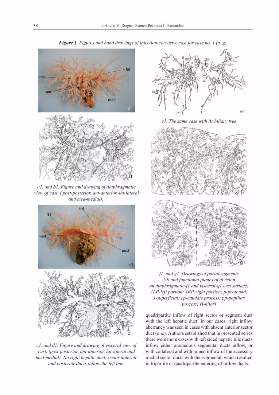
tic duct, (Figure 1. a1, b1, c1, d1 and e1). In two cases
the right hepatic duct with absent anterior sector duct
was confluent for both sector and segment duct inflow.
The left (liver) hepatic duct (on contrary to the
right one) was found at approximately 2/3 (64,70%) of
cases to persist as confluent of the both (lateral and me-
dial) sector ducts. Just one case had divergent inflow of
sector ducts (5,9%).
There were similar findings in the results in com-
parison with Couinaud’s (7) and Ohkubo’s et al. (13)
classification. They also had observed anomalous (ab-
errant) inflow of different order ducts, i.e. convergent
inflow of sector and hepatic duct, or segments with
sectors etc. But there was not any case of tripartite or
APPLIED 3-D ANATOMY OF LIVER BILE DUCTS IN INJECTION-CORROSION CASTS 37
Table 7. Aberrant confluence of the sector ducts
Type (constituent ducts) number of cases %
Consecutive confluence of posterior and anterior sector ducts
in the left hepatic duct5 71,40
Extrahepatic confluence of anterior and then posterior sector
ducts in the left hepatic duct1 14,30
Extrahilar connection of the right posterior with lateral
sector ducts, where the latter enters the medial and anterior
sector ducts
1 14,30
Total 7 100,00
Kolmogorov-Smirnov’s Dmax = 0,38 < D (7 and 0,05) = 0,48
quadripartite inflow of right sector or segment duct
with the left hepatic duct. In our cases, right inflow
aberrancy was seen in cases with absent anterior sector
duct (one). Authors established that in presented series
there were more cases with left sided hepatic bile ducts
inflow either anomalous segmental ducts inflow, or
with collateral and with joined inflow of the accessory
medial sector ducts with the segmental, which resulted
in tripartite or quadripartite entering of inflow ducts.
38 Jurkovikj M. Dragica, Korneti-Pekevska L. Kostandina
e1. The same case with its biliary tree
f1. and g1. Drawings of portal segments
1-9 and functional planes of division
on diaphragmatic-f1 and visceral-g1 cast surface;
1LP-left portion; 1RP-right portion; p-profound;
s-superficial; cp-caudate process; pp-papillar
process; H-hilar)
a1. and b1. Figure and drawing of diaphragmatic
view of cast. ( post-posterior, ant-anterior, lat-lateral
and med-medial).
Figure 1. Figures and hand drawings of injection-corrosive cast for case no. 1 (a–g)
c1. and d1. Figure and drawing of visceral view of
cast. (post-posterior, ant-anterior, lat-lateral and
med-medial). No right hepatic duct, sector anterior
and posterior ducts inflow the left one.
Our results were in comparison with Couinaud’s
(7) establishing that partial or total duplication of the
right hepatic duct was quite frequent-46,73%.
The authors standpoint for “aberrancy” is differ-
ent compared with that reported by Taourel et al. (8)
that a bile duct draining of one or more Couinaud’s
segments 5–8 joining the cystic duct or common hepa-
tic duct below the bile duct bifurcation was an aberrant
right hepatic duct.
Kim et al. (15), also come to similar findings in li-
ving donors of right lobe by MR cholangiography.
APPLIED 3-D ANATOMY OF LIVER BILE DUCTS IN INJECTION-CORROSION CASTS 39
c2. and d2. Figure and drawing of visceral view
of cast. The right posterior sector duct joins the left
lateral sector duct which gets inflow of the remaining
ones (right anterior and left medial sectors ducts).
e2. The same case with its biliary tree
f2 and g2. Drawings of portal segments 1-9 and
functional planes of division on diaphragmatic-f2
and visceral-g2 cast surface; 1LP-left portion;
1RP-right portion; p-profound; s-superficial;
cp-caudate process; pp-papillar process; H-hilar
Figure 2. Figures and hand drawings of injection-corrosive cast for case no. 2 (a–g)
a2. and b.2 Figure and drawing of diaphragmatic
view of cast. (post-posterior, ant-anterior and
lat-lateral). Notice the tortuosity and apparent
shortening of portal branches
They estimated that 53,3% of them had normal, 40% of
them were with aberrant and 6,7 % were with undeter-
mined anatomy of biliary ducts. Details of the biliary
anatomy included the configuration of the main divi-
sion (bifurcation versus trifurcation) and the drainage
of second order biliary radicals (anterior and posterior
right sectoral branches). All grafts from donors with
aberrant anatomy had two ducts.
As a rare observed was the extrahillar connection
of the right posterior with lateral sector duct, where the
latter entered the medial and anterior sector ducts (Fig-
ure 2. a2, b2, c2, d2 and e2) that may be compared with
type E reported by Ayuso et al. (12) as an absence of a
defined hepatic duct confluence, with all the sectoral
ducts joining separately.
The most important anomalies of biliary ducts to
related to anatomic variations of hepatic ducts conflu-
ence which were described by Couinaud (18) and also
recently cited by Doklestic at all. (17), was the conflu-
ence anatomy, as follows: the typical confluence of
right and left hepatic ducts was found in 57%; the tri-
furcation of left hepatic duct, posterior and anterior
right sectoral ducts in 12%; an ectopic drainage of right
sectoral duct into common hepatic duct in 24% or in-
flow into left hepatic duct in 7%; the absence of conflu-
ence in 4% and the absence of right hepatic duct in 2%.
Preoperative mapping of biliary anatomy is one of
the critical assessments for donor selection and surgi-
cal planning. Donor livers with aberrant drainage of
the right ductal system, such as trifurcation and right
posterior draining into left main duct, require two sepa-
rate biliary anastomoses in order to prevent postopera-
tive biliary leakage and long-term segmental atrophy
in recipients-Limanond et al. (11).
Considering right and left graft liver transplanta-
tion, Uchida et al. (19) had classified each branching
pattern of the hepatic vasculatures (portal vein and bili-
ary duct) into group A (1 anastomosis in reconstruc-
tion) or group B (possible multiple or technically com-
plicated anastomoses), using 3-dimensional computed
tomography. In right lobe graft, group B biliary duct
were detected in 50% of donors.
There was a difference in biliary drainage in cases
with aberrant or absent biliary channels compared to
the anatomo-morphological aspect of the 9-segmented
pattern portal system.
The majority of aberrant cases with lack of a cer-
tain duct to come into being as a congenital anomalies,
in spite of cases with total absent left portion duct and
9d Sub-segment ducts owing to a fibro-obliterative
changes in the liver diseases.
We also agree Hadziselimovic’s (20) experienced
opinion that the object one researches is unique in sha-
pe and structure in any individual.
Also, presented specimens partly at the segmental
level a different range of pruning and shortening of
porto-biliary branches (Figure 1. a1, b1, c1, d1, e1, f1
and g1; Figure 2. a2, b2, c2, d2, e2, f2 and g2), as well
as, a finding of small regenerative collaterals and reti-
cular bridging shown or tortuosity and apparent shorte-
ning caused by neighbouring tissue pressure (Figure 2.
a2, b2, c2, d2, e2, f2 and g2). Similar pathologic chan-
ges were the visible finding on the MR and CT evalua-
tions of liver morphology (21–23).
The use of Kolmogorov-Smirnov’s test of agree-
ment for one sample (cluster sample) shown statisti-
cally significant differences between cumulated relati-
ve frequencies obtained with investigation and same
which were expected for single modalities of confluen-
ce pattern segmental ducts into sectoral, at a level of
all-4 sectoral ducts. On contrary to this, at a level of he-
patic ducts there were statistically significant differen-
ces only to confluence pattern of sectoral ducts into left
hepatic duct. Hence, variations in modalities of conflu-
ence pattern segmental ducts into sectoral and sectoral
into left hepatic duct wasn’t accidental, they were a ca-
use of factor counteract during rate of growth and de-
velopment. For modalities of confluence pattern of
right sectoral ducts into right hepatic duct and differen-
ces between frequencies of their aberrant inflow into
the left hepatic duct no statistically significant differ-
ences were indicated.
As to the specimens with incomplete fill of acrylic
material into biliary elements, the first and second we-
re obstructed at the liver hilum, whereas the third was
filled until the sectoral order duct with discontinuities
and overrunning into vascular area of segment 4 and
retrograde in middle hepatic vein. In accordance with
this finding we think that incomplete fill was caused by
fibroobliterative or obstructive changes of liver and
vasculobiliary elements in all-three cases.
From the others investigators which used injec-
tion-corrosive method the date to similar disadvantage
was found in a study done by Uflacker et al. (24) as ap-
plied liver anatomy contribution to the placement of
TIPS-Transjugular Intrahepatic Portosystemic Shunts.
The authors didn’t comment the causes of incomplete
fill, but it is important to note that they used twenty-fi-
ve macroscopically normal livers obtained at necropsy
from cadavers age ranged from 20 to 70, all died from
causes not related to the liver.
Our experience with the use of the acrylic sub-
stance for corrosion casts resulted with managing its
optimal viscosity for injection, so that even the 4th or-
der of bile ducts constituents was visible. The additio-
nal ability to have the portal segmentation at the same
case made the advantage of the use of this method in
this and previous authors studies (25, 26, 27).
40 Jurkovikj M. Dragica, Korneti-Pekevska L. Kostandina
CONCLUSION
Corrosion liver cast presents the entire portal tree,
which voluminosity creates the anatomo-functional struc-
ture and segmentation of the liver. The bile channels are
concordant with portal branches at segmental level, beca-
use in lower one, there are varieties in their confluence.
According to one confluent pattern of the bile
ducts (segmental to sectors and further to hepatic one)
the conventional and most frequent type was in more
than one half cases as for the right sector ducts respec-
tively in less than one half in the left (sectors) ones. On
contrary in relation to the hepatic ducts, more than one
half of cases had numerous aberrances and inflow vari-
ations and anomalies between the left hepatic ducts of
different order respectively such cases in the right liver
they were in less than one half of cases.
The injection-corrosion method and the corrosion
acrylic casts provide instructive samples which can be
used in basic science education, as well as in diagnos-
tic, interventional and surgical procedures. Knowledge
of both anatomic variations in portal branching and in
bile ducts confluence, and the segmental anatomy of
the liver, is an essential prerequisite for developing sur-
gical skills for the reduced-size liver resection and
transplantation.
APPLIED 3-D ANATOMY OF LIVER BILE DUCTS IN INJECTION-CORROSION CASTS 41
Sa`etak
PRIMENJENA 3-D ANATOMIJA @U^NIH KANALA JETRE
NA INJEKCIONO-KOROZIVNIM MODELIMA
Jurkovi} M. Dragica, Korneti-Pekevska L. Kostandina
Institut za Anatomiju, Medicinski fakultet, Univerzitet „Sveti Kiril i Metodij“, Skoplje, R. Makedonija
Na 20 post-autopsionih adultnih izolovanih pri-
meraka jetre od pacijenata oba pola, starosne dobi od
29 do 88 godina (17 mu{karaca i 3 `ene), primenjena ja
injekciono-koroziona metoda. Obojeni akrilat je injek-
ciran u bilijarni sistem, a neobojeni u portnu venu.
Ukupno 17 akrilnih modela su bili zadovoljavaju}eg
kvaliteta. Unutar 9 portnih segmenata, utvr|eni su ana-
tomska determinacija i kvantitet, kao i modaliteti sliva-
nja intrajetrenih `u~nih kanala. Na|eni su diferentni
modaliteti sliva `u~nih kanala u sektorne i jetrene ka-
nale. Pored najfrekventnijih nalaza sa uobi~ajenim sli-
vom `u~nih kanala bilo je i aberantnih modaliteta bili-
jarne drena`e kod 8 slu~ajeva. Me|u njima 5 slu~ajeva
su imali sliv zadnjeg pa prednjeg sektornog kanala u
levi jetreni kanal, à 1 slu~aj prvo sliv prednjeg, a zatim
zadnjeg sektornog kanala u levi jetreni kanal. Tako|e
je na|ena kod 1 slu~aja ekstrahilusna konekcija zad-
njeg desnog sa levim lateralnim u zajedni~ki jetreni ka-
nal, gde kasnije ulaze medijalni pa prednji sektorni ka-
nal. Bilo je redom slivanje kanala sa 8-og i 5-og seg-
menta kod jednog (1) slu~aja, a sa lateralnog i medijal-
nog sektora sa ili bez repnog re`nja kod 3 slu~aja. Za-
jedni~ki (kod 4) i izdvojeni (kod 2) sliv kanala leve i
desne porcije repnog re`nja u levi drena`ni sistem bio
je kod 6 slu~ajeva, dok u oba, u levi i desni drena`ni si-
stem je na|en kod 7 slu~ajeva. Retko, na|en je aberan-
tan jedini kanal sa desne porcije kod jednog (1) slu~aja,
kao i prisustvo i bilijarna drena`a samo sa leve porcije
1-og segmenta. @u~ni kanali 9-tog segmenta drenirali
su sva 3 subsegmenta (b, c i d) kod 10 slu~ajeva, a sa-
mo dva (c i d) kod 3, kao i samo dva (c i b) od prisutnih
3 subsegmenata kod preostala 3 slu~aja. Isto, bio je je-
dan slu~aj sa prisutnim 9d portnim subsegmentom, a
bez kanala sa 9-tog segmenta. Ovi modaliteti su od in-
teresa za primenjivanje i preciznu interpretaciju di-
jagnosti~kih i interventnih procedura, kao i za seg-
mentnu, sektornu i semiheparnu resekciju kod jetrene
hirurgije.
Klju~ne re~i: jetra, intraheparni `u~ni kanal, port-
na vena, injekciono-koroziona metoda, segmentacija.
REFERENCES
1. Korneti K, Kargovska A, Ugrinski P. Prikaz na metoda za
morfoloshko ispituvanje na krvnite sadovi. X Kongres na MLD;
1978; Skopje, Republika Makedonija. Zb trud. 1978; 17–9.
2. Korneti K. Vaskularizacija na nodus sinuatrialis Keith
Flack ŠMagisterski trud¹. Medicinski fakultet: Univerzitet vo
Skopje; 1980.
3. Korneti K, Kargovska A,Ugrinski P, Josifov J,Tosovska
D. Morphologic demonstration of renal vasculature by the injec-
tion-corrosion method. Mac Med Review. 1982; 36: 52–3.
4. Lazarova D, Kargovska A, Dzidrova D, Papazova M,
Strateska A. Hepatovenski segmenti vo crniot drob kaj chovec-
hki fetus. XI Kongres na lekarite na SRM; 1982; Struga, Repu-
blika Makedonija. Zb trud. 1982; 519–21.
5. Jurkovikj D. Determination of portal vascular segmen-
tation in liver ŠMaster’s thesis¹. Medical faculty: University in
Skopje; 1994.
6. Ilieski V, Jovevska S, Jurkovikj D.Injection-corrosive
method of making anatomic preparations with acrylates. Mac
Vet Review. 1995; 24(1–2): 89–92.
42 Jurkovikj M. Dragica, Korneti-Pekevska L. Kostandina
Correspondence to/Autor za korespondenciju:
Prof. dr Kostandina Korneti-Pekevska
ul. „11 Oktomvri“ br. 19/1–7
1000 Skopje,
R. Macedonia
E-mail: kostandina_kornetiªyahoo.com
7. Couinaud C. Tell me more about liver anatomy. 2nd
ed. Paris: Karger; 1999.
8. Taourel P, Bret MP, Reinhold C, Barkun NA, Atri M.
Anatomic Variants of the Biliary Tree: Diagnosis with MR Cho-
langiopancreatography. Radiology. 1996; 199: 521–7.
9. Bak T, Wachs M, Trotter J, et al. Adult-to-Adult Liv-
ing Donor Liver Transplantation Using Right-Lobe Grafts: Re-
sults and Lessons Learned From a Single-Center Experience.
Liver Transpl. 2001; 7(8): 680–6.
10. Lee SV, Krinsky AG, Nazzaro AC. Defining Intrahe-
patic Biliary Anatomy in Living Liver Transplant Donor Candi-
dates at Mangafodipir Trisodium-enhanced MR Cholangio-
graphy versus Conventional T2-weighted MR Cholangio-
graphy. Radiology. 2004; 233:659-66.
11. Limanond P, Raman SS, Ghobrial MR, Busuttil WR,
Lu SKD. The Utility of MRCP in Preoperative Mapping of Bili-
ary Anatomy in Adult-to-Adult Living Related Liver Transplant
Donors. J Magn Reson Imaging. 2004; 19: 209–15.
12. Ayuso RJ, Ayuso C, Bombuy E, et al. Preoperative
Evaluation of Biliary Anatomy in Adult Live Liver Donors with
Volumetric Mangafodipir Trisodium Enhanced Magnetic Reso-
nance Cholangiography. Liver Transpl. 2004; 10(11): 1391–7.
13. Ohkubo M, Nagino M, Kamiya J, et al. Surgical Anat-
omy of the Bile Ducts at the Hepatic Hilum as Applied to Living
Donor Liver Transplantation. Ann Surg. 2004; 239: 82–6.
14. Liu LC, Lo MC, Chan CS, Tso KW, Fan TS. The Right
May Not Be Always Right: Biliary Anatomy Contraindicates
Right Lobe Live Donor Liver Transplantation. Liver Transpl.
2004; 10(6): 811–2.
15. Kim DR, Sakamoto S, Haider AM. Role of Magnetic
Resonance Cholangiography in Assessing Biliary Anatomy in
Right Lobe Living Donors. Transplantation. 2005; 27: 1417–21.
16. Strasberg MS. Nomenclature of hepatic anatomy and
resection: a review of the Brisbane 2000 system. J Hepatobiliary
Pancreat Surg. 2005; 12: 351–5.
17. Doklestic K, Detanac Dz, Detanac Dz, Ceranic A, Ka-
ramarkovic A. Functional liver anatomy-Surgical impact. Sana-
med. 2011; 6(1): 35–43.
18. Couinaud C. Liver anatomy: portal (and suprahepatic)
or biliary segmentation. Dig Surg. 1999; 16 (6): 459–67.
19. Uchida K, Taniguchi M, Shmamura T, et al. Three-Di-
mensional Computed Tomography Scan Analysis of Hepatic
Vasculatures in the Donor Liver for Living Donor Liver Trans-
plantation: Original Article. Liver Transpl. 2010; 16: 1062–8.
20. Hadziselimovic H. Krvni sudovi srca. 2. izd. Zagreb:
JUMENA-Jugoslovenska Medicinska Naklada; 1981.
21. Bader RT, Beavers LK, Semelka CR. MR Imaging Fe-
atures of Primary Sclerosing Cholangitis: Patterns of Cirrhosis
in Relationship to Clinical Severity of Disease. Radiology.
2003; 226: 675–85.
22. Ito K, Mitchel GD, Outwater KE, Blasbalg. Primary
Sclerosing Cholangitis: MR Imaging Features. AJR. 1999; 172:
1527–33.
23. Dodd III DG, Baron LR, Oliver III HJ, Federle PM.
End-Stage Primary Sclerosing Cholangitis: CT Findings of He-
patic Morphology in 36 Patients: Gastrointestinal Imaging. Ra-
diology. 1999; 211: 357–62.
24. Uflacker R, Reichert P, D’ Albuquerque CL, de Olivei-
ra e Silva A. Liver Anatomy Applied to the Placement of Tran-
sjugular Intrahepatic Portosystemic Shunts. Radiology. 1994;
191: 705–12.
25. Jurkovikj D, Korneti-Pekevska K. Left Lateral Liver
Section (Anatomo-surgical aspects of modal type of porto-bili-
ary elements). Mac Med Review. 2009; 63(3): 39–46.
26. Jurkovikj D. Important Biliaru Drainage Variations of Left
Liver Lobe. Contributions. Sec Biol Med Sci. 2009; 30(2): 81–92.
27. Jurkovikj D, Korneti-Pekevska K. Aberrant Confluen-
ce of Intrahepatic Biliary Ducts-A Case Report. Maced J Med
Sci. 2013; 6(1): 69–73.
Related Documents


![BiliaryEpithelialApoptosis,Autophagy,andSenescencein … · 2017. 11. 11. · necroinflammatory activity of small bile ducts and hepato-cytes [38]. 4.ImmunopathologyofPBC Mechanisms](https://static.cupdf.com/doc/110x72/5fdfe07dcf21c6201d25fb17/biliaryepithelialapoptosisautophagyandsenescencein-2017-11-11-necroiniammatory.jpg)
