IAWA Journal, Vol. 31 (3), 2010: 317–331 REFLECTED LIGHT MICROSCOPY AS A NON-INVASIVE IDENTIFICATION TOOL FOR WOODEN ARTEFACTS Flavio Ruffinatto 1,* , Nicola Macchioni 2 , Guido Boetto 1 , Pieter Baas 3 and Roberto Zanuttini 1 SUMMARY Species identification is a crucial step in the study of wooden artefacts, but sampling is frequently impossible. The objective of this study is to evaluate the efficacy of reflected light microscopy, including the use of polarising and narrow-band blue filters, as a non-invasive identification tool. Different surfacing and finishing techniques copying historical manufacturing methods were applied to selected species. The visibility of anatomical features was evaluated on the basis of a four level scale. Two indexes were created: an Identifiable Anatomical Features index (IAF) to evaluate the effect of treatments on the complex of microstruc- tural characters, and a Feature Recognition Index (FRI) to estimate the susceptibility of each anatomical feature towards different treatments. Surfacing affected the visibility of anatomical features to different degrees of severity depending both on the technique used and on the species. The visibility could be partially improved or decreased by the presence of finishes, depending on their transparency. Each anatomical feature showed different susceptibilities towards treatments. Both polarising and narrow-band blue filters considerably increased visibility of several anatomical features. Possibilities to recognise individual character states were encouraging, except when obscured by low transparency finishes. Much diagnostic anatomical information can be obtained by the use of non-invasive, reflected light microscopy, although the step from feature recognition to species identification may still require further analysis. Key words: Reflected light microscopy, wooden artefacts, non-invasive wood identification, surfacing, finishes. INTRODUCTION Both conservation and restoration of wooden works of art or rare archaeological artefacts require an in-depth diagnostic process. Species identification is a crucial step of that procedure (Fioravanti 1994; Hoadley 1994; Nardi Berti & Berti 1983; Wheeler & Baas 1998; Uzielli 1989). On the one hand the species is the key to physical and mechanical 1) Department of Agronomy, Forest and Land Management (AgROSELvITER), University of Turin, Italy. 2) CNR–IvALSA, Sesto Fiorentino, Italy. 3) Netherlands Centre for Biodiversity Naturalis (section NHN), Leiden, The Netherlands. *) Corresponding author [E-mail: flavio.ruffi[email protected]].

Welcome message from author
This document is posted to help you gain knowledge. Please leave a comment to let me know what you think about it! Share it to your friends and learn new things together.
Transcript

IAWA Journal, Vol. 31 (3), 2010: 317–331
Reflected light micRoscopy as a non-invasive identification tool foR wooden aRtefacts
Flavio Ruffinatto1,*, Nicola Macchioni2, Guido Boetto1, Pieter Baas3 and Roberto Zanuttini1
SUMMARY
Species identification is a crucial step in the study of wooden artefacts, but sampling is frequently impossible. The objective of this study is to evaluate the efficacy of reflected light microscopy, including the use of polarising and narrow-band blue filters, as a non-invasive identification tool. Different surfacing and finishing techniques copying historical manufacturing methods were applied to selected species. The visibility of anatomical features was evaluated on the basis of a four level scale. Two indexes were created: an Identifiable Anatomical Features index (IAF) to evaluate the effect of treatments on the complex of microstruc-tural characters, and a Feature Recognition Index (FRI) to estimate the susceptibility of each anatomical feature towards different treatments. Surfacing affected the visibility of anatomical features to different degrees of severity depending both on the technique used and on the species. The visibility could be partially improved or decreased by the presence of finishes, depending on their transparency. Each anatomical feature showed different susceptibilities towards treatments. Both polarising and narrow-band blue filters considerably increased visibility of several anatomical features. Possibilities to recognise individual character states were encouraging, except when obscured by low transparency finishes. Much diagnostic anatomical information can be obtained by the use of non-invasive, reflected light microscopy, although the step from feature recognition to species identification may still require further analysis.
Key words: Reflected light microscopy, wooden artefacts, non-invasive wood identification, surfacing, finishes.
INTRODUCTION
Both conservation and restoration of wooden works of art or rare archaeological artefacts require an in-depth diagnostic process. Species identification is a crucial step of that procedure (Fioravanti 1994; Hoadley 1994; Nardi Berti & Berti 1983; Wheeler & Baas 1998; Uzielli 1989). On the one hand the species is the key to physical and mechanical
1) Department of Agronomy, Forest and Land Management (AgROSELvITER), University of Turin, Italy.
2) CNR–IvALSA, Sesto Fiorentino, Italy.3) Netherlands Centre for Biodiversity Naturalis (section NHN), Leiden, The Netherlands.*) Corresponding author [E-mail: [email protected]].

IAWA Journal, Vol. 31 (3), 2010318
properties of a wooden object, on the other hand it represents information of historical interest about ancient trade, uses, and manufacturing techniques. For these reasons species identification is a requirement of the standards pertaining to wooden cultural heritage in many countries. In Italy, UNI 11118 states the criteria to be adopted for species identification on wooden artefacts, while UNI 11161 includes species identification amongst the essential requirements for a conservation, restoration or maintenance plan. So far, as a rule, species identification of wooden artefacts has been carried out through sampling, specimen preparation and observation of its anatomical features using either light or scanning electron microscopy. By contrast faster methods, such as hand lens identification (e.g. Forest Products Research 1960, 1963; venet 1986; Hoadley 1990; Ilic 1990, 1991; CITES 2002), are not or little used as they are com-monly inappropriate because species identification often requires seeing more features than are visible with a hand lens and the surfaces of cultural artefacts may not lend themselves well for hand lens inspection. Sampling for wood anatomical analysis is, however, invasive and thus affected by restrictions. Certain types of artefacts such as small wood carvings or marquetry, cannot be sampled adequately, and in any case invasive and destructive sampling does not guarantee that the wood can be accurately identified. Accuracy of identification depends in fact on different factors which have been analysed by Wheeler and Baas (1998) and can be briefly summarised as: access to auxiliary information such as geographical origin (which is, however, often one of the unknowns requested together with a botanical identification); sample dimension and state of conservation; access to literature and/or reference samples; knowledge on variability within species. Non-invasive wood identification is therefore an absolute necessity for certain valu-able wooden objects. The objective of the present study is to evaluate if reflected light microscopy (RLM) applied to unmodified surfaces of wooden artefacts can be useful for the observation of anatomical features. To our knowledge only sporadic attempts have been made with RLM on a few artefacts (M. Fioravanti, personal communication), but a methodological study to evaluate its efficacy is still wanting. On the other hand RLM is routinely used in anthracology for charcoal identification (Leney & Casteel 1975; Figueiral et al. 2002; Dias Leme et al. 2010). Here we explore, in selected wood species, how different techniques of surfacing and finishing can influence the visibility of anatomical features with RLM, and thus how powerful the application of RLM can be in wood identification of wooden artefacts.
MATERIALS AND METHODS
Materials Marquetry furniture was chosen as a model for this pilot study, representative as it is of the problem under investigation. Six temperate species were selected: Acer pseu-doplatanus, Buxus sempervirens, Juglans regia, Olea europaea, Prunus avium, Pyrus communis. Tropical species commonly used in marquetry were avoided because they belong to notoriously difficult taxa to identify, such as Dalbergia (Gasson et al. 2010).
319Ruffinatto et al. — Non-invasive wood identification
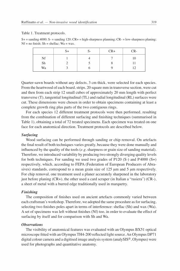
Quarter-sawn boards without any defects, 3 cm thick, were selected for each species. From the heartwood of each board, strips, 20 square mm in transverse section, were cut and then from each strip 12 small cubes of approximately 20 mm length with perfect transverse (T), tangential longitudinal (TL) and radial longitudinal (RL) surfaces were cut. These dimensions were chosen in order to obtain specimens containing at least a complete growth ring plus parts of the two contiguous rings. For each species 12 different treatment protocols were then performed, resulting from the combination of different surfacing and finishing techniques (summarised in Table 1), obtaining a total of 72 treated specimens. Each specimen was treated on one face for each anatomical direction. Treatment protocols are described below.
Surfacing Wood surfacing can be performed through sanding or chip removal. On artefacts the final result of both techniques varies greatly, because they were done manually and influenced by the quality of the tools (e.g. sharpness or grain size of sanding material). Therefore, we introduced variability by producing two strongly diverging quality levels for both techniques. For sanding we used two grades of P120 (S-) and P4000 (S+) respectively, which, according to FEPA (Federation of European Producers of Abra-sives) standards, correspond to a mean grain size of 125 μm and 5 μm respectively. For chip removal, one treatment used a planer accurately sharpened in the laboratory just before planing (CR+), the other used a card scraper (in Italian a “rasiera”) (CR-), a sheet of metal with a burred edge traditionally used in marquetry.
Finishing The composition of finishes used on ancient artefacts commonly varied between each craftsman’s workshop. Therefore, we adopted the same procedure as for surfacing, selecting two finishes poles apart in terms of interference: shellac (Sh) and wax (Wa). A set of specimens was left without finishes (Nf) too, in order to evaluate the effect of surfacing by itself and for comparison with Sh and Wa.
Observations The visibility of anatomical features was evaluated with an Olympus BX51 optical microscope fitted with an Olympus TH4-200 reflected light source. An Olympus DP71 digital colour camera and a digitised image analysis system (analySIS®, Olympus) were used for photographs and quantitative anatomy.
S+ S- CR+ CR-
Nf 1 4 7 10 Sh 2 5 8 11 Wa 3 6 9 12
Table 1. Treatment protocols. S+ = sanding 4000; S- = sanding 120; CR+ = high-sharpness planning; CR- = low-sharpness planing; Nf = no finish; Sh = shellac; Wa = wax.

IAWA Journal, Vol. 31 (3), 2010320
As references for wood anatomical species descriptions we used Schweingruber 1978, 1990 and InsideWood 2004-onwards. Terminology and survey principles gener-ally followed the IAWA list of microscopic features for hardwood identification (IAWA Committee 1989); however, ray height was measured, although the IAWA list only records whether rays are >1 mm (feature 102). Table 2 lists the characters recorded, for each species and surface. A scale of four levels was adopted to classify the visibility of anatomical characters: level A: the character state is visible and identifiable throughout the entire specimen
Table 2. Observed anatomical features with (in brackets) the related range of IAWA codes. For each feature the table also reports species and surface(s) on which observations were performed. T = transverse face; TL = tangential longitudinal face; RL = radial longitudinal face; A = Acer pseudoplatanus; B = Buxus sempervirens; P = Prunus avium; J = Juglans regia; O = Olea euro-paea; Py = Pyrus communis.
Character Feature Survey face(s) Species
Growth rings boundaries (1-2) T A, B, P, J, Py
Vessels porosity (3-5) Te A, B, P, J, O, Py arrangement (6-8) T A, J, O groupings (9-11) T A, B, P, J, Py solitary vessel outline angular (12) T B perforation plates (13-19) RL A, B, P, J, O, Py intervessel pits arrangement and size (20-28) TL A, B, P, J, O, Py vessel-ray pitting (30-35) RL A, B, P, J, O, Py helical thickenings (36-39) TL, RL A, P, Py tangential diameter of vessel lumina (40-45) T A, B, P, J, O, Py per square millimetre (46-51) T A, B, P, J, O, Py element length (52-55) TL, RL A, B, P, J, O, Py gums and other deposits in heartwood vessels (58) T, TL, RL P, O, Py
Axial parenchyma type (75-89) T A, B, P, J, O, Py cell type/strand length (90-95) TL A, B, P, J, O, Py
Rays width (96-100) TL A, B, P, J, O, Py rays of two distinct sizes (103) TL A cellular composition (104-109) RL A, B, P, J, O, Py per millimetre (114-116) T, TL A, B, P, J, O, Py height TL A, B, P, J, O, Py
Fibres wall thickness (68-70) T A, B, P, J, O, Py ground tissue fibres (61-64) RL A, B, P, J, O, Py ground tissue fibres (61-64) TL B, P, Py
Mineral inclusions other crystal types RL O

321Ruffinatto et al. — Non-invasive wood identification
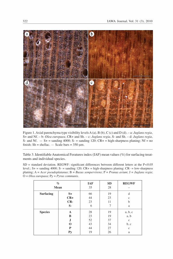
surface; level B: the character is visible throughout the entire specimen surface but its state can be identified only on some portions of the surface; level C: the character is visible throughout the specimen surface but its state is hardly identifiable and then only on rare portions of the surface; level D: on the entire specimen surface the anatomical feature is not sufficiently visible to allow the character state to be identified. Each time that the visibility of an anatomical feature was not graded as “A”, a po-larising filter and an Olympus U-MNB2 narrow-band blue filter were used in order to evaluate if a better visibility level was attainable. Examples of visibility levels and the effect of filters are respectively shown in Figures 1 and 2. Since only the A and B levels allowed recognition of characters and character states, the probability of feature identification was expressed by means of a Feature Recogni-tion Index (FRI), representing the percentage of all observations that resulted in feature identification as follows:
FRI represents the degree to which the visibility of an anatomical feature is affected by treatment. The total number of observations was different for each anatomical feature, depend-ing on the number of species in which it was present (Table 2). On the basis of data analysis, arbitrary classes were then created to give FRI a qualitative ranking: – low if ≤ 50%; – good if >50% and <75%; – high if ≥75%. Treatment effects were hence evaluated through an index called “Identifiable Ana-tomical Features” (IAF), calculated as:
which is the percentage of identified anatomical features. This index does not represent the probability of species identification, because that depends again on the combination of specific anatomical characters that is characteristic for a given species. For statistical analysis differences in values arcsine-transformed were tested at the P< 0.05 significance level with the Ryan-Einot-gabriel-Welsch F test (REgWF) (Hsu 1996).
RESULTS AND DISCUSSION
Surfacing The IAF indices differed significantly depending on the technique (Table 3), and ranged from overall poor recognisability for the coarsely sanded surfaces to relatively
A + BFRI = ––––––––––––– × 100A + B + C + D
ΣA + BIAF = ––––––––––––––– × 100ΣA + B + C + D
IAWA Journal, Vol. 31 (3), 2010322
Table 3. Identifiable Anatomical Feratures index (IAF) mean values (%) for surfacing treat-ments and individual species. SD = standard deviation. REgWF: significant differences between different letters at the P<0.05 level.; S+ = sanding 4000; S- = sanding 120; CR+ = high-sharpness planing; CR- = low-sharpness planing; A = Acer pseudoplatanus; B = Buxus sempervirens; P = Prunus avium; J = Juglans regia; O = Olea europaea; Py = Pyrus communis.
% iaf sd Regwf Mean 35 28 –
Surfacing s+ 66 19 d cR+ 44 23 c cR- 23 11 b s- 6 7 a
Species a 28 19 a, b, c B 23 19 a, b J 52 37 c o 43 34 b, c p 44 27 c Py 19 26 a
Figure 1. Axial parenchyma type visibility levels A (a), B (b), C (c) and D (d). – a: Juglans regia, S+ and Nf. – b: Olea europaea, CR+ and Sh. – c: Juglans regia, S- and Sh. – d: Juglans regia, S- and Nf. — S+ = sanding 4000; S- = sanding 120; CR+ = high-sharpness planing; Nf = no finish; Sh = shellac. — Scale bars = 350 μm.

323Ruffinatto et al. — Non-invasive wood identification
high IAF values for finely sanded surfaces, with the two planed surfaces intermediate. These differences are due to the fact that different surfacing techniques produce different cellular damage, and leave various amounts of damaged cell wall material projecting from the wood surface (giordano 1981; Hoadley 2000). The resulting layer of partially damaged cells on the surface reflects light diffusely (Marschner et al. 2005), causing the difference between “invisible” and “identifiable”. IAF values also differed significantly among species. Species with the coarser texture, such as Juglans regia or Prunus avium, were less affected by the absence of perfect sur- facing and had the highest IAF values, while species with a fine texture, such as Pyrus communis or Buxus sempervirens, were the most affected and registered low IAF values. Not surprisingly, anatomical features by themselves also showed a great variability (Fig. 3). On the whole, over three quarters of all anatomical features had a low probabil-ity of being recognised and none had a 100% one. All features that were relatively easy to identify, i.e. with an FRI ≥50%, belong to the transverse face and this is in fact the anatomical section mostly used by hand-lens identification methods (e.g. CITES 2002; Forest Products Research 1960, 1963; Hoadley 1990; Ilic 1990, 1991; venet 1986).
Figure 2. visibility levels for anatomical features in absence and presence of filters. Tangential surfaces, finely sanded, without finish. From left to right: Intervessel pit arrangement and heli-cal thickenings in Prunus avium, level C (a) and level A (b, polarizing filter); ray width in Acer pseudoplatanus, level B (c) and level A (d, blue filter). — Scale bars a, b = 20 μm, c, d = 90 μm.

IAWA Journal, Vol. 31 (3), 2010324
Finishes variation in IAF and FRI values caused by the presence of shellac and wax are compared with those for unfinished surfaces in Table 4. Shellac coating resulted in a remarkable overall improvement of IAF by 14%, while wax had the opposite effect, with an 8% decrease of IAF. The application of finishes interacted differently with dif-ferent types of surfacing: IAF improvement by shellac was greatest in coarsely surfaced samples; coating with wax on the contrary resulted in the greatest deterioration in the most perfect surfaces (finely sanded or planed with sharpened tools). Shellac and wax finishes reduced the surfacing-induced variability of IAF (compare Tables 3 and 4): shellac through a marked improvement on the coarsest surfaces; wax by reducing the visibility of features on more perfect surfaces. Shellac and wax finishes also had diverging effects on different species (Table 4), al- though the grouping of species as based on IAF values remained rather similar to that of untreated surfaces. visibility in fine-grained Buxus sempervirens suffered most from waxing.
vessel arrangementrays per mm (T)
growth ring boundariesporosity
fibre wall thicknessdiameter of vessel lumina
rays per mm (TL)vessel groupings
deposits in vessels (RL)deposits in vessels (TL)
deposits in vessels (T)ray width
vessels per square mmray cellular composition
fibre type (RL)axial parenchyma type
vessel length (RL)helical thickenings in v. (RL)
vessel length (TL)ray height
perforation platesintervessel pits arrangementhelical thickenings in v. (TL)
axial par. strand lengthintervessel pits size
fibre type (TL)vessel–ray pitting
0 10 20 30 40 50 60 70 80 FRI (%)
Figure 3. Ranking of anatomical features observed in absence of finishes based on the FRI index (%).

325Ruffinatto et al. — Non-invasive wood identification
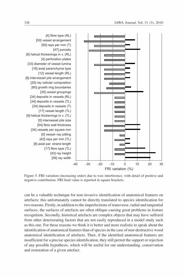
Figure 4 shows that FRI values for individual wood anatomical features increased in shellac-finished surfaces in over half of all features, and was only significantly nega-tive for “deposits in vessels”. Treatment with wax, on the other hand, decreased FRI of most features, with the exception of ray width and height, and fibre type as seen in tangential surface (Fig. 5). Finishes acted as filters altering light reflection in consequence of their physical interaction with wood surface. They reduced reflection because of the absorption of light by the finish (Xie et al. 2006) and affected the reflection angle depending on its refractive index. The refractive index of a clear finish (such as shellac and wax) is close to that of the wood, and the first surface layer of damaged cells is effectively eliminated so that the reflection of wood originates from the less damaged structure of the subsur-face (Marschner et al. 2005). The differences registered in the overall effect between shellac and wax were due to their different transparency. Some preliminary tests on artificially Uv-aged specimen revealed that degradation of finishes did not substantially alter their effect on the visibility of anatomical features (results not shown).
Filters The enhancement of IAF and FRI by filters is summarised in Tables 5 and 6. The narrow-band blue filter worked well on finished and unfinished surfaces. The highest benefit was observed in shellac-finished surfaces (which already started from the best IAF values) with a mean increase of +14%. A much lower contribution was found for waxed surfaces which already had the lowest original IAF values. Polarised filters did not work in the presence of finishing. On non-finished surfaces (detailed results not shown) its positive effect was greater on cut surfaces than on sanded
Table 4. Identifiable Anatomical Feature index (IAF) variation and final mean values due to shellac and wax interference. REgWF, see Table 3. Sh = shellac; Wa = wax; S+ = sanding 4000; S- = sanding 120; CR+ = high-sharpness planing; CR- = low-sharpness planing; A = Acer pseudoplatanus; B = Buxus sempervirens; P = Prunus avium; J = Juglans regia; O = Olea europaea; Py = Pyrus communis.
Variation Final value REGWF IAF (%) ––––––––––––––––––––––––––––––––––––––––––––––––––––––––––– sh wa sh wa sh wa
Surfacing s+ +6 -17 72 49 c b cR+ +13 -19 57 25 b,c a,b cR- +15 -3 38 20 a,b a s- +22 +6 28 12 a a
Species a +23 -5 51 23 b,c a,b,c B +10 -22 33 1 a,b a J +20 -1 72 51 c c o +17 -8 60 35 c b,c p +9 -9 53 35 b,c b,c Py +6 -6 25 13 a a,b
Mean +14 -8 49 27 – –

IAWA Journal, Vol. 31 (3), 2010326
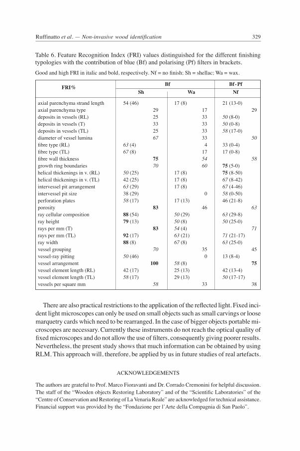
ones and in Acer and Prunus the increase of IAF was much greater than in the other species. Combined use of blue filters and polarising filters on non-finished surfaces showed remarkable results in most species, close to those achieved on shellac-finished specimens. Table 6 summarises FRI values when using different finishings. The blue filter had a markedly positive effect on recognising “ray cellular composition”, while for the other anatomical features the effects depended on finishing. Polarised filters greatly improved the visibility of helical thickenings and intervessel pit features, while visibility of other features were hardly affected. The best overall results were observed with shellac finish, where only “deposits in vessels”, “axial parenchyma type” and “intervessel pits size” were left with a low FRI, while FRI for “vessel element length” and “helical thickenings” remained low (≤ 50%) for only one of the two possible planes of observation. All the features relating to rays, “porosity”, “vessel arrangement” and
[58] fibre type (TL)[80] ray width
[67] ray height[58] fibre type (RL)
[75] rays per mm (TL)[100] vessel arrangement
[70] vessel groupings[42] perforation plates
[84] porosity[59] vessels per square mm
[75] fibre wall thickness[67] diameter of vessel lumina
[42] vessel length (TL)[84] rays per mm (T)
[33] intervessel pits arrangement[70] growth ring boundaries[33] ray cellular composition[29] axial parenchyma type
[25] vessel length (RL)[17] helical thickenings in v. (TL)
[8] intervessel pits size[8] axial par. strand length
[4] vessel–ray pitting[25] helical thickenings in v. (RL)
[33] deposits in vessels (T)[25] deposits in vessels (RL)[25] deposits in vessels (TL)
-20 -10 0 10 20 30 40 50 FRI variation (%)
Figure 4. FRI variation (decreasing order) due to shellac interference, with detail of positive and negative contribution. FRI final value is reported in square brackets.

327Ruffinatto et al. — Non-invasive wood identification
Bf – pf Bf
IAF (%) Nf Sh Wa + f.v. + f.v. + f.v.
Surfacing S+ 13 79 17 89 11 60 cR+ 27 71 18 74 9 34 cR- 15 38 12 50 3 23 s- 7 13 8 36 2 14
Species a 31 59 6 57 14 36 B 9 32 23 56 6 7 J 13 65 13 85 7 58 o 7 50 10 70 2 38 p 18 62 22 75 3 38 Py 15 34 13 38 7 20
Mean 16 51 14 63 6 33
Table 5. Identifiable Anatomical Feature index (IAF) enhancement (+) and final values (f.v.) by blue (Bf) and polarising (Pf) filters.Nf = no finish; Sh = shellac; Wa = wax; S+ = sanding 4000; S- = sanding 120; CR+ = high-sharp-ness planing; CR- = low-sharpness planing; A = Acer pseudoplatanus; B = Buxus sempervirens; P = Prunus avium; J = Juglans regia; O = Olea europaea; Py = Pyrus communis.
“fibre wall thickness” showed high FRIs, partly due to high initial values, partly to appreciable improvement by the use of filters. The use of filters for non-finished sur-faces showed satisfactory results for most features. Only axial parenchyma, fibre type and some vessel features did not come up to good FRI values. FRIs of waxed surfaces remained low, despite some enhancement due to the use of blue filter. For both types of filter the visibility is enhanced by improved contrast. Blue filters amplify a very narrow band of wave lengths and attenuates all the others; polarising filters enhance the visibility of birefringent material, a well-known property of crystal-line cellulose microfibrils, and dependent on their direction and thus on the angle of observation (Donaldson 2008).
CONCLUSIONS
The finest surfacing techniques for the manufacturing of artefacts do not achieve the results provided by thin sections for transmitted light microscopy. The absence of a perfect surface preparation seriously affects the visibility of anatomical features with the RLM and hence their probability of identification, but with different severity depending both on the technique used, the individual feature, and the specific texture of the timber. The interference due to finishes is different depending on their transparency. Wax is without doubt a problem as it strongly decreases visibility of most features, while shellac generally greatly enhances visibility. The use of filters greatly increases the visibility of many anatomical features. As a result, for most features the probabilities of recognis-ing them are relatively high on non-finished and shellac-treated surfaces. Thus RLM
IAWA Journal, Vol. 31 (3), 2010328
can be a valuable technique for non-invasive identification of anatomical features on artefacts; this unfortunately cannot be directly translated to species identification for two reasons. Firstly, in addition to the imperfections of transverse, radial and tangential surfaces, the surfaces of artefacts are often oblique causing great problems in feature recognition. Secondly, historical artefacts are complex objects that may have suffered from other deteriorating factors that are not easily reproduced in a model study such as this one. For these reasons we think it is better and more realistic to speak about the identification of anatomical features than of species in the case of non-destructive wood anatomical identification of artefacts. Then, if the identified anatomical features are insufficient for a precise species identification, they still permit the support or rejection of any possible hypothesis, which will be useful for our understanding, conservation and restoration of a given artefact.
[4] fibre type (RL)[50] vessel arrangement
[50] rays per mm (T)[47] porosity
[8] helical thickenings in v. (RL)[4] perforation plates
[33] diameter of vessel lumina[16] axial parenchyma type
[12] vessel length (RL)[8] intervessel pits arrangement
[20] ray cellular composition[60] growth ring boundaries
[35] vessel groupings[34] deposits in vessels (RL)[34] deposits in vessels (TL)
[34] deposits in vessels (T)[17] vessel length (TL)
[9] helical thickenings in v. (TL)[0] intervessel pits size
[54] fibre wall thickness[34] vessels per square mm
[0] vessel–ray pitting[42] rays per mm (TL)
[8] axial par. strand length[17] fibre type (TL)
[42] ray height[59] ray width
-40 -30 -20 -10 0 10 20 30 FRI variation (%)
Figure 5. FRI variation (increasing order) due to wax interference, with detail of positive and negative contribution. FRI final value is reported in square brackets.
329Ruffinatto et al. — Non-invasive wood identification
There are also practical restrictions to the application of the reflected light. Fixed inci- dent light microscopes can only be used on small objects such as small carvings or loose marquetry cards which need to be rearranged. In the case of bigger objects portable mi- croscopes are necessary. Currently these instruments do not reach the optical quality of fixed microscopes and do not allow the use of filters, consequently giving poorer results. Nevertheless, the present study shows that much information can be obtained by using RLM. This approach will, therefore, be applied by us in future studies of real artefacts.
ACKNOWLEDgEMENTS
The authors are grateful to Prof. Marco Fioravanti and Dr. Corrado Cremonini for helpful discussion. The staff of the “Wooden objects Restoring Laboratory” and of the “Scientific Laboratories” of the “Centre of Conservation and Restoring of La venaria Reale” are acknowledged for technical assistance. Financial support was provided by the “Fondazione per l’Arte della Compagnia di San Paolo”.
Table 6. Feature Recognition Index (FRI) values distinguished for the different finishing typologies with the contribution of blue (Bf) and polarising (Pf) filters in brackets. good and high FRI in italic and bold, respectively. Nf = no finish; Sh = shellac; Wa = wax.
fRi% Bf Bf-pf sh wa nf
axial parenchyma strand length 54 (46) 17 (8) 21 (13-0) axial parenchyma type 29 17 29 deposits in vessels (RL) 25 33 50 (8-0) deposits in vessels (T) 33 33 50 (0-8) deposits in vessels (TL) 25 33 58 (17-0) diameter of vessel lumina 67 33 50 fibre type (RL) 63 (4) 4 33 (0-4) fibre type (TL) 67 (8) 17 17 (0-8) fibre wall thickness 75 54 58 growth ring boundaries 70 60 75 (5-0) helical thickenings in v. (RL) 50 (25) 17 (8) 75 (8-50) helical thickenings in v. (TL) 42 (25) 17 (8) 67 (8-42) intervessel pit arrangement 63 (29) 17 (8) 67 (4-46) intervessel pit size 38 (29) 0 58 (0-50) perforation plates 58 (17) 17 (13) 46 (21-8) porosity 83 46 63 ray cellular composition 88 (54) 50 (29) 63 (29-8) ray height 79 (13) 50 (8) 50 (25-0) rays per mm (T) 83 54 (4) 71 rays per mm (TL) 92 (17) 63 (21) 71 (21-17) ray width 88 (8) 67 (8) 63 (25-0) vessel grouping 70 35 45 vessel-ray pitting 50 (46) 0 13 (8-4) vessel arrangement 100 58 (8) 75 vessel element length (RL) 42 (17) 25 (13) 42 (13-4) vessel element length (TL) 58 (17) 29 (13) 50 (17-17) vessels per square mm 58 33 38
IAWA Journal, Vol. 31 (3), 2010330
REFERENCES
CITES. 2002. CITES Identification guide – Tropical Woods. Wildlife Enforcement and Intelli-gence Division, Enforcement Branch, Environment Canada.
Dias Leme, C.L., C. Cartwright & P. gasson. 2010. Anatomical changes to the wood of Mimosa ophthalmocentra and Mimosa tenuiflora when charred at different temperatures. IAWA J. 31: 333–351 (this issue).
Donaldson, L. 2008. Microfibril angle: measurement, variatiom and relationships – A review. IAWA J. 29: 345–386.
Figueiral, I., v. Mosbrugger, N.P. Rowe, T. Utescher, T.P. Jones & F. von der Hocht. 2002. Role of charcoal analysis for interpreting vegetation change and paleoclimate in Miocene Rhine Embayment (germany). Palaios 17: 347–365.
Fioravanti, M. 1994. Le specie legnose dei supporti: implicazioni per la conoscenza, la conserva-zione ed il restauro dei dipinti su tavola. In: L. Uzielli & O. Casazza (ed.), Conservazione dei dipinti su tavola: 83–107. Nardini editore, Firenze.
Forest Products Research. 1960. Identification of hardwoods. A lens key. For. Prod. Res. Bull. No. 25, London.
Forest Products Research. 1963. An atlas of end-grain photomicrographs for the identification of hardwoods. For. Prod. Res. Bull. No. 26, London.
gasson, P., R. Miller, D.J. Stekel, F. Whinder & K. Zieminska. 2010. Wood identification of Dalbergia nigra (CITES Appendix I) using quantitative wood anatomy, principal components analysis and naïve Bayes classification. Ann. Bot. 105: 45–56.
giordano, g. 1981. Tecnologia del legno. vol. I. La materia prima. Unione Tipografico, Editrice Torinese.
Hoadley, R.B. 1990. Identifying wood: Accurate results with simple tools. The Taunton Press, Newtown, CT.
Hoadley, R.B. 1994. Wood as a physical surface for paint application. In: v. Dorge & F.C. How- lett (eds.), Painted wood: history and conservation: 2–16. The getty Conservation Institute, Los Angeles.
Hoadley, R.B. 2000. Understanding wood. The Taunton Press, Newtown, CT.Hsu, J.C. 1996. Multiple comparisons: theory and methods. Chapman & Hall, UK.IAWA Committee. 1989. IAWA list of microscopic features for hardwood identification. IAWA
Bull. n.s. 10: 219–332.Ilic, J. 1990. CSIRO macro key for hardwood identification. CSIRO, Australia.Ilic, J. 1991. CSIRO atlas of hardwoods. Springer-verlag, Berlin.InsideWood. 2004–onwards. Published on the Internet. http://insidewood.lib.ncsu.edu [accessed
2008].Leney, L. & R.W. Casteel. 1975. Simplified procedure for examining charcoal specimens for iden-
tification. J. Archeol. Sci. 2: 153–159.Marschner, S.R., S.H. Westin, A. Arbree & J.T. Moon. 2005. Measuring and modelling the ap-
pearance of finished wood. ACM Transactions on graphics (SIggRAPH 2005 Proceedings) 24: 727–734.
Nardi Berti, R. & S. Berti. 1983. Principali specie legnose impiegate nelle strutture e nei manufatti del passato e criteri per il loro riconoscimento. In: g. Tampone (ed.), Legno nel restauro e restauro del legno. vol. II: 45–55. Palutan editrice, Milano.
Schweingruber, F.H. 1978. Mikroskopische Holzanatomie. Anatomie microscopique du bois. Microscopic wood anatomy. Structural variability of stems and twigs in recent and subfos-sil woods from Central Europe. Swiss Fed. Inst. For. Res., Birmensdorf. Edition Zürcher: Zug, Switzerland.

331Ruffinatto et al. — Non-invasive wood identification
Schweingruber, F.H. 1990. Anatomie europäischer Hölzer. Anatomy of European woods. verlag Paul Haupt, Bern, Stuttgart.
UNI 11118. 2004. Cultural heritage. Wooden artefacts. Criteria for the identification of the wood species.
UNI 11161. 2005. Cultural heritage. Wooden artefacts. guidelines for conservation, restoration, maintenance.
Uzielli, L. 1989. valutazione tecnologica del degrado e degli interventi di risanamento in una strut-tura lignea antica: l’esempio di una copertura seicentesca del Castello del valentino, a Torino. In: g. Tampone (ed.), Il restauro del legno. vol. I: 191–199. Nardini editore, Firenze.
venet, J. 1986. Identification et classement des bois français (2ème édition revue par R. Keller). ENgREF ed., Nancy.
Wheeler, E.A. & P. Baas. 1998. Wood identification – a review. IAWA J. 19: 241–264.Xie, Y., A. Krause, H. Militz & C. Mai. 2006. Coating performance of finishes on wood modified
with an N-methylol compound. Progress in Organic Coatings 57: 291–300.
Related Documents


