Application of Positron Emission Tomography ( PET ) in Colorectal Cancer Dr Chan Wai Keung Department of Surgery Ruttonjee and Tang Shiu Kin Hospitals

Application of Positron Emission Tomography ( PET ) in Colorectal Cancer Dr Chan Wai Keung Department of Surgery Ruttonjee and Tang Shiu Kin Hospitals.
Dec 22, 2015
Welcome message from author
This document is posted to help you gain knowledge. Please leave a comment to let me know what you think about it! Share it to your friends and learn new things together.
Transcript

Application of PositronEmission Tomography ( PET ) in Colorectal Cancer
Dr Chan Wai KeungDepartment of SurgeryRuttonjee and Tang Shiu Kin Hospitals

• What is PET
• Liver metastasis
• Extrahepatic metastasis
• Elevated CEA
• Local recurrence
• Monitor resposnse to therapy
PET - Background
• Positron emission tomography ( PET ) in use for 20 years
• Initially for research purposes
• Clinical application since 90s
• Wide clinical uses: carcinomas, melanoma, lymphoma,
epilepsy, dementia, cerebrovascular disease, coronary
artery disease and others


PET - Basic Principles
• A PET tracer is administered and takes part in physiological processes
• Different concentrations at different locations
• The PET scanner detects signals
• The resulting images showed functional information

Anhilation

PET - Basic Principles
• Metabolically active cells can take up the tracer
• Enhanced activity seen in brain, skeletal muscle, bowel, myocardium, genitourinary tract, thyroid and others
• “Functional imaging”help detection at earlier stage than cross sectional imaging
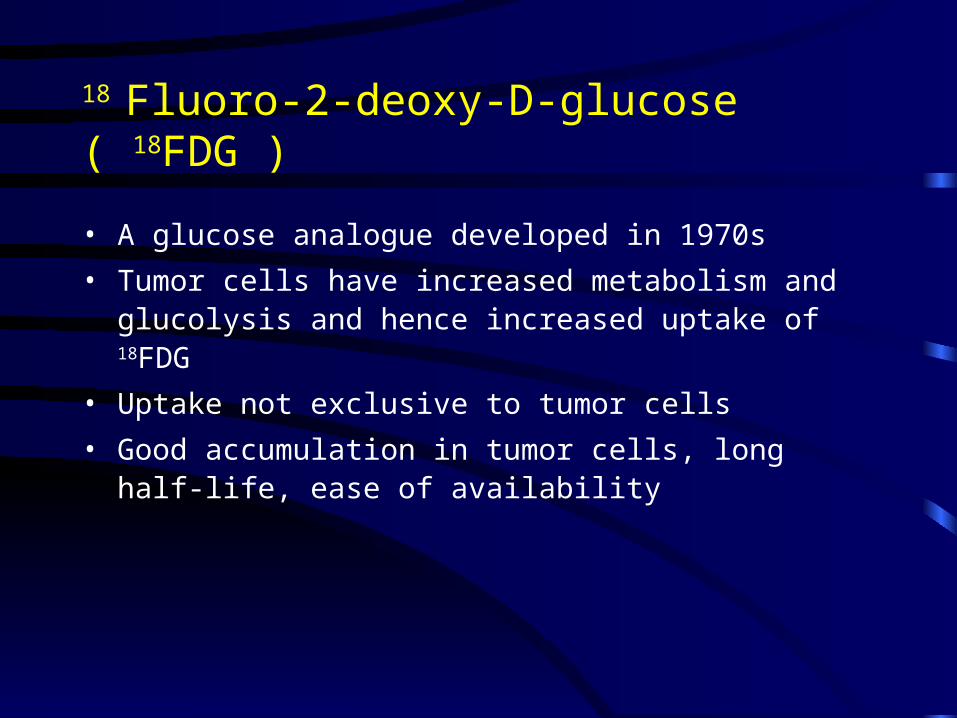
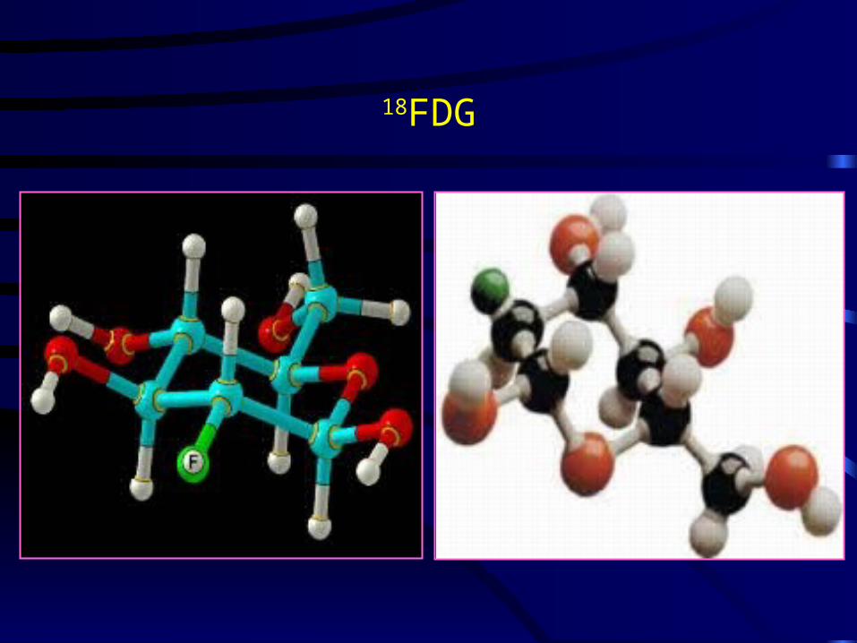
18 Fluoro-2-deoxy-D-glucose ( 18FDG )
• A glucose analogue developed in 1970s
• Tumor cells have increased metabolism and glucolysis and hence increased uptake of 18FDG
• Uptake not exclusive to tumor cells
• Good accumulation in tumor cells, long half-life, ease of availability

Glucose
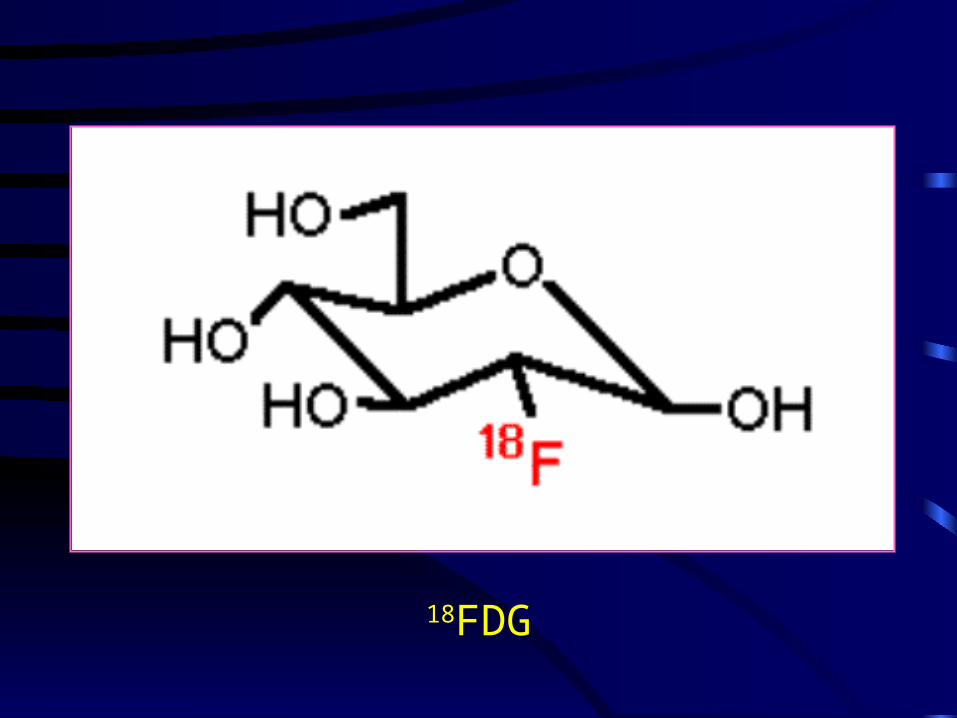
18FDG
a
18FDG
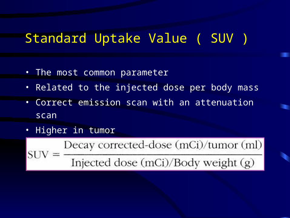
Standard Uptake Value ( SUV )
• The most common parameter
• Related to the injected dose per body mass
• Correct emission scan with an attenuation scan
• Higher in tumor


• Sensitivity : TP / ( TP + FN )
• Specificity : TN / ( FP + TN )
• Positive predictive value : TP / ( TP + FP )
• Negative predictive value : TN / ( TN + FN )

Pre-operative Diagnosis
• A study of 48 patients with established or suspicious
diagnoses of colorectal cancer
• PET scan detected all lesions
• Positive and negative predective values of 90% and 100%
Abdel-Nabi H., Radiology, 1998
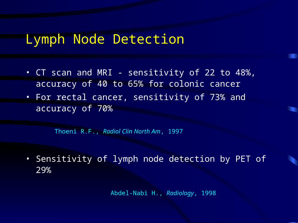
Lymph Node Detection
• CT scan and MRI - sensitivity of 22 to 48%, accuracy of 40 to 65% for colonic cancer
• For rectal cancer, sensitivity of 73% and accuracy of 70%
Thoeni R.F., Radiol Clin North Am, 1997
• Sensitivity of lymph node detection by PET of 29%
Abdel-Nabi H., Radiology, 1998

Colorectal Liver Metastasis
• 25% have liver metastasis at diagnosis
• Another 20% will have liver metastasis
• 30 to 40% have 5-years survival after hepatectomy
• Patient selection - anatomical resectability and no extra-
hepatic involvement

PET in Liver Metastasis
• Superiority of PET over CT in detecting liver metastasis
not eastablished
• No adequate spatial information about metastases
• The main role into detect extrahepatic involvement
Arulampalam T.H.A., 2003

a
Para-caval LNPara-aortic LNLiver

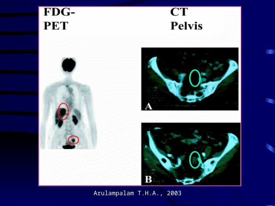
Extrahepatic Disease in Liver Metastasis
• 51 patients analyzed for resection for colorectal liver
metastasis
• PET result in 20% change of management because of
unexpected extrahepatic involvement
Ruers T.J., J Clin Oncol, 2002

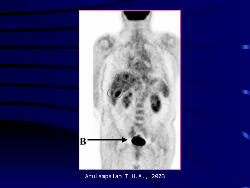
PET showed identical lesions to CT scan

Spinal metastasis
• Spinal metastasis detected by PET but not by CT
• Spinal cord compression 3 months after hepatectomy

Hepatic and Extrahepatic Lesions

PET and Liver Resectability
• 43 patients for hepatectomy for liver metastasis
• 6 patients spared of surgery due to extrahepatic disease
• Hepatectomy in 35 out of 37 patients
• 95% resectability rate of hepatic metastasis with PET in
addition to other imaging techniques
• At 3 years 77% overall and 40% disease free survival
Strasberg S.M., Ann Surg, 2000
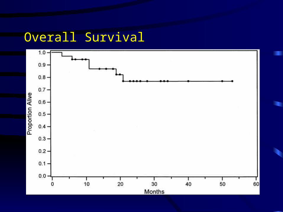
Overall Survival
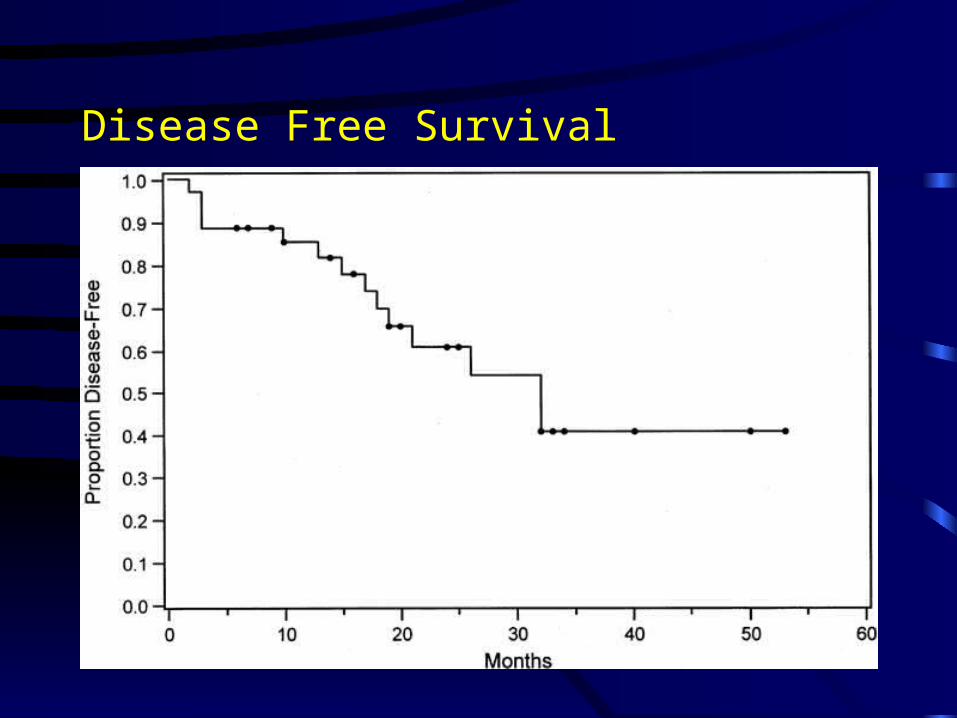
Disease Free Survival

Elevated CEA
• Investigated by conventional imaging modalities and colonoscopy - still some have negative imaging
• CEA directed laparotomy: low resectability rate of 44 to 58% because of unexpected presence of extensive disease
Minton J.P., Cancer, 1985
Martin E.W.Jr., Am J Surg, 1979

Elevated CEA
• PET for 32 patients with elevated CEA
• Histological diagnosis, serial CT and clinical follow-up as standards
• Sensitivity - 90%, specificity 92%
• Positive predicitive value 95%
• Negative predicitive value 85%
Valk P.E., Arch Surg, 1999


Elevated CEA with Normal Imaging
• 22 patients with elevated CEA and normal conventional imaging
• 17 recurrent lesions found - histological confirmation in 7, recurrence on follow up in 8, false positive in 2
• No recurrence in those with negative PET
Flanagan F.L., Ann Surg,
1999

Metastatic Disease - PET vs CT
• 41 patients had laparotomy for metastatic colorectal cancer
• All have pre-op PET and CT
• Sensitivity : liver ( 100% vs 69% ), extraheaptic ( 90% vs 52% ), abdomen ( 87% vs 61%), pelvis ( 87% vs 61%)
Johnson K., Dis Colon Rectum,
2001


Local Recurrent Disease - PET vs CT
• 70 patients with suspected locally recurrent colorectal cancer
• PET compared with CT / Colonoscopy
• Sensitivity : 90% vs 71%
• PPV and NPV: PET - 88% and 92%
CT - 79% and 79%
Whiteford M.H., Dis Colon Rectum, 2000
Arulampalam T.H.A., 2003

Monitoring Therapy of Colorectal Cancer
• Response to chemotherapy and regional therapy
monitored by PET
• FDG uptake decreased in responding lesions
• To separate responsders from non-responders
• Residual uptake help to guide further therapy

Pre Chemotherapy
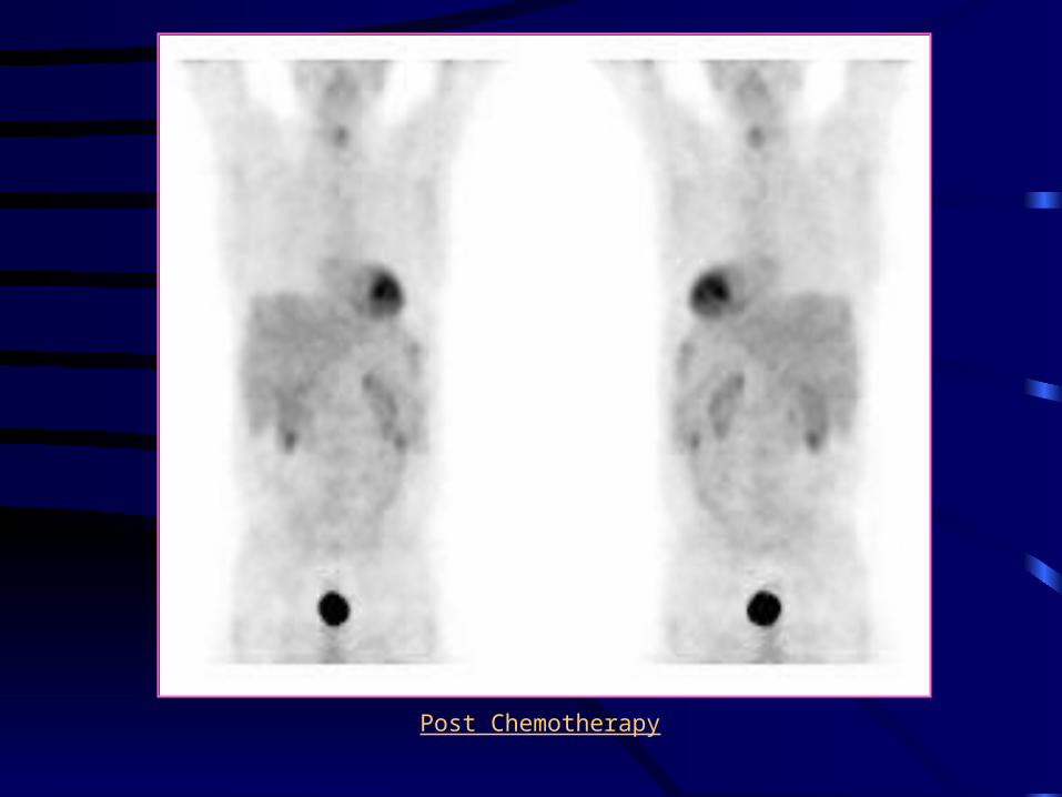
Post Chemotherapy

Limitation of PET
• Detectability depends on size and degree of uptake
• False -ve in small lesion and necrotic lesions
• Low sensitivity in mucinous adenocarcinoma
• False +ve in inflammed tissue
• Usual FDG activity at gastrointestinal tract

Impact on Management
• Early detection of abnormal tissue metabolism
• Detection of tumor at usual and unexpected sites
• Avoid unnecessary surgery
• Allow earlier treatment by diagnosing recurrence earlier
• Monitor treatment response
• PET is a power imaging modality but its use needs to be refined
Conclusion
• PET is a functional imaging technique
• It detects hepatic and extrahepatic lesions, and help to
avoid unnecessay surgery by detecting extrahepatic
disease
• It detects recurrent disease in patients with elevated CEA
and negative imaging
• Its helps to monitor treatment and guide further treatment

Thank You !
Related Documents