Application of Electrochemical Techniques to the Solution of Problems in Medicine P. N. Sawyer Electrochemical and Biophysical Laboratories, Transplantation Service, and Vascular Surgical Services, Departments o] Surgery and Surgical Research, State University of New York, Downstate Medical Center, Brooklyn, New York 11203 I should like to thank the members of the Electro- chemical Society for the privilege of addressing you today. As you all know, I am a surgeorL I became one originally because it was obvious to me that the ob- jective which I had in mind could only be fulfilled by applying the solution to problems in medicine to pa- tients. By the time I completed medical school it seemed possible to solve various problems in medicine by the application of physical chemical techniques (1). It was equally apparent that once the problems were solved, the solutions would probably not be applied to man unless the person who had produced the solution Fig. lb. It was also shown that a conductive balloon of this type, as a balloon in a Foley catheter, would produce hemostasis in a bleeding prostatic bed. Fig. lc. Lastly, we were presented with a young hemophiliac, with uncontrollable bleeding into a massive pseudo-tumor of the left thigh. Insertion of positive current electrodes was used to produce hemostasis in the pseudo-tumor, prior to disarticulation of the left hlp. Following disarticulation, hemostasis was produced in the flaps of the wound, again using direct current coagulation techniques, as shown here. It is not necessary to use these tech- niques anymore now that Factor Eight has been purified, but, in this instance, application of the d-c coagulation technique led to the first successful disarticulation in a hemophiliac with preserva- tion of life reported in modern society. Fig. la. The fact that an electrode pair would produce clotting was soon shown to produce hemostasis in bleeding wounds in man. It was also shown that it would produce hemostasis in esophageal varices by passing current through a conductive balloon tube to thrombos. ~ all of the vessels in the local area. This proved to be the undoing of this technique because it was so effective in occluding esophageal varices that it ablated one source of collateral flow around a portal occlusion. was able to apply them to a patient population over which he had control. This pragmatic philosophical principal has, in general, been the case in medicine. The group which I represent, past, present, and, I hope, future, has discerned problems in the patient, thought with a phiIosophic scientific attitude about the prob- lems, decided what approaches might practically lead 419C ) unless CC License in place (see abstract). ecsdl.org/site/terms_use address. Redistribution subject to ECS terms of use (see 54.39.17.49 Downloaded on 2018-04-01 to IP

Welcome message from author
This document is posted to help you gain knowledge. Please leave a comment to let me know what you think about it! Share it to your friends and learn new things together.
Transcript
Application of Electrochemical Techniques to the Solution of Problems in Medicine
P. N. Sawyer Electrochemical and Biophysical Laboratories, Transplantation Service, and Vascular Surgical Services,
Departments o] Surgery and Surgical Research, State University of New York, Downstate Medical Center, Brooklyn, New York 11203
I should l ike to t hank the member s of the Elec t ro- chemical Socie ty for the pr iv i lege of address ing you today. As you al l know, I am a surgeorL I became one or ig ina l ly because i t was obvious to me that the ob- jec t ive which I had in mind could only be fulfi l led by app ly ing the solut ion to p rob lems in medic ine to p a - t ients.
By the t ime I completed medica l school i t seemed possible to solve var ious problems in medic ine by the appl ica t ion of physical chemical techniques (1). I t was equa l ly appa ren t that once the p rob lems were solved, the solut ions would p robab ly not be appl ied to m a n unless the person who had produced the solut ion
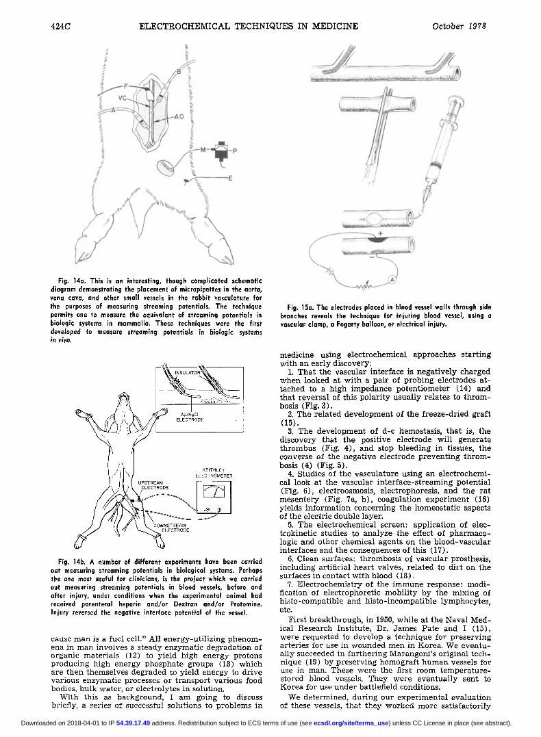
Fig. lb. It was also shown that a conductive balloon of this type, as a balloon in a Foley catheter, would produce hemostasis in a bleeding prostatic bed.
Fig. lc. Lastly, we were presented with a young hemophiliac, with uncontrollable bleeding into a massive pseudo-tumor of the left thigh. Insertion of positive current electrodes was used to produce hemostasis in the pseudo-tumor, prior to disarticulation of the left hlp. Following disarticulation, hemostasis was produced in the flaps of the wound, again using direct current coagulation techniques, as shown here. It is not necessary to use these tech- niques anymore now that Factor Eight has been purified, but, in this instance, application of the d-c coagulation technique led to the first successful disarticulation in a hemophiliac with preserva- tion of life reported in modern society.
Fig. la. The fact that an electrode pair would produce clotting was soon shown to produce hemostasis in bleeding wounds in man. It was also shown that it would produce hemostasis in esophageal varices by passing current through a conductive balloon tube to thrombos. ~ all of the vessels in the local area. This proved to be the undoing of this technique because it was so effective in occluding esophageal varices that it ablated one source of collateral flow around a portal occlusion.
was able to app ly them to a pa t ien t popula t ion over which he had control. This p ragmat ic phi losophical pr inc ipa l has, in general , been the case in medicine. The group which I represent , past, present , and, I hope, future, has discerned problems in the pat ient , thought wi th a phiIosophic scientific a t t i tude about the p r o b - lems, decided wha t approaches might p rac t i ca l ly lead
419C
) unless CC License in place (see abstract). ecsdl.org/site/terms_use address. Redistribution subject to ECS terms of use (see 54.39.17.49Downloaded on 2018-04-01 to IP
420C E L E C T R O C H E M I C A L T E C H N I Q U E S IN M E D I C I N E October 1978
ION DiSTRIBUTiON boundary conc. cone. profiles
ION FLUXES
EL-POTENTIAL
EL -CONDUCTANCE
_H20 - EFFECTS swelling
anomalous osmosis electro - osmosis
fixed charge density LI permselectivity ~ B 0 - }1 adsorption,, sieving t:~ I partition "~
Oonnan ion selectivity ~r =, ~ .. interactions 0 7 0
fRm oat x 6 5 E i , %
_: - -~+ '~"~ Donnan - Planck I-- 6 0
~ ~ o n Z t ~ $ ~ zeta - potential o 0_
~ surface= l - (:3 convection j~ edna. CO
en
swelling pressure 0
.1-. ~',.,~.7.~ :' H20 streaming potential
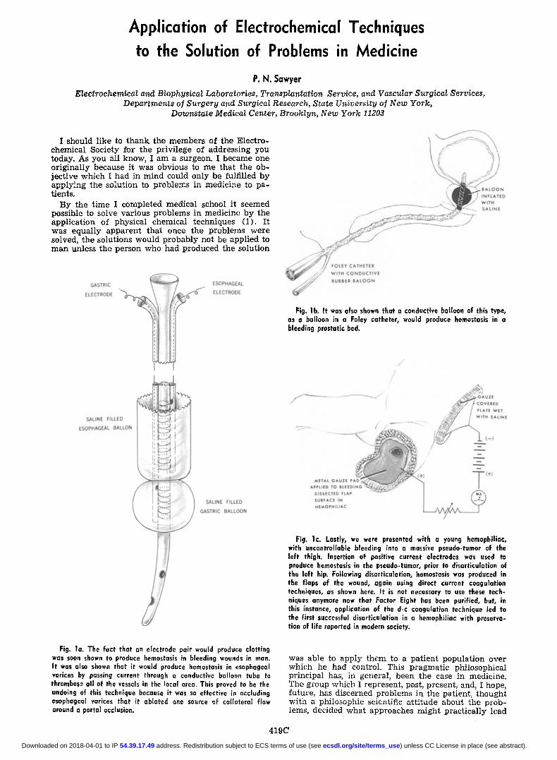
Fig. 2. This series of cartoons, drawn by Torsten Terrell, reflects I - his understanding of the significance of the fact that several elec- ~- (.9 trochemical phenomena occur at solid-liquid interfaces. He handled only those phenomena related to solid-liquid interfaces in this communication, although there are several other types of communication between the skin surface and the surrounding air envelope, which are even more complicated than those shown here. If one crosses from a bulk-water area through the ordered mole- cules of the membrane, multiple energy and transport phenomena occur relating even to. such a simple problem as ion distribution. Ion fluxes, that is, the act of transporting an ion across a barrier, requires energy of an ordered type, requiring geometric arrange- ment of enzymes at the interface. These fluxes are known to occur across virtually all solid-liquid interfaces and have been studied by literally thousands of scientists in modern physiologic chemistry. Fluxes relate to the attempt of the biologic organism to order intercellular and extracellular metabolism, water fluxes and, as well, preserve the "internal milieu." These fluxes produce electrical potential differences, relate to various forms of changes in conduction with change in transport. Other sophisticated phe- nomena involve pressure equivalent electroosmosis across mem- brane pores, as shown in the bottom cartoon.
55 - /
5 0 o
4 5 -
4 0 -
:55-
5 0 "
2 5 -
2 0 - o
15-
I0-
5 -
0 I .5
QUANTITY of ELECTRICITY
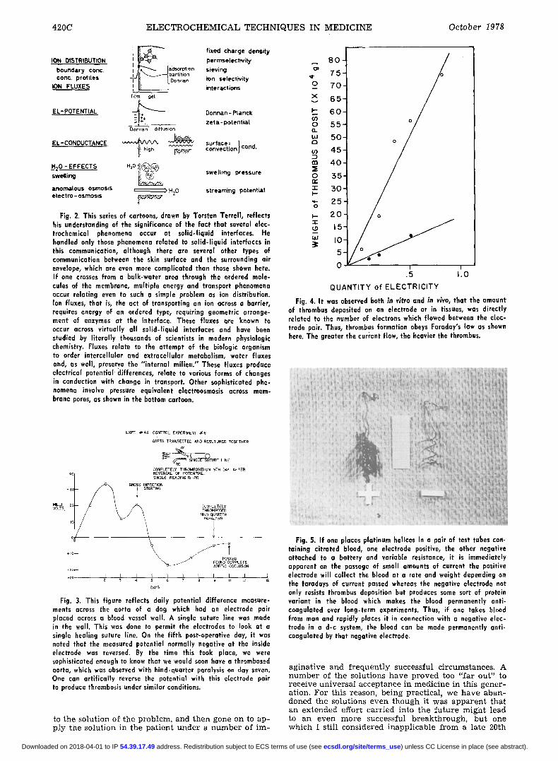
Fig. 4. It was observed both in vitro and in viva, that the amount of thrombus deposited on an electrode or in tissues, was directly related to the number of electrons which flowed between the elec- trode pair. Thus, thrombus formation obeys Faraday's law as shown here. The greater the current flow, the heavier the thrombus.
MILLI VOLTS
EXR~ .#43 CONTROL EXPERIMENT ~6
AORTA TRANSECTED AND RESUTURED TOGETHER
~ T U ~ E LINE COMPLETELY THROMBOSEDON 9TH DAY AFTER REVERSAL OF POTENTIAL. SINGLE READING RI-RO
COMPLETELY TF~ROMBOSED
HIND QUARTER PARAiYSIS
i z 3 4 s 6 T a ~ m ~t
DAYS
Fig. 3. This figure reflects daily potential difference measure- ments across the aorta of a dog which had an electrode pair placed across a blood vessel wall. A single suture line was made in the wall. This was done to permit the electrodes to look at a single healing suture line. On the fifth post-operatlve day, it was noted that the measured potential normally negative at the inside electrode was reversed. By the time this took place, we were sophisticated enough to know that we would soon have a thrombosed aorta, which was observed with hind-quarter paralysis on day seven. One can artifically reverse the potential with this electrode pair to produce thrombosis under similar conditions.
to the solution of the problem, and then gone on to ap- ply the solution in the pat ient under a number of im-
Fig. 5. i f one places platinum helices in a pair of test tubes cart. taining citrated blood, one electrode positive, the other negative attached to a battery and variable resistance, it is immediately apparent on the passage of small amounts of current the positive electrode will collect the blood at a rate and weight depending on the faradays of current passed whereas the negative electrode not only resists thrombus deposition but produces some sort of protein variant in the blood which makes the blood permanently anti- coagulated over long-term experiments. Thus, if one takes blood from man and rapidly places it in connection with a negative elec- trode in a d-c system, the blood can be made permanently anti- coagulated by that negative electrode.
aginat ive and f requent ly successful circumstances. A number of the solutions have proved too "far out" to receive universal acceptance in medicine in this gener- ation. For this reason, being practical, we have aban- doned the solutions even though it was apparent that an extended effort carr ied into the fu ture might lead to an even more successful breakthrough, but one which I still considered inapplicable from a late 20th
) unless CC License in place (see abstract). ecsdl.org/site/terms_use address. Redistribution subject to ECS terms of use (see 54.39.17.49Downloaded on 2018-04-01 to IP
Vol. 125, No, I0 J. EIectrochem. Soc.: REVIEWS AND NEWS 421C
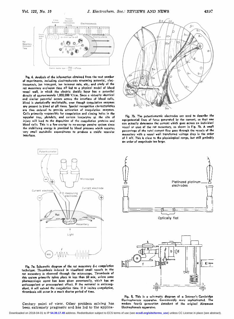
Fig. 6. Analysis of the information obtained from the vast number of experiments, incl~dlng eiectrophoresis streaming potential, eIec- troosmosis, ion transport, ion turnover rate, etc., and study of the rat mesentery occlusion time all led to a physical model of blood vessel wall, in which the electric double layer has a potential density of approximately 1,000,000 V/cm. Since a virtually identical and similar potential occurs across the interface of blood cells, blood is statistically unclottable, even though coagulation enzymes are present in blood at all times. Special recognition characteristics are thus ordered to provide activation of Coagulation enzymes. Cells primarily responsible for coagulation and closing holes in the vascular tree, platelets, and certain leucocytes at the site of injury will lead to the deposition of the coagulation proteins and blood cells. This is a low-energy to no-energy passive system since the stabilizing energy is provided by blood pressure which requires very small metabolic expenditures to produce a stable vascular interface.
Fig. 7b. The potentlametric electrodes are used to describe the equipotential lines of force generated by the current, so that one can actually determine the current which goes across an individual vessel or area of the rat mesentery, as shown in Fig. 7b. A small percentage of the total current flew goes through the vessels of the mesentery with a vessel wet[ transferred ~oltoge drop in the order of 1 mV. This is close to the physiological range, but still probably an order of magnitude too large.
Platinized plat inum electrodes
Optical ly f lat
Fig. 7a. Schematic diagram of the rat mesentery d-c coagulation technique. Thrombosis induced in visualized small vessels in the rat mesentery is observed through the microscope. Thrombosis of this system primarily takes place in less than 60 min, unless some pharmacologic agent has been given parenterally, which has an anticoagulant or proceagulant effect. If the material is anticoag- ulant', i t will extend the coagulation time. If it incites coagulation, thrombosis will occur in a much shorter period of time.
Century point of view. Other problem solving has beel~ extremely pragmatic and has led to the applica-
k
d 0 . . . .
a e
Fig. 8. Th~s is a schematic diagram of a Seaman's-Cambridge Electrophoresls apparatus. Considerably more sophisticated. The modern fourth generation decedent of the original Abramson Electrophoresis apparatus.
) unless CC License in place (see abstract). ecsdl.org/site/terms_use address. Redistribution subject to ECS terms of use (see 54.39.17.49Downloaded on 2018-04-01 to IP

422C ELECTROCHEMICAL TECHNIQUES IN MEDICINE October 1978
Fig. 9. Electrophoresis of a series of blood cells, erythro- cytes, and leucocytes from dogs treated with the antipregnancy hormones indicated at the top of the columns. These agents made the cell surfaces more positive in female dogs, but had a much less significant effect an the electrophoresis of erythrocytes and platelets in male dogs. This is an extremely interesting find- ing and presents information sug- gesting the reason why the anti- pregnancy hormones produce their procaagulant and throm- botic effects in females.
CONTROL/ CHEESE/ 1.5 GM/KG
19"67
~ 16.8
NI I
w _>
I
0 ~
BEN YL PO'PROVERA ~LC( IOL 22.5 MG/KG iOMq iKG
l i l l I A ! I A
F~] ELECTROPHORESIS RBC ~ PLATELETS
IORGI GTRKE( )RGE 0.09 .09 I + E1 IYNII E2Tla DIOL ).009 IG/K
012 45~6 112
I A I
0 ~ORTA
A I A
NORETHINORON 0.2l MG/K2
0123456
LILi ! A
ELECTROOSMOSI$
I I VENA CAVA I= INTIMA
MESTRANOL 2.1MG/KG +
INORETHINDRONE
p-. ~ L o 2 12456 WEEKS " ' ~ ~
0 ~. vl.I "1" E
~ i1.0 r a.9 ~
I ~ Ln. d 0 1234 WEEKS-'-
I A
A = ADVENTmA ]
I I
Fig. 10a. In experiments carried out with Drs. Walter Brattan and Philip Bodd, using the Rutherford cell, it was determined that erythrocytes, leucocytes, and platelets from mammalia tend to precipitate out on the surface of an electrode of known character- istic at a potential, approximating 250-300 mV more positive than zero with respect to the normal hydrogen electrode.
tion in the patient of a number of conceptual ideas which are now so common place that today nobody even bothers to acknowledge the source.
Looked at from another point of view, it must be realized and accepted that we have never insisted that our problem solv ing of a given problem was complete in, and of itself. We have been extremely practical in our day- to-day relationship with these problems. Us- ing this approach, the members of the laboratory have opened up a large number of fields in bioelectrochem- istry, the consideration of biological tissues as fuel
Table I. The cataphoresis of platelets in plasma
T h e speed of p o l y m o r p h o n u e l e a r l e u c o c y t e s is g ive n in the last co lumn. A l t h o u g h red ce l l s and smal l l y m p h o c y t e s have d i f ferent ve loc i t ies , no te that p la te le t s and polyraorphonuelear l e u c o c y t e s
have the same ve loc i ty ( s ix h o r s e s ) .
POTENTIAL DEPENDENT PLATELET ADHESION
c ~ 8 0 - ,,~
0 CONTROL OC
70- [] DEFIBRINOGENATED PLASMA co
I.-: A IODOACETIC ACID , a 0
,,, 6 0 - o o -1- I..-
o 50 " (._9 Z
I
c0 30- _j DECREASE
o 2 0 - i, I
W ] 0 - 9 8 % DECREASE nn ~-- , )
Z 0 , ~ i , ,
-6oo -4oo -2oo o +200 +400 +600 +800 SURFACE POTENTIAL IN MV NHE
Fig. 10b. It was also known that heparln prevents electrode "precipitation." It should be noted in Fig. 9 that the blood cells placed in the physiologic solution next to the precipitating elec- trode would begin to stick on with potentials more negative than the critical potential. Art the critical potential, there was a rapid increase in the rate of precipitation. The potential's more nega- tive than zero NHE, reversion toward a more negative potential would lead to detachment of the cells precipitated on the elec- trode if these were erythrocytes or leucocytes. Platelets, o,ce stuck, tended to show viscous metamorphosis.
Platelets Age Vo V
Plasma (hr) (n/sec/V/cm) (~/sec/V/cm)
Polymorpho- nuc lear
l e u c o c y t e s V ( ~ / s e c / V / c m )
1 30 0.71 0.41 0.46 2 6 0.82 0.69 0.57 3 30 0.76 0.55 0.60 4 6 0.65 0.46 0.52 5 6 0.57 0.40 0.43 6 6 0.67 0.51 0.53
6 0.68 0.46 0.54
Mean exc lud ing No. 2 0.45 0.49 p o t e n t i a l (mV) 12 13 (26.5 ~< ~ / s e c / V / c m )
cells and the electrokinetic characteristics of biological systems.
Philosophical We have used what help we could obtain from a
number of extraordinary physical chemists, physicists, and physician surgeons to attempt to solve problems (1). We are all in their debt.
These include various e lements of the electrochem- istry of thrombosis (2) beginning with rat mesentery technique (3), the induction and prevention of throm- bosis in experimental animals and man (4), the elec-
) unless CC License in place (see abstract). ecsdl.org/site/terms_use address. Redistribution subject to ECS terms of use (see 54.39.17.49Downloaded on 2018-04-01 to IP
Vol. 125, No. 10 3. Electrochem. Soc.: R E V I E W S A N D N E W S 423C
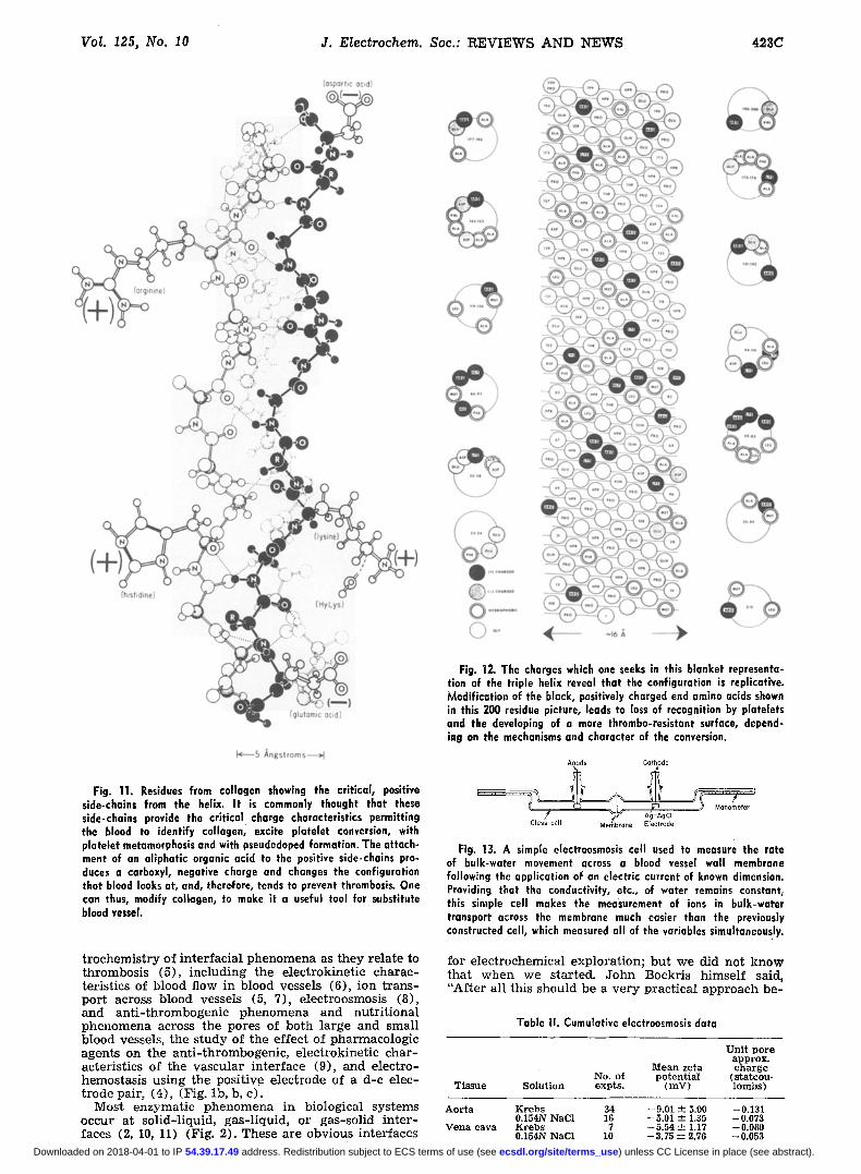
Fig. 11. Residues from collagen showing the critical, positive side-chains from the helix. It is commonly thought that these side-chains provide the critical charge characteristics permitting the blood to identify collagen, excite platelet conversion, with plateht metamorphosis and with pseudodeped formation. The attach- ment of an aliphatic organic acid to the positive side-chains pro- duces a carboxyl, negative charge and changes the configuration that blood looks at, and, therefore, tends to prevent thrombosis. One can thus, modify collagen, to make it a useful tool for substitute blood vessel.
t rochemis t ry of in te r rac ia l phenomena as they re la te to thrombosis (5), inc luding the e lec t rokinet ic charac- ter is t ics of blood flow in blood vessels (6), ion t rans- por t across blood vessels (5, 7), electroosmosis (8), and an t i - th rombogen ic phenomena and nut r i t iona l phenomena across the pores of both la rge and smal l b lood vessels, the s tudy of the effect of pharmacologic agents on the an t i - th rombogenic , e lec t rokinet ic char - acter is t ics of the vascular in ter face (9), and e lec t ro- hemostasis using the posi t ive e lec t rode of a d-c elec- t rode pair , (4), (Fig. lb , b, c) .
Most enzymat ic phenomena in biological systems occur at sol id- l iquid, gas- l iquid , or gas-sol id in t e r - faces (2, 10, 11) (Fig. 2). These are obvious interfaces
Fig. 12. The charges which one seeks in this blanket representa- tion of the triple helix reveal that the configuration is replicative. Modification of the black, positively charged end amino acids shown in this 200 residue picture, leads to loss of recognition by platelets and the developing of a more thrombo-resistant surface, depend- ing on the mechanisms and character of the conversion.
Anode Cathode
Fig. 13. A simple electroosmosis cell used to measure the rate of bulk-water movement across a blood vessel wall membrane following the application of an electric current of known dimension. Providing that the conductivity, etc., of water remains constant, this simple cell makes the measurement of ions in bulk-water transport across the membrane much easier than the previously constructed cell, which measured all of the variables simultaneously.
for e lectrochemical explora t ion; but we did not know tha t when we star ted. John Bockris h imself said, "Af te r al l this should be a ve ry prac t ica l approach be-
Table II. Cumulative electroosmosls data
Uni t po re approx.
Mean zeta c h a r g e No. of po ten t ia l (s ta tcou-
Tissue Solut ion expts . ( m V ) l ambs )
Aor t a Krebs 34 -9 .01 ± 5.00 -0.131 0.154N NaC1 16 -5 .01 ± 1.35 -0.073
Vena eava Krebs 7 -5 .54 +--- 1.17 --0.080 0.154N NaCI 10 --3.75 -- 2.70 -0.053
) unless CC License in place (see abstract). ecsdl.org/site/terms_use address. Redistribution subject to ECS terms of use (see 54.39.17.49Downloaded on 2018-04-01 to IP
424C E L E C T R O C H E M I C A L T E C H N I Q U E S IN M E D I C I N E October 1978
Fig. 14a. This is an interesting, though complicated schematic diagram demonstrating the placement of micropipettes in the aorta, vena cava, and other small vessels in the rabbit vasculature for the purposes of measuring streaming potentials. The technique permits one to measure the equivalent of streaming potentials in biologic systems in mammalia. These techniques were the first developed to measure streaming potentials in biologic systems in vivo.
Fig. 14b. A number of different experiments have been carried out measuring streaming potentials in biological systems. Perhaps the one most useful for clinicians, is the project which we carried out measuring streaming potentials in blood vessels, before and after injury, under conditions when the experimental animal hod received parenteral heparin and/or Dextron and/or Protomine. Injury reversed the negative interface potential of the vessel.
cause man is a fuel cell." Al l energy-u t i l i z ing phenom- ena in man involves a s teady enzymat ic degrada t ion of organic mater ia l s (12) to yield high ene rgy protons producing high energy phosphate groups (13) which are then themselves degraded to yie ld energy to dr ive var ious enzymat ic processes or t r anspor t var ious food bodies, bu lk water, or e lectrolytes in solution.
Wi th this as background, I am going to discuss briefly, a series of successful solut ions to problems in
Fig. 15a. The electrodes placed in blood vessel walls through side branches reveals the technique for injuring blood vessel, using a vascular clamp, a Fogarty balloon, or electrical injury.
medic ine using e lect rochemical approaches s ta r t ing wi th an ea r ly discovery:
1. That the vascular in terface is nega t ive ly charged when looked at wi th a pa i r of probing electrodes a t - tached to a h igh impedance po ten t iomete r (14) and tha t reversa l of this po la r i ty usua l ly re la tes to th rom- bosis (Fig. 3).
2. The re la ted deve lopment of the f reeze -dr ied graf t (15).
3. The deve lopment of d-c hemostasis, tha t is, the discovery that the posi t ive e lec t rode wil l genera te th rombus (Fig. 4), and stop b leeding in tissues, the converse of the nega t ive e lec t rode p reven t ing t h rom- bosis (4) (Fig. 5).
4. Studies of the vascula ture using an e lec t rochemi- cal look at the vascular in te r face - s t r eaming potent ia l (Fig. 6), electroosmosis, electrophoresis , and the ra t mesen te ry ( F i g . 7a, b) , coagulat ion expe r imen t (16) yields in format ion concerning the homeostat ic aspects of the electr ic double layer .
5. The e lect rochemical screen: appl ica t ion of elec- t rokine t ic s tudies to analyze the effect of pha rmaco- logic and o ther chemical agents on the b lood-vascu la r interfaces and the consequences of this (17).
6. Clean surfaces: thrombosis of vascular prosthesis, including artificial hear t valves, re la ted to di r t on the surfaces in contact wi th blood (18).
7. E lec t rochemis t ry of the immune response: modi- fication of e lect rophoret ic mobi l i ty by the mix ing of h is to-compat ib le and h i s to - incompat ib le lymphocytes , etc.
F i r s t b reakthrough, in 1950, whi le at the Naval Med- ical Research Inst i tute, Dr. James Pate and I !15), were requested to develop a technique for p reserv ing ar ter ies for use in wounded men in Korea. We even tu- a l ly succeeded in fur ther ing Marangoni ' s or ig inal tech- nique (19) by preserv ing homograf t h u m a n vessels for use in man. These were the first room t empe ra tu r e - s tored blood vessels. They were even tua l ly sent to Korea for use under bat t lef ie ld conditions.
We determined, dur ing our exper imen ta l evaluat ion of these vessels, tha t t hey worked more sa t i s fac tor i ly
) unless CC License in place (see abstract). ecsdl.org/site/terms_use address. Redistribution subject to ECS terms of use (see 54.39.17.49Downloaded on 2018-04-01 to IP
Vol. 125, No. I0 J. Electrochem. Soc.: R E V I E W S A N D N E W S 425C
Effects of Injury and Dextran 4 0 - Dextran 75 on In Vivo Streaming Potentials in Canine Blood Vessels
o • CONTROL o • HEMOSTAT CRUSH INJURY
A FOGARTY CATHETER INJURY o • ELECTRICAL INJURY, D C , I M A / 2 HRS
A POSITIVE STREAMING POTENTIAL iNDICATES A NEGATIVE VASCULAR SURFACE CHARGE AND HENCE A NEGATIVE pOTENTIAL ACROSS THE BLOOD VESSEL WALL BLOOD rNTERFACE
Fig. 15b. This figure shows that injury reverses the potential of blood vessel wall. Heparin reverses the potential again so that th~ interface is again more negative. Dextran does almost nothing tu reverse interfacial potentials of the wall; it has a different physical chemical effect on the blood cells themselves. I t displaces the electric double layer, half the width of a Dextran molecule, so that then the interface recognition effects are significantly ablated. Protamine makes the interface more positive.
Fig. 15c. Under these conditions, platelets cannot see either the wall or each other. Therefore, platelet activated intravascular thrombosis is, to a very large extent, blocked by the presence 'of Dextran at appropriate levels in the blood stream. No platelets are seen on these injured surfaces by the scanning electron micrograph, recognition has been blocked by Dextran 40.
than did fresh, unp rese rved canine counte rpar t s . Since these tissues were dead struts , we suggested tha t the ingrowth of t issues f rom the rec ip ient might be more sa t i s fac tor i ly car r ied out wi th a f reeze-dr ied , r a the r than f resh vessel succept ible to the rec ip ient ' s immune response. Since one of us, PNS, had or ig ina l ly been work ing wi th e lectrodes measur ing e lect r ica l and in- tes t inal potent ials , e lect rodes we re placed across the rec ip ient blood vessels (14), (Fig. 3), and inser ted grafts. This led to the first observat ion: the in t imal sur - face of blood vessels is nega t ive ly charged wi th r e - spect to the deep surfaces, the advent i t ia , and the con- ta ined blood.
Since A b r a m s o n (Table 1)1 had p rev ious ly de t e r - mined tha t blood cells were nega t ive ly charged and, in fact, e lec t rophore t ica l ly catophoret ic , i t was obvious to suggest tha t l ike charge repulses, and is one of the mechanisms opera t ive in the p reven t ion of in t r avascu- l a r thrombosis under convent ional c i rcumstances (20) (Fig. 4, 8, and 9).
W e now know tha t this process is much more sophis- t ica ted than suggested at tha t t ime. Much of the work in subsequent years has~ in itself, been electrochemical , such as the measuremen t of p rec ip i ta t ion or p la t ing- out potent ia ls of blood, proteins, and cells (21, 22), (Fig. 10a, b) . The s t ruc ture of the p ro te in deposi ted on blood vessel wal ls is also re la ted to the charge cha r - acter is t ics of the surface, as wel l as the s t ruc ture of the charge seen by those pro te ins and enzymes respon-
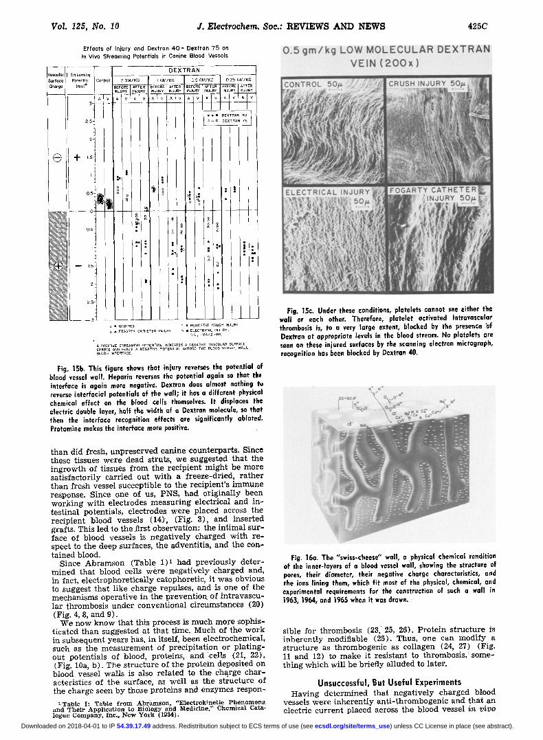
1Table I: Table from Abramson, "Electrokinetic Phenomena and Their Application to Biology and Medicine," Chemical Cata- logue Company, Inc., New York (1934).
Fig. 16a. The "swiss-cheese" wall, a physical chemical rendition of the inner-layers of a blood vessel wall, showing the structure of pores, their diameter, their negative charge characteristics, and the ions lining them, which fit most of the physical, chemical, and experimental requirements for the construction of such a wall in 1963, 1964, and 1965 when it was drawn. •
sible for thrombosis (23, 25, 26). P ro te in s t ruc tu re is i nhe ren t ly modif iable (25). Thus, one can modi fy a s t ruc ture as thrombogenic as col lagen (24, 27) (Fig. 11 and 12) to make i t res is tant to thrombosis, : some- th ing which wil l be br ief ly a l luded to la ter .
Unsuccessfu l , But Usefu l Exper iments Having de te rmined tha t nega t ive ly charged blood
vessels were inheren t ly an t i - th rombogen ic and tha t an electr ic cur ren t p laced across the blood vessel in vivo
) unless CC License in place (see abstract). ecsdl.org/site/terms_use address. Redistribution subject to ECS terms of use (see 54.39.17.49Downloaded on 2018-04-01 to IP
426C ELECTROCHEMICAL TECHNIQUES IN MEDICINE October 1978
Fig. 17b. Here the technique is slightly different. This is an elution experiment. The tissues are first loaded with an isotope of a known ion. Then the rate at which i t is turned over is determined by placing it sequentially into a series of test tubes which contain identical, but isotope free, solution. Thus the rate in which the "hot" ion turns over in the blood vessel wall can be measured using simple techniques and calculations.
Fig. 16b. This is a moderately good physical, chemical model of blood vessel wall, as shown in Fig. 16b, which is a scanning densitogram of blood vessel wall. It reveals that more than 60% blood vessel wall is extracellular space.
Fig. 17a. Ion transport across blood vessel walls is measured very simply using two half-cells whose interfaces are divided by blood vessel wall. Oxygen or other gases are provided through bubblers. The isotope elements are pipetted sequentially with the passage of time, dried, and measured in a beta counter to determine the rate at which the ions move across the blood vessel wall.
would lead to deposition of blood on an electrode (28), we next sought out a solution in the early days t rying to determine the dimensions of current which produced the observed effects. A n u m b e r of years were spent looking at the size, dimensions, and characteristics of current flow or suspected in ju ry currents wi thin bio- logical systems to determine if these were capable of producing int ravascular thrombosis (29, 30). The avail- able evidence suggests that they are. However, it ap- pears that thrombosis p e r se is kinet ical ly activated by
E u
E
x
57
54
51
4 8
4 5
4 2
39
36
33
30
27
24
21
18
15
12
g
6
3
C
~ ~ "-,,- A ~1"
I ~ r ' ~ ' A O R T I C INNER LAYER / / / PREPARATION
~ . ~ ~ A ~T
,....,../. . ,~. "r ~ A . . . . .
AORTIC OUTER LAYER PREPARATION
I I I I I 2 3 4
T I M E (hrs,)
Fig. 18. If one separates the inner layers, the intima, from the outer layers, the adventitia, of blood vessel wall, it is immediately possible to reveal in an ion transport cell that the inner layer is responsible for that ion transport across the aorta. This is not as simple a determination as one would wish for, however, in many in- stances, the aortic inner layers will transport ions in the opposite direction from that which is normally found with the, intact aorta. A simple explanation for this is that the aorta, when torn apart, no longer displays normal uninjured characteristics. This might explain the reversal in transport. However, chemical enzymatic inhibition studies, when carried out, were equivocal with respect to this explanation. In any event, i t is obvious that the inner layers of the vascular tree are most responsible for the modulation of both ion transport turnover and port "sluice gate" manipulation-- something which one would anticipate.
modification of the interfacial potential ra ther than by an " in jury current" p e r se (21, 28). Thus, thrombo- sis activation appears to be "low energy," interracial potential related ra ther than "high energy" current re- lated.
) unless CC License in place (see abstract). ecsdl.org/site/terms_use address. Redistribution subject to ECS terms of use (see 54.39.17.49Downloaded on 2018-04-01 to IP

Vol. 125, No. 10 ELECTROCHEMICAL TECHNIQUES IN MEDICINE 427C
% Flux Flux in#umoles cm=
0,40 73-92
0-58 70-22
0-36 66-53
0-34~ 6?:85
0-~52 59"14
0-30' 55.44
0 -28 51,T4
0-26 48-05
0-24 44-35
C~22 ~0.66
0-20 ~6-96
0-18 ~326
O'l 6 29"5Y
0"I 4 25"87
0,12J22-t8
O.IO 18"48
/
/
/
/
/ , #
efflux
~ in f lux efflux
~ i n f l u x
Fresh Vena Cava
Oz & Nz
__Cl 3= - - NaZZ
oi Ne Oz ~ N z ~
I 2 3 4 5 6 7 hours
Fig. 19. If one removes all oxygen from the cell, ion transport falls, and, in fact, undergoes an abnormal change in direction as shown in Fig. 20.
Modern Experiments A number o~ electrometric experiments previously
referred to were carried out from 1961-1965. These in- cluded electrophoresis of blood cells (Fig. 8 and 9), electroosmosis (8) (Fig. 13) (Table II),S and streaming potential of blood vessel walls.
~Table IT: This table illustrates the calculations of zeta poten- tials calculated from alectroosmotic streaming potentials across the pores of both canine aorta and vena cava. The table is in- teresting for two reasons: (i) The zeta potential across veins is one-half that for arteries, (ii) If sodium chloride rather than sea water (Krebs) is used to bathe the vessels, both measured streaming potentials and calculated zeta potentials are markedly diminished, indicating the common observation that calcium and potassium, and other ions have to be available to blood vessels to maintain their normal functional characteristics.
Table III . Effect of boiling aorta and vena cava on the zeta potential
Mean ze ta C h a r g e / e r a = No. of po ten t i a l po re su r f ace
Tissues Solut ion expts. (mV) ( s t a tcou lombs)
A o r t a 0.154N NaC1 26 - 2.12 • 0.41 1.3 • 0.2 x l0 s Vena eava 0.154N NaCI 16 -0.70 • 0.14 0.5 • 0.1 x los
The first measurements of ion t r anspor t and tu rn - over ra te in blood vessel wal ls s t r eaming potent ia ls in biological systems including t r ansmura l electroosmosis were car r ied out in the a t t empt to de te rmine the s ig- nificance of s t ructure , size, t issue pore dimensions, and v iab i l i ty in vivo, as wel l as in vitro (6) (Fig. 14-20) Table HI) . 8 The studies were in te res t ing and useful
Tab le III : Boiling des t roys po re su r f ace charge . I f one p lunges e i t he r the ao r t a or the v e n a eava into boi l ing wa te r , t he s t r eam- ing potent ia ls and ca lcu la ted ze ta po ten t ia l s app roach zero. Con- clusion: T h e vascu l a r t r e e is not m a d e of i n a n i m a t e pipe.
100%-
8 _J
~0O,,o. o
"X~-.y.. X " k "'X..,...:~__..;,,,,,,,,~( FULLTHICKNESS AORTIC GRAFT
I% 2's ~o ;s ~6o i~s i~o 175 TIME IN MINUTES
Fig. 20. Calcium turnover is markedly different when the aortic wall is compared to the aortic graft following implantation. Thus, an aortic graft never displays conventional metabolism, even at the simplest level, such as ion transport, which is found in normol, healthy blood vessel wall, including the aorta.
Fig. 21a. A positively charged collagen tube implanted in the canine aorta for 2 hr, showing very rapid thrombosis containing all formed blood elements, coag- ulation enzymes, and proteins.
) unless CC License in place (see abstract). ecsdl.org/site/terms_use address. Redistribution subject to ECS terms of use (see 54.39.17.49Downloaded on 2018-04-01 to IP
428C J. EIectrochem. Sot.: R E V I E W S A N D N E W S October 1978
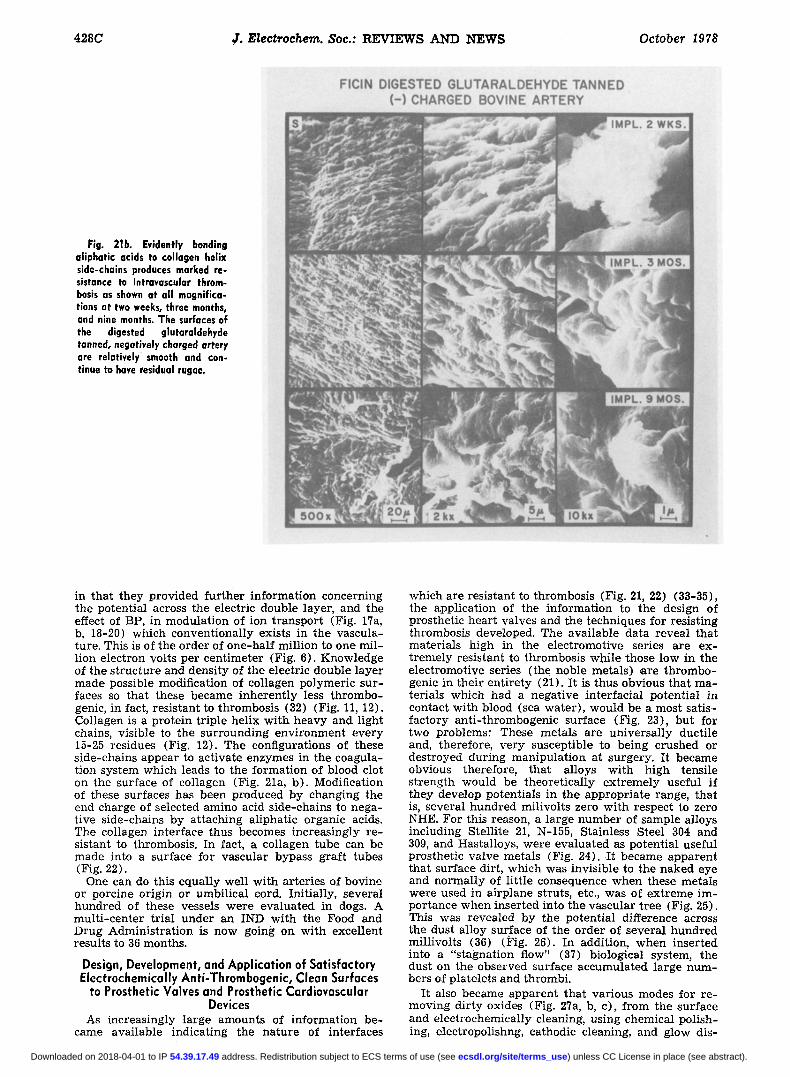
Fig. 21b. Evidently bonding aliphatic acids to collagen helix side-chains produces marked re- sistance to intravascular throm- bosis as shown at all magnifica- tions at two weeks, three months, and nine months. The surfaces of the digested glutaraldehyde tanned, negatively charged artery are relatively smooth and con- tinue to have residual rugae.
in tha t they provided fu r the r informat ion c o n c e r n i n g the potent ia l across the electr ic double layer , and the effect of BP, in modula t ion of ion t r anspor t (Fig. 17a, b, 18-20) which convent ional ly exists in the vascula- ture. This is of the o rder of one-ha l f mi l l ion to one mi l - l ion e lec t ron volts per cen t imeter (Fig. 6). Knowledge of the s t ruc tu re and dens i ty of the electr ic double l aye r made possible modification of col lagen polymer ic sur - faces so tha t these became inhe ren t ly less t h rombo- genic, in fact, res i s tan t to thrombosis (32) (Fig. 11, 12). Collagen is a p ro te in t r ip le hel ix wi th heavy and l ight chains, v is ible to the su r rounding env i ronment eve ry 15-25 residues (Fig. 12). The configurations of these s ide-chains appear to act ivate enzymes in the coagula- t ion sys tem which leads to the fo rmat ion of blood clot on the surface of col lagen (Fig. 21a, b) . Modification of these surfaces has been produced by c h a n g i n g the end charge of selected amino acid s ide-chains to nega- t ive s ide-chains by a t taching al iphat ic organic acids. The col lagen in ter face thus becomes increas ing ly r e - s is tant to thrombosis . In fact, a col lagen tube can be made into a surface for vascular bypass graf t tubes (Fig. 22).
One can do this equa l ly wel l wi th ar ter ies of bovine or porcine or igin or umbi l ica l cord. In i t ia l ly , severa l hundred of these vessels were eva lua ted in dogs. A mul t i - cen te r t r ia l under an IND wi th the Food and Drug Admin i s t r a t ion is now going on wi th excel lent results to 36 months.
Design, Development, and Application of Satisfactory Electrochemically Anti-Thrombogenic, Clean Surfaces
to Prosthetic Valves and Prosthetic Cardiovascular Devices
As increas ingly la rge amounts of informat ion be- came avai lab le indica t ing the na tu re of interfaces
which are res is tant to thrombosis (Fig. 2I, 22) (33-35), the appl ica t ion of the informat ion to the design of prosthet ic hear t valves and the techniques for res is t ing thrombosis developed. The ava i lab le da ta revea l that mater ia l s high in the e lec t romot ive series a re ex- t r eme ly res is tant to thrombosis whi le those low in the e lec t romot ive series ( the noble meta ls) ,are th rombo- genic in the i r en t i re ty (21). I t is thus obvious tha t ma- ter ia ls which had a negat ive in te r fac ia l po ten t ia l in contact wi th blood (sea water ) , would be a most sat is- fac tory an t i - th rombogenic surface (Fig. 23), bu t for two problems: These meta l s a re un iversa l ly ducti le and, therefore, ve ry suscept ible to being crushed or des t royed dur ing manipu la t ion at surgery . I t became obvious therefore, that al loys wi th high tensi le s t rength would be theore t i ca l ly ex t r eme ly useful if t hey develop potent ia ls in the app rop r i a t e range, tha t is, severa l hundred mil ivol ts zero wi th respect to zero NHE. For this reason, a l a rge number of sample al loys including Ste l l i te 21, N-155, Stainless Steel 304 and 309, and Hastal loys, were eva lua ted as potent ia l useful prosthet ic va lve meta ls (Fig. 24). I t became appa ren t tha t surface dirt , which was invis ible to the naked eye and no rma l ly of l i t t le consequence when these meta ls were used in a i rp lane struts, etc., was of ex t r eme im- por tance when inser ted into the vascular t ree (Fig. 25). This was revealed by the potent ia l difference across the dust a l loy surface of the o rde r of severa l hundred mil l ivol ts (36) (Fig. 26). In addit ion, when inser ted into a "s tagnat ion flow" (37) biological system, the dust on the observed surface accumula ted la rge num- bers of p la te le ts and thrombi.
I t also became appa ren t tha t var ious modes for re - moving d i r ty oxides (Fig. 27a, b, c), f rom the sur face and e lec t rochemical ly cleaning, using chemical pol i sh- ing, electropolishng, cathodic cleaning, and glow dis-
) unless CC License in place (see abstract). ecsdl.org/site/terms_use address. Redistribution subject to ECS terms of use (see 54.39.17.49Downloaded on 2018-04-01 to IP
VoL 125, No. 10 ELECTROCHEMICAL TECHNIQUES IN MEDICINE 429C
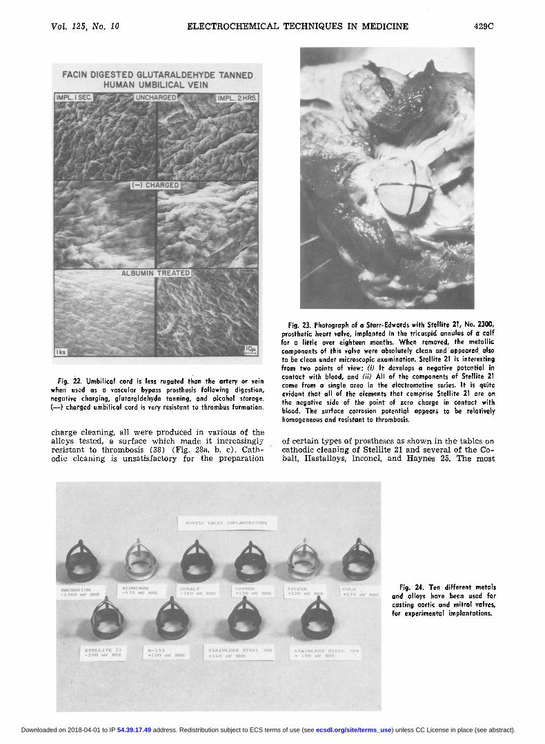
Fig. 22. Umbilical cord is less rugated than the artery or vein when used as a vascular bypass prosthesis following digestion, negative charging, glutaraldehyde tanning, and alcohol storage. (w) charged umbilical cord is very resistant to thrombus formation.
charge cleaning, all were produced in various of the alloys tested, a surface which made it increasingly resistant to thrombosis (38) (Fig. 28a, b, c). Cath- odic cleaning is unsatisfactory for the preparation
Fig. 23. Photograph of a Starr-Edwards with Stellite 21, No. 2300, prosthetic heart valve, implanted in the tricuspid annulus of a calf far a little over eighteen months. When removed, the metallic components of this valve were absolutely clean and appeared also to be clean under microscopic examination. Stellite 21 is interesting from two points of view: (i) It develops a negative potential in contact with blood, and ( i i) AJI of the components of Stellite 21 come from a single area in the electromotive series. It is quite evident that all of the elements that comprise Stellite 21 a,re on the negative side of the point of zero charge in contact with b~ood. The surface corrosion potential appears to be relatively homogeneous and resistant to thrombosis.
of certain types of prostheses as shown in the tables on cathodic cleaning of Stellite 21 and several of the Co- balt, Hastalloys, Inconel, and Haynes 25. The most
Fig. 24. Ten different metals and alloys have been used far casting aortic and mitral valves, far experimental implantations.
) unless CC License in place (see abstract). ecsdl.org/site/terms_use address. Redistribution subject to ECS terms of use (see 54.39.17.49Downloaded on 2018-04-01 to IP

430C J. Electrochem. Sac.: REVIEWS AND NEWS October 1978
Fig. 25. Surface contamination and thrombogenicity of N-155 alloy heart valves. The unusual mixed alloy N-155, when used as a heart valve material, although high in iron, and therefore expected to be highly thrombogenie, will serve as a nonthrombogenic surface unless contaminated by touching with a surgically gloved hand, or subjected to steam autoclave sterilization. If so contaminated, chemical cleans- ing may return the anti-thrombogenic characteristics but not with absolute assurance.
METHOD OF IN VlVO MEASUREMENT OF INTERFACIAL POTENTIAL
VOLTMETER
Fig. 26. This figure is a simple schematic of the techniques one uses to measure interface p~tentials of a prosthetic heart valve placed in the heart of a calf. The techniques of measurement are simple.
thrombogenic of these surfaces were those which be- came progressively more cathodic with the passage of t ime under a fixed voltage approaching minus ( - - ) 150 to 300 millivolts cathodic (38). It can be seen as the cathodic potential increased current for the various surfaces became increasingly stable approaching zero in mil l iamperes for satisfactorily clean alloys (Fig. 27b, c). With the unsatisfactory alloys, the current flow tended to increase, indicating a poor passivation range and a very high dissolution current for those specimens which were apparent ly unsatisfactory.
Not too strangely, hydrochloric acid polishing, which produces a chlorodized reduced chemical surface on the valves, proves to be a satisfactory technique for polish- ing many of the metal surfaces, par t icular ly a luminum, Stellite 21, copper, and the Noble metals tested (39).
Thus, in one ten year stand, the technology of con- ductive metals and alloys useful to the production of
DIRTY
~
16N HNO 3
I
ION HCf,
t
IMPLANTATION IN CALF
Fig. 27a. Various modes of cleaning heart valves ore shown
heart valves and as critical parameters and production techniques were elucidated by a very simple series of determinat ions involving both in viva and in vitro studies, using implanta t ion techniques as well as scan- ning electron microscopic and sophisticated electro- chemical polishing techniques in an integrated pa t te rn star t ing from the simplest approaches avai lable (38, 39).
Elect rok inet ic Studies of the I m m u n e Response Several years ago we set up an experiment using a
very rapid liver rejection technique described herein, or a heterotopic ex viva perfusion. The model per-
) unless CC License in place (see abstract). ecsdl.org/site/terms_use address. Redistribution subject to ECS terms of use (see 54.39.17.49Downloaded on 2018-04-01 to IP
Vo/. 125, No. 10 431C E L E C T R O C H E M I C A L T E C H N I Q U E S I N M E D I C I N E
CATHODIC CLEANING ( POTE NTIOSTATIC )
~ ) 6 o 5 o
(z 4
u~ 2 rY
:E 0 <~
o cJ o p- 6 <~ (J
I0
+ 210 -Z~
+ 60-STARR EDWAROS -ST" ZI 0 2 4 6 ~ ' I / ~ . - - & - - l I I I I I I I ,: ' 1 / / I T I M E
+ 2 9 0 & ~ - 9 0 8 I0 12 ]4 16 18 25 3 0 (m[n.)
-- ~ -20-STARR EDWARDS-ST- 21 + i 6 0 0 O ~
+ 1 4 0 -e ~ ' ~ 0"--
",,, ./ POTENTIALS ARE WITH REFERENCE TO NORMAL HYDROGEN
ELECTRODE IN MILLIVOLTS ( ELECTROLITE 0,09 % Na Ci }
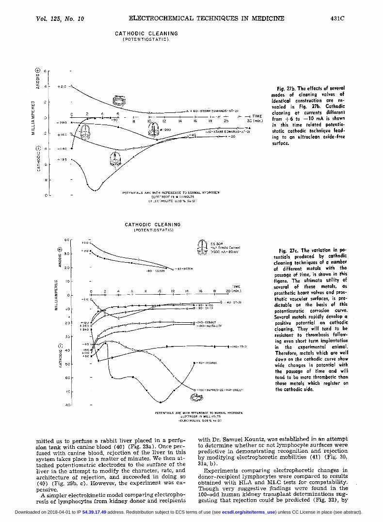
Fig. 27b. The effects of several modes of cleaning valves of identical construction are re- vealed in Fig. 27b. Cathodic cleaning at currents different from + 6 to - -10 mA is shown in this time related potentio- static cathodic technique lead- ing to an ultraclean oxide-free surface.
40
| _~ 3 o
2O
~0
l.l.I
:E 0 < ..J -1
I0
2O
3 0
0 4 0
03 2:
50
6 0
70
8 0
C A T H O D I C C L E A N I N G (POTENTIOSTATIC)
4 . o ~
- ~ . _ ~ . - ~ o -- 65- $5509
SS 3 0 4 High Anodic Current > 5 0 0 m A - 80 rnV
T IME 0 2 4 G 8 I I0 12 14 16 18 20 (miD.) -J I I . ? I I [ I f I
-,,o~\+A . t . . . . . ! . . . . ~ + ~ ' ~ + - - + _ _ $ : L t ~ + - T , 0
+ 2 5 0 ~> ~ 1 7 6 . . . . . . . . ~...._.~. +. 3,~Oo-_%%Loy 4-240 # | ~ . _ _ + / e - -
. . . . . . R , / ~ o~o...., ~ + + + ..........
. . . . . . . . . . . .
1 +m - 160 - HAYNES-25 - KAY-SHI LEY
POTENTIALS ARE WITH REFERENCE TO NORMAL HYDROGEN ELECTRODE IN MILLIVOLTS
(ELECTROLtTE 0.09 % No CI)
Fig. 27e. The variation in po- tentials produced by cathodic cleaning techniques of a number of different metals with the passage of time, is shown in this figure. The ultimate utility of several of these metals, as prosthetic heart valves and pros- thetic vascular surfaces, is pre- dictable on the basis of this potentiostatic corrosion curve. Several metals rapidly develop a positive potential On cathodic cleaning. They will tend to be resistant to thrombosis follow- ing even short term imp!antution in the experimental animal. Therefore, metals which are well down on the cathodic curve show wide changes in potential with the passage of time and will tend to be more thrombotic than those metals which register on the cathodic side.
mit ted us to perfuse a r abb i t l ive r p laced in a pe r fu - sign t ank wi th canine blood (40) (Fig. 23a). Once per - fused wi th canine blood, re jec t ion of the l iver in this sys tem takes place in a ma t t e r of minutes. We then a t - tached potent iometr ic electrodes to the surface of the l iver in the a t t empt to modi fy the character , rate, and archi tec ture of rejection, and succeeded in doing so (40) (Fig. 29b, c). However , the exper imen t was ex- pensive.
A s impler e lec t rokinet ic model compar ing e lec t ropho- resis of lymphocytes f rom k idney donor and recipients
wi th Dr. Samuel Kountz, was es tabl ished in an a t t empt to de te rmine whe the r or not lymphocy te surfaces were pred ic t ive in demons t ra t ing recogni t ion and re jec t ion by modify ing e lec t rophoret ic inabi l i t ies (41) (Fig. 30, 31a, b) .
Exper iments compar ing e lec t rophoret ic changes in donor - rec ip ien t lymphocytes were compared to resul ts obta ined wi th HLA and MLC tests for comparabi l i ty . Though ve ry suggest ive findings were found in the 100-odd human k idney t r ansp lan t de te rmina t ions sug- gest ing tha t re jec t ion could be pred ic ted (Fig. 32), by
) unless CC License in place (see abstract). ecsdl.org/site/terms_use address. Redistribution subject to ECS terms of use (see 54.39.17.49Downloaded on 2018-04-01 to IP
432C J. EZectrochem. Sot.: R E V I E W S A N D N E W S October I978
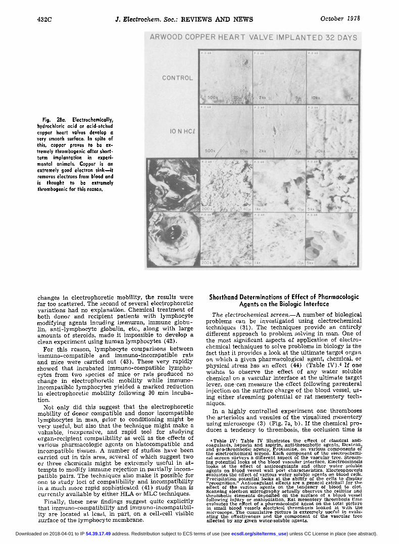
Fig. 28a. Electrochemically, hydrochloric acid or acid-etched copper heart valves develop a very smooth surface. In spite of this, copper proves to be ex- tremely thrombogenic after short- term implantation in experi- mental animals. Copper is an extremely good electron sinkuit removes electrons from blood and is thought to be extremely thrombogenic for this reason.
c h a n g e s in e l e c t r o p h o r e t i c m o b i l i t y , t h e r e s u l t s w e r e f a r too s ca t t e r ed . T h e s e c o n d of s e v e r a l e l e c t r o p h o r e t i c v a r i a t i o n s h a d no e x p l a n a t i o n . C h e m i c a l t r e a t m e n t of b o t h d o n o r a n d r e c i p i e n t p a t i e n t s w i t h l y m p h o c y t e m o d i f y i n g a g e n t s i n c u d i n g i m m u r a n , i m m u n e g l o b u - l in, a n t i - l y m p h o c y t e g lobu l in , etc., a l o n g w i t h l a r g e a m o u n t s of s t e ro ids , m a d e i t i m p o s s i b l e to d e v e l o p a c l e a n e x p e r i m e n t u s i n g h u m a n l y m p h o c y t e s (42) .
F o r t h i s r eason , l y m p h o c y t e c o m p a r i s o n s b e t w e e n i m m u n o - c o m p a t i b l e a n d i m m u n o - i n c o m p a t i b l e r a t s a n d mice w e r e c a r r i e d o u t (43) . T h e s e v e r y r a p i d l y s h o w e d t h a t i n c u b a t e d i m m u n o - c o m p a t i b l e l y m p h o - cy tes f r o m two spec ies of m i c e o r r a t s p r o d u c e d no c h a n g e in e l e c t r o p h o r e t i c m o b i l i t y w h i l e i m m u n o - i n c o m p a t i b l e l y m p h o c y t e s y i e l d e d a m a r k e d r e d u c t i o n in e l e c t r o p h o r e t i c m o b i l i t y f o l l o w i n g 30 r a i n i n c u b a - t ion.
No t o n l y d id t h i s s u g g e s t t h a t t h e e l e c t r o p h o r e t i c m o b i l i t y of d o n o r c o m p a t i b l e a n d d o n o r i n c o m p a t i b l e l y m p h o c y t e s i n m a n , p r i o r to c o n d i t i o n i n g m i g h t b e v e r y usefu l , b u t also t h a t t h e t e c h n i q u e m i g h t m a k e a v a l u a b l e , i n e x p e n s i v e , a n d r a p i d tool f o r s t u d y i n g o r g a n - r e c i p i e n t c o m p a t i b i l i t y as w e l l as t h e effects of v a r i o u s p h a r m a c o l o g i c a g e n t s o n h i s t o c o m p a t i b l e a n d i n c o m p a t i b l e t i ssues . A n u m b e r of s t ud i e s h a v e b e e n c a r r i e d ou t in t h i s a rea , s e v e r a l of w h i c h s u g g e s t t w o or t h r e e c h e m i c a l s m i g h t b e e x t r e m e l y u s e f u l in a t - t e m p t s to m o d i f y i m m u n e r e j e c t i o n in p a r t i a l l y i n c o m - p a t i b l e pa i rs . T h e t e c h n i q u e s also m a k e i t pos s ib l e for one to s t u d y loci of c o m p a t i b i l i t y a n d i n c o m p a t i b i l i t y in a m u c h m o r e r a p i d s o p h i s t i c a t e d (41) s t u d y t h a n is c u r r e n t l y a v a i l a b l e b y e i t h e r H L A or M L C t e c h n i q u e s .
F ina l l y , t h e s e n e w f ind ings sugges t q u i t e e x p l i c i t l y t h a t i m m u n o - c o m p a t i b i l i t y a n d i m m u n o - i n c o m p a t i b i l - i t y a r e l oca t ed a t least , in pa r t , o n a ce l l - ce l l v i s i b l e s u r f a c e of t h e l y m p h o c y t e m e m b r a n e .
Shorthand Determinations of Effect of Pharmacologic Agents on the Biologic Interface
The electrochemical screen.--A n u m b e r of b io log i ca l p r o b l e m s c a n be i n v e s t i g a t e d u s i n g e l e c t r o c h e m i c a l t e c h n i q u e s (31). T h e t e c h n i q u e s p r o v i d e a n e n t i r e l y d i f f e r e n t a p p r o a c h to p r o b l e m s o l v i n g in m a n . One of t h e m o s t s ign i f i can t a spec t s of a p p l i c a t i o n of e l e c t r o - c h e m i c a l t e c h n i q u e s to so lve p r o b l e m s in b i o l o g y is t he f ac t t h a t i t p r o v i d e s a look a t t h e u l t i m a t e t a r g e t o r g a n on w h i c h a g i v e n p h a r m a c o l o g i c a l agen t , c h e m i c a l , or p h y s i c a l s t r e s s h a s a n effect (44) ( T a b l e IV) .4 I f one w i s h e s to o b s e r v e t h e effect of a n y w a t e r so lub l e c h e m i c a l o n a v a s c u l a r i n t e r f a c e a t t h e u l t i m a t e t a r g e t l ever , o n e c a n m e a s u r e t h e effect f o l l o w i n g p a r e n t e r a l i n j e c t i o n o n t h e s u r f a c e c h a r g e o f t h e b l o o d vessel , u s - ing e i t h e r s t r e a m i n g p o t e n t i a l o r r a t m e s e n t e r y t e c h - n iques .
I n a h i g h l y c o n t r o l l e d e x p e r i m e n t o n e t h r o m b o s e s t h e a r t e r i o l e s a n d v e n u l e s of t h e v i s u a l i z e d m e s e n t e r y u s i n g m i c r o s c o p e (3) (Fig. 7a, b ) . I f t h e c h e m i c a l p r o - duces a t e n d e n c y to t h r o m b o s i s , t h e occ lu s ion t i m e is
~Table IV: Table IV illustrates the effect of classical anti- coagulants, heparin and aspirin, anti-thrombotic agents, Dextran, and pro-thrombotic agency, Protamine, on various components of the electrochemical screen. Each component of the electrochemi- cal screen surveys a different aspect of the vascular tree. Stream- ing potential looks at the blood vascular interface. Electroosmosis looks at the effect of anticoagulants and other water soluble agents on blood vessel wall port characteristics. Electrophoresis evaluates the effect of various water soluble agents on blood cells. Precipitation potential looks at t h e ability of the cells to display "recognition." Anticoagulant effects are a general catchall for the effect of the various agents on the tendency of blood to clot. Scanning electron micrography actually observes the cellular and thrombotic elements deposited on the surface of a blood vessel following injury or manipulation. Rat mcsentery thrombosis time evaluates the effect of a pharmacologic agent on the total picture in small blood vessels electrical thrombosis looked at w~th the microscope. The cumulative picture is extremely useful in evalu- ating the effectiveness and the component of the vascular tree af f ec ted by any given water-soluble agents.
) unless CC License in place (see abstract). ecsdl.org/site/terms_use address. Redistribution subject to ECS terms of use (see 54.39.17.49Downloaded on 2018-04-01 to IP
VoI. 125, No. 10 E L E C T R O C H E M I C A L T E C H N I Q U E S IN M E D I C I N E 433C
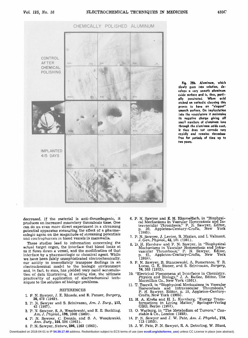
Fig. 28b. Aluminum, which slowly goes into solution, de- velops a very smooth aluminum oxide surface and is, thus, parti- ally possivated. When acid etched on cathodic cleaning this proves to have an "elegant" smooth surface. On implantation into the vasculature it maintains its negative charge giving off small numbers of aluminum ions through the aluminum oxide coat, it thus does not corrode very rapidly and remains thrombus- free for periods of time up to two years.
decreased. I f the ma te r i a l is an t i - th rombogenic , i t produces an increased mesen te ry thrombosis time. One can do an even more d i rec t exper imen t in a s t r eaming potent ia l appara tus measur ing the effect of a p h a r m a - cologic agent on the magn i tude of s t r eaming potent ia ls and eiectrophoresis in blood vessels in mammal ia .
These studies l ead to in format ion concerning the ac tual t a rge t organ, the in ter face tha t blood looks at as it flows down a vessel, and the modification of tha t in ter face by a pharmacologic or chemical agent. Whi le we have been f a i r ly unsophis t ica ted e lectrochemical ly , our ao i i i ty to immed ia t e ly t ranspose findings in an e lec t rochemical model to the biologic env i ronment and, in fact, to man, has y ie lded ve ry rap id accumula- t ion of da ta i l lus t ra t ing, if no,thing else, the u l t ima te p rac t i ca l i ty of appl ica t ion of e lec t rochemical tech- niques to the solut ion of biologic problems.
REFERENCES 1. P. N. Sawyer , J. E. Rhoads, and R. Panzer , Surgery,
26, 479 (1949). 2. P. N. Sawye r and S. Sr inivasan, Am. J. Surg., 113,
42 (1967). 3. P. N. Sawyer , S. A. Wesolowski, and E. E. Suckling,
Am. J. Physiol., 198, 1006 (1960). 4. P. N. Sawyer , C. Dennis, and S. A. Wesolowski ,
Ann. Surg., 154, 556 (1961). 5. P. N. Sawyer , Nature, 2@6, 1162 (1965).
6. P. N. Sawyer and E. H. Himmelfa rb , in "Biophysi - cal Mechanisms in Vascular Homeostasis and In- t r avaseu la r Thrombosis ," P. N. Sawyer , Editor, p. 30, App le ton -Cen tu ry -Crof t s , New York (1965).
7. P. N. Sawyer , J. Levine, R. Mazlen, and I. Valmont, J. Gen. Physiol., 45, 181 (1961).
8. D. H. Har shaw and P. N: Sawyer , in "Biophysical Mechanisms in Vascular Homeostasis and In t r a - vascular Thrombosis ," P. N. Sawyer , Editor, p. 61, App le ton-Cen tu ry -Crof t s , New York (1965).
9. P. N. Sawyer , B. Stanczewski , A. Pomerance, T. R. Lucas, G. E. Stoner, and S. Sr inivasan, Surgery, 74, 263 (1973).
10. "Elect r ica l Phenomena at In ter faces in Chemistry, Physics and Biology," J. A. Butler, Editor, The Macmil lan Co., New York (1951).
11. T. Teorell , in "Biophysical Mechanisms in Vascular Homeostasis and In t r avascu la r Thrombosis ," P. N. Sawyer , Editor, p. 19, A p p l e t o n - C e n t u r y - Crofts, New York (1965).
12. H. A. Krebs and H. L. Kornberg , "Energy Trans- format ions in Living Mat ter ," Sp r inge r -Ver l ag OHG, Ber l in (1957).
13. O. Warburg , in "The Metabol i sm of Tumors," Con- s table & Co., London (1930).
14. P. N. Sawyer and J. W. Pate, Am. J. Physiol., 175, 113 (1953).
15. J. W. Pate, P. N. Sawyer , R. A. Deter l ing, W. Blunt,
) unless CC License in place (see abstract). ecsdl.org/site/terms_use address. Redistribution subject to ECS terms of use (see 54.39.17.49Downloaded on 2018-04-01 to IP

434C J. Electrochem. Soc.: REVIEWS AND N E W S October 1978
Fig. 28c. Stellite 21 proves to be very resistant to thrombosis. All of the elements of Stellite 21 come from one area in the elec- tromotive series. The metal has proved to be one of the best materials for construction of cardiovascular prostheses be- cause of its resistance to throm- bosis when implanted in the ultroclean state.
Stainless steel - control
~urrent on f lux)
Fig. 29a. Application of oriented electrodes to a heterotopic ex vivo liver delayed the rate of total rejection of the liver.
and M. S. Parshley, Surg. Forum, 3, 147 (1952). 16. P. N. Sawyer , S. Sr inivasan, B. Stanczewski,
N. Ramasamy, and W. Ramsey, This Journal, 121, 221C (1974).
17. P. N. Sawyer , B. Stanczewski, N. Ramasamy, and J. S. Keates, in "Coronary A r t e r y Medicine and Surgery, Concepts and Controversies," J. C. Nor - man, Editor, p. 920, App le ton-Cen tu ry -Crof t s , New York (1975).
18. P. N. Sawyer , B. Stanczewski, S. Sr inivasan, J. G. S tempak and G. W. Kammlot t , J. Thorac. Cardio- vasc. Surg., 67, 24 (1974).
19. A. G. Mar rangon i and L. P. Cecchini, Ann. Surg., 134, 977 (1951).
20. H. W. Abrahamson, "EIectrophoresis of Ceils and
Fig. 29b. The configuration of rejection was markedly altered according to the imposed charge distribution.
Proteins," Hafner Publ ishing Co., New York (1968).
21. P. J. Boddy, W. H. Brat ta in , and P. N. Sawyer , in "Biophysical Mechanisms in Vascular Homeos ta- sis and In t r avascu la r Thrombosis," P. N. Sawyer , Editor , p. 30, App le ton-Cen tu ry -Crof t s , New York (1965).
22. B. W. Morrissey, L. E. Smith, R. R. St romberg, and
) unless CC License in place (see abstract). ecsdl.org/site/terms_use address. Redistribution subject to ECS terms of use (see 54.39.17.49Downloaded on 2018-04-01 to IP
VoL 125, No. 10 ELECTROCHEMICAL TECHNIQUES IN MEDICINE 435C
ELECTROPHORETIC MOBILITY (E.M.) OF HUMAN PERIPHERAL BLOOD LYMPHOCYTES FROM ABO INCOMPATIBLE SUBJECTS BEFORE AND
AFTER MIXING
Fig. 29c. The configuration of rejection was markedly altered according to the imposed charge distribution.
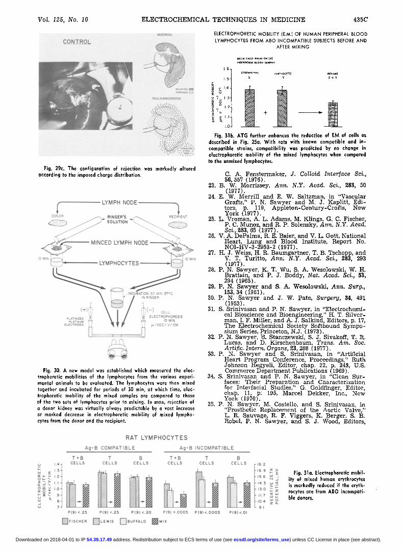
Fig. 30. A new model was established which measured the elec- trophoretic mohilities of the lymphocytes from the various experi- mental animals to be evaluated. The lymphoeytes were then mixed together and incubated for periods of 30 min, at which time, elec- trophoretic mobility of the mixed samples are compared to those of the two sets of lymphocytes prior to mixing. In man, rejection of a donor kidney was virtually always predictable by a vast increase or marked decrease in elcctrophoretic mobility of mixed lympho- cytes from the donor and the recipient.
MEAN VALUE BASED ON (30) INDEPENDENT BROOD SAMPLES
- 6 1 LYMPHOCYT~ S LYMPHOCYTE s
1.51 x Y
- 151 'g
~ m I.R i '> +
1.0
MIXIURE
X + Y
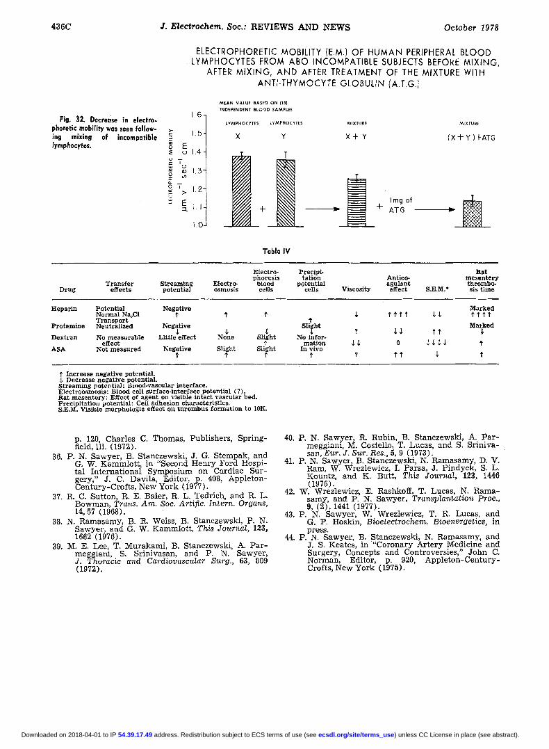
Fig. 31b. ATG further enhances the reduction of EM of ceils as described in Fig. 25a. With rats with known compatible and in. compatible strains, compatibility was predicted by no change in electrophoretic mobility of the mixed lymphocytes when compared to the unmixed lymphocytes.
C. A. Fenstermaker, J. Colloid Interlace Sci., G6, 557 (1976).
23. B. W. Morrissey, Ann. N.Y. Acad. Sci., 283, 50 (1977).
24. E. W. Merrill and E. W. Saltzman, in "Vascular Grafts," P. N. Sawyer and M. J. Kaplitt, Edi- tors, p. 119, Appleton-Century-Crofts, New York (1977).
25. L. Vroman, A. L. Adams, M. Klings, G. C. Fischer, P. C. Munoz, and R. P. Solensky, Ann. N.Y. Acad. Sci., 283, 65 (1977).
26. V. A. DePalma, R. E. Baier, and V. L. Gott, National Heart, Lung and Blood Institute, Report No. NO1-HV-3-2953-2 (1977).
27. H. J. Weiss, H. R. Baumgartner, T. B. Tschopp, and V. T. Turitto, Ann; N.Y. Acad. Sci., 283, 293 (1977).
28. P. N. Sawyer, K. T. Wu, S. A. Wesolowski, W. H. Brattain, and P. J. Boddy, Nat. Acad. Sci., 53, 294 (1965).
29. P. N. Sawyer and S. A. Wesolowsld, Ann. Surg., 1G3, 34 (1961).
30. P. N. Sawyer and J. W. Pate, Surgery, 34, 491 (1953).
31. S. Srinivasan and P. N. Sawyer, in "Electrochemi- cal Bioscience and Bioengineering," H. T. Silver- man, I. F. Miller, and A. J. Salkind, Editors, p. 17, The Electrochemical Society Softbound Sympo- sium Series, Princeton, N.J. (1973).
32. P. N. Sawyer, B. Stanczewski, S. J. Sivakoff, T. R. Lucas, and D. Kirschenbaum, Trans. Am. Soc. Artific. Intern. Organs, 23, 288 (1977).
33. P. N. Sawyer and S. Srinivasan, in "Artificial Heart Program Conference, Proceedings," Ruth Johnson Hegyeli, Editor, chap. 22, p. 243, U.S. Commerce Department Publications (1969).
34. S. Srinivasan and P. N. Sawyer, in "Clean Sur- faces: Their Preparation and Characterization for Interracial Studies," G. Goldfinger, Editor, chap. 11, p: 195, Marcel Dekker, Inc., New York (1970).
35. P. N. Sawyer, M. Costello, and S. Srinivasan, in "Prosthetic Replacement of the Aortic Valve," L. R. Sauvage, R. F. Viggers, K. Berger, S. B. Robel, P. N . Sawyer, and S. J. Wood, Editors,
Fig. 31a. Electrophoretic mobil- ity of mixed human erythrocytes is markedly reduced if the eryth- rocytes are from ABO incompati- ble donors.
) unless CC License in place (see abstract). ecsdl.org/site/terms_use address. Redistribution subject to ECS terms of use (see 54.39.17.49Downloaded on 2018-04-01 to IP
436C d. Electrochem. Soc.: REVIEWS AND NEWS October 1978
Fig. 32. Decrease in electro. phoretic mobility was seen follow- ing mixing of incompatible lymphocytes.
ELECTROPHORETIC MOBILITY (I:.M.) OF HUMAN PERIPHERAL BLOOD LYMPHOCYTES FROM ABO INCOMPATIBLE SUBJECTS BEFORE MIXING,
AFTER MIXING, AND AFTER TREATMENT OF THE MIXTURE WITH ANTI-THYMOCYTE GLOBUUN (A.T.GI
1.6-
1,5- ..%
I 8 Er. 4_
o ~ 1.3
~,~ 1.2- , j
~ E. ~ 1 . 1 -
i.O-
MEAN VALUE BASED ON (15) INDEPENDENT BLGOD SAMPLES
LYMPHOCYTES LYMPHOCYTES MIXTURE MIXTURE
X Y X+ Y (X+Y)+ATG
I mg of + ATG ~-
Table IV
Transfer Streaming Electro- Drug effects potential osmosis
Electro- Precipi- Rat phoresis tat ion Antico- mesentery
blood potentiaI agulant thrombo- cells cells Viscosity effect S.E.M.* sis t ime
Heparin Potential Negative Normal Na, CI t t Transport
Protamine Neutralized Negative $ $ Dextran No measurable Little effect None
effect ASA Not measured Negative Slight
t t
Marked t J, t t t t $$ t t t t
t g Marked $ Sliht ~. $ $ t t $ Slight No infor-
t mation $ $ O $ $ $ $ t Slight In vivo
t t ? t t $ t
I Increase negative potential. Decrease negative potential.
Streaming potential: ~iood-vascular interface. Electroosmosis: Blood cell surface-interface potential (?). Rat mesentery: Effect of agent on visible intact vascular bed. Precipi tat ion potential: Ceii adhesion characteristics. S.E.M. Visible morphologie effect on thrombus formation to 10K.
p. 120, Charles C. Thomas, Publishers, Spring- field, i l l (1972).
36. P. N. Sawyer, B. Stanczewski, J. G. Stempak, and G. W. Kammlott , in "Second Henry Ford Hospi- tal In te rna t iona l Symposium on Cardiac Sur- gery," J. C. Davila, Ed i to r , p. 408, Appleton- Century-Crofts , New York (1977).
37. R. C. Sutton, R. E. Baler, R. L. Tedrich, and R. L. Bowman, Trans. Am. Soc. Artific. Intern. Organs, 14, 57 (1968).
38. N. Ramasamy, B. R. Weiss, B. Stanczewski, P. N. Sawyer, and G. W. Kammlott , This Journal, 123, 1662 (1976).
39. M. E. Lee, T. Murakami, B. Stanczewski, A. Pa r - megglam, S. Srinivasan, and P. N. Sawyer, J. Thoracic and Cardiovascular Surg., 63, 809 (1972).
40. P. N. Sawyer, R. Rubin, B. Stanezewski, A. Par - meggiani, l~L Costello, T. Lueas, and S. Sr in iva- san, Eur. J. Sur. Res., 5, 9 (1973).
41. P. N. Sawyer, B. Stanczewski, N. Ramasamy, D. V. Ram, W. Wrezlewicz, I. Parsa, J. Pindyck, S. L. Kountz, and K. Butt, This Journal, 123, 1446 (1976).
42. W. Wrezlewicz, E. Rashkoff, T. Lucas, N. Rama- samy, and P. N. Sawyer, Transplantation Prec., 9, (2), 1441 (1977).
43. P. N. Sawyer, W. Wrezlewicz, T. R. Lucas, and G. P. Hoskin, Bioelectrochem. Bioenergetics, in press.
44. P. N. Sawyer, B. S~anczewski, N. Ramasamy, and J. S. Keates, in "Coronary Ar tery Medicine and Surgery, Concepts and Controversies," John C. Norman, Editor, p. 920, Apple ton-Century- Crofts, New York (1975).
) unless CC License in place (see abstract). ecsdl.org/site/terms_use address. Redistribution subject to ECS terms of use (see 54.39.17.49Downloaded on 2018-04-01 to IP
Related Documents