1 Application of Electroactive Polymers to Cardiovascular Flows Dave Morgan Department of Mechanical Engineering, Concordia University, Montreal, Canada Abstract The ability of electroactive polymers (EAPs) to replicate the function of biological muscles, including large actuation strain, makes them an ideal choice for actuators to be used in vivo, as well as in cardiac simulators. Certain types of EAPs require a low activation energy (1-2 volts) and thrive in the wet and saline environment of the inside of the human body and, in particular, around the myocardium of the heart. Completely implantable cardiac assist devices are presented, which would reduce potential infection and increase patient mobility. EAPs can also form a substrate to which cardiac tissues can attach themselves.

Welcome message from author
This document is posted to help you gain knowledge. Please leave a comment to let me know what you think about it! Share it to your friends and learn new things together.
Transcript
1
Application of Electroactive Polymers to Cardiovascular Flows
Dave Morgan
Department of Mechanical Engineering, Concordia University, Montreal, Canada
Abstract
The ability of electroactive polymers (EAPs) to replicate the function of biological muscles, including
large actuation strain, makes them an ideal choice for actuators to be used in vivo, as well as in cardiac
simulators. Certain types of EAPs require a low activation energy (1-2 volts) and thrive in the wet and
saline environment of the inside of the human body and, in particular, around the myocardium of the
heart. Completely implantable cardiac assist devices are presented, which would reduce potential
infection and increase patient mobility. EAPs can also form a substrate to which cardiac tissues can
attach themselves.

2
1. Introduction
This paper will examine the application of electroactive polymers (EAP) to cardiac flows. The importance
of understanding and being able to repair damaged parts of the heart is underscored by the staggering
cost of cardiovascular disease and stroke in the United States, estimated at over US$300 billion
(Shahinpoor, 2009). The treatments vary from repairing damaged valves to complete transplants, and
are often time sensitive, with the added possibility that the patient’s body will reject the new heart at a
rate of 50% (Shahinpoor, 2009).
EAPs are polymers that, when electrically stimulated, can bend, stretch or contract. The ability of EAPs
to replicate the function of biological muscles (also referred to as biomimetic), including large actuation
strain (Bar-Cohen, 2002), makes them an ideal choice for actuators to be used in vivo, as well as in
cardiac simulators. For the purpose of this paper, the manner in which the EAPs are manufactured will
not be discussed.
There are two primary categories of EAP, depending on their activation mechanism: electronic and ionic
(Bar-Cohen, 2002). The former is driven by Coulomb forces and activated by a DC voltage that, while
applied, can cause the material to maintain its displacement, whereas the latter uses the diffusions of
ions and consists of two electrodes and an electrolyte. The activation energy for each type of EAP is
different. In some cases, electronic EAPs can require activation fields greater than 100 V/µm, whereas
ionic EAPs require as little as 1-2 V (Bar-Cohen, 2002).
The response times of EAPs are also of great importance when dealing with the heart; a response that is
insufficient to drive a heart at normal biological is impractical. An advantage of the electronic EAPs is
that while they require high voltages, they exhibit a response time on the magnitude of milliseconds

3
(Bar-Cohen, 2004). Conversely, ionic EAPs require a low voltage, but possess a slower response time, on
the order of fractions of a second.
One other consideration for the choice of EAP is the environmental conditions in which they operate.
Electronic EAPs can work in air, but ionic EAPs, which include gels and polymer-metal composites, “rely
on ion and solvent transport to effect volume change; they are therefore ideal for operation in biofluids”
(Smela, 2003).
2. Current Research
At present, there is research being conducted at various institutions to develop a form of electroactive
polymer suitable for use with the heart. This chapter will present some of the works in this field.
2.1. Heart Compression / Assist Device
As mentioned, the possibility of a patient rejecting a heart transplant is 50%. Instead of replacing a weak
or defective heart, Shahinpoor proposes a minimally invasive device that is implantable and can assist a
weak heart as seen in Figure 1. This system would be comprised of multiple fingers (number 3 in Figure
1) that can selectively assist the ventricles, and would be actuated by soft ionic polymeric artificial
muscles that “thrive in the wet and saline environment of the inside of the human body and, in
particular, around the myocardium of the heart” (Shahinpoor, 2009). The muscles themselves have been
developed, and are composed of ionic polymer-metal nanocomposites (IPMNC). That is, if IMPNC are
used as sensors or actuators, they can used to actuate or sense at the nanometer level with small
applied voltages, on the order of micro-volts (Shahinpoor, 2009). However, the heart assist device is still
in the development phase.
4
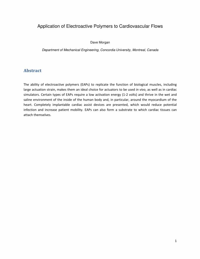
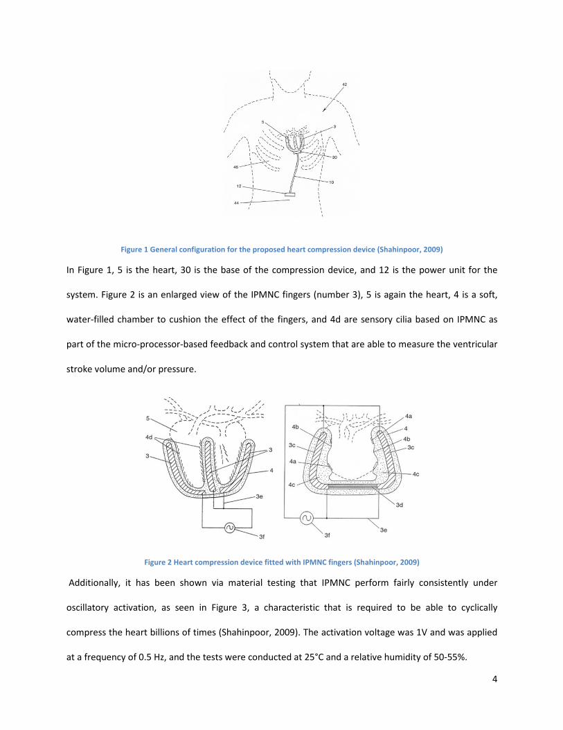
Figure 1 General configuration for the proposed heart compression device (Shahinpoor, 2009)
In Figure 1, 5 is the heart, 30 is the base of the compression device, and 12 is the power unit for the
system. Figure 2 is an enlarged view of the IPMNC fingers (number 3), 5 is again the heart, 4 is a soft,
water-filled chamber to cushion the effect of the fingers, and 4d are sensory cilia based on IPMNC as
part of the micro-processor-based feedback and control system that are able to measure the ventricular
stroke volume and/or pressure.
Figure 2 Heart compression device fitted with IPMNC fingers (Shahinpoor, 2009)
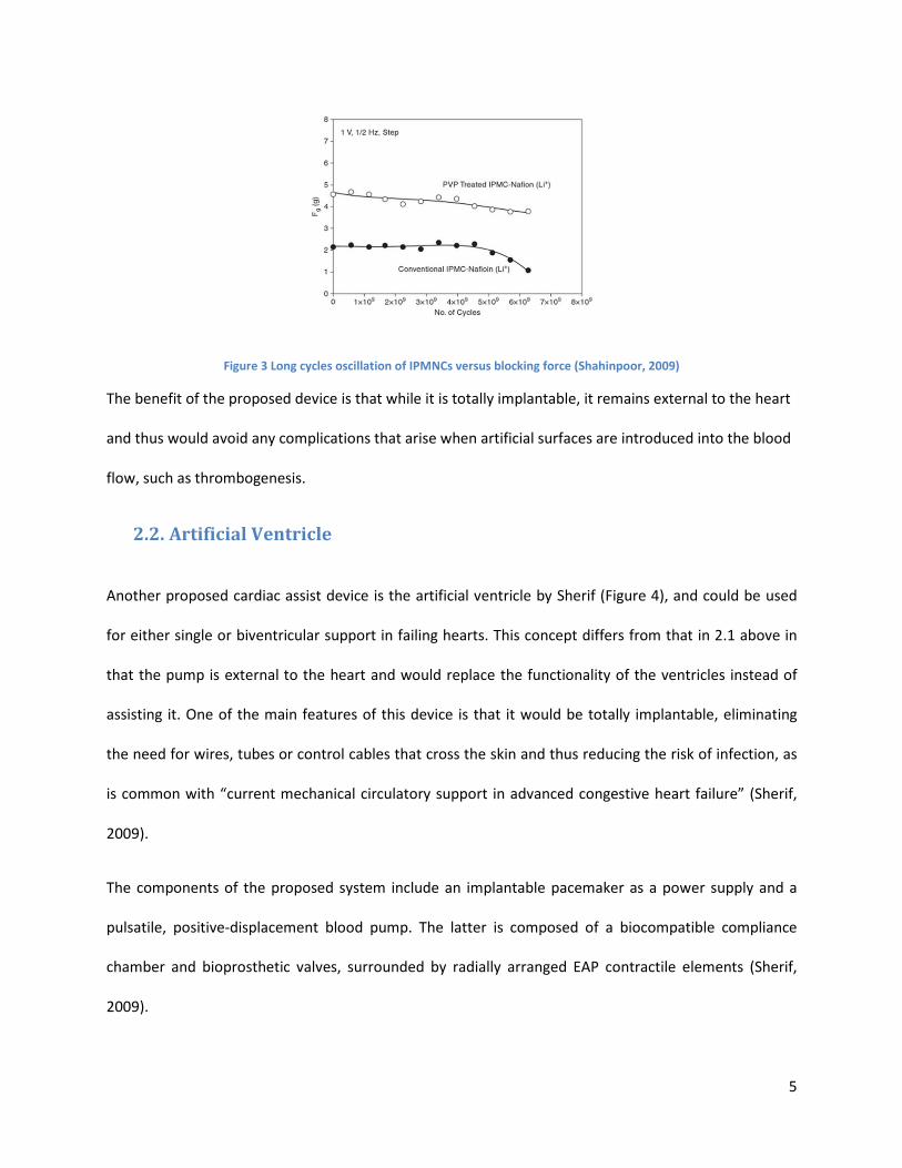
Additionally, it has been shown via material testing that IPMNC perform fairly consistently under
oscillatory activation, as seen in Figure 3, a characteristic that is required to be able to cyclically
compress the heart billions of times (Shahinpoor, 2009). The activation voltage was 1V and was applied
at a frequency of 0.5 Hz, and the tests were conducted at 25°C and a relative humidity of 50-55%.
5
Figure 3 Long cycles oscillation of IPMNCs versus blocking force (Shahinpoor, 2009)
The benefit of the proposed device is that while it is totally implantable, it remains external to the heart
and thus would avoid any complications that arise when artificial surfaces are introduced into the blood
flow, such as thrombogenesis.
2.2. Artificial Ventricle
Another proposed cardiac assist device is the artificial ventricle by Sherif (Figure 4), and could be used
for either single or biventricular support in failing hearts. This concept differs from that in 2.1 above in
that the pump is external to the heart and would replace the functionality of the ventricles instead of
assisting it. One of the main features of this device is that it would be totally implantable, eliminating
the need for wires, tubes or control cables that cross the skin and thus reducing the risk of infection, as
is common with “current mechanical circulatory support in advanced congestive heart failure” (Sherif,
2009).
The components of the proposed system include an implantable pacemaker as a power supply and a
pulsatile, positive-displacement blood pump. The latter is composed of a biocompatible compliance
chamber and bioprosthetic valves, surrounded by radially arranged EAP contractile elements (Sherif,
2009).
6
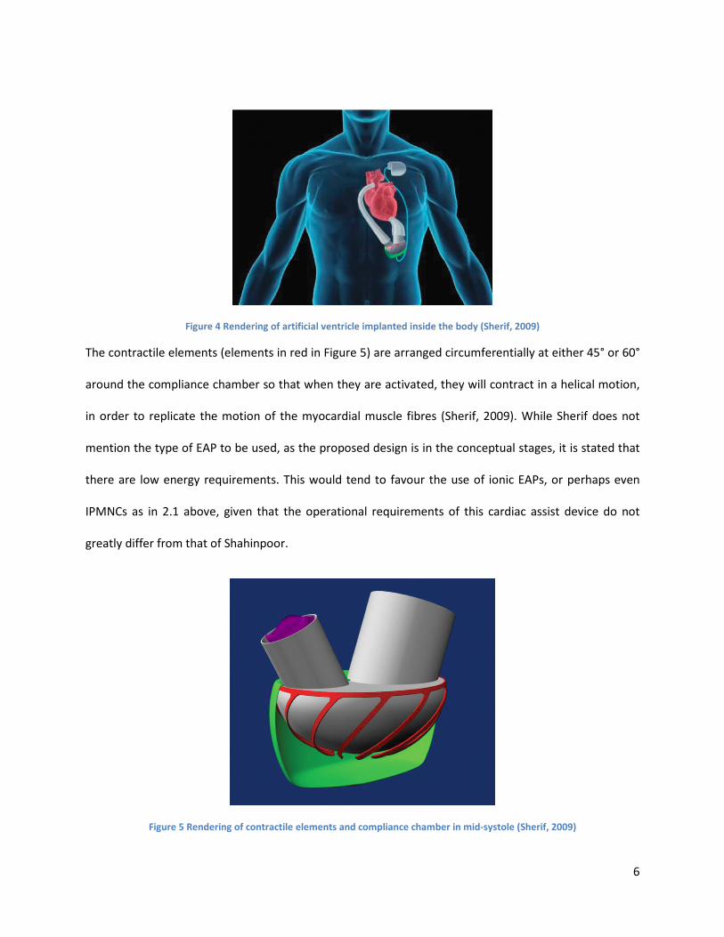
Figure 4 Rendering of artificial ventricle implanted inside the body (Sherif, 2009)
The contractile elements (elements in red in Figure 5) are arranged circumferentially at either 45° or 60°
around the compliance chamber so that when they are activated, they will contract in a helical motion,
in order to replicate the motion of the myocardial muscle fibres (Sherif, 2009). While Sherif does not
mention the type of EAP to be used, as the proposed design is in the conceptual stages, it is stated that
there are low energy requirements. This would tend to favour the use of ionic EAPs, or perhaps even
IPMNCs as in 2.1 above, given that the operational requirements of this cardiac assist device do not
greatly differ from that of Shahinpoor.
Figure 5 Rendering of contractile elements and compliance chamber in mid-systole (Sherif, 2009)

7
Figure 6 shows the effect of the EAP contracting on the compliance chamber, after it has received a
signal from the pacemaker; blood would then be ejected from the chamber and into the arterial side of
the circulation. This pacemaker would operate as a permanent pulse generator, and also be able to
automatically regulate the pumping rate depending on the physiological needs of the patient (Sherif,
2009).
Figure 6 Cut-out rendering of closed inlet valve and the compliance chamber in the late systole (Sherif, 2009)
Despite the device still being in the conceptual stage, the design presents several merits, such as
reduced risk of infection and increased patient mobility. The design is also conducive for use in cardiac
simulators, as it can theoretically reproduce pulsatile flow and remove the need for bulky pumps and
plumbing.
Work by Hanson et al. is focused on the development of a control strategy for a similar cardiac assist
device, one that consists of “an artificial muscle wrap consisting of contractile bands” (Hanson, et al.,
2005). They have developed a simulator to test the response of their model, and have determined that
an impedance control strategy will work to control the assist device as it can “provides an assistance
level that automatically adjusts as appropriate to the natural ability of the heart” (Hanson, et al., 2005).

8
Both the work of Sherif and Hanson et al. contribute to the feasibility of using EAP to provide varying
levels of assistance to a damaged heart.
2.3. Biocompatible EAP & Tissue Engineering
The work of Bidez et al. explores another use of EAPs, which are the potential surfaces and matrices for
cell and tissue growth. They have investigated both the “adhesion and proliferation properties of H9c2
cardiac myoblasts on a conductive polyaniline substrate” (Bidez, et al., 2006) and found that E-PANi
forms of polyaniline were biocompatible and also maintained their electrical conductivity. E-PANi is the
conductive salt form of polyaniline, which is a commercially available polymer with electroactive
properties. As seen in Figure 7, the percent of H9c2 attachment on E-PANi differs statistically by
approximately 7% from the tissue-culture-treated polystyrene (TCP) control (Bidez, et al., 2006).
Figure 7 H9c2 cell adhesion on PANi, E-PANi and TCP controls (Bidez, et al., 2006)

9
Additionally, in recent in vivo studies, polyaniline did not result in inflammation in the rodent subjects,
which suggests that there is the possibility of good bio-compatibility (Bidez, et al., 2006). Combined with
the work of Bidez et al., it can be seen that polyaniline is a suitable substrate for potential cardiac tissue
engineering.
In an article from the Journal of the Royal Society Interface, Ravichandran et al. further support the
potential of using H9c2 cardiac cells by presenting a more recent work by Bidez et al. This work shows
that by using conducting polymer scaffolds (a substrate of EAP on which tissue attaches itself), “H9c2
cardiac cell viability was preserved and that biocompatibility was enhanced” (Ravichandran, et al.,
2010).
3. Conclusion and Future Work
It has been shown that electroactive polymers lend themselves to use in the cardiovascular system. The
power requirements of EAPs are relatively low (as little as 1-2 volts), and the power plants for such
systems could likely exist in vivo as well as in vitro for simulators and testing. Despite the fact that much
of the work is still theoretical, the potential for EAPs to provide assistance to cardiac flows as external
compression devices remains. EAPs have also been shown to be biocompatible, and indeed can form a
substrate for cardiac tissue growth, which could one day form the basis for replacement sections of the
heart. They also lend themselves to use in cardiac simulators as the driving force for the simulated
heart, instead of pumps and complicated plumbing, and it has been shown that a suitable control
strategy exists.

10
4. References
Bar-Cohen, Y. (2002). Electro-active polymers: current capabilities and challenges. Paper 4695-02,
Proceedings of the SPIE Smart Structures and Materials Symposium. San Diego: SPIE.
Bar-Cohen, Y. (2004, December). Electroactive Polymers (EAP). Retrieved March 22, 2011, from
Electrochemistry Encyclopedia: http://electrochem.cwru.edu/encycl/art-p02-elact-pol.htm
Bidez, P. R., Macdiarmid, A. G., Venancio, E. C., Wei, Y., Lelkes, P. I., & Li, S. (2006). Polyaniline, an
electroactive polymer, supports adhesion and proliferation of cardiac myoblasts. Journal of Biomaterials
Science, Polymer Edition, 17(1-2), 199–212.
Hanson, B., Richardson, R., Davies, G., Watterson, K., Levesley, M., & Walker, P. (2005). Control of a Non-
Blood Contacting Cardiac Assist Device. Biomedical Engineering (pp. 679-684). Innsbruck: ACTA Press.
Ravichandran, R., Sundarrajan, S., Venugopal, J., Mukherjee, S., & Ramakrishna, S. (2010). Applications
of conducting polymers and their issues in biomedical engineering. 7(Supplement 5).
Shahinpoor, M. (2009). Implantable Heart-Assist and Compression Devices Employing an Active Network
of Electrically-Controllable Ionic Polymer-Metal Nanocomposites. In F. Carpi, & É. Smela (Eds.),
Biomedical Applications of Electroactive Polymer Actuators (pp. 137-160). Chichester, United Kingdom:
John Wiley & Sons Ltd.
Sherif, H. M. (2009). The artificial ventricle: A conceptual design for a novel mechanical circulatory
support system. Minimally Invasive Therapy & Allied Technologies, 18(3), 178-180.
Smela, E. (2003, March 17). Conjugated Polymer Actuators for Biomedical Applications. Advanced
Materials, 15(6), 481-494.
Related Documents


![synthesis of semiconducting polymers for possible application in [autosaved]](https://static.cupdf.com/doc/110x72/55656de8d8b42a7b518b4b89/synthesis-of-semiconducting-polymers-for-possible-application-in-autosaved.jpg)


