Appendix D.2 Acute RELs and toxicity summaries using the previous version of the Hot Spots Risk Assessment guidelines (OEHHA 1999) SUBSTANCE PAGE Acrylic Acid .................................................................................................................................... 3 Ammonia......................................................................................................................................... 8 Benzene ......................................................................................................................................... 17 Benzyl Chloride ............................................................................................................................ 26 Carbon Disulfide ........................................................................................................................... 31 Carbon Monoxide ......................................................................................................................... 40 Carbon Tetrachloride .................................................................................................................... 46 Chlorine......................................................................................................................................... 53 Chloroform.................................................................................................................................... 62 Chloropicrin .................................................................................................................................. 68 Metallic Copper And Copper Compounds ................................................................................... 73 1,4-Dioxane................................................................................................................................... 79 Epichlorohydrin ............................................................................................................................ 84 Ethylene Glycol Monobutyl Ether ................................................................................................ 89 Ethylene Glycol Monoethyl Ether ................................................................................................ 95 Ethylene Glycol Monoethyl Ether Acetate ................................................................................. 101 Ethylene Glycol Monomethyl Ether ........................................................................................... 106 Hydrogen Chloride...................................................................................................................... 111 Hydrogen Cyanide ...................................................................................................................... 118 Hydrogen Fluoride ...................................................................................................................... 130 Hydrogen Selenide ...................................................................................................................... 137 Hydrogen Sulfide ........................................................................................................................ 143 Isopropyl Alcohol ....................................................................................................................... 152 Methanol ..................................................................................................................................... 157 Methyl Bromide .......................................................................................................................... 165 Methyl Chloroform ..................................................................................................................... 174 Methyl Ethyl Ketone ................................................................................................................... 179 Methylene Chloride .................................................................................................................... 186 Nitric Acid .................................................................................................................................. 192 Nitrogen Dioxide ........................................................................................................................ 200 Ozone .......................................................................................................................................... 212 Perchloroethylene ....................................................................................................................... 219 Phenol ......................................................................................................................................... 225 Phosgene ..................................................................................................................................... 231 Propylene Oxide.......................................................................................................................... 238 Sodium Hydroxide ...................................................................................................................... 242 Styrene ........................................................................................................................................ 247 Sulfates........................................................................................................................................ 257 Sulfur Dioxide............................................................................................................................. 263 Sulfuric Acid and Oleum ............................................................................................................ 270 Toluene ....................................................................................................................................... 276 Appendix D2 1

Welcome message from author
This document is posted to help you gain knowledge. Please leave a comment to let me know what you think about it! Share it to your friends and learn new things together.
Transcript

Appendix D.2 Acute RELs and toxicity summaries using the previous version of the Hot Spots Risk Assessment guidelines (OEHHA 1999)
SUBSTANCE PAGE Acrylic Acid .................................................................................................................................... 3 Ammonia......................................................................................................................................... 8 Benzene ......................................................................................................................................... 17 Benzyl Chloride ............................................................................................................................ 26 Carbon Disulfide ........................................................................................................................... 31 Carbon Monoxide ......................................................................................................................... 40 Carbon Tetrachloride .................................................................................................................... 46 Chlorine......................................................................................................................................... 53 Chloroform .................................................................................................................................... 62 Chloropicrin .................................................................................................................................. 68 Metallic Copper And Copper Compounds ................................................................................... 73 1,4-Dioxane ................................................................................................................................... 79 Epichlorohydrin ............................................................................................................................ 84 Ethylene Glycol Monobutyl Ether ................................................................................................ 89 Ethylene Glycol Monoethyl Ether ................................................................................................ 95 Ethylene Glycol Monoethyl Ether Acetate ................................................................................. 101 Ethylene Glycol Monomethyl Ether ........................................................................................... 106 Hydrogen Chloride...................................................................................................................... 111 Hydrogen Cyanide ...................................................................................................................... 118 Hydrogen Fluoride ...................................................................................................................... 130 Hydrogen Selenide ...................................................................................................................... 137 Hydrogen Sulfide ........................................................................................................................ 143 Isopropyl Alcohol ....................................................................................................................... 152 Methanol ..................................................................................................................................... 157 Methyl Bromide .......................................................................................................................... 165 Methyl Chloroform ..................................................................................................................... 174 Methyl Ethyl Ketone ................................................................................................................... 179 Methylene Chloride .................................................................................................................... 186 Nitric Acid .................................................................................................................................. 192 Nitrogen Dioxide ........................................................................................................................ 200 Ozone .......................................................................................................................................... 212 Perchloroethylene ....................................................................................................................... 219 Phenol ......................................................................................................................................... 225 Phosgene ..................................................................................................................................... 231 Propylene Oxide.......................................................................................................................... 238 Sodium Hydroxide ...................................................................................................................... 242 Styrene ........................................................................................................................................ 247 Sulfates ........................................................................................................................................ 257 Sulfur Dioxide ............................................................................................................................. 263 Sulfuric Acid and Oleum ............................................................................................................ 270 Toluene ....................................................................................................................................... 276
Appendix D2 1

Triethylamine .............................................................................................................................. 285 Vanadium Pentoxide ................................................................................................................... 289 Vinyl Chloride ............................................................................................................................ 295 Xylenes ....................................................................................................................................... 303
Appendix D2 2

ACUTE TOXICITY SUMMARY
ACRYLIC ACID
CAS Registry Number: 79-10-7 I. Acute Toxicity Summary (for a 1-hour exposure) Inhalation reference exposure level 6,000 µg/m³ Critical effect(s) nasal irritation Hazard Index target(s) Respiratory System; Eyes II. Physical and Chemical Properties (HSDB, 1994) Description colorless liquid Molecular formula C3H4O2 Molecular weight 72.06 Density 1.0497 g/cm3 @ 20°C (liquid) Boiling point 141°C Melting point 14°C Vapor pressure 52 mm Hg @ 20°C Flashpoint 54°C, open cup Explosive limits unknown Solubility soluble in benzene and acetone; miscible with water,
alcohol and several ethers. Odor threshold 0.06 ppm-1.0 ppm Odor description acrid, irritating Metabolites carbon dioxide and a short chain fatty acid (possibly
3-hydroxypropionic acid) Conversion factor 1 ppm in air = 2.95 mg/m³ III. Major Uses and Sources The most common industrial production process is the oxidation of acrolein, which is then further oxidized to acrylic acid. Acrylic acid is used in the manufacture of plastics, molding powder for signs, construction units, decorative emblems and insignias, emulsion in polymers, paint formulations, leather finishing and paint coatings. IV. Acute Toxicity to Humans Acrylic acid vapors have been reported to cause nasal and eye irritation in workers, although no concentrations were given in these reports (ACGIH 1986, 1991). Contact with the liquid may produce skin and eye burns and blindness.
Appendix D2 3 Acrylic Acid

Predisposing Conditions for Acrylic Acid Toxicity Medical: Persons with severe uncorrected vision or chronic lung disease may be at increased
risk for the adverse effects of acrylic acid (HSDB, 1994). Chemical: Acrylic acid is a skin sensitizing agent as determined by the guinea pig maximization
test, but not by the Draize test (ACGIH, 1991). This finding may indicate a sensitizing potential of acrylic acid in humans.
V. Acute Toxicity to Laboratory Animals Inhalation exposure of rats to 2,000 ppm (6,000 mg/m³) acrylic acid for 4 hours resulted in no mortality (Carpenter et al., 1974). All animals (6) died at twice this concentration (4,000 ppm). An inhalation LC50 of 1,200 ppm (3,500 mg/m³) acrylic acid was determined for rats exposed for 4 hours (Majka et al., 1974). In rats exposed to acrylic acid aerosol for 30 minutes, the LC50 is 8,612 ppm (25,400 mg/m³) and the LC01 is 1,203 ppm (3,550 mg/m³) (Hagan, 1988). For a 60-minute exposure the LC50 is 3,750 ppm (11,100 mg/m³) and the LC01 is 2,180 ppm (6,430 mg/m³); for a 2-hour exposure, the LC50 is 2,502 ppm (7,381 mg/m³) and the LC01 is 928 ppm (2,740 mg/m³). Treatment related signs of toxicity included eye squinting, lacrimation, rhinorrhea, salivation, gasping, difficulty in breathing and corneal opacities. In addition to these signs, following the 2-hour exposure, rales, loss of righting reflex, ataxia, lethargy and prostration were reported. Rats exposed to acrylic acid vapors ranging from 928 to 2,142 ppm (2,740 to 6,319 mg/m³) for 60 minutes showed signs similar to the animals exposed at the same concentrations of aerosol. However, unlike the aerosol, recovery was more rapid and no deaths occurred following exposure to vapors. A single 5-hour exposure to 6,000 ppm (18,000 mg/m³) acrylic acid in rats produced nasal and eye irritation, respiratory difficulties, unresponsiveness and death in 1 of 4 animals (Gage, 1970). An autopsy revealed lung hemorrhage and degeneration of the liver and kidney tubules. Four 6-hour exposures to 1,500 ppm (4,400 mg/m³) in 8 animals produced nasal discharge, lethargy, weight loss and congested kidneys. Nasal irritation, lethargy and reduced weight gain were observed after twenty 6-hour exposures at 300 ppm (900 mg/m³). Histopathological examination showed no damage to tissues. No toxic signs were observed in 8 rats exposed 20 times to 80 ppm (240 mg/m³) for 6 hours. Histological examinations were performed in ten rats and ten mice exposed to acrylic acid vapor 6 hours per day, 5 days per week for 2 weeks at 25, 75, and 225 ppm (74, 220, and 664 mg/m³) (Miller et al., 1981). At 25 ppm, very slight olfactory tissue effects (unspecified) were observed in mice. Slight focal degeneration of the olfactory tissue without metaplasia was found in mice at 75 ppm. No adverse effects were noted in rats at this dose. Labored breathing and apparent nasal irritation during exposure occurred in mice exposed to 225 ppm. Slight focal squamous metaplasia of the olfactory epithelium was observed in both rats and mice at this concentration. The investigators noted that since rodents are obligate nasal breathers, irritation of the nasal mucosa was likely to be pronounced in these animals. Majka et al. (1974) observed that exposure of rats to 240 ppm (710 mg/m³) acrylic acid, 4 hours per day for 5 weeks resulted in
Appendix D2 4 Acrylic Acid

reduced body weight gain, increased reticulocyte count, and irritation with irreversible changes to the skin and eyes. The instillation of 0.5 ml of a 1% solution of acrylic acid caused severe irritation and corneal burns in the eyes of rabbits (Union Carbide Corp., 1977). VI. Reproductive or Developmental Effects Four groups of 5 female rats were injected intraperitoneally with 2.5, 4.7, or 8 mg/kg body weight acrylic acid three times on days 5, 10, and 15 of gestation (Singh et al., 1972). Skin abnormalities (hemangiomas) were observed in the offspring of animals from the two highest dose groups. Skeletal abnormalities and embryotoxicity were observed in the litter from the highest dose group. DePass et al. (1983) conducted a one-generation reproduction study in rats. Animals were exposed to doses ranging from 83 to 750 mg/kg/day acrylic acid in the drinking water throughout gestation and lactation. No statistically significant changes in reproductive indices were observed. Pregnant rats were exposed via inhalation to concentrations of acrylic acid ranging from 40 to 360 ppm (120-1,060 mg/m³) on days 6 through 15 of gestation (Klimisch et al., 1983). Decreased body weight and feed consumption were observed in the dams exposed to 120 or 360 ppm acrylic acid. No embryotoxic or teratogenic effects were observed. VII. Derivation of Acute Reference Exposure Level and Other Severity Levels
(for a 1-hour exposure) Reference Exposure Level (protective against mild adverse effects): 2 ppm (6,000 µg/m³) Study Gage, 1970 Study population groups of 4-8 rats Exposure method inhalation Critical effects nasal irritation LOAEL 300 ppm NOAEL 80 ppm Exposure duration 6 hours/day (on 20 occasions) Extrapolation to 1 hour Cn * T = K, where n = 2 (ten Berge et al., 1986) Extrapolated 1 hour concentration 200 ppm (802ppm * 6 h = C2 * 1 h) LOAEL uncertainty factor 1 Interspecies uncertainty factor 10 Intraspecies uncertainty factor 10 Cumulative uncertainty factor 100 Reference Exposure Level 2 ppm (6 mg/m³; 6,000 µg/m³)
Appendix D2 5 Acrylic Acid

Level Protective against Severe Adverse Effects No recommendation is made due to the limitations of the database. Slight focal degeneration of the olfactory tissue was observed in mice exposed to 75 ppm (225 mg/m³) acrylic acid, 6 hours per day for 10 days (Miller et al., 1981). An ERPG-2 of 50 ppm (150 mg/m³) was recommended based on this study (AIHA, 1991). The AIHA document stated that strong odors and slight eye irritation may be present at this level but that escape would not be impaired. The document incorrectly states that no effects were seen at 75 ppm. Because no safety factors were used in the derivation of this value, it should be reevaluated. Therefore, no recommendation can be made. Level Protective against Life-threatening Effects No recommendation is made due to the limitations of the database. NIOSH does not list an IDLH for acrylic acid. An acute inhalation study in rats determined a 1-hour LC01 of 2,180 ppm (6,430 mg/m³) acrylic acid aerosol (Hagan, 1988). In addition, no deaths were observed in rats exposed for 6 hours per day for 4 days to 1,500 ppm (4,400 mg/m³) acrylic acid (Gage, 1970). AIHA (1991) derived an ERPG-3 value of 763 ppm (2,250 mg/m³). Because the ERPG-3 value was based on a personal communication (Hagan, 1988) with little supporting documentation, no recommendation can be made. VIII. References (ACGIH) American Conference of Governmental Industrial Hygienists. Documentation of the Threshold Limit Values and Biological Exposure Indices. 6th ed. Cincinnati (OH): ACGIH; 1991. p. 26-29. (ACGIH) American Conference of Governmental Industrial Hygienists. Documentation of the Threshold Limit Values and Biological Exposure Indices. 5th ed. Cincinnati (OH): ACGIH; 1986. p. 14. (AIHA) American Industrial Hygiene Association. Emergency response planning guidelines. Akron (OH): AIHA; 1991. Carpenter CP, Weil CS, Smyth HF Jr. Range finding toxicity data: list VIII. Toxicol Appl Pharmacol 1974; 28:313-319. Depass LR, Woodside MD, Garman RH, Weil C.S. Subchronic and reproductive toxicology studies on acrylic acid in the drinking water of the rat. Drug Chem Toxicol 1983; 6:1-20. Gage JC. The subacute inhalation toxicity of 109 industrial chemicals. Br J Ind Med 1970; 27:1-18. Hagan JV. Acrylic acid concentration time mortality response inhalation toxicity study in rats. Personal communication. Rohm and Haas Co. 1988. [Cited in AIHA, 1991.]
Appendix D2 6 Acrylic Acid

(HSDB) Hazardous Substances Data Bank. National Library of Medicine, Bethesda, Maryland (CD-ROM version). Denver (CO): Micromedex, Inc.; 1993. (Edition expires 11/31/93). (IARC) International Agency for Research on Cancer. IARC monograph on the evaluation of the carcinogenic risk of chemicals to man: some monomers, plastics and synthetic elastomers, and acrolein. Vol. 19. Lyon: IARC; 1979. p. 47-71. Klimisch HJ, Merkle J, Hildebrand B. Prenatal toxicity study of acrylic acid after inhalation in Sprague-Dawley rats. Vol. 1. #83RC-1002. Dept of Toxicology. Ludwigshafen/Rhine: BASF Aktiengesellschaft; 1983. [Cited in U.S.EPA, 1990.] Majka J, Knobloch K, Stetkiewicz J. Evaluation of acute and subacute toxicity of acrylic acid. Med Pracy (Polish) 1974; 25(5):427-435. Miller RR, Ayres JA, Jersey GC, McKenna MJ. Inhalation toxicity of acrylic acid. Fundam Appl Toxicol 1981; 1:271-277. Singh AR, Lawrence WH, Autian J. Embryonic-fetal toxicity and teratogenic effects of a group of methacrylate esters in rats. J Dent Res 1972; 51:1632-1638. Ten Berge WF, Zwart A, Appelman LM. Concentration-time mortality response relationship of irritant and systemically acting vapours and gases. J Hazard Mater 1986; 13:301-309. Union Carbide Corp. Toxicology studies-acrylic acid, glacial. 2 May. Industrial Medicine and Toxicology Department. New York: Union Carbide; 1977. [Cited in IARC, 1979.] U.S.EPA. Acrylic acid. Reference Concentration for chronic inhalation exposure (RfC). U.S.EPA Integrated Risk Information Service (IRIS); 1990.
Appendix D2 7 Acrylic Acid

ACUTE TOXICITY SUMMARY
AMMONIA
(anhydrous ammonia, aqueous ammonia)
CAS Registry Number: 7664-41-7 I. Acute Toxicity Summary (for a 1-hour exposure) Inhalation reference exposure level 3,200 µg/m³ Critical effect(s) eye and respiratory irritation Hazard Index target(s) Eyes; Respiratory System II. Physical and Chemical Properties (HSDB, 1994 except as noted) Description colorless gas Molecular formula NH3 Molecular weight 17.03 Density 0.695 g/L @ 25°C Boiling point -33.5°C Melting point -77.7°C Vapor pressure 6,460 mm Hg @ Flashpoint unknown Explosive limits unknown Solubility very soluble in water, alcohol and ether Odor threshold 17 ppm (geometric mean) (AIHA, 1989) Odor description sharp and very irritating Metabolites unknown Conversion factor 1 ppm = 0.71 mg/m³ @ 25°C III. Major Uses or Sources Ammonia is a strongly alkaline chemical which is widely used in industry as a feed stock for nitrogen based chemicals such as fertilizers, plastics and explosives (ATSDR, 1990). Nationwide, ammonia is the third most common chemical to be released accidentally (U.S.EPA, 1989). Among hazardous material incidents such as intentional and threatened releases, those involving ammonia are the sixth most common. The volatility of ammonia, along with its common method of storage as large quantities under pressure, results in a potential for release of large amounts of ammonia gas (NRC, 1987).
Appendix D2 8 Ammonia

IV. Acute Toxicity to Humans Ammonia vapors cause irritation of the eyes and respiratory tract. Higher concentrations cause conjunctivitis, laryngitis, and pulmonary edema, possibly accompanied by a feeling of suffocation (OSHA, 1989). Contact with the skin causes burns and blistering. The eye is especially sensitive to alkali burns. Ammonia combines with moisture in the eyes and mucous membranes to form ammonium hydroxide. Ammonium hydroxide causes saponification and liquefaction of the exposed, moist epithelial surfaces of the eye and can easily penetrate the cornea and damage the iris and the lens (CCOHS, 1988; Way et al., 1992). Damage to the iris may eventually lead to cataracts (CCOHS, 1988). Inhalation exposure to ammonia may result in an increase in systemic arterial blood pressure (Zitnik et al., 1969). Exposure can also cause a decrease in minute ventilation volume (Cole et al., 1977). Ammonia gas is especially irritating to upper respiratory passages, which prompts exposed victims to attempt escape from the fumes as quickly as possible. MacEwen and Vernot (1972) described pulmonary edema as the most frequent cause of death in humans exposed to ammonia. Silverman and coworkers (1949) exposed 7 volunteers to 500 ppm (355 mg/m³) ammonia for 30 minutes using an oral-nasal mask. Symptoms due to ammonia inhalation varied widely among the 7 subjects. All seven subjects experienced upper respiratory irritation, which was graded as severe in 2 subjects. Only 2 subjects were able to continue nasal breathing throughout the 30 minute exposure. Reactions included irritation of the nose and throat, hypoesthesia of the exposed skin, and lacrimation. In two subjects, the nasopharyngeal irritation persisted for 24 hours after the exposure. One of the 7 subjects was only exposed to ammonia for 15 minutes rather than the full 30 minutes. The reason for this deviation in the exposure regimen was not given. In a previous experiment, brief exposure to 1,000 ppm reportedly resulted in immediate coughing in human subjects. Ferguson and coworkers (1977) used six human subjects to demonstrate that a tolerance to ammonia exposure of 100 ppm (71 mg/m³) can be developed with a two-to-three week inurement period during which volunteers were exposed to lesser concentrations. The results tended to support the belief that persons with no recent history of ammonia exposure are more sensitive to the irritating effects than those who are acclimated to the noxious gas. Verberk (1977) exposed sixteen subjects, eight previously exposed and eight naive, for two hours to ammonia in concentrations of 50, 80, 110, and 140 ppm (36, 57, 78, 99 mg/m³). The naive group could not tolerate 140 ppm for two hours and had several complaints during exposure to 110 ppm for 1 hour. None of the subjects in the study demonstrated a decrease in measured pulmonary function tests, including vital capacity, forced expiratory volume (1 second), and forced inspiratory volume (1 second), following ammonia exposure. The results showed a greater sensitivity to ammonia exposure for the naive group for responses of smell, eye irritation, cough, general discomfort, headache, and irritation of the chest. At the end of the initial 30 minutes of the 2-hour exposure period, nuisance level smell, eyes, nose, or throat irritation, or cough urge were reported by 7 of 16 (44%), 9 of 16 (56%), 12 of 16 (75%), or 15 of 16 (94%) individuals at concentrations of 50, 80, 110, or 140 ppm, respectively.
Appendix D2 9 Ammonia

MacEwen et al. (1970) exposed groups of 5 and 6 human subjects to respective ammonia concentrations of 30 and 50 ppm (21 and 36 mg/m³). The volunteers subjectively rated irritation for the 10-minute exposures. No moderate or higher irritation was discerned by the group at the lower exposure level; however, 4 of the 6 subjects rated the 10 minute exposure at 50 ppm as causing moderate irritation. The Industrial Bio-Test Laboratories (1973) evaluated ten human subjects for the irritation threshold of ammonia from exposures to ammonia gas at four different concentrations: 32, 50, 72, and 134 ppm (23, 36, 51, and 95 mg/m³). Irritation was taken to be any annoyance to the eyes, nose, mouth, throat, or chest which persisted throughout the 5-minute exposure period. At 72 ppm three subjects experienced eye irritation, two had nasal irritation, and three had throat irritation. At 134 ppm, five of the ten subjects experienced lacrimation and eye irritation, seven complained of nasal irritation, eight had throat irritation, and one experienced chest irritation. The authors only used 5-minute exposure durations; and it is possible that irritation symptoms could have developed with longer exposure durations at the lower exposures. The authors discounted the significance of nasal dryness reported at the two lowest levels. Douglas and Coe (1987) determined a lachrymatory threshold of 55 ppm for ammonia following approximately 15 second exposures of volunteers via tight-fitting goggles. The threshold for bronchoconstriction, determined as a 20% increase in airway resistance, was slightly higher at 85 ppm following 10 breaths of ammonia via mouthpiece. Estimates of odor thresholds for ammonia vary from 0.04-103 ppm (0.03-73 mg/m³) (Ferguson et al., 1977; Henderson and Haggard, 1943; Ruth, 1986). Near the odor threshold, persons exposed to ammonia can experience annoyance and believe the odor to be a nuisance. Exposure to ammonia may result in an exacerbation of preexisting asthma. Shim and Williams (1986) surveyed 60 patients with a history of asthma worsened by certain odors. Nearly 80% of these patients claimed to have an exacerbation of asthma following exposure to household cleaners containing ammonia. Predisposing Conditions for Ammonia Toxicity Medical: Persons with asthma and other respiratory ailments including underlying
cardiopulmonary disease (Shim and Williams, 1986) and persons with no tolerance, developed from recent exposures to ammonia (Ferguson et al. 1977), may be more susceptible to the toxic effects of ammonia.
Chemical: Chronic high dose aspirin therapy and therapy with valproic acid elevate blood
ammonia levels (Reprotext, 1999). V. Acute Toxicity to Laboratory Animals The pulmonary lesions observed following acute, potentially lethal, inhalation of ammonia are similar in man and experimental animals (Withers, 1986; Payne et al., 1990). Male rats and mice were determined to be more sensitive to the lethal effects of ammonia than the females of either species (Appelman et al., 1982; Stupfel et al., 1971).
Appendix D2 10 Ammonia

Several animal lethality studies published dose-response data from which the MLE05 (maximum likelihood estimate corresponding to 5% lethality) and BC05 (benchmark dose at the 95% lower confidence interval of the MLE05) could be determined (see Table 1). Table 1. Animal Lethality Effective and Benchmark Dose Levels for Ammonia Reference Species Time (min) MLE05 (ppm) BC05 (ppm)
MacEwen & Vernot (1972) rat 60 5,999 4,908
MacEwen & Vernot (1972) mouse 60 4,006 3,406
Kapeghian et al. (1982) mouse 60 3,664 3,366
Appelman et al. (1982) rat (10)* 11,862 9,950
Appelman et al. (1982) rat (20)* 13,010 10,206 Appelman et al. (1982) rat (40)* 11,137 4,881
Silver and McGrath (1948) mouse (10)* 2,846 2,298
* Exposure time was adjusted to 60 min using a modification of Haber’s Law to facilitate
comparisons of MLE05 and BC05 values. Exponent n = 2 was determined, based on Appelman et al. (1982) rat lethality data, by varying the term in a log-normal probit analysis (Crump, 1984; Crump and Howe, 1983).
Appelman et al. (1982) observed signs of restlessness, wet noses and nasal discharge in rats immediately after the start of inhalation exposure to ammonia. Mouth breathing and dyspnea occurred soon after the start of exposure. Eye discharge began about 30 minutes into the exposure, and signs of eye irritation after 60 minutes of exposure. Dose versus exposure time varied from 7,000 ppm (4,970 mg/m³) for 60 minutes to 26,850 ppm (19,064 mg/m³) for 10 minutes. VI. Reproductive or Developmental Toxicity There are no confirmed studies which show conclusively that reproductive or developmental toxicity can be linked experimentally or epidemiologically to ammonia exposure (Reprotext, 1999).
Appendix D2 11 Ammonia

VII. Derivation of Acute Reference Exposure Level and Other Severity Levels (for a 1-hour exposure)
Reference Exposure Level (protective against mild adverse effects): 3,200 µg/m³
Study Industrial Biotest Laboratories, 1973; MacEwen et al., 1970; Silverman et al., 1949; Verberk, 1977
Study population humans Exposure method inhalation Critical effects eye and respiratory irritation LOAEL varied (see Section IV of text) NOAEL varied (see Section IV of text) Exposure duration varied (see Section IV of text) Extrapolated 1 hour concentration 13.6 ppm (BC05) LOAEL uncertainty factor not needed in BC approach Interspecies uncertainty factor 1 Intraspecies uncertainty factor 3 Cumulative uncertainty factor 3 Reference Exposure Level 4.5 ppm (3.2 mg/m³; 3,200 µg/m³) The exposure concentrations from the 4 studies were adjusted to 1-hour durations using the formula Cn x T = K (Table 2). The value for the exponent n was empirically derived from the preceding data sets. The value of n (in the formula Cn x T = K) was sequentially varied for the log-normal probit relationship analysis. Using a chi-square analysis, a value of n = 4.6 was found to be the best fit. The REL was calculated by a benchmark concentration (BC) approach using a log-normal probit analysis (Crump and Howe, 1983; Crump. 1984). The 95% lower confidence limit of the concentration expected to produce a response rate of 5% is defined as the BC05. The maximum likelihood estimate for a 5% response was 20.1 ppm and the 95% LCL on this value (BC05) for ammonia from this analysis was 13.6 ppm.
Response rate MLE (ppm) 95% LCL (ppm) 1% 13.4 7.8 5% 20.1 13.6 (BC05)
An uncertainty factor (UF) of 3 was used to account for intraspecies variation in the human population. Refer to section IX of this toxicity summary for the graphic representation of benchmark dose derivation.
Appendix D2 12 Ammonia

Table 2. Ammonia, Human Irritation, 60 Minute Exposures (adjusted), ppm
Study Concentration
32 30 50 50 72 50 80 134 110 140 500
Exposure Time (min.)
5 10 5 10 5 120 120 5 60 60 30
60 min. adjusted Concentration
19 20 29 34 42 43 69 78 95 120 430
Response 0/10 0/5 0/10 4/6 3/10 7/16 9/16 8/10 12/16 15/16 7/7 Study 2 3 2 3 2 1 1 2 1 1 4
Table adapted from: (1) Verberk, 1977; (2) Industrial Biotest Laboratories, 1973; (3) MacEwen et al., 1970; (4) and Silverman et al., 1949. The two lowest concentrations were combined for the log-probit analysis since this improved the fit of the data. Level Protective against Severe Adverse Effects Exposure to 140 ppm (99.4 mg/m³) ammonia was considered ‘unbearable’ resulting in termination of exposure by all of 8 non-expert student volunteers after 30 to 75 minutes (Verberk, 1977). These exposures were tolerated for the full 2-hour exposure period by all 8 expert volunteers who were familiar with irritant vapors. Based on these findings in which ammonia inhalation resulted in a subjective response of panic or the need in naive subjects to take shelter, a 2-hour NOAEL of 110 ppm and a 30-minute LOAEL of 140 ppm were noted. Short exposures to ammonia did not result in increased nasal resistance of atopic subjects when compared to nonatopic subjects (McLean et al., 1979). The non-expert group was considered to be more like the general public in their response. The final value to protect against severe adverse effects from ammonia exposure is thus 110 ppm (78 mg/m³). Level Protective against Life-threatening Effects Kapeghian et al. (1982) determined a 1-hour LC50 of 4,230 ppm and a 1-hour no observed lethality level of 3,440 ppm in male mice. The MLE05 and BC05 were estimated as 3,664 and 3,366 ppm (Table 1), respectively. The report by Kapeghian et al. (1982) provides one of the most detailed exposure and monitoring methods used for ammonia among the various animal lethality reports reviewed. In addition, a sensitive experimental animal species was used for the experiments (MacEwen & Vernot, 1972). An uncertainty factor of 1 was applied to account for animal to human extrapolation since (1) the BC accounts for some degree of variation and (2) OEHHA’s comparison of human irritation thresholds with concentrations lethal to mice suggests humans are not more susceptible than mice to ammonia toxicity. That is, in examining the Verberk (1977) study and comparing it to the mouse lethality study, additional uncertainty factors to the mouse study results in a concentration below the Verberk (1977) human study. A factor of 10 was applied to account for individual human variation. The cumulative uncertainty factor was 10. The resulting level for ammonia to protect against life-threatening effects is 340 ppm (240 mg/m³). VIII. References
Appendix D2 13 Ammonia

Agency for Toxic Substances and Disease Registry (ATSDR). Toxicological profile for ammonia. Atlanta (GA): ATSDR, US Public Health Service; 1990.
American Industrial Hygiene Association (AIHA). Odor thresholds for chemicals with established occupational health standards. Akron (OH): AIHA; 1989. p. 13.
Appelman LM, Ten Berge WF, Reuzel PGJ. Acute inhalation toxicity study ammonia in rats with variable exposure periods. Am Ind Hyg Assoc J 1982; 43:662-665.
Canadian Centre for Occupational Health and Safety (CCOHS). Chemical Hazard Summary, Ammonia. CCOHS number C88-1E. ISBN 1988; 0-660-12738-5.
Cole T, Cotes J, Johnson G, de V Martin H, Reed J, Saunders M. Ventilation, cardiac frequency and pattern of breathing during exercise in men exposed to o-chlorobenzylidene malononitrile (CS) and ammonia gas in low concentrations. Qtrly J Exp Phys 1977; 62:341-351.
Crump KS, Howe R. Probit-A computer program to extrapolate quantile animal toxicological data to low doses. Ruston (LA): KS Crump & Company, Inc.; 1983.
Crump K. A new method for determining allowable daily intakes. Fundam Appl Toxicol 1984; 4:860-866.
Douglas RB, Coe JE. The relative sensitivity of the human eye and lung to irritant gases. Ann Occup Hyg 1987; 31(2):265-267.
Ferguson WS, Koch WC, Webster LB, Gould JR. Human physiological response and adaptation to ammonia. J Occup Med 1977; 19(5):319-326.
Hazardous Substances Data Bank (HSDB). National Library of Medicine, Bethesda (MD) (CD-ROM version) Denver (CO): Micromedex, Inc.; 1994. (Edition expires 11/31/94).
Henderson Y, Haggard HW. Noxious gases. 2nd ed. New York: Reinhold Publishing Corporation; 1943.
Industrial Bio-Test Laboratories, Inc. Report to International Institute of Ammonia Refrigeration: Irritation threshold evaluation study with ammonia. IBT No 1973; 663-03161 (March 23, 1973).
Kapeghian J, Mincer H, Jones A, Verlanger A, Water I. Acute inhalation toxicity of ammonia in mice. Bull Environ Contam Toxicol 1982; 29:371-38.
MacEwen J, Vernot E. Annual Technical Report. AMRL-TR-72-62, (NTIS AD755-358) Wright-Patterson Air Force Base (OH): Aerospace Medical Research Laboratory, Toxic Hazards Research Unit; 1972.
MacEwen J, Theodore J, Vernot EH. Human exposure to EEL concentration of monomethylhydrazine. AMRL-TR- 1970; 70-102, 23. Wright-Patterson Air Force Base (OH): SysteMed Corp.; 1970.
Appendix D2 14 Ammonia
McLean JA, Mathews KP, Solomon WR, Brayton PR, Bayne NK. Effect of ammonia on nasal resistance in atopic and nonatopic subjects. Ann Otol Rhinol Laryngol 1979; 88(2 Pt 1):228-34.
National Research Council (NRC). Committee on Toxicology. Emergency and Continuous Exposure Guidance Levels for selected airborne contaminants. Vol. 7. Washington (DC): National Academy Press; 1987. p. 7-15.
Occupational Safety and Health Administration (OSHA). Industrial exposure control strategies and technologies for OSHA regulated hazardous substances. Vol. 1. Cincinnati: OSHA; 1989.
Payne MP, Delic J, Turner RM. 1990. Ammonia. In: Toxicology of substances in relation to major hazards. London: HMSO, Crown Publishing; 1990. p. 1-17.
Reprotext ® System. Dabney BJ, editor. Denver (CO): Micromedex, Inc.; 1999. (Edition expires 1/31/1999).
Ruth JH. Odor thresholds and irritation levels of several chemical substances: a review. Am Ind Hyg Assoc J 1986; 47(3):A142-A151.
Shim C, Williams MH. Effect of odors in asthma. Am J Med 1986; 80:18-22.
Silver SD, McGrath FP. A comparison of acute toxicities of ethylene imine and ammonia in mice. J Ind Hyg Toxicol 1948;30(1):7-9.
Silverman L, Whittenberger JL, Muller J. Physiological response of man to ammonia in low concentrations. J Ind Hyg Toxicol 1949;31:74-78.
Stupfel M, Roman R, Magnier M, Powers J. Comparative acute toxicity in male and female mice of some air pollutants. Automobile gas, nitrogen oxides, sulphur dioxide, ozone, ammonia, carbon dioxide. C R Soc Biol 1971;165:1869-1872.
United States Environmental Protection Agency (U.S.EPA). Why accidents occur: insights from the accidental release information program OSWER-89-008.1. Washington: U.S.EPA; 1989.
Verberk M. Effects of ammonia in volunteers. Int Arch Occup Health 1977;39:73-81.
Way J, Baxter L, McGuinn D, Zitzer A, Petrikovics I. Ch. 9. Occupational hazards and ocular toxicity. In: Chiou GCY, editor. Ophthalmic toxicology. Raven Press; 1992.
Withers J. The lethal toxicity of ammonia. A report to the MHAP. North Western Branch Papers, No. 1. Institution of Chemical Engineers; 1986. p. 6.1-6.27.
Zitnik R, Burchell H, Shepard J. Hemodynamic effects of inhalation of ammonia in man. Am J Cardiol 1969;24:187-190.
Appendix D2 15 Ammonia

IX. Graphic Representation of Benchmark Concentration Determination
Appendix D2 16 Ammonia

ACUTE TOXICITY SUMMARY
BENZENE
(benzol; benzole; cyclohexatriene)
CAS Registry Number: 71-43-2
I. Acute Toxicity Summary (for a 6-hour exposure) Inhalation reference exposure level 1,300 µg/m³
Critical effect(s) Reproductive/developmental toxicity Hazard Index target(s) Reproductive/developmental; Immune System;
Hematologic System; II. Physical and Chemical Properties (HSDB, 1994 except as noted) Description colorless liquid Molecular formula C6H6 Molecular weight 78.1 Density 0.879 g/cm3 @ 25°C Boiling point 80.1°C Melting point 5.5°C Vapor pressure 100 mm Hg @ 26.1°C Flashpoint -11°C Explosive limits upper = 8.0% by volume in air lower = 1.4% by volume in air Solubility soluble in ethanol, chloroform, ether, carbon
disulfide, acetone, oils, and glacial acetic acid; slightly soluble in water
Odor threshold 0.875 ppm (2.8 mg/m³) (Haley, 1977) Odor description sweet Metabolites hydroquinone, quinone, catechol, phenol Conversion factor 1 ppm = 3.24 mg/m³ III. Major Uses or Sources Benzene has been widely used as a multipurpose organic solvent. This use is now discouraged due to its high toxicity. Present uses include benzene as a raw material in the synthesis of styrene, phenol, cyclohexane, aniline, and alkyl benzenes and in the manufacture of various plastics, resins, and detergents. Synthesis of many pesticides and pharmaceuticals also involves benzene as a chemical intermediate (HSDB, 1994). Benzene is emitted in large quantities from refineries and petroleum storage facilities. The tire industry and shoe factories use benzene extensively. Annual demand in the U.S. was estimated to be 6 million tons in 1990 (HSDB, 1994).
Appendix D2 17 Benzene

IV. Acute Toxicity to Humans Deaths from acute exposure to benzene are often related to physical exertion and release of epinephrine with subsequent cardiac failure. Frequently, the person trying to rescue a collapsed victim will die during the effort of lifting the unconscious person (HSDB, 1994). Anesthesia may develop at concentrations above 3,000 ppm (9,600 mg/m³) (Reprotext, 1993). At exposures of greater than 1,000 ppm (3,200 mg/m³) (duration unspecified), CNS symptoms include giddiness, euphoria, nausea, and headaches; heightened cardiac sensitivity to epinephrine-induced arrhythmias may develop (Snyder, 1987). These effects may be accompanied by symptoms of mild irritation to the eyes and mucous membranes. Acute hemorrhagic pneumonitis is highly likely if benzene is aspirated into the lung (HSDB, 1994). Respiratory tract inflammation, pulmonary hemorrhages, renal congestion, and cerebral edema have been observed at autopsy in cases of acute benzene poisoning (IARC, 1987). In these cases, blood levels of 2 mg/ml benzene were not associated with hematological changes (Winek and Collom, 1971). Systemic poisoning by benzene can occasionally result in neuroretinal edema and in retinal and conjunctival hemorrhage (Grant, 1986). Additionally, petechial hemorrhages of the brain, pleura, pericardium, urinary tract, mucous membranes, and skin may occur in cases of fatal, acute benzene poisoning (Haley, 1977). Major concerns of systemic benzene toxicity include aplastic anemia and acute myelogenous leukemia (IARC, 1987; Reprotext, 1993). Both of these conditions are typically seen in the chronic and subchronic exposures, but may be of concern following acute exposures as well. Myeloid and erythroid components of the bone-marrow are specific targets of benzene toxicity, which leads to aplastic anemia (IARC, 1982). In men and women exposed to benzene for 4 hours, 46.9% of the inhaled dose was absorbed. Of this absorbed fraction, 30.1% was retained and 16.8% was excreted unchanged in the expired air (Nomiyama and Nomiyama, 1974). Most of the catechol and phenol metabolites are excreted within 24 hours in the urine, while hydroquinone requires 48 hours (Teisinger et al., 1952). Exposure at the odor threshold (0.875 ppm or 2.8 mg/m³) for a brief duration is reported to enhance the electropotential of the brain (Haley, 1977). Predisposing Conditions for Benzene Toxicity Medical: People with existing hematologic disorders and cellular anemias may be more
sensitive to the acute toxicity of benzene to the bone-marrow (Reprotext 1993, 1999). People with heart conditions may also be at increased risk for cardiac arrhythmias induced by exposure to high levels of benzene. Administration of epinephrine is known to potentiate the cardiac toxicity of benzene (Reprotext, 1993).
Females may be more sensitive to benzene toxicity than males due to higher average body fat content, which serves as a storage reservoir for the chemical
Appendix D2 18 Benzene
(Reprotext, 1993). Similarly, obese individuals of either sex may be more sensitive to benzene toxicity.
Chemical: Previous acute exposure to toluene inhibits benzene metabolism to toxic
metabolites, and may reduce toxicity (Reprotext, 1993). Consumption of ethanol potentiates the bone-marrow toxicity of inhaled benzene in mice (Baarson et al., 1982).
V. Acute Toxicity to Laboratory Animals The oral LD50 in rats is reported to be 3.4 g/kg in young rats and 4.9 g/kg in older rats (Kimura et al., 1971). Mortality was observed in 2 out of 10 rats exposed to 33,000 mg/m³ (10,300 ppm) for 12.5-30 minutes daily for either 1 or 12 days (IARC, 1982). A 4-hour LC50 of 13,700 ppm (43,800 mg/m³) was reported in female rats (IARC, 1982). An LCLo of 45,000 ppm (144,000 mg/m³) is reported in rabbits (RTECS, 1994). In mice, an LC50 of 9,800 ppm (31,400 mg/m³) is reported (RTECS, 1994). Leukopenia has been demonstrated to occur in rabbits exposed to 240 ppm (767 mg/m³) for 10 hours/day for 2 weeks (IARC, 1982). Brief inhalation of air saturated with benzene vapor (concentration unknown) resulted in ventricular extrasystole in cats and primates, with periods of ventricular tachycardia that occasionally terminated in ventricular fibrillation (Clayton and Clayton, 1981). An attempt by Nielsen and Alarie (1982) to determine the inhalation RD50 for benzene was not successful. These investigators showed that inhalation of 5,800 ppm (18,800 mg/m³) benzene in mice caused an increase in respiratory rate beginning at 5 minutes following onset of exposure. They speculated that the stimulation of respiratory rate resulted from the action of benzene on the central nervous system. In this study, benzene was not irritating to the upper airways of the animals. The pharmacokinetics of benzene in the rat reportedly follows a 2-compartment model. The rapid phase has an elimination half-life (t1/2) of 0.7 hours, and the t1/2 for the longer phase is 13.1 hours (Rickert et al., 1979). The long elimination half-life for benzene is due to the formation of catechol, quinone, and hydroquinone in the bone marrow. These reactive metabolites are not readily excreted, and are cytotoxic to the germinal cells in the bone marrow (Greenlee et al., 1981). A 3-compartment model was fitted to human data on benzene disposition and bone-marrow metabolism (Watanabe et al., 1994). The general relationship between cumulative quantity of metabolites produced and inhalation concentration was not linear, but was S-shaped, inflecting upward at low concentrations, and saturating at high concentrations. Mice, particularly the DBA/2 strain, are more sensitive to myelotoxicity from benzene than are rats or rabbits (IARC, 1982). Colony-forming unit cells (CFUs; leukocyte precursors) were depleted in bone-marrow cultures taken from mice exposed to 4,610 ppm (14,950 mg/m³) benzene for 8 hours. Recovery of CFUs was noted 7 days after exposure (IARC, 1982). In addition to myelotoxicity, acute exposure to benzene may disrupt erythropoiesis and result in genotoxicity. Erythropoiesis, as measured by uptake of radiolabeled iron in the bone-marrow,
Appendix D2 19 Benzene

has been shown to be inhibited by subcutaneous injection of 10 mmol/kg benzene in mice (Bolcsak and Nerland, 1983). Results from subacute exposures further illustrate the hematotoxic effects of benzene and the potential for immunotoxicity. Inhalation of 103 ppm (334 mg/m³) benzene for 6 hours/day for 7 days by mice caused decreased spleen and marrow cellularities and decreased spleen weights (Green et al., 1981). Benzene inhalation at concentrations of 0, 10, 30, 100, and 300 ppm (0, 32.4, 97.3, 324, and 973 mg/m³) for 6 hours/day for 5 days resulted in a decreased host-resistance to bacterial infection by Lysteria monocytogenes (Rosenthal and Snyder, 1985). The numbers of L. monocytogenes bacteria isolated from the spleen were increased in a dose-dependent manner on day 4 of infection. The total numbers of T- and B-lymphocytes in the spleen and the proliferative ability of the splenic lymphocytes were decreased in a dose-dependent manner by benzene exposures of 30 ppm (97.3 mg/m³) or greater. In this study, no decrement in host resistance or immune response was observed at 10 ppm (32 mg/m³) benzene. Later studies in mice have also shown that exposure to 10 ppm for a subacute duration does not significantly alter hematological parameters in blood, spleen, thymus, or bone marrow (Farris et al., 1996; 1997). Farris et al. (1997) reported the hematological consequences of benzene inhalation in B6C3F1 mice exposed to 1, 5, 10, 100, and 200 ppm benzene for 6 hr/day, 5 days/week for 1, 2, 4, or 8 weeks and a recovery group. There were no significant effects on hematopoietic parameters from exposure to 10 ppm benzene or less. Thus 10 ppm was a NOAEL for 1 week of exposure (and longer). Exposure to 100 and 200 ppm benzene reduced the number of total bone marrow cells, progenitor cells, differentiating hematopoietic cells, and most blood parameters. Replication of primitive progenitor cells in the bone marrow was increased during the exposure period as a compensation for the cytotoxicity. At 200 ppm, the primitive progenitor cells maintained an increased percentage of cells in S-phase through 25 days of recovery compared with controls. Inhalation of 3 ppm (9.6 mg/m³) benzene for 6 hours by rats resulted in a significant increase over controls in the frequency of sister chromatid exchanges in peripheral blood lymphocytes (Erexson et al., 1986). Evans et al. (1981) observed an increase in active behavior in the form of eating and grooming in mice following exposure to 300 ppm (960 mg/m³) benzene for 6 hours. VI. Reproductive or Developmental Toxicity Coate et al. (1984) exposed groups of 40 female rats to 0, 1, 10, 40, and 100 ppm (0, 3.24, 32.4, 129.6, or 324 mg/m³) benzene for 6 hours/day during days 6-15 of gestation. In this study, teratologic evaluations and fetotoxic measurements were done on the fetuses. A significant decrease was noted in the body weights of fetuses from dams exposed to 100 ppm (324 mg/m³). No effects were observed at a concentration of 40 ppm (129.6 mg/m³). Keller and Snyder (1986) reported that exposure of pregnant mice to concentrations as low as 5 ppm (16 mg/m³) benzene on days 6-15 of gestation (6 hr/day) resulted in bone-marrow
Appendix D2 20 Benzene

hematopoietic changes in the offspring that persisted into adulthood. However, the hematopoietic effects (e.g., bimodal changes in erythroid colony-forming cells) in the above study were of uncertain clinical significance. In a similar, later study, Keller and Snyder (1988) found that exposure of mice in utero to 20 ppm (64 mg/m³) benzene on days 6-15 of gestation resulted in neonatal suppression of erythropoietic precursor cells and persistent, enhanced granulopoiesis. This effect was considered significant bone-marrow toxicity by the authors. No hematotoxicity was seen in this study at 10 ppm (32 mg/m³). An exposure of 500 ppm (1,600 mg/m³) benzene through days 6-15 of gestation was teratogenic in rats while 50 ppm (160 mg/m³) resulted in reduced fetal weights on day 20 of gestation. No fetal effects were noted at an exposure of 10 ppm (Kuna and Kapp, 1981). An earlier study by Murray et al. (1979) showed that inhalation of 500 ppm benzene for 7 hours/day on days 6-15 and days 6-18 of gestation in mice and rabbits, respectively, induced minor skeletal variations. Tatrai et al. (1980) demonstrated decreased fetal body weights and elevated liver weights in rats exposed throughout gestation to 150 mg/m³ (47 ppm). VII. Derivation of Acute Reference Exposure Level and Other Severity Levels
(for a 1-hour exposure) Level protective against mild adverse effects: While benzene exposure results in decreased immune response and hematopoietic effects in laboratory animals following 5 day exposures, it was problematic to extrapolate from these repeated dose studies for these endpoints. Thus, no level protective against mild adverse effects for one-hour is being recommended. The REL is based on developmental toxicity, a severe adverse effect. Reference Exposure Level for a 6-hour exposure (Level Protective against Severe Adverse Effects): 1,300 µg/m3 Because of the uncertainty of extrapolating from repeated exposures to a one-hour concentration, we have chosen to use a single day exposure in the reproductive studies with no time extrapolation as an REL. In the case of benzene, the REL is for a 6-hour exposure.
Study Coate et al., 1984; (supported by Kuna and Kapp, 1981; Keller and Snyder, 1988)
Study population pregnant female rats Exposure method inhalation of 0, 1, 10, 40, or 100 ppm Critical effects decreased fetal body weights LOAEL 100 ppm NOAEL 40 ppm Exposure duration 6 hours per day (for 5 days) LOAEL uncertainty factor 1 Interspecies uncertainty factor 10 Intraspecies uncertainty factor 10 Cumulative uncertainty factor 100 Reference Exposure Level 0.4 ppm (1.3 mg/m³; 1,300 µg/m³)
Appendix D2 21 Benzene

Pregnant female rats (40 per group) were exposed for 6 hours/day on days 6-15 of gestation to benzene concentrations of 0, 1, 10, 40, and 100 ppm (0, 3.24, 32.4, 129.6, and 324 mg/m³) (Coate et al., 1984). The mean fetal weights from the females treated with 100 ppm benzene were significantly decreased (p < 0.05) compared to controls. No teratogenic, fetotoxic, or maternally toxic effects were observed in rats exposed to 40 ppm (129.6 mg/m³) benzene or less. The 40 ppm (129.6 mg/m³) concentration is considered a NOAEL for reduced fetal weight. The value of 40 ppm for a 6-hour exposure was extrapolated to a 1-hour exposure using the equation Cn * T = k, where n = 2. The resulting 100 ppm extrapolated value was used to determine the level protective against severe adverse effects using uncertainty factors of 10 for intraspecies and 10 for interspecies variation. The level protective against severe adverse effects for benzene is therefore 1.0 ppm or 3.24 mg/m³. Kuna and Kapp (1981) found direct teratogenic effects measured as decreased crown-rump length, exencephaly, and angulated ribs in rats when pregnant females were exposed 6 hours/day during days 6-15 of gestation to a concentration of 500 ppm. In this study, a concentration of 50 ppm during gestation resulted in lower fetal weights measured on day 20 of gestation. No fetal effects were noted at an exposure of 10 ppm (32 mg/m³). Keller and Snyder (1988) reported a NOAEL of 10 ppm for developmental hematopoietic effects in mice. The highest reported NOAEL (i.e., 40 ppm) consistent with reported LOAEL values was chosen for the derivation of the Reference Exposure Level (severe adverse effect level, in this case) for benzene. Level Protective against Life-threatening Effects Svirbely et al. (1943) exposed mice for 7 hours to various benzene concentrations. They determined a NOAEL (0/18 animals) for lethality of 4,980 ppm and a LOAEL (3/18 animals) of 7,490 ppm. A benchmark concentration derived (BC05) using a log-normal model with these data is 5,650 ppm (MLE = 6,550 ppm). A life-threatening level was calculated using these data with an uncertainty factor of 30 (10 for individual variability, and 3 for interspecies uncertainty using the BC05 as the starting point for the calculation). The level protective against life-threatening effects is therefore 5,650 ppm ÷ 30 = 190 ppm (620 mg/m³). VIII. References Baarson KA, Snyder CA, Green JD, Akumar AS, Goldstein BD, Albert RE. The hematotoxic effects of inhaled benzene on peripheral blood, bone marrow, and spleen cells are increased by ingested ethanol. Toxicol Appl Pharmacol 1982;64:393-404. Bolcsak LE, Nerland DE. Inhibition of erythropoiesis by benzene and benzene metabolites. Toxicol Appl Pharmacol 1983;69:363-368. Clayton GD, Clayton FE. Industrial hygiene and toxicology. 3rd ed. revised. Vol. IIB. Toxicology. New York (NY): John Wiley and Sons; 1981. p. 3260-3283.
Appendix D2 22 Benzene

Coate WB, Hoberman AM, Durloo RS. Inhalation teratology study of benzene in rats. In: MacFarland HN, editor. Advances in modern environmental toxicology, Vol VI. Applied toxicology of petroleum hydrocarbons. Princeton (NJ): Princeton Scientific Publishers, Inc; 1984. p. 187-198. Cronkite EP, Drew RT, Inoue T, Hirabayashi Y, Bullis JE. Hematotoxicity and carcinogenicity of inhaled benzene. Environ Health Perspect 1989;82:97-108. Erexson GL, Wilmer JL, Steinhagen WH, Kligerman AD. Induction of cytogenetic damage in rodents after short-term inhalation of benzene. Environ Mutagen 1986;8:29-40. Evans HL, Dempster AM, Snyder CA. Behavioral changes in mice following benzene inhalation. Neurobehav Toxicol Teratol 1981;3:481-485. Farris GM, Robinson SN, Gaido KW, Wong BA, Wong VA, Leonard L, Shah R. Effects of low concentrations of benzene on mouse hematopoietic cells in vivo: a preliminary report. Environ Health Perspect 1996;104(6):1275-1276. Farris GM, Robinson SN, Gaido KW, Wong BA, Wong VA, Hahn WP, Shah R. Benzene-induced hematotoxicity and bone marrow compensation in B6C3F1 mice. Fundam Appl Toxicol 1997;36(2):119-129. Flury F. Toxicities in modern industry: pharmacological-toxicological aspects of intoxicants in modern industry (German). Arch Exp Pathol Pharmakol 1928;138:71. Gerarde HW. Benzene. In: Toxicology and biochemistry of aromatic hydrocarbons. Elsevier Monographs. Amsterdam, the Netherlands: Elsevier; 1960. Grant WM. Toxicology of the eye. Springfield (IL): CC Thomas; 1986. p. 140-141. Green JD, Snyder CA, LoBue J, Goldstein BD, Albert RE. Acute and chronic dose/response effect of benzene inhalation on the peripheral blood, bone marrow, and spleen cells of CD-1 male mice. Toxicol Appl Pharmacol 1981;59:204-214. Greenlee WF, Sun JD, Bus JS. A proposed mechanism of benzene toxicity: formation of reactive intermediates from polyphenol metabolites. Toxicol Appl Pharmacol 1981;59:187-195. Haley TJ. Evaluation of the health effects of benzene inhalation. Clin Toxicol 1977;11(5):531-548. (HSDB) Hazardous Substances Data Bank. National Library of Medicine, Bethesda, MD (CD-ROM version). Denver (CO): Micromedex, Inc.; 1994. (Edition expires 1/31/94).
Appendix D2 23 Benzene

(IARC) International Agency for Research on Cancer. IARC monographs on the evaluation of the carcinogenic risk of chemicals to humans. Vol. 29. Some industrial chemicals and dyestuffs. Lyon: IARC; 1982. p. 93-148. Keller KA, Snyder CA. Mice exposed in utero to low concentrations of benzene exhibit enduring changes in their colony forming hematopoietic cells. Toxicology 1986;42:171-181. Keller KA, Snyder CA. Mice exposed in utero to 20 ppm benzene exhibit altered numbers of recognizable hematopoietic cells up to seven weeks after exposure. Fundam Appl Toxicol 1988;10:224-232. Kimura ET, Ebert DM, Dodge PW. Acute toxicity and limits of solvent residue for sixteen organic solvents. Toxicol Appl Pharmacol, 1971;19:699-704. Kuna R., Kapp RW. The embryotoxic/teratogenic potential of benzene vapor in rats. Toxicol Appl Pharmacol 1981;57:1-7. Murray FJ, John JA, Rampy L., Kuna RA, Schwetz BA. Embryotoxicity of inhaled benzene in mice and rabbits. Am Ind Hyg Assoc J 1979;40:993-998. Nomiyama K, Nomiyama H. Respiratory elimination of organic solvents in man. Benzene, toluene, n-hexane, trichloroethylene, acetone, ethyl acetate and ethyl alcohol. Int Arch Arbeitsmed. 1974;32:85-91 Nielsen GD, Alarie Y. Sensory irritation, pulmonary irritation, and respiratory stimulation by alkyl benzene and alkylbenzenes: prediction of safe industrial exposure levels and correlation with their thermodynamic properties. Toxicol Appl Pharmacol 1982;65:459-477. (RTECS®) Registry of Toxic Effects of Chemical Substances. National Institute of Occupational Safety and Health, Cincinnati, OH (CD-ROM version). Denver (CO): Micromedex, Inc.; 1994. (Edition expires 1/31/94). Reprotext® System. Dabney BJ, editor. Denver (CO): Micromedex, Inc.; 1993. (Edition expires 11/31/93). Reprotext ® System. Dabney BJ, editor. Denver (CO): Micromedex, Inc.; 1999. (Edition expires 1/31/1999). Rickert D, Baker, TS, Bus JS, Barrow CS, Irons RD. Benzene disposition in the rat after exposure by inhalation. Toxicol Appl Pharmacol 1979;49:417-423. Rosenthal GJ, Snyder CA. Modulation of the immune response to Listeria monocytogenes by benzene inhalation. Toxicol Appl Pharmacol 1985;80:502-510. Snyder CA. Benzene, In: Snyder R, editor. Ethyl Browning’s toxicity and metabolism of industrial solvents. Amsterdam: Elsevier; 1987. p. 3-37.
Appendix D2 24 Benzene

Svirbely JL, Dunn RL, von Oettingen WF. The acute toxicity of vapors of certain solvents containing appreciable amounts of benzene and toluene. J Ind Hyg Toxicol 1943;25:366-373. Tatrai E, Ungvary GY, Hudak A, Rodics K, Lorincz M, Barcza GY. Concentration dependence of the embryotoxic effects of benzene inhalation in CFY rats. J Hyg Epidemiol Microbiol Immunol 1980;24(3):363-371. Teisinger J, Bergerova-Fiserova V, Kudrna J. The metabolism of benzene in man (Pol). Pracov Lek 1952;4:175-188. [Cited in International Agency for Research on Cancer (IARC) monographs. Vol. 29. 1987. p. 117.] Watanabe KH, Bois FY, Daisey JM, Auslander DM, Spear RC. Benzene toxicokinetics in humans: exposure of bone marrow to metabolites. Occup Environ Med 1994;51(6):414-420. Winek CL, Collom WD. Benzene and toluene fatalities. J Occup Med 1971;13:259-261. [Cited in International Agency for Research on Cancer (IARC) monographs. Vol. 29. 1987. p. 116.]
Appendix D2 25 Benzene

ACUTE TOXICITY SUMMARY
BENZYL CHLORIDE
(α-chlorotoluene, chloromethylbenzene, tolyl chloride)
CAS Registry Number: 100-44-7 I. Acute Toxicity Summary (for a 1-hour exposure) Inhalation reference exposure level 240 µg/m³ Critical effect(s) eye and nose irritation in rats and mice Hazard Index target(s) Eyes; Respiratory System II. Physical and Chemical Properties (HSDB, 1994 except as noted) Description colorless to slightly yellow liquid Molecular formula C7H7Cl Molecular weight 126.58 Density 1.1 g/cm3 @ 20°C Boiling point 179° C Melting point -43 to -48°C Vapor pressure 1 mm Hg @ 22°C Flashpoint 67°C, closed cup; 74°C, open cup Explosive limits upper = unknown lower = 1.1% by volume in air Solubility insoluble in water; miscible with most organic
solvents Odor threshold 0.041 ppm (240 µg/m³) (geometric mean)
(AIHA, 1989) Odor description pungent (AIHA, 1989) Metabolites benzyl mercapturic acid, benzoic acid Conversion factor 1 ppm = 5.2 mg/m³ @ 25°C III. Major Uses or Sources Benzyl chloride is a chemical intermediate in the manufacture of dyes, plasticizers, lubricants, gasoline additives, pharmaceuticals, tanning agents, and quaternary ammonium compounds (HSDB, 1994). Benzyl chloride can react with water or steam to produce corrosive and toxic fumes. It reacts vigorously with oxidizing materials, decomposes rapidly, and liberates heat and hydrochloric acid when exposed to all common metals, except lead and nickel. When heated, it may form phosgene (Hazardtext, 1993).
Appendix D2 26 Benzyl Chloride

IV. Acute Toxicity to Humans Benzyl chloride is extremely irritating to the eyes, nose, and throat, is a potent lacrimator, and is capable of causing pulmonary edema (Smyth, 1956). Exposure to 31 ppm (160 mg/m³) benzyl chloride for 5 minutes was reported to be unbearably irritating to the eyes and respiratory tract; a 5-minute exposure to 1.2-1.5 ppm (6-8 mg/m³) benzyl chloride resulted in “slight conjunctivitis” (Mikhailova, 1983). Skin burns or irritation may result from direct contact with vapors or liquid (Meditext, 1993). Human volunteers exposed to benzyl chloride vapor for a single breath reported that the odor was perceptible at 8 ppm (42 mg/m³), very unpleasant at 17 ppm (88 mg/m³), painfully strong at 37 ppm (190 mg/m³), and intolerable at 79 ppm (410 mg/m³) (Katz and Talbert, 1930). Occupational exposure to 2 ppm (10 mg/m³) benzyl chloride was reported to result in neurological symptoms and liver dysfunction; these effects most likely reflect chronic exposure, although the duration of exposure was not reported (Mikhailova, 1983). Little or no information was reported on the number of workers examined in the original studies cited, on the range of exposure, or on possible concomitant exposures. Predisposing Conditions for Benzyl Chloride Toxicity Medical: Those individuals with preexisting eye, skin, allergic, liver or kidney disease or
preexisting respiratory conditions including underlying cardiopulmonary disease may be more sensitive to the effects of benzyl chloride exposure (Reprotext, 1999).
Chemical: Persons exposed to other irritants might be more sensitive (Reprotext, 1999). V. Acute Toxicity to Laboratory Animals The 2-hour LC50 for benzyl chloride is reported as 0.39 mg/l (80 ppm) and 0.74 mg/l (150 ppm) in mice and rats, respectively (Mikhailova, 1965). The same study reports that rats and mice exposed to concentrations exceeding 0.1 mg/l (20 ppm) benzyl chloride for 2 hours exhibited irritation of the eyes, nose, and throat and decreased respiratory rate. Two cats exposed for 8 hours per day for 6 days to 95 ppm (500 mg/m³) benzyl chloride exhibited eye and respiratory irritation and decreased appetite (Wolf, 1912). VI. Reproductive or Developmental Toxicity No adverse reproductive effects were observed in rats administered 50 or 100 mg/kg/day benzyl chloride orally on days 6-15 of gestation (Skowronski and Abdel-Rahman, 1986). A non-statistically significant increase in sternebral defects was observed in the 100 mg/kg/day exposure group. No maternal toxicity was observed.
Appendix D2 27 Benzyl Chloride

VII. Derivation of Acute Reference Exposure Level and Other Severity Levels (for a 1-hour exposure)
Reference Exposure Level (protective against mild adverse effects): 46 ppb (240 µg/m³)
Study Mikhailova, 1965 Study population rats and mice Exposure method inhalation chamber Critical effects signs of irritation of eyes and nasal passages;
decreased respiratory rate LOAEL 20 ppm NOAEL not observed Exposure duration 2 hours Extrapolated 1 hr concentration 28 ppm (202 * 2 h = C2 * 1 h ) (see Table 12 for information on “n”) LOAEL uncertainty factor 6 Interspecies uncertainty factor 10 Intraspecies uncertainty factor 10 Cumulative uncertainty factor 600 Reference exposure level 46 ppb (240 µg/m³)
An animal study was used for the derivation of the REL because the available human data (Smyth, 1956; Mikhailova, 1983; Katz and Talbert, 1930) were not adequate for the determination of this level; the original human data were anecdotal and the exposure conditions were not well defined. Level Protective against Severe Adverse Effects No recommendation is made due to the limitations of the database. Level Protective against Life-threatening Effects No recommendation is made due to the limitations of the database. NIOSH lists an IDLH of 10 ppm. However, NIOSH admits: “Very little data are available on the acute effects of exposure to benzyl chloride.” NIOSH also states: “ACGIH (1971) reported that in 1 minute an exposure to 16 ppm is intolerable to man (Flury and Zernik, 1931). ILO (1972) reported that 20 ppm will render the atmosphere irrespirable in 1 minute. ILO (1971) reported that 50 to 100 mg/m³ (10 to 19 ppm) immediately causes weeping and twitching of the eyelids, while 160 mg/m³ (30 ppm) causes effects that are intolerable to the eyes and nasal mucous membranes. Based on this data, an IDLH of 10 ppm is assumed in order to avoid difficulties in escape in the event of respirator failure.” The level makes no allowance for sensitive individuals and therefore can not be recommended for use for the general public.
Appendix D2 28 Benzyl Chloride

VIII. References (ACGIH) American Conference of Governmental Industrial Hygienists. Benzyl chloride. In: Documentation of the threshold limit values for substances in workroom air. 3rd ed. Cincinnati (OH): ACGIH; 1971. p. 24. (ACGIH) American Conference of Governmental and Industrial Hygienists. Documentation of the Threshold Limit Values and Biological Exposure Indices. 6th ed. Cincinnati (OH): ACGIH, 1991. p. 132-136. (AIHA) American Industrial Hygiene Association. Odor thresholds for chemicals with established occupational health standards. Akron (OH): AIHA; 1989. p. 14. Flury F , Zernik F. Schadliche Gase - dampfe, nebel, rauch- und staubarten. Berlin, Germany: Verlag von Julius Springer; 1931. Hazard Management: HAZARDTEXT. Hall AH, Rumack BH, editors. TOMES® Information System. Denver (CO): Micromedex, Inc.; 1993. (Edition expires 11/31/93). (HSDB) Hazardous Substances Data Bank. National Library of Medicine, Bethesda (MD). (CD-ROM Version). Denver (CO): Micromedex, Inc.; 1994 (Edition expires 4/30/94). ILO. Benzyl chloride. In: Encyclopaedia of occupational health and safety. 2nd ed. Vol. I (A-K). Geneva, Switzerland: International Labour Office; 1971. p. 169-170. ILO. Toluene and derivatives. In: Encyclopaedia of occupational health and safety. Vol. II (L-Z). 2nd ed. Geneva, Switzerland: International Labour Office; 1972. p. 1414-1415. Katz SH, Talbert EJ. Intensities of odors and irritating effects of warning agents for inflammable and poisonous gases. Technical paper 480. US Department of Commerce, Bureau of Mines; 1930. p. 37 [cited in NIOSH, 1978.] Medical Management: MEDITEXT. Hall AH, Rumack BH, editors. TOMES Information System. Denver (CO): Micromedex, Inc.; 1993 (Edition expires 11/31/93). Mikhailova TV. Comparative toxicity of chlorine derivatives of toluene: benzyl chloride, benzal chloride and benzotrichloride. Fed Proc Trans Suppl 1965;24(5):877-880. Mikhailova TV. Benzyl chloride. In: International Labour Office Encyclopaedia of Occupational Health and Safety. 3rd ed. Vol. 1. Geneva: ILO; 1983. p. 262. (The author’s name is incorrectly spelled in this reference as “Mihajlova”. The correct spelling is that which appears above.) (NIOSH) National Institute for Occupational Safety and Health. Chemical listing and documentation of revised IDLH values (as of March 1, 1995). Available at http://www.cdc.gov/niosh/intridl4.html.
Appendix D2 29 Benzyl Chloride

Reprotext® System. Dabney BJ, editor. Denver (CO): Micromedex, Inc.; 1999. (Edition expires 1/31/1999). Skowronski G, Abdel-Rahman MS. Teratogenicity of benzyl chloride in the rat. J Toxicol Environ Health 1986;17:51-56. Smyth HF Jr. Improved communication-Hygienic standards for daily inhalation. Am Ind Hyg Assoc Q 1956;17(2):129-185. Wolf W. Concerning the effect of benzyl chloride and benzal chloride in the animal organism (in German). Doctoral dissertation. Wurzburg: Royal Bavarian Julius-Maximilians University, Franz Staudenraus Book Printing; 1912. [Cited in NIOSH, 1978.]
Appendix D2 30 Benzyl Chloride

ACUTE TOXICITY SUMMARY
CARBON DISULFIDE
(carbon bisulfide, carbon sulfide, dithiocarbonic anhydride)
CAS Registry Number: 75-15-0 I. Acute Toxicity Summary (for a 6-hour exposure) Inhalation reference exposure level 6,200 µg/m³ Critical effect(s) significant reductions in fetal body weight Hazard Index target(s) Reproductive/developmental; Nervous System II. Physical and Chemical Properties (HSDB, 1993 except as noted) Description colorless to faintly yellow liquid Molecular formula CS2 Molecular weight 76.14 Density 1.2632 g/cm3 @ 20° C Boiling point 46.5°C at 760 mm Hg Melting point -11.5°C Vapor pressure 297 mm Hg @ 20°C Flashpoint -30°C (closed cup) (AIHA, 1992) Explosive limits: upper = 50% (AIHA, 1992) lower = 1.25% Solubility soluble in chloroform, alcohol, ether, benzene,
slightly soluble in water Odor threshold 0.1-0.2 ppm (ACGIH, 1991) Odor description Commercially pure CS2 has a sweetish aromatic odor;
industrial grade CS2 has a rotten cabbage or radish odor (Coppock et al., 1981).
; Metabolites inorganic sulfates such as thiourea Conversion factor 1 ppm = 3.11 mg/m³@ 25°C III. Major Uses or Sources (HSDB, 1993) The most prominent industrial use of CS2 is in the production of viscose rayon fibers; it is also used in the production of carbon tetrachloride and cellophane. Carbon disulfide is used as a solvent for rubber, sulfur, oils, resins, and waxes, and has been used for soil fumigation and insect control in stored grain. Industrial processes that produce carbon disulfide as a by-product include coal blast furnaces and oil refining.
Appendix D2 31 Carbon Disulfide

IV. Acute Toxicity to Humans CS2 is primarily a neurotoxic poison; symptoms indicate both central nervous system (CNS) and peripheral nervous system (PNS) damage. Acute inhalation toxicity, after accidental exposure to very high concentrations, is usually characterized by excitation followed by sulfocarbonic inebriation, similar to drunkenness, and narcosis with extinction of cornea and tendon reflexes (Teisinger, 1971; Bashore and Staley, 1938). Death may occur due to respiratory depression. Recovery from acute exposure may result in motor agitation and disorientation. Other symptoms of acute inhalation toxicity are headache, nausea, garlicky breath, vomiting, dizziness, fatigue, abdominal pain, weak pulse, and palpitations (HSDB, 1993). Hallucinations of sight, smell, hearing, and taste have also been reported following massive vapor exposures. However, many case reports of so-called “acute” poisonings were actually acute exposure and acute onset of symptoms superimposed on chronic inhalation exposure (Gordy and Trumper, 1938). Therefore, it is unknown whether all of the effects described above are due entirely to acute exposure to CS2. Eye and mucous membrane irritation are also reported as symptoms of acute CS2 exposure (HSDB, 1993). However, experimental exposure to the pure gas has not resulted in this effect (Beauchamp et al., 1983; Du Pont, 1981). It is likely that the irritant effects attributed to CS2 are due to its combustion products (carbonyl sulfide [COS] and sulfur dioxide [SO2]) when it burns or to hydrogen sulfide, a known eye and mucous membrane irritant commonly found in workplace air with CS2 in viscose rayon facilities (Beauchamp et al., 1983; Bashore and Staley, 1938; Spyker et al., 1982). Chronic, subchronic, and, in some cases, subacute inhalation exposure to relatively low concentrations of CS2 have resulted in severe CNS and PNS effects with sequelae different from those seen with acute exposure. The vast majority of the published literature on CS2 exposure describes long-term or occupational toxicity rather than acute toxicity. Vigliani (1954) reported that viscose rayon workers developed severe CS2 toxicity from 4-5 hour daily exposures to 1-2 mg/l (322-643 ppm) for as little as 2 months. Symptoms included polyneuritis, psychosis, gastric disturbances, headaches, vertigo, impotence, tremors, sleep disturbances, and myopathy. Concentrations of 0.40 to 0.50 mg/l (129-161 ppm) caused toxicity after 1 or more years of work, while exposure to 0.15-0.20 mg/l (48-64 ppm) did not result in cases of toxicity. Paluch (1948) reported that viscose rayon workers occupationally exposed to 283-370 ppm CS2 (daily exposure duration unknown) developed serious CNS and PNS effects such as severe headaches, paresthesia of the upper and lower extremities, marked polyneuritis, and neurotic/psychotic behavior. However, one worker exposed to this level of CS2 for only 8 days experienced severe headaches, psychotic behavior, and optical hallucinations. Because of improvements in technology and hygienic conditions in viscose rayon factories in developed countries, there have been few, if any, recent reports of acute or chronic toxicity due to occupational exposure from these countries (Teisinger, 1971). Spyker et al. (1982) reported an exposure that occurred following an accidental spill in which a railroad tank car that was leaking CS2 caught fire. Twenty-seven people, mainly first responders (police and firefighters), were subsequently admitted to a hospital due to exposure. Symptoms included (in order of frequency) headache, dizziness, nausea, burning of throat, lips, or skin, shortness of breath or chest pain, impotence, and vomiting. No significant changes were
Appendix D2 32 Carbon Disulfide

observed in FEV1, FVC, or diffusing capacity and all subjective complaints were transient. However, changes in slow (i.e., not rushed or forced) vital capacity and arterial partial pressure of oxygen were observed, which suggest mild inflammation in small airways. Airborne CS2 levels of 20 ppm were measured at a nearby undisclosed site during transfer of the chemical to an intact railroad tank car. While the effects reported may have been due to CS2, it is likely that some or all the effects, particularly the throat, skin, and pulmonary irritation, were due to combustion products such as SO2 and COS (Spyker et al., 1982; Beauchamp et al., 1983). Predisposing Conditions for Carbon Disulfide Toxicity Medical: Persons with disorders of the central nervous system, eyes, cardiovascular
system, kidneys, and liver may be more sensitive to CS2 (Reprotext, 1999). Persons taking disulfiram (Antabuse) may be more sensitive to CS2 (Brugnone et al, 1992; Caroldi et al, 1994) since disulfiram is metabolized to CS2.
Chemical: Human subjects exposed for 6 hours to 10 ppm (30 mg/m³) CS2 exhibited
an inhibition of oxidative N-demethylation (Mack et al., 1974). In persons using drugs such as analgesics, hypnotics, antidiabetics, and anticonvulsants, which are metabolized by oxidative N-demethylation, critical elevations in the plasma levels of these agents may be observed following exposure to CS2. Persons exposed to other neurotoxicants may be at increased risk during carbon disulfide exposure (Reprotext, 1999).
V. Acute Toxicity to Laboratory Animals Izmerov (1982) reports a 2 hour LC50 of 10,000 mg/m³ (3,215 ppm) in mice. Kuljak et al. (1974) reports that an “average” lethal concentration (LCm) of 4,500 ppm over 30 minutes resulted in 17 deaths out of 30 mice. Exposure to 3,000 ppm for 30 minutes/day for 3 days resulted in 21 deaths out of 30 mice. An unpublished report (PPG Industries, 1978) observed a 1-hour LC50 of 15,500 ppm in rats. Intraperitoneal injection of 400 mg/kg CS2 in male guinea pigs resulted in the death of 3 of the 4 test animals within 24 hours (Divincenzo and Krasavage, 1974). In an unpublished study, exposure of 6 rats to 3,000 ppm CS2 for 4 hours resulted in no deaths during the exposure or during the 14 day post-exposure observation period (DuPont, 1966). Adverse effects during exposure included tachypnea, ptosis (drooping of eyelids), incoordination, chromodacyorrhea (red fluid emanating from the eyes), and gasping. Weight loss, hyperexcitability, and dyspnea were noted 24 hours post-exposure. Exposure of 6 rats to 3,500 ppm for 4 hours resulted in death of all animals during exposure or before 2 hours post-exposure. Adverse effects similar to the ones previously mentioned were noted, in addition to salivation, aimless wandering, and prostration. Autopsy of 2 rats revealed pleural effusion, dark red and edematous lungs, petechial lung hemorrhages, and pulmonary hyperemia. Changes in other organs were seen but not reported. In another acute inhalation study by the same laboratory, head-only exposure of rats (4 per group) to 1,660, 8,760, 35,100, or 81,100 ppm CS2 for 10 minutes did not result in significant respiratory rate depression or overt clinical signs of
Appendix D2 33 Carbon Disulfide
toxicity (DuPont, 1981). Therefore, CS2 was not considered by the investigators to be a direct-acting respiratory irritant. In a range finding portion for a reproductive/developmental toxicity study, 6 pregnant rabbits were exposed to 3,000 ppm CS2 for 6 hours on day 6 of gestation (PAI, 1991). Four of 6 animals died during exposure and the other 2 were moribund at the end of exposure and were euthanized. No gross lesions were observed but the rabbits exhibited tremors, labored breathing, and apparent anoxia. The 4 animals that died during exposure did not struggle or convulse prior to death. Pregnant rabbits exposed daily (6 hours/day on gestation days 6-18) to 1,000 ppm CS2 showed only occasional transient signs of toxicity, including ataxia, tremors, and decreased food consumption. Rabbits exposed to 600 ppm or lower showed no signs of exposure-related effects. Several subchronic studies have reported acute effects in experimental animals soon after initiation of exposure. Wilmarth et al. (1993) observed a narcotic-like stupor in rats during 10-hour exposure to 600 or 800 ppm CS2. Eight-hour exposure of dogs to 404 ppm resulted in drowsiness, stumbling, staggering, and tremors immediately after leaving the exposure chamber (Lewey et al., 1941). Exposure of cats to a nominal concentration of 8-10 mg/l (2,560-3,210 ppm) for 2-3 hours resulted in restlessness and excitement early in the exposure; apathy, and occasionally coma, occurred later (Ferraro et al., 1941). Other signs of CS2 toxicity during exposure were salivation, dyspnea, tremors, and muscular jerks. Vomiting was seen occasionally but convulsions were observed in only one instance. VI. Reproductive or Developmental Toxicity Carbon disulfide is listed under California Proposition 65 (Cal/EPA, Safe Drinking Water and Toxic Enforcement Act of 1986) as a reproductive hazard with male, female, and developmental endpoints. No teratogenic effects were observed in rats and rabbits exposed to 40 ppm (120 mg/m³) CS2 for 6 hours per day on days 1-19 or 1-24 of gestation, respectively (Hardin et al., 1981). The U.S.EPA NOAEL, from which the Proposition 65 NOAEL for developmental endpoints was adopted, is based on these data (IRIS, 1994). In a reproductive toxicity study, groups of 15 female rats were exposed to 125, 250, and 500 ppm carbon disulfide 6 hours per day from 14 days prior to mating through day 19 of gestation (CMA, 1993). A concurrent control group of 24 female rats was included in the study. The dams were allowed to deliver normally and both pups and dams were observed through day 21 of lactation. Signs of irritation (clear fluid around the eyes and reddening around the nose) were observed in dams immediately following exposure to 500 ppm. A slight decrease in food consumption was observed between days 15-20 of gestation in dams exposed to 500 ppm. Difficulty with delivery (dystocia) was observed in 2 dams and total litter loss was observed in 3 dams from the 500 ppm group. Increased pup mortality, decreased pup viability, and decreased mean litter size were also observed in this group. In another study, pregnant rats (17-22 per exposure group; 40 controls) were exposed to 0, 100, 200, 400, or 800 ppm carbon disulfide 6 hours per day on days 6-20 of gestation (Saillenfait et al., 1989). A statistically significant reduction in maternal body weight gain was observed in rats exposed to 400 or 800 ppm carbon disulfide. Fetal body weights were also statistically
Appendix D2 34 Carbon Disulfide

significantly reduced in these exposure groups. A statistically significant increase in the incidence of unossified sternebrae was observed at 800 ppm. An increase in the incidence of club foot at 400 and 800 ppm was not statistically significant. In a developmental toxicity study conducted by PAI (1991), pregnant rabbits in groups of 24 were exposed to 0, 60, 100, 300, 600, or 1,200 ppm carbon disulfide 6 hours per day on days 6-18 of gestation. In dams exposed to 1,200 ppm, statistically significant decreases in maternal weight gain and clinical signs of toxicity including ataxia, low food consumption, labored respiration, wheezing, tremors, and abortion with bloody excretion involving the death of two animals, were observed. No exposure-related signs of maternal toxicity were observed in does of the other dose groups. In this study, post implantation loss had a significantly higher incidence in does exposed to 600 or 1,200 ppm. Total resorption was observed in 2/22 and 14/21 litters of the 600 ppm and 1,200 ppm exposure groups, respectively. Mean fetal body weight was significantly reduced in the 600 and 1200 ppm exposure groups. In the 1,200 ppm group, the total incidence of skeletal and visceral malformations was significantly increased; however, no single malformation accounted for this increase. In the lower dose groups, significant increases in skeletal malformations were observed in the incidences of rudimentary 13th ribs, extra ribs, extrathoracic vertebrae, or hypoplastic pubis. The malformations in the lower dose groups did not appear to be dose-related and were within the range of historical control data presented by the authors. In a multigenerational reproductive study, pregnant rats (F0) inhaled 0.03-200 mg/m³ (0.01-60 ppm) CS2 for 8 hours per day for the duration of gestation (Tabacova et al., 1983). When the healthy pregnant female offspring of the F0 rats (F1) were exposed to CS2 during gestation at levels identical to their prenatal exposure, the progeny of F1 (F2) had significantly more malformations than the F1 generation or the progeny of unexposed rats. For example, exposure at 0.03 mg/m³ was non-teratogenic in the F1 generation yet had teratogenic effects on the F2 generation. At the highest level of exposure (200 mg/m³), 38% of the first generation (F1) exhibited some malformations and 53% of the progeny of this generation (F2) were malformed while no malformations were observed in controls. The LOAEL for teratogenic effects in the first generation was 100 mg/m³ (30 ppm). Teratogenic endpoints observed in a dose dependent manner within the same generation included gross malformations such as club foot and hypognathia, and CNS abnormalities such as hydrocephalus and microcephalus, in addition to decreased levels of hepatic aniline hydroxylase and aminopyrine N-demethylase. The purity of the CS2 was not reported, nor was the method of air sampling. It is not clear from the paper if concentrations were measured from the input lines and whether there was potential condensation on fur, cage walls, or food. The study design and the toxicological endpoints observed may be valid, but the dose levels may not have been adequately determined. Male rats exposed to approximately 610 ppm (1,900 mg/m³) CS2 for 6 hours per day, 5 days per week for 10 weeks resulted in significant changes in copulatory behavior by the fourth week and reduction in sperm counts by the seventh week (Zenick et al., 1984). Caudal epididymal sperm counts were not depressed and the testes appeared histologically normal. These findings suggest that CS2 does not exert a direct effect on the testes, but may instead interfere with sperm transport and ejaculation. No significant adverse effects on male rat reproductive parameters were observed following 1 week of exposure to 610 ppm CS2.
Appendix D2 35 Carbon Disulfide

Few reproductive studies exist of human CS2 exposures. Studies of rayon worker groups suggest that occupational exposure to carbon disulfide may result in reproductive abnormalities. In a cross-sectional study, the rate of spontaneous abortion in women employed in the viscose rayon industry was found to be elevated compared to the rate among women employed in other industrial production, excluding paper products or chemical factories (Hemminki and Niemi, 1982). In this study, women whose husbands worked in the viscose rayon industry also had increased rates of spontaneous abortion. However, this study was exploratory in nature and has yet to be validated. VII. Derivation of Acute Reference Exposure Level and Other Severity Levels (for a
1-hour exposure) Mild Adverse Effect Level Because the most sensitive endpoint found in the literature was developmental toxicity, a potentially disabling effect, there is no mild adverse effect level available for CS2. Reference Exposure Level for a 6 hour exposure (protective against severe adverse effects):
2.0 ppm (6,200 µg/m³) Study Saillenfait et al., 1989 Study population pregnant rats Exposure method inhalation of 0, 100, 200, 400, and 800 ppm on days 6-20 of gestation Critical effects significant reductions in fetal body weight LOAEL 400 ppm NOAEL 200 ppm Exposure duration 6 hours LOAEL uncertainty factor 1 Interspecies uncertainty factor 10 Intraspecies uncertainty factor 10 Cumulative uncertainty factor 100 Reference Exposure Level 2.0 ppm (6.2 mg/m³; 6,200 µg/m³) Level Protective against Life-threatening Effects Animal data suggest that subchronic exposure to 1,000 ppm or less does not result in life-threatening effects while exposure to 3,000 ppm or more for several hours can be lethal. However, the experimental animal lethality studies do not provide enough data for a reliable 1-hour life threatening level. Kuljak et al. (1974) reported a 30 minute LCm of approximately 4,500 ppm in mice. However, since the methods used were relatively primitive and the resulting LCm does not agree with any other animal exposure data, this study was not considered for life threatening level determination. The studies by DuPont (1966, 1981) observed a steep dose-response for lethality in rats following 4-hour exposure to 3,000 (0% lethality) or 3,500 ppm (100% lethality) CS2. However, 10 minute exposure of rats to 81,100 ppm did not result in any
Appendix D2 36 Carbon Disulfide

observable effects. A 1-hour LC50 of 15,500 ppm in rats is reported, but no other information is provided (PPG Industries, 1978). In pregnant rabbits, 3,000 ppm for 6 hours produced high mortality while exposure to 1,000 ppm during gestation (6 hours/day) produced little or no effects. Because of the steep dose-response curve for lethality and the lack of lethality data approximating 1 hour of exposure, the life threatening level is based on high-level human occupational exposures to CS2 that may result in non-lethal effects. These effects were considered comparable to a NOAEL for lethality. Vigliani (1954) reported that occupational exposure to 322-643 ppm CS2 4-5 hours/day may result in severe CNS effects such as polyneuritis, psychosis, gastric disturbances, headaches, vertigo, impotence, tremors, sleep disturbances, and myopathy within 2 months. However, no life-threatening effects were reported. Therefore, exposure to 643 ppm for 5 hours, the highest reported human exposure, represents a free-standing NOAEL for life threatening level effects (lethality) in humans. An equivalent 1-hour exposure concentration was estimated from the 5-hour NOAEL using the equation Cn x T = K, where n = 2, resulting in a 1-hour level of 1,438 ppm. An uncertainty factor of 10 was applied to account for sensitive individuals. The resulting level protective against life-threatening effects of 144 ppm (448 mg /m³) is appropriately health protective, based on occupational studies (Paluch, 1948; Vigliani, 1954; Toyama and Sukurai, 1967) for a 1-hour exposure to CS2.
VIII. References ACGIH (American Conference of Governmental Industrial Hygienists). Documentation of Threshold Limit Values and Biological Exposure Indices. 6th ed. Cincinnati (OH): ACGIH; 1991. p. 224-227. AIHA (American Industrial Hygiene Association). Emergency response planning guidelines. Akron (OH): AIHA; 1992. Bashore RM, Staley AL. Survey of carbon disulphide and hydrogen sulphide hazards in the viscose rayon industry. Bulletin 46. Harrisburg (PA): Occupational Disease Prevention Division, Department of Labor and Industry, Commonwealth of Pennsylvania; 1938. Beauchamp RO Jr, Bus JS, Popp JA, Boreiko CJ, Golberg L. A critical review of the literature on carbon disulfide toxicity. CRC Crit Rev Toxicol 1983;11(3):169-278. Brugnone F, Maranelli G, Zotti S, Zanella I, De Paris P, Caroldi S, and Betta A. Blood concentration of carbon disulphide in “normal” subjects and in alcoholic subjects treated with disulfiram. Br J Ind Ned 1992;49:658-663. Cal /EPA (California Environmental Protection Agency), Safe Drinking Water and Toxic Enforcement Act of 1986 (Proposition 65). Status Report. Sacramento: Office of Environmental Health Hazard Assessment; 1993.
Appendix D2 37 Carbon Disulfide

Caroldi S, De Paris P, Zotti S, Zanella I, Brugnone F. Effects of disulfiram on serum dopamine-β-hydroxylase and blood carbon disulphide concentrations in alcoholics. J Applied Toxicol 1994;14(2):77-80. CMA (Chemical Manufacturers Association). An assessment of reproduction in female rats exposed to CS2 via inhalation. Study conducted by Wil Research Laboratories (Sept. 2, 1992). Study Project #Wil-186001. 1993. [Copy of study provided by CMA]. Coppock RW, Buck WB, Mabee RL. Toxicology of carbon disulfide: a review. Vet Hum Toxicol 1981;23(5):331-336. Divincenzo GD, Krasavage WJ. Serum ornithine carbamyl transferase as a liver response test for exposure to organic solvents. Am Ind Hyg Assoc J 1974 January:21-29. (DuPont) EI Du Pont De Nemours and Company. Acute inhalation toxicity - progress report. Haskell Laboratory Report No. 161-66. Newark (DE): Haskell Laboratory; 1966. (DuPont) EI Du Pont De Nemours and Company. Upper respiratory tract irritation in rats. Haskell Laboratory Report No. 367-81. Newark (DE): Haskell Laboratory; 1981. Ferraro A, Jervis GA, Flicker DJ. Neuropathologic changes in experimental carbon disulfide poisoning in cats. Arch. Pathol 1941;32:723-738. Gordy ST, Trumper M. Carbon disulfide poisoning. With a report of six cases. JAMA 1938;110(29):1543-1549. Hardin BD, Bond GP, Sikov MR, Andrew FD, Beliles RP, Niemeier RW. Testing of selected workplace chemicals for teratogenic potential. Scand J Work Environ Health 1981;7(4 Suppl):66-75. HSDB (Hazardous Substances Data Bank). National Library of Medicine, Bethesda, Maryland (CD-ROM Version). Denver (CO): Micromedex, Inc; 1993. (Edition expires 11/31/93). Hemminki K, Niemi M. Community study of spontaneous abortions: relation to occupation and air pollution by sulfur dioxide, hydrogen sulfide and carbon disulfide. Int Arch Occup Environ Health 1982;51:55-63. IRIS (Integrated Risk Information System). US Environmental Protection Agency, Washington, DC (CD-ROM Version). Denver (CO): Micromedex, Inc.; 1993. (Edition expires 11/31/93). Izmerov NF, Sanotsky IV, Siderov KK. Toxicometric parameters of industrial toxic chemicals under single exposure. Moscow, Russia: Centre of International Projects, GKNT; 1982. p. 32. Kuljak S, Stern P, Ratkovic D. Contribution of the action of CS2 in the central nervous system. Med Lav 1974;65(5-6)193-201.
Appendix D2 38 Carbon Disulfide
Lewey FH, Alpers BJ, Bellet S, Creskoff AJ, Drabkin DL, Ehrich WE, et al. Experimental chronic carbon disulfide poisoning in dogs. J Ind Hyg Toxicol 1941;23(9):415-436. Mack T, Freundt KJ, Henschler D. Inhibition of oxidative n-demethylation in man by low doses of inhaled carbon disulfide. Biochem Pharmacol 1974;32:607-614. PAI (Pathology Associates Inc). Developmental toxicology report: Developmental inhalation toxicity study of carbon disulfide in the New Zealand white rabbit. Project #2100-202. Final report. Jan. 31, 1991. Paluch EA. Two outbreaks of carbon disulfide poisoning in rayon staple fiber plants in Poland. J Ind Hyg Toxicol 1948;30(1):37-42. PPG Industries. Material Safety Data Sheet on carbon disulfide. Pittsburgh (PA): PPG Industries; 1978 Reprotext (Reprotext® System). Dabney BJ, editor. Denver (CO): Micromedex, Inc; 1999. (Edition expires 1/31/1999). Saillenfait AM, Bonnet P, deCeaurriz J. Effects of inhalation exposure to carbon disulfide and its combination with hydrogen sulfide on embryonal and fetal development in rats. Toxicol Lett 1989;48:57-66. Spyker DA, Gallanosa AG, Suratt PM. Health effects of acute carbon disulfide exposure. J Toxicol Clin Toxicol 1982;19(1):87-93. Tabacova S, Nikiforov B, Balabaeva L. Carbon disulfide intrauterine sensitization. J Appl Toxicol 1983;3(5):223-229. Teisinger J. Carbon disulphide. In: International Labour Office. Encyclopaedia of occupational health and safety. Vol. 1. New York: McGraw-Hill; 1971. p. 252-253. Toyama T, Sukurai H. Ten-year changes in exposure level and toxicological manifestations in CS2 workers. In: Brieger H, Teisinger J, editors. Excerpta Medica. Proceedings of a symposium, Prague, September 15th-17th, 1966. New York (NY): Excerpta Medica; 1967. p. 197-204. Vigliani EC. Carbon disulfide poisoning in viscose rayon factories. Br J Ind Med 1954;11:235-244. Wilmarth KR, Viana ME, Abou-Donia MB. Carbon disulfide inhalation increases Ca2+/calmodulin-dependent kinase phosphorylation of cytoskeletal proteins in the rat central nervous system. Brain Res 1993;628:293-300. Zenick H, Blackburn K, Hope E, Baldwin D. An evaluation of the copulatory, endocrinologic, and spermatotoxic effects of carbon disulfide in the rat. Toxicol Appl Pharmacol 1984;73: 275-283.
Appendix D2 39 Carbon Disulfide

ACUTE TOXICITY SUMMARY
CARBON MONOXIDE
(carbon monoxide)
CAS Registry Number: 630-08-0
I. Acute Toxicity Summary (for a 1-hour exposure) Inhalation reference exposure level 23 mg/m³ Critical effect(s) angina in persons with known cardiovascular diseases who are exercising heavily Hazard Index target(s) Cardiovascular System II. Physical and Chemical Properties (HSDB, 1994) Description colorless gas Molecular formula CO Molecular weight 28.01 Density 1.25 g/L @ 0°C Boiling point -191.5°C Melting point -205°C Vapor pressure >760 mm Hg @ 20°C Flashpoint not applicable Explosive limits not applicable Solubility soluble in benzene, ethyl acetate, chloroform,
acetic acid Odor threshold not applicable Odor description odorless Metabolites unknown Conversion factor 1 ppm = 1.15 mg/m³ @ 25°C III. Major Uses or Sources Carbon monoxide (CO) is formed during the incomplete combustion of organic substances including gasoline, diesel, natural gas, wood, coal, tobacco, and other vegetation. The California Air Resources Board (CARB) Staff Report (1989) estimated that approximately 70% of the CO present in California urban atmospheres was due to emissions from mobile sources. Solid waste combustion, agricultural burning, and various industrial processes accounted for most of the remaining urban CO.
Appendix D2 40 Carbon Monoxide

IV. Acute Toxicity to Humans The severity of symptoms due to CO exposure increases with the blood carboxyhemoglobin (COHb) level. The first signs of CO exposure include mild headache and breathlessness with moderate exercise (HSDB, 1994). Continued exposure may lead to more severe headache, irritability, impaired judgment and memory, and rapid onset of fatigue (Winter and Miller, 1976). Persons with existing cardiovascular conditions, such as angina pectoris, are likely to be more sensitive to the effects of CO exposure. Earlier onset of angina was reported in exercising subjects with coronary heart disease exposed to 100 ppm (120 mg/m³) carbon monoxide (resulting in 2.9% blood COHb level) (Kleinman et al., 1989). In another study, men with confirmed coronary artery disease and stable exertional angina were exposed to air with or without one of two levels of CO for 1 hour while at rest. They then exercised until the onset of angina (Allred et al., 1989). A 4.2% decrease in time to angina compared to control exercise periods (p = 0.03; 95% CI = 0.4-8.74) was observed following a 1-hour exposure to a mean concentration of 117 ppm (135 mg/m³) CO (resulting in 2% blood COHb level). Similarly, a 1-hour exposure to a mean concentration of 253 ppm (291 mg/m³) CO resulted in 4% blood COHb level and a 7.1% decrease in time to onset of angina compared to control exercise periods (p = 0.002; 95% CI = 5.18-14.46). The California Ambient Air Quality Standard (CAAQS) for CO is based on the conclusion of the California Air Resources Board (CARB) (1982, 1989) that “exposure to carbon monoxide has been clearly demonstrated to cause aggravation of angina and other cardiovascular diseases. Carbon monoxide exerts its effect primarily by binding to hemoglobin and forming carboxyhemoglobin (COHb), thereby reducing the oxygen-carrying capacity of the blood. These effects are considered to be adverse and have been shown to occur at COHb levels in the range of 2.0 to 3.0 percent COHb.” Aronow (1981) reported that the lowest demonstrated effect level for aggravation of angina was as low as 2% COHb. In double blinded exposures (Benignus et al., 1987), 18 nonsmoking, young men at rest were exposed to high levels of CO in order to elevate COHb to levels of 15-20% in 3-5 minutes, followed by continued exposure to 232 ppm CO in order to maintain a constant COHb level for a total of 130 minutes, which resulted in COHb values of 16-23% (average = 19%). These values did not produce significantly more symptoms such as headache, dizziness, and nausea (as reported in open-ended questioning of the subjects) than in the control group (n = 23) exposed to air. The authors theorized that neurological symptoms reported for similar levels of COHb in the discussion of CO poisoning in medical standard references (cited in Benignus et al., 1987) may have resulted (1) from CO exposure in combination with exposure to other substance(s), (2) from stress, or (3) from higher COHb levels before the initial blood sample to measure COHb was taken. Predisposing Conditions for Carbon Monoxide Toxicity Medical: Persons with cardiovascular disease, including those with angina, persons
with chronic obstructive pulmonary disease, persons with anemia, and fetuses may be more sensitive to the adverse effects of carbon monoxide
Appendix D2 41 Carbon Monoxide

exposure (CARB, 1982). The fetuses of pregnant women, especially those mothers exercising vigorously, may be especially vulnerable due to the much higher affinity of fetal hemoglobin for CO compared to adult hemoglobin.
Chemical: Persons exposed to methylene chloride are more sensitive to the effects of
CO exposure because CO is a metabolite of methylene chloride. Smokers will experience an additional burden of COHb since their carboxyhemoglobin levels are already elevated by smoking.
V. Acute Toxicity to Laboratory Animals Four-hour LC50s for rats, mice, and guinea pigs are 1,807, 2,444, and 5,718 ppm (2,078, 2,811, and 6,576 mg/m³) CO, respectively (Rose et al., 1970). The lowest reported lethal concentration in dogs (the level at which one dog in the group died) was 4,000 ppm (4,600 mg/m³) CO for a 46-minute exposure (RTECS, 1994). Anesthetized, open-chested dogs were exposed for 2 hours to air or to 100 ppm (120 mg/m³) CO (Aronow et al., 1979). Postexposure blood COHb levels were 6.5%. Electrical shocks of varying amplitude were applied to the myocardium to induce ventricular fibrillation. A decrease in the ventricular fibrillation threshold was observed in CO-exposed dogs compared to controls. A dose-dependent decrement in performance was observed in maze running in rats following a 30-minute exposure to 2,000, 3,000, 3,500, or 4,000 ppm (2,300, 3,500, 4,030, or 4,600 mg/m³) CO (Annau, 1987). As exposure concentration increased, a greater proportion of rats failed to reach the goal and there was a decrease in goal directed behavior. The authors compare these results to lethargy and confusion observed in human victims following smoke inhalation. VI. Reproductive or Developmental Toxicity Carbon monoxide is listed under California Proposition 65 (Cal/EPA, Safe Drinking Water and Toxic Enforcement Act of 1986) as a chemical known to the State to cause developmental toxicity. A prospective study of pregnancy outcomes reported an increased risk of fetal neurologic disorders following maternal CO poisoning. This resulted in blood COHb levels of 21% or greater with symptoms including, but not limited to, disorientation, depressed sensorium, limited and inappropriate response to simple commands, and coma (Koren et al., 1991). Pregnant rats were exposed to 150 ppm (170 mg/m³) CO continuously for the duration of gestation (Fechter and Annau, 1980). The offspring of the CO exposed rats exhibited decreased birth weights and decreased growth rates prior to weaning. Behavioral testing revealed decreased performance on negative geotaxis (performing a 180° turn to face the top of an incline plane) and homing (orientation by the rat pup towards its home cage) tests in offspring of CO-exposed rats compared to controls.
Appendix D2 42 Carbon Monoxide
Pregnant mice were exposed to 65, 125, 250, or 500 ppm (75, 144, 290, or 580 mg/m³) CO continuously on days 7-18 of gestation (Singh and Scott, 1984). A significant increase in fetal mortality was observed following maternal exposure to 500 ppm CO. A significant decrease in fetal body weight was observed following maternal exposure to CO at concentrations of 125 ppm or greater. Delayed ossification was observed in all dose groups but was not statistically significant or dose-dependent. No significant developmental effects were observed following maternal exposure to 65 ppm CO. VII. Derivation of Acute Reference Exposure Level and Other Severity Levels (for a 1-hour exposure) Level Protective Against mild adverse effects) Because angina is a severe effect, there is no level protective against mild adverse effects. Reference Exposure Level (level protective against severe adverse effects): 20 ppm (23 mg/m3) Study Aronow, 1981 Study population humans Exposure method inhalation Critical effects aggravation of angina and other cardiovascular diseases LOAEL 2% carboxyhemoglobin in blood NOAEL 1.1%-1.3% carboxyhemoglobin in blood (corresponds to 20 ppm CO,
calculated toxicokinetically) Exposure duration 1 hour LOAEL uncertainty factor 1 Interspecies uncertainty factor 1 Intraspecies uncertainty factor 1 Cumulative uncertainty factor 1 Reference Exposure Level 20 ppm (23 mg/m³, 23,000 µg/m³) Level Protective against Life-threatening Effects The NRC (1984) selected an EEGL of 400 ppm (460 mg/m³). The NRC document states that 400 ppm (460 mg/m³) was determined as the concentration of CO to which a 1-hour exposure would result in a carboxyhemoglobin (COHb) level of less than 10% in resting individuals. The committee cautions that sensitive individuals, such as persons with angina or heart disease, should not be exposed to concentrations approaching the EEGL as they may incur serious adverse health effects. The Coburn model (Coburn et al., 1965) estimates that only at a low ventilation rate (e.g., 5 liters/ min) would a 1-hour exposure to 400 ppm CO result in a COHb of less than 10%. At a ventilation rate of 15 liters/min, the same exposure would be expected to result in 16% COHb (Shusterman, 1994). The NRC (1984) acknowledged that at the EEGL of 400 ppm physical activity might increase the COHb to 20% or higher by 1 hour. The exposure
Appendix D2 43 Carbon Monoxide

level of 400 ppm may not protect sensitive subpopulations, since persons with cardiovascular disease would experience serious health effects such as angina pectoris (Aronow, 1981; Allred et al., 1989). According to NRC (1984), “It must also be stressed that, in people with atherosclerosis, the danger of myocardial infarction, angina pectoris, or even sudden death might be increased by exposure to CO.” The EEGL of 400 ppm is recommended as the level protective against life-threatening effects with a cautionary note that people with heart disease, as noted by NRC, may not be protected. In addition, the NRC notes that the EEGL is derived for resting individuals. Individuals engaged in activities other than resting will achieve a higher COHb level and will beat increased risk. VIII. References Allred EN, Bleecker ER, Chaitman BR, Dahms TE, Gottlieb SO, Hackney JD, et al. Acute effects of carbon monoxide exposure on individuals with coronary artery disease. Health Effects Institute (HEI) Research Report No 25. Cambridge (MA): HEI, 1989. Annau Z. Complex maze performance during carbon monoxide exposure in rats. Neurotoxicol Teratol 1987;9:151-155. Aronow WS, Stemmer EA, Zweig S. Carbon monoxide and ventricular fibrillation threshold in normal dogs. Arch Environ Health 1979;34(3)184-186. Aronow WS. Aggravation of angina pectoris by two percent carboxyhemoglobin. Am Heart J 1981;101:154-157. Benignus VA, Kafer ER, Muller KE, Case MW. Absence of symptoms with carboxyhemoglobin levels of 16-23%. Neurotoxicol Teratol 1987;9(5):345-348 (CARB) California Air Resources Board. California Ambient Air Quality Standards for carbon monoxide (sea level). Sacramento: CARB; 1982. (CARB) California Air Resources Board. Adequacy of the statewide carbon monoxide ambient air quality standard: The impact of recent health effects studies. Staff report. Sacramento: CARB; December 1989. Coburn RF, Forster RE, Kane PB. Considerations of the physiology and variables that determine the blood carboxyhemoglobin concentration in man. J Clin Invest 1965;44(11):1899-1910. Fechter LD, Annau Z. Prenatal carbon monoxide exposure alters behavioral development. Neurobehavioral Toxicol 1980;2:7-11. (HSDB) Hazardous Substances Data Bank. National Library of Medicine, Bethesda (MD) (CD-ROM Version). Denver (CO): Micromedex, Inc.; 1994. (Edition expires 7/31/94).
Appendix D2 44 Carbon Monoxide

Kleinman MT, Davidson DM, Vandagriff RB, Caiozzo VJ, Whittenberger JL. Effects of short-term exposure to carbon monoxide in subjects with coronary artery disease. Arch Environ Health 1989;44(6):361-369. Koren G, Sharav T, Pastuszak A, Garrettson LK, Hill K, Samson I, et al. A multicenter, prospective study of fetal outcome following accidental carbon monoxide poisoning in pregnancy. Reprod Toxicol 1991;5:397-403. (NIOSH) National Institute for Occupational Safety and Health. Chemical listing and documentation of revised IDLH values. 1995. (http://www.cdc.gov/niosh/intridl4.html) (NRC) National Research Council. Committee on Toxicology. Emergency and continuous exposure limits for selected airborne contaminants. Vol 4. Washington (DC): National Academy Press; 1984. (RTECS®) Registry of the Toxic Effects of Chemical Substances. National Institute for Occupational Safety and Health, Cincinnati (OH) (CD-ROM Version). Denver (CO): Micromedex, Inc; 1994. (Edition expires 7/31/94). Rose CS, Jones RA, Jenkins LJ, Siegel J. The acute hyperbaric toxicity of carbon monoxide. Toxicol Appl Pharmacol 1970;17:752-760. Shusterman D. Personal communication. Aug 6, 1994. Singh J, Scott LH. Threshold for carbon monoxide induced fetotoxicity. Teratology 1984;30:253-257. Stewart RL. The effect of carbon monoxide on humans. Annu Rev Pharmacol 1975;15:409-423. Winter PM, Miller JN. Carbon monoxide poisoning. JAMA 1976;236(13):1502-1504.
Appendix D2 45 Carbon Monoxide

ACUTE TOXICITY SUMMARY
CARBON TETRACHLORIDE
(carbon chloride; carbon tet; Freon 10; Halon-104; methane tetrachloride; necatrine; tetrachlorocarbon; tetrachloromethane; tetraform; tetrasol; univerm)
CAS Registry Number: 56-23-5
I. Acute Toxicity Summary (for a 7-hour exposure) Inhalation reference exposure level 1,900 µg/m³ Critical effect(s) toxicity to the developing fetus Hazard Index target(s) Reproductive/developmental; Nervous System; Alimentary Tract II. Physical and Chemical Properties (HSDB, 1994 except as noted) Description colorless liquid Molecular formula CCl4 Molecular weight 153.24 Density 1.59 g/cm3 @ 20°C Boiling point 76.54°C Melting point -23°C Vapor pressure 91.3 mm Hg @ 20°C Flashpoint critical temperature = 283.1°C Explosive limits not found Solubility soluble in acetone, ethanol, benzene, carbon
disulfide; moderately soluble in water Odor threshold 96 ppm (604 mg/m³) (Amoore and Hautala,
1983) Odor description sweet, chloroform-like odor Metabolites chloroform; carbene radical, carbon
monoxide (Ahr et al., 1980) Conversion factor 1 ppm = 6.3 mg/m³ III. Major Uses or Sources Carbon tetrachloride is used as a solvent for the recovery of tin in tin-plating waste and in the manufacture of semiconductors. It is also used in petrol additives, refrigerants, metal degreasing, and as a catalyst in the production of polymers. Carbon tetrachloride is also used as a chemical intermediate in the production of fluorocarbons and pesticides (HSDB, 1994).
Appendix D2 46 Carbon Tetrachloride

IV. Acute Toxicity to Humans Hepatotoxicity is the most sensitive and best studied toxic endpoint for CCl4 exposure (Andrews and Snyder, 1991). The human data on hepatic effects of CCl4 are based on numerous clinical case reports with poorly defined exposure conditions. The hepatotoxic effects, which may occur more readily in persons regularly consuming alcohol, are often reversible over the course of several weeks (Fry et al., 1959). Bioactivation of CCl4 into reactive metabolites by hepatic cytochrome P-450 enzymes results in hepatic centrilobular degeneration and necrosis (Andrews and Snyder, 1991). After a single dose of CCl4, evidence of centrilobular necrosis is visible within 12 hours and obvious necrosis occurs by 24 hours. If no further injury occurs, the lesions begin to repair after 24 hours, and may be restored to normal after 14 days recovery. A reduction of P450 activity in the liver also occurs and is due to irreversible binding by reactive metabolites and subsequent inhibition of the P450 enzymes that metabolize CCl4 (Andrews and Snyder, 1991). Mucosal irritation and CNS effects have also been reported following CCl4 exposure. In one of the first controlled human studies on the effects of CCl4, Davis (1934) observed headaches, nausea, and vomiting in subjects exposed to 317 ppm (1,997 mg/m³) for 30 minutes. Stewart et al. (1961) exposed 6 human volunteers to 49 ppm (309 mg/m³) CCl4 for 70 minutes, or to 10-11 ppm (63-69 mg/m³) CCl4 for 3 hours. The subjects reported no irritation to the eyes or respiratory tract. A Romberg test and a heel to toe test (tests of central nervous system function) were normal in these subjects immediately following exposure, but one individual had elevated urine urobilinogen levels 7 days following exposure. The magnitude of the elevation of urinary urobilinogen was not given. Two of 4 individuals exposed to 49 ppm (309 mg/m³) CCl4 also exhibited decreased serum iron, although in one the decrease was still within the normal range. Predisposing Conditions for CCl4 Toxicity Medical: Individuals with compromised liver function may be more susceptible to CCl4-
induced hepatotoxicity. Chemical: Co-exposure to ethanol, acetone, or isopropanol is known to potentiate the
toxicity of carbon tetrachloride (Charbonneau et al., 1986; Cornish and Adefuin, 1966). Exposure to other chlorinated compounds, such as chlordecone, also potentiates the toxicity of CCl4 (Curtis et al., 1979).
V. Acute Toxicity to Laboratory Animals A concentration of 7,300 ppm (45,990 mg/m³) CCl4 was reported to be lethal to 1 out of 10 rats after a single, 2-hour exposure (Adams et al., 1952). In this study, one rat out of 30 died after a 10-hour exposure to 3,000 ppm (18,900 mg/m³). Delayed effects from these exposures included weight loss, abnormal behavior and appearance, and additional mortality. Rats surviving these exposures exhibited liver injury evidenced by serum phosphatase, increased prothrombin clotting time, fatty degeneration, and enlargement of the liver.
Appendix D2 47 Carbon Tetrachloride

A 4-hour inhalation exposure to 250 ppm (1,575 mg/m³) CCl4 resulted in increased serum glutamic-oxalacetic transaminase (SGOT) activity in rats, indicative of hepatic damage (Cornish and Block, 1960). Exposure of these rats to a concentration of 100 ppm for 4 hours did not result in changes in SGOT activity. Kim and coworkers (1990) showed that administration of a range of concentrations from 10 mg/kg to 1,000 mg/kg CCl4 by gavage to rats resulted in a dose-dependent increase in serum levels of hepatic enzymes, decrease in hepatic cytochrome P-450 activity, and an increase in centrilobular lesions in the liver. In this study, 10 mg/kg was the LOAEL for hepatocellular changes and elevated serum enzymes. At low doses, the hepatocellular effects of CCl4 are reversible, while at higher doses necrosis and irreversible damage occur (Gerhard et al., 1970). In addition to its hepatocellular toxicity, CCl4 also has been shown to affect the immune system. Mice exposed orally to 500 mg/kg CCl4 exhibited suppressed T-cell dependent immune responses as measured by decreased splenic antibody forming cells. These mice also had elevated plasma interleukin-2 and transforming growth factor-β1 measured 24 and 48 hours after exposure (Delaney et al., 1994). However, a previous study showed that hepatotoxicity from CCl4 occurs at much lower concentrations than does toxicity to the immune system (Smialowitz et al., 1991). A physiologically-based pharmacokinetic model for carbon tetrachloride has been developed in the rat (Paustenbach et al., 1988). In this model, it was estimated that 60% of inhaled CCl4 is metabolized, and that 96% of the metabolized CCl4 forms biological adducts which degrade slowly with a half-life of 24 hours. The remaining 4% of the metabolized CCl4 becomes CO2. VI. Reproductive or Developmental Toxicity No studies were available on the reproductive effects of CCl4 in humans. Significant decreases in fetal body weight, crown-rump length, and ossification of sternebrae were observed in rats exposed to 300 ppm (1,890 mg/m³) CCl4 on days 6-15 gestation (Schwetz et al., 1974). High doses (0.3 ml/100 g body weight) of CCl4 injected intraventricularly caused marked histological injury to the chorionic epithelium of the placenta in rats (Tsirel’nikov and Tsirel’nikova, 1976). Carbon tetrachloride has not been listed as a developmental toxicant under Proposition 65. VII. Derivation of Acute Reference Exposure Level and Other Severity Levels
(for a 1-hour exposure) Mild Adverse Effect Level Because the most sensitive endpoint found in the inhalation toxicity literature was developmental toxicity, a potentially disabling effect, there is no mild adverse effect level available for CCl4.
Appendix D2 48 Carbon Tetrachloride

Reference Exposure Level (protective against severe adverse effects for a 7 hour exposure): 0.3 ppm (1,900 µg/m³) Because of the uncertainty in extrapolating from repeated dose studies to a one-hour concentration, for the reproductive/developmental endpoint, we have chosen to use a single day exposure as the basis for the REL. Thus, the REL for CCl4 is for a 7 hour exposure.
Study Schwetz et al. (1974) Study population pregnant rats
Exposure method inhalation exposure to 0, 300, or 1,000 ppm for 7 hours/day on days 6-15 of gestation
Critical effects fetal growth retardation (decreased crown-rump length and body weight)
LOAEL 300 ppm NOAEL not determined in this study Exposure duration 7 hours/day LOAEL uncertainty factor 10 Interspecies uncertainty factor 10 Intraspecies uncertainty factor 10 Cumulative uncertainty factor 1,000 Reference Exposure Level 0.3 ppm (1.9 mg/m³; 1,900 µg/m³) Level Protective against Life-threatening Effects Rats (5-30 per group, males and females) were exposed to single concentrations of CCl4 for durations varying from 6 minutes to 10 hours (Adams et al., 1952). Concentrations used ranged from 3,000 to 19,000 ppm (45,990 to 119,700 mg/m³) CCl4. Mortality was measured for several weeks following the exposure. 1-Hour Mortality Data in Rats from CCl4 Inhalation
Concentration (ppm x 103)
7.3
8.4
9.4
10.8
12.0
12.0
15.4
17.8
19.0
24.0
Response
0/20 0/20 1/10 1/10 3/10 4/10 7/10 8/10 9/19 20/20
Adams et al. (1952) A benchmark dose approach used a log-normal probit analysis (Crump, 1983) of rat lethality data from Adams et al. (1952). Exposure durations from 1-4 hours were included in the analysis. Concentrations of CCl4 were adjusted to approximate equivalent 1-hour concentrations using the equation Cn * T = K, where n = 2.8 (ten Berge et al., 1986). The concentration associated with a 5% incidence of lethality was 8,557 ppm (53,909 mg/m³); the benchmark concentration (BC05) for this response, the 95% lower confidence limit on this concentration, was 7,010 ppm (44,163 mg/m³). An uncertainty factor (UF) of 3 was applied to the BC05 to account for
Appendix D2 49 Carbon Tetrachloride

interspecies variation since the BC05 accounts for some degree of individual variation and a UF of 10 was used to account for human individual variability. The total UF was 30.
level protective against life-threatening effects= BC05 /(UF) The level protective against life-threatening effects for CCl4 is therefore 234 ppm (1,470 mg/m³). The maximum likelihood estimates (MLE) and 95% lower confidence limits (LCL) for 1% and 5% response rates are compared below. Refer to section IX of this toxicity summary for the graphic representation of benchmark concentration derivation.
Response rate MLE (ppm) 95% LCL (ppm)
1% 6,646 4,976 5% 8,557 7,010
NIOSH (1995) lists a revised IDLH of 200 ppm based on acute inhalation toxicity in humans. VIII. References Adams EM, Spencer HC, Rowe VK, McCollister DD, Irish DD. Vapor toxicity of carbon tetrachloride determined by experiments on laboratory animals. Arch Ind Hyg Occup Med 1952; 6:50-66. Ahr HJ, King LJ, Nastainczyk W, Ullrich V. The mechanism of chloroform and carbon monoxide formation from carbon tetrachloride by microsomal cytochrome P-450. Biochem Pharmacol 1980; 29:2855-2861. Amoore JE, Hautala E. Odor as an aid to chemical safety: Odor thresholds compared with threshold limit values and volatilities for 214 industrial chemicals in air and water dilution. J Appl Toxicol 1983; 3(6):272-290. Andrews LS, Snyder R. Toxic effects of solvents and vapors. In: Amdur MO, Doull J, Klaassen CD, editors. Cassarett and Doull’s Toxicology. 4th ed. New York: McGraw Hill; 1991. p. 693-694. Charbonneau M, Brodeur J, Souich P, Plaa GL. Correlation between acetone-potentiated CCl4-induced liver injury and blood concentrations after inhalation or oral administration. Toxicol Appl Pharmacol 1986; 84:286-294. Cornish HH, Adefuin J. Ethanol potentiation of halogenated aliphatic solvent toxicity. Am Ind Hyg Assoc 1966; 27:57-61. Cornish HH, Block WD. A study of carbon tetrachloride. I The effect of carbon tetrachloride inhalation on rat serum enzymes. Arch Ind Health 1960;21:549-554. Crump KS and Co, Inc. Probit (Log-Normal) software for the IBM-PC. Ruston (LA): KS Crump & Co.; 1983.
Appendix D2 50 Carbon Tetrachloride

Curtis LR, Williams WL, Mehendale HM. Potentiation of the hepatotoxicity of carbon tetrachloride following pre-exposure to chlordecone (Kepone) in the male rat. Toxicol Appl Pharmacol 1979;51:283-293. Davis P. Carbon tetrachloride as an industrial hazard. JAMA 1934;103:962-966. Delaney B, Strom SC, Collins S, Kaminski NE. Carbon tetrachloride suppresses T-cell-dependent immune responses by induction of transforming growth factor-β1. Toxicol Appl Pharmacol 1994;126:98-107. Fry WA, Smith JM, Suker JR. Acute carbon tetrachloride intoxication. Northwestern University Med School Quarterly Bulletin 1959;33:346-351. Gerhard H, Schultze B, Maurer W. Autoradiographische Untersuchung uber die Proteinsynthesestorung in der Mauseleber nach CCl4-Intoxikatien. Virchows Arch Abt B Zellpath 1970;6:38-56. (HSDB) Hazardous Substances Data Bank. National Library of Medicine, Bethesda (MD) (CD-ROM version). Denver (CO): Micromedex, Inc.; 1994. (Edition expires 1/31/94). Kim HJ, Odend’hal S, Bruckner JV. Effect of oral dosing vehicles on the acute hepatotoxicity of carbon tetrachloride in rats. Toxicol Appl Pharmacol 1990;102:34-49. (NIOSH) National Institute for Occupational Safety and Health. Chemical listing and documentation of revised IDLH values. 1995. (http://www.cdc.gov/niosh/intridl4.html) Paustenbach DJ, Clewell HJ, Gargas ML, Andersen ME. A physiologically-based pharmacokinetic model for inhaled carbon tetrachloride. Toxicol Appl Pharmacol 1988;96:191-211. Schwetz BA, Leong BK, Gehring PJ. Embryo- and fetotoxicity of inhaled carbon tetrachloride, 1,1-dichloroethane and methyl ethyl ketone in rats. Toxicol Appl Pharmacol 1974;28:452-464. Smialowitz RJ, Simmons JE, Luebke RW, Allis JW. Immunotoxicologic assessment of subacute exposure of rats to carbon tetrachloride with comparison to hepatotoxicity and nephrotoxicity. Fundam Appl Toxicol 1991;17:186-196. Stewart RD, Gay HH, Erley DS, Hake CL, Peterson JE. Human exposure to carbon tetrachloride vapor. J Occup Med 1961;3:586-590. Ten Berge WF, Zwart A, Appelman LM. Concentration-time mortality response relationship of irritant and systemically acting vapours and gases. J Hazard Mater 1986;13:301-309.
Appendix D2 51 Carbon Tetrachloride
Tsirel’nikov NI, Tsirel’nikova TG. Morphohistochemical study of the rat placenta after exposure to carbon tetrachloride at different stages of pregnancy. Bull Exp Biol Med 1976;82(1):1262-1267. U.S. Environmental Protection Agency. Interim methods for development of inhalation Reference Concentrations. EPA 600/8-90/066A. Environmental Criteria and Assessment Office, Office of Research and Development. Cincinnati (OH): U.S.EPA; 1990.
Appendix D2 52 Carbon Tetrachloride

ACUTE TOXICITY SUMMARY
CHLORINE
(bertholite)
CAS Registry Number: 7782-50-5
I. Acute Toxicity Summary (for a 1-hour exposure) Inhalation reference exposure level 210 µg/m³ Critical effect(s) throat irritation Hazard Index target(s) Respiratory System; Eyes II. Physical and Chemical Properties (HSDB, 1994 except as noted) Description yellow/green gas Molecular formula Cl2 Molecular weight 70.9 Density 2.9 g/L Boiling point -34.6°C Melting point -101°C Vapor pressure 5 atm @ 10.3°C Flashpoint not applicable Explosive limits not applicable Solubility slightly soluble in water Odor threshold 0.2 ppm Odor description bleachy, pungent odor (Ruth, 1986) Metabolites N-chloro-derivatives of biomolecules; reacts
with water to form hypochlorous acid, hydrochloric acid
Conversion factor 1 ppm = 2.9 mg/m³ Chlorine, although non-combustible by itself, reacts explosively with many chemicals including: acetylene, acetaldehyde, alcohols, alkyl isothiourea, salts, ammonia, benzene, t-butanol, carbon disulfide, diborane, diethyl ether, and glycerin. III. Major Uses or Sources Chlorine is used in the manufacture of rubber and plastics, pesticides, and other chlorinated hydrocarbons. It is also extensively used to bleach wood pulp and paper and is used as chlorinated lime in the bleaching of all kinds of fabrics. It is used in the cleaning of dairy equipment and as a disinfectant in laundries and dishwater. It is also used in odor control and as a demulsifying agent in the treatment of water. When acid is mixed with household bleach (a dilute solution of sodium hypochlorite) in an attempt to increase the cleaning power of bleach, chlorine gas is released. Internationally, chlorine gas is the major source of toxic release incidents (Davis et al., 1989).
Appendix D2 53 Chlorine
IV. Acute Toxicity to Humans Chlorine exposure in the range of 3-6 ppm (9-17 mg/m³) results in stinging or burning sensations from irritation and corrosion of mucous membranes including the eyes, skin, and the respiratory system (Baxter et al., 1989; Wither and Lees, 1985). In high concentrations, inhalation may result in necrosis of the tracheal and bronchial epithelium as well as in pulmonary edema. Delayed pulmonary edema may also develop up to 24 hours following acute exposure. Death at high exposure is mainly from respiratory failure or cardiac arrest due to toxic pulmonary edema. Bronchopneumonia may be a common and potentially lethal complication of pulmonary edema. The odor threshold is not an adequate warning sign for overexposure to chlorine since the sense of smell rapidly accommodates at low concentrations, near the ACGIH 8-hour TLV of 0.5 ppm (1.45 mg/m³) (Reprotext, 1999). Anglen (1981) showed that exposure of “up to” 29 volunteer subjects to chlorine resulted in concentration- and time-dependent severity of irritation to the eyes and throat. In this study, volunteers were exposed for 4 or 8 hours to chlorine concentrations of 0, 0.5, 1.0, and 2.0 (4 hour exposures only) ppm. Severity of irritation was subjectively measured by questionnaires from the subjects every 15-60 minutes, and was divided into 5 categories, which ranged from barely perceptible to clearly objectionable. A consistent, statistically significant increase in throat irritation in subjects exposed to 1.0 ppm chlorine began at 1 hour into exposure. Consistent throat irritation was not observed in subjects during a 4-hour exposure to 0.5 ppm. However, 0.5 ppm chlorine produced throat irritation and an urge to cough after a 4-hour exposure (Anglen et al., 1980). A statistically significant decrease in group mean FEV1 (-15.3%) was observed following 8-hour exposure to 1.0 ppm chlorine. D’Alessandro et al. (1996) studied 10 subjects, five with and five without airway hyperresponsiveness (HR) after exposure to 1.0 ppm chlorine and five persons, all with HR, to 0.4 ppm chlorine for 1 hour by mouth-breathing facial mask. After inhalation of 1.0 ppm, there was a significant fall in FEV1 immediately following exposure among both normal and HR subjects. The fall was greater among the HR subjects compared with the normals (p = 0.04). Specific airway resistance (SRaw) increased to a greater degree among the HR group compared with normal subjects (p = 0.04). Among all 10 subjects, the proportional change in FEV1 after exposure to 1.0 ppm chlorine correlated with baseline reactivity (Spearman rank correlation r = 0.64, p < 0.05). At 24-h follow-up, there were no significant chlorine-related pulmonary function deficits. After 0.4 ppm chlorine inhalation by the 5 persons with HR, there was no significant pulmonary function effect. These data indicated that persons with hyperreactive airways manifest a clinically significant, exaggerated airway response to chlorine at 1.0 ppm, but not at 0.4 ppm. Rotman et al. (1983) studied clinically significant changes in pulmonary function tests (PFTs) following controlled chlorine exposures. Using a group of 9 volunteers (8 normal volunteers plus 1 volunteer with allergic rhinitis), data were collected on several PFTs following 4- and 8-hour exposures to 0, 0.5, and 1.0 ppm (0, 1.45, and 2.9 mg/m³) chlorine. The subject with allergic rhinitis was excluded from the final group mean statistical analysis due to the severity of his response to chlorine exposure. Although 8-hour exposure to 1 ppm chlorine resulted in
Appendix D2 54 Chlorine
clinically significant decreases in FEV1 (4 subjects) and clinically significant increases in specific airway resistance (SRaw) (4 subjects), there were no reports of respiratory distress among the normal subjects (Rotman et al., 1983; Rotman, 1994). The NOAEL for a clinically significant increase (100%) in SRaw and clinically significant decrease (20%) in FEV1 was 1 ppm for a 4-hour exposure. The subject with allergic rhinitis developed shortness of breath and wheezing following 4-hour exposure to 1 ppm chlorine and left the exposure chamber due to development of shortness of breath and wheezing (Rotman et al., 1983). Pulmonary function tests showed that this subject had a clinically significant increase in pulmonary SRaw and a clinically significant decrease in FEV1 when compared to sham exposure of 8 healthy subjects and when compared to the subject’s own sham control values. The subject also had compromised lung function relative to the 8 healthy subjects during sham exposures. The pulmonary tests under sham control conditions also showed that exposure of the sensitive subject to 0.5 ppm chlorine for 8 hours, but not 4 hours, resulted in a clinically significant, greater than 100% increase in SRaw and clinically significant, greater than 20% decrease in FEV1. However, no clinical symptoms and no apparent indication of bronchoconstriction were reported at this concentration. The Rotman study is supported by two earlier human studies, which suggest that some test subjects develop respiratory distress at similar concentrations of chlorine. In a study by Rupp and Henschler (1967), the concentration of chlorine was gradually increased from 0 to 1.3 ppm over a 50 minute period. One subject developed shortness of breath and a severe headache following exposure to 1.0 to 1.3 ppm chlorine for 35 to 50 minutes. NIOSH (1976) suggested that this subject was sensitive to the irritant effects of chlorine. In a study by Beck (1959), 1 out of 10 subjects judged a 20 minute exposure to 1 ppm chlorine as unbearable due to sensory skin and conjunctival irritation, headache, and slight respiratory distress. It was not indicated in the study if this was a “sensitive” individual and it was unclear if clinical symptoms indicative of bronchoconstriction had actually occurred. In another human exposure study, 6-8 healthy ‘expert’ volunteers (people familiar with irritant gases and laboratory exposure situations) easily tolerated exposure to 0.5, 1, and 2 ppm chlorine for 2 hours (Joosting and Verberk, 1975). Exposure of 3 expert volunteers to 4 ppm chlorine for 2 hours was considered a limit, due mainly to throat irritation. One of the 3 volunteers actually left the exposure chamber after 75 minutes, but it was unclear if this was due to throat irritation. However, no significant changes in ventilatory capacity (VC, FEV, and FIV) were noted at any concentration following exposure. The researchers considered 4 ppm to be unbearable for non-informed (non-expert) healthy subjects. In a human poisoning case, a young male with a questionable history of asthma was exposed to 0.05 ounce/1,000 ft3 (1/20 ounce per 1,000 cubic feet (equivalent to 19 ppm)) of chlorine for several minutes (Monto and Woodall, 1944). Immediately following exposure, the patient did not complain of any unusual irritation or shortness of breath. Several hours later, however, the subject was hospitalized with dyspnea and wheezing, with rales over the chest area. The diagnosis was pulmonary edema. The patient’s past history included one questionable asthmatic attack in which he was subsequently told that he was sensitive to dust. Predisposing Conditions for Chlorine Toxicity
Appendix D2 55 Chlorine

Medical: Persons with skin, eye, respiratory, cardiovascular or neurologic conditions and
smokers may be more sensitive to chlorine (Reprotext, 1999). Persons who are sensitive to irritants, such as those with RADS, may react strongly to chlorine.
Chemical: Smokers may be more sensitive to the effects of chlorine gas (Das and Blanc,
1993). V. Acute Toxicity to Laboratory Animals One of the most comprehensive acute lethality studies for chlorine was performed by Zwart and Woutersen (1988). Lethality data were collected for 4 exposure durations (5, 10, 30, and 60 minutes) in rats and 2 exposure durations (10 and 30 minutes) in mice. Clinical observations during exposure included restlessness, eye irritation, dyspnea, and nasal discharge. Nearly all rats that died during the course of the investigation did so during exposure or up to 1 week after exposure. However, many mice died during the second week post-exposure, which suggested that these delayed deaths were the result of secondary infection (Zwart and Woutersen, 1988). Post-mortem examination noted swollen lungs and increased lung weights in exposed rats and mice, indicative of pulmonary edema. For studies that published adequate lethality data, the LC50, MLE05 (maximum likelihood estimate corresponding to 5% lethality), BD05, and BD01 (benchmark dose at the 95% lower confidence interval of the MLE05 and MLE01, respectively) were determined by log-normal probit analysis (Crump, 1984; Crump and Howe, 1983) and are shown in Table 1. Table 1. Animal Lethality Benchmark Dose Determinations in ppm for Chlorine
Reference Species Exposure Time (min)
LC50 60 min1
MLE05 60 min1
BD05 60 min1
BD01 60 min1
MacEwen & Vernot, 1972 rat 60 294 233 197 169 mouse 60 134 102 73 58 Zwart & Woutersen, 1988 rat 60 483 383 311 265 mouse 30 285 164 -2 -2 mouse varied3 816 494 363 265 Schlagbauer & Hensc., 1967 mouse 30 105 76 54 43 Underhill, 1920 dog 30 500 228 111 66 1 Exposure time was extrapolated to 60 minutes using a modification of Haber’s equation
(Cn * T = K) where n = 2.8 for rats and 1.3 for mice. 2 The 30 minute mouse lethality data were insufficient for benchmark dose determination. 3 Lethality data for 2 durations (10 and 30 minutes) were pooled and normalized to a 1-hour
exposure using the equation Cn * T = K, where n = 1.3. The values in Table 1 were extrapolated to equivalent 60-minute exposures, where needed, using a modification of Haber’s equation, Cn * T = K. The exponent “n” was determined from the lethality data provided by Zwart and Woutersen (1988) for each species by varying the term n in
Appendix D2 56 Chlorine

a log-normal probit analysis (Crump, 1984; Crump and Howe, 1983) until the lowest chi-square value was achieved. The lethality data for chlorine indicate that the exponent is dependent on exposure duration (“n” increases with increasing exposure time). The rat data provide an n = 2.8 for extrapolation from 30 minute to 1 hour exposures. However, for exposures of 5-10 minutes in duration, the rat data indicate that n = 1 for extrapolation to 1-hour exposure. Extrapolation of the 10 and 30 minute mouse lethality data to 1-hour provides an n = 1.3. The lethality data by Zwart and Woutersen (1988) probably provide the most accurate estimation of the BD05 for acute chlorine exposure. Inspection of the values in Table 1 suggests that mice are more sensitive to the lethal effects of chlorine. However, the 30-minute mouse data generated by the Zwart and Woutersen (1988) study were not usable for determining low dose lethality, as the variability of the dose-response slope was too high. By extrapolating the 10- and 30-minute mouse lethality data to 1-hour and pooling the values, a benchmark dose can be estimated from the mouse data (see Table 1). Calculating the BD05 by this method for mice results in a value similar to that determined for rats using data by the same authors. Zwart and Woutersen (1988) determined lethality values higher than previous studies. However, the authors felt that the earlier studies were deficient, partly due to high fluctuations in chlorine concentration at each dose. In other acute animal studies, a dose-response study for chlorine exposure in rabbits was performed by Barrow and Smith (1975). While the number of rabbits per group was not included in the report for all dose levels, a 30-minute LOAEL (500 ppm) and NOAEL (250 ppm) for lethality were determined. The study also identified a non-lethal, 30-minute LOAEL and NOAEL of 100 and 50 ppm, respectively, for severe pulmonary function changes and development of pulmonary edema. Exposure of rats and mice to 9-11 ppm for 6 hours produced severe lesions in specific locations in both olfactory and respiratory epithelia of the nasal passages with a widespread loss of cilia (Jiang et al., 1983). In order to develop an animal model of the asthma-like abnormality known as reactive airways dysfunction syndrome (RADS; acute, irritant-induced asthma), Demnati et al. (1995) evaluated the effects of exposure to various levels of chlorine on airway mucosa and lung parenchyma. Seventy-four Sprague-Dawley rats were exposed to air (controls) or to 50, 100, 200, 500, and 1,500 ppm of chlorine for 2 to 10 minutes. Histological assessment was performed at 1, 3, 6, 12, 24, and 72 hours after exposure. Exposure to 500 ppm did not induce significant histological changes. Exposure to 1,500 ppm for 2 minutes induced perivascular edema and the appearance of focal mild inflammation, whereas exposure to 1,500 ppm for 10 minutes caused profound histological changes, including airspace and interstitial edema associated with bronchial epithelial sloughing at 1 hour; decreased edema and the appearance of mucosal polymorphonuclear leukocytes at 6 to 24 hours (maximal at 12 hours); and epithelial regeneration, manifested by hyperplasia and goblet cell metaplasia, at 72 hours. Demnati et al. (1995) concluded that acute exposure to chlorine at 1500 ppm for 10 minutes induces significant airway mucosal abnormalities that vary over a short period of time.
Appendix D2 57 Chlorine

Winternitz et al. (1920) report severe lung edema and desquamation of the trachea and bronchial epithelium in dogs exposed to chlorine gas at lethal concentrations (concentration not reported). Bronchial constriction from the irritant properties was noted. VI. Reproductive or Developmental Toxicity No information is available on reproductive toxicity of chlorine in humans. Meier et al. (1985) determined that chlorine, predominantly in the form of hypochlorite, causes sperm head abnormalities when given in the drinking water at 4 mg/kg per day in mice. However, these effects were observed after three weeks exposure but were not present after five weeks. VII. Derivation of Acute Reference Exposure Level and Other Severity Levels (for a 1-hour exposure) Reference Exposure Level (protective against mild adverse effects):
0.07 ppm (210 µg/m³) Study Anglen, 1981 Study population 29 adult volunteers Exposure method inhalation of 1.0 ppm chlorine for up to 8 hr Critical effects itching or burning of the throat LOAEL not determined NOAEL 1 ppm Exposure duration 30 minutes Extrapolated 1 hour concentration 0.71 ppm (12ppm * 0.5 h = C2 * 1 h )
(see Table 12 for information on “n”) LOAEL uncertainty factor 1 Interspecies uncertainty factor 1 Intraspecies uncertainty factor 10 Cumulative uncertainty factor 10 Reference Exposure Level 0.07 ppm (0.21 mg/m³; 210 µg/m³) A published value of 3.5 for “n” is based on animal lethality data for chlorine (Ten Berge et al., 1986). However, in this case, a value of 2 for “n” appears to be more appropriate based on graphic representation of the human throat irritation data in the Anglen study. Level Protective against Severe Adverse Effects In the D’Alessandro et al. (1996) study, after inhalation of 1.0 ppm chlorine for 1 hour, there was a significant fall in FEV1 among subjects with hyperreactive airways. After 0.4 ppm chlorine inhalation by the 5 persons with hyperreactive airways, there was no significant effect on pulmonary function. The data indicated that persons with hyperreactive airways, a sensitive subpopulation, manifest a clinically significant, exaggerated airway response to chlorine at 1.0 ppm, but not at 0.4 ppm. Since the exposure was for 1 hour in a sensitive population, no time adjustment or uncertainty factor is applied. Thus 0.4 ppm (1.2 mg/m³) is a level protective against severe adverse effects for chlorine. (The sensitive individual in the study by Rotman et
Appendix D2 58 Chlorine

al. (1983) and less reliable evidence for respiratory distress in sensitive individuals from 3 other studies (Rupp and Henschler, 1967; Beck, 1959; Monto and Woodall, 1940) resulted in a severe adverse effect level of 1 ppm after time extrapolation from 4 hours to 1 hour. The D’Alessandro et al. (1996) study tested sensitive subjects. The overall uncertainty was lower, thus it was selected as the key study.) Level Protective against Life-threatening Effects The comprehensive chlorine lethality study conducted in rats and mice by Zwart and Woutersen (1988) provides the best data for estimation of the life threatening level. The results suggested that the “n” for the equation Cn x T = K is dependent on exposure duration. However, the 1-hour lethality data for rats were sufficient for determining a BD05. Data from earlier lethality studies in the literature (see Table 1) produced lower LD50s and BDs than those produced by Zwart and Woutersen (1988). The researchers felt that continuous monitoring of the chlorine gas concentration to keep the concentration extremely stable during exposure produced higher, but more accurate, values. Based on probit analysis of the data, a BC05 of 311 ppm was determined in rats for 1-hour exposure to chlorine. Uncertainty factors of 3 to account for interspecies differences and 10 to account for the increased susceptibility of sensitive human individuals were applied to the BC05.
level protective against life-threatening effects = BC05/(UF) The total uncertainty factor was 30. Incorporation of these factors resulted in a level protective against life-threatening effects of 10 ppm (29 mg/m³) for 1-hour exposure to chlorine. NIOSH (1995) reports a (revised) IDLH for chlorine of 10 ppm based on acute inhalation toxicity data in humans. VIII. References Anglen DM. Sensory response of human subjects to chlorine in air [PhD dissertation]. Ann Arbor (MI): University Microfilms International; 1981. Anglen DM, Smith RG, Byers DH, Hecker LH. Sensory response of human subjects to low levels of chlorine in air [abstract 159]. American Industrial Hygiene Conference Abstracts. May 27-June 1, 1980. Chicago (IL). 1980. p. 92. Barrow RE, Smith RG. Chlorine-induced pulmonary function changes in rabbits. Am Ind Hyg Assoc J 1975;36:398-403. Baxter PJ, Davies PC, Murray V. Medical planning for toxic releases into the community: the example of chlorine gas. Br J Ind Med 1989;46:277-285. Beck H. Experimental determination of the olfactory thresholds of some important irritant gases (chlorine, sulfur dioxide, ozone, nitrous gases) and symptoms induced in humans by low concentrations. Inaugural Dissertation. Wuerzburg, Germany: Julius-Maximilians-Universität, 1959. p. 1-12.
Appendix D2 59 Chlorine

Crump K. A new method for determining allowable daily intakes. Fundam Appl Toxicol 1984;4:860-866. Crump KS, Howe R. Probit-A computer program to extrapolate quantile animal toxicological data to low doses. Ruston (LA): KS Crump & Company, Inc.; 1983. D’Alessandro A, Kuschner W, Wong H, Boushey HA, Blanc PD. Exaggerated responses to chlorine inhalation among persons with nonspecific airway hyperreactivity. Chest 1996;109(2):331-337. Das R, Blanc PD. Chlorine gas exposure and the lung: a review. Toxicol Ind Health 1993;9(3):439-455. Davis DS, DeWolf GB, Ferland KA, et al. Accidental releases of air toxics: prevention, control, and mitigation. Park Ridge (NJ): Noyes Data Corp; 1989. p. 6-9. [Cited in Das and Blanc, 1993.] Demnati R, Fraser R, Plaa G, Malo JL. Histopathological effects of acute exposure to chlorine gas on Sprague-Dawley rat lungs. J Environ Pathol Toxicol Oncol 1995;14(1):15-19. (HSDB) Hazardous Substances Data Bank. National Library of Medicine, Bethesda, MD (CD-ROM version). Denver (CO): Micromedex, Inc.; 1994. (Edition expires 4/30/94). Jiang KZ, Buckley LA, Morgan KT. Pathology of toxic responses to the RD50 concentration of chlorine gas in the nasal passages of rats and mice. Toxicol Appl Pharmacol 1983;71:225-236. Joosting PE, Verberk MM. Emergency population exposure: a methodical approach (with a report on a human experiment with chlorine). In: Proceedings International Symposium. Recent advances in the assessment of the health effects of environmental pollution. Paris, 24-28 June 1974. Vol. 4. Luxembourg: CEC; 1975. p. 2005-2029. MacEwen J, Vernot E. Annual Technical Report: AMRL-TR-72-62 (NTIS AD755-358). Wright-Patterson Air Force Base (OH): Aerospace Medical Research Laboratory, Toxic Hazards Research Unit; 1972. Meier JR, Bull RJ, Stober JA, Cimino MC. Evaluation of chemicals used for drinking water disinfection for production of chromosomal damage and sperm-head abnormalities in mice. Environ Mutagen 1985;7(2):201-211. Monto RW, Woodall PS. Mediastinal emphysema resulting from exposure to a pulmonary irritant. War Med 1944;6:251-252. (NIOSH) National Institute for Occupational Safety and Health. Criteria for a recommended standard...Occupational exposure to chlorine. DHEW Pub. NIOSH 76-170. Cincinnati: NIOSH; 1976.
Appendix D2 60 Chlorine

(NIOSH) National Institute for Occupational Safety and Health. Chemical listing and documentation of revised IDLH (as of March 1, 1995). Available at http://www.cdc.gov/niosh/intridl4.html. Reprotext® System. Dabney BJ, editor. Denver (CO): Micromedex, Inc.; 1999. (Edition expires 1/31/1999). Rotman HH, Fliegelman MJ, Moore T, Smith RG, Anglen DM, Kowalski CJ, et al. Effects of low concentrations of chlorine on pulmonary function in humans. J Appl Physiol 1983;54(4):1120-1124. Rotman HH. Written communication. 1994. Rupp H, Henschler D. Wirkungen geringer Chlor- und Bromkonzentrationen auf den Menschen. Int Arch Gewerbepathol Gewerbehyg 1967; 23:79-90. Ruth J. Odor thresholds and irritation levels of several chemical substances: A review. Am Ind Hyg Assoc J 1986; 47:142-151. Schlagbauer M, Henschler D. Toxicitat von Chlor und Brom bei einmaliger und wiederholter Inhalation. Int Arch Gewerbepathol Gewerbehyg 1967; 23:91-98. Ten Berge WF, Zwart A, Appelman LM. Concentration-time mortality response relationship of irritant and systemically acting vapours and gases. J Hazard Mater 1986;13:301-309. Underhill FP. The use of lethal war gases. The lethal war gases, physiology and experimental treatment. New Haven (CT): Yale University Press; 1920. Chapters 1 and 2. p. 1-8 and 183-192. Winternitz MC, Lambert RA, Jackson L, Smith GH. The pathology of chlorine poisoning. In: Collected Studies on the Pathology of War Gas Poisoning. MC Winternitz, ed. New haven: Yale University Press,1920. Withers RMJ, Lees FP. The assessment of major hazards: the lethal toxicity of chlorine. Part 1. Review of information on toxicity. J Hazard Mater 1985;12:231-282. Zwart A, Woutersen RA. Acute inhalation toxicity of chlorine in rats and mice: time-concentration-mortality relationships and effects on respiration. J Hazard Mater 1988;19(2):195-208.
Appendix D2 61 Chlorine

ACUTE TOXICITY SUMMARY
CHLOROFORM
(trichloromethane, formyl trichloride, methenyl trichloride, methyl trichloride)
CAS Registry Number: 67-66-3
I. Acute Toxicity Summary (for a 7-hour exposure) Inhalation reference exposure level 150 µg/m³ Critical effect(s) histological changes in the nasal epithelium Hazard Index target(s) Respiratory System; Nervous System; Reproductive/developmental II. Physical and Chemical Properties (HSDB, 1993 except as noted) Description colorless liquid Molecular formula CHCl3 Molecular weight 119.49 Density 1.483 g/cm3 @ 20°C Boiling point 61°C Melting point -63.5°C Vapor pressure 200 mm Hg @ 25°C Flashpoint not applicable; non-flammable liquid, vapor
will burn at high temperatures Explosive limits not applicable Solubility soluble in water, carbon tetrachloride, carbon
disulfide, alcohols, benzene, ethers, oils Odor threshold 192 ppm (geometric mean) (AIHA, 1989) Odor description sweet, suffocating (AIHA, 1989) Metabolites carbon dioxide, phosgene Conversion factor 1 ppm = 4.88 mg/m³ @ 25°C III. Major Uses or Sources Chloroform (CHCl3) is used in industry and laboratory settings as a solvent for adhesives, pesticides, fats, oils, and rubbers. It is also used as a chemical intermediate for fluorocarbon 22, dyes, pesticides, and tribromomethane. It is produced as a byproduct of water and sewage chlorination. Chloroform is also produced in large quantities as a byproduct of wood pulp chlorination in the production of paper products.
Appendix D2 62 Chloroform

IV. Acute Toxicity to Humans In humans, pulmonary excretion was found to be the major means of elimination following a single oral dose of 0.5 or 1.0 g CHCl3 (Fry et al., 1972). Up to 68% of the unchanged CHCl3 and up to 50.6% of the metabolite carbon dioxide were found in the expired air within eight hours of administration. Chloroform in the urine accounted for less than 1% of the oral dose. Signs of acute CHCl3 toxicity include fainting, vomiting, dizziness, salivation, fatigue, headache, respiratory depression, and coma (IRIS, 1993). Few reports were found in the literature on the acute toxicity of CHCl3 to humans in chamber studies. However, a number of case reports exist stemming from its use an anesthetic. Cardiac arrhythmia, brachycardia, and cardiac arrest resulting in death have been reported following the use of CHCl3 as an anesthetic in concentrations of approximately 8,000 to 22,500 ppm (39,000 to 110,000 mg/m³) (Payne, 1981). Severe liver and kidney damage were noted in an adult male following fatal suicidal ingestion of approximately 6 ounces of CHCl3 (Piersol et al., 1933). The incidence of liver enlargement and jaundice was increased in workers exposed to 2-204 ppm (10-995 mg/m³) CHCl3 for at least one year (Bomski et al., 1967). Jaundice was reported in 31 workers occupationally exposed to 14-400 ppm (68-1,952 mg/m³) CHCl3 for 6 months or less (Phoon et al., 1983). Predisposing Conditions for Chloroform Toxicity Medical: Persons with skin, eye, respiratory, liver, kidney or neurological conditions may
be more sensitive to the effects of chloroform (Reprotext, 1999). Chemical: Epinephrine (e.g., in bronchodilators) may potentiate the cardiac effects of
chloroform exposure (Reprotext, 1999). Concurrent exposure to barbiturates has been shown to increase chloroform toxicity by induction of liver cytochrome P-450 activity (Cornish et al., 1973). The potentiation of chloroform-induced hepatotoxicity and nephrotoxicity by various alcohols and ketones is well documented (Cowlen et al., 1984; Iijima et al., 1983; Brown and Hewitt, 1984.)
V. Acute Toxicity to Laboratory Animals Beagle dogs exposed to 14,500 ppm (70,800 mg/m³) CHCl3 survived an average of 202 minutes (Von Oettingen et al., 1949). The oral LD50 in male and female adult Sprague-Dawley rats is reported as 908 mg CHCl3/kg and 1,117 mg CHCl3/kg, respectively (Chu et al., 1980). Hepatocellular necrosis was observed in adult female mice following a single 4-hour exposure to 200 ppm (976 mg/m³) CHCl3 (Kylin et al., 1963). Hepatic fatty infiltration was noted following a single 4-hour exposure to 100 ppm (488 mg/m³) CHCl3. Some studies report that chloroform renal toxicity is gender-dependent, while hepatotoxicity is similar in both sexes (Smith et al., 1983 and 1984; Hill et al., 1975; Pohl et al., 1984; Taylor et al., 1974).
Appendix D2 63 Chloroform

Cytochrome P-450-mediated metabolism of CHCl3 in the liver and kidneys has been demonstrated to produce phosgene in rats (Pohl et al., 1979). Hepatotoxicity following chloroform exposure is thought to be due largely to phosgene and other reactive CHCl3 metabolites. Metabolism of CHCl3 to phosgene is also responsible for the nephrotoxicity of CHCl3 (Bailie et al., 1984). Male rats were exposed to 1, 3, 10, 30, 100, or 300 ppm CHCl3 6 hours per day for 7 days (Mery et al., 1994). Statistically significant, concentration-dependent, bony proliferation was observed in the ethmoid turbinates of rats exposed to 10 ppm CHCl3 or greater. Cellular hypertrophy and proliferation in the nasal pharyngeal and olfactory mucosal regions were also increased in a concentration dependent manner in rats exposed to 10 ppm CHCl3 or greater. No adverse effects were observed following exposure to 3 ppm (15 mg/m³) CHCl3. VI. Reproductive or Developmental Toxicity Pregnant rats were exposed to 30, 100, or 300 ppm (150, 500, or 1,500 mg/m³) CHCl3 for 7 hours per day on days 6-15 of gestation (Schwetz et al., 1974). A significant increase in the number of fetal resorptions and a decrease in fetal body weights and crown-rump lengths were observed in those animals exposed to 300 ppm CHCl3. Following maternal exposure to 100 ppm CHCl3, fetuses exhibited a significant increase in malformations including acaudia, imperforate anus, missing ribs and delayed sternal ossification. An increase in the incidence of wavy ribs and delayed skull ossification, as well as reduced fetal crown-rump length, were observed following maternal exposure to 30 ppm CHCl3. Maternal toxicity was observed in all three exposure groups. The incidence of abnormal sperm was significantly increased in male mice exposed to 400 ppm (1,952 mg/m³) CHCl3 for 4 hours/day for 5 days (Land et al., 1981). Chloroform has not been listed as a developmental or reproductive toxicant under Proposition 65.
Appendix D2 64 Chloroform
VII. Derivation of Acute Reference Exposure Level and Other Severity Levels Reference Exposure Level (level protective against severe adverse effects; estimated for 7 hour exposure): 0.03 ppm (150 µg/m3)
Study Schwetz et al (1974) Study population pregnant rats Exposure method inhalation exposures to 30, 100, 300 ppm
for 7 h/d, days 6-15 of gestation Critical Effect fetotoxicity LOAEL 30 ppm NOAEL not determined Exposure duration 7 hours/day LOAEL uncertainty factor 10 Interspecies uncertainty factor 10 Intraspecies uncertainty factor 10 Cumulative uncertainty factor 1000 Reference Exposure Level (7 h) 0.03 ppm (0.15 mg/m3; 150 µg/m3) The study by Schwetz et al. (1974) is the only published developmental toxicity study of chloroform. Exposure of pregnant rats to 30 ppm (150 mg/m³) CHCl3 for 7 hours per day on days 6-15 of gestation resulted in fetotoxicity as indicated by decreased crown-rump length and increased incidences of wavy ribs and skeletal ossification defects. Maternal toxicity was also observed. An abstract by Dilley et al. (1977) indicates an absence of teratological effects in rats exposed to 20,000 mg/m³ CHCl3 on days 7-14 of gestation. The data from this study were not available for review, therefore, the Schwetz et al. study is used in developing the severe adverse effect level for chloroform. A NOAEL was estimated from the reported LOAEL using an uncertainty factor of 10. An additional uncertainty factor of 100 was applied to account for inter- and intraspecies differences. The level protective against severe adverse effects for a 7 hour exposure is estimated as 0.03 ppm (0.15 mg/m³). Level Protective Against Life-threatening Effects No recommendation is made due to the limitations of the database. NIOSH (1995) lists a (revised) IDLH of 500 ppm based on acute inhalation toxicity in humans but the selection of the level is somewhat arbitrary and the IDLH does not make allowance for sensitive individuals. VIII. References (ATSDR) Agency for Toxic Substances and Disease Registry. Toxicological profile for chloroform. Prepared by Syracuse Research Corporation under subcontract to Clement International Corporation Contract No 205-88-0608. Prepared for US Department of Health and Human Services, Public Health Service, ATSDR; 1991. (AIHA) American Industrial Hygiene Association. Odor thresholds for chemicals with established occupational health standards. Akron (OH): AIHA; 1989. p. 15.
Appendix D2 65 Chloroform
Bailie MB, Smith JH, Newton JF, Hook JB. Mechanism of chloroform nephrotoxicity. IV. Phenobarbitol potentiation of in vitro chloroform metabolism and toxicity in rabbit kidneys. Toxicol Appl Pharmacol 1984;74:285-292. Bomski H, Sobolewska A, Strakowski A. Toxische Schadigung der Leber durch Chloroform bei Chemiebetriebswerkern [Toxic damage of the liver by chloroform in chemical industry workers]. Int Arch Arbeitsmed 1967;24:127-134. Brown ES, Hewitt WR. Dose response relationships in ketone-induced potentiation of chloroform hepato- and nephrotoxicity. Toxicol Appl Pharmacol 1984;76:437-453. Cowlen MS, Hewitt WR, Schroeder F. Mechanisms in 2-hexanone potentiation of chloroform hepatotoxicity. Toxicol Lett 1984;22:293-299. Chu I, Secours V, Marino L, Villenevue D. The acute toxicity of four trihalomethanes in male and female rats. Toxicol Appl Pharmacol 1980;52(2):351-353. Cornish HH, Ling B, Barth M. Phenobarbitol and organic solvent toxicity. Am Ind Hyg Assoc J 1973;34:487-492. Dilley JV, Chernoff N, Kay D, Winslow N, Newell GW. Inhalation teratology studies of five chemicals in rats [abstract]. Toxicol Appl Pharmacol 1977;41:196. Fry BJ, Taylor T, Hathaway DE. Pulmonary elimination of chloroform and its metabolite in man. Arch Int Pharmacodyn Ther 1972;196:98-111. Hill RN, Clemens T, Liu D, Vesell E. Genetic control of chloroform toxicity in mice. Science 1975;198:159-160. (HSDB) Hazardous Substances Data Bank. National Library of Medicine, Bethesda (MD) (CD-ROM Version). Denver (CO): Micromedex, Inc. 1993. (Edition expires 11/31/93). Iijima M, Cote MG, Plaa GI. A semiquantitative morphologic assessment of chlordecone-potentiated chloroform hepatotoxicity. Toxicol Lett 1983;17:307-314. (IRIS) Integrated Risk Information System. U.S. Environmental Protection Agency, Washington (DC) (CD-ROM Version). Denver (CO): Micromedex, Inc.; 1993. (Edition expires 11/31/93). Kylin B, Reichard H, Sumegi L, Yllner S. Hepatotoxicity of inhaled trichloroethylene, tetrachloroethylene, and chloroform: single exposure. Acta Pharmacol Toxicol 1963;20:16-26. Land PC, Owen E, Linde H. Morphological changes in mouse spermatozoa after exposure to inhalational anesthetics during early spermatogenesis. Anesthesiology 1981;54:53-56.
Appendix D2 66 Chloroform

Mery S, Larson JL, Butterworth BE, Wolf DC, Harden R, Morgan KT. Nasal toxicity of chloroform in male F-344 rats and female B6C3F1 mice following a 1-week inhalation exposure. Toxicol Appl Pharmacol 1994;125:214-227. (NIOSH) National Institute for Occupational Safety and Health. Chemical listing and documentation of revised IDLH values. 1995. (http://www.cdc.gov/niosh/intridl4.html) Payne JP. Chloroform in clinical anaesthesis. Br J Anaesth 1981;53 Suppl 1:11s-15s. [cited in ATSDR, 1991.] Phoon WH, Goh K, Lee L, Tan Kwok S. Toxic jaundice from occupational exposure to chloroform. Med J Malaysia 1983;38(1):31-34. Piersol GM, Tuman H, Kau L. Fatal poisoning following the ingestion of chloroform. Med Clin N Amer 1933;17:587-601. Pohl LR, George JW, Martin JL, Krisha G. Deuterium isotope effect in in vivo bioactivation of chloroform to phosgene. Biochem Pharmacol 1979;28:561-563. Pohl LR, George JW, Satoh H. Strain and sex differences in chloroform-induced nephrotoxicity. Different rates of metabolism of chloroform to phosgene by the mouse kidney. Drug Metab Dispos 1984;12(3):304-308. Reprotext® System. Dabney BJ, editor. Denver (CO): Micromedex, Inc.; 1999. (Edition expires 1/31/1999). Schwetz B, Leong B, Gehring P. Embryo- and fetotoxicity of inhaled chloroform in rats. Toxicol Appl Pharmacol 1974;28:442-451. Smith JH, Maita K, Sleigh S, Hook J. Mechanism of chloroform toxicity. 1. Time course of chloroform toxicity in male and female mice. Toxicol Appl Pharmacol 1983;70:467-479. Smith JH, Maita K, Sleigh S, Hook J. Effect of sex hormone status on chloroform nephrotoxicity and renal mixed function oxidases in mice. Toxicology 1984;30:305-316. Taylor DC, Brown D, Keeble R, Langley P. Metabolism of chloroform-11. A sex difference in the metabolism of [14C] chloroform in mice. Xenobiotica 1974;4(3):165-174. Von Oettingen W, Powell C, Alford W, Pecora L. 11 Animal experiments. NIH Bulletin #191. 1949;5-6,28-35.
Appendix D2 67 Chloroform

ACUTE TOXICITY SUMMARY
CHLOROPICRIN
(trichloronitromethane; nitrochloroform; nitrochloromethane)
CAS Registry Number: 76-06-2 I. Acute Toxicity Summary (for a 1-hour exposure) Inhalation reference exposure level 29 µg/m³ Critical effect(s) mild respiratory irritation Hazard Index target(s) Respiratory System; Eyes II. Physical and Chemical Properties (HSDB, 1994 except as noted) Description colorless to faint yellow liquid Molecular formula CCl3NO2 Molecular weight 164.4 Density 1.65 g/cm3 @ 20°C Boiling point 112.4°C Melting point -64°C Vapor pressure 5.7 mm Hg @ 0°C Flashpoint not applicable Explosive limits: upper = not applicable lower = not applicable Solubility 0.16 g/100 mL water; miscible in benzene, absolute alcohol, and carbon disulfide Odor threshold 1.1 ppm (7.3 mg/m³) Odor description pungent, sweet, irritating odor resembling flypaper
(Prentiss, 1937) Metabolites unknown; photodegrades into phosgene Conversion factor 1 ppm = 6.72 mg/m³ @ 25°C III. Major uses or sources Chloropicrin is used as a fumigant for warehouses, cereals and grains. It is also a soil insecticide, and is added in trace amounts as a warning agent in odorless gases such as methyl bromide. Previously, it was used as a chemical warfare agent by the military because of its strong irritancy and potency in inducing lacrimation.
Appendix D2 68 Chloropicrin
IV. Acute Toxicity to Humans Data on the effects of chloropicrin on humans were collected post-World War I. The symptomatology in humans following an exposure to 50 mg/m³ for 1 hour includes intolerable irritation to the eyes and upper respiratory tract (Prentiss, 1937). The probable oral lethal dose in humans is between 5 and 50 mg/kg (HSDB, 1994). Inhalation of 2,000 ppm (13,340 mg/m³) for 10 minutes is reported to be lethal (HSDB, 1994). Lethality was also reported following a 10 minute exposure to 2.0 mg/L (2,000 mg/m³) chloropicrin (Prentiss, 1937). Death is due to acute effects on the upper and lower airways. Chloropicrin affects the medium and small bronchi primarily, but also injures the alveoli, resulting in pulmonary edema, which is often the cause of lethality (Clayton and Clayton, 1982; Gonmori et al., 1987). Flury and Zernik (1931) reported that exposure to a concentration of 26.8 mg/m³ (4 ppm) chloropicrin for a few seconds renders a person unfit for military action, although no clinical details were provided. Exposure to 1 ppm (6.7 mg/m³) chloropicrin causes immediate lacrimation and eye irritation (Grant, 1986). Systemically, chloropicrin reacts with sulfhydryl groups on hemoglobin to interfere with oxygen transport. Predisposing Conditions for Chloropicrin Toxicity Chemical: Persons with preexisting eye, skin, respiratory, or asthmatic conditions might be
more sensitive (Reprotext, 1999). Medical: Individuals with a high level of carboxyhemoglobin (e.g., smokers) may be more
susceptible to the effects of chloropicrin on oxygen transport. Persons with underlying cardiopulmonary disease may be more sensitive to the irritant effects on the lung. Persons exposed to other lacrimators or irritants or with prevoius exposure to chloropicrin might be more sensitive (Reprotext, 1999).
V. Acute Toxicity in Laboratory Animals In guinea pigs and cats the inhalation LCLo is 800 mg/m³ for 20 min (HSDB, 1994). For rats, the LC50 is 96 mg/m³ for 4 hours, and the LC50 for mice is 9.9 ppm (66 mg/m³) for 4 hours (HSDB, 1994). The RD50 is the concentration of a chemical in air which is associated with a 50% decrease in respiratory rate. The RD50 in animals has a predictable relationship to irritation in man (Kane et al., 1979). The RD50 in mice for chloropicrin is 53-60 mg/m³ (8-9 ppm) (Kane et al., 1979; TeSlaa et al., 1986). Chloropicrin exposure at the RD50 concentration caused lesions in both the upper and lower respiratory tract in mice (Buckley et al., 1984). Lambert and Jackson (1920) reported on the pathology of chloropicrin poisoning in the dog. Concentrations of 900 to 1000 mg/m3 for 30 minutes killed more than half the dogs. Extreme lung edema, severe necrosis of the bronchi, congestion of the lung and dilatation of the heart were observed at necropsy. These authors described lethal concentrations in several different species (no sample size reported) to range from 370 (in the cat) to 740 mg/m3 (in the dog) for 30 minutes.
Appendix D2 69 Chloropicrin
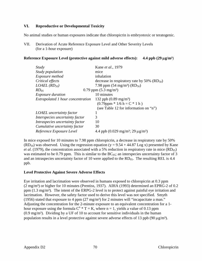
VI. Reproductive or Developmental Toxicity No animal studies or human exposures indicate that chloropicrin is embryotoxic or teratogenic. VII. Derivation of Acute Reference Exposure Level and Other Severity Levels
(for a 1-hour exposure)
Reference Exposure Level (protective against mild adverse effects): 4.4 ppb (29 µg/m³) Study Kane et al., 1979 Study population mice Exposure method inhalation Critical effects decrease in respiratory rate by 50% (RD50) LOAEL (RD50) 7.98 ppm (54 mg/m³) (RD50) RD05 0.79 ppm (5.3 mg/m³) Exposure duration 10 minutes Extrapolated 1 hour concentration 132 ppb (0.89 mg/m³) (0.79ppm * 1/6 h = C * 1 h ) (see Table 12 for information on “n”) LOAEL uncertainty factor 1 Interspecies uncertainty factor 3 Intraspecies uncertainty factor 10 Cumulative uncertainty factor 30 Reference Exposure Level 4.4 ppb (0.029 mg/m³; 29 µg/m³) In mice exposed for 10 minutes to 7.98 ppm chloropicrin, a decrease in respiratory rate by 50% (RD50) was observed. Using the regression equation (y = 9.54 + 44.87 Log x) presented by Kane et al. (1979), the concentration associated with a 5% reduction in respiratory rate in mice (RD05) was estimated to be 0.79 ppm. This is similar to the BC05; an interspecies uncertainty factor of 3 and an intraspecies uncertainty factor of 10 were applied to the RD05. The resulting REL is 4.4 ppb. Level Protective Against Severe Adverse Effects Eye irritation and lacrimation were observed in humans exposed to chloropicrin at 0.3 ppm (2 mg/m³) or higher for 10 minutes (Prentiss, 1937). AIHA (1993) determined an EPRG-2 of 0.2 ppm (1.3 mg/m³). The intent of the ERPG-2 level is to protect against painful eye irritation and lacrimation. However, the safety factor used to derive this level was not specified. Smyth (1956) stated that exposure to 4 ppm (27 mg/m³) for 2 minutes will “incapacitate a man.” Adjusting the concentration for the 2-minute exposure to an equivalent concentration for a 1-hour exposure using the formula Cn * T = K, where n = 1, yields a value of 0.13 ppm (0.9 mg/m³). Dividing by a UF of 10 to account for sensitive individuals in the human population results in a level protective against severe adverse effects of 13 ppb (90 µg/m³).
Appendix D2 70 Chloropicrin
Level Protective Against Life-threatening Effects No recommendation is made due to the limitations of the database. Exposure of mice to 336 mg/m³ (50 ppm) chloropicrin for 15 minutes caused death after 10 days (Clayton and Clayton, 1982). Brief exposures to 27 mg/m³ (4 ppm) chloropicrin may cause severe respiratory irritation in addition to vertigo, fatigue, gastrointestinal cramps, and diarrhea in humans (Fairhall, 1949). Application of the standard safety factors (1,000) to the value reported by Clayton and Clayton (1982), and time extrapolation to a 1-hour exposure would yield a concentration for a life threatening level that is lower than the EPRG-3 level of 3.0 ppm (20 mg/m³) recommended by AIHA (1992). NIOSH (1995) lists an IDLH of 2 ppm based on acute inhalation toxicity data in workers and animals. NIOSH also mentions that 4 ppm for a few seconds renders a worker unfit for activity. The IDLH makes no allowance for sensitive individuals. VIII. References (AIHA) American Industrial Hygiene Association. Emergency response planning guidelines. Set 1. Akron (OH): AIHA; 1992. Buckley LA, Jiang XZ, James RA, Morgan KT, Barrow CS. Respiratory tract lesions induced by sensory irritants at the RD50 concentration. Toxicol Appl Pharmacol 1984;74:417-429. Clayton G, Clayton F, editors. Patty’s Industrial Hygiene and Toxicology. 3rd ed. New York: John Wiley and Sons; 1982. p. 4164-4166. Fairhall L. Industrial toxicology. Baltimore (MD): Williams and Wilkins; 1949. [cited in Smyth HF Jr. Improved communication - hygienic standards for daily inhalation. Am Ind Hyg Assoc J 1956;17:129-185.] Flury F, Zernik F. Schadliche Gase. Berlin: Springer; 1931. Gonmori K, Muto H, Yamamoto T, Takahashi K. A case of homicidal intoxication by chloropicrin. Am J Forensic Med Pathol 1987;8(2):135-138. Grant WM. Toxicology of the eye. 3rd ed. Springfield (IL): CC Thomas; 1986. (HSDB) Hazardous Substances Data Bank. National Library of Medicine, Bethesda, MD (CD-ROM version). Denver (CO): Micromedex, Inc.; 1994. (Edition expires 4/30/94). Kane LE, Barrow CS, Alarie Y. A short-term test to predict acceptable levels of exposure to airborne sensory irritants. Am Ind Hyg Assoc J 1979;40:207-229.
Appendix D2 71 Chloropicrin

Lambert RA, Jackson L. The pathology of chloropicrin poisoning. In: Collected Studies on the Pathology of War Gas Poisoning. MC Winternitz, ed. New Haven: Yale University Press; 1920 pp 69-90 (NIOSH) National Institute for Occupational Safety and Health. Chemical listing and documentation of revised IDLH values. 1995. (http://www.cdc.gov/niosh/intridl4.html) Prentiss A. Chemicals in war. New York: McGraw-Hill; 1937. p. 143. Reprotext ® System. Dabney BJ, editor. Denver (CO): Micromedex, Inc.; 1999. (Edition expires 1/31/1999). Smyth HF Jr. Improved communication - hygienic standards for daily inhalation. Am Ind Hyg Assoc J 1956:17:129-185. TeSlaa G, Kaiser M, Biederman L. Chloropicrin toxicity involving animal and human exposure. Vet Hum Toxicol 1986;28(4):323-324.
Appendix D2 72 Chloropicrin
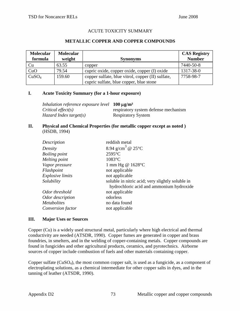
TSD for Noncancer RELs June 2008
ACUTE TOXICITY SUMMARY
METALLIC COPPER AND COPPER COMPOUNDS
Molecular formula
Molecular weight
Synonyms
CAS Registry Number
Cu 63.55 copper 7440-50-8 CuO 79.54 cupric oxide, copper oxide, copper (I) oxide 1317-38-0 CuSO4 159.60 copper sulfate, blue vitrol, copper (II) sulfate,
cupric sulfate, blue copper, blue stone 7758-98-7
I. Acute Toxicity Summary (for a 1-hour exposure) Inhalation reference exposure level 100 µg/m³ Critical effect(s) respiratory system defense mechanism Hazard Index target(s) Respiratory System II. Physical and Chemical Properties (for metallic copper except as noted )
(HSDB, 1994) Description reddish metal Density 8.94 g/cm3 @ 25°C Boiling point 2595°C Melting point 1083°C Vapor pressure 1 mm Hg @ 1628°C Flashpoint not applicable Explosive limits not applicable Solubility soluble in nitric acid; very slightly soluble in
hydrochloric acid and ammonium hydroxide Odor threshold not applicable Odor description odorless Metabolites no data found Conversion factor not applicable III. Major Uses or Sources Copper (Cu) is a widely used structural metal, particularly where high electrical and thermal conductivity are needed (ATSDR, 1990). Copper fumes are generated in copper and brass foundries, in smelters, and in the welding of copper-containing metals. Copper compounds are found in fungicides and other agricultural products, ceramics, and pyrotechnics. Airborne sources of copper include combustion of fuels and other materials containing copper. Copper sulfate (CuSO4), the most common copper salt, is used as a fungicide, as a component of electroplating solutions, as a chemical intermediate for other copper salts in dyes, and in the tanning of leather (ATSDR, 1990).
Appendix D2 73 Metallic copper and copper compounds

TSD for Noncancer RELs June 2008
Copper oxide (CuO) is another common copper salt. It is used in insecticides, fungicides, and catalysts (HSDB, 1994). CuO is also used in fuel additives, cement, and wood preservatives. IV. Acute Toxicity to Humans Following occupational exposures to copper dust, commonly reported reactions include metallic or sweet taste, upper respiratory tract irritation, and nausea (Whitman, 1962). An unpublished letter regarding occupational exposure to copper fumes reported that levels of 0.02-0.40 mg/m³ copper did not “cause complaints” while exposure to 1.0-3.0 mg/m³ copper for “short periods of time” resulted in a “sweet taste in the mouth” but no nausea (Whitman, 1957). Inhalation exposure to copper fumes, usually from welding or smelting operations, may result in “metal fume fever.” This condition results in headache, dryness of the mouth and throat, chills, fever, and muscle aches, usually beginning 4-8 hours after exposure to the oxides of various metals, including copper. Symptoms and signs spontaneously subside within 24-36 hours (ATSDR, 1990; Seaton and Morgan, 1984). Symptoms consistent with metal fume fever were reported by workers in a facility with airborne copper dust at concentrations of 0.03-0.12 mg/m³ (Gleason, 1968). Upper respiratory irritation has been reported, in addition to symptoms consistent with metal fume fever (fever, dyspnea, chills, headache, nausea, myalgia, cough, shortness of breath, a sweet metallic taste, and vomiting), in factory workers exposed to copper fumes for 1 to 10 hours as a result of cutting pipes known to contain copper (Armstrong et al., 1983). The sweet taste experienced by workers from the Whitman (1957) report above is consistent with the onset of symptoms of metal fume fever. Factory workers exposed to copper dust, CuO, and several other copper salts reported symptoms of eye, nose, and throat irritation, anorexia, and nausea (Askergren and Mellgren, 1975; Suciu et al., 1981). Occasional diarrhea was also reported by these workers. Predisposing Conditions for Copper and Copper Compound Toxicity Medical: Persons with Wilson’s disease, a genetic disorder affecting copper
homeostasis, may be more sensitive to the effects of copper exposure (Schroeder et al., 1966; ATSDR, 1990). Persons with glucose-6-phosphate dehydrogenase deficiency, anemic, allergic, liver or kidney conditions moght be more sensitive (Reprotext, 1999). Infants and children less than 1-year of age may be more sensitive to the effects of copper exposure because homeostatic mechanisms for clearing copper from the body are not yet developed
Chemical: Persons exposed to molybdenum might be less sensitive to copper, since
molybdewnum is antagonistic to copper toxicity (Reprotext, 1999).
Appendix D2 74 Metallic copper and copper compounds

TSD for Noncancer RELs June 2008
V. Acute Toxicity to Laboratory Animals Rats were dosed by intratracheal instillation with 2.5, 5, 10, 20, 30, 50, and 100 mg Cu/rat and pulmonary clearance of CuO was measured over time (Hirano et al., 1993). The CuO particles were cleared from the lung with a half-time of 37 hours. A 54% and 70% increase in mortality in male and female mice, respectively, over controls was observed following challenge with aerosolized streptococci after a 3-hour exposure to 0.56 mg/m³ Cu as CuSO4 (Drummond et al., 1986). Pulmonary bactericidal activity was not measured for this exposure group. The effects of copper sulfate (and other metal sulfate) aerosols on respiratory defense mechanisms were studied in male hamsters (Skornik and Brain, 1983). Pulmonary macrophage phagocytic rates were measured by determining the in vivo uptake of radioactive colloidal gold 1, 24, or 48 hours after a single 4-hour inhalation exposure to 0, 0.3, 3.2, 4.0, 5.8 and 7.1 mg Cu/m³. When hamsters were exposed for 4 h to greater than or equal to 3.2 mg Cu/m³, macrophage endocytosis was significantly reduced 1 h after exposure compared with that in unexposed control animals. The reduction was dose-dependent. At 24 h after exposures to the higher concentrations of Cu the percent of gold ingested by pulmonary macrophages remained depressed but less than at 1 hour. (By 48 h, the rate of macrophage endocytosis in hamsters returned to control levels except in hamsters exposed to 3.2 and 5.8 mg Cu/m³.) VI. Reproductive or Developmental Toxicity Copper is known to be spermicidal (U.S.EPA, 1987). Copper absorbed from copper intrauterine loops or wires has been shown to prevent mammalian embryogenesis. Conversely, terata have been observed in the offspring of experimental animals deficient in dietary copper. Inhibited spermatogenesis and testicular atrophy were observed in male rats exposed to 0.1-1.0 mg/m³ CuO (Ginoian, 1976). The same study also reported that the number of fetuses was reduced in a dose-related manner in females exposed to CuO. Because the original article was not available for review, key experimental details, including duration of exposure, are unknown.
Appendix D2 75 Metallic copper and copper compounds

TSD for Noncancer RELs June 2008
VII. Derivation of Acute Reference Exposure Level and Other Severity Levels (for a 1-hour exposure)
Reference Exposure Level (protective against mild adverse effects): 100 µg/m³ Study ACGIH, 1991; Gleason, 1968; Whitman, 1957, 1962 Study population workers Exposure method inhalation Critical effects metal fume fever LOAEL unkown NOAEL 1 mg Cu/m³ Exposure duration unknown Extrapolated 1 hour concentration no extrapolation LOAEL uncertainty factor 1 Interspecies uncertainty factor 1 Intraspecies uncertainty factor 10 Cumulative uncertainty factor 10 Reference Exposure Level 0.1 mg Cu/m³ (100 µg/m³) The ACGIH-TLV is based on an unpublished letter which reported that exposure to 1 - 3 mg/m3 copper fume for “short periods” resulted in a “sweet taste in the mouth” and that exposure to 0.02 - 0.4 mg/m3 did not result in any symptoms (Whitman, 1957). However, it was not clear from the letter if or how actual copper levels were measured. Another author reported that symptoms of metal fume fever were observed in workers exposed for an unspecified number of weeks to 0.03 - 0.12 mg/m3 copper dust (Gleason, 1968). The latter exposure was not designed to determine the level of copper responsible for the symptoms; it was meant to justify the implementation of exhaust controls. Therefore, the air samples were not directly compared to worker exposure or worker symptoms. The current REL is based on the ACGIH-TLV of 1 mg/m3 copper dust. The TLV of 1 mg/m3 is a NOAEL based on the report of Whitman (1957) indicating that exposure to copper dust was detectable by taste but that no other symptoms occurred following exposure to 1 - 3 mg/m3 for an unknown duration. An uncertainty factor of 10 was applied to the NOAEL to account for variability in individual response. No time extrapolation was applied because the duration of exposure was not clearly specified by either of the available reports. Because of the limitations of the existing data, reevaluation of the REL for copper is recommended when better methods or data are available. Level Protective Against Severe Adverse Effects No recommendation is made due to the limitations of the database.
Appendix D2 76 Metallic copper and copper compounds

TSD for Noncancer RELs June 2008
Level Protective Against Life-threatening Effects No recommendation is made due to the limitations of the database. NIOSH (1995) lists an IDLH of 100 mg/m³ but it is based on studies of lethality by the oral route in animals and man. VIII. References (ATSDR) Agency for Toxic Substances and Disease Registry. Toxicological profile for copper. Prepared by Syracuse Research Corporation under Subcontract No. ATSDR-88-0608-02. Prepared for ATSDR, US Public Health Service, 1990. (ACGIH) American Conference of Governmental Industrial Hygienists. Documentation of the Threshold Limit Values and Biological Exposure Indices. 6th ed. Cincinnati (OH): ACGIH; 1991. p. 336-337. Armstrong CW, Moore LW, Hackler RL, Miller GB, Stroub RB. An outbreak of metal fume fever: diagnostic use of urinary copper and zinc determinations. J Occup Med 1983;25:886-888. Askergren A, Mellgren M. Changes in the nasal mucosa after exposure to copper salt dust. Scand J Work Environ Health 1975;1:45-49. Drummond JG, Aranyi C, Schiff LJ, Fenters JD, Graham JA. Comparative study of various methods used for determining health effects of inhaled sulfates. Environ Res 1986;41:514-528. Ginoian MM. [Experimental data on the hygienic substantiation of the maximum permissible concentration of cupric oxide in the atmosphere][Russian].Gig Sanit 1976;6:8-12. [cited in Reprotext, 1999.] Gleason RP. Exposure to copper dust. Am Ind Hyg Assoc J 1968;29:461-462. (HSDB) Hazardous Substances Data Bank. National Library of Medicine, Bethesda (MD) (CD-ROM Version). Denver (CO): Micromedex, Inc.; 1994. (Edition expires 4/30/94). Hirano S, Ebihara H, Sakai S, Kodama N, Suzuki KT. Pulmonary clearance and toxicity of intratracheally instilled cupric oxide in rats. Arch Toxicol 1993;67:312-317. Johansson A, Curstedt T, Robertson B, Camner P. Lung morphology and phospholipids after experimental inhalation of soluble cadmium, copper, and cobalt. Environ Res 1984;34(2):295-309. Lundborg M, Camner P. Lysozyme levels in rabbit lung after inhalation of nickel, cadmium, cobalt, and copper chlorides. Environ Res 1984;34(2):335-342
Appendix D2 77 Metallic copper and copper compounds

TSD for Noncancer RELs June 2008
(NIOSH) National Institute for Occupational Safety and Health. Chemical listing and documentation of revised IDLH values. 1995. (http://www.cdc.gov/niosh/intridl4.html) Reprotext® System. Dabney BJ, editor. Denver (CO): Micromedex, Inc.; 1999. (Edition expires 1/31/1999). Schroeder WH, Nason AP, Tipton IH. Essential trace metals in man: copper. J Chron Dis 1966;19:1007-1034. Seaton A, Morgan WKC. Toxic gases and fumes. In: Morgan WKC, Seaton A, editors. Occupational lung diseases. Philadelphia: WB Saunders Co.; 1984. p. 609-642. Skornik WA, Brain JD. Relative toxicity of inhaled metal sulfate salts for pulmonary macrophages. Am Rev Respir Dis 1983;128(2):297-303 Suciu I, Prodan L, Lazar V, Ilena E, Cocirla A, Olinici L, et al. Research on copper poisoning. Med Lav 1981;72:190-197. (U.S.EPA) United States Environmental Protection Agency. Summary review of the health effects associated with copper. Cincinnati (OH): Environmental Criteria and Assessment Office, Office of Health and Environmental Assessment, Office of Research and Development, U.S.EPA; 1987. Whitman NE. Letter to TLV Committee from Industrial Health Engineering. Bethlehem (PA): Bethlehem Steel Co; 1957 (March 12, 1957). Whitman NE. Letter to TLV Committee from Industrial Health Engineering. Bethlehem (PA): Bethlehem Steel Co; 1962 (April 24, 1962).
Appendix D2 78 Metallic copper and copper compounds

TSD for Noncancer RELs June 2008
ACUTE TOXICITY SUMMARY
1,4-DIOXANE
(diethylene oxide; p-dioxane; glycolethylene ether; tetrahydro-p-dioxin)
CAS Registry Number: 123-91-1 I. Acute Toxicity Summary (for a 1-hour exposure) Inhalation reference exposure level 3,000 µg/m³ Critical effect(s) Nasal and eye irritation in healthy human volunteers Hazard Index target(s) Respiratory System; Eyes II. Physical and Chemical Properties (ACGIH, 1991 except as noted) Description colorless liquid Molecular formula C4H8O2 Molecular weight 88.1 Density 1.0329 g/cm3 @ 20°C Boiling point 101.1°C @ 760 mm Hg Melting point 11.8°C Vapor pressure 29 mm Hg @ 20°C Flash point 12.22°C (closed cup) Explosive limits 2 - 22 % by volume in air Solubility soluble in water and most organic solvents Odor threshold 24 ppm (ACGIH, 1991);
1.8 ppm (Hellman and Small, 1974) Odor description ethereal odor (Buffler et al., 1978) Metabolites hydroxyethoxyacetic acid (Braun and Young, 1977) Conversion factor 1 ppm = 3.6 mg/m³ III. Major Uses or Sources 1,4 - Dioxane is used as a solvent for oils, resins, waxes, adhesives, cellulose esters and ethers. It is also used as a stabilizer in chlorinated solvents (ACGIH, 1991). As much as 90% of U.S. production of dioxane has been used to stabilize chlorinated solvents. As a stabilizer it is present as a few percent by volume. IV. Acute Toxicity to Humans There are case reports of lethal hemorrhagic nephritis in workers exposed to unspecified high concentrations of 1,4-dioxane for several days (Barber, 1934; Johnstone, 1959).
Appendix D2 79 Dioxane

TSD for Noncancer RELs June 2008
1,4-Dioxane was irritating to the eyes, nasal passages, and the throat of adult volunteers following a 10-minute exposure to 1,600 ppm (Yant et al., 1930). In this study, no control subjects were tested concomitantly. A similar study of 4-6 volunteers by Fairly et al. (1934) showed that inhalation exposure to a concentration of 1,000 ppm (3,600 mg/m³) for five minutes caused a warm sensation in the throat and chest, but no noticeable irritation. However, in a more recent study, four healthy adult male volunteers exposed in a chamber for 6 hours to 50 ppm (180 mg/m³) dioxane exhibited eye irritation and 2 of the 4 subjects reported olfactory fatigue after 4 and 5 hours (Young et al., 1977). Predisposing Conditions for 1,4-Dioxane Toxicity Medical: Persons with preexisting skin, eye, respiratory, neurological, and liver and kidney
conditions might be more sensitive (Reprotext, 1999). Chemical: Unknown V. Acute Toxicity to Laboratory Animals Inhalation by guinea pigs and rats of 10,000 ppm (36,000 mg/m³) 1,4-dioxane for two 1.5-hour exposures was lethal (Fairley et al., 1934). 1,4-Dioxane affects the rat central nervous system as measured by a significant decrease in avoidance behavior following a 4-hour exposure to 3,000 ppm (10,800 mg/m³) (Goldberg et al., 1964). Nasal irritation was indicated by behavioral signs in guinea pigs exposed to 1,000 ppm (3,600 mg/m³) 1,4-dioxane for 4 hours (Yant et al., 1930); behavioral signs of eye irritation were evident at concentrations of 2,000 ppm (7,200 mg/m³) 1,4-dioxane or greater. Slight hyperemia was observed in the lungs, large air passages, and the brain in the animals exhibiting mild irritation. No histological changes were noted in control animals (unexposed to 1,4-dioxane). The absence of pathological lesions in the brain and lungs in exposed animals 9-10 days after 1,4-dioxane exposure led the authors to conclude that the histological effects of dioxane exposure were transient at the concentrations and exposure duration tested. Based on pharmacokinetic data, rats appear to be the most appropriate animal model for metabolism of 1,4-dioxane in humans (Young et al., 1978). In a comparative toxicity study on rats, mice, guinea pigs, and rabbits, Fairley et al. (1934) showed that all species became drowsy after a 1.5 hour exposure to 1,000 ppm (3,600 mg/m³) 1,4-dioxane. In this study, guinea pigs were the most sensitive species to organ-specific histopathological lesions, which included: acute vascular congestion in the lungs, patchy cell degeneration and hemorrhage of the renal cortex, and hepatic necrosis. Schrenk and Yant (1936) showed that nasal irritation was evident in guinea pigs immediately following brief exposure to 1,000 ppm (3,600 mg/m³) 1,4-dioxane. No behavior indicative of eye irritation or lacrimation was observed at this concentration. Drew et al. (1978) showed that a single 4-hour inhalation of 1,000 ppm (3,600 mg/m³) 1,4-dioxane by rats resulted in immediate elevation of serum glutamic-oxaloacetic transaminase activity. Alanine aminotransferase and ornithine carbamyl transaminase activities were elevated 24 hours following the 4-hour 1,000 ppm (3,600 mg/m³) exposure. The elevations of these hepatic enzymes indicated that 1,4-dioxane is hepatotoxic in rats.
Appendix D2 80 Dioxane

TSD for Noncancer RELs June 2008
VI. Reproductive or Developmental Toxicity Pregnant rats treated with 0, 0.25, 0.5, or 1.0 mL dioxane/kg body weight by gavage on days 6-15 of gestation showed no differences in the number of implanted fetuses, live fetuses, post-implantation loss, or major malformations. Slight maternal toxicity in the form of weight loss was observed at the 1.0 mL/kg dose (Giavini et al., 1985). No data on human reproductive toxicity were available. VII. Derivation of Acute Reference Exposure Level and Other Severity Levels (for a 1-hour exposure) Reference Exposure Level (protective against mild adverse effects): 0.8 ppm (3,000 µg/m³) Study Young et al., 1977 Study population 4 healthy human male volunteers Exposure method chamber Critical effects subjective reports of eye irritation LOAEL 50 ppm NOAEL not reported Exposure duration 6 hours Extrapolation to 1 hour not used (see below) Extrapolated 1-hour concentration 50 ppm LOAEL uncertainty factor 6 (mild irritation) Interspecies uncertainty factor 1 Intraspecies uncertainty factor 10 Cumulative uncertainty factor 60 Reference Exposure Level 0.8 ppm (3 mg/m³, 3,000 µg/m³) The volunteers complained of eye irritation throughout the exposure. Two of the subjects were not able to perceive the odor of dioxane after 4 and 5 hours exposure, respectively. A time-adjustment factor for the 6-hour exposure was not used since the individuals complained of eye irritation throughout the exposure. Level Protective Against Severe Adverse Effects No recommendation is made due to the limitations of the database. Level Protective Against Life-threatening Effects No recommendation is made due to the limitations of the database. NIOSH (1995) lists a (revised) IDLH for 1,4-dioxane of 500 ppm based on acute inhalation toxicity data in animals. NIOSH derived 30 minute LC50s from several studies of cats, rats, mice and guinea pigs, then divided the lowest 30 minute LC50 by 10 to determine an IDLH for humans. NIOSH stated that no relevant human data were available for the IDLH estimation.
Appendix D2 81 Dioxane

TSD for Noncancer RELs June 2008
VII. References (ACGIH) American Conference of Governmental and Industrial Hygienists. Documentation of the Threshold Limit Values and Biological Exposure Indices. 6th ed. Vol. I. Cincinnati: ACGIH; 1991. p. 512-515. Barber H. Haemorrhagic nephritis and necrosis of the liver from dioxane poisoning. Guy’s Hosp Rep 1934;84:267-280. Braun WH, Young JD. Identification of hydroxyethoxyacetic acid as the major urinary metabolite of 1,4-dioxane in the rat. Fundam Appl Toxicol 1977;39:33-38. Buffler PA, Wood SM, Suarez L, Kilian DJ. Mortality follow-up of workers exposed to 1,4-dioxane. J Occup Med 1978;20(4):255-259. Drew RT, Patel JM, Lin FN. Changes in serum enzymes in rats after inhalation of organic solvents singly and in combination. Toxicol Appl Pharmacol 1978;45:809-819. Fairley A, Linton EC, Forde-Moore AH. The toxicity to animals of 1,4-dioxan. J Hyg 1934;34:486-501. Giavini E, Vismara C, Broccia ML. Teratogenesis study of dioxane in rats. Toxicol Lett 1985;26:85-88. Goldberg ME, Johnson HE, Pozzani UC, Smyth HF Jr. Effect of repeated inhalation of vapors of industrial solvent on animal behavior. 1. Evaluation of nine solvent vapors on pole-climb performance. Am Ind Hyg Assoc J 1964;25:369-375. Hellman TM, Small FH. Characterization of the odor properties of 101 petrochemicals using sensory methods. J Air Poll Cont Assoc 1974;24(10):979-982. Johnstone RT. Death due to dioxane? AMA Arch Ind Health 1959;20:445-447. Klimmer O. Beitrag zur toxikologischen Wirkung technischer Losungsmittel (German) [dissertation]. Wurzburg, Germany; 1937. [cited in: Spector WS. Handbook of toxicology. Vol I. Acute toxicities. Philadelphia (PA): Saunders Company; 1956. p. 334-5.] (NIOSH) National Institute for Occupational Safety and Health. Pocket guide to chemical hazards. DHEW/NIOSH Pub. No. 93-114. Washington (DC): Government Printing Office; 1993. (NIOSH) National Institute for Occupational Safety and Health. Chemical listing and documentation of revised IDLH values. 1995. (http://www.cdc.gov/niosh/intridl4.html) Reprotext ® System. Dabney BJ, editor. Denver (CO): Micromedex, Inc.; 1999. (Edition expires 1/31/1999).
Appendix D2 82 Dioxane

TSD for Noncancer RELs June 2008
Spector WS. Handbook of toxicology. Vol. I. Acute toxicities. Philadelphia (PA): Saunders Company; 1956. p. 334-335. Schrenk HH, Tant WP. Toxicity of dioxan. J Ind Hyg Toxicol 1936;18:448-460. Yant WP, Schrenk HH, Waite CP, Patty FA. Acute response of guinea pigs to vapors of some new commercial organic compounds. Pub Health Rep 1930;45:2023-2032. Young JD, Braun WH, Gehring PJ. Dose-dependent fate of 1,4-dioxane in rats. J Toxicol Environ Health 1978;4:709-726. Young JD, Braun WH, Rampy LW. Pharmacokinetics of 1,4-dioxane in humans. J Toxicol Environ Health 1977;3:507-520.
Appendix D2 83 Dioxane

TSD for Noncancer RELs June 2008
ACUTE TOXICITY SUMMARY
EPICHLOROHYDRIN
(1-chloro-2,3-epoxy-propane)
CAS Registry Number: 106-89-8 I. Acute Toxicity Summary (for a 1-hour exposure) Inhalation reference exposure level 1,300 µg/m³ Critical effect(s) eye and nasal irritation in human volunteers Hazard Index target(s) Eyes; Respiratory System II. Physical and Chemical Properties (HSDB, 1994)
Description colorless liquid Molecular formula C3H5ClO Molecular weight 92.5
Density 1.181 g/cm3 @ 20°C Boiling point 117.9°C Melting point -25.6°C Vapor pressure 13 mm Hg @ 20°C Flash point 33.9°C Explosive limits 3.3% - 14.5 % by volume in air Solubility slightly soluble in water, soluble in most organic
solvents Odor threshold 0.93 ppm (chloroform-like, irritating odor) Metabolites N-acetyl-S-(3-chloro-2-hydroxypropyl)-L-cysteine Conversion factor 1 ppm = 4 mg/m³
III. Major Uses or Sources Epichlorohydrin is a major raw material used in the manufacture of epoxy and phenoxy resins. It is also used as a solvent and in the synthesis of glycerol. Other uses include that of insect fumigation and as a chemical intermediate for the formation of glycidyl acrylate derivatives such as those used in the formation of eyeglass lenses (HSDB, 1994). IV. Acute Toxicity to Humans Case reports of exposure to epichlorohydrin in the workplace, either through inhalation or dermal contact, describe symptoms including burning sensations of the nose and throat, chest congestion, running nose, eye tenderness, and headache followed by nausea, in addition to reddening and burning sensations of the exposed skin, which persist for several days to 2 months (Wexler, 1971, as cited in NIOSH, 1976). Epichlorohydrin is a strong skin sensitizer following dermal contact (U.S.EPA, 1984). Epichlorohydrin is a reactive epoxide and a known mutagen.
Appendix D2 84 Epichlorohydrin

TSD for Noncancer RELs June 2008
In vitro exposure of human lymphocytes to 10-11 to 10-4 M epichlorohydrin resulted in dose-dependent chromatid and chromosomal breaks (HSDB, 1994). Predisposing Conditions for Epichlorohydrin Toxicity Medical: Asthmatics may be more sensitive to the irritant effects of inhaled
epichlorohydrin. Chemical: Unknown V. Acute Toxicity to Laboratory Animals A six-hour exposure to epichlorohydrin with a 14-day follow-up showed the median lethal concentration to be 360 ppm (1,440 mg/m³) in rats (Laskin et al., 1980). An LC50 of 445 ppm (1,780 mg/m³) for four hours was reported for rabbits (HSDB, 1994). An eight-hour exposure to 250 ppm (1,000 mg/m³) killed two-thirds of the rats exposed (sample size not given) (LeFaux, 1968). A single subcutaneous injection of 75 mg/kg resulted in swelling of proximal renal tubular epithelium in male rats (Kluwe et al., 1983). Deaths occurred in rats exposed chronically to a concentration of 68 ppm (272 mg/m³) epichlorohydrin for an unknown duration (IRIS, 1994). Tumors induced by chronic epichlorohydrin exposure are typically local to the area of initial exposure (U.S.EPA, 1984). Nasal carcinomas are among the tumors known to occur following epichlorohydrin exposure (U.S.EPA, 1984). VI. Reproductive or Developmental Toxicity Fetotoxicity and toxicity to dams were reported in mice exposed to 120 mg/kg/day epichlorohydrin via gavage during days 6-15 of gestation; however, no teratogenic effects were noted (Marks et al., 1982). Teratology studies in rats and rabbits yielded negative results for embryotoxicity and teratogenicity (John et al., 1983a). Maternal toxicity, as measured by a decrease in body weight and food consumption, was demonstrated in pregnant rats following exposure to 25 ppm (100 mg/m³) epichlorohydrin for 7 hours/day on days 6-18 of gestation (John et al., 1983a). Additionally, exposure of male rats to 25 ppm for 5 days/week for 10 weeks resulted in a transient loss in fertility (John et al., 1983b). Injury to epididymal tissue, testicular atrophy, and increases in the number of sperm with abnormal morphology have been observed in male rats exposed via single subcutaneous injection to 75 mg/kg epichlorohydrin (Kluwe et al., 1983). Although animal studies indicate that male fertility is affected by exposure to high doses of epichlorohydrin, a human epidemiologic study showed no changes in male fertility rates among workers (HSDB, 1994).
Appendix D2 85 Epichlorohydrin

TSD for Noncancer RELs June 2008
VII. Derivation of Acute Reference Exposure Level and Other Severity Levels (for a 1-hour exposure) Reference Exposure Level (protective against mild adverse effects): 0.33 ppm (1,300
µg/m³) Study Wexler (1971) as cited in NIOSH, 1976 Study population occupationally exposed workers Exposure method during work-shifts (occupation not given) Critical effects irritation of eyes and nasal passages LOAEL 20 ppm NOAEL not reported Exposure duration 1 hour Extrapolated 1 hour concentration 20 ppm LOAEL uncertainty factor 6 (mild irritation) Interspecies uncertainty factor 1 Intraspecies uncertainty factor 10 Cumulative uncertainty factor 60 Reference Exposure Level 0.33 ppm (1.3 mg/m³, 1,300 µg/m³) The Wexler (1971) study represents the only human data but it was not available for review. The report by NIOSH (1976), which reviewed the Wexler study, was therefore used as the basis for the REL. Level Protective Against Severe Adverse Effects No recommendation is made due to the limitations of the database. Exposure of 8 rats for 6 hours/day, 5 days/week for 19 days to 17 ppm epichlorohydrin resulted in no pulmonary histopathological abnormalities as compared to controls (Gage, 1959). The ERPG documentation for epichlorohydrin (AIHA, 1992) erroneously refers to Laskin et al. (1980) as a teratology study instead of a carcinogenicity study. In addition, the extrapolation of sub-chronic animal exposures in the Gage study to acute human exposures involves considerable uncertainty that is not accounted for in the ERPG document. The ERPG-2 value of 20 ppm (76 mg/m³) is therefore poorly substantiated. Level Protective Against Life-threatening Effects No recommendation is made due to the limitations of the database. Subacute exposures of rats and mice (5/sex) to 100 ppm epichlorohydrin 6 hours/day, 5 days/week, 9 exposures in 12 days, resulted in focal pneumonitis and inflammation and degeneration of nasal epithelium in addition to decreased weight gain (Quast et al., 1979a, b). Kidney toxicity was seen in the rats exposed to 100 ppm. No lethality was observed. It was concluded that acute exposure to 100 ppm would not cause fatality in humans. Thus AIHA
Appendix D2 86 Epichlorohydrin

TSD for Noncancer RELs June 2008
(1992) selected 100 ppm (380 mg/m³) as the ERPG-3 for epichlorohydrin. This value can be considered a subchronic NOAEL for lethality in mice, but the lack of uncertainty factors for the extrapolation of animal to human exposures, in addition to those required for consideration of sensitive individuals, dictate that this value should be reevaluated. The small sample sizes in the rodent studies, and the absence of peer-reviewed data used to derive the NOAEL, further weaken the scientific validity of this value. The ERPG-3 value is based on severe, non-lethal effects and not on lethality data. An inhalation LC50 in mice of 2,998 mg/m³ for 2 hours is reported by the World Health Organization (1992). VIII. References (AIHA) American Industrial Hygiene Association. Emergency response planning guidelines for epichlorohydrin. Set 5. Akron: AIHA; 1992. Gage JC. The toxicity of epichlorohydrin vapour. Br J Ind Med 1959;16:11-14. (HSDB) Hazardous Substances Data Bank. National Library of Medicine, Bethesda (MD) (CD-ROM version). Denver (CO): Micromedex, Inc.; 1994. (Edition expires 4/30/94). (IRIS) Integrated Risk Information System. U.S. Environmental Protection Agency, Washington (DC) (CD-ROM version). Denver (CO): Micromedex, Inc.; 1994. (Edition expires 10/31/94). John JA, Gushow TS, Ayres JA, Hanley TR, Quast JF, Rao KS. Teratologic evaluation of inhaled epichlorohydrin and allyl chloride in rats and rabbits. Fundam Appl Toxicol 1983a;3:437-442. John JA, Quast JF, Murray FJ, Calhoun LG, Staples RE. Inhalation toxicity of epichlorohydrin: effects on fertility in rats and rabbits. Toxicol Appl Pharmacol 1983b;68:415-423. Kluwe WM, Gupta BN, Lamb JC. The comparative effects of 1,2-dibromo-3-chloropropane (DBCP) and its metabolites, 3-chloro-1,2-propanediol (alphachlorohydrin), and oxalic acid, on the urogenital system of male rats. Toxicol Appl Pharmacol 1983;70:67-86. Laskin S, Sellakumar AR, Kuschner M, Nelson N, La Mendola S, Rusch GM, et al. Inhalation carcinogenicity of epichlorohydrin in noninbred Sprague-Dawley rats. J Natl Cancer Inst 1980;65(4):751-757. LeFaux R. Practical toxicology of plastics. London: Scripta Technica Ltd; 1968. p. 108. Marks TA, Gerling FS, Staples RE. Teratogenic evaluation of epichlorohydrin in the mouse and rat and glycidol in the mouse. J Toxicol Environ Health 1982;9:87-96. (NIOSH) National Institute for Occupational Safety and Health. Criteria for a recommended standard: occupational exposure to epichlorohydrin. Cincinnati: DHEW (NIOSH); 1976;76-202.
Appendix D2 87 Epichlorohydrin

TSD for Noncancer RELs June 2008
Quast JF, Henck JW, Postma BJ, Schuetz DJ, McKenna MJ. Epichlorohydrin - subchronic studies. I. A 90-day inhalation study in laboratory rodents. Toxicology Research Laboratory, Dow Chemical, USA. Midland (MI); 1979a. (unpublished). Quast JF, Lederer TS, Postma BJ, Schuetz DJ, John JA, McKenna MJ. Epichlorohydrin - subchronic studies II. A 12-day inhalation study in laboratory rodents. Toxicology Research Laboratory, Dow Chemical, USA. Midland (MI); 1979b. (unpublished). Wexler B. [Determination of epichlorohydrin contamination in an industrial facility for the manufacturing of epoxy resins.] (Rum) Mater Plast (Bucharest) 1971;8:322-333. U.S. Environmental Protection Agency. 1984. Health assessment for epichlorohydrin. EPA-600/8-83-032F, Washington (DC): U.S.EPA; 1979b. World Health Organization. Chemical review: epichlorohydrin. In: Dangerous properties of industrial materials report. 1992;12(2):150-170.
Appendix D2 88 Epichlorohydrin

TSD for Noncancer RELs June 2008
ACUTE TOXICITY SUMMARY
ETHYLENE GLYCOL MONOBUTYL ETHER
(2-butoxyethanol, butyl cellosolve, butyl glycol)
CAS Registry Number: 111-76-2 I. Acute Toxicity Summary (for a 1-hour exposure)
Inhalation reference exposure level 14,000 µg/m³ Critical effect(s) irritation Hazard Index target(s) Respiratory System
II. Physical and Chemical Properties (HSDB, 1994 except as noted) Description colorless liquid Molecular formula C6H14O2 Molecular weight 118.20 Density 0.90 g/cm3 @ 20°C Boiling point 171°C Melting point -70°C Vapor pressure 0.76 mm Hg @ 20°C Flashpoint unknown Explosive limits unknown Solubility soluble in water, acetone, benzene, carbon tetrachloride,
ethyl ether; miscible with ketones, ethers, alcohols and halogenated hydrocarbons
Odor threshold 0.10 ppm (geometric mean) (AIHA, 1989) Odor description sweet, ester-like, musty (AIHA, 1989) Metabolites butoxyacetic acid (Johanson et al., 1986) Conversion factor 1 ppm = 4.84 mg/m³ @ 25°C III. Major Uses or Sources Ethylene glycol monobutyl ether (EGBE) is used as a coupling agent to stabilize immiscible ingredients in metal cleaners, textile lubricants, and cutting oils (HSDB, 1994). It is also used as a solvent for nitrocellulose resins, spray lacquers, enamels, and varnish removers. EGBE is also found in hydraulic fluids. IV. Acute Toxicity to Humans Two adult male volunteers were exposed to 113 ppm (550 mg/m³) EGBE for 4 hours. Eye, nose and throat irritation, taste disturbances, and headache and nausea were reported (Carpenter et al., 1956). Erythrocyte osmotic fragility and urinalysis were normal in the subjects during and after
Appendix D2 89 Ethylene Glycol Monobutyl Ether

TSD for Noncancer RELs June 2008
exposure. In this study, 8-hour exposures at the same concentrations resulted in similar reports of discomfort. Four volunteers were exposed either mouth-only or skin-only, by a mouthpiece or a respirator in a chamber, to 50 ppm EGBE for 2 hours (Johanson and Boman, 1991). Capillary blood samples were taken at regular intervals to determine rate of uptake from dermal and inhalation (mouth-only) exposure. The experiment was done under both normal and raised humidity conditions. The authors concluded that dermal uptake of EGBE from air is approximately four times greater than respiratory uptake. The authors also note that dermal uptake increased with air temperature and humidity. Seven healthy male adults were exposed to 20 ppm (100 mg/m³) EGBE in a chamber experiment designed to assess pulmonary uptake and metabolism of EGBE. Butoxyacetic acid was the primary metabolite found in the urine (Johanson et al., 1986). The authors report that 57% of the inhaled dose was absorbed in the respiratory tract. The authors report that none of the subjects complained or showed any adverse effects from exposure for 2 hours to 20 ppm EGBE. Although increased erythrocyte fragility has been observed in rodents following exposure to EGBE (Carpenter et al., 1956), recent studies found no increase in the fragility of human erythrocytes taken from normal and susceptible individuals (persons with hereditary spherocytosis or sickle cell disease and older persons) following a 4-hour incubation with butoxyacetic acid (Udden, 1994; Udden and Patton, 1994). Predisposing Conditions for EGBE Toxicity Medical: Persons with preexisting neurological, blood or kidney conditions might
be more sensitive (Reprotext, 1999). Chemical: Unknown V. Acute Toxicity to Laboratory Animals A 7-hour LC50 for mice was reported as 700 ppm (3,000 mg/m³) EGBE (Werner et al., 1943). Severe hemoglobinuria was observed; hepatic focal necrosis and splenic lymphoid hyperplasia were noted at necropsy. An 8-hour LC50 in rats was reported as 564 ppm (2,800 mg/m³) EGBE (Pozzani et al., 1959). No mortality or other clinical signs of toxicity were observed in 5 male and 5 female guinea pigs exposed to 691 or 633 ppm EGBE, respectively, for one hour (Nachreiner, 1994). Further, no signs of toxicity were observed during the 14-day post-exposure period or at necropsy. Rats were exposed to 867, 523, or 202 ppm EGBE for four hours (Dodd et al., 1983). Exposure was lethal to all animals in the 867 ppm group and to 2/6 males and 3/6 females in the 523 ppm group. No deaths were observed in the 202 ppm EGBE exposure group. Rats exposed to 867 ppm exhibited loss of coordination and shallow breathing and had a red discharge around the urogenital area. Red-stained fluid in the urinary bladder and enlarged and discolored kidneys
Appendix D2 90 Ethylene Glycol Monobutyl Ether

TSD for Noncancer RELs June 2008
were observed at necropsy of the animals that died during or following exposure to 867 or 523 ppm EGBE. Increased erythrocyte fragility was observed in rats exposed for 4 hours to 62 ppm (300 mg/m³) EGBE (Carpenter et al., 1956). No significant increase in erythrocyte fragility was observed following a 4-hour exposure to 32 ppm (150 mg/m³) EGBE. Corley et al. (1994) developed a physiologically based pharmacokinetic model to describe in rats and humans the disposition of EGBE and its major metabolite, 2-butoxyacetic acid (BAA); BAA is the agent that causes lysis of red blood cells. The model predicted that rats metabolize EGBE and eliminate BAA faster per kg body weight than humans do. The balance of the two processes in addition to physiological differences between species resulted in higher predicted peak blood concentrations for rats as well as total areas under the blood concentration (AUC) time curves for BAA. The species differences in kinetics coupled with the fact that human blood is significantly less susceptible than rat blood (and mouse blood and probably rabbit blood) to the hemolytic effects of BAA (Udden et al., 1994a,b) indicate that there is less risk for hemolysis in humans as a result of exposure to EGBE than predicted solely by standard rat toxicity studies. VI. Reproductive or Developmental Toxicity No studies on the developmental and reproductive toxicity of EGBE in humans were located in the literature. Pregnant rats were exposed to 0, 25, 50, 100, or 200 ppm EGBE 6 hours per day on days 6-15 of gestation (Tyl et al., 1984). A significant increase in the incidence of delayed skeletal ossification was observed in the offspring of rats exposed to 100 or 200 ppm EGBE. Maternal toxicity, as indicated by decreased body weight gain, decreased food consumption, and significantly decreased erythrocyte indices, was observed in rats exposed to 100 or 200 ppm EGBE. It is not clear whether the reported delayed ossification effects indicate distinct developmental toxicity since there was concurrent maternal toxicity (RCHAS, 1994). The same study exposed pregnant rabbits to 0, 25, 50, 100, or 200 ppm EGBE 6 hours per day on days 6-18 of gestation. Treatment-related increases in maternal deaths, spontaneous abortions, and decreased body weight were observed in does exposed to 200 ppm EGBE. Embryotoxicity, indicated by reduced gravid uterine weight and a concomitant reduction in total and viable fetuses, was observed at 200 ppm. Hematological parameters in the does were normal. However, rabbit erythrocytes resemble rat erythrocytes and are therefore also sensitive to the hemolytic effects of the reactive metabolite of EGBE (Ghanayem et al., 1992). The study indicates a LOAEL of 200 ppm and a NOAEL of 100 ppm for maternal and embryotoxicity in rabbits. EGBE has not been listed as a developmental or reproductive toxicant under Proposition 65.
Appendix D2 91 Ethylene Glycol Monobutyl Ether

TSD for Noncancer RELs June 2008
VII. Derivation of Acute Reference Exposure Level and Other Severity Levels (for a 1-hour exposure)
Reference Exposure Level (protective against mild effects): 14,000 µg/m³
Study Carpenter et al., 1956; Johanson et al., 1986 Study population human volunteers 2 in Carpenter; 7 in Johanson et al.) Exposure method inhalation of 113 ppm for 4 hours (2 men) in
Carpenter et al. (1956); inhalation of 20 ppm in Johanson et al. (1986)
Critical effects mucous membrane irritation of the nose and eyes LOAEL 113 ppm NOAEL 20 ppm for 2 hours Exposure duration 2 or 4 hours Equivalent 1-hour concentration 28 ppm (202 * 2 hours = C2 * 1 hour)) LOAEL uncertainty factor 1 Interspecies uncertainty factor 1 Intraspecies uncertainty factor 10 Cumulative uncertainty factor 10 Reference Exposure Level 2.8 ppm (14 mg/m³; 14,000 µg/m³)
Two human volunteers were exposed to 113 ppm EGBE for 4 hours (Carpenter et al., 1956). Symptoms observed included nasal and ocular irritation, disagreeable metallic taste, and a slight increase in nasal mucus discharge. The time to onset of symptoms was not specified; thus no time adjustment was made. Volunteers exposed to 98 ppm for 8 hours with a 30-minute break and 3 volunteers exposed to 195 ppm for 8 hours showed similar symptoms. The 3 exposed to the highest level agreed that it was too high for comfort. In Johansen et al. (1986) 7 healthy adults were exposed to 20 ppm in a study designed to look at the toxicokinetics of EGBE. The authors report that the subjects did not complain of adverse effects. Thus, this level can be identified as a freestanding NOAEL. Level protective against severe adverse effects No recommendation is made due to the limitations of the database. Tyl et al. (1984) exposed pregnant rabbits to 0, 25, 50, 100, or 200 ppm EGBE 6 hours per day on days 6-18 of gestation. Treatment-related increases in maternal deaths, spontaneous abortions, and decreased body weight were observed in does exposed to 200 ppm EGBE. Embryotoxicity, indicated by reduced gravid uterine weight and a concomitant reduction in total and viable fetuses, was observed at 200 ppm. The study indicates a LOAEL of 200 ppm and a NOAEL of 100 ppm for maternal and embryotoxicity in rabbits. Rabbit erythrocytes resemble rat erythrocytes and are therefore also sensitive to the hemolytic effects of the reactive metabolite of EGBE (Ghanayem et al., 1992). Hematologic parameters in the does were normal but there was evidence in their cages of hematuria. Therefore, it is not clear if the reproductive and fetal toxicity were secondary to hematological effects. No adverse effects to does or fetuses were observed following exposure to 0, 25, 50 or 100 ppm EGBE. This study indicates a LOAEL of
Appendix D2 92 Ethylene Glycol Monobutyl Ether

TSD for Noncancer RELs June 2008
200 ppm and a NOAEL of 100 ppm for maternal toxicity and embryotoxicity in rabbits. The pharmacokinetic model of Corley et al. (1994), as well as other evidence in humans and incubated human erythrocytes, indicates that that there is considerably less risk for hemolysis in humans as a result of exposure to EGBE than predicted solely by standard animal toxicity studies. Level Protective Against Life-threatening Effects
No recommendation is made due to the limitations of the database. Data on lethal effects of EGBE in species resistant to the hemolytic effects of EGBE were not available other than a 1-hour free-standing NOAEL of 633-691 ppm in guinea pigs (5 per sex) (Nachreiner, 1994). The only lethality study providing dose-response data had been conducted in mice (Werner et al., 1943). Both rats and mice have been shown to be sensitive to hemolysis following EGBE exposure. This effect is not observed in humans, including sensitive human subpopulations such as the elderly or those persons with sickle cell disease or hereditary spherocytosis (Udden and Patton, 1994; Udden, 1994). Therefore, the use of mouse lethality data may not accurately reflect the risk of potentially lethal effects in humans following EGBE exposure. VIII. References (AIHA) American Industrial Hygiene Association. Odor thresholds for chemicals with established occupational health standards. Akron (OH): AIHA; 1989. p. 14 Carpenter CP, Pozzani UC, Weil CS, Nair JH, Keck GA, Smyth HF Jr. The toxicity of butyl cellosolve solvent. Arch Ind Health 1956;14:114-131. Corley RA, Bormett GA, Ghanayem BI. Physiologically based pharmacokinetics of 2-butoxyethanol and its major metabolite, 2-butoxyacetic acid, in rats and humans.Toxicol Appl Pharmacol 1994;129(1):61-79 Crump KS and Co, Inc. Probit (Log-Normal) software for the IBM-PC. Ruston (LA); 1983. Crump KS. A new method for determining allowable daily intakes. Fundam Appl Toxicol 1984;4:854-871. Dodd DE, Snellings WM, Maronpot RR, Ballantyne B. Ethylene glycol monobutyl ether: Acute, 9-day, and 90-day vapor inhalation studies in Fischer 344 rats. Toxicol Appl Pharmacol 1983;68:405-414. Ghanayem BI, Ward S, Wall C. Effects of 1-butoxyethanol (BE) and its toxic metabolite, 2-butoxyacetic acid (BAA) on blood from various mammals in vivo and in vitro [abstract]. Toxicologist 1992;12, 282.
Appendix D2 93 Ethylene Glycol Monobutyl Ether

TSD for Noncancer RELs June 2008
(HSDB) Hazardous Substances Data Bank. National Library of Medicine, Bethesda (MD) (CD-ROM Version). Denver (CO): Micromedex, Inc.; 1994. (Edition expires 4/30/94). Johanson G, Boman A. Percutaneous absorption of 2-butoxyethanol vapour in human subjects. Br J Ind Med 1991;48:788-792. Johanson G, Kronborg H, Naslund PH, Nordquist MB. Toxicokinetics of inhaled 2-butoxyethanol (ethylene glycol monobutyl ether) in man. Scand J Work Environ Health 1986;12:594-602. Nachreiner DJ. Ethylene glycol butyl ether: Acute vapor inhalation toxicity study in guinea pigs. Project ID 94N1392. Export (PA):Bushy Run Research Center (BRRC), Union Carbide Corporation; 1994. Sponsored by the Chemical Manufacturers Association, Washington (DC). Pozzani UC, Weil CS, Carpenter CP. The toxicological basis of threshold limit values: 5. The experimental inhalation of vapor mixtures by rats, with notes upon the relationship between single dose inhalation and single dose oral data. Ind Hyg Assoc (no volume #) 1959:364-369. (RCHAS) Reproductive and Cancer Hazard Assessment Section. Memorandum on ethylene glycol monobutyl ether reproductive toxicity to George Alexeeff, Air Toxicology and Epidemiology Section (ATES) from Jim Donald, Reproductive Toxicology Unit. July 20, 1994. Reprotext ® System. Dabney BJ, editor. Denver (CO): Micromedex, Inc.; 1999. (Edition expires 1/31/1999). Tyl RW, Millicovsky G, Dodd DE, Pritts IM, France KA, Fischer LC. Teratologic evaluation of ethylene glycol monobutyl ether in Fischer 344 rats and New Zealand white rabbits following inhalation exposure. Environ Health Perspect 1984;57:47-68. Udden MM. Hemolysis and deformability of erythrocytes exposed to butoxyacetic acid, a metabolite of 2-butoxyethanol: II. Resistance in red blood cells from humans with potential susceptibility. J Appl Toxicol 1994;14(2):97-102. Udden MM, Patton CS. Hemolysis and deformability of erythrocytes exposed to butoxyacetic acid, a metabolite of 2-butoxyethanol: I. Sensitivity in rats and resistance in normal humans. J Appl Toxicol 1994;14(2):91-96. Werner HW, Mitchell JL, Miller JW, Von Oettingen WF. The acute toxicity of vapors of several monoalkyl ethers of ethylene glycol. J Ind Hyg Toxicol 1943;25:157-163.
Appendix D2 94 Ethylene Glycol Monobutyl Ether

TSD for Noncancer RELs June 2008
ACUTE TOXICITY SUMMARY
ETHYLENE GLYCOL MONOETHYL ETHER
(2-ethoxyethanol, Cellosolve)
CAS Registry Number: 110-80-5 I. Acute Toxicity Summary (for a 6-hour exposure) Inhalation reference exposure level 370 µg/m³ Critical effect(s) specific skeletal defects Hazard Index target(s) Reproductive/developmental II. Physical and Chemical Properties (HSDB, 1994 except as noted) Description colorless liquid Molecular formula C4H10O2 Molecular weight 90.12 Density 0.931 g/cm3 @ 20°C Boiling point 135°C Melting point -70°C (solidifies) Vapor pressure 3.8 mm Hg @ 20°C (ACGIH, 1991) Flashpoint 44°C, closed cup Explosive limits upper = 15.6% lower = 1.7% Solubility miscible with water and organic solvents Odor threshold 2.7 ppm (geometric mean) (AIHA, 1989) Odor description sweet, fruity, ester-like (AIHA, 1989) Metabolites ethoxyacetic acid (Groeseneken et al., 1986) Conversion factor 1 ppm = 3.69 mg/m³ @ 25°C III. Major Uses or Sources Ethylene glycol monoethyl ether (EGEE) is used as a solvent for nitrocellulose, and natural and synthetic resins. It is used in lacquers, varnish removers, and cleaning solutions and as an antifreeze in jet fuel. EGEE is also used in the dyeing and printing of textiles. IV. Acute Toxicity to Humans Investigators conducting an animal experiment on the acute toxicity of EGEE intentionally exposed themselves to 6,000 ppm EGEE for “a few seconds” and reported eye irritation and a “disagreeable odor” (Waite et al., 1930).
Appendix D2 95 Ethylene Glycol Monoethyl Ether

TSD for Noncancer RELs June 2008
Reports of acute human toxicity following EGEE inhalation were not found in the literature. Cyanosis, pulmonary edema, and tonic-clonic spasms were reported in a woman who accidentally ingested approximately 40 ml EGEE (Reprotext, 1999). Resting individuals exposed to EGEE retained 64% of the inhaled dose (Groeseneken et al., 1986). The main metabolite of EGEE detectable in the urine of exposed persons is ethoxyacetic acid (Veulemans et al., 1987). The incidence of anemia and granulocytopenia was significantly increased in shipyard painters exposed to low levels (below the TLV of 5 ppm (20 mg/m³)) of EGEE for a mean of 8 years as compared to controls (Welch and Cullen, 1988). Concomitant exposure to lead and benzene may have occurred, but the authors report that the approximate exposure levels of these toxicants during the study period were negligible. Predisposing Conditions for EGEE Toxicity Medical: Persons with preexisting eye, skin, kidney or blood conditions may be more
sensitive (Reprotext, 1999). Chemical: Persons with concomitant exposure to ethylene glycol or other glycol ethers may
be more sensitive to the effects of EGEE exposure (Reprotext, 1999) since ethoxyacetic acid is a common metabolite among glycol ethers.
V. Acute Toxicity to Laboratory Animals A 7-hour LC50 in mice of 1,820 ppm EGEE has been reported (Werner et al., 1943). Four of six guinea pigs exposed to 6,000 ppm EGEE for 24-hours died; one of six guinea pigs exposed to 6,000 ppm EGEE for 8-hours died (Waite et al., 1930). One of six guinea pigs exposed to 1,000 ppm EGEE for either 16 or 24-hours died following exposure. Pulmonary edema, hyperemia in the kidneys, abdominal distention, and discoloration of the stomach contents were noted at necropsy of the above animals. VI. Reproductive or Developmental Toxicity EGEE is listed under California Proposition 65 (Cal/EPA, Safe Drinking Water and Toxic Enforcement Act of 1986) as a reproductive hazard. An increased prevalence of oligospermia and azoospermia and an increased odds ratio (OR 1.85; 95% CI = 0.6-5.6) for lower sperm count were observed in a study of shipyard painters exposed to a mean of 0.8 ppm EGEE for an average of 8 years compared to unexposed workers (Welch et al., 1988). Lower sperm count was also reported in workers exposed to a geometric mean air concentration of 6.6 ppm EGEE for at least one month (Ratcliffe et al., 1989).
Appendix D2 96 Ethylene Glycol Monoethyl Ether

TSD for Noncancer RELs June 2008
Exposure of male rats by gavage to 936, 1,872, and 2,808 mg EGEE/kg/day for 5 consecutive days was reported to result in reversible impairment of testicular function as indicated by significantly decreased sperm counts and increased abnormal sperm morphology (Oudiz et al., 1984). Pregnant rats were exposed to 10, 50, and 250 ppm (40, 200, and 920 mg/m³) EGEE 6 hours per day on days 6-15 of gestation (Tinston et al., 1983). Maternal toxicity as indicated by reduced hemoglobin, hematocrit, and mean cell volume in red blood cells was observed in rats exposed to 250 ppm EGEE. A significant reduction in the number of live fetuses was observed in rats exposed to 10 and 250 ppm, and a reduction in total litter weight was observed in rats exposed to 10 ppm and 50 ppm. Statistically significant pre-implantation loss was observed in all exposed groups and was statistically significant at 10 and 50 ppm EGEE. However, a dose-response relationship was not observed. Furthermore, since the first exposure to EGEE occurred on the expected day of implantation (gestational day 6), there was some question as to whether any increase in pre-implantation loss was exposure-related. Intergroup comparison showed significantly increased incidence of total minor skeletal defects in fetuses in the 250 ppm dose group; delayed ossification was the most common abnormality observed at this dose. Specific skeletal defects, including delayed ossification of the cervical vertebrae and sternebrae and the presence of extra ribs, were significantly increased in both the 50 and 250 ppm dose groups. VII. Derivation Acute Reference Exposure Level and Other Severity Levels (for a 1-hour exposure) Mild Adverse Effect Level Because the most sensitive effect observed is developmental toxicity, a severe adverse effect, and since this effect is observed at or below the threshold for a less serious effect, no mild adverse effect level is recommended. Reference Exposure Level for 6 hour exposure (protective against severe adverse effects): 0.1 ppm (370 µg/m³) Because of uncertainty in extrapolating from a repeated dose study to a one-hour concentration, for the reproductive/developmental endpoint we have chosen to use one day’s exposure as the basis for the REL. Thus, the REL for EGEE is for a 6 hour exposure.
Appendix D2 97 Ethylene Glycol Monoethyl Ether

TSD for Noncancer RELs June 2008
Study Tinston et al., 1983; Doe, 1984 Study population pregnant rats Exposure method inhalation 6 hours per day on days 6-15 of gestation
Critical effects specific skeletal defects, including delayed ossification of the cervical vertebrae and sternebrae and extra ribs
LOAEL 50 ppm NOAEL 10 ppm Exposure duration 6 hours per day LOAEL uncertainty factor 1 Interspecies uncertainty factor 10 Intraspecies uncertainty factor 10 Cumulative uncertainty factor 100 Reference Exposure Level 0.1 ppm (0.37 mg/m³; 370 µg/m³) Level Protective Against Life-threatening Effects Mice were exposed to concentrations of 1,130-6,000 ppm EGEE for a single 7-hour exposure (Werner et al., 1943). Mortality during and up to 3 weeks following exposure was recorded. The following data were used for benchmark calculation:
EGEE concentration (ppm)
7-hour data 1,130 1,580 1,740 1,830 2,210 2,800 5,500 1-hour equivalent 2,990 4,180 4,604 4,842 5,847 7,408 14,552 Mortality 2/16 4/16 6/14 9/16 11/16 15/16 16/16
A benchmark dose approach employed a log-normal probit analysis (Crump, 1983) of 7-hour mouse lethality data from Werner et al. (1943). The 7-hour exposure concentrations were extrapolated to 1-hour exposure equivalents using the equation Cn * T = K, where n = 2. From the 1-hour data, the concentration associated with a 5% incidence of lethality (ED05) was 3,307 ppm; the lower confidence limit (LCL) on this concentration [the BC05] was 2,223 ppm. An uncertainty factor (UF) of 30 was applied to the BC05 of 2,223 ppm (3 to account for interspecies variability and 10 for interindividual human variation).
level protective against life-threatening effects = BC05 /(UF)
The final level protective against life-threatening effects for EGEE is therefore 74 ppm (270 mg/m³). The maximum likelihood estimates (MLE) and 95% lower confidence limits (LCL) for the 1% and 5% response rates are indicated below. Refer to section IX of this toxicity summary for the graphic representation of benchmark dose derivation.
Appendix D2 98 Ethylene Glycol Monoethyl Ether

TSD for Noncancer RELs June 2008
Comparison of benchmark concentrations (1% vs 5%)
Response rate MLE (ppm) 95% LCL (ppm)
1% 2,766 1,635
5% 3,307 2,223
VIII. References (ACGIH) American Conference of Governmental Industrial Hygienists. Documentation of the Threshold Limit Values and Biological Exposure Indices. 6th ed. Cincinnati (OH): ACGIH; 1991. p. 564-566. (AIHA) American Industrial Hygiene Association. Odor thresholds for chemicals with established occupational health standards. Akron (OH): AIHA; 1989. p. 18. Crump KS and Co., Inc. Probit (Log-Normal) software for the IBM-PC. Ruston (LA); 1983. Doe JE. Ethylene glycol monoethyl ether and ethylene glycol monoethyl ether acetate teratology studies. Environ Health Perspect 1984;57:33-41 Groeseneken H, Veulemans H, Masschelein R. Respiratory uptake and elimination of ethylene glycol monoethyl ether after experimental human exposure. Br J Ind Med 1986;43:544-549. (HSDB) Hazardous Substances Data Bank. National Library of Medicine, Bethesda (MD) (CD-ROM Version). Denver (CO): Micromedex, Inc.; 1994. (Edition expires 4/30/94). Oudiz DJ, Zenick H, Niewenhuis RJ, McGinnis PM. Male reproductive toxicity and recovery with acute ethoxyethanol exposure in rats. J Toxicol Environ Health 1984;13:763-775. Ratcliffe JM, Schrader SM, Clapp DE, Halpern WE, Turner TW, Hornung RW. Semen quality in workers exposed to 2-ethoxyethanol. Br J Ind Med 1989;46:399-406. Reprotext® System. Dabney BJ, editor. Denver (CO): Micromedex, Inc.; 1999. (Edition expires 1/31/1999). Tinston DJ, Doe JE, Godley MJ, Head LK, Killick M, Litchfield MH, et al. Ethylene glycol monoethyl ether (EE): teratogenicity study in rats. Imperial Chemical Industries PLC. Report No. CTL/P/761 to Chemical Manufacturers Association 1983. Veulemans H, Groeseneken D, Massachelein R, Van Vlem E. Field study of the urinary excretion of ethoxyacetic acid during repeated daily exposure to the ethyl ether of ethylene glycol and the ethyl ether of ethylene glycol acetate. Scand J Work Environ Health 1987;13:239-242.
Appendix D2 99 Ethylene Glycol Monoethyl Ether

TSD for Noncancer RELs June 2008
Waite CP, Patty FA, Yant WP. Acute response of guinea pigs to vapors of some new commercial organic compounds. Pub Health Rep 1930;45:1459-1466. Welch LS, Cullen MR. Effect of exposure to ethylene glycol ethers on shipyard painters: III. Hematologic effects. Am J Ind Med 1988;14:527-536. Welch LS, Schrader SM, Turner TW, Cullen MR. Effects of exposure to ethylene glycol ethers on shipyard painters: II. Male reproduction. Am J Ind Med 1988;14:509-526. Werner HW, Mitchell JL, Miller JW, Von Oettingen WF. The acute toxicity of vapors of several monoalkyl ethers of ethylene glycol. J Ind Hyg Toxicol 1943;25:157-163.
Appendix D2 100 Ethylene Glycol Monoethyl Ether
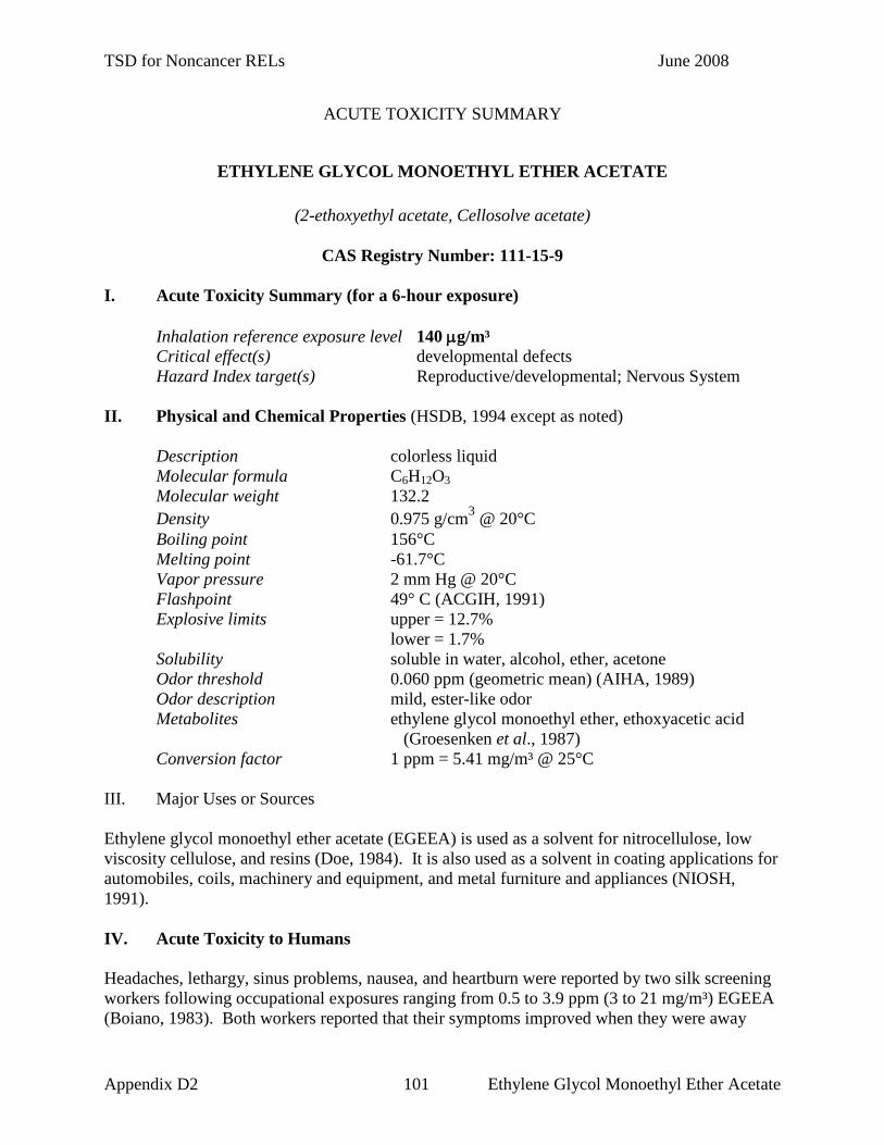
TSD for Noncancer RELs June 2008
ACUTE TOXICITY SUMMARY
ETHYLENE GLYCOL MONOETHYL ETHER ACETATE
(2-ethoxyethyl acetate, Cellosolve acetate)
CAS Registry Number: 111-15-9 I. Acute Toxicity Summary (for a 6-hour exposure) Inhalation reference exposure level 140 µg/m³ Critical effect(s) developmental defects Hazard Index target(s) Reproductive/developmental; Nervous System II. Physical and Chemical Properties (HSDB, 1994 except as noted) Description colorless liquid Molecular formula C6H12O3 Molecular weight 132.2 Density 0.975 g/cm3 @ 20°C Boiling point 156°C Melting point -61.7°C Vapor pressure 2 mm Hg @ 20°C Flashpoint 49° C (ACGIH, 1991) Explosive limits upper = 12.7% lower = 1.7% Solubility soluble in water, alcohol, ether, acetone Odor threshold 0.060 ppm (geometric mean) (AIHA, 1989) Odor description mild, ester-like odor Metabolites ethylene glycol monoethyl ether, ethoxyacetic acid (Groesenken et al., 1987) Conversion factor 1 ppm = 5.41 mg/m³ @ 25°C III. Major Uses or Sources Ethylene glycol monoethyl ether acetate (EGEEA) is used as a solvent for nitrocellulose, low viscosity cellulose, and resins (Doe, 1984). It is also used as a solvent in coating applications for automobiles, coils, machinery and equipment, and metal furniture and appliances (NIOSH, 1991). IV. Acute Toxicity to Humans Headaches, lethargy, sinus problems, nausea, and heartburn were reported by two silk screening workers following occupational exposures ranging from 0.5 to 3.9 ppm (3 to 21 mg/m³) EGEEA (Boiano, 1983). Both workers reported that their symptoms improved when they were away
Appendix D2 101 Ethylene Glycol Monoethyl Ether Acetate

TSD for Noncancer RELs June 2008
from work. Dermal absorption and concomitant exposure to other organic solvents may have contributed to the observed symptoms. It was reported in a human pharmacokinetic study that EGEEA was converted to ethylene glycol ethyl ether (EGEE) by plasma esterases and subsequently metabolized to ethoxyacetic acid (Groeseneken et al., 1987). Ethoxyacetic acid, accounting for 22.2% of the absorbed dose, was found in the urine of EGEEA exposed subjects. Predisposing Conditions for EGEEA Toxicity Medical: Persons with preexisting eye, respiratory, or neurologic conditions may be more
sensitive to the effects of EGEEA exposure (Reprotext, 1999). Chemical: Persons with concurrent exposure to ethylene glycol monoethyl ether (EGEE) or
to ethylene glycol may be more sensitive to the effects of EGEEA exposure because EGEE is a metabolite of EGEEA (Reprotext, 1999).
V. Acute Toxicity to Laboratory Animals An 8-hour LC50 in female rats is reported as 2,200 ppm (12,000 mg/m³) EGEEA (Pozzani et al., 1959). However, the lethality data were generated using chemical mixtures, not EGEEA alone. Hemoglobinuria and hematuria were observed in rabbits following a 4-hour exposure to 2,000 ppm (11,000 mg/m³) EGEEA (Truhaut et al., 1979). No other signs of toxicity were noted either during a post-exposure observation period or at necropsy. Osmotic fragility was compared in the erythrocytes of EGEEA exposed animals and unexposed animals (Carpenter et al., 1956). The erythrocytes of rats exposed to 62 ppm (340 mg/m³) EGEEA for 4-hours exhibited increased osmotic fragility as compared to the erythrocytes of unexposed rats. No increase in erythrocyte fragility was observed following a 4-hour exposure to 32 ppm (170 mg/m³) EGEEA. VI. Reproductive or Developmental Toxicity EGEEA is listed under California Proposition 65 (Cal/EPA, Safe Drinking Water and Toxic Enforcement Act of 1986) as a reproductive hazard. Tinston and colleagues (1983) exposed pregnant rabbits to 25, 100, or 400 ppm (140, 500, or 2,000 mg/m³) EGEEA 6 hours per day on days 6-18 of gestation. Significant maternal toxicity, as indicated by decreased food consumption and body weight, and a significant reduction in hemoglobin concentration were observed in the rabbits exposed to 400 ppm EGEEA. One fetus in the 25 ppm EGEEA exposed group had agenesis of the left kidney. Right kidney agenesis was observed in one fetus in the 400 ppm EGEEA exposed group. A review of the data is presented by Doe (1984).
Appendix D2 102 Ethylene Glycol Monoethyl Ether Acetate
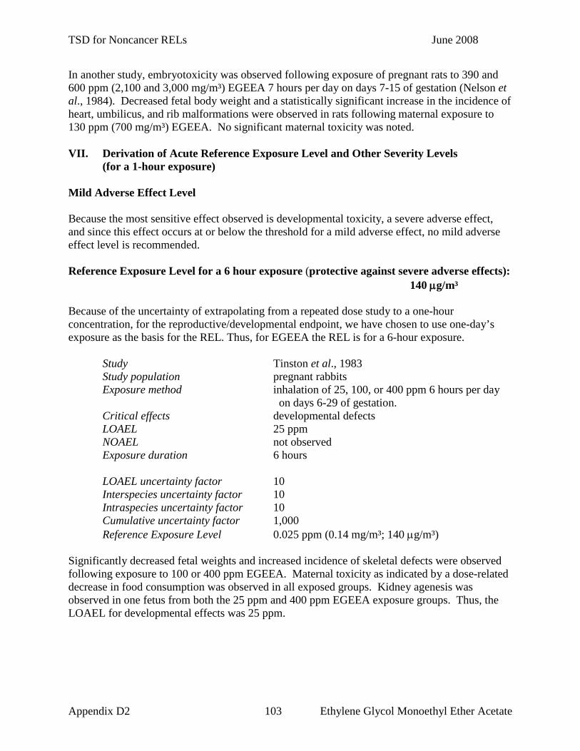
TSD for Noncancer RELs June 2008
In another study, embryotoxicity was observed following exposure of pregnant rats to 390 and 600 ppm (2,100 and 3,000 mg/m³) EGEEA 7 hours per day on days 7-15 of gestation (Nelson et al., 1984). Decreased fetal body weight and a statistically significant increase in the incidence of heart, umbilicus, and rib malformations were observed in rats following maternal exposure to 130 ppm (700 mg/m³) EGEEA. No significant maternal toxicity was noted. VII. Derivation of Acute Reference Exposure Level and Other Severity Levels
(for a 1-hour exposure) Mild Adverse Effect Level Because the most sensitive effect observed is developmental toxicity, a severe adverse effect, and since this effect occurs at or below the threshold for a mild adverse effect, no mild adverse effect level is recommended. Reference Exposure Level for a 6 hour exposure (protective against severe adverse effects): 140 µg/m³ Because of the uncertainty of extrapolating from a repeated dose study to a one-hour concentration, for the reproductive/developmental endpoint, we have chosen to use one-day’s exposure as the basis for the REL. Thus, for EGEEA the REL is for a 6-hour exposure. Study Tinston et al., 1983 Study population pregnant rabbits Exposure method inhalation of 25, 100, or 400 ppm 6 hours per day on days 6-29 of gestation. Critical effects developmental defects LOAEL 25 ppm NOAEL not observed Exposure duration 6 hours LOAEL uncertainty factor 10 Interspecies uncertainty factor 10 Intraspecies uncertainty factor 10 Cumulative uncertainty factor 1,000 Reference Exposure Level 0.025 ppm (0.14 mg/m³; 140 µg/m³) Significantly decreased fetal weights and increased incidence of skeletal defects were observed following exposure to 100 or 400 ppm EGEEA. Maternal toxicity as indicated by a dose-related decrease in food consumption was observed in all exposed groups. Kidney agenesis was observed in one fetus from both the 25 ppm and 400 ppm EGEEA exposure groups. Thus, the LOAEL for developmental effects was 25 ppm.
Appendix D2 103 Ethylene Glycol Monoethyl Ether Acetate

TSD for Noncancer RELs June 2008
Level Protective against Life-threatening Effects No recommendation is made due to the limitations of the database. NIOSH (1995) lists a (revised) IDLH of 500 ppm for 2-ethoxyethyl acetate, based on acute inhalation toxicity (specifically lethality) data in animals (Pozzani et al., 1959; Smyth et al., 1941; Truhaut et al., 1979), but states that it may be a conservative value due to the lack of relevant acute inhalation toxicity data for workers. VIII. References (ACGIH) American Conference of Governmental Industrial Hygienists. Documentation of the Threshold Limit Values and Biological Exposure Indices. 6th ed. Cincinnati (OH): ACGIH; 1991. p. 567-568. (AIHA) American Industrial Hygiene Association. Odor thresholds for chemicals with established occupational health standards. Akron (OH): AIHA; 1989. p. 18. Boiano JM. Health hazard evaluation determination report: Downing Displays, Incorporated, Cincinnati (OH). NIOSH Report No. HETA 1983;82-330-1252. Cincinnati (OH): US Department of Health, Education, and Welfare, Public Health Service, Centers for Disease Control, National Institute for Occupational Safety and Health; 1983. [cited in NIOSH, 1991.] Carpenter CP, Pozzani UC, Weil CS, Nair JH III, Keck GA, Smyth HF Jr. The toxicity of butyl Cellosolve solvent. Arch Ind Health 1956;14:114-131. Doe JE. Ethylene glycol monoethyl ether and ethylene glycol monoethyl ether acetate teratology studies. Environ Health Perspect 1984;57:33-44. Groeseneken D, Veulemans H, Masschelein R, Van Vlem E. Ethoxyacetic acid: a metabolite of ethylene glycol monoethyl ether acetate in man. Br J Ind Med 1987;44:488-493. (HSDB) Hazardous Substances Data Bank. National Library of Medicine, Bethesda (MD) (CD-ROM Version). Denver (CO): Micromedex Inc.; 1994. (Edition expires 4/30/94). (NIOSH) National Institute for Occupational Safety and Health. Criteria for a recommended standard. Occupational exposure to ethylene glycol monomethyl ether, ethylene glycol monoethyl ether, and their acetates. Cincinnati (OH): US Department of Health and Human Services, Public Health Service, Centers for Disease Control, NIOSH, Division of Standards for Development and Technology Transfer; 1991. (NIOSH) National Institute for Occupational Safety and Health. Chemical listing and documentation of revised IDLH values. 1995. (http://www.cdc.gov/niosh/intridl4.html)
Appendix D2 104 Ethylene Glycol Monoethyl Ether Acetate

TSD for Noncancer RELs June 2008
Nelson BK, Setzer JV, Brightwell WS, Mathinos PR, Kuczuk MH, Weaver TE, Goad PT. Comparative inhalation teratogenicity of four glycol ether solvents and an amino derivative in rats. Environ Health Perspect 1984;57:261-271. Pozzani UC, Weil CS, Carpenter CP. The toxicological basis of threshold limit values: 5. The experimental inhalation of vapor mixtures by rats, with notes upon the relationship between single dose inhalation and single oral dose data. Ind Hyg J 1959:364-369. Reprotext® System. Dabney B.J, editor. Denver (CO): Micromedex, Inc.; 1999. (Edition expires 1/31/1999). Smyth HF Jr, Seaton J, Fischer L. The single dose toxicity of some glycols and derivatives. J Ind Hyg Toxicol 1941;23:259-268 Ten Berge WF, Zwart A, Appelman LM. Concentration-time mortality response relationship of irritant and systemically acting vapors and gases. J Hazard Mater 1983;13:301-309. Tinston DJ, Doe JE, Killick ME, Thomas MR. Ethylene glycol monoethyl ether acetate (EEAc): inhalation teratogenicity study in rabbits. Imperial Chemical Industries PLC, Report No. CTL/P/840 to Chemical Manufacturers Association, 1983 [cited in Doe, 1984]. Truhaut R, Dutertre-Catella H, Phu-Lich N, Huyen VN. Comparative toxicological study of ethylglycol acetate and butylglycol acetate. Toxicol Appl Pharmacol 1979;51:117-127.
Appendix D2 105 Ethylene Glycol Monoethyl Ether Acetate

TSD for Noncancer RELs June 2008
ACUTE TOXICITY SUMMARY
ETHYLENE GLYCOL MONOMETHYL ETHER
(2-methoxyethanol, 1-hydroxy-2-methoxyethane, methyl cellosolve)
CAS Registry Number: 109-86-4 I. Acute Toxicity Summary (for a 6-hour exposure) Inhalation reference exposure level 93 µg/m³ Critical effect(s) teratogenic effects Hazard Index target(s) Reproductive/developmental II. Physical and Chemical Properties (HSDB, 1994 except as noted) Description colorless liquid Molecular formula C3H8O2 Molecular weight 76.09 Density 0.965 g/cm3 @ 20°C Boiling point 125°C Melting point -85.1°C Vapor pressure 6.2 mm Hg @ 20°C Flashpoint 41.7° C (closed cup) (ACGIH, 1991) Explosive limits upper = 19.8% (ACGIH, 1991) lower = 2.5% (ACGIH, 1991) Solubility miscible with water, alcohol, benzene, ether, acetone Odor threshold 2.3 ppm (Amoore and Hautala, 1983) Odor description mild ethereal odor Metabolites methoxyacetic acid, carbon dioxide (Miller et al., 1983) Conversion factor 1 ppm = 3.1 mg/m³ @ 25°C III. Major Uses or Sources Ethylene glycol monomethyl ether (EGME) is used as a solvent for cellulose acetate and resins (HSDB, 1994). It is also used in dyeing leather and in the manufacture of photographic film. EGME is used as an antifreeze in jet fuels. Quick drying varnishes, enamels, nails polishes and wood stains may also contain EGME. IV. Acute Toxicity to Humans Acute overexposure to EGME may cause irritation of the eyes, nose, and throat, drowsiness, dizziness, headache, nausea, vomiting, disorientation, and loss of consciousness (HSDB, 1994). Fatigue and hematologic effects including decreased white and red blood cell counts, and decreased hemoglobin, hematocrit and platelet levels, were observed in a microfilm manufacturing worker following daily inhalation exposure for approximately 9 months to a mean concentration of 35 ppm EGME and substantial but unquantified dermal exposure (Cohen,
Appendix D2 106 Ethylene Glycol Monomethyl Ether

TSD for Noncancer RELs June 2008
1984). Concomitant exposure to methyl ethyl ketone and propylene glycol monomethyl ether was also reported. Retention of EGME was reported to be 76% in seven male volunteers who inhaled 5 ppm EGME for 4 hours (Groeseneken et al., 1989). The average elimination half-life was 77 hours. The majority (85%) of the inhaled dose was metabolized to methoxyacetic acid. Predisposing Conditions for EGME Toxicity Medical: Persons with eye, neurologic, or hematologic conditions may be more sensitive to
the effects of EGME exposure (Reprotext, 1999). Chemical: Persons exposed to other bone marrow suppressants or substances affecting the
nervous system may be more sensitive to the effects of EGME exposure (Reprotext, 1999).
V. Acute Toxicity to Laboratory Animals A 7-hour LC50 in mice of 1,480 ppm (4,736 mg/m³) was reported (Werner et al., 1943). Rats were exposed to 100, 300, or 1,000 ppm (320, 960, or 3,200 mg/m³) EGME for 6 hours per day for 9 days (Miller et al., 1981). Reduced bone marrow cellularity, severe degeneration and necrosis of the germinal epithelium in the testes, and severe lymphoid depletion in the cortex of the thymus were observed at necropsy following exposure to 1,000 ppm (3,200 mg/m³) EGME. Red and white blood cell counts and hemoglobin levels were significantly reduced in female rats exposed to 300 or 1,000 ppm, and in male rats exposed to 100, 300, or 1,000 ppm EGME. Methoxyacetic acid and carbon dioxide were the main metabolites measured in the urine, feces and exhaled air of male rats following oral exposure to EGME (Miller et al., 1983). The majority of the metabolites were recovered in the urine, with smaller amounts in the exhaled air and feces. VI. Reproductive or Developmental Toxicity EGME is listed under California Proposition 65 (Cal/EPA, Safe Drinking Water and Toxic Enforcement Act of 1986) as a reproductive hazard with male reproductive toxicity and developmental endpoints. Hanley and colleagues (1984) exposed pregnant rats and rabbits to 3, 10, or 50 ppm (9.6, 32, or 160 mg/m³) EGME for 6 hours per day on days 6-15 (rats) or 6-18 (rabbits) of gestation. Pregnant mice were exposed to 10 or 50 ppm (32 or 160 mg/m³) EGME for 6 hours per day on days 6-15 of gestation. A statistically significant increase in the incidence of skeletal variations was observed in rats and mice following maternal exposure to 50 ppm EGME. Gross soft tissue and skeletal teratogenic effects and significantly decreased fetal body weights were observed in rabbits following maternal exposure to 50 ppm EGME. In rabbits, a significant increase in the rate of fetal resorption was observed in the 10 ppm exposure group. Thus 10 ppm was considered a LOAEL for increased resorptions and 3 ppm a NOAEL. Although the authors
Appendix D2 107 Ethylene Glycol Monomethyl Ether

TSD for Noncancer RELs June 2008
attribute the statistical significance of this effect to an unusually low rate of resorptions in controls compared to historical controls, historical control data were not presented. Maternal toxicity as indicated by decreased body weight gain was observed in all three species exposed to 50 ppm. Pregnant rats exposed to EGME exhibited statistically significant lower mean hemoglobin levels and packed cell volumes at all 3 exposure levels. Thus 3 ppm was selected as a LOAEL for these 2 hematologic effects. A NOAEL was not identified. A lower mean red blood cell count was observed in rat dams exposed to 50 ppm EGME. In another study, male rats were exposed to 30, 100, and 300 ppm (96, 320, and 960 mg/m³) EGME for 6 hours per day, 5 days per week for 13 weeks before mating with unexposed female rats (Rao et al., 1983). A decrease in fertility, body and testes weights, and an increase in the incidence of gross and microscopic testicular and epididymal lesions were observed in the male rats exposed to 300 ppm (960 mg/m³). Complete resorption of all fetuses was observed in the unexposed females mated with the males exposed to 300 ppm EGME. A male reproductive NOAEL of 100 ppm (320 mg/m³) EGME was observed. VII. Derivation of Acute Reference Exposure Level and Other Severity Levels
(for a 1-hour exposure) Level Protective against Mild Adverse Effects: Because the most sensitive effect observed is developmental toxicity, a severe adverse effect, and since this effect is observed at or below the threshold for a less serious effect, no mild adverse effect level is recommended. Reference Exposure Level for 6 hr exposure (Protective Against Severe Adverse Effects):
0.03 ppm (93 µg/m³) Study Hanley et al., 1984 Study population pregnant rabbits Exposure method inhalation of 3, 10, or 50 ppm EGME 6 hours per day on days 6-15 of gestation Critical effects gross soft tissue and skeletal teratogenic effects and
significantly decreased fetal body weights LOAEL 10 ppm NOAEL 3 ppm Exposure duration 6 hours LOAEL uncertainty factor 1 Interspecies uncertainty factor 10 Intraspecies uncertainty factor 10 Cumulative uncertainty factor 100 Reference Exposure Level 0.03 ppm (0.093 mg/m³; 93 µg/m³) Pregnant rabbits were exposed to 3, 10, or 50 ppm EGME 6 hours per day on days 6-18 of gestation (Hanley et al., 1984). Maternal toxicity, as indicated by decreased body weight gain, was observed only in rabbits exposed to 50 ppm EGME. The authors report that the hematologic parameters of EGME exposed rabbits were not altered at any dose. Gross soft tissue and skeletal
Appendix D2 108 Ethylene Glycol Monomethyl Ether

TSD for Noncancer RELs June 2008
teratogenic effects and significantly decreased fetal body weight were observed in rabbits following maternal exposure to 50 ppm EGME. Statistically significant increases in fetal resorption rates were observed following maternal exposure to 10 or 50 ppm EGME. A NOAEL of 3 ppm for increased resorptions was used to develop the REL. An uncertainty factor of 100 was applied to account for inter- and intraspecies differences. Dividing this by 100 gives a level protective against severe adverse effects for a 6 hour exposure of 0.03 ppm (0.093 mg/m3; 93 µg/m³). Level Protective Against Life-threatening Effects Mice were exposed to EGME at concentrations of 930 to 6,800 ppm for a single 7-hour exposure (Werner et al., 1943). The mortality during exposure and up to three weeks following were recorded. The NOAEL was 930 ppm and was extrapolated from 7-hour to 1-hour exposure using a modification of Haber’s equation, Cn * T = K, where n = 2. An uncertainty factor (UF) of 100 was applied to the time-adjusted NOAEL of 2,461 ppm to account for interspecies variability and individual human variation. The final 1-hour level protective against life-threatening effects for EGME is 25 ppm. (A benchmark dose approach (Crump, 1984; Crump and Howe, 1983) could not be employed because log-normal probit analysis of the lethality data was shown to be too heterogeneous.) NIOSH (1995) lists an IDLH of 200 ppm derived by multiplying the current NIOSH REL of 0.1 ppm by 2,000, an assigned protection factor for respirators. VIII. References (ACGIH) American Conference of Governmental Industrial Hygienists. Documentation of the Threshold Limit Values and Biological Exposure Indices. 6th ed. Cincinnati (OH): ACGIH; 1991. p. 913-921. Amoore JE, Hautala E. Odor as an aid to chemical safety: odor thresholds for compounds with threshold limit values and volatilities for 24 industrial chemicals in air and water dilution. J Appl Toxicol 1983;3(6):272-290. Cohen R. Reversible subacute ethylene glycol monomethyl ether toxicity associated with microfilm production. A case report. Am J Ind Med 1984;6:441-446. Crump KS. A new method for determining allowable daily intakes. Fundam Appl Toxicol 1984;4:854-871. Crump KS, Howe R. Probit-A computer program to extrapolate quantile animal toxicological data to low doses. Ruston (LA): KS Crump & Company Inc.; 1983. Groeseneken D, Veulemans H, Masschelein R, Van Vlem E. Experimental human exposure to ethylene glycol monomethyl ether. Int Arch Occup Environ Health 1989;61:243-7.
Appendix D2 109 Ethylene Glycol Monomethyl Ether

TSD for Noncancer RELs June 2008
Hanley TR Jr, Yano BL, Nitschke KD, John JA. Comparison of the teratogenic potential of inhaled ethylene glycol monomethyl ether in rats, mice, and rabbits. Toxicol Appl Pharmacol 1984;75:409-422. (HSDB) Hazardous Substances Data Bank. National Library of Medicine, Bethesda (MD) (CD-ROM Version). Denver (CO): Micromedex, Inc.; 1994. (Edition expires 4/30/94). Miller RR, Ayres JA, Calhoun LL, Young JT, McKenna MJ. Comparative short-term inhalation toxicity of ethylene glycol monomethyl ether and propylene glycol monomethyl ether in rats and mice. Toxicol Appl Pharmacol 1981;61:368-377. Miller RR, Hermann EA, Largvardt PW, McKenna MJ, Schwetz BA. Comparative metabolism and disposition of ethylene glycol monomethyl ether and propylene glycol monomethyl ether in male rats. Toxicol Appl Pharmacol 1983;67:229-237. (NIOSH) National Institute for Occupational Safety and Health. Chemical listing and documentation of revised IDLH values. 1995. (http://www.cdc.gov/niosh/intridl4.html) Rao KS, Cobel-Geard SR, Young JT, Hanley TR Jr, Hayes WC, John JA. Ethylene glycol monomethyl ether. II. Reproductive and dominant lethal studies in rats. Fundam Appl Toxicol 1983;3:80-85. Reprotext® System. Dabney BJ, editor. Denver (CO): Micromedex, Inc.; 1999. (Edition expires 1/31/1999). Ten Berge WF, Zwart A, Appelman LM. Concentration-time mortality response relationship of irritant and systemically acting vapors and gases. J Hazard Mater 1983;13:301-309. Werner HW, Mitchell JL, Miller JW, Von Oettingen WF. The acute toxicity of vapors of several monoalkyl ethers of ethylene glycol. J Ind Hyg Toxicol 1943;25:157-163.
Appendix D2 110 Ethylene Glycol Monomethyl Ether

TSD for Noncancer RELs June 2008
ACUTE TOXICITY SUMMARY
HYDROGEN CHLORIDE
(hydrogen chloride, anhydrous hydrogen chloride, muriatic acid)
CAS Registry Number: 7647-01-1 I. Acute Toxicity Summary (for a 1-hour exposure) Inhalation reference exposure level 2,100 µg/m³ Critical effect(s) upper respiratory symptoms Hazard Index target(s) Respiratory System; Eyes II. Physical and Chemical Properties (HSDB, 1994 except as noted) Description colorless gas Molecular formula HCl Molecular weight 36.46 Density 1.49 g/L @ 25°C Boiling point -84.9°C Melting point -114.8°C Flashpoint not applicable Explosive limits unknown Solubility soluble in water, alcohol, benzene, ether; insoluble in
hydrocarbons Odor threshold 0.26-10.0 ppm (AIHA, 1989a) Odor description sharp, irritating (AIHA, 1989a) Metabolites not applicable Conversion factor 1 ppm = 1.49 mg/m³ @ 25°C III. Major Uses or Sources Hydrogen chloride (HCl) is used in the manufacture of vinyl chloride, fertilizers, dyes, artificial silk, and pigments for paints. It is also used in electroplating, soap refining, and leather tanning. Other consumers of HCl include the photographic, textile and rubber industries (HSDB, 1994). Hydrogen chloride is produced in large quantities during combustion of most materials and especially chlorine-containing materials. Thus, HCl is a major product formed during the thermal decomposition of polyvinyl chloride, a commonly used plastic polymer (Burleigh-Flayer et al., 1985). It is also released in large quantities during the test firing of some rocket and missile engines (Wohlslagel et al., 1976).
Appendix D2 111 Hydrogen Chloride

TSD for Noncancer RELs June 2008
IV. Acute Toxicity to Humans Inhalation exposure to high concentrations of HCl fumes may result in coughing, choking sensation, burning of the respiratory tract, and pulmonary edema (Proctor et al., 1991). Dental erosion has been reported in workers chronically exposed to low levels of gaseous hydrogen chloride (Finkel, 1983). Reactive Airway Dysfunction Syndrome (RADS; acute, irritant-induced asthma) was reported in three male police officers (36-45 years old) who responded to a roadside chemical spill (Promisloff et al., 1990). Other reports of RADS include individual occupational cases (Boulet, 1988; Turlo and Broder, 1989). Young adult asthmatic subjects (18-25 years, 5 of each sex) were exposed by a half-face mask to filtered air, 0.8 ppm HCl, and 1.8 ppm HCl during three separate 45-minute exposures (Stevens et al., 1992). The exposure protocol included two 15-minute exercise periods separated by a 15-minute rest period. Tests of pulmonary function included forced expiratory volume in 1 second, forced expiratory volume, maximal flow at 50% and 75% of expired vital capacity, and total respiratory resistance and peak flow. Nasal work of breathing was also measured pre- and post exposure. No significant changes in these parameters were observed following exposure to HCl at 0.8 or 1.8 ppm. There was no exposure-related increases in severity of upper respiratory, lower respiratory, or other symptoms reported by participants. Because exposure occurred by half-face mask, effects on the ocular mucosae were not addressed. Predisposing Conditions for HCl Toxicity Medical: Persons with preexisting skin, eye, gastrointestinal tract (including ulcers) or
respiratory conditions or underlying cardiopulmonary disease may be more sensitive to the effects of HCl exposure (Reprotext, 1999).
Chemical: Persons also exposed to formaldehyde might be at increased risk for developing
cancer (Reprotext, 1999). V. Acute Toxicity to Laboratory Animals A single baboon exposed for 5-minutes to 16,570 ppm (24,690 mg/m³) HCl was dyspneic until death 18 days following exposure (Kaplan et al., 1985). Pneumonia, pulmonary edema, tracheitis, and epithelial erosion were noted at autopsy. Baboons exposed for 15-minutes to 500, 5,000 or 10,000 ppm (750, 7,500, or 15,000 mg/m³) HCl exhibited a concentration-related increase in respiratory rate and minute volume (Kaplan et al., 1988). A marked decrease in arterial blood oxygenation was observed in baboons exposed to 5,000 or 10,000 ppm. Pulmonary function parameters measured 3 days and 3 months following exposure were not significantly different from pre-exposure measurements. However, the animals were anesthetized with Ketamine which could reduce airway resistance and bronchospasm (Bovill et al., 1971). Histopathologic examination performed 12 months post-exposure (Kaplan et al., 1993a) found pulmonary hemorrhage, edema, fibrosis, and bronchiolitis in the medial right lung of one of three animals exposed to 10,000 ppm. In another of the three animals zonal atelectasis and focal multiple hemorrhages were observed in the right lung. In each of the three animals exposed to 5,000 ppm and examined, focal, patchy hemorrhages were observed.
Appendix D2 112 Hydrogen Chloride

TSD for Noncancer RELs June 2008
A 30-minute LC50 in rats and mice is reported as 5,666 ppm (8,442 mg/m³) and 2,142 ppm (3,192 mg/m³) HCl aerosol, respectively (Darmer et al., 1974). Alveolar emphysema, atelectasis, and pulmonary edema were noted at necropsy of animals that died either during or within 7 days following exposure. Bloody nasal discharge, indicative of purulent bronchitis, was observed in animals of both species surviving the exposure. A 1-hour LC50 of 2,810 ppm in rats was reported by Hartzell and colleagues (1985). Rats were exposed to concentrations of HCl ranging from 1,793-4,854 ppm HCl for one hour and the mortality following exposure was recorded over a 14-day observation period. Hartzell et al. also reported LC50s of 15,900 ppm, 8,370 ppm, 6,920 ppm, 5,920 ppm and 3,715 ppm, for rats exposed for 5 minutes, 10 minutes, 15 minutes, 22.5 minutes, and 30 minutes, respectively. A decrease in respiratory rate was observed in guinea pigs exposed to 320 ppm (480 mg/m³) HCl for 6-minutes and to 680 ppm (1,010 mg/m³) HCl for less than 1-minute (Burleigh-Flayer et al., 1985). The RD50 is the concentration of a chemical in air that is associated with a 50% decrease in respiratory rate, and is used as a measure of irritancy. The RD50 in animals has a predictable relationship to irritation in man (Kane et al., 1979). The RD50 in mice was reported as 309 ppm (460 mg/m³) for a 10-minute exposure (Kane et al., 1979). In addition to respiratory irritation, HCl exerts ocular effects. Corneal opacities were observed in guinea pigs following a 30-minute exposure to HCl concentrations of 680 ppm (1,010 mg/m³ ; 1 of 4), 1,040 ppm (1,550 mg/m³; 4 of 6) and 1,380 ppm (5 of 5), but not 320 ppm (480 mg/m³ ). Cloudy corneas were also reported 90 days post-exposure by Kaplan et al. (1993b) in guinea pigs exposed for 15 minutes to 4,200 ppm, but not at 500 ppm (Burleigh-Flayer et al., 1985). Coughing, frothing at the mouth, excess salivation, and blinking and rubbing of the eyes were observed in baboons following a 5-minute exposure to 810 ppm (1,210 mg/m³) HCl (Kaplan et al., 1985). No signs of irritation were observed following a 5-minute exposure to 190 ppm (280 mg/m³) HCl. In another study conducted in exercising guinea pigs (Malek and Alarie, 1989), a concentration of 107 ppm for 30 minutes was irritating and a concentration of 140 ppm was incapacitating at 16.5 minutes. VI. Reproductive or Developmental Toxicity The reproductive hazard of hydrogen chloride to humans is unknown (Reprotext, 1999). Few studies on the reproductive effects of HCl exposure were found in the literature. Maternal exposure to a high concentration of a strong acid could result in metabolic acidosis and subsequent fetal acidemia which has been linked with low Apgar scores, neonatal death, and seizures. However, there is no evidence linking HCl exposure to fetal acidemia (Reprotext, 1999). Pregnant rats exposed to 300 ppm (450 mg/m³) HCl for 1 hour on the 9th day of gestation exhibited signs of severe dyspnea and cyanosis (Pavlova, 1976; 1978). The exposure was lethal to one-third of the exposed rats (number of rats exposed not reported). Increased mortality was also observed in the progeny of the exposed rats compared to that of controls. The author
Appendix D2 113 Hydrogen Chloride

TSD for Noncancer RELs June 2008
implies that organ functional abnormalities in the progeny resulted from in utero exposure. However, the lack of key experimental details and the ambiguity of organ function tests make this conclusion difficult to validate. VII. Derivation of Acute Reference Exposure Level and Other Severity Levels
(for a 1-hour exposure) Reference Exposure Level (protective against mild adverse effects): 1.4 ppm (2,100 µg/m³) Study Stevens et al., 1992 Study population 10 asthmatics aged 18-25 Exposure method inhalation via half face mask to 0.8 or 1.8 ppm HCl Critical effects upper respiratory system symptoms of sore throat; nasal discharge LOAEL not observed NOAEL 1.8 ppm Exposure duration 45 minutes Extrapolated 1 hour concentration 1.4 ppm (1.81 ppm* 0.75 h = C1 * 1 h ) (see Table 12 for information on “n”) LOAEL uncertainty factor 1 Interspecies uncertainty factor 1 Intraspecies uncertainty factor 1 Cumulative uncertainty factor 1 Reference Exposure Level 1.4 ppm (2.1 mg/m³; 2,100 µg/m³) No significant effects on pulmonary function (forced expiratory volume in one second, forced expiratory volume, maximal flow at 50% and 75% of expired vital capacity, and total respiratory resistance and peak flow) or nasal work of breathing were observed in asthmatics aged 18-25 years exposed via half-face mask to 0.8 or 1.8 ppm HCl for 45 minutes, including 30 minutes of exercise. Additionally, there was no association between HCl exposure and upper respiratory symptoms of sore throat and nasal discharge. There was no association between HCl exposure and lower respiratory symptoms of cough, chest pain, burning, dyspnea and wheezing. The lack of effects on the pulmonary functions measured is not surprising because of the extreme water-solubility of HCl. The high water solubility of HCl supports upper airway effects as the most sensitive target endpoint since the HCl would dissolve there. While the animal studies summarized in this document suggest that HCl does penetrate and affect the lower respiratory system, this would be expected to occur mostly at higher concentrations of HCl. Level Protective Against Severe Adverse Effects The RD50 in mice for a 10-minute exposure to HCl is reported as 309 ppm (460 mg/m³). NRC applied an uncertainty factor of 10 to the RD50 to account for interspecies differences yielding a 1-hour EEGL of 31 ppm. The EEGL was further reduced to 20 ppm (29.8 mg/m³) because “of the paucity of human data.”
Appendix D2 114 Hydrogen Chloride

TSD for Noncancer RELs June 2008
A 1-hour SPEGL (Short-term Public Emergency Planning Level) of 1 ppm is also recommended by NRC. The rationale states “...in connection with community exposure during space shuttle launches, the Committee recommends lower concentrations, to avoid adverse effects that might occur in a more sensitive population...” (NRC, 1987). While it appears that no supporting data are cited to justify the value, the SPEGL essentially incorporates an additional 20-fold safety factor to protect sensitive subpopulations and is an excessively low value, lower than the acute REL recommended to protect against mild adverse effects. However, since the development of the SPEGL, that relied largely on expert judgment since the database was poor (NRC, 1987), the Stevens et al. (1992) human study has become available, in addition to a number of additional animal studies. For this reason, we recommend the EEGL of 20 ppm as a level protective against severe adverse effects. The levels should be reevaluated when more data become available. Level Protective Against Life-threatening Effects Groups of 6 rats were exposed to the following concentrations of HCl for a single 1-hour period: 1,793, 2,281, 2,600, 4,277, 4,460, and 4,854 ppm (Hartzell et al., 1985). Mortality during and up to 14 days following exposure was reported. Rat Mortality Data from Hartzell et al., 1985
HCl Concentration (ppm) 1,793 2,281 2,600 4,277 4,460 4,854
Mortality 0/6 3/6 1/6 7/8 6/6 6/6 The rat study was chosen since it was considered to be of greatest quality based on the number of doses and time points tested. Furthermore, Kaplan et al. (1987 and 1993b) suggest fairly similar lethality responses between baboons and rats for HCl exposure. A benchmark dose approach was employed using a log-normal probit analysis (Crump, 1983) of 60-minute lethality data from Hartzell et al. (1985). The concentration associated with a 5% incidence of lethality (ED05) was 1,772 ppm; the lower 95% confidence limit (LCL) on this concentration [the BC05] was 1,271 ppm. A total uncertainty factor of 30 was applied to the BC05 of 1,271 ppm to account for interspecies variability (3) and individual variation (10) in response.
level protective against life-threatening effects = BC05/(UF)
The final level protective against life-threatening effects for HCl is therefore 42 ppm (63 mg/m³). The maximum likelihood estimates (MLE) and 95% lower confidence limits (LCL) for the 1% and 5% response rates are compared below. Refer to section IX of this toxicity summary for the graphic representation of benchmark dose derivation. Comparison of benchmark calculations (1% vs 5%)
Response rate MLE (ppm) 95% LCL (ppm)
1% 1,464 946
5% 1,772 1,271
Appendix D2 115 Hydrogen Chloride

TSD for Noncancer RELs June 2008
VIII. References (AIHA) American Industrial Hygiene Association. Emergency response planning guidelines. Hydrogen chloride. Akron (OH): AIHA; 1989. (AIHA) American Industrial Hygiene Association. Odor thresholds for chemicals with established occupational health standards. Akron (OH): AIHA; 1989a. p. 20 Boulet L-P. Increases in airway responsiveness following acute exposure to respiratory incidents: Reactive airway dysfunction syndrome or occupational asthma? Chest 1988; 94: 476-481. Bovill JG, Clarke RSJ, Davis EA, Dundee JW. Some cardiovascular effects of ketamine in man. Br J Pharmacol 1971;41:411P-412P. Burleigh-Flayer H, Wong KL, Alarie Y. Evaluation of the pulmonary effects of HCl using CO2 challenges in guinea pigs. Fundam Appl Toxicol 1985;5:978-985. Crump KS & Co., Inc. Probit (Log-Normal) software for the IBM-PC. Ruston (LA); 1983. Darmer KI, Kinkead ER, Di Pasquale LC. Acute toxicity in rats and mice exposed to hydrogen chloride gas and aerosol. Am Ind Hyg Assoc J 1974;35:623-631. Finkel AJ, editor. Hamilton and Hardy’s industrial toxicology. Boston: John Wright, PSG Inc.; 1983. p. 179. Hartzell GE, Packham SC, Grand AF, Switzer WG. Modeling of toxicological effects of fire gases: III. Quantification of post-exposure lethality of rats from exposure to HCl atmospheres. J Fire Sci 1985;3:195-207. (HSDB) Hazardous Substances Data Bank. National Library of Medicine, Bethesda (MD) (CD-ROM version). Denver (CO): Micromedex, Inc.; 1994. (Edition expires 4/30/94). Henderson Y, Haggard HW. Noxious gases. New York: Reinhold Publishing Corp; 1943. p. 126. (IRIS) Integrated Risk Information System. US Environmental Protection Agency, Washington (DC) (CD-ROM Version). Denver (CO): Micromedex Inc.; 1994. (Edition expires 4/30/94). Kane LE, Barrow CS, Alarie Y. A short-term test to predict acceptable levels of exposure to airborne sensory irritants. Am Ind Hyg Assoc J 1979;40:207-229. Kaplan HL, Grand AF, Switzer WG, Mitchell DS, Rogers WR, Hartzell GE. Effects of combustion gases on escape performance of the baboon and rat. J Fire Sci 1985;3:228-244. Kaplan HL, Anzueto A, Switzer WG, Hinderer RK. Effects of hydrogen chloride on respiratory response and pulmonary function of the baboon. J Toxicol Environ Health 1988;23:473-493.
Appendix D2 116 Hydrogen Chloride

TSD for Noncancer RELs June 2008
Kaplan HL, Switzer WG, Hinderer RK, Anzueto A. A study on the acute and long-term effects of hydrogen chloride on respiratory response and pulmonary function and morphology in the baboon. J Fire Sci 1993a; 11: 459-484. Kaplan HL, Switzer WG, Hinderer RK, Anzueto A. Studies of the effects of hydrogen chloride and polyvinyl chloride (PVC) smoke in rodents. J Fire Sci 1993b; 11: 512-552. (NRC) National Research Council. Committee on Toxicology. Emergency and continuous exposure limits for selected airborne contaminants. Vol. 7. Washington (DC): National Academy Press; 1987. p. 17-30. Pavlova TE. [Disorders in the development of offspring following exposure of rats to hydrogen chloride] [Russian]. Biull Eksp Biol Med 1976;82:866-868. Pavlova TE. Disturbance of development of the progeny of rats exposed to hydrogen chloride. Bull Exp Biol Med 1978;2:1078-1081. Proctor NH, Hughes JP, Hathaway GJ, Fischman ML, editors. Proctor and Hughes’ chemical hazards of the workplace. 3rd ed. New York: Van Nostrand Reinhold; 1991. p. 322. Promisloff RA, Lenchner GS, Phan A, Cichelli AV. Reactive airway dysfunction syndrome in three police officers following a roadside chemical spill. Chest 1990;98:928-929. Reprotext ® System. Dabney BJ, editor. Denver (CO): Micromedex, Inc.; 1999. (Edition expires 1/31/1999). Stevens B, Koenig JQ, Rebolledo V, Hanley QS, Covert DS. Respiratory effects from the inhalation of hydrogen chloride in young adult asthmatics. J Occup Med 1992;34(9):923-929. Ten Berge WF, Zwart A, Appelman LM. Concentration-time mortality response relationship of irritant and systemically acting vapors and gases. J Hazard Mater 1983;13:301-309. Turlo SM, Broder I. Irritant-induced occupational asthma. Chest 1989; 96: 297-300. Wohlslagel J, Di Pasquale LC, Vernot EH. Toxicity of solid rocket motor exhaust of HCl, HF, and alumina on rodents. J Combust Toxicol 1976;3:61-70.
Appendix D2 117 Hydrogen Chloride

TSD for Noncancer RELs June 2008
ACUTE TOXICITY SUMMARY
HYDROGEN CYANIDE
(formonitrile; hydrogen cyanide; prussic acid)
CAS Registry Number: 74-90-8 I. Acute Toxicity Exposure Levels (for a 1-hour exposure) Inhalation reference exposure level 340 µg/m³ Critical effect(s) loss of coordination and loss of consciousness, due to cellular hypoxia of the central nervous system Hazard Index target(s) Nervous System II. Physical and Chemical Properties (HSDB, 1994 except as noted) Description colorless gas Molecular formula HCN Molecular weight 27.03 Density 1.1 g/L @ 25°C Boiling point 25.6°C Melting point -13.4°C Vapor pressure 630 mm Hg @ 20°C Flashpoint -17.8°C (closed cup) Explosive limits upper = 40% by volume in air lower = 5.6% by volume in air Solubility miscible in water, alcohol, slightly soluble in ether Odor threshold 0.58 ppm (w/w) (Amoore and Hautala, 1983) Odor description faint, bitter almond odor Metabolites thiocyanate, 2-aminothiazo-line-4-carboxylic acid, cyanocobalamin (Vitamin B12) (Ansell and Lewis, 1970) Conversion factor 1 ppm = 1.13 mg/m³ III. Major Uses or Sources Hydrogen cyanide (HCN) is used in a variety of syntheses, including the production of adiponitrile (for nylon), methyl methacrylate, sodium cyanide, cyanuric chloride, chelating agents, pharmaceuticals, and other specialty chemicals. Manufacturing activities producing HCN include electroplating, metal mining, metallurgy, and metal cleaning processes. Additionally, HCN has some insecticide and fungicide applications (ATSDR, 1993). Fires involving some nitrogen-containing polymers, often found in fibers used in fabrics, upholstery covers, and padding, also produce HCN (Tsuchiya and Sumi, 1977).
Appendix D2 118 Hydrogen Cyanide

TSD for Noncancer RELs June 2008
Another common source of HCN is cigarette smoke. Levels in inhaled mainstream cigarette smoke range from 10 to 400 µg per cigarette and decrease to levels ranging from 0.06 to 108 µg in secondary or sidestream smoke (Fiksel et al., 1981). IV. Acute Toxicity to Humans Cyanide toxicity results from cytochrome oxidase inhibition which prevents cellular utilization of oxygen. The respiratory, cardiovascular, and central nervous systems are the primary target organs of acute cyanide toxicity. Acute effects from inhalation of HCN are characterized by altered sense of smell, headache, tachypnea, nausea, loss of coordination, loss of consciousness, palpitations, convulsions, respiratory distress, and asphyxiation (Chandra et al., 1980; Blanc et al., 1985; Peden et al., 1986; ATSDR, 1993). Eye or dermal contact with liquid HCN, a weak acid, may cause some mild local irritation (Anon., 1970). However, dermal and ocular absorption leading to systemic effects is clearly more cause for concern than possible local irritation. Even though the signs and symptoms of HCN poisoning are recognized, the acute dose-response relationship has not been well defined. Lethality data from case report studies exist, but specific exposure concentrations are often lacking. As reported by McNamara (1976), several commonly reported inhalation values given as human toxicity data (Kobert, 1912; Henderson and Haggard, 1927; Flury and Zernick, 1931; Dudley et al., 1942; Moore and Gates, 1946; Fassett, 1963) may actually be based on pre-1920 animal data. One estimate of the average fatal inhaled dose for humans, 546 ppm (617 mg/m³), is based on minimal human data and relies on multiple unsubstantiated assumptions including: (1) human susceptibility to HCN is similar to the relatively resistant monkey and goat, and (2) animal data, such as breathing rates, can be substituted for human parameters (McNamara, 1976). In an accidental human poisoning, a workman collapsed 3 minutes after entering a tank for inspection and cleaning (Bonsall, 1984). The workman was exposed for an additional 3 minutes before being fitted with a breathing apparatus and taken to a hospital, where he later recovered. Later analysis of the tank revealed an HCN concentration of 500 mg/m³ (442 ppm). In a fatal human poisoning, a workman cleaning the bottom of a silver plating tank was found unconscious by workmates (Singh et al., 1989). The duration of exposure was unknown but subsequent analysis of the air in the tank revealed a concentration of 200 ppm HCN. The onset and progression of severe health effects are similar among humans and experimental animals (ATSDR, 1993, Ballantyne, 1987; Wexler et al., 1947, Purser et al., 1984). These effects are hyperventilation, followed by loss of consciousness, depressed respiration, and bradycardia. Blanc et al. (1985) studied 36 former workers who had been exposed to HCN in a silver-reclaiming facility. A significant dose-response trend was observed between proximity of work to the CN- source and prevalence of symptoms consistent with CN- toxicity including headache, dizziness, nausea or vomiting, dyspnea, and syncope (unconsciousness). A 24-hour time-weighted average air concentration of 15 ppm was recorded 1 day after the plant had been closed because of a death from cyanide exposure. Due to poor hygienic conditions at the plant, dermal
Appendix D2 119 Hydrogen Cyanide

TSD for Noncancer RELs June 2008
and oral exposure also occurred. The researchers considered the time-weighted average of 15 ppm to be a low estimate of the occupational exposure due to multiple potential routes of exposure and the retrospective analysis of the air concentration. Predisposing Conditions for HCN Toxicity Medical: Individuals with some motor neuron diseases, such as amyotrophic lateral
sclerosis, have a decreased ability to convert cyanide to thiocyanate and may be predisposed to HCN toxicity (Kato et al., 1985). Individuals with Leber’s hereditary optic atrophy, a rare neuroopthalmologic condition, may have low activity of the enzyme rhodanese, an enzyme responsible for converting cyanide to thiocyanate (Wilson, 1983).
Up to 20% to 40% of the population cannot detect the bitter almond odor of
cyanide and may therefore be at greater risk for toxicity following exposure (Brown and Robinette, 1967).
Chemical: Individuals taking megadoses of ascorbic acid may diminish the availability of
cysteine, an amino acid important in the detoxification of cyanide, thus increasing susceptibility to HCN poisoning (Basu, 1983).
V. Acute Toxicity to Laboratory Animals The progression of severe health effects is similar among humans and experimental animals (ATSDR, 1993, Kulig and Ballantyne, 1993; Curry, 1992; Ballantyne, 1987; Wexler et al., 1947, Purser et al., 1984). These effects are characterized by hyperventilation, followed by loss of coordination and consciousness, depressed respiration, bradycardia, convulsions, asphyxiation, and respiratory failure. In work by Purser (1984), 4 monkeys exposed to 60 ppm HCN developed electroencephalogram (EEG) patterns characteristic of early onset of CNS depression (increased slow wave [delta] activity and decreased fast wave [beta] activity) and increased respiratory rate near the end of the 30 minute exposure period. While both results are indicative of early onset of cellular hypoxia, none of the monkeys lost consciousness. However, with exposures to 80 ppm and above, incapacitation (semi-conscious state with loss of muscle tone) did result within 30 minutes (Purser et al., 1984). Time-to-incapacitation, as a function of HCN concentration, has been measured in mice (Sakurai, 1989), rats (Hartzell et al., 1985), monkeys (Purser et al., 1984; Purser, 1984), and goats (Barcroft, 1931). The tests used by Barcroft (1931) and Purser et al. (1984) essentially defined incapacitation as a semi-conscious state with loss of muscle tone, whereas Sakurai (1989) and Hartzell et al. (1985) defined incapacitation as complete loss of consciousness. A linear relationship between gas concentration and mean incapacitation time can be shown as:
C = (a/t) + b
Appendix D2 120 Hydrogen Cyanide

TSD for Noncancer RELs June 2008
where C = gas concentration (ppm), t = incapacitation time (min), and a, b = coefficients for HCN gas.
The HCN concentration producing a mean incapacitation time of 30 minutes, using the equation C = (a/t) + b, is shown in Table 1. Table 1. Tabulation of modeling constants for use in the equation C = (a/t) + b for various
experimental animal species and determination of HCN concentration resulting in incapacitation following 30 minute exposure to HCN.
Reference Species a (slope) b (y-intercept) Concentration (ppm)1
Sakurai (1989) mouse 491 25 42
Hartzell et al. (1985) rat 698 92 115
Purser et al. (1984) monkey 685 66 89
Barcroft (1931) goat 885 152 182 1 Concentration of HCN producing a mean incapacitation time of 30 minutes. While the above equation can estimate the mean time-to-incapacitation for a given concentration of HCN, it cannot provide a NOAEL for incapacitation. However, the coefficient b (y-intercept) could be viewed as the concentration of HCN below which incapacitation will not occur in normal experimental animals. In mice, Sakurai (1989) has shown that exposure to HCN concentrations of approximately 150 ppm and above results in incapacitation and apnea at about the same time, within 5 minutes. However, exposures to lower HCN concentrations (approximately 150 ppm or less) result in incapacitation in about one-third the time required to cause apnea. This latter situation is observed when incapacitation occurs at 10 minutes or later into exposure to HCN. Rats inhaling 64 ppm HCN were incapacitated after a mean duration of 35 minutes, while those inhaling 184 ppm HCN were incapacitated after a mean of 5 minutes (Chaturvedi et al., 1995). Blood cyanate levels did not predict incapacitation onset, since the blood cyanate at incapacitation following 184 ppm HCN inhalation was half that seen upon incapacitation following 64 ppm HCN inhalation. In rats, Levin et al. (1987) observed that incapacitating levels were approximately 65% of lethal levels for exposure durations ranging from 1 to 10 minutes. Also in rats, Hartzell et al. (1985) observed that time-to-lethality was about 2 to 6-fold greater for a given concentration of HCN that produces incapacitation within 1 to 21 minutes. For exposures that produced mean incapacitation times of 10.9 and 21.0 minutes (165 and 127 ppm, respectively), the mean time-to-lethality was 3- to 4-fold greater. Purser et al. (1984) noted that a monkey exposed to 147 ppm HCN was incapacitated at 8 minutes and developed apnea at 27 minutes, a 3.4 fold difference. Other monkeys exposed to similar or lower levels of HCN did not develop apnea.
Appendix D2 121 Hydrogen Cyanide

TSD for Noncancer RELs June 2008
Therefore, there is a clear (though steep) dose-response effect for HCN exposure resulting in incapacitation (a severe adverse effect) followed by apnea (a life-threatening effect) and death. Numerous citations were located in the literature that contained LC50 determinations for HCN at various exposure durations in experimental animals, but many of the studies did not include the raw mortality data from which to estimate an MLE05 (maximum likelihood estimate corresponding to 5% lethality) and BC05 (benchmark dose at the 95% lower confidence interval of the MLE05) These citations and their respective LC50s are shown in Table 2.
Table 2. Experimental Animal LC50s for Hydrogen Cyanide
Reference Species Exposure
Time (min)1 LC50 ppm
(95% Confidence Interval)
Post-exposure Time
Ballantyne (1983) rat 5 436 (329-585) NR2 30 153 (141-171) NR 60 140 (127-154) NR rabbit 5 362 (284-405) NR 35 184 (136-244) NR Ballantyne (1984) rat 30 133 NR Levin et al. (1987)
rat 5 570 (460-710) 24 hr
10 290 (250-340) 24 hr 20 170 (160-180) 24 hr 30 110 (95-130) 24 hr 30 160 (140-180) none 60 90 24 hr Moore & Gates (1946)
mouse 10 204 NR
30 165 NR rabbit 10 283 NR Esposito & Alarie (1988)
mouse 30 177 (157 -199) 10 min
Hartzell et al. (1985)
rat 30 170 NA4
Smith et al. (1976)
rat 7.9 + 2.03 450 NA4
1 LC50 determinations for exposure durations of less than 5 minutes were not included in the
table. 2 Not reported 3 Mean time to death (± SD) at 450 ppm HCN 4 Not applicable, time to death experiment
Appendix D2 122 Hydrogen Cyanide

TSD for Noncancer RELs June 2008
Table 3 contains the studies which provided adequate data from which an MLE05 and BC05 could be determined. The MLE05 and BC05 in Table 3 were extrapolated to 60- minute exposure using a modification of Haber’s equation, Cn * T = K, where n = 1. The value of n = 1 was based on the lethality studies of Levin et al. (1995) and Sato et al. (1955) for extrapolation from exposure durations of less than 1 hour to 1-hour exposure. An exponent n = 2.7 was determined by ten Berge et al. (1986) based on lethality data from Barcroft (1931). However, the Barcroft study used static HCN exposure conditions based mainly on nominal concentration estimates; the HCN concentration decreased during exposure and sampling of the HCN concentration was apparently not done on a consistent basis. Groups of 10 rats inhaled hydrogen cyanide for 30 minutes and were observed over the next 24 hours (Lynch, 1975). Deaths noted occurred within 1 hour of exposure. No deaths were reported following exposure to 60 or 68 mg/m³. Some but not all rats survived exposure to HCN at concentrations between 90 and 166 mg/m³. There were no survivors following exposure to 168 or 192 mg/m³. Table 3. Animal Lethality Benchmark Dose Determinations for Hydrogen Cyanide
Reference
Species
Exposure Time (min)1
MLE05 (ppm)
60 min2
BC05 (ppm)
60 min2
Post-exposure
Time
Lynch (1975) rat 30 35 29 24-hr Bhattacharya et al. (1991) mouse 30 337 169 24 hr Matijak-Shaper et al. (1982) mouse 30 51 25 10 min Sato et al. (1955) mouse varied 35 26 NA3 Higgins et al. (1972) mouse 5 19 16 7 days rat 5 28 24 7 days Levin et al. (1985) rat 30 87 73 none 1 Exposure durations of less than 5 minutes were not included in the table. 2 Exposure time was extrapolated to 60 minutes using a modification of Haber’s equation (Cn * T = K), where n = 1. 3 Not applicable Experimental animals incapacitated and brought near death during HCN exposure can appear to recover quickly following cessation of exposure (Purser et al., 1984). However, while most deaths occur during the exposure period, Levin et al. (1987) noted that deaths of additional experimental animals may occur within 24 hours of exposure. Therefore, LC50 studies without a post-exposure period may overestimate the exposure necessary to cause death. Similarly, time to death studies (Hartzell et al., 1985; Smith et al., 1976; Sato et al., 1955) may also overestimate the concentration of HCN necessary to produce death. One mortality study reported an inhalation NOAEL of 16 ppm (18.1 mg/m³) for rats and mice exposed for 16 hours (Weedon et al., 1940). Of the four experimental HCN concentrations (1,000, 250, 63, and 16 ppm, or 1,130, 282, 71, and 18 mg/m³, respectively), only 16 ppm
Appendix D2 123 Hydrogen Cyanide

TSD for Noncancer RELs June 2008
produced no distress (excitement, loss of coordination, or respiratory difficulties) throughout the exposure period. However, no other physiological indicators or measures of toxicity were used. Necropsy revealed lung and coronary artery changes in one of the two rats exposed to 16 ppm HCN. Continuous exposure of rabbits to 0.5 ppm HCN (0.57 mg/m³), for either 1 or 4 weeks, produced no microscopically detectable morphological changes in the lung parenchyma, pulmonary arteries, coronary arteries, or aorta (Hugod, 1979; 1981). Due to the lipophilic nature of HCN, dermal absorption during exposure to high atmospheric concentrations of HCN can occur. Moore and Gates (1946) exposed mice, cats, and dogs to body-only exposure to HCN gas, which resulted in 10 minute lethality at concentrations of 20,000 mg/m³ (17,700 ppm), 50,000 mg/m³ (44,250 ppm) and 100,000 mg/m³ (88,500 ppm), respectively. Dermal exposure through whole body or shaved region exposures of guinea pigs, rabbits, and dogs also resulted in systemic signs and symptoms of HCN poisoning (Walton and Witherspoon, 1926; Fairley et al., 1934). VI. Reproductive or Developmental Toxicity No information is available regarding developmental and reproductive effects in humans for any route of exposure to HCN. Also, no animal studies utilizing inhalation or dermal exposure have been reported for either HCN or cyanide salts. Certain plants, such as cassava, contain naturally occurring cyanide compounds, cyanogenic glycosides, that produce HCN when hydrolyzed. Hamsters fed a cassava diet exhibited adverse effects, such as stunted growth and decreased ossification (Frakes et al., 1986). However, rats fed cassava or cassava supplemented with potassium cyanide failed to display this toxicity (Tewe and Maner, 1981). Furthermore, no reproductive or developmental effects were reported in hamsters fed cassava during gestation (Frakes et al., 1986). VII. Derivation of Acute Reference Exposure Level and Other Severity Levels
(for a 1-hour exposure) Mild Adverse Effect Level The most sensitive, measurable endpoints, loss of coordination and consciousness, are potentially disabling (severe adverse effects). Acute symptoms of HCN toxicity which may qualify as mild adverse effects, such as headache, dizziness, and nausea or vomiting, have been described in humans (ATSDR, 1993; Blanc et al., 1985). Flury and Zernik (1931) described similar symptoms in humans following exposure to 45 ppm. However, no adequate acute dose-response trends can be determined from these data to develop a mild adverse effect level.
Appendix D2 124 Hydrogen Cyanide

TSD for Noncancer RELs June 2008
Reference Exposure Level (protective against severe adverse effects): 340 µg/m³ Study Purser, 1984; Purser et al., 1984 Study population 4 cynomolgus monkeys Exposure method inhalation Critical effects CNS depression/incapacitation LOAEL 80 ppm NOAEL 60 ppm (68 mg/m³) Exposure duration 30 minutes Extrapolated 1 hour concentration 30 ppm (601ppm* 0.5 h = C1 * 1 h ) (see Table 12 for information on “n”) LOAEL uncertainty factor 1 Interspecies uncertainty factor 10 Intraspecies uncertainty factor 10 Cumulative uncertainty factor 100 Reference Exposure Level 0.30 ppm (0.34 mg/m³; 340 µg/m³) This value of 0.30 ppm protective against severe adverse effects is consistent with the conclusion of a review by Kaplan and Hartzell (1984), which determined that HCN exhibits a steep dose-response effect with incapacitating doses of HCN about one-third to one-half of those required to effect death (see below). Level Protective Against Life-threatening Effects From Table 3, the best estimate of the BC05 is 66.1 mg/m³ for 30 minute exposures and is derived from the Lynch (1975) data. This study included 9 exposure groups, 10 animals per group, and an adequate post-exposure observation period (24 hours), which made the data superior to that of other data presented in Table 3. Uncertainty factors of 3 to account for interspecies differences and 10 to account for increased susceptibility of sensitive human individuals were applied to the 60 minute BC05 (33 ppm).
level protective against life-threatening effects = BC05 /(UF) Incorporation of these factors (cumulative uncertainty factors = 30) yielded a level protective against life-threatening effects of 1.1 ppm (1.2 mg/m³) for a 1-hour HCN exposure. VIII. References
Appendix D2 125 Hydrogen Cyanide

TSD for Noncancer RELs June 2008
Amoore JE, Hautala E. Odor as an aid to chemical safety: odor thresholds compared with threshold limit values and volatilities for 214 industrial chemicals in air water dilution. J Appl Toxicol 1983;3:272-290. Anonymous. Hydrogen cyanide. Am Ind Hyg Assoc J 1970;31:116-119. Ansell M, Lewis FAS. A review of cyanide concentrations found in human organs: A survey of literature concerning cyanide metabolism, ‘normal’, non-fatal and fatal body cyanide levels. J Forensic Med 1970;17:148-155. ATSDR (Agency for Toxic Substances and Disease Registry). Toxicological profile for cyanide. TP-92/09. Atlanta: U.S. Department of Health and Human Services; 1993. Ballantyne B. The influence of exposure route and species on the acute lethal toxicity and tissue concentrations of cyanide. In: Hayes AW, Schnell RC, Miya TS, editors. Developments in the science and practice of toxicology. Amsterdam, The Netherlands: Elsevier Science Publishers BV; 1983. p. 583-586. Ballantyne B. Relative toxicity of carbon monoxide and hydrogen cyanide in combined atmospheres [abstract]. Toxicologist 1984;4:69. Ballantyne B. Toxicology of cyanides. In: Ballantyne B, Marrs T, editors. Clinical and experimental toxicology of cyanides. Bristol: Wright; 1987. p. 41-126. Barcroft J. The toxicity of atmospheres containing hydrocyanic acid gas. J Hyg 1931;31(1):1-34. Basu TK. High-dose ascorbic acid decreases detoxification of cyanide derived from amygdalin (laetrile): studies in guinea pigs. Can J Physiol Pharmacol 1983;61:1426-1430. Bhattacharya R, Vijayaraghavan R. Cyanide intoxication in mice through different routes and its prophylaxis by α-ketoglutarate. Biomed Environ Sci 1991;4:452-459. Blanc P, Hogan M, Mallin K, Hryhorczuk D, Hessl S, Bernard B. Cyanide intoxication among silver-reclaiming workers. JAMA 1985;253:367-371. Bonsall J. Survival without sequelae following exposure to 500 mg/m³ of hydrogen cyanide. Human Toxicol 1984;3:57-60. Brown KS, Robinette RR. No simple pattern of inheritance in ability to smell solutions of cyanide. Nature (London) 1967;215:406-408. Chandra H, Gupta BN, Bhargave SH, Clerk SH, Mahendra PN. Chronic cyanide exposure - a biochemical and industrial hygiene study. J Anal Toxicol 1980;4:161-165.
Appendix D2 126 Hydrogen Cyanide

TSD for Noncancer RELs June 2008
Chaturvedi AK, Sanders DC, Endecott BR, Ritter RM. Exposures to carbon monoxide, hydrogen cyanide and their mixtures: interrelationship between gas exposure concentration, time to incapacitation, carboxyhemoglobin and blood cyanide in rats. J Appl Toxicol 1995;15(5):357-363. Curry SC. Hydrogen cyanide and inorganic cyanide salts. In: Sullivan J, Krieger G, editors. Hazardous materials toxicology, clinical principles of environmental health. Baltimore (MD): Williams and Wilkins; 1992. p. 698-710. Dudley HC, Sweeney TR, Miller JW. Toxicology of acrylonitrile (vinyl cyanide). II. Studies of effects of daily inhalation. J Ind Hyg Toxicol 1942;24:255-258. Esposito FM, Alarie Y. Inhalation toxicity of carbon monoxide and hydrogen cyanide gases released during the thermal decomposition of polymers. J Fire Sci 1988;6(3):195-242. Fairley A, Linton EC, Wild FE. The absorption of hydrocyanic acid vapour through the skin: with notes on other matters relating to acute cyanide poisoning. J Hyg 1934;34(3):283-294. Fassett DW. Cyanides and nitriles. In: Patty, FA, editor. Industrial hygiene and toxicology. Chapter XLIV. Vol. II. 2nd ed. New York (NY): Interscience Publishers; 1963. Fiskel J, Cooper C, Eschenroeder A. Exposure and risk assessment for cyanide. EPA/440/4-85/008. NTIS PB85-220572. Springfield (VA) NTIS; 1981. Flury F, Zernik F. [Hydrogen cyanide]. In: [Noxious Gases--Vapors, Mist, Smoke- and Dust Particles]. Berlin: Verlag von Julius Springer; 1931. p. 340-493. Frakes RA, Sharma RP, Willhite CC, Gomez G. Effect of cyanogenic glycosides and protein content in cassava diets on hamster prenatal development. Fundam Appl Toxicol 1986;7:191-198. Hartzell GE, Priest DN, Switzer WG. Modeling of toxicological effects of fire gases: II. Mathematical modeling of intoxication of rats by carbon monoxide and hydrogen cyanide. J Fire Sci 1985;3:115-128. Henderson Y, Haggard HW. Noxious gases and the principles of respiration influencing their action. New York: The Chemical Catalog Co., Inc.; 1927. p. 110-112. Higgins EA, Fiorca V, Thomas AA, Davis HV. Acute toxicity of brief exposures to HF, HCl, NO2 and HCN with and without CO. Fire Technol 1972;8:120-130. (HSDB) Hazardous Substances Data Bank. National Library of Medicine, Bethesda (MD) (CD-ROM version). Denver (CO): Micromedex, Inc.; 1994. (Edition expires 4/30/94).
Appendix D2 127 Hydrogen Cyanide

TSD for Noncancer RELs June 2008
Hugod C. Effect of exposure to 0.5 ppm hydrogen cyanide singly or combined with 200 ppm carbon monoxide and/or 5 ppm nitric oxide on coronary arteries, aorta, pulmonary artery, and lungs in the rabbit. Int Arch Occup Environ Health 1979;44:13-23. Hugod C. Myocardial morphology in rabbits exposed to various gas-phase constituents of tobacco smoke. Artherosclerosis 1981;40:181-190. Kaplan HL, Hartzell GE. Modeling of toxicological effects of fire gases: I. Incapacitating effects of narcotic fire gases. J Fire Sci 1984;2:286-305. Kato T, Kameyama M, Nakamura S, Inada M, Sugiyama H. Cyanide metabolism in motor neuron disease. Acta Neurol Scand 1985;72:151-156. Kobert R. Kompendium der Praktischen Toxikologie zum Gebrauche für Ärzte, Studierende und Medizinalbeamte. Stuttgart, Germany: Ferdinand Enke (publisher); 1912. Kulig KW, Ballantyne B. Cyanide toxicity. Am Family Physician 1993;48(1):107-114. Levin BC, Gurman JL, Paabo M, Baier L, Holt T. Toxicological effects of different time exposures to the fire gases: carbon monoxide or hydrogen cyanide or to carbon monoxide combined with hydrogen cyanide or carbon dioxide. 9th Joint Panel Meeting of the UJNR Panel on Fire Research and Safety, May, 1987. U.S. Department of Commerce; 1987. p. 368-385. Levin BC, Braun E, Navarro M, Paabo M. Further development of the N-gas mathematical model: An approach for predicting the toxic potency of complex combustion mixtures. In: Nelson GL, editor. Fire and polymers II: Materials and tests for hazard prevention. ACS Symposium Series No. 599. American Chemical Society; 1985. p. 293-311. Lynch RD. On the non-existence of synergism between inhaled hydrogen cyanide and carbon monoxide. Fire Research Note No. 1035. Borehamwood, Hertfordshire, WD6 2BL, England: Fire Research Station; 1975. Matijak-Schaper M, Alarie Y. Toxicity of carbon monoxide, hydrogen cyanide and low oxygen. J Combust Toxicol 1982;9:21-61. McNamara BP. Estimates of the toxicity of hydrocyanic acid vapors in man. Edgewood Arsenal Technical Report. EB-TR-78023. Edgewood (MD): Dept of the Army; 1976. Moore S, Gates M. Hydrogen cyanide and cyanogen chloride. In: Summary Technical Report of Division 9, NDRC, Volume 1: Chemical warfare agents and related chemical problems, Parts I-II, PB-158 508; 1946. p.7-16. Peden NR, Taha A, McSorley PD, Bryden GT, Murdoch IB, Anderson JM. Industrial exposure to hydrogen cyanide: implications for treatment. BMJ 1986;293:538.
Appendix D2 128 Hydrogen Cyanide

TSD for Noncancer RELs June 2008
Purser DA. A bioassay model for testing the incapacitating effects of exposure to combustion product atmospheres using cynomolgus monkeys. J Fire Sci 1984;2:20-36. Purser DA, Grimshaw P, Berrill KR. Intoxication by cyanide in fires: a study in monkeys using polyacrylonitrile. Arch Environ Health 1984;39(6):394-400. Sakurai T. Toxic gas tests with several pure and mixed gases using mice. J Fire Sci 1989;7(1):22-77. Sato T, Fukuyama T, Yamada M. The study of allowable concentration of hydrogen cyanide in air. Bull Inst Publ Health (Jap) 1955;4(4):3-5. Singh BM, Coles N, Lewis P, Braithwaite RA, Nattrass M, FitzGerald MG. The metabolic effects of fatal cyanide poisoning. Postgrad Med J 1989;65:923-925. Smith PW, Crane CR, Sanders DC, Abbott JK, Endecott B. Effects of exposure to carbon monoxide and hydrogen cyanide. In: Physiological and toxicological aspects of combustion products: International Symposium. Washington (DC): National Academy of Sciences; 1976. p. 75-84. ten Berge WF, Zwart A, Appelman LM. Concentration-time mortality response relationship of irritant and systematically acting vapours and gases. J Hazard Mater 1986;13:301-309. Tewe OO, Maner JH. Long-term and carry-over effect of dietary inorganic cyanide (KCN) in the life cycle performance and metabolism of rats. Toxicol Appl Pharmacol 1981;58:1-7. Tsuchiya Y, Sumi K. Thermal decomposition products of polyacrylonitrile. J Appl Polym Sci 1977;21:975-980. Walton DC, Witherspoon MG. Skin absorption of certain gases. J Pharmacol Exp Ther 1926;26:315-324. Weedon FR, Hartzell A, Setterstrom C. Toxicity of ammonia, chlorine, hydrogen cyanide, hydrogen sulphide, and sulphur dioxide gases. V. Ann Contri Boyce Thompson Inst 1940;11:365-385. Wexler J, Whittenberger J, Dumke P. The effect of cyanide on the electrocardiogram of man. Am Heart J 1947;34:163-173. Wilson J. Cyanide in human disease: a review of clinical and laboratory evidence. Fundam Appl Toxicol 1983;3:397-399.
Appendix D2 129 Hydrogen Cyanide

TSD for Noncancer RELs June 2008
ACUTE TOXICITY SUMMARY
HYDROGEN FLUORIDE
(hydrofluoric acid (aqueous solution); hydrogen fluoride (gas))
CAS Registry Number: 7664-39-3
I. Acute Toxicity Summary (for a 1-hour exposure) Inhalation reference exposure level 240 µg/m³ Critical effect(s) irritation to the eyes, nose, and throat Hazard Index target(s) Respiratory System; Eyes II. Physical and Chemical Properties (HSDB, 1994 except as noted) Description colorless liquid or gas Molecular formula HF Molecular weight 20.01 Density 0.818 g/L @ 25°C (gas) Boiling point 19.51°C Melting point -83.55°C Vapor pressure 760 mm Hg @ 19.5°C Flashpoint not applicable Explosive limits not applicable Solubility soluble in water and alcohol Odor threshold 0.042 ppm (geometric mean) (Amoore and Hautala, 1983) Odor Description strong, irritating odor Metabolites F- (fluoride) Conversion factor 1 ppm = 0.83 mg/m³ @ 25°C III. Major Uses or Sources Hydrofluoric acid (HF) is a colorless, fuming liquid with a sharp, penetrating odor (Fairhall, 1949). This acid is used in the glass etching, electronic, and chemical industries (Bertolini, 1992). These industries use HF in the manufacture of such things as metal cans, plastics, refrigerant chemicals, inorganic chemicals, soaps and detergents, high octane gasoline, and aircraft parts (Wohlslagel et al., 1976; Wing et al., 1991). IV. Acute Toxicity to Humans Hydrogen fluoride, an inorganic acid of fluorine, can cause both severe burns and systemic toxicity. Hydrogen fluoride produces dehydration and corrosion of tissues mediated by free hydrogen ions. In addition, the dissociated fluoride ion, F-, also produces severe toxicity. The
Appendix D2 130 Hydrogen Fluoride

TSD for Noncancer RELs June 2008
fluoride ion complexes certain bivalent cations, primarily calcium and magnesium, to form insoluble salts. This interferes with the calcium metabolism in the underlying soft and bony tissues and results in cell destruction and severe pain. With severe HF burns, systemic toxicity may also result; hypocalcemia and hypomagnesemia are the most common manifestations (Bertolini, 1992). Inhalation of HF causes coughing, choking, and chills lasting 1-2 hours after exposure; following an asymptomatic period of 1-2 days, pulmonary edema can occur with cough, chest tightness, rales, and cyanosis (Dreisbach and Robertson, 1987). Fatalities from HF inhalation may be due to pulmonary edema (ATSDR, 1993) and bronchial pneumonia (Dreisbach and Robertson, 1987). Acute aspiration of HF following facial splashes can cause bronchiolar ulceration, pulmonary hemorrhage and edema, and death (ATSDR, 1993). Dermal exposures have resulted in death when as little as 2.5% of the body surface has come into contact with HF (Bertolini, 1992; Dreisbach and Robertson, 1987). Largent (1961) describes the effects on 5 human volunteers of low-level HF exposures lasting 6 hours a day for 10-50 days. Each subject received a range of overlapping concentrations. The lowest concentration, 1.42 ppm (1.18 mg/m³), produced no noticeable effects in one individual. Concentrations ranging from 2.59 to 4.74 ppm (2.15-3.93 mg/m³) caused slight irritation of the face, nose and eyes, in addition to facial erythema apparently during the exposures. At 3.39 ppm (2.81 mg/m³) “...an upper respiratory cold made the nasal passages hyper-irritable for a short time, and during this period burning in the nose produced by HF was the source of considerable discomfort” (Largent, 1961). Wing et al. (1991) noted that hydrofluoric acid, in the form of a mist, can cause severe irritation of the eyes and respiratory tract, resulting in intense lacrimation, sore throat, cough, lower airway inflammation, and possible airway edema. Lund et al. (1997) investigated eye and airway symptoms and lung function (forced expiratory volume in one second (FEV1) and forced vital capacity (FVC)) during and after a one hour exposure to hydrogen fluoride. Twenty healthy male volunteers were exposed in a chamber to constant HF concentrations that ranged from 0.2 to 5.2 mg/m³. ( Such concentrations occur among potroom workers in the primary aluminum industry, according to the authors.) The volunteers were asked to report itching or soreness of the eyes and upper airways and to grade these subjective responses on a scale from 1 to 5 with a standardized questionnaire. Lower airway symptoms of chest tightness and soreness, coughing, expectoration, and wheezing were similarly reported and graded by the volunteers. For the purposes of analysis the authors grouped the subjects into exposure groups of 0.2-0.6 mg/m³ (low), 0.7-2.4 mg/m³ (medium), and 2.5-5.2 mg/m³ (high). Lower airway scores were not significantly different for any concentration range. The upper airway and total symptom score was significantly increased (p<0.05) at the end of exposure for the highest exposure range (2.5-5.2 mg/m³, n=7) and for all exposures considered as a single group (0.2-5.2 mg/m³, n=23). The total symptom score was also significantly increased at the end of exposure for the lowest concentration range (0.2-0.6 mg/m³, n=9), although individual scores for eye irritation, upper respiratory irritation, and lower respiratory irritation were not significantly different comparing before and after exposure.
Appendix D2 131 Hydrogen Fluoride

TSD for Noncancer RELs June 2008
Almost all the symptoms had disappeared four hours after the end of exposure. Symptom scores from the upper airways were significantly correlated with the HF concentration (r = 0.62, p = 0.002), the change in plasma fluoride concentration (delta C) (r = 0.51, p = 0.01), and the maximum plasma fluoride concentration (Cmax) (r = 0.42, p = 0.05). A significant correlation was found between total symptom score for airways and the HF concentration (p = 0.009). No significant changes occurred in FEV1 following exposure at any concentration. A statistically significant decrease in FVC (-0.02 L, 95% CI -0.5 to 0.06) was found in the group exposed at the lowest concentration range (0.2-0.6 mg/m³, n = 9). However, no dose-response relationship was evident and no lower airway symptoms were reported. The 0.7-2.4 mg/m³ range was considered to be a NOAEL and the range of 2.5-5.2 mg/m³ was deemed to be a LOAEL for upper airway irritation. Predisposing Conditions for HF Toxicity Medical: People with underlying cardiopulmonary disease may be more at risk from the
irritating properties of HF at high concentrations on the lower airway. Chemical: Unknown V. Acute Toxicity to Laboratory Animals In a study of the lethal effects of HF in mice, Higgins et al. (1972) determined a 5-minute LC50 of 6,427 ppm (5,334 mg/m³) while no lethality was observed after exposure to 2,430 ppm (2,017 mg/m³). The authors observed pulmonary edema in varying degrees of severity in most of the exposed mice. Pulmonary hemorrhage was a common finding in animals that died during, or shortly after, exposure to concentrations above the LC50 value. Higgins and colleagues also exposed rats to high concentrations of HF for 5-minute periods. Exposure of rats to 12,440 ppm (10,325 mg/m³) HF resulted in 10% mortality and exposure to 25,690 ppm (21,323 mg/m³) resulted in 100% mortality. Wohlslagel and colleagues (1976) exposed rats and mice to HF for 60 minute durations. The 1-hour LC50 in mice, the most sensitive species, was 342 ppm (284 mg/m³), while no lethality was observed at 263 ppm (218 mg/m³). An exposure of 1,087 ppm (902 mg/m³) resulted in no lethality in rats, while 100% mortality was observed at 1,765 ppm (1,464 mg/m³). Wohlslagel et al. (1976) noted symptoms in both rats and mice which included eye and mucous membrane irritation, respiratory distress, corneal opacity, and erythema of exposed skin. Rosenholtz et al. (1963) showed that rats and guinea pigs exhibited dose- and duration-dependent toxic effects from exposure to concentrations as low as 103 ppm (85 mg/m³) for 60 minutes. At this concentration, HF produced signs of irritation in rats, including pawing of the eyes and blinking. No histological damage to nasal or pulmonary epithelium, liver, or kidney was observed upon necropsy at this concentration. The signs resolved shortly after removal of the animals from the exposure chamber. Exposure to a concentration of 126 ppm (104 mg/m³) resulted in general discomfort, pawing at the nose, and tearing from the eyes. Most of the signs were mild and lasted for a few hours after exposure. Consequently, it was concluded that 103 ppm (85 mg/m³) represented a NOAEL for severe or disabling effects.
Appendix D2 132 Hydrogen Fluoride

TSD for Noncancer RELs June 2008
VI. Reproductive or Developmental Toxicity There are no data available which describe reproductive effects in humans or animals, resulting from acute inhalation exposure to HF. Exposure of female rats to HF at 0.2 mg/m³ (0.24 ppm) was reported to be embryotoxic and teratogenic (Kenchenko and Saripova, 1974). The original study was not available for review. VII. Derivation of Acute Reference Exposure Level and Other Severity Levels
(for a 1-hour exposure) Reference Exposure Level (protective against mild adverse effects): 0.3 ppm (240 µg/m³) Study Lund et al. (1997) Study population 20 healthy, male volunteers Exposure method inhalation of 0.2 to 5.2 mg/m³ HF (range) in an exposure chamber Critical effects upper respiratory tract membrane irritation LOAEL 2.5-5.2 mg/m³ NOAEL 0.7-2.4 mg/m³ Exposure duration 1 hour Extrapolated 1 hour concentration 2.4 mg/m³ (3 ppm) LOAEL uncertainty factor 1 Interspecies uncertainty factor 1 Intraspecies uncertainty factor 10 Cumulative uncertainty factor 10 Reference Exposure Level 0.24 mg/m³ (240 µg/m³; 0.3 ppm) Self-reported upper airway and eye irritation occurred after one hour of exposure to HF at 0.2-0.6 mg/m3 with 4/9 subjects reporting low symptom scores. However, the scored symptoms were not statistically significantly different comparing before-exposure reported symptoms to after-exposure reported symptoms until concentrations exceeded 2.5 mg/m3. The 0.7-2.4 mg/m³ range was considered to be a NOAEL and the range of 2.5-5.2 mg/m³ was deemed to be a LOAEL. While there were no changes in FEV1, there was a slight decrease in FVC after exposure at the medium concentration range. However, OEHHA staff did not consider the changes in FVC to be significant adverse effects since there was no dose-response relationship and they were unaccompanied by changes in FEV1 (see Section 3.2.1.1 in main text). Level Protective Against Severe Adverse Effects Following a 60-minute exposure to 103 ppm (85 mg/m³) HF, rats exhibited signs of mild irritation that resolved shortly after removal from exposure (Rosenholtz et al., 1963). Higher concentrations produced increasingly severe responses that persisted for hours after exposure. The 103 ppm (85 mg/m³) exposure was considered a NOAEL for severe effects. Application of an uncertainty factor of 100 to account for interspecies and individual (human intraspecies) variation results in a level protective against severe adverse effects of 1.0 ppm (0.85 mg/m³).
Appendix D2 133 Hydrogen Fluoride

TSD for Noncancer RELs June 2008
The ERPG-2 for HF (20 ppm) is based on a report by Machle and Evans (1940) that workmen were exposed to HF in the range of 13-26 ppm (11-22 mg/m³) over a period of 9 years. The ERPG document also considered the animal lethality data from Machle et al. (1934) for development of the ERPG-2. The studies that form the basis for the ERPG-2 for HF are inappropriate. The study on workers by Machle and Evans (1940) did not examine irritation, kidney, liver, or lung function, but only skeletal fluorosis. In addition, the animal lethality data from Machle et al. (1934) is inappropriate for use as a basis for the ERPG-2, which is intended to protect nearly all individuals from serious or irreversible health effects. For these reasons, the ERPG-2 was rejected for use as a severe adverse effect level. In comparison with the severe adverse effect level for HF, an alternative analysis yielded a level of 2 ppm that is protective against severe effects from a single 1-hour exposure to HF (Alexeeff et al., 1993). The results in this published paper provide support for the 1 ppm value calculated above to be protective against severe adverse effects. Level Protective Against Life-threatening Effects The ERPG-3 value for HF of 50 ppm (AIHA, 1992) is based on essentially two reports. The first, Machle et al. (1934), indicated that no deaths in rabbits or guinea pigs were observed following 30-minute exposures to 1,220 ppm (1,013 mg/m³) HF. The second report, an unpublished communication in the ERPG document, describes dangerous serum fluoride concentrations in humans exposed to 50 ppm (41.5 mg/m³) HF (Smith, 1988). However, the unpublished personal communication from Smith (1988) is not described in the ERPG documentation in sufficient detail for evaluation. There are some data indicating that mice and rats may be more sensitive to the acute lethal effects of HF than rabbits and guinea pigs (Wohlslagel et al., 1976). We did not choose to use the ERPG-3 as the level protective against life-threatening effects because of the inadequate explanation in the ERPG documentation. In contrast to the qualitative estimate of the ERPG-3, the benchmark dose (BD) approach is presented below as a quantitative derivation. Wohlslagel et al. (1976) exposed mice to varying concentrations of HF for 60-minute intervals. The 1-hour LC50 value was determined to be 342 ppm (284 mg/m³) in mice. With these data, an exposure level was calculated by a BD approach using a log-normal probit analysis (Crump, 1983). The 95% LCL of the concentration expected to produce a response (in this case, lethality) rate of 5% was defined as the benchmark concentration (BC05). The resulting BC05 from this analysis was 204 ppm (170 mg/m³). A UF of 3 was applied to account for animal to human (interspecies) extrapolation since use of the BC accounts for some degree of variation and a UF of 10 to account for human individual variation (intraspecies extrapolation).
level protective against life-threatening effects = BC/(UF) The resulting value is 6.8 ppm (5.6 mg/m³). Based on comparison with the available literature on human studies, discussed above, this value appears to be an overly protective life-threatening effect level even for sensitive subpopulations. The appropriate level is probably between 7 and 50 ppm. Since neither value appears to be entirely appropriate, we chose a single point estimate
Appendix D2 134 Hydrogen Fluoride
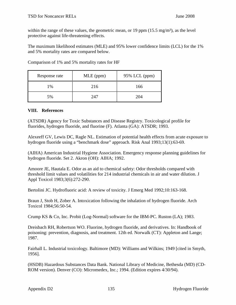
TSD for Noncancer RELs June 2008
within the range of these values, the geometric mean, or 19 ppm (15.5 mg/m³), as the level protective against life-threatening effects. The maximum likelihood estimates (MLE) and 95% lower confidence limits (LCL) for the 1% and 5% mortality rates are compared below. Comparison of 1% and 5% mortality rates for HF
Response rate MLE (ppm) 95% LCL (ppm)
1% 216 166
5% 247 204
VIII. References (ATSDR) Agency for Toxic Substances and Disease Registry. Toxicological profile for fluorides, hydrogen fluoride, and fluorine (F). Atlanta (GA): ATSDR; 1993. Alexeeff GV, Lewis DC, Ragle NL. Estimation of potential health effects from acute exposure to hydrogen fluoride using a “benchmark dose” approach. Risk Anal 1993;13(1):63-69. (AIHA) American Industrial Hygiene Association. Emergency response planning guidelines for hydrogen fluoride. Set 2. Akron (OH): AIHA; 1992. Amoore JE, Hautala E. Odor as an aid to chemical safety: Odor thresholds compared with threshold limit values and volatilities for 214 industrial chemicals in air and water dilution. J Appl Toxicol 1983;3(6):272-290. Bertolini JC. Hydrofluoric acid: A review of toxicity. J Emerg Med 1992;10:163-168. Braun J, Stob H, Zober A. Intoxication following the inhalation of hydrogen fluoride. Arch Toxicol 1984;56:50-54. Crump KS & Co, Inc. Probit (Log-Normal) software for the IBM-PC. Ruston (LA); 1983. Dreisbach RH, Robertson WO. Fluorine, hydrogen fluoride, and derivatives. In: Handbook of poisoning: prevention, diagnosis, and treatment. 12th ed. Norwalk (CT): Appleton and Lange; 1987. Fairhall L. Industrial toxicology. Baltimore (MD): Williams and Wilkins; 1949 [cited in Smyth, 1956]. (HSDB) Hazardous Substances Data Bank. National Library of Medicine, Bethesda (MD) (CD-ROM version). Denver (CO): Micromedex, Inc.; 1994. (Edition expires 4/30/94).
Appendix D2 135 Hydrogen Fluoride

TSD for Noncancer RELs June 2008
Higgins EA, Fiorca V, Thomas AA, Davis HV. Acute toxicity of brief exposures to HF, HCl, NO2, and HCN with and without CO. Fire Technol 1972;8:120-130. Jenkins GN, Venkateswarlu P, Zipkin I. Physiological effects of small doses of fluoride. In: Fluorides and human health. Geneva: World Health Organisation; 1970. p. 173. Kenchenko VG, Saripova NP. Vopy Eksp Klin Ter Profil Prom Intoksikatsi: 1974;160-164. [cited in ReproRisk Vol 21. Denver (CO): Micromedex, Inc; 1994.] Largent EJ, Fluorosis: The health aspects of fluorine compounds. Columbus (OH): Ohio State University Press; 1961. Lund K, Ekstrand J, Boe J, Sostrand P, Kongerud J. Exposure to hydrogen fluoride: an experimental study in humans of concentrations of fluoride in plasma, symptoms, and lung function. Occup Environ Med 1997;54:32-37. Machle W, Evans EE. Exposure to fluorine in industry. J Ind Hyg Toxicol 1940;22:213-217. [cited in AIHA, 1992.] Machle W, Thamann F, Kitzmiller K, Cholak J. The effects of the inhalation of hydrogen fluoride. I. The response following exposure to high concentrations. J Ind Hyg 1934;16(2):129-145. National Institute for Occupational Safety and Health (NIOSH). Criteria for a recommended standard for occupational exposure to hydrogen fluoride. Washington (DC): US Government Printing Office; 1976. Rosenholtz MJ, Carson TR, Weeks MH, Wilinski F, Ford D, Oberst F. A toxicopathologic study in animals after brief single exposures to hydrogen fluoride. Ind Hyg J 1963;24:253-261. Smith FA. 1988. Written communication to AIHA ERPG Review Committee. AIHA, 475 Wolf Ledges Pkwy., Akron, OH 44311-1087. [cited in AIHA, 1992.] Smyth HF Jr. Improved communication - hygienic standards for daily inhalation. Am Ind Hyg Assoc J 1956;17:129-185. (U.S.EPA) United States Environmental Protection Agency. Interim methods for development of inhalation Reference Concentrations. Office of Research and Development. EPA/600/8-90//066A. Washington (DC): U.S.EPA; 1990. Wing JS, Brender JD, Sanderson LM, Perrotta DM, Beauchamp RA. Acute health effects in a community after a release of hydrofluoric acid. Arch Environ Health 1991;46(3):155-160. Wohlslagel J, DiPasquale LC, Vernot EH. Toxicity of solid rocket motor exhaust: effects of HCl, HF, and alumina on rodents. J Combust Toxicol 1976;3:61-70.
Appendix D2 136 Hydrogen Fluoride

TSD for Noncancer RELs June 2008
ACUTE TOXICITY SUMMARY
HYDROGEN SELENIDE
(hydrogen selenide, selenium hydride)
CAS Registry Number: 7783-07-5
I. Acute Toxicity Summary (for a 1-hour exposure) Inhalation reference exposure level 5 µg/m³
Critical effect(s) signs of eye and respiratory irritation in guinea pigs during exposure. (Difficulty in breathing and inactivity were observed after the exposure.)
Hazard Index target(s) Eyes; Respiratory System II. Physical and Chemical Properties (HSDB, 1994 except as noted) Description gas
Molecular formula H2S Molecular weight 80.98 Density 3.31 g/L @ 25°C Boiling point -41.3oC Melting point -65.73oC Flashpoint not applicable Explosive limits not applicable Solubility soluble in water, carbonyl chloride and carbon
disulfide Odor threshold 0.3 ppm (AIHA, 1989) Odor description garlic odor (AIHA, 1989) Metabolites trimethylselenonium (Palmer et al., 1970) Conversion factor 1 ppm = 3.31 mg/m³ @ 25°C
III. Major Uses or Sources Selenium occurs in four distinct valence forms: selenates (6+), selenite (4+), selenides (2-), and elemental (0) (Amdur et al., 1991). Selenite (4+) compounds and elemental selenium are believed to be of low toxicity because of their insolubility in biological media. Selenates are more acutely toxic due to their greater solubility. The most acutely toxic selenium compound reported is hydrogen selenide (H2Se). Hydrogen selenide is formed by the reaction of acids or water with metal selenides or by the contact of nascent hydrogen with soluble selenium compounds (Clayton and Clayton, 1982). Hydrogen selenide has no reported commercial use.
Appendix D2 137 Hydrogen Selenide

TSD for Noncancer RELs June 2008
Selenium compounds are used as a decolorizing agent in the glass industry, as a vulcanizing agent in the rubber industry, in insecticides, and in photoelectric cells. Selenium compounds are also found in the toning baths used in photography and xerography. Selenium sulfide (SeS) is used in shampoos as an antidandruff agent. Up to 90% of the selenium content in ambient air is emitted during the burning of fossil fuels (Kut and Sarikaya, 1981). The most widely used selenium compound in industry is selenium dioxide (SeO2) (HSDB, 1994). It is produced by the oxidation of Se with nitric acid followed by evaporation or by burning Se in oxygen. Selenium is an essential trace element in many species, including humans (Amdur et al., 1991). However, the dose differential between acute toxicity and chronic deficiency is slight. While the lower limit for acute oral selenium toxicity is reported to be 200 µg Se/day in humans, the “normal” oral intake is reported as 70 µg Se/day, and the oral level associated with disease due to chronic deficiency is 20 µg Se/day. IV. Acute Toxicity to Humans Eye, nose and throat irritation and headaches were reported by workers briefly exposed to high, but unquantitated, concentrations of selenium fume (Clinton, 1947). One worker reported delayed symptoms of sore throat and dyspnea 8-12 hours following exposure. In a review of the literature and a report of five cases, Buchan (1947) reported that signs of acute intoxication following exposure to 0.21 ppm (0.7 mg/m³) H2Se included irritation of the respiratory tract, severe bronchitis, bronchial pneumonia, and pulmonary edema. This report reflects occupational exposure; the exact duration of exposure was not specified. In another report, workers accidentally exposed to selenium oxide reported initial symptoms of bronchospasms, irritation of the upper respiratory passages, violent coughing, and gagging with nausea and vomiting (Wilson, 1962). Late onset symptoms observed 2 or more hours following exposure included fever, chills, headache, and dyspnea. Symptoms of bronchitis persisted for four days. Predisposing Conditions for Selenium Toxicity Medical: Persons with preexisting eye, skin, or respiratory conditions (including allergies)
may be more sensitive to the effects of exposure to H2Se (Reprotext, 1999). Chemical: Persons exposed to multiple selenium compounds over time may be more
sensitive to the effects of additional Se exposure (Reprotext, 1999). V. Acute Toxicity to Laboratory Animals The 2-hour LC50 in guinea pigs is 3.6 ppm (12 mg/m³) H2Se (Dudley and Miller, 1941). No increase in mortality was observed in rabbits and guinea pigs exposed to 33 mg/m³ Se dust for 4 hours every other day for 8 days (total duration of exposure of 16 hours) (Hall et al., 1951).
Appendix D2 138 Hydrogen Selenide

TSD for Noncancer RELs June 2008
Moderate interstitial pneumonitis and congestion of the lungs was noted in both species at necropsy. A 10% mortality rate was observed in rats exposed to the same concentration of Se dust for a total of 8 hours; mild pneumonitis was noted at necropsy. Signs of nasal and ocular irritation, including nasal discharge and pawing of the eyes and nose, were observed in guinea pigs exposed to 0.9-57 ppm (3-190 mg/m³) H2Se for 60-minutes (Dudley and Miller, 1937). Decreased activity, marked difficulty in breathing, and decreased food intake were noted in those animals surviving the exposure. No significant increase in mortality as compared to controls was observed in guinea pigs exposed to 3 mg/m³ H2Se for 1 hour. (Three of the 32 control animals died during the 30 day observation period following exposure while 1 of 16 animals exposed to either 3 or 4 mg/m³ H2Se died during the observation period). No histological changes or other signs of toxicity were observed in rats following a 1-hour exposure to 1,607, 4,499, or 8,034 ppm (7,200, 20,000, or 36,000 mg/m³) dimethylselenide vapor (equivalent to 5,200, 15,000, or 26,000 mg Se/m³) (Al-Bayati et al., 1992). Microorganisms in the soil and plant products can methylate selenium to form dimethylselenide and, subsequently, dimethylselenide has been shown to be released as a vapor from acidic soil. Rats were exposed to 2.6 mg/m³ Se0 for 10 minutes and sacrificed 4 hours later; 57% of the Se deposited in the lungs had been absorbed into the blood (Medinsky et al., 1981). The single largest fraction of the excreted Se (20-28%) was found in the urine. VI. Reproductive or Developmental Toxicity Female Japanese rectifier workers known to be exposed to selenium reported irregular menstrual bleeding (Nagaii, 1959). The original article was not available for review and no additional information was reported in the secondary source (Friberg et al., 1986). No other reports of human reproductive or developmental toxicity following exposure to Se were available. A dose-dependent increase in fetal malformations was observed following a single oral administration of 90, 100, or 110 mg/kg sodium selenate (Na2SeO4) to pregnant hamsters on the 8th day of gestation (Ferm et al., 1990). A significant decrease in fetal body weight and crown-rump length were observed following a single maternal oral dose of 110 mg/kg Na2SeO4. Maternal toxicity, as indicated by a significant weight loss, was observed following a single oral dose of 110 mg/kg Na2SeO4; approximately 30% of the dams in this group died following administration of the dose. Dose-dependent injury to the testes of male rats was observed following a 90-day intraperitoneal administration of 2, 6, or 10 mg/day selenium dioxide (SeO2) (Chowdhury and Venkatakrishna-Bhatt, 1983). Statistically significant decreases in relative testes weight, seminiferous tubular diameter, and Leydig cell population were observed following exposure to 6 or 10 mg SeO2/day. Significant testicular degeneration and testicular atrophy were observed following administration of the higher dose.
Appendix D2 139 Hydrogen Selenide

TSD for Noncancer RELs June 2008
VII. Derivation of Acute Reference Exposure Level and Other Severity Levels (for a 1-hour exposure)
Reference Exposure Level (protective against mild adverse effects): 5 µg/m³
Study Dudley and Miller, 1937; Dudley and Miller, 1941 Study population groups of 16 guinea pigs; 32 controls Exposure method inhalation in a chamber Critical effects signs of eye and respiratory irritation, with
persistent coughing after exposure, for several days.
LOAEL 0.9 ppm (3 mg/m³) NOAEL not observed Exposure duration 1 hour Extrapolated 1 hour concentration 0.9 ppm (3 mg/m³) LOAEL uncertainty factor 6 Interspecies uncertainty factor 10 Intraspecies uncertainty factor 10 Cumulative uncertainty factor 600 Reference Exposure Level 0.0015 ppm (0.005 mg/m³; 5 µg/m³)
Guinea pigs exposed to 0.9 ppm (3 mg/m³) H2Se for 1 hour exhibited acute eye and nasal irritation (indicated by pawing of the nose and eyes) during the exposure and marked difficulty breathing and decreased activity following the exposure. The range of exposure concentrations was 0.9-57 ppm (3-190 mg/m³) and a 30 day observation period followed the exposure. Nearly 100% of the animals were dead within 30 days of exposure to concentrations of H2Se of 6 ppm (20 mg/m³) and greater. No increase in mortality was observed in animals exposed to 3 or 4 mg/m³ H2Se compared to control animals. The LOAEL for irritant effects is 0.9 ppm (3 mg/m³) H2Se. The signs reported by the authors indicate that the irritation experience by the animals was at least moderate and may have approached a severe level. Dudley and Miller (1941) exposed guinea pigs to hydrogen selenide for periods of 2, 4, or 8 hours. The 8-hour exposure resulted in 8/16 (50%) mortality in the animals when exposed to a concentration of 1 mg/m³. The dose-response is very steep for hydrogen selenide. Since H2Se is reported to be the most acutely toxic selenium compound (Amdur et al., 1991), this level is considered to be protective against adverse effects from other selenium compounds as well. Use of this value for some selenium compounds will overestimate health risks. Thus, its use should be restricted to evaluating emissions of hydrogen selenide. OEHHA will continue to evaluate the literature for other selenium compounds for the development of RELs for selenium salts. Level Protective Against Severe Adverse Effects No recommendation is made due to the limitations of the database.
Appendix D2 140 Hydrogen Selenide

TSD for Noncancer RELs June 2008
Level Protective Against Life-threatening Effects No recommendation is made due to the limitations of the database. NIOSH (1995) lists an IDLH of 1 mg Se/m³ based on acute toxicity data in animals. “This may be a conservative value for selenium compounds in general since it is based on sodium selenite, which is orders of magnitude more toxic than many other selenium compounds. Further, this may also be a conservative value due to the lack of relevant acute toxicity data for workers.” Due to the uncertainty this value cannot be recommended. VIII. References Al-Bayati MA, Raabe OG, Teague SV. Effect of inhaled dimethylselenide in the Fischer 344 male rat. J Toxicol Environ Health 1992;37:549-557. Amdur MO, Doull J, Klaassen CD, editors. Casarett and Doull’s Toxicology. 4th ed. New York: Pergamon Press; 1991. p. 658-660. (ACGIH) American Conference of Governmental Industrial Hygienists . Documentation of the Threshold Limit Values and Biological Exposure Indices. 6th ed. Cincinnati (OH): ACGIH; 1991. p. 1354-1360. (AIHA) American Industrial Hygiene Association. Odor thresholds for chemicals with established occupational health standards. Akron (OH): AIHA; 1989. p. 20. Buchan RF. Industrial selenosis: a review of the literature, report of five cases and a general bibliography. Occup Med 1947;3:439-456. Chowdhury AR, Venkatakrishna-Bhatt H. Effect of selenium dioxide on the testes of the rat. Indian J Physiol Pharmacol 1983;27:237-240. Clayton GD, Clayton FE, editors. Patty’s industrial hygiene and toxicology. 3rd ed. Revised. Vol. II. Toxicology. New York (NY): John Wiley Sons; 1982. p. 2133. Clinton M Jr. Selenium fume exposure. J Ind Hyg Toxicol 1947;29:225. Dudley HC, Miller JW. Toxicology of selenium. IV. Effects of exposure to hydrogen selenide. US Public Health Rep 1937;52:1217-1231. Dudley HC, Miller JW. Toxicology of selenium. VI. Effects of subacute exposure to hydrogen selenide. J Ind Hyg Toxicol 1941;23:470-477. Ferm VH, Hanlon DP, Willhite CC, Choy WN, Book SA. Embryotoxicity and dose-response relationships of selenium in hamsters. Reproductive Toxicol 1990;4(3):183-190.
Appendix D2 141 Hydrogen Selenide

TSD for Noncancer RELs June 2008
Friberg L, Nordberg GF, Vouk VB, editors. Handbook on the toxicology of metals. Vol II. Specific metals. 2nd ed. New York: Elsevier; 1986. p. 482-520. Hall RH, Leskin S, Frank P, Maynard EA, Hodge HC. Preliminary observations on toxicity of elemental selenium. Arch Ind Hyg Occup Med 1951;4:458-464. (HSDB) Hazardous Substances Data Bank. National Library of Medicine, Bethesda (MD) (CD-ROM version). Denver (CO): Micromedex, Inc.; 1994. (Edition expires 7/31/94). Kut D, Sarikaya Y. Determination of selenium in atmospheric particulate material of Ankara and its possible sources. J Radioanal Chem 1981;62:161-170. [cited in Friberg et al., 1986.] Medinsky MA, Cuddihy RG, McClellan RO. Systemic absorption of selenious acid and elemental selenium aerosols in rats. J Toxicol Environ Health 1981;8:917-928. Nagaii I. Igaku Kenkyu 1959;29:1505-1532 (Japanese). [cited in Friberg et al., 1986.] NIOSH. Chemical listing and documentation of revised IDLH values (as of March 1, 1995). Available at http://www.cdc.gov/niosh/intridl4.html. Palmer IS, Gunsalus RP, Halverson AW, Olson OE. Trimethylselenonium ion as a general excretory product from selenium metabolism in the rat. Biochim Biophys Acta 1970;208:260-266. Reprotext® System. Dabney BJ, editor. Denver (CO): Micromedex, Inc.; 1999. (Edition expires 1/31/1999). Wilson HM. Selenium oxide poisoning. N Carolina Med J 1962;23:73-75.
Appendix D2 142 Hydrogen Selenide
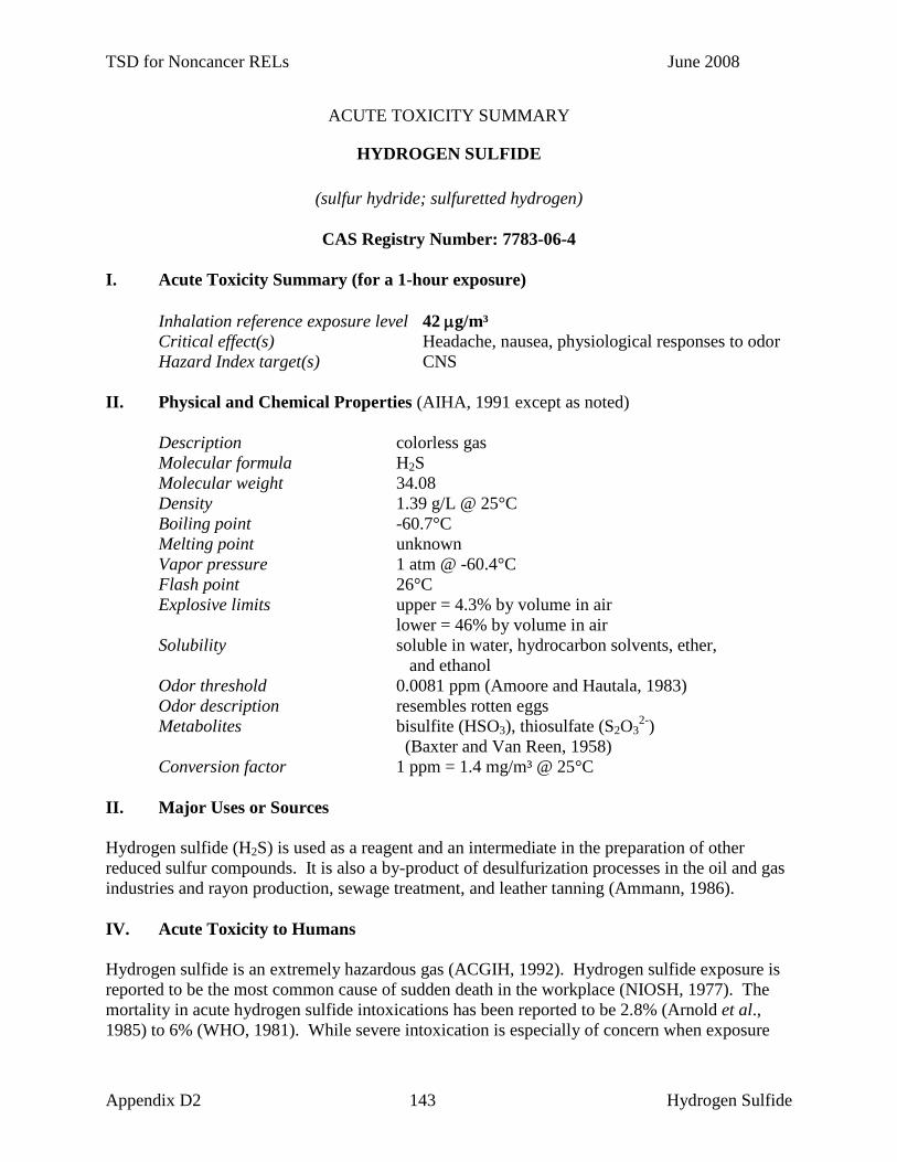
TSD for Noncancer RELs June 2008
ACUTE TOXICITY SUMMARY
HYDROGEN SULFIDE
(sulfur hydride; sulfuretted hydrogen)
CAS Registry Number: 7783-06-4 I. Acute Toxicity Summary (for a 1-hour exposure) Inhalation reference exposure level 42 µg/m³ Critical effect(s) Headache, nausea, physiological responses to odor Hazard Index target(s) CNS II. Physical and Chemical Properties (AIHA, 1991 except as noted) Description colorless gas Molecular formula H2S Molecular weight 34.08 Density 1.39 g/L @ 25°C Boiling point -60.7°C Melting point unknown Vapor pressure 1 atm @ -60.4°C Flash point 26°C Explosive limits upper = 4.3% by volume in air lower = 46% by volume in air Solubility soluble in water, hydrocarbon solvents, ether,
and ethanol Odor threshold 0.0081 ppm (Amoore and Hautala, 1983) Odor description resembles rotten eggs Metabolites bisulfite (HSO3), thiosulfate (S2O3
2-) (Baxter and Van Reen, 1958) Conversion factor 1 ppm = 1.4 mg/m³ @ 25°C II. Major Uses or Sources Hydrogen sulfide (H2S) is used as a reagent and an intermediate in the preparation of other reduced sulfur compounds. It is also a by-product of desulfurization processes in the oil and gas industries and rayon production, sewage treatment, and leather tanning (Ammann, 1986). IV. Acute Toxicity to Humans Hydrogen sulfide is an extremely hazardous gas (ACGIH, 1992). Hydrogen sulfide exposure is reported to be the most common cause of sudden death in the workplace (NIOSH, 1977). The mortality in acute hydrogen sulfide intoxications has been reported to be 2.8% (Arnold et al., 1985) to 6% (WHO, 1981). While severe intoxication is especially of concern when exposure
Appendix D2 143 Hydrogen Sulfide

TSD for Noncancer RELs June 2008
occurs in confined spaces, an accidental release of hydrogen sulfide into the air surrounding industrial facilities can cause very serious effects. For example, at Poza Rica, Mexico 320 people were hospitalized and 22 died (WHO, 1981). An inhalation LCLo of 600 and 800 ppm (840 and 1,120 mg/m³) for 30 and 5 minutes, respectively, is reported (Hazardtext, 1994). A lethal exposure was documented for a worker exposed to approximately 600 ppm H2S for 5-15 minutes (Simson and Simpson, 1971). Inhalation of 1,000 ppm (1,400 mg/m³) is reported to cause immediate respiratory arrest (ACGIH, 1992). Concentrations greater than 200 ppm (280 mg/m³) H2S are reported to cause direct irritant effects on exposed surfaces and can cause pulmonary edema following longer exposures (Spiers and Finnegan, 1986). The mechanism of H2S toxicity, cellular hypoxia caused by inhibition of cytochrome oxidase, is similar to that for cyanide and can be treated by induction of methemoglobin or with hyperbaric oxygen (Elovaara et al., 1978; Hsu et al., 1987). At concentrations exceeding 50 ppm (70 mg/m³), olfactory fatigue prevents detection of H2S odor. Exposure to 100-150 ppm (140-210 mg/m³) for several hours causes local irritation (Haggard, 1925). Exposure to 50 ppm for 1 hour causes conjunctivitis with ocular pain, lacrimation, and photophobia; this can progress to keratoconjunctivitis and vesiculation of the corneal epithelium (ACGIH, 1992). Bhambhani and Singh (1991) showed that 16 healthy subjects exposed to 5 ppm (7 mg/m³) H2S under conditions of moderate exercise exhibited impaired lactate and oxygen uptake in the blood. Bhambhani and Singh (1985) reported that exposure of 42 individuals to 2.5 to 5 ppm (3.5 to 7 mg/m³) H2S caused coughing and throat irritation after 15 minutes. In another study, ten asthmatic volunteers were exposed to 2 ppm H2S for 30 minutes and pulmonary function was tested (Jappinen et al., 1990). All subjects reported detecting “very unpleasant” odor but “rapidly became accustomed to it.” Three subjects reported headache following exposure. No significant changes in mean FVC or FEV1 were reported. Although individual values for specific airway resistance (SRaw) were not reported, the difference following exposure ranged from -5.95% to +137.78%. The decrease in specific airway conductance, SGaw, ranged from -57.7% to +28.9%. The increase in mean SRaw and the decrease in mean SGaw were not statistically significant. However, significantly increased airway resistance and decreased airway conductance were noted in two of ten asthmatic subjects which may be biologically significant. Hydrogen sulfide is noted for its strong and offensive odor. Based on a review of 26 studies, the average odor detection threshold ranged from 0.00007 to 1.4 ppm (Amoore, 1985). The geometric mean of these studies is 0.008 ppm. In general, olfactory sensitivities decrease by a factor of 2 for each 22 years of age above 20 (Venstrom and Amoore, 1968); the above geometric mean is based on the average age of 40. For hydrogen sulfide, concentrations that substantially exceed the odor threshold result in the annoying and discomforting physiological symptoms of headache or nausea (Amoore, 1985; Reynolds and Kauper 1985). The perceived intensity of the odor of hydrogen sulfide depends on the longevity of the concentration, and the intensity increases 20% for each doubling concentration (Amoore, 1985). Several studies have been conducted to establish the ratio of discomforting annoyance threshold to detection threshold for unpleasant odors (Winneke, 1975;
Appendix D2 144 Hydrogen Sulfide

TSD for Noncancer RELs June 2008
Winneke and Kastka, 1977; Hellman and Small, 1974; Adams et al., 1968; and NCASI, 1971). The geometric mean for these studies is 5, indicating that when an unpleasant odor reaches an average concentration of 5 times its detection threshold, the odor will result in annoying discomfort. Applying the 5-fold multiplier to the mean detectable level, 0.008 ppm, results in a mean annoyance threshold of 0.04 ppm. At the current California Ambient Air Quality Standard (CAAQS) of 0.03 ppm, the level would be detectable by 83% of the population and would be discomforting to 40% of the population. These estimates have been substantiated by odor complaints and reports of nausea and headache (Reynolds and Kauper 1985) at 0.03 ppm H2S exposures from geyser emissions. The World Health Organization (WHO) reports that in order to avoid substantial complaints about odor annoyance among the exposed population, hydrogen sulfide concentrations should not be allowed to exceed 0.005 ppm (7 µg/m3), with a 30-minute averaging time (WHO, 1981; National Research Council, 1979; Lindvall, 1970). Predisposing Conditions for Hydrogen Sulfide Toxicity Medical: Unknown Chemical: Ethanol has been shown to potentiate the effects of H2S by shortening the mean
time-to-unconsciousness in mice exposed to 800 ppm (1,120 mg/m³) H2S (Beck et al., 1979).
V. Acute Toxicity to Laboratory Animals A median lethal concentration (LC50) in rats exposed to H2S for 4 hours was estimated as 440 ppm (616 mg/m³) (Tansy et al., 1981). An inhalation LCLo of 444 ppm for an unspecified duration is reported in rats, and a lethal concentration of 673 ppm (942 mg/m³) for 1 hour is reported in mice (RTECS, 1994). In another study, mortality was significantly higher for male rats (30%), compared to females (20%), over a range of exposure times and concentrations (Prior et al., 1988). A concentration of 1,000 ppm (1,400 mg/m³) caused respiratory arrest and death in dogs after 15-20 minutes (Haggard and Henderson, 1922). Inhalation of 100 ppm (140 mg/m³) for 2 hours resulted in altered leucine incorporation into brain proteins in mice (Elovaara et al., 1978). Kosmider et al. (1967) reported abnormal electrocardiograms in rabbits exposed to 100 mg/m³ (71 ppm) H2S for 1.5 hours. Khan et al. (1990) exposed groups of 12 male Fischer 344 rats to 0, 10, 50, 200, 400, or 500-700 ppm hydrogen sulfide for 4 hours. Four rats from each group were sacrificed at 1, 24, or 48 hours post-exposure. Cytochrome c oxidase activity in lung mitochondria was significantly (p<0.05) decreased at 50 ppm (15%), 200 ppm (43%), and 400 ppm (68%) at 1-hour post-exposure compared to controls. A NOAEL of 10 ppm was identified in this study for effects on lung mitochondrial cytochrome c oxidase activity. VI. Reproductive or Developmental Toxicity Xu et al. (1998) conducted a retrospective epidemiological study in a large petrochemical complex in Beijing, China in order to assess the possible association between petrochemical exposure and spontaneous abortion. The facility consisted of 17 major production plants which
Appendix D2 145 Hydrogen Sulfide

TSD for Noncancer RELs June 2008
are divided into separate workshops, allowing for the assessment of exposure to specific chemicals. Married women (n = 2853), who were 20-44 years of age, had never smoked, and who reported at least one pregnancy during employment at the plant, participated in the study. According to their employment record, about 57% of these workers reported occupational exposure to petrochemicals during the first trimester of their pregnancy. There was a significantly increased risk of spontaneous abortion for women working in all of the production plants with frequent exposure to petrochemicals compared with those working in nonchemical plants. Also, when a comparison was made between exposed and non-exposed groups within each plant, exposure to petrochemicals was consistently associated with an increased risk of spontaneous abortion (overall odds ratio (OR) = 2.7 (95% confidence interval (95% CI) = 1.8 to 3.9) after adjusting for potential confounders). When the analysis was performed with the exposure information obtained from the women' interview responses for (self reported) exposures, the estimated OR for spontaneous abortions was 2.9 (95% CI = 2.0 to 4.0). The analysis was repeated by excluding those 452 women who provided inconsistent reports between recalled exposure and work history, and a comparable risk of spontaneous abortion (OR 2.9; 95% CI 2.0 to 4.4) was found. In analyses for exposure to specific chemicals, an increased risk of spontaneous abortion was found with exposure to most chemicals. There were 106 women (3.7% of the study population) exposed only to hydrogen sulfide, and the results for hydrogen sulphide (OR 2.3; 95% CI = 1.2 to 4.4) were significant. No hydrogen sulfide exposure concentration was reported. VII. Derivation of Acute Reference Exposure Level and Other Severity Levels (for a 1-hour exposure) Reference Exposure Level (protective against mild adverse effects): 42 µg/m³ (California Ambient Air Quality Standard) Study California State Department of Public Health, 1969; CARB, 1984; Reynolds and Kamper, 1985; Amoore, 1985 Study population panel of 16 people; general population Exposure method inhalation of increasing concentrations of H2S Critical effects headache, nausea LOAEL 0.012-0.069 ppm (range of odor threshold) NOAEL ≤ 0.01 ppm Exposure duration not stated (tested until odor detected) Extrapolated 1 hour concentration 0.012-0.069 ppm (geometric mean = 0.03 ppm) (1 hour = minimum duration for an air standard) LOAEL uncertainty factor not used Interspecies uncertainty factor 1 Intraspecies uncertainty factor 1 Cumulative uncertainty factor 1 Reference Exposure Level 0.03 ppm (0.042 mg/m³; 42 µg/m³)
Appendix D2 146 Hydrogen Sulfide

TSD for Noncancer RELs June 2008
The 1-hour California Ambient Air Quality Standard (AAQS) for hydrogen sulfide was originally based on an olfactory perception study by the California State Department of Public Health (1969). Sixteen individuals were each exposed to increasing concentrations of H2S until his or her odor threshold was reached. The range of the odor thresholds was 0.012-0.069 ppm, and the geometric mean was 0.029 ppm (geometric standard deviation = 0.005 ppm). The mean odor threshold (rounded to 0.03 ppm) was selected as the AAQS for H2S. However, others have reported that the odor threshold is as low as 0.0081 ppm (Amoore and Hautala, 1983). In 1984 CARB reviewed the AAQS for H2S and found that the standard was necessary not only to reduce odors, but also to reduce the physiological symptoms of headache and nausea. (CARB, 1984). Furthermore, Amoore (1985) conducted a study that estimated 40% of the population would find 0.03 ppm (0.042 mg/m³) to be an objectionable concentration. In public testimony before the ARB it was stated that some people reported headaches and other symptoms at the standard (Reynolds and Kamper, 1985). Thus this recommended level protective against mild adverse effects may be need to be reexamined as more data become available. Level Protective Against Severe Adverse Effects No recommendation can be made due to the limitations of the database. An ERPG-2 of 30 ppm (AIHA, 1991) was based on experimental data showing that exposure of rats to 45 ppm (63 mg/m³) H2S for 4 hours resulted in no deaths (Rogers and Ferin, 1981). In addition, rabbits exposed to 71 ppm (100 mg/m³) H2S for 1.5 hours developed cardiac irregularities, measured by electrocardiogram, and decreased myocardial ATP phosphorylase (Kosmider et al., 1967). The rationale for the margin of safety used for the ERPG-2 is not presented. Level Protective Against Life-threatening Effects No recommendation can be made due to the limitations of the database. The AIHA ERPG-3 for hydrogen sulfide of 100 ppm (AIHA, 1991) was based on case reports of conjunctivitis, respiratory irritation, and unconsciousness in humans exposed to estimated concentrations of 200-300 ppm (280-420 mg/m³) H2S for 20 minutes to 1 hour (Ahlborg, 1951; Yant, 1930). In addition, a 1-hour LC50 of 712 ppm (997 mg/ m3) in rats is cited (CIIT, 1983). The case reports cited in the ERPG document are inadequate to establish acute exposure levels in humans because the concentrations and durations of exposure are only estimates. In addition, there are no LC50 data in the CIIT (1983) report. Rats (5 female and 5 male) exposed to H2S concentrations ranging from 400-600 ppm (560-840 mg/m³) for 4 hours showed dose-dependent lethality rates ranging from 30% - 100% (Tansy et al., 1981). On the other hand, two of three rhesus monkeys exposed to a concentration of 500 ppm (700 mg/m³) for only 35 minutes or less died, which suggests that primates are more sensitive to the lethal effect of H2S than rats (Lund and Wieland, 1966). The rationale for the margin of safety used for the ERPG-3 was not presented.
Appendix D2 147 Hydrogen Sulfide

TSD for Noncancer RELs June 2008
NIOSH (1995) reports a (revised) IDLH for hydrogen sulfide of 100 ppm based on acute inhalation toxicity data in humans and animals, but the values from animals appear to be more heavily weighted than the human data in the selection of the IDLH. VII. References Adams DF, Young FA, Lahr RA. Evaluation of odor perception threshold test facility. TAPPI 1968;51(13):62A-67A. Ahlborg G. Hydrogen sulfide poisoning in shale oil industry. AMA Arch Ind Hyg Occup Med 1951;3:247-266. [cited in: AIHA; 1991.] Alberta Health. Report on H2S Toxicity. Alberta Health 1990. (ACGIH) American Conference of Governmental Industrial Hygienists. Documentation of the Threshold Limit Values and Biological Exposure Indices. 6th ed. Vol II. Cincinnati: ACGIH; 1991. p. 786-788. (AIHA) American Industrial Hygiene Association. Emergency response planning guideline for hydrogen sulfide. Set 6. Akron: AIHA; 1991. Ammann HM. A new look at physiologic respiratory response to H2S poisoning. J Hazard Mater 1986;13:369-374. Amoore JE. The perception of hydrogen sulfide odor in relation to setting an ambient standard. California Air Resources Board Contract A4-046-33. April 1985. Amoore JE, Hautala E. Odor as an aid to chemical safety: Odor thresholds compared with threshold limit values and volatilities for 214 industrial chemicals in air and water dilution. J Appl Toxicol 1983;3(6):272-290. Arnold IM, Dufresne RM, Alleyne BC, Stuart PJ. Health implications of occupational exposures to hydrogen sulfide. J Occup Med 1985;27:373-376. Baxter CF, Van Reen R. Some aspects of sulfide oxidation by rat-liver preparations. Biochim Biophys Acta 1958;28:567-573. Beck JF, Cormier F, Donini JC. The combined toxicity of ethanol and hydrogen sulfide. Toxicol Lett 1979;311-313. Bhambhani Y, Singh M. Effects of hydrogen sulphide on selected metabolic and cardio-respiratory variables during rest and exercise. Report submitted to Alberta Worker’s Health and Safety and Compensation. June, 1985. [cited by Alberta Health; 1990.] Bhambhani Y, Singh M. Physiological effects of hydrogen sulfide inhalation during exercise in healthy men. J Appl Physiol 1991;71:1872-1877.
Appendix D2 148 Hydrogen Sulfide

TSD for Noncancer RELs June 2008
California Air Resources Board. Report of the committee regarding the review of the AAQS for hydrogen sulfide. Memorandum from CARB to G. Duffy, 1984. California State Department of Public Health. Recommended Ambient Air Quality Standards. (Statewide standards applicable to all California Air Basins). 1969;HS-3. (CIIT) Chemical Industry Institute of Toxicology. Ninety day vapor inhalation toxicity study of H2S in Fischer-344 rats. Docket #22063. Research Triangle Park (NC): Chemical Industry Institute of Toxicology; 1983. Elovaara E, Tossavainen A, Savolainen H. Effects of subclinical hydrogen sulfide intoxication on mouse brain protein metabolism. Exp Neurol 1978;62:93-98. Haggard HAW. The toxicology of hydrogen sulphide. J Ind Hyg 1925;7:113-121. Haggard HW, Henderson Y. The influence of hydrogen sulfide on respiration. Am J Physiol 1922;61:289-297. HAZARDTEXT. Hall AH, Rumack BH, editors. Denver (CO): Micromedex, Inc.; 1994. (Edition expires 4/30/94). (HSDB) Hazardous Substances Data Bank. National Library of Medicine, Bethesda (MD) (CD-ROM version). Denver (CO): Micromedex, Inc.; 1994. (Edition expires 4/30/94). Hellman TM, Small FH. Characterization of the odor properties of 101 petrochemicals using sensory methods. J Air Pollut Control Assoc 1974;24:979-982. Hsu P, Li HW, Lin Y. Acute hydrogen sulfide poisoning treated with hyperbaric oxygen. J Hyperbaric Med 1987;2(4):215-221. Jappinen P, Vilka V, Marttila O, Haahtela T. Exposure to hydrogen sulfide and respiratory function. Br J Ind Med 1990;47:824-828. Khan AA, Schuler MM, Prior MG et al. (1990) Effects of hydrogen sulfide exposure on lung mitochondrial respiratory chain enzymes in rats. Toxicol Appl Pharmacol 103: 482-490. Kosmider S, Rogala E, Pacholek A. Electrocardiographic and histochemical studies of the heart muscle in acute experimental hydrogen sulfide poisoning. Arch Immunol Ther Exp 1967;15:731-740. Lindvall T On sensory evaluation of odorous air pollutant intensities. Nord Hyg Tidskr 1970;Suppl 2:1-181. Lund OE, Wieland H. Pathologic-anatomic findings in experimental hydrogen sulfide poisoning: A study on rhesus monkeys. Int Arch Gewerbepathol Gewerbehyg 1966;22:46-54.
Appendix D2 149 Hydrogen Sulfide

TSD for Noncancer RELs June 2008
NCASI. Evaluation of the use of humans in measuring the effectiveness of odor control technology at the source. Atmospheric Quality Improvement Technical Bulletin No. 56. New York: National Council of Paper Industry for Air and Steam Improvement; 1971. (NIOSH) National Institute for Occupational Safety and Health. Criteria for a recommended standard...Occupational exposure to hydrogen sulfide, DHEW (NIOSH) #77-158. Cincinnati (OH): National Institute for Occupational Safety and Health; 1977. (NIOSH) National Institute for Occupational Safety and Health. Chemical listing and documentation of revised IDLH values (as of March 1, 1995). Available at http://www.cdc.gov/niosh/intridl4.html. National Research Council. Hydrogen sulfide. Baltimore: University Park Press: 1979. Prior MG, Sharma AK, Yong S, Lopez A. Concentration-time interactions in hydrogen sulphide toxicity. Can J Vet Res 1988;52:375-379. Reynolds R L, Kamper RL. Review of the State of California Ambient Air Quality Standard for Hydrogen Sulfide (H2S). Lakeport (CA): Lake County Air Quality Management District; 1984. (RTECS) Registry of Toxic Effects of Chemical Substances. National Institute of Occupational Safety and Health, Cincinnati (OH) (CD-ROM version). Denver (CO): Micromedex, Inc.; 1994. (Edition expires 4/30/94). Rogers RE, Ferin J. Effect of hydrogen sulfide on bacterial inactivation in the rat lung. Arch Environ Health 1981;36:261-264. [cited in AIHA, 1991.] Simson RE, Simpson GR. Fatal hydrogen sulphide poisoning associated with industrial waste exposure. Med J Austral 1971;2:331-334. Spiers M, Finnegan OC. Near death due to inhalation of slurry tank gases. Ulster Med Soc 1986;55(2):181-183. Tansy MF, Kendall FM, Fantasia J, Landlin WE, Oberly R, Sherman W. Acute and subchronic toxicity of rats exposed to vapors of methyl mercaptan and other reduced-sulfur compounds. J Toxicol Environ Health 1981;8(1-2):71-88. Venstrom P, Amoore JE. Olfactory threshold in relation to age, sex or smoking. J Food Sci 1968;33:264-265. Winkler K. Zur Diskussion Gestellt Imissionsgrenzwerte Zur Vehrinderung von. Geruchsbelastigungan Wasser Luft Betrieb 1975;19:411.
Appendix D2 150 Hydrogen Sulfide

TSD for Noncancer RELs June 2008
Winneke G, Kastka J. Odor pollution and odor annoyance reactions in industrial areas of the Rhine-Ruhr region. In: Olfaction and Taste VI. J Le Magnen, P MacLeod, editors. pp. 471-479. London: Information Retrieved; 1977. (WHO) World Health Organization. Hydrogen sulfide. Environmental Health Criteria No. 19. Geneva: WHO; 1981. Xu X, Cho SI, Sammel M, You L, Cui S, Huang Y, et al. Association of petrochemical exposure with spontaneous abortion. Occup Environ Med 1998;55(1):31-36. Yant WP. Hydrogen sulphide in industry: Occurrence, effects, and treatment. Am J Publ Health 1930;20:598-608.
Appendix D2 151 Hydrogen Sulfide

TSD for Noncancer RELs June 2008
ACUTE TOXICITY SUMMARY
ISOPROPYL ALCOHOL
(isopropanol, 2-propanol, dimethylcarbinol, propyl alcohol)
CAS Registry Number: 67-63-0
I. Acute Toxicity Summary (for a 1-hour exposure) Inhalation reference exposure level 3,200 µg/m³ Critical effect(s) irritation of the eyes, nose and throat. Hazard Index target(s) Eyes; Respiratory System II. Physical and Chemical Properties (HSDB, 1993 except as noted) Description colorless liquid Molecular formula C3H8O Molecular weight 60.09 Density 0.78505 g/cm3 @ 20°C Boiling point 82.5°C @ 760 mm Hg Melting point -88.5°C Vapor pressure 44.0 mm Hg @ 25°C Flashpoint 11.7°C (closed cup) Explosive limits upper = 12.0% lower = 2.0% Solubility soluble in benzene, miscible with most organic
solvents, slightly soluble in water, alcohol, and acetone Odor threshold 19 ppm (geometric mean) (AIHA, 1989) Odor description sharp (AIHA, 1989) Metabolites acetone Conversion factor 1 ppm = 2.45 mg/m³ @ 25°C III. Major Uses or Sources Isopropyl alcohol has wide use in consumer products such as mild skin disinfectants and astringents. It is also used as a solvent for cellulose nitrate. IV. Acute Toxicity to Humans Symptoms of acute poisoning include dizziness, incoordination, headache, and confusion. Vomiting, hematemesis, diarrhea, and hypotension may occur following ingestion of large quantities of isopropyl alcohol. Late manifestations include aspiration pneumonia and kidney and liver dysfunction (Reprotext, 1993). The oral LOAEL for isopropyl alcohol is reported as 233 mg/kg (RTECS, 1993).
Appendix D2 152 Isopropyl Alcohol

TSD for Noncancer RELs June 2008
Irritation of the mucous membranes of the upper respiratory tract may occur following inhalation of isopropyl alcohol. Ten human subjects were exposed for 3-5 minutes to 400 or 800 ppm (1,000 or 2,000 mg/m³) isopropyl alcohol (Nelson et al., 1943). Exposure to 400 ppm isopropyl alcohol produced mild irritation of the eyes, nose, and throat. When exposed to 800 ppm the majority of the subjects declared the atmosphere unsuitable for a prolonged exposure. The subjects indicated, however, that prolonged exposure to 200 ppm would not be objectionable. Predisposing Conditions for Isopropyl Alcohol Toxicity Medical: Persons with eye, skin, respiratory or neurological conditions and diabetics may
be more sensitive to the toxic effects of isopropyl alcohol (Reprotext, 1999). Chemical: Individuals exposed to acetone, carbon tetrachloride, or n-hexane may be at
increased risk for adverse effects when exposed simultaneously to isopropyl alcohol (Reprotext, 1999).
V. Acute Toxicity to Laboratory Animals A 4-hour rat LCLo of 16,000 ppm (39,000 mg/m³) isopropyl alcohol is reported (Carpenter et al., 1949). Reduced ciliary activity and epithelial damage in the nasal mucosa of guinea pigs were observed following a 24-hour exposure to 400 ppm (1,000 mg/m³) isopropanol. Complete recovery from the exposure occurred within 2 weeks. Exposure at 5,500 ppm (13,000 mg/m³) resulted in similar damage requiring more than two weeks for complete recovery (Ohashi et al., 1988). A 10-minute RD50 of 17,693 ppm (43,000 mg/m³) for mice has been reported (Kane at al., 1980). VI. Reproductive or Developmental Toxicity No human reproductive studies and only a limited number of animal studies on the effects of isopropyl alcohol were available. Pregnant rats exposed to 3,500, 7,000, and 10,000 ppm (8,600, 17,000, and 25,000 mg/m³) isopropanol for 7 hours per day on days 1-19 of gestation exhibited signs of maternal toxicity, indicated by retarded weight gain, following exposure to 7,000 ppm or greater. Signs of narcosis were observed in the dams exposed to 10,000 ppm. Fetal weight was reduced in all three exposed groups in a dose-dependent manner; increased skeletal and visceral malformations were observed following exposure to 7,000 ppm (Nelson et al., 1988).
Appendix D2 153 Isopropyl Alcohol

TSD for Noncancer RELs June 2008
VII. Derivation of Acute Reference Exposure Level and Other Severity Levels (for a 1-hour exposure)
Reference Exposure Level (protective against mild adverse effects) 1.3 ppm (3,200 µg/m³) Study Nelson et al., 1943 Study population ten human subjects Exposure method 400 ppm for 3-5 minutes Critical effects mild irritation of the eyes, nose and throat LOAEL 400 ppm NOAEL 200 ppm (implied) Exposure duration 4 minutes Extrapolated 1 hour concentration 13 ppm (2001ppm * 0.067 h = C1 * 1 h ) (see Table 12 for information on “n”) LOAEL uncertainty factor 1 Interspecies uncertainty factor 1 Intraspecies uncertainty factor 10 Cumulative uncertainty factor 10 Reference Exposure Level 1.3 ppm (3.2 mg/m³; 3,200 µg/m³) Ten human subjects, exposed for 3-5 minutes to 400 ppm (1,000 mg/m³) isopropyl alcohol, reported mild irritation of the eyes, nose and throat. The study indicates a 4 minute LOAEL of 400 ppm. The subjects indicated that exposure to 200 ppm would be tolerable, which implies a NOAEL of 200 ppm. This 4 minute NOAEL was time adjusted to 1 hour. An uncertainty factor of 10 was applied to the 200 ppm NOAEL to account for the susceptibility of sensitive individuals. Level Protective Against Severe Adverse Effects Rats were exposed for 6 hours to 0, 500, 1,500, 5,000, or 10,000 ppm isopropyl alcohol (Gill et al., 1995). Signs of narcosis and concentration-related decreases in motor activity were observed in rats exposed to 5,000 or 10,000 ppm. Slight but statistically significant decreases in motor activity were observed in male, but not female, rats exposed to 1,500 ppm isopropyl alcohol. No adverse effects were observed in rats exposed to 500 ppm isopropyl alcohol. Narcosis during isopropanol exposure at 1,500 and 5,000 ppm was also noted in a chronic inhalation study by Burleigh-Flayer et al. (1994). A 6-hour NOAEL of 500 ppm is defined from this study. An uncertainty factor of 10 was applied to account for interspecies differences. An additional uncertainty factor of 10 was applied to account for sensitive individuals. An equivalent 1-hour exposure concentration was estimated from the reported 6-hour NOAEL using the equation Cn * T = K, where n = 2. The resulting level protective against severe adverse effects is 12 ppm (29 mg/m³). A TLV-TWA of 400 ppm is reported by ACGIH (1991) based on findings by Nelson et al. (1943); the NRC-EEGL of 400 ppm is based on the TLV (NRC, 1984). However, the reported 3-5-minute exposure to 400 ppm was not extrapolated to a 1-hour equivalent by NRC. Using the equation Cn * T = K, where n = 1, the equivalent 1-hour exposure is 20 ppm. This is consistent
Appendix D2 154 Isopropyl Alcohol

TSD for Noncancer RELs June 2008
with our use of the animal studies. In addition, the recent data described above (Gill et al., 1995) were not available to ACGIH or NRC when determining these values. Level Protective Against Life-threatening Effects No recommendation can be made due to the limitations of the database. NIOSH (1995) lists an IDLH of 2,000 ppm (4,900 mg/m³). The IDLH is based strictly on safety considerations and is 10% of the lower explosive limit of 2%. VIII. References American Conference of Governmental Industrial Hygienists. Documentation of the Threshold Limit Values and Biological Exposure Indices. 6th ed. Cincinnati (OH): ACGIH; 1991. p. 828-830. (AIHA) American Industrial Hygiene Association. Odor thresholds for chemicals with established occupational health standards. Akron (OH): AIHA; 1989. p. 21. Burleigh-Flayer HD, Gill MW, Strother DE, Masten LW, McKee RH, Tyler TR, Gardiner TH. Isopropanol 13-week vapor inhalation study in rats and mice with neurotoxicity evaluation in rats. Fundam Appl Toxicol 1994; 23:421-428. Carpenter CP, Smyth HF Jr, Pozzani UC. The assay of acute vapor toxicity, and the grading and interpretation of results on 96 chemical compounds. J Ind Hyg Toxicol 1949;31(6):343-346. Gill MW, Burleigh-Flayer HD, Strother DE, Masten LW, McKee RH, Tyler TR, Gardiner TH. Isopropanol: Acute vapor inhalation neurotoxicity study in rats. J Appl Toxicol 1995;15(2):77-84. (HSDB) Hazardous Substances Data Bank. National Library of Medicine, Bethesda (MD) (CD-ROM Version). Denver (CO): Micromedex, Inc.; 1993. (Edition expires 11/31/93). Kane LE, Dombroske R, Alarie Y. Evaluation of sensory irritation from some common industrial solvents. Am Ind Hyg Assoc J 1980;41:451-455. (NRC) National Research Council. Committee on Toxicology. Emergency and continuous exposure limits for selected airborne contaminants. Vol 2. Washington (DC): National Academy Press; 1984. Nelson BK, Brightwell WS, MacKenzie-Taylor DR, Khan A, Burg JR, Weigel WW. Teratogenicity of n-propanol and isopropanol administered at high inhalation concentrations to rats. Food Chem Toxicol 1988;26(3):247-254. Nelson KW, Ege JF Jr, Ross M, Woodman LE, Silverman L. Sensory response to certain industrial solvent vapors. J Ind Hyg Toxicol 1943;25(7):282-285.
Appendix D2 155 Isopropyl Alcohol
TSD for Noncancer RELs June 2008
(NIOSH) National Institute for Occupational Safety and Health. Chemical listing and documentation of revised IDLH values (as of March 1, 1995). Available at http://www.cdc.gov/niosh/intridl4.html. Ohashi Y, Nakai Y, Koshimo H, Esaki Y, Ikeoka H, Horiguchi S, et al. Toxicity of isopropyl alcohol exposure in the nasal mucociliary system in the guinea pig. Environ Res 1988;46:25-38. (RTECS) Registry of the Toxic Effects of Chemical Substances. National Institute for Occupational Safety and Health, Cincinnati (OH) (CD-ROM Version). Denver (CO): Micromedex, Inc.; 1993. (Edition expires 11/31/93). Reprotext ® System. Dabney BJ, editor. Denver (CO): Micromedex, Inc.; 1999. (Edition expires 1/31/1999). Smyth HF Jr. Improved communication--Hygienic standards for daily inhalation. Am Ind Hyg Assoc Q 1956;17(2):129-185.
Appendix D2 156 Isopropyl Alcohol
TSD for Noncancer RELs June 2008
ACUTE TOXICITY SUMMARY
METHANOL
(methyl alcohol, wood spirit, carbinol, wood alcohol, wood naphtha)
CAS Registry Number: 67-56-1 I. Acute Toxicity Summary (for a 1-hr exposure) Inhalation reference exposure level 28,000 µg/m³ Critical effect(s) subtle impairment in the performance of complicated tasks Hazard Index target(s) Nervous System II. Physical and Chemical Properties (HSDB, 1993 except as noted) Description colorless liquid Molecular formula CH3OH Molecular weight 32.04
Density 0.7915 g/cm3 @ 20°C Boiling point 64.5°C Melting point -97.8°C Vapor pressure 92 mm Hg @ 20°C Flashpoint 12°C, closed cup Explosive limits lower = 7.3% upper = 36% Solubility methanol is miscible with water, ethanol,
ether and many other organic solvents Odor threshold 160 ppm (geometric mean) (AIHA, 1989) Odor description sour/sweet (AIHA, 1989) Metabolites metabolized to formaldehyde, then formate
Conversion factor 1 ppm = 1.31 mg/m³ @ 25°C III. Major Uses and Sources Originally distilled from wood, methanol is now manufactured synthetically from carbon oxides and hydrogen. Methanol is primarily used for the manufacture of other chemicals and as a solvent. It is also added to a variety of commercial and consumer products such as windshield washing fluid and de-icing solution, duplicating fluids, solid canned fuels, paint remover, model airplane fuels, embalming fluids, lacquers, inks and as alternative motor fuel. Methanol is released in large quantities from pulp and paper mills.
Appendix D2 157 Methanol

TSD for Noncancer RELs June 2008
IV. Acute Toxicity to Humans Methanol is easily absorbed following ingestion, inhalation, or dermal exposure and is metabolized by the liver to formaldehyde, then formate. The latter metabolite is responsible for the metabolic acidosis and ocular effects characteristic of acute methanol poisoning. Odor and irritation are not adequate warnings of overexposure to methanol (Reprotext, 1999). Upon ingestion or inhalation, methanol initially has a narcotic effect followed by an asymptomatic period of approximately 10 to 15 hours (Rowe and McCollister, 1978). After this period, methanol may produce nausea, vomiting, dizziness, headaches, vertigo, respiratory difficulty, lethargy, abdominal pain, pain in the extremities, visual disturbances, and metabolic acidosis (ATSDR, 1993; NIOSH, 1976). The visual disturbances vary from spots or cloudiness of vision to complete blindness (Grant, 1986). Methanol toxicity can result in coma and death by respiratory or cardiac arrest. In one study, symptoms of blurred vision, headaches, dizziness, nausea, and skin problems were reported in teachers’ aides who were exposed to duplicating fluid containing 99% methanol while working with “spirit duplicators” (Frederick et al., 1984). A dose-response relationship was observed between the amount of time spent at the duplicator and the incidence of symptoms. The concentrations of methanol in the breathing zones near the machines in 12 schools ranged from 485 to 4,096 mg/m³ (365 to 3,080 ppm) for a 15 minute sample. Employees working in the proximity of direct process duplicating machines complained of frequent headaches and dizziness (Kingsley and Hirsch, 1954). Air concentrations of methanol ranged from 15 ppm (20 mg/m³) to 375 ppm (490 mg/m³). In a pilot study, 12 young, paid, male volunteers were exposed to filtered air and to 250 mg/m³ (192 ppm) methanol vapor for 75 minutes and were administered a battery of 20 neurobehavioral and neurophysiological tests before, during, and after exposure (Cook et al., 1991). Methanol had no significant effect on the subjects’ performance for all but two of the tests. Although statistically significant effects were observed in one test measuring fatigue and concentration (fatigue scale score, p = 0.02) and a trend was observed in a test measuring the latency of visual evoked potentials (P200 component of event-related potentials, p = 0.02), both the effects were small and, according to the authors, did not exceed the normal range during the sham exposures. A trend was observed for decreased performance of the Sternberg memory task following exposure to methanol (p = 0.055) although it is of borderline statistical significance. Consistent with this finding, subjects reported higher levels of fatigue and there was a trend toward decreased ability to concentrate and less vigor when exposed to methanol vapors compared to control conditions. According to the authors, these changes did not affect the subjects’ ability to maintain vigilance or to respond quickly to stimuli. Predisposing Conditions for Methanol Toxicity Medical: Persons with skin, eye, respiratory or neurological conditions may be more
sensitive to the adverse effects of methanol (Reprotext, 1999). There is a great range of individual response to the toxic effects of methanol, probably due to the
Appendix D2 158 Methanol

TSD for Noncancer RELs June 2008
variability in individual capacity to generate toxic metabolites (Bennet, 1953; NIOSH, 1976).
Chemical: Persons simultaneously exposed to formaldehyde or formic acid may be more
sensitive. Those ingesting ethanol may be less sensitive to methanol toxicity (Reprotext, 1999).
V. Acute Toxicity to Laboratory Animals With the exception of non-human primates, the signs of methanol toxicity in laboratory animals are quite different from the signs observed in humans (Gilger and Potts, 1955). The major effect of methanol in non-primates is CNS depression similar to that produced by other alcohols. Metabolic acidosis and ocular toxicity are not observed. The differences in toxicity are attributed to the ability of non-primates to more efficiently metabolize formate than humans and other primates (Tephly, 1991). The lethal oral dose of methanol in humans is estimated at approximately 1/3 and 1/9 the equivalent oral dose in monkeys and rats, respectively (Gilger and Potts, 1955). In one poorly described study, 11 rhesus monkeys, 12 rabbits, and 46 rats were exposed by inhalation to methanol concentrations ranging from 1,000 ppm to 40,000 ppm (1,300 to 52,400 mg/m³) for 1-18 hours/day for up to 41 hours (McCord, 1931). Of the species studied, monkeys were the most sensitive to the effects of methanol. Some animals (number and species unidentified) died after exposure to 1,000 ppm for at least 41 hours. Exposure at 40,000 ppm for 4 hours led to immediate death in all animals. A 1-hour exposure at this concentration led to “sickness in [all] animals within 2-3 days and eventually to death.” Twenty-four cynomolgus monkeys were exposed by inhalation to methanol vapor at concentrations up to 6,650 mg/m³ (5,010 ppm) for 6 hours per day, 5 days per week for 4 weeks (Andrews et al., 1987). No deaths occurred and no treatment-related effects, including ocular damage, were observed. Methanol has been shown to be a mild irritant to the eyes and skin of rabbits when applied topically (Rowe and McCollister, 1978). Additionally, NIOSH (1976) cites studies by Flury and Wirth (1933) which reported a Lowest Lethal Concentration (LCLo) in cats of 33,082 ppm after a 6-hour exposure, and by Izmerov et al. (1982) which reported an LCLo in mice of 37,594 ppm after a 2-hour exposure. VI. Reproductive or Developmental Toxicity Exposure to methanol along with other solvents is believed to cause central nervous system birth defects in humans (Holmberg, 1979). However, because of mixed or inadequate exposure data, it is not considered a known human teratogen. In two separate studies in male rats, inhalation exposure to methanol at concentrations ranging from 260 to 13,000 mg/m³ (200 to 9,900 ppm) for 6 to 8 hours per day for either 1 day or 1, 2, 4,
Appendix D2 159 Methanol

TSD for Noncancer RELs June 2008
or 6 weeks resulted in a significant reduction in circulating testosterone levels (Cameron et al., 1984; 1985). However, a dose-response relationship was not observed. Pregnant rats were exposed by inhalation to methanol at concentrations ranging from 5,000 to 20,000 ppm (6,600 to 26,000 mg/m³) for 7 hours per day on days 1-19 of gestation, and days 7-15 for the highest dose group (Nelson et al., 1985). A dose-related decrease in fetal weight and increases in extra or rudimentary cervical ribs and in urinary and cardiovascular defects were observed. Exencephaly and encephalocele were observed in the 20,000 ppm dose group. The no observable adverse effect level (NOAEL) was 5,000 ppm. Rogers et al., (1993) exposed pregnant mice to methanol vapors at concentrations ranging from 1,000 to 15,000 ppm (1,300 to 20,000 mg/m³) for 7 hours per day on days 6-15 of gestation. Increased embryonic and fetal death, including an increase in full-litter resorptions, was observed at 7,500 ppm (9,800 mg/m³) and higher. Significant increases in the incidence of exencephaly and cleft palate were observed at 5,000 ppm (6,600 mg/m³) and higher. A dose-related increase in the number of fetuses per litter with cervical ribs (usually small ossification sites lateral to the seventh cervical vertebra) was observed at 2,000 ppm (2,600 mg/m³) and above. The NOAEL was 1,000 ppm (1,300 mg/m³) methanol. VII. Derivation of Acute Reference Exposure Level and Other Severity Levels
(for a 1-hour exposure) Reference Exposure Level (protective against mild adverse effects): 21 ppm (28,000 µg/m³) Study Cook et al., 1991 Study population twelve healthy male volunteers Exposure method inhalation of 192 ppm (250 mg/m³) Critical effects subtle impairment in the performance of complicated tasks LOAEL not observed NOAEL 192 ppm Exposure duration 75 minutes Extrapolated 1 hour concentration 214 ppm (1922ppm * 1.25 h = C2 * 1 h ) (see Table 12 for information on “n”) LOAEL uncertainty factor 1 Interspecies uncertainty factor 1 Intraspecies uncertainty factor 10 Cumulative uncertainty factor 10 Reference Exposure Level 21 ppm (28 mg/m³; 28,000 µg/m³) The only exposure concentration tested, 250 mg/m³ (192 ppm), was considered a free-standing NOAEL for subtle neurologic effects. Reevaluation of the mild adverse effect level is recommended when a study of the neurobehavioral effects of methanol using a larger sample size becomes available.
Appendix D2 160 Methanol
TSD for Noncancer RELs June 2008
Level Protective Against Severe Adverse Effects A NOAEL of 1,000 ppm (1,300 mg/m³) for congenital malformations was observed in mice exposed for 7 hours/day on days 6 through 15 of gestation (Rogers et al., 1993). The investigators calculated maximum likelihood estimates (MLEs) and benchmark concentrations (BC, the lower 95% confidence limit of the MLEs) for both 1% and 5% added risks above background. The most sensitive developmental toxicity endpoint was an increase in the incidence of cervical ribs. The MLE01 and BC01 for cervical ribs were 302 ppm (393 mg/m³) and 58 ppm (75 mg/m³), respectively. The MLE05 and BC05 for this endpoint were 824 ppm (1,072 mg/m³) and 305 ppm (397 mg/m³), respectively. The use of a quantitative dose-response model to estimate a benchmark dose has been described by Crump (1984). The recommended serious adverse effect level was calculated by adjusting the BC05 by an uncertainty factor (UF) of 30, 3 to account for interspecies variation since the BC approach accounts for some degree of variation and 10 to account for intraspecies extrapolation.
7-hour level = BC05/(UF) The 7-hour value was used as the basis for the level protective against severe adverse effects. The resulting level protective against severe adverse effects is 10 ppm (13 mg/m³), and is designed for a 7-hour exposure. Revision of this level, designed to protect against serious adverse effects is recommended when a primate reproductive study is available. Level Protective Against Life-threatening Effects No recommendation is made due to the limitations of the database. NIOSH (1995) lists a (revised) IDLH for methyl alcohol of 6,000 ppm (7,860 mg/m³) based on the Izmerov et al. (1982) mouse acute inhalation toxicity data. NIOSH used the LCLo of 37,594 ppm from that study to calculate an adjusted 0.5-hour Lethal Concentration value of 60,150 ppm using a Correction Factor (CF) of 1.6, which was then divided by a safety factor of 10 to provide the IDLH value of 6,000 ppm (7,860 mg/m³). NIOSH asserts that this may be a conservative value due to the lack of relevant acute toxicity data for workers exposed to concentrations between 1,000 and 30,000 ppm. Additionally, NIOSH (1995) notes that the lethal human oral dose for methanol has been reported as being between 143 and 6,422 mg/kg, which they found equivalent to a 70-kg worker being exposed to about 7,000 to 225,000 ppm for 30 minutes, assuming a breathing rate of 50 liters per minute and 100% absorption. Assuming a 1-hour exposure and a breathing rate of 20 m3/day, the equivalent lethal inhalation exposure would be 3,864 - 124,200 ppm. Thus, the IDLH of 6,000 ppm may not be adequate protection for the general public.
Appendix D2 161 Methanol
TSD for Noncancer RELs June 2008
VIII. References (ATSDR) Agency for Toxic Substances and Disease Registry. Methanol toxicity. American Family Physician 1993;January:163-171. (ACGIH) American Conference of Governmental Industrial Hygienists. Documentation of the Threshold Limit Values. 5th ed. Cincinnati (OH): ACGIH; 1986. p. 372-373. (AIHA) American Industrial Hygiene Association. Odor thresholds for chemicals with established occupational health standards. Akron (OH): AIHA; 1989. p. 23. Andrews LS, Clary JJ, Terrill JB, Bolte HF. Subchronic inhalation toxicity of methanol. J Toxicol Environ Health 1987;20:117-124. Bennett I, Cary F, Mitchell G, Cooper M. Acute methyl alcohol poisoning: a review based on experiences in an outbreak of 323 cases. Medicine 1953;32:431-463. Cameron AM, Nilsen OG, Huag E, Eik-Nes KB. Circulating concentrations of testosterone, luteinizing hormone and follicle stimulating hormone in male rats after inhalation of methanol. Arch Toxicol Suppl 1984;7:441-443. [cited in Kavet and Nauss, 1990.] Cameron AM, Zahlsen K, Haug E, Nilsen OG, Eik-Nes KB. Circulating steroids in male rats following inhalation of n-alcohols. Arch Toxicol Suppl 1985;8:422-424. [cited in Kavet and Nauss, 1990.] Cook MR, Bergman FJ, Cohen HD, Gerkovich MM, Graham C, Harris RK. Effects of methanol vapor on human neurobehavioral measures. Health Effects Institute (HEI) Research Report No. 42. Cambridge (MA): HEI; 1991. Crump KS. A new method for determining allowable daily intakes. Fundam Appl Toxicol 1984;4:854-871. Flury F, Wirth W. Zur Toxikologie der Lösungsmittel (Verschieden Ester, Aceton, Methylalkohol). Arch Gewerbepath Gewerbehyg 1933;5:1-90 (in German). Frederick LJ, Schulte PA, Apol A. Investigation and control of occupational hazards associated with the use of spirit duplicators. Am Ind Hyg Assoc J 1984;45:51-55. Gilger AP, Potts AM. Studies on the visual toxicity of methanol. V. The role of acidosis in experimental methanol poisoning. Am J Ophthalmol 1955;39:63-86. Grant WM, editor. Toxicology of the eye. Springfield (IL): CC Thomas Publishing; 1986. p. 832-838. (HSDB) Hazardous Substances Data Bank. National Library of Medicine, Bethesda (MD) (CD-ROM Version). Denver (CO): Micromedex, Inc. 1993. (Edition expires 11/31/93).
Appendix D2 162 Methanol

TSD for Noncancer RELs June 2008
Health Effects Institute. Automotive methanol vapors and human health: an evaluation of existing scientific information and issues for future research. Report of the Institute’s Health Research Committee. Cambridge (MA): Health Effects Institute; 1987. Holmberg PC. Central nervous system defects in children born to mothers exposed to organic solvents during pregnancy. Lancet 1979;2:177-179. Izmerov NF, Sanotsky IV, Sidorov KK. Toxicometric parameters of industrial toxic chemicals under single exposure. Moscow, Russia: Centre of International Projects, GKNT; 1982. p. 80. [cited by NIOSH, 1996.] Kavet R, Nauss KM. The toxicity of inhaled methanol vapors. CRC Crit Rev Toxicol 1990;21(10):21-50. Kingsley WH, Hirsch FG. Toxicologic considerations in direct process spirit duplicating machines. Comp Med 1954;6:7-8. McCord CP. Toxicity of methyl alcohol (methanol) following skin absorption and inhalation. Ind Eng Chem 1931;23(8):931-936 (NIOSH) National Institute for Occupational Safety and Health. Criteria document for methyl alcohol. Cincinnati (OH): NIOSH; 1976. (NIOSH) National Institute for Occupational Safety and Health. IDLH documentation: methyl alcohol. Cincinnati (OH): NIOSH; 1995. Nelson BK, Brightwell WS, MacKenzie DR, Khan A, Burg JR, Weigel WW. Teratological assessment of methanol and ethanol at high inhalation levels in rats. Fundam Appl Toxicol 1985;5:727-736. Patty FA, editor. Industrial hygiene and toxicology. 2nd revised ed. Vol II. Toxicology. New York, NY: Interscience Publishing; 1963. p. 1984-1987. [cited in NIOSH, 1976.] Reprotext® System. Dabney BJ, editor. Denver (CO): Micromedex, Inc.; 1999. (Edition expires 1/31/1999). Rogers JM, Mole ML, Chernoff N, Barbee BD, Turner CI, Logsdon TR, et al. The developmental toxicity of inhaled methanol in the CD-1 mouse, with quantitative dose-response modeling for estimation of benchmark doses. Teratology 1993;47:175-188. Rowe RK, McCollister SB. Alcohols. In: Clayton GE, Clayton FD, editors. Patty’s industrial hygiene and toxicology. Vol. IIIc. New York: Wiley; 1978. p. 4527-4541. Smyth HF Jr. Unpublished work by Chemical Hygiene Fellowship, Mellon Institute, Pittsburgh. 1937-1955. [cited in Smyth, 1956]
Appendix D2 163 Methanol

TSD for Noncancer RELs June 2008
Smyth HF Jr. Improved communication - hygienic standards for daily inhalation. Am Ind Hyg Assoc Q 1956;17:129-185. Tephly TR. Minireview. The toxicity of methanol. Life Sciences 1991;48:1031-1041.
Appendix D2 164 Methanol

TSD for Noncancer RELs June 2008
ACUTE TOXICITY SUMMARY
METHYL BROMIDE
(bromomethane; monobromomethane)
CAS Registry Number: 74-83-9
I. Acute Toxicity Summary (for a 1-hour exposure) Inhalation reference exposure level 3,900 µg/m Critical effect(s) serious CNS effects: labored breathing,
prostration, decreased activity, tremors, lacrimation
Hazard Index target(s) Nervous System; Respiratory System; Reproductive/developmental II. Physical and Chemical Properties (HSDB, 1994) Description colorless gas Molecular formula CH3Br Molecular weight 94.95 Density 3.88 g/L @ 25°C Boiling point 3.6°C Melting point -93.7°C Vapor pressure 1,420 mm Hg @ 20°C Flashpoint unknown Explosive limits unknown Solubility soluble in ethanol, benzene, carbon
disulfide, and 1.75% (w/w) in water Odor threshold 20.6 ppm Odor description sweetish odor Metabolites methanol, bromide, 5-methylcysteine Conversion factor 1 ppm = 3.89 mg/m³ @ 25°C III. Major Uses or Sources Methyl bromide (MeBr), introduced in the U.S. from Europe in the 1920s, was used historically as an industrial fire extinguishing agent. Current uses of MeBr include the fumigation of homes and other structures for termites and other pests. Methyl bromide is also used to fumigate soil before planting, and fruits and vegetables after harvest. In 1981, 6.3 million pounds of MeBr were reported to have been used in California (Alexeeff and Kilgore, 1983). In 1991, its use had grown to 18.7 million pounds in the state (Cal/EPA, 1993).
Appendix D2 165 Methyl Bromide

TSD for Noncancer RELs June 2008
IV. Acute Toxicity to Humans Symptoms (in approximate increasing severity) following acute exposure to MeBr include: 1. dizziness and headache; 2. anorexia, nausea, vomiting, and abdominal pain; 3. lassitude, profound weakness, slurring of speech, and staggering gait; 4. transient blurring of vision, diplopia, and even temporary blindness; 5. mental confusion, mania, tremors, and epileptiform convulsions; 6. rapid respiration, associated with signs of severe pulmonary edema, cyanosis, pallor, and collapse; 7. coma, areflexia, and death from respiratory and circulatory collapse (HSDB, 1994). Low-level subchronic vapor exposures have produced a syndrome of persistent numbness in the hands and legs, impaired superficial sensation, muscle weakness, unsteadiness of gait, and absent or hypoactive distal tendon reflexes. Late sequelae include bronchopneumonia, renal failure with anuria due to tubular degeneration, and severe weakness with or without evidence of paralysis (HSDB, 1994). Acute fatal exposures of unspecified duration to airborne levels of 300-400 ppm (1,164 -1,552 mg/m³) have been reported (HSDB, 1994). A lethal concentration of at least 60,000 ppm (233 g/m³) MeBr for two hours was reported. Toxic effects preceding death included convulsions, in addition to nausea or vomiting (Wyers, 1945). The lowest lethal level was reported in a child exposed to 257 ppm (1,000 mg/m³) MeBr for 2 hours; marked exposure-related changes in clotting factors were found after death (HSDB, 1994). The absence of warning qualities, the severity of symptoms, the poor prognosis of the patients, and the variety of CNS effects possible make this compound of particular concern for health effects (Alexeeff and Kilgore, 1983). During a two-week manufacturing operation, 90 persons were exposed to concentrations of methyl bromide generally less than 35 ppm (136 mg/m3) (Watrous, 1942). Toxic symptoms developed sometime during the workshift, for example, following a few hours of exposure. In others, the symptoms were delayed and did not develop until several hours following the shift. The symptoms occurred in 33 of the 90 workers and were described as mild systemic symptoms primarily of anorexia, nausea and headache. Anorexia (reported by 25 of the 90 workers) was a common symptom and in some cases lasted for a week or more post-exposure, but without marked weight-loss. In some cases, the symptoms progressed to vomiting. Headache was a fairly common symptom (16 of 90) which disappeared when exposure ceased. While exposure was measured in a crude fashion using a “Frigidaire Leak Detector” (measures halides by color of flame), extensive monitoring was conducted throughout the manufacturing operation. In general, concentrations were at or below the limit of detection of 35 ppm. A study by Garnier et al (1996) found that two workers similarly exposed to methyl bromide (about 17,000 mg/m3) exhibited substantially different symptoms. Glutathione-s-transferase (GsT) was measured in the erythrocytes of both patients. The patient with severe poisoning possessed GsT and was therefore a conjugator. The second patient who exhibited only mild symptoms lacked measurable GsT activity in the erythrocytes and was therefore classed as a nonconjugator. The genetic polymorphism of GsT is not restricted to the erythrocytes. Conjugators appear to be homozygous or heterozygous bearers of the gene for GsT. As cited by
Appendix D2 166 Methyl Bromide

TSD for Noncancer RELs June 2008
Garnier et al (1996), the gene is lacking in 20.4 % of whites, 21.8% of African-Americans, 64.6% of Chinese-Americans, 60.2% of Korean-Americans, and 9.7% of Mexican-Americans. Thus, conjugation of methyl bromide with glutathione may be a toxifying step for neurotoxicity and the ability to conjugate may reflect susceptibility to neurotoxicity. Conjugation apparently protects against the cytogenetic effects of methyl bromide (Hallier et al (1993). These latter investigators note that about one-quarter of the human population does not possess GsT activity in erythrocytes, and that this enzyme is not found in erythrocytes of laboratory animals (rats and mice). For this reason, studies in laboratory rodents may underestimate the neurotoxicity of methyl bromide. Predisposing Conditions for Methyl Bromide Toxicity Medical: Individuals with psychiatric or neurologic disorders, or those with lung, liver, or
kidney disorders may be more sensitive to the toxic effects of methyl bromide (Reprotext, 1993). In addition, a wide variability in response to methyl bromide in the human population is suggested by the studies of Hallier et al (1993) and Garnier et al (1996), due to the impact of the polymorphisms for glutathione-s-transferase in the population. People with high glutathione-s-transferase activity in eruthrocytes may be more sensitive to the neurotoxic effects of methyl bromide due to metabolism to a neurotoxic metabolite than those with low to no levels of this enzyme in the erythrocytes.
Chemical: Methyl bromide exposure may prolong the period of somnolence associated with
barbiturates (Honma et al., 1985). V. Acute Toxicity to Laboratory Animals An LCLo of 300 ppm (1,167 mg/m³) for 9 hours is reported in guinea pigs (U.S. Public Health Service, 1929). A 30-minute LC50 in rats was 2,828 ppm (11,000 mg/m³) (Bakhishev, 1973). An 8-hour LC50 in rats was 302 ppm (1,175 mg/m³), with significant decreases in body weight gains noted at concentrations of 125 ppm (486 mg/m³) or higher. Thiopental sleep-time was also increased in rats exposed to 63 ppm (245 mg/m³) or higher (Honma et al., 1985). In mice, the LC50 is 1,164 ppm (4,700 mg/m³) for 1 hour (Alexeeff et al., 1985) and 396 ppm (1,540 mg/m³) for 2 hours (Izmerov et al., 1982). Mice exposed to 200 ppm (778 mg/m³) methyl bromide for 6 hours/day, 5 days/week, for 14 days showed a survival rate of 25% (males 1/10, females 4/10) (NTP, 1992). In five short-term studies, dogs were exposed to methyl bromide for one (233-394 ppm), four (55-283 ppm), 23-24 (25-100 ppm), 30 (10 ppm, then 150 ppm), or 34 (5 ppm) exposure days for 7 hours per day, 5 days per week (Pharmaco LSR, Inc., 1994). One day exposure of 6 dogs to concentrations of methyl bromide between 233 and 394 ppm resulted in CNS effects (tremors, decreased activity, excessive salivation) within 3-7 hours of initiation of exposure. Signs of respiratory effects (labored breathing and gasping) were also observed in 2 dogs. The post-exposure observation period lasted anywhere from 4 to 14 days. However, all dogs appeared to recover from the CNS and pulmonary effects by the second day following exposure.
Appendix D2 167 Methyl Bromide

TSD for Noncancer RELs June 2008
In the 4 day study, no effects were observed during exposure in the 55 ppm group. Dogs (one of each sex per group) exposed to 156 or 268 ppm methyl bromide showed no effects after one day of exposure. However, all dogs in both groups began exhibiting CNS effects during the second (268 ppm group) or third (156 ppm group) day of exposure. In dogs exposed to 283 ppm, 1 of 3 animals exhibited CNS (excessive salivation and emesis) and pulmonary (labored breathing) effects within 6 hours of exposure on day one. Dogs at the 2 highest concentrations were sacrificed after 2 days of exposure due to severe signs of neurotoxicity, including delirium, thrashing and vocalization, tremors, traumatizing behavior (defined as slamming the head and body into cage walls), depression, ataxia, and irregular gait. Labored breathing was also observed in most of these dogs. Organ weights (brain, kidneys, adrenals, liver, lungs, testes) were not affected and no brain lesions were detected microscopically in animals at any exposure level. The spinal cord and peripheral nerves were not examined microscopically. In the 30-day exposure study, dogs (4 animals/sex/group) previously exposed to 10 ppm for 24 days without signs of toxicity were exposed to 150 ppm for 6 days. The dogs showed decreased activity starting on the second day of exposure to 150 ppm and were in a poor condition during the final (6th) exposure. The next day, 3 of the 150 ppm males had to be euthanized due to severe neurotoxicity. Histological examinations indicated brain lesions in all treated dogs. In the 23-24 exposure day study, dogs exposed to 103 ppm began exhibiting signs of neurotoxicity (mainly decreased activity) on day 9, but apparently did not progress to more severe CNS effects before the end of the study. No effect was observed at 50 ppm. VI. Reproductive or Developmental Toxicity Data on human reproductive or developmental toxicity from methyl bromide exposure are presently unavailable. No maternal toxicity was observed in pregnant rats exposed to methyl bromide up to 70 ppm (272 mg/m³) from gestation days 1 to 19 (Sikov et al., 1981). The only developmental effect was an increase in the incidence of delayed skull ossification of the supraoccipital plate. The NOAEL was 20 ppm methyl bromide for developmental effects. Rats exposed to methyl bromide up to 90 ppm 5 days per week at premating and during gestation showed decreased fertility in the dams (American Biogenics Corp., 1986). Pups born to these dams showed decreased body weights postnatally. Since the pups were not directly exposed to methyl bromide until after weaning, the decreased body weight may be due to in utero exposure. The NOAEL for these effects was 3 ppm. In an abbreviated developmental toxicity study, pregnant rabbits exposed to 70 ppm starting on gestation day 1 showed severe neurotoxicity and mortality after 1 week of exposure (Sikov et al., 1981). Exposure of the rabbits was stopped after gestation day 15 (Hardin et al., 1981). No developmental effects were observed in the fetuses of the one survivor. A NOAEL for developmental toxicity cannot be determined from this study since it was terminated prematurely. The NOAEL for maternal toxicity was 20 ppm. In two subsequent studies, pregnant New Zealand white rabbits exposed to methyl bromide at 80 ppm from gestation days 7 to 19 showed neurotoxicity and decreased body weight (Breslin et al., 1990). Developmental effects observed in the fetuses of the 80 ppm group included gall bladder
Appendix D2 168 Methyl Bromide

TSD for Noncancer RELs June 2008
agenesis, fused sternebrae, and decreased fetal body weight. No effects on the fetuses and does were observed at 40 ppm (155 mg/m³). After consideration of the above studies showing developmental effects in rabbits and rats, the California Department of Pesticide Regulation concluded that these effects were significant and warranted regulation on the use of methyl bromide to decrease human exposure. However, the California Developmental and Reproductive Toxicity (DART) Committee for Proposition 65 concluded the animal evidence insufficient in meeting the listing standard of “clearly shown to cause developmental or reproductive toxicity” for the purposes of Proposition 65. VII. Derivation of Acute Reference Exposure Level and Other Severity Levels
(for a 1-hour exposure) Reference Exposure Level (protective against mild adverse effects): 1 ppm (3,900 µg/m³)
Study Watrous, 1942 Study population humans, 90 workers Exposure method acute inhalation of 35 ppm Critical effects anorexia, nausea, headache LOAEL 35 ppm NOAEL not available Exposure duration 2 hours Extrapolated 1 hour concentration 59 ppm
C1.33 (2 hr) = C1.33 (1 hr) LOAEL uncertainty factor 6 Interspecies uncertainty factor 1 Intraspecies uncertainty factor 10 Cumulative uncertainty factor 60 Reference Exposure Level 1 ppm (3.9 mg/m³; 3,900 µg/m³)
The evaluation by Watrous (1942) of 90 workers indicated that symptoms developed during the workshift. We thus assumed a 2 hour exposure was sufficient to cause the symptoms to occur. Using the value for the exponent “n” in the modified Haber’s Law equation Cn x T = K of 1.33, derived by Zwart et al. (1992) from the data of Irish et al. (1940), we extrapolated to a one-hour LOAEL of 59 ppm. Applying an uncertainty factor of 6 for extrapolation of a LOAEL to a NOAEL for mild adverse effects, and an additional uncertainty factor of 10 for intraindividual variability yields an acute REL of 1 ppm. Level Protective Against Severe Adverse Effects CNS and pulmonary effects were observed within 7 hours in dogs exposed individually to concentrations of methyl bromide between 233 and 394 ppm. Signs of toxicity included tremors, decreased activity, excessive salivation, labored breathing, and gasping. In the 4-day study (exposed 7 hours/day), 1 of 3 dogs exposed to 283 ppm exhibited similar signs of CNS and pulmonary toxicity on the first day of exposure. Dogs (2 per group) exposed to 156 and 268
Appendix D2 169 Methyl Bromide

TSD for Noncancer RELs June 2008
ppm showed signs of neurotoxicity during the second or third day of exposure. Lacrimation was observed after 5 hours exposure to 156 ppm in one dog, and lacrimation combined with labored breathing, prostration, and decreased activity was observed in both dogs on days 3 and 4. At 233 ppm, trembling extremities, panting, rapid eye blink, and tremors were observed after 5 hours. Dogs exposed to 103 ppm exhibited no adverse effects after a single exposure, and less severe signs of neurotoxicity on day 9. A 7-hour (1 day) exposure to 103 ppm was therefore chosen as the NOAEL for this study. Applying the value of 1.33 for the exponent “n” in the modified Haber’s equation yields a one-hour concentration of 445 ppm. Dividing by a cumulative uncertainty factor of 100 (10 for interspecies and 10 for intraindividual variability) yields a level protective against severe adverse effects of 4.45 ppm. Level Protective Against Life-threatening Effects Dogs exposed individually to concentrations of methyl bromide between 233 and 394 ppm (233, 314, 345, 350, or 394 ppm) did not show signs of CNS or pulmonary toxicity by day 2 of the post-exposure observation period (Pharmaco LSR, Inc., 1994). However, the observation period was inconsistent from animal to animal, lasting from 4 to 14 days. In the 4-day study, 1 of 3 dogs was “humanely sacrificed” following one 7-hour exposure to 283 ppm methyl bromide due to “extreme clinical (CNS and pulmonary) signs.” These signs included delirium, thrashing and vocalization, tremors, traumatizing behavior (defined as slamming the head and body into cage walls), depression, ataxia, and irregular gait, rales, and a cachectic appearance. After the second day of exposure, all the dogs in the 268 ppm group and the other 2 dogs in the 283 ppm group were sacrificed due to extreme clinical signs. Dogs exposed to 156 ppm for 4 days had irregular gait, decreased activity, and labored breathing. However, the post-exposure observation time before necropsy was unspecified. Based on these results, the highest nonlethal level observed in dogs was 268 ppm for a 7-hour exposure. Dogs exposed for longer durations at this level or exposed to higher concentrations were humanely sacrificed due to severe CNS and pulmonary toxicity. The CNS toxicity was severe enough to be considered life-threatening. A comparison of the toxicity data for mice and dogs suggests that dogs are more sensitive to methyl bromide, even though the dogs were humanely sacrificed before they actually died from exposure. The CNS and pulmonary effects at concentrations higher than the NOAEL (268 ppm) were severe enough in the dogs to be considered life-threatening effects. Extrapolation to a one-hour concentration using modified Haber’s Law and an exponent of 1.33, yields a one-hour concentration of 1157 ppm. Using an uncertainty factor of 100 for interspecies and intraspecies extrapolation, the level protective against life-threatening effects is 115 ppm (447 mg/m³). Comparison with studies in mice By using data from an LC50 study in Swiss-Webster mice (Alexeeff et al., 1985), a benchmark concentration could be determined for 1-hour exposure to MeBr. Exposure concentrations ranged from 870 to 5,929 mg/m³ (224 to 1,524 ppm ) and clinical signs of toxicity were observed for up to 7 days following exposure. Dose-dependent mortality was observed at the 4 highest concentrations (1/6, 4/6, 5/6, and 5/6 deaths, respectively for the 3,824, 4,696, 5,770, and 5,929 mg/m³ groups). A log-normal probit analysis (Crump, 1983) of the 1-hour mouse lethality
Appendix D2 170 Methyl Bromide
TSD for Noncancer RELs June 2008
data was employed to determine a benchmark concentration. The maximum likelihood estimate (MLE) associated with a 5% incidence of lethality was 896 ppm. The 95% lower confidence limit (LCL) on the concentration resulting in 5% lethality (BC05) was 747 ppm (2,906 mg/m³). An uncertainty factor of 3 to account for interspecies variability since the BC05 accounts for some degree of variability and an additional uncertainty factor of 10 to account for individual variation among people were applied to the LCL of the BC05.
level protective against life-threatening effects = BC05/(UF) The total uncertainty factor was 30. The final level for MeBr based on mice was therefore 747 ppm/30 = 25 ppm (97 mg/m³). However, since dogs are the most sensitive species, we recommend using the value of 2.7 ppm as the level protective against life-threatening effects. The MLE and 95% lower confidence limits (LCL) for the 1% and 5% response rates are compared below.
Response rate MLE (ppm) 95% LCL (ppm) Level (ppm)
5% 896 747 25
1% 790 618 21 VIII. References Alexeeff GV, Kilgore WW, Munoz P, Watt D. Determination of acute toxic effects in mice following exposure to methyl bromide. J Toxicol Environ Health 1985;15(1):109-123. Alexeeff GV, Kilgore WW. Methyl bromide. In: Residue Reviews. Vol. 88. New York: Springer-Verlag; 1983. p. 102-153. American Biogenics Corp. Two-generation reproductive study via inhalation in albino rats using methyl bromide. Study no. 450-1525. DPR Vol. 1986:123-82 #58196. Bakhishev GN. Relative toxicity of aliphatic halohydrocarbons to rats. Farmakologiya i Toxicologiya. 1973;8:140-142. Breslin WJ, Zablotny CL, Brabley GL, Lomax LG. Methylbromide inhalation teratology study in New Zealand white rabbits (Parts I & II). Study No. K-000681-033. OTS Number 8EHQ-1189-0844 S. Toxicology Research Laboratory, Health and Environmental Sciences. Midland (MI): Dow Chemical Company; 1990. (Cal/EPA) California Environmental Protection Agency. Letter to Carol Browner, Administrator, U.S.EPA, on methyl bromide uses in California. May, 1993. Crump KS and Co., Inc. Probit (log-normal) software for the IBM-PC. Ruston (LA): KS Crump and Co.; 1983.
Appendix D2 171 Methyl Bromide

TSD for Noncancer RELs June 2008
Department of Pesticide Regulation, Medical Toxicology Branch. Toxicology Study Evaluation Worksheet on “A four week inhalation toxicity study of methyl bromide in the dog”. Doc.# 123-164. Sacramento: Department of Pesticide Regulation; 1994. Garnier R, Rambourg-Schepens M-O, Muller A, Hallier E. Glutathione transferase activity and formation of macromolecular adducts in two cases of acute methyl bromide poisoning. Occup Environ Med 1996;53:211-215. Hallier E, Langhof T, Dannappel D, Leutbecher M, Schroder K, Goergens HW, Muller A, Bolt HM. Polymorphism of glutathione conjugation of methyl bromide, ethylene oxide and dichloromethane in human blood: influence on the induction of sister chromatid exchanges (SCE) in lymphocytes. Arch Toxicol 1993;67:173-178. Hardin BD, Bond GP, Sikov MR, Andrew FD, Beliles RP, Niemeier RW. Testing of selected workplace chemicals for teratogenic potential. Scand J Work Environ Health 1981;4:66-75. (HSDB) Hazardous Substances Data Bank. National Library of Medicine, Bethesda (MD) (CD-ROM version). Denver (CO): Micromedex, Inc.; 1994. (Edition expires 4/30/94). Hezemans-Boer M, Toonstra J, Meulenbelt J, Zwaveling J, Sangster B, Van Vloten WA. Skin lesions due to exposure to methyl bromide. Arch Dermatol 1988;124:917-921. Honma T, Miyagawa M, Sato M, Hasegawa H. Neurotoxicity and metabolism of methyl bromide in rats. Toxicol Appl Pharmacol 1985;81:183-191. ICF Kaiser, Inc. Toxicology Risk Assessment Program. Tox_Risk, Version 3.5. Ruston (LA): ICF Kaiser; 1993. Izmerov NF, Sanotsky IV, Siderov KK. Toxicometric parameters of industrial toxic chemicals under single exposure. Moscow: United Nations Environment Programme, Centre of International Projects, GKNT; 1982. (NTP) National Toxicology Program. Toxicology and carcinogenesis studies of methyl bromide in B6C3F1 mice. NTP TR 385. Research Triangle Park (NC): U.S. Dept. of Health and Human Services, Public Health Service, National Institutes of Health, NTP; 1992. Pharmaco:LSR, Inc. An up-and-down acute inhalation toxicity study of methyl bromide in the dog (four exposure phase). E. Millstone (NJ): Pharmaco:LSR, Inc.; 1994. Reprotext® System. Dabney BJ, editor. Denver (CO): Micromedex, Inc. (Edition expires 11/31/93). Sikov MR, Cannon WC, Carr DR, Miller RA, Montgomery LF, Phelps DW. Teratologic assessment of butylene oxide, styrene oxide, and methyl bromide. NIOSH Technical Report, Publication No. 81-124. Washington (DC): U.S. Government Printing Office; 1981.
Appendix D2 172 Methyl Bromide

TSD for Noncancer RELs June 2008
US Public Health Service. Public Health Bulletin No. 185. Physiological response attending exposure to vapors of methyl bromide, methyl chloride, ethyl bromide and ethyl chloride. Washington (DC): US Public Health Service; 1929. Watrous RM. Methyl bromide. Ind Med 1942;11(12):575-579. Wyers H. Methyl bromide intoxication. Br J Ind Med 1945;2:24-29.
Appendix D2 173 Methyl Bromide

TSD for Noncancer RELs June 2008
ACUTE TOXICITY SUMMARY
METHYL CHLOROFORM
(1,1,1-trichloroethane, methyltrichloromethane)
CAS Registry Number: 71-55-6 I. Acute Toxicity Summary (for a 1-hour exposure) Inhalation reference exposure level 68,000 µg/m³ Critical effect(s) subtle impairment of the central nervous system Hazard Index target(s) Nervous System II. Physical and Chemical Properties (HSDB, 1994 except as noted) Description colorless liquid Molecular formula C2H3Cl3 Molecular weight 133.42 Density 1.3376 g/cm3 @ 20°C Boiling point 74.1°C Melting point -30.4°C Vapor pressure 127 mm Hg @ 25°C Flashpoint not applicable Explosive limits upper = 10.5% lower = 8.0% Solubility soluble in acetone, benzene, methanol, carbon
tetrachloride Odor threshold 390 ppm (geometric mean) (AIHA, 1989) Odor description sweet, chloroform-like odor Metabolites trichloroethanol, trichloroacetic acid (ACGIH, 1991) Conversion factor 1 ppm = 5.46 mg/m³ @ 25°C III. Major Uses or Sources Methyl chloroform is used as a solvent for adhesives and for metal degreasing (ACGIH, 1991). It is also used in the manufacture of vinylidene chloride. Methyl chloroform is also used in textile processing and dry cleaning. IV. Acute Toxicity to Humans Cardiac arrhythmia resulting from heightened cardiac sensitivity to epinephrine has been reported in several case reports of high acute inhalation exposures to methyl chloroform (ATSDR, 1990). There are case reports of arrhythmias persisting for two weeks or more after cessation of exposure to methyl chloroform (McLeod et al., 1987).
Appendix D2 174 Methyl Chloroform

TSD for Noncancer RELs June 2008
Twelve human volunteers were exposed to 250, 350, 450, and 550 ppm (1,400, 1,900, 2,500, and 3,000 mg/m³) methyl chloroform sequentially for 30-minutes per concentration for a total of 2 hours (Gamberale and Hultengren, 1973). Tests to measure manual dexterity, perceptual speed, and reaction time were administered during each of the four exposures. No adverse effects were observed during a 30-minute exposure to 250 ppm (1,400 mg/m³) methyl chloroform. A statistically significant reduction in task performance was observed during the subsequent 30-minute exposure to 350 ppm (1,900 mg/m³) methyl chloroform. Equilibrium and coordination were impaired as indicated by an abnormal Romberg test and an abnormal Flannagan Aptitude Classification test (a test of coordination) in three of four human subjects exposed to 920 ppm (5,000 mg/m³) methyl chloroform for 70-75 minutes (Torkelson et al., 1958). Slight eye irritation and light-headedness were reported by the subjects. Six male volunteers were exposed to 35 and 350 ppm (190 and 1,900 mg/m³) methyl chloroform for 6 hours on two separate occasions (Nolan et al., 1984). Absorption was determined to be 25% of the inhaled dose. Of the absorbed dose, 91% was excreted unchanged in the expired air. Although the odor was perceptible for the duration of the exposure, no subjective symptoms were reported by the volunteers. Transient eye irritation was reported in 3 of 6 human volunteers exposed to a mean concentration of 500 ppm (3,000 mg/m³) methyl chloroform for 78 minutes (Stewart et al., 1961). Predisposing Conditions for Methyl Chloroform Toxicity Medical: Persons with preexisting eye, skin, respiratory, liver or cardiovascular disease
may have increased sensitivity. Those persons using epinephrine-containing bronchodilators may be at greater risk of developing cardiac arrhythmias when exposed to methyl chloroform (Reprotext, 1999).
Chemical: Alcohol use concurrent with methyl chloroform exposure has been shown to
potentiate methyl chloroform toxicity in rats (Reprotext, 1999). V. Acute Toxicity to Laboratory Animals A 1-hour LC50 of 18,400 ppm (1 x 10⁵ mg/m³) methyl chloroform was reported in mice (Moser and Balster, 1985). A separate study exposed mice continuously to 13,500 ppm (7.4 x 10⁴ mg/m³) methyl chloroform (Gehring, 1968). The onset of anesthesia and death were noted as a function of time. The duration of exposure responsible for the onset of anesthetic effects in 50% of the test population (ET50) is reported as 16.3 minutes. The duration of exposure lethal to 50% of the test population (LT50) is reported as 595 minutes. Heightened cardiac sensitivity to epinephrine following exposure to methyl chloroform has been observed in dogs (Rennick et al., 1949). Sensitivity to methyl chloroform induced arrhythmias was not found to be greater in dogs with experimentally induced myocardial infarctions (Trochimowicz et al., 1976).
Appendix D2 175 Methyl Chloroform

TSD for Noncancer RELs June 2008
A dose-related increase in response time on a discrimination task was observed in 4 baboons exposed to 1,400, 1,800, or 2,100 ppm (7,600, 9,800, or 11,000 mg/m³) methyl chloroform for 4 hours (Geller et al., 1982). No effect on response time was noted following a 4-hour exposure to 700 ppm (4,000 mg/m³) methyl chloroform. VI. Reproductive or Developmental Toxicity No human reproductive studies were located in the literature (Reprotext, 1999). Pregnant rats were exposed to 2,100 ppm (11,000 mg/m³) methyl chloroform 6 hours per day on days 1-20 of gestation (York et al., 1982). Decreased fetal body weight and a significant increase in skeletal and soft tissue variation were observed. No lasting developmental effects were observed as measured by body weight and neurobehavioral tests during postnatal evaluation. No significant adverse reproductive or developmental effects were observed following the exposure of pregnant rats and mice to 875 ppm (4,780 mg/m³) methyl chloroform 7 hours per day on days 6 through 15 of gestation (Schwetz et al., 1975). VII. Derivation of Acute Reference Exposure Level and Other Severity Levels
(for a 1-hour exposure)
Reference Exposure Level (protective against mild adverse effects): 68,000 µg/m³ Study Gamberale and Hultengren, 1973 Study population twelve human volunteers Exposure method inhalation of methyl chloroform
Critical effects reduced performance in manual dexterity, perceptual speed, and reaction time
LOAEL 350 ppm NOAEL 250 ppm Exposure duration 30-minutes (see Table 12 for information on “n”) Extrapolated 1 hour concentration 125 ppm (2501ppm * 0.5 h = C1 * 1 h ) LOAEL uncertainty factor 1 Interspecies uncertainty factor 1 Intraspecies uncertainty factor 10 Cumulative uncertainty factor 10 Reference Exposure Level 12.5 ppm (68 mg/m³; 68,000 µg/m³) Level Protective Against Severe Adverse Effects No recommendation is made due to the limitations of the database.
Appendix D2 176 Methyl Chloroform

TSD for Noncancer RELs June 2008
Level Protective Against Life-threatening Effects NIOSH (1995) lists an IDLH of 700 ppm. An abnormal Romberg test was observed in one of three human volunteers exposed to 900 ppm (5,000 mg/m³) methyl chloroform for 20 minutes (Stewart et al., 1961). Two of three subjects reported lightheadedness. In another study, equilibrium and coordination were impaired in three of four human subjects exposed to 920 ppm (5,000 mg/m³) methyl chloroform for 70-75 minutes (Torkelson et al., 1958). Slight eye irritation and light-headedness were reported by the subjects. Although incoordination and loss of equilibrium are non-lethal effects, the NIOSH-IDLH uses these endpoints because such effects could be potentially lethal in the workplace. Thus, the level protective against life-threatening effects is 700 ppm. This level should be re-evaluated when better data become available. VIII. References (ATSDR) Agency for Toxic Substances and Disease Registry. Toxicological profile for 1,1,1-trichloroethane. Prepared by Syracuse Research Corporation under subcontract to Clement Associates, Inc under contract No. 205-88-0608. Atlanta: ATSDR, US Public Health Service, 1990. (ACGIH) American Conference of Governmental Industrial Hygienists. Documentation of the Threshold Limit Values and Biological Exposure Indices. 6th ed. Cincinnati (OH): ACGIH; 1991. p. 958-964. (AIHA) American Industrial Hygiene Association. Odor thresholds for chemicals with established health standards. Akron (OH): AIHA; 1989. p. 23. Bowen SE, Balster RL. Effects of inhaled 1,1,1-trichloroethane on locomotor activity in mice. Neurotoxicol Teratol 1996;18(1):77-81. Gamberale F, Hultengren M. Methylchloroform exposure. II. Psychophysiological functions. Work Environ Health 1973;10:82-92. Gehring PJ. Hepatotoxic potency of various chlorinated hydrocarbon vapors relative to their narcotic and lethal potencies in mice. Toxicol Appl Pharmacol 1968;13:287-298. Geller I, Mendez V, Hartmann RJ, Gause E, Rippstein WJ. Effects of 1,1,1-trichloroethane on a match-to-sample discrimination task in the baboon. J Toxicol Environ Health 1982;9:783-795. (HSDB) Hazardous Substances Data Bank. National Library of Medicine, Bethesda (MD) (CD-ROM Version). Denver (CO): Micromedex, Inc.; 1994. (Edition expires 4/30/94). McLeod AA, Marjot R, Monaghan MJ. Chronic cardiac toxicity after inhalation of 1,1,1-trichloroethane. BMJ 1987;294:727-729.
Appendix D2 177 Methyl Chloroform

TSD for Noncancer RELs June 2008
Moser VC, Balster RL. Acute motor and lethal effects of inhaled toluene, 1,1,1-trichloroethane, halothane and ethanol in mice: effects of exposure duration. Toxicol Appl Pharmacol 1985;77:285-291. (NIOSH) National Institute for Occupational Safety and Health. Chemical listing and documentation of revised IDLH values (as of March 1, 1995). Available at http://www.cdc.gov/niosh/intridl4.html. Nolan RJ, Freshour NL, Rick DL, McCarty LP, Saunders JH. Kinetics and metabolism of inhaled methyl chloroform (1,1,1-trichloroethane) in human volunteers. Fundam Appl Toxicol 1984;4:654-662. Rennick BR, Malton SD, Moe GK, Seevers MH. Induction of idioventricular rhythms by 1,1,1-trichloroethane and epinephrine [abstract]. Fed Proc 1949;8:327. Reprotext® System. Dabney BJ, editor. Denver (CO): Micromedex, Inc.; 1999. (Edition expires 1/31/1999). Schwetz BA, Leong BKJ, Gehring PJ. The effect of maternally inhaled trichloroethylene, perchloroethylene, methyl chloroform, and methylene chloride on embryonal and fetal development in mice and rats. Toxicol Appl Pharmacol 1975;32:84-96. Stewart RD, Gay HH, Erley DS, Hake CL, Schaffer AW. Human exposure to 1,1,1-trichloroethane vapor: relationship of expired air and blood concentrations to exposure and toxicity. Ind Hyg J 1961;22:252-262. Torkelson TR, Oyen F, McCollister DD, Rowe VK. Toxicity of 1,1,1-trichloroethane as determined on laboratory animals and human subjects. Indust Hyg J 1958;19:353-362. Trochimowicz HJ, Reinhardt CF, Mullin LS, Azar A, Karrh BW. The effect of myocardial infarction on the cardiac sensitization potential of certain halocarbons. J Occup Med 1976;18(1):26-30. York RG, Sowry BM, Hastings L, Manson JM. Evaluation of teratogenicity and neurotoxicity with maternal inhalation exposure to methyl chloroform. J Toxicol Environ Health 1982;9:251-266.
Appendix D2 178 Methyl Chloroform
TSD for Noncancer RELs June 2008
ACUTE TOXICITY SUMMARY
METHYL ETHYL KETONE
(2-butanone, 3-butanone, methyl acetone, ethyl methyl ketone)
CAS Registry Number: 78-93-3 I. Acute Toxicity Summary (for a 1-hour exposure)
Inhalation reference exposure level 13,000 µg/m³ Critical effect(s) eye, nose and throat irritation in human volunteers Hazard Index target(s) Eyes; Respiratory System
II. Physical and Chemical Properties (HSDB, 1993 except as noted) Description colorless liquid Molecular formula C4H8O Molecular weight 72.10 Density 0.805 g/cm3 @ 20°C Boiling point 79.6°C Melting point -86.3°C Vapor pressure 77.5 mm Hg @ 20°C Flashpoint -9°C (closed cup) Explosive limits 1.4% - 11.4% Solubility soluble in alcohol, ether, acetone benzene and water Odor threshold 16 ppm (geometric mean) range = 2-85 ppm (AIHA, 1989) Odor description sweet, sharp odor (AIHA, 1989) Metabolites 2-butanol, 2,3-butanediol (NIOSH, 1978) Conversion factor 1 ppm = 2.94 mg/m³ @ 25°C III. Major Uses or Sources Methyl ethyl ketone (MEK) is a solvent often found in mixtures with acetone, ethyl acetate, n-hexane, toluene, or alcohols. MEK has applications in the surface coating industry and in the dewaxing of lubricating oils. MEK is used in the manufacture of colorless synthetic resins, artificial leather, rubbers, lacquers, varnishes, and glues. IV. Acute Toxicity to Humans Symptoms of acute MEK exposure include irritation of the eyes, nose, and throat (HSDB, 1993). In human case studies, inhalation of MEK for its euphoric effect has also resulted in slight excitement, followed by somnolence or unconsciousness at higher concentrations (Glatt, 1977). Humans occupationally exposed to MEK have also complained of mild neurologic effects including headaches, dizziness, and nausea (Markey, 1991). However, these exposures were to multiple solvents. Human volunteers exposed to pure MEK did not report these symptoms.
Appendix D2 179 Methyl Ethyl Ketone

TSD for Noncancer RELs June 2008
In a chamber study, ten human subjects exposed to 100 ppm (300 mg/m³) MEK for 3 to 5 minutes experienced mild throat and nose irritation (Nelson et al., 1943). Mild eye irritation was reported by subjects exposed to 200 ppm (600 mg/m³) for the same duration. Another chamber study exposed 4 subjects to an increasing concentration of MEK (90 to 270 ppm) over a period of 2 hours (Nakaaki, 1974). The average concentration was 150 ppm for the 2-hour exposure. A relatively strong odor was noted at 90 ppm, upon entry into the room. The odor was described as unpleasant and irritating, but apparently was never offensive enough for the subjects to consider leaving the room early. Irritation of eyes, nose, and throat became more severe as the concentration increased, which eventually led to lacrimation and sneezing sometime during the exposure. Volunteer subjects were exposed to 200 ppm MEK for 5 minutes followed by 4 hours air or 200 ppm MEK for a total of 4 hours (Dick et al., 1992). Neurobehavioral tests were performed at 2 and 4 hours of exposure and 90 minutes post-exposure. No consistent, statistically significant, neurobehavioral effects were observed. Data on sensory and irritant effects show a significant increase only in perception of strong odor. Therefore, this study identifies a 2-hour free-standing NOAEL of 200 ppm. In an earlier chamber study by the same research group, human subjects exposed to 200 ppm (600 mg/m³) MEK for 4 hours showed no significant effects as measured by psychomotor, sensorimotor, neurophysiological, and psychological tests (Dick et al., 1989). Effects of exposure on mucous membrane irritation or symptoms such as headache or nausea were not examined in this study. Predisposing Conditions for Methyl Ethyl Ketone Toxicity Medical: Persons with preexisting eye or neurologic or skin or respiratory conditions may
be more sensitive to the toxic effects of MEK (Reprotext, 1999). Chemical: Persons exposed to isobutanol may be more sensitive to MEK exposure because
MEK is a metabolite of isobutanol (Reprotext, 1999). MEK can potentiate the neurotoxic effects of n-hexane and methyl butyl ketone. MEK may also potentiate the hepatotoxic effects of carbon tetrachloride.
V. Acute Toxicity to Laboratory Animals The 5-minute RD50 in mice for MEK is reported as 10,745 ppm (32,000 mg/m³) (De Ceaurriz, 1981). Pozzani et al. (1959) determined an 8-hour LC50 in rats to be 23.5 mg/l (7,993 ppm). A 2-hour LC50 of 40,000 ppm in mice has been reported (Izmerov et al., 1982). In a time-to-incapacitation and time-to-death study by Patty et al. (1935), exposure to 10,000 ppm of MEK produced incoordination in guinea pigs 90 minutes into exposure. Unconsciousness occurred in all animals between 240 and 280 minutes into exposure. At 33,000 ppm, incoordination occurred 18-30 minutes into exposure and unconsciousness occurred 48-90 minutes into exposure. At 100,000 ppm, incoordination was observed in 3-5 minutes and
Appendix D2 180 Methyl Ethyl Ketone

TSD for Noncancer RELs June 2008
narcosis in 10-11 minutes. All guinea pigs (6 animals per group) exposed to 33,000 and 100,000 ppm MEK died 200-260 and 45-55 minutes into exposure, respectively. However, lower concentrations (3,300 and 10,000 ppm) did not cause any deaths during exposures up to 13.5 hours. There were no delayed deaths in the guinea pigs that survived exposure (i.e., all deaths occurred during exposure). Death was due to narcosis; lung edema was cited as secondary to the narcosis. Congestion of liver, kidneys and other organs were also noted at lethal concentrations of MEK. VI. Reproductive or Developmental Toxicity No studies on the reproductive effects of MEK in humans were available. An increase in the incidence of congenital central nervous system defects was observed among women exposed to a mixture of organic solvents during the first trimester of pregnancy; but MEK alone was not implicated (Holmberg, 1979). Pregnant rats were exposed to 0, 1,000, or 3,000 ppm MEK for 7 hours per day on days 6-15 of gestation (Schwetz et al., 1974). Statistically significant reductions in fetal body weight and in crown-rump length were observed in the 1,000 ppm group but not in the 3,000 ppm group. The incidence of skeletal anomalies was 95% (21 of 23 litters affected) in the 1,000 ppm group. In the 3,000 ppm exposure group, 4 of 21 litters exhibited gross anomalies (two brachygnathous and two acaudate fetuses) which were significantly elevated as compared to controls. A statistically significant increased incidence in delayed sternebral ossification was also observed in the 3,000 ppm exposure group as was a statistically significant increase in total soft tissue anomalies. No signs of maternal toxicity were observed. To confirm the findings by Schwetz et al. (1974), the same research group conducted a similar, but more extensive, developmental study in which pregnant rats were exposed to 0, 400, 1,000, or 3,000 ppm MEK for 7 hours per day on gestational days 6-15 (Deacon et al., 1981). The dams exhibited decreased weight gain and increased water consumption during exposure. Two types of minor skeletal malformations were observed in litters of rats exposed to 3,000 ppm MEK, which indicated slight fetotoxicity at this level. No adverse effects were observed in either generation following exposure to 400 or 1,000 ppm. Taken together, the authors of the 2 studies (Schwetz et al. 1974; Deacon et al., 1981) concluded that the LOAEL for developmental toxicity in rats was 3,000 ppm. A later study (Schwetz et al., 1991) exposed pregnant mice to 0, 400, 1,000, or 3,000 ppm MEK 7 hours per day on days 6-15 of gestation. Relative liver and kidney weights were statistically significantly increased in dams exposed to 3,000 ppm MEK. Decreased fetal weight was also observed in this exposure group; significant decreases were observed in the male fetuses only. A statistically significant trend in the incidence of misaligned sternebrae (a developmental variation) was observed. The authors concluded that the effects observed in mice were similar and not contradictory to those observed in rats.
Appendix D2 181 Methyl Ethyl Ketone

TSD for Noncancer RELs June 2008
VII. Derivation of Acute Reference Exposure Level and Other Severity Levels (for a 1-hour exposure)
Reference Exposure Level (protective against mild adverse effects): 13,000 µg/m³
Study Nakaaki (1974) Study population 4 healthy human volunteers Exposure methods inhalation chamber Critical effects subjective reports of eye, nose, and throat irritation;
lacrimation and sneezing LOAEL 270 ppm NOAEL not reported Exposure duration 2 hours Extrapolated 1-hour concentration 270 ppm (not extrapolated; see below) LOAEL uncertainty factor 6 (mild irritation) Interspecies uncertainty factor 1 Intraspecies uncertainty factor 10 Cumulative uncertainty factor 60 Reference Exposure Level 4.5 ppm (13 mg/m³; 13,000 µg/m³)
Nakaaki (1974) reported that eye, nose, and throat irritation was produced in subjects exposed to an increasing concentration of MEK (90 to 270 ppm) over a 2-hour period. Lacrimation and sneezing also occurred during exposure but the precise duration and concentration required to produce these effects were unspecified. Because of the uncertainties in determining a precise duration of exposure leading to onset of symptoms, no time-adjustment was used. In another study, no consistent significant neurobehavioral effects were observed in human volunteers exposed to 0 or 200 ppm MEK for a total of 4 hours (Dick et al., 1992). Neurobehavioral tests were administered after 2 and 4 hours of exposure and 90-minutes post-exposure. This study identifies a 4-hour free-standing NOAEL for irritation and neurobehavioral effects of 200 ppm. Personal communications with the principal author indicated that this study should not be used, since it was not designed to address irritation thresholds. In addition, the result from the Dick et al. (1992) study contradicts the findings of Nelson et al. (1943) which reported a 3-minute LOAEL for irritation of 200 ppm. However, the Dick et al. (1992) study contained more accurate measurements of MEK, a longer duration of exposure, and a more sophisticated evaluation of irritation than Nelson et al. (1943). Control incidences were very high in the Dick et al. study and may preclude the determination of a nuisance effect due to MEK. While it is apparent that the subjects in the Nakaaki study experienced mucous membrane irritation from MEK during exposure, the nature of the study complicates the determination of the NOAEL and LOAEL for irritant effects. It is unclear from the study whether the lowest concentrations of MEK, starting at 90 ppm, resulted in anything other than odor perception. However, by the end of the 2-hour exposure it was clear that the subjects were experiencing mucous membrane irritation. Based on the known concentration of MEK at the end of exposure (270 ppm), it can be reliably determined that a 2-hour exposure to this concentration will
Appendix D2 182 Methyl Ethyl Ketone

TSD for Noncancer RELs June 2008
produce mild irritant effects. Therefore, the LOAEL for the Nakaaki study is 270 ppm while the NOAEL is undetermined. Level Protective Against Severe Adverse Effects Based on the findings of the three developmental toxicity studies (Schwetz et al., 1974; Deacon et al., 1981; Schwetz et al., 1991), the NOAEL and LOAEL for maternal and fetal toxicity in rats and mice were determined to be 1,000 and 3,000 ppm, respectively. Maternal toxicity consisted of decreased weight gain and increased water consumption in rats, and increased relative liver and kidney weights in mice. Fetal toxicity consisted of increased incidences of gross and skeletal anomalies and delayed sternebral ossification in rats, and decreased fetal weight in mice. The highest actual time-weighted-average NOAEL among the three studies was 1,126 ppm (Schwetz et al., 1974). The 7-hour per day exposure concentration was used as the basis for the level protective against severe adverse effects with no time extrapolation. An uncertainty factor of 10 was applied to the adjusted NOAEL to account for interspecies differences. An additional uncertainty factor of 10 was applied to account for sensitive individuals, which results in a level protective against severe adverse effects of 11 ppm (32 mg/m³) for 7-hour exposure to MEK. Level Protective Against Life-threatening Effects Human exposure data relevant to a life-threatening level determination for MEK could not be found in the literature. Therefore, LC50 studies in experimental animals provided the best source for a life-threatening effect level in humans. Only one citation (LaBelle and Brieger, 1955) was located in the literature that contained sufficient mortality data from which to estimate an LC50, MLE05 (maximum likelihood estimate, corresponding to 5% mortality), BC05, and BC01 (benchmark dose at the lower 95% confidence interval expected to produce a response rate of 5% and 1%, respectively) by log-normal analysis (Crump, 1984; Crump and Howe, 1983). The results are shown below in Table 1. Rats (4 to 8 per group) were exposed for 4 hours by inhalation to concentrations of MEK ranging from 7,850 to 20,200 ppm. Acute toxicity resulted in narcosis with most deaths occurring immediately (i.e., occurring during exposure). Table 1. Rat Lethality Benchmark Dose Determination from LaBelle and Brieger (1955)
for 4-hour Methyl Ethyl Ketone Exposure.
LC50 MLE05 BC05 BC01 BC05 (ppm) (ppm) (ppm) (ppm) (ppm) Adjusted to 1 hour
11,600 8,559 7,062 5,790 14,124 Based on log-normal probit analysis of the lethality data by LaBelle and Brieger (1955), the BC05 was determined to be 7,062 ppm (see Table 1). The BC05 was then adjusted to 1 hour exposure using a modification of Haber’s equation (Cn * T = K), where the exponent n = 2 (for extrapolation of exposure duration greater than 1 hour to 1 hour exposure). The resulting concentration at the BC05 for 1 hour exposure was 14,124 ppm. An uncertainty factor of 3 was applied to account for interspecies differences because the BC05 likely accounts for some degree of variability and an uncertainty factor of 10 was applied to account for the increased susceptibility of sensitive human individuals. The total UF was 30.
Appendix D2 183 Methyl Ethyl Ketone

TSD for Noncancer RELs June 2008
level protective against life-threatening effects = BC05/(UF)
Incorporation of these factors results in a level protective against life-threatening effects of 471 ppm (1,385 mg/m³) for 1-hour exposure to MEK. VIII. References (AIHA) American Industrial Hygiene Association. Odor thresholds for chemicals with established occupational health standards. Akron (OH): AIHA; 1989. p. 23. Crump K. New method for determining allowable daily intakes. Fundam Appl Toxicol 1984;4:860-866. Crump KS, Howe R. Probit-A computer program to extrapolate quantile animal toxicological data to low doses. Ruston (LA): KS Crump & Company Inc.; 1983. Deacon MM, Pilny MD, John JA, Schwetz BA, Murray FJ, Yakel HO, Kuna RA. Embryo- and fetotoxicity of inhaled methyl ethyl ketone in rats. Toxicol Appl Pharmacol 1981;59:620-622. De Ceaurriz JC, Micillino JC, Bonnet P, Guenier JP. Sensory irritation caused by various industrial airborne chemicals. Toxicol Lett 1981;9:137-143 Dick RB, Krieg EF Jr, Setzer J, Taylor B. Neurobehavioral effects from acute exposure to methyl isobutyl ketone and methyl ethyl ketone. Fundam Appl Toxicol 1992;19:453-473. Dick RB, Setzer JV, Taylor BJ, Shukla R. Neurobehavioral effects of short duration exposures to acetone and methyl ethyl ketone. Br J Ind Med 1989;46:111-121. Dyro FM. Methyl ethyl ketone polyneuropathy in shoe factory workers. Clinical Toxicol 1978;13(3):371-376. Glatt MM. Abuse of solvents “for kicks”. Lancet 1977;1(8009):485. (HSDB) Hazardous Substances Data Bank. National Library of Medicine, Bethesda (MD) (CD-ROM Version). Denver (CO): Micromedex, Inc.; 1993. (Edition expires 11/31/93). Holmberg P. Central-nervous-system defects in children born to mothers exposed to organic solvents during pregnancy. Lancet 1979;2:177-179. Izmerov NF, Sanotsky IV, Siderov KK. Toxicometric parameters of industrial toxic chemicals under single exposure. Moscow: Centre of International Projects; 1982. p. 83.
Appendix D2 184 Methyl Ethyl Ketone

TSD for Noncancer RELs June 2008
LaBelle CW, Brieger H. The vapor toxicity of a composite solvent and its principal components. AMA Arch Ind Health 1955;12:623-627. Markey TF. Health effects and risk assessment of methyl ethyl ketone: derivation of an ambient air guideline. Concord (NH): New Hampshire Division of Public Health Services; 1991. Nakaaki K. Experimental study on the effect of organic solvent vapors on human subjects. J Labor Sci (Japanese) 1974;50(2):89-96. (NIOSH) National Institute for Occupational Safety and Health. Criteria for a recommended standard: occupational exposure to ketones. DHEW (NIOSH) Publication No. 78-173. Cincinnati: US Department of Health, Education and Welfare, Public Health Service, Centers for Disease Control, National Institute for Occupational Safety and Health; 1978. p. 115. Nelson KW, Ege JF, Ross M, Woodman LE, Silverman L. Sensory response to certain industrial solvent vapors. J Ind Hyg Toxicol 1943;25:282-285. Patty FA, Schrenk HH, Yant WP. Acute response of guinea pigs to vapors of some new commercial organic compounds. VIII. Butanone. US Treas Publ Health Rep 1935;50:1217-1228. Pozzani UC, Weil CS, Carpenter CP. The toxicological basis of threshold limit values: 5. The experimental inhalation of vapor mixtures by rats, with notes upon the relationship between single dose inhalation and single oral dose data. Am Ind Hyg Assoc J 1959;20:364-369. Reprotext® System. Dabney BJ, editor. Denver (CO): Micromedex, Inc.; 1999. (Edition expires 1/31/1999). Ruth JH. Odor thresholds and irritation levels of several chemical substances: a review. Am Ind Hyg Assoc J 1986;47:142-151. Schwetz BA, Leong BKJ, Gehring PJ. Embryo- and fetotoxicity of inhaled carbon tetrachloride, 1,1-dichloroethane and methyl ethyl ketone in rats. Toxicol Appl Pharmacol 1974;28:452-464. Schwetz BA, Mast TJ, Weigel RJ, Dill JA, Morrissey RE. Developmental toxicity of inhaled methyl ethyl ketone in Swiss mice. Fundam Appl Toxicol 1991;16:742-748. Smyth HF Jr, Carpenter CP, Weil CS, Pozzani UC, Striegel JA. Range-finding toxicity data: List VI. Am Ind Hyg Assoc J 1962;23:95-107.
Appendix D2 185 Methyl Ethyl Ketone
TSD for Noncancer RELs June 2008
ACUTE TOXICITY SUMMARY
METHYLENE CHLORIDE
(dichloromethane, methylene dichloride)
CAS Registry Number: 75-09-2 I. Acute Toxicity Summary (for a 1-hour exposure) Inhalation reference exposure level 14,000 µg/m³ Critical effect(s) subtle impairment of the central nervous system Hazard Index target(s) Nervous System II. Physical and Chemical Properties (HSDB, 1994 except as noted) Description colorless liquid Molecular formula CH2Cl2 Molecular weight 84.93 Density 1.32 g/cm3 @ 20°C (ACGIH, 1991) Boiling point 39.75°C Melting point -95.1°C (ACGIH, 1991) Vapor pressure 400 mm Hg @ 24.1°C Flashpoint unknown Explosive limits upper = 66.4% lower = 15.5% Solubility miscible with most organic solvents, slightly soluble in
water (ACGIH, 1991) Odor threshold 160 ppm (geometric mean) (AIHA, 1989) Odor description sweet, pleasant, chloroform-like odor Metabolites carbon monoxide (Reprotext, 1999) Conversion factor 1 ppm = 3.47 mg/m³ @ 25°C III. Major Uses or Sources Methylene chloride (MC) is used in paint and varnish remover, in aerosols as a cosolvent or vapor pressure depressant, and in solvent degreasing and metal cleaning. It is also used in plastics processing and in extraction of fats and oils from food products. IV. Acute Toxicity to Humans Frequently reported effects following acute inhalation exposure to MC include CNS depression at concentrations of 1,000 ppm (3,500 mg/m³) or more and increased blood carboxyhemoglobin (COHb) content at lower concentrations due to metabolism of MC to carbon monoxide (Stewart et al., 1972).
Appendix D2 186 Methylene Chloride

TSD for Noncancer RELs June 2008
Twelve healthy adult volunteers exposed to 195 ppm (680 mg/m³) MC for 4 hours exhibited impaired performance on dual-task and auditory vigilance tests (Putz et al., 1976). The dual task test required sustained attention divided between two sources of visual stimuli, and the auditory vigilance test required subjects to report relative auditory signal intensity. Statistically significant decrements in performance were first noted after 90 minutes of exposure; increasing decrements in performance were observed with prolonged exposure. Blood COHb levels rose from 1.35% pre-exposure to 5.1% post-exposure. The study did not address subjective symptoms such as headache, nausea, or irritation of the nose and throat. In another study, blood COHb levels were significantly elevated (approximately 1% pre-exposure to a mean of 10.1% one hour following cessation of exposure) in three subjects exposed to a mean airborne concentration of 986 ppm (3,400 mg/m³) MC for 2 hours (Stewart et al., 1972). All three subjects exhibited an altered visual evoked response, as compared to pre-exposure measurements; one of the subjects reported mild light-headedness and another reported speech difficulties. In one case report, use of a MC-based tile remover in a poorly ventilated room resulted in acute renal tubular necrosis and elevated liver enzymes levels indicative of possible hepatotoxicity (Miller et al., 1985). Although COHb levels were not measured, in the opinion of the authors kidney biopsy findings indicated that mitochondrial anoxic damage may have been caused by substantially elevated COHb levels. Buie et al. (1986) described a case of diffuse pulmonary edema, pleural effusions, and hypoxia in a 34-year-old man following the use of furniture stripper containing MC. Although animal studies have shown COHb-induced cardiovascular effects following MC exposure (Aviado et al., 1977), no such reports exist for humans. Studies of men with coronary artery disease and exercise-induced angina report a decrease in time to onset of exercise-induced angina following exposure to carbon monoxide (CO) at concentrations sufficient to result in blood COHb levels of about 2% (Kleinman et al., 1989; Allred et al., 1989). From a physiologically based pharmacokinetic model of MC and CO it was estimated that a 1-hour exposure to 340 ppm (1,200 mg/m³) MC at a ventilation rate of 9 liters/min would result in a peak blood COHb level of 2% (Andersen et al., 1991; Reitz, 1994). The California Ambient Air Quality Standard for CO is based on a blood COHb level of 2% (CARB, 1982). Predisposing Conditions for Methylene Chloride Toxicity Medical: Pregnant women and fetuses may be at increased risk for adverse effects
following methylene chloride exposure due to the greater affinity of fetal hemoglobin for CO. Persons with pre-existing cardiovascular disease might have increased sensitivity (Reprotext, 1999).
Chemical: Tobacco smokers typically have chronically elevated COHb levels and may not
be able to tolerate higher levels of CO resulting from methylene chloride exposure.
Appendix D2 187 Methylene Chloride

TSD for Noncancer RELs June 2008
V. Acute Toxicity to Laboratory Animals The 20-minute LC50 for mice is 27,000 ppm (94,000 mg/m³) MC (Aviado et al., 1977). The 6-hour LC50 for guinea pigs is 12,000 ppm (42,000 mg/m³) MC (Balmer et al., 1976). Hepatocyte lesions were observed at necropsy in mice exposed continuously for 12 hours to 5,000 ppm (20,000 mg/m³) MC (Weinstein et al., 1972). Rats exposed for 24-hours to 1,000 ppm MC exhibited significant decreases in duration of REM sleep compared to pre-exposure measurements (Fodor and Winneke, 1971). Non-significant deviations from pre-exposure sleep patterns were observed in rats exposed for 24 hours to 500 ppm (2,000 mg/m³) MC. Mortality in mice challenged with an aerosolized streptococcal infection following exposure to 100 ppm (350 mg/m³) MC for a single 3-hour period was significantly greater than in unexposed mice (Aranyi et al., 1986). Significantly reduced pulmonary bactericidal activity, thought to be due to impaired macrophage function, was observed in mice following a 3-hour exposure to 100 ppm MC. No such effects were observed in mice exposed to 50 ppm (170 mg/m³) MC for a single 3-hour period. Persistent myocardial arrhythmia and decreased cardiac output were observed in anesthetized open-chested dogs following a 5-minute inhalation exposure to 87 mg/m³ (25 ppm) MC (Aviado et al., 1977). VI. Reproductive or Developmental Toxicity An increased odds ratio (OR) of borderline significance (OR 2.3; 95% CI = 1.0-5.7) for spontaneous abortion was reported among female pharmaceutical workers exposed to MC (Taskinen et al., 1986). The range of exposure concentration was not reported. Increased liver weights were noted in female rats exposed to 4,500 ppm (16,000 mg/m³) MC 6 hours per day for 3 weeks prior to mating and during the first 17 days of gestation (Hardin and Manson, 1980). Blood COHb levels were elevated to 7.2-10.1% (baseline measurements were not reported). Fetuses from exposed rats exhibited significantly decreased birth weights compared to controls, but no significant increases in soft tissue or skeletal anomalies were observed. In rats and mice exposed for 7 hours per day on days 6-15 of gestation to 1,250 ppm (4,300 mg/m³) MC, a significant increase in maternal blood COHb levels was observed after the third 7-hour exposure (Schwetz et al., 1975). The incidence of delayed sternebral ossification was significantly greater in exposed rat pups compared to controls. Of note, control rat pups exhibited a greater number of litters with delayed ossification of lumbar ribs or spurs than exposed rats. In exposed mice, a significant number of litters contained pups with a single extra center of ossification in the sternum. The previously mentioned effects reflect developmental variation and are not adverse effects. No teratogenic effects were observed.
Appendix D2 188 Methylene Chloride
TSD for Noncancer RELs June 2008
VII. Derivation of Acute Reference Exposure Level and Other Severity Levels (for a 1-hour exposure)
Reference Exposure Level (protective against mild adverse effects): 4 ppm (14,000 µg/m³) Study Putz et al., 1976 Study population twelve healthy adult volunteers Exposure method inhalation of 195 ppm methylene chloride Critical effects impaired performance on dual-task and auditory vigilance tests LOAEL 195 ppm NOAEL (LOEL) not observed Exposure duration 90 minutes Extrapolated 1 hour concentration 240 ppm (1952ppm* 1.5 h = C2 * 1 h ) (see Table 12 for information on “n”) LOAEL uncertainty factor 6 Interspecies uncertainty factor 1 Intraspecies uncertainty factor 10 Cumulative uncertainty factor 60 Reference Exposure Level 4 ppm (14 mg/m³; 14,000 µg/m³) In twelve healthy adult volunteers exposed to 195 ppm (680 mg/m³) MC (Putz et al., 1976), significant decrements in performance were first noted after 90 minutes of exposure with increasing decrements in performance observed after prolonged exposure. Blood COHb levels rose from 1.35% pre-exposure to 5.1% post-exposure. No subjective symptoms, such as headache, nausea, or irritation of the nose and throat were reported. . The 90-minute exposure to 195 ppm MC is a LOAEL. An uncertainty factor of 6 was applied to the LOAEL to develop a NOAEL and a factor of 10 was applied to the NOAEL to account for individual variability in response. An equivalent 60-minute exposure was estimated from the 90-minute exposure using the equation Cn * T = K, where n = 2. Cardiac effects resulting from COHb formation following MC exposure, such as those observed in sensitive human populations following carbon monoxide exposure (Kleinman et al., 1989; Allred et al., 1989), were considered as a possible endpoint of MC toxicity not yet identified in the human toxicological literature. A 2% COHb level results in decreased time to onset of exercise-induced angina in coronary artery disease patients. Based on an available model of blood COHb formation following MC exposure (Andersen et al., 1991), exposure to MC at a concentration of 340 ppm (1200 mg/m³) for 1 hour would result in a blood COHb level of 2%. This is higher than the exposure concentration resulting in the behavioral effects reported by Putz et al. (1976). Since angina is considered a severe adverse effect, this latter concentration should be considered when there is a sufficent database to derive a severe adverse effect level. Level Protective Against Severe Adverse Effects No recommendation is made due to the limitations of the database.
Appendix D2 189 Methylene Chloride

TSD for Noncancer RELs June 2008
Level Protective Against Life-threatening Effects No recommendation is made due to the limitations of the database. NIOSH (1995) lists a (revised) IDLH for methylene chloride of 2,300 ppm. However, NIOSH notes one report that a 10-minute exposure at 2,330 ppm produced vertigo and also quotes another reliable source which reported no feeling of dizziness after 1 hour of exposure to 2,300 ppm. NIOSH further states that 2 other authors report that no dizziness, but slight nausea, is caused by exposure to 2,300 ppm for 1 hour and that methylene chloride is not lethal at 25,000 ppm, but the citation gives only the authors’ names. Thus it was not possible to refer to the original articles. VIII. References (ATSDR) Agency for Toxic Substances and Disease Registry. Toxicological Profile for Methylene Chloride. Prepared by: Life Systems, Inc for ATSDR Contract No. 68-02-4228. Atlanta: ATSDR; 1989. Allred EN, Bleecker ER, Chaitman BR, Dahms TE, Gottlieb SO, Hackney JD, et al. Acute effects of carbon monoxide exposure on individuals with coronary artery disease. Health Effects Research Institute (HEI) Research Report No. 25. Cambridge (MA): HEI; 1989. (ACGIH) American Conference of Governmental Industrial Hygienists. Documentation of Threshold Limit Values and Biological Exposure Indices. 6th ed. Cincinnati (OH): ACGIH; 1991. p. 981-987. (AIHA) American Industrial Hygiene Association. Odor thresholds for chemicals with established occupational health standards. Akron (OH): AIHA; 1989. p. 23. Andersen ME, Clewell HJ III, Gargas ML, MacNaughton MG, Reitz RH, Nolan RJ. Physiologically based pharmacokinetic modeling with dichloromethane, its metabolite, carbon monoxide, and blood carboxyhemoglobin in rats and humans. Toxicol Appl Pharmacol 1991;108:14-27. Aranyi C, O’Shea WJ, Graham JA, Miller FJ. The effects of inhalation of organic chemical air contaminants on murine lung host defenses. Fundam Appl Toxicol 1986;6:713-720. Aviado DM, Zakhari S, Watanabe T. Methylene chloride. In: Golberg L, editor. Non-fluorinated propellants and solvents for aerosols. Cleveland (OH): CRC Press; 1977. p. 19-45. Balmer FM, Smith FA, Leach LJ, Yuile CL. Effects in the liver of methylene chloride inhaled alone and with ethyl alcohol. Am Ind Hyg Assoc J 1976;37:345-352. [cited in ATSDR, 1989.]
Appendix D2 190 Methylene Chloride

TSD for Noncancer RELs June 2008
Buie SE, Pratt DS, May JJ. Diffuse pulmonary injury following paint remover exposure. Am J Med 1986;81:702-704. (CARB) California Air Resources Board. California Ambient Air Quality Standards for carbon monoxide (sea level). Sacramento: CARB; 1982. Fodor GG, Winneke H. Nervous system disturbances in men and animals experimentally exposed to industrial solvent vapors. Proceedings of the 2nd International Clean Air Congress. New York: Academic Press; 1971 p. 238-243. (HSDB) Hazardous Substances Data Bank. National Library of Medicine, Bethesda (MD) (CD-ROM Version). Denver (CO): Micromedex, Inc.; 1994. (Edition expires 1/31/94). Hardin BA, Manson JM. Absence of dichloromethane teratogenicity with inhalation exposure in rats. Toxicol Appl Pharmacol 1980;52:22-28. Kleinman MT, Davidson DM, Vandigraff RB, Caiozzo VJ, Whittenberger JL. Effects of short-term exposure to carbon monoxide in subjects with coronary artery disease. Arch Environ Health 1989;44(6):361-369. Miller L, Pateras V, Friederici H, Engel G. Acute tubular necrosis after inhalation exposure to methylene chloride. Arch Intern Med 1985;145:145-146. (NIOSH) National Institute for Occupational Safety and Health. IDLH documentation. Cincinnati (OH): NIOSH; 1995. (available at http://www.cdc.gov/niosh/intridl4.html). Putz VR, Johnson BL, Setzer JV. A comparative study of the effects of carbon monoxide and methylene chloride on human performance. J Environ Path Toxicol 1976;2:97-112. Reitz RH. Personal communication. August 5, 1994. Reprotext® System. Dabney BJ, editor. Denver (CO): Micromedex, Inc.; 1999. (Edition expires 1/31/1999). Schwetz BA, Leong BKJ, Gehring PJ. The effect of maternally inhaled trichloroethylene, perchloroethylene, methyl chloroform, and methylene chloride on embryonal and fetal development in mice and rats. Toxicol Appl Pharmacol 1975;32:84-86. Stewart RD, Fisher TN, Hosko MJ, Peterson JE, Baretta ED, Dodd HC. Experimental human exposure to methylene chloride. Arch Environ Health 1972;25(5):342-348. Taskinen H, Lindbohm M-L, Hemminki K. Spontaneous abortions among women in the pharmaceutical industry. Br J Ind Med 1986;43:199-205. Weinstein RS, Boyd DD, Back KC. Effects of continuous inhalation of dichloromethane in the mouse: Morphologic and functional observations. Toxicol Appl Pharmacol 1972;23:660-679.
Appendix D2 191 Methylene Chloride

TSD for Noncancer RELs June 2008
ACUTE TOXICITY SUMMARY
NITRIC ACID
(aqua fortis, hydrogen nitrate)
CAS Registry Number: 7697-37-2 I. Acute Toxicity Summary (for a 1-hour exposure) Inhalation reference exposure level 86 µg/m³ Critical effect(s) small increases in airway resistance, especially in asthmatics Hazard Index target(s) Respiratory System II. Physical and Chemical Properties (HSDB, 1993 except as noted) Description colorless or yellow liquid Molecular formula HNO3 Molecular weight 63.02 Density 1.50269 g/cm3 @ 25°C Boiling point 83° C with decomposition Melting point -41.59°C Vapor pressure 62 mm Hg @ 25°C Flashpoint not applicable Explosive limits not applicable Solubility miscible in water Odor threshold 0.27 ppm (AIHA, 1989) Odor description choking odor Metabolites oxides of nitrogen, particularly NO2 and NO (ACGIH, 1991) Conversion factor 1 ppm = 2.58 mg/m³ @ 25°C III. Major Uses or Sources Nitric acid (HNO3) is the most common strong acid and also a strong oxidizing agent. It is used to dissolve gold and platinum and in the etching and cleaning of metals. It is also used to make nitrates and nitro compounds, especially organic compounds, many of which are commercial or military explosives. HNO3 is also used to destroy residues of organic matter. The primary use of nitric acid is the production of ammonium nitrate fertilizer. Nitric acid solutions generally range in strengths from 50 to 99%, with variable amounts of dissolved NO2 (NIOSH, 1976). White fuming nitric acid (WFNA) contains about 97.5% nitric acid by weight while red fuming nitric acid (RFNA) contains 82.4-85.4%. The percentages of NO2 content in WFNA and RFNA are about 0.5 and 14%, respectively. Decomposition of HNO3 releases nitrogen dioxide (NO2) and nitric oxide (NO). In practice, HNO3 is usually found in conjunction with NO2 which appears to be more hazardous (ACGIH, 1991).
Appendix D2 192 Nitric Acid

TSD for Noncancer RELs June 2008
IV. Acute Toxicity to Humans HNO3 can be corrosive to the eyes, skin, nose, mucous membranes, respiratory tract, gastrointestinal tract, or any other tissue with which it comes in contact. Severe injury and deaths have resulted from exposure of humans to vapors and gases originating from nitric acid solutions, which ranged in concentration from 34 to 68% (Rossano, 1945; Hejela et al., 1990; Schmid, 1974). Exposure durations were occasionally recorded in the human case reports but concentrations were unknown. Symptoms of respiratory tract irritation following acute HNO3 exposure include coughing, gagging, chest pain, and dyspnea (Hall and Cooper, 1905; Trieger and Przypyszny, 1947). Cyanosis and acute pulmonary edema have been reported following high acute exposure. Severe pulmonary sequelae due to inhalation of vapors and gases originating from nitric acid solutions have been divided into three categories: (1) immediate fatalities from very high concentrations, (2) delayed effects occurring within 48 hours, and (3) mild immediate effects followed by a short recovery period, but culminating in pneumonia (NIOSH, 1976; Hamilton and Hardy, 1974). Inhalation of gases and vapors originating from nitric acid can be extremely dangerous because they do not set up a violent respiratory reflex, such as occurs with chlorine and ammonia, which serves as a warning property. Inhalation effects from “nitric acid fumes” are due to a mixture of nitric acid vapor and oxides of nitrogen (NOX), mainly nitrogen dioxide (NO2) and nitric oxide (NO). The toxic effects of nitric acid in humans cannot be isolated from those of its reaction products, since contact with air, organic matter and some metals immediately liberates NOX (Durant et al., 1991). Inhalation of NO2 originating from nitric acid is considered more hazardous than inhalation of nitric acid vapor itself (Procter et al., 1988). Therefore, caution must be used when estimating exposure to vapors and gases emitted by nitric acid. Factors that affect the NOX content of nitric acid, and hence its toxicity, include temperature, humidity, and other materials with which the fumes make contact (Henschler, 1992). Adolescent asthmatics exposed to 0.05 ppm (0.13 mg/m³) HNO3 via mouthpiece for 40 minutes, including 10 minutes of moderate exercise, exhibited a 4% decrease in FEV1 (forced expiratory volume) and a 23% increase in total respiratory resistance (Koenig et al., 1989a). A later study by the same author found no significant changes in pulmonary function of adolescent asthmatics following exposure to the same concentration of HNO3, even though total exercise time during exposure was increased (Koenig, 1989b). The author cautioned that the lack of response to HNO3 in the latter experiment contradicts the results of two earlier studies performed in her laboratory during the summer months; the results suggest a seasonal variation in pulmonary response. No significant changes in pulmonary function or symptoms of sensory or respiratory irritation were observed in 10 “ozone-sensitive” adults exposed 2 hours to a fog containing 0.2 ppm (0.5 mg/m³) HNO3 (Aris et al., 1991). Exercise during exposure at a ventilatory rate of 40 L/min was also part of the protocol. Ozone sensitivity was defined as an FEV1 decrement of > 10% of the baseline value after 3 hour exposure to ozone. A later study by the same research group exposed 10 healthy athletic subjects to 0.21 ppm nitric acid gas for 4 hours during moderate exercise (ventilatory rate = 40 L/min) (Aris et al., 1993). No significant changes in pulmonary function or symptom scores were observed during or immediately after exposure. In addition,
Appendix D2 193 Nitric Acid

TSD for Noncancer RELs June 2008
bronchoalveolar lavage and bronchial biopsy specimens taken the following day revealed no evidence of proximal airway inflammation. In other studies, 9 healthy volunteers were exposed to 0.08 ppm nitric acid for 2 hours with 100 minutes of moderate exercise (ventilation rate = 42 L/min) (Becker et al., 1992). Pulmonary function tests, as measured by FEV1, FVC, and airway resistance, remained unchanged following exposure. Bronchoalveolar lavage fluid, collected 18 hours after exposure, did not present any indicators of inflammation. However, phagocytic activity and antiviral activity (to respiratory syncytial virus) of alveolar macrophages had significantly increased. Sackner and Ford (1981) exposed 5 normal subjects to 1.6 ppm nitric acid via mouthpiece for 10 minutes. Vital capacity, total respiratory resistance, and FEV1 were unaffected over a 1 hour follow-up period. In another study, 12 mildly asthmatic subjects inhaled hypoosmolar fog (30 mOsm, pH 2) containing nitric acid via mouthpiece until specific airway resistance increased 100% above baseline (Balmes et al., 1989). Inhalation of nitric acid fog (1.05 g/min) for 3 minutes resulted in increased airway resistance. Actual nitric acid concentrations were not reported. Diem (1907) describes a study in which the author and a colleague exposed themselves to a concentration of nitric acid fumes between 11.5 and 12.2 ml/m³ (ppm) for 1 hour. Initial symptoms included irritation of nasal mucosa resulting in sneezing, moderate burning of eyes resulting in lacrimation, marked secretion from the nose and salivary glands, and burning and itching of the facial skin. Deep inhalation resulted in a feeling of pressure in the chest, slight stabbing pains in the trachea and larynx, and coughing, so the researchers kept their breathing shallow. A mild frontal headache developed and nasal secretion became more marked after 20 minutes of exposure. However, the other symptoms became more tolerable. Many of the symptoms remained for about 1 hour after cessation of exposure. Tiredness, especially in the legs, and the feeling of dry skin of the hands were late or delayed symptoms of the exposure. The researcher concluded that exposure longer than 1 hour to this concentration of nitric acid cannot be tolerated without risk of adverse effects on health. Exposure to 84 ppm nitric acid could only be tolerated for 2 to 3 minutes by the author. Symptoms were the same as the previous exposure, but at a much greater intensity. All symptoms persisted beyond the end of exposure. Predisposing Conditions for Nitric Acid Toxicity Medical: Persons with preexisting eye, skin, or respiratory conditions including underlying
cardiopulmonary disease may be more sensitive to the irritative effects of nitric acid. Persons with preexisting disorders of the blood which result in decreased oxygen carrying capacity such as anemia and those with liver or kidney disorders might have increa sed sensitivity (Reprotext, 1999).
Chemical: Persons who are exposed to other inorganic nitrates or nitrites, or those who drink
water with high nitrate content may be more sensitive to the effects of nitric acid exposure (Reprotext, 1993).
Appendix D2 194 Nitric Acid

TSD for Noncancer RELs June 2008
V. Acute Toxicity to Laboratory Animals Abraham et al. (1982) reported that exposure to 1.6 ppm HNO3 vapor for 4 hours did not induce significant changes in airway reactivity to aerosolized carbachol in normal sheep but resulted in mild airway hyperreactivity (up to 52% increase in specific pulmonary resistance) within 24 hours following exposure in allergic animals. Sheep that were considered allergic reacted with bronchospasm to inhalation of Ascaris suum extract. Diem (1907) exposed rabbits and cats individually to various concentrations and durations of nitric acid fumes by warming concentrated acid. A rabbit exposed to 191.2 ppm nitric acid for 100 minutes showed few visible signs of dyspnea but appeared ‘apathetic’. Autopsy 1 week later revealed inflammation in the larynx and trachea, hyperemia, and hypostasis in the lower lung. Two rabbits exposed to lower concentrations (15.3 and 68.8 ppm) had no remarkable signs of toxicity. One cat each was exposed individually to 9 different concentrations of nitric acid ranging from 15.3 to 336.5 ppm for varying lengths of time. The highest NOAEL for severe injury or death was ascertained to be 164.4 ppm for 90 minutes. This animal exhibited salivation, nasal secretion, lacrimation, progressive dyspnea, gulping and retching, and clonic convulsions in trunk and extremities. The cat was prostrate and panting at end of exposure. However, the cat appeared to have completely recovered 1 day later. Higher concentrations (191.2 to 336.5 ppm) resulted in death or severe pulmonary injury requiring 8 days to recover. Extensive lung edema was observed in animals that died. Cats exposed to concentrations below 164.4 ppm showed little or no grossly observable effects from exposure; autopsy revealed little to no pulmonary edema in these cats. In the only other study investigating the lethal effect of nitric acid, a 30 minute and a 4 hour LC50 were determined for red fuming nitric acid in male albino rats to be 138 and 67 ppm, respectively (Gray et al., 1954). The 30 minute LC50 for white fuming nitric acid was estimated to be 244 ppm. However, these LC50’s represent only the concentration of NO2 during exposure; therefore the concentration generated by nitric acid was likely considerably higher than the measured values. A third group of rats was exposed to pure NO2 with a post-exposure observation period of 3 days. Similar lethality tests conducted with NO2 indicated that the primary toxic constituent of red and white fuming nitric acid was NO2, with nitric acid playing a secondary role as a lung irritant. In all cases, death was due to pulmonary edema. Skin burns were observed on hairless parts of rats exposed to WFNA only. The authors indicated that the 30 minute LC50’s were probably low. Rats from several sources (i.e., different rat strains) were used for the lethality tests. The different rat strains were subsequently found to have widely varying susceptibilities to nitric acid exposure. Based upon molecular weights and the percentage of NO2 in white and red fuming nitric acid, NIOSH (1976) determined the total concentration of gases and vapors emitted by nitric acid for the 30 minute LC50 values presented in Gray et al. (1954). The LC50 for NO2 gas (174 ppm) was below the LC50 for both red and white fuming nitric acid, approximately 310 and 334 ppm, respectively (Table 1). NIOSH (1976) stated that, based on the data by Gray et al. (1954), nitric acid vapor is approximately half as toxic as NO2 under acute exposure conditions.
Appendix D2 195 Nitric Acid
TSD for Noncancer RELs June 2008
Table 1. Thirty minute LC50s of male rats exposed to nitric acid (red fuming and white fuming) and NO2
1.
Chemical agent LC50 in ppm (95% confidence limits)
for NO2 concentration only LC50 (ppm) in total
concentration of nitric acid and NO2
Red Fuming Nitric Acid 138 (123-155) 310
White Fuming Nitric Acid 244 (none)2 334
NO2 174 (154-197) 174
1 Adapted from Gray et al. (1954) and NIOSH (1976). 2 Confidence limits could not be established for WFNA due to the unpredictability of deaths.
VI. Reproductive or Developmental Toxicity No direct evidence of reproductive toxicity following HNO3 exposure has been reported (Reprotext, 1993). Females working in the photolithographic and diffusion areas in a semiconductor manufacturing plant were found to be at increased risk for spontaneous abortions (Pastides et al., 1988). Silicon ingots are cleaned with acid baths and solvents; however HNO3 was not specifically mentioned in this study. VII. Derivation of Acute Reference Exposure Level and Other Severity Levels
(for a 1-hour exposure) Reference Exposure Level (protective against mild adverse effects): 0.033 ppm (86 µg/m³) Study Koenig et al., 1989a Study population 9 adolescent asthmatics Exposure method inhalation of 0.05 ppm for 40 minutes with 10 minutes of moderate exercise Critical effects decrease in FEV1 and increase in total respiratory resistance LOAEL not observed NOAEL 0.05 ppm Exposure duration 40 minutes Extrapolated 1 hour concentration 0.033 ppm (0.051ppm * 2/3 h= C1 * 1 h) (see Table 12 for information on “n”) LOAEL uncertainty factor 1 Interspecies uncertainty factor 1 Intraspecies uncertainty factor 1 Cumulative uncertainty factor 1 Reference Exposure Level 0.033 ppm (0.086 mg/m³; 86 µg/m³)
Appendix D2 196 Nitric Acid

TSD for Noncancer RELs June 2008
Level Protective Against Severe Adverse Effects No recommendation is made due to the limitations of the database. Level Protective Against Life-threatening Effects No recommendation is made due to the limitations of the database. The lethality results in cats (Diem, 1907) provide the only estimate of a NOAEL for life-threatening effects. No raw mortality data or NOAELs were provided by Gray et al. (1954) or NIOSH (1976). The cause of death in experimental animals from Diem (1907) and Gray et al. (1954) was due to pulmonary edema. Adjustment of the NOAEL (164.4 ppm for 90 minutes) to a 1 hour exposure, using a modification of Haber’s equation (Cn * T = K; n = 2), yields an adjusted NOAEL of 201 ppm. Uncertainty factors of 10 each, applied to account for interspecies differences and for increased susceptibility of sensitive human individuals, yield a life-threatening level of 2 ppm (5.2 mg/m³; 5.2x10³ µg/m³). Exposure to 2 ppm would likely cause only mild symptoms of irritation in normal humans (Diem, 1907; Sackner and Ford, 1981). Until this issue can be resolved, this derivation is meant for illustrative purposes only and is not a recommended value. NIOSH (1995) lists a (revised) IDLH of 25 ppm based on acute toxicity data in humans and animals. NIOSH states that this may be a conservative value due to the lack of relevant acute inhalation toxicity data for workers. VIII. References Abraham WM, Kim MM, Oliver W, Yerger L. Effects of nitric acid on carbachol reactivity of the airways in normal and allergic sheep. Arch Environ Health 1982;37(1):36-40. (ACGIH) American Conference of Governmental Industrial Hygienists. Documentation of the Threshold Limit Values and Biological Exposure Indices. 6th ed. Cincinnati (OH): ACGIH; 1991. p. 1088-1089. (AIHA) American Industrial Hygiene Association. Odor thresholds for chemicals with established occupational health standards. Akron (OH): AIHA; 1989. p. 25. Aris R, Christian D, Tager I, Ngo L, Finkbeiner WE, Balmes JR. Effects of nitric acid gas alone or in combination with ozone on healthy volunteers. Am Rev Respir Dis 1993;148:965-973. Aris R, Christian D, Sheppard D, Balmes JR. The effects of sequential exposure to acidic fog and ozone on pulmonary function in exercising subjects. Am Rev Respir Dis 1991;143:85-91. Balmes JR, Fine JM, Gordon T, Sheppard D. Potential bronchoconstrictor stimuli in acid fog. Environ Health Perspect 1989;79:163-166.
Appendix D2 197 Nitric Acid

TSD for Noncancer RELs June 2008
Becker S, Roger LJ, Devlin RB, Koren HS. Increased phagocytosis and antiviral activity of alveolar macrophages from humans exposed to nitric acid [abstract]. Am Rev Respir Dis 1992;145(4 part 2):A429. Diem L. Experimentelle Untersuchungen über die Einatmung von Saltpetersäuer-Dämpfen. Thesis. D-8700 Würzburg, Germany; 1907. [cited in Henschler, 1992.] Durant WS, Craig DK, Vitacco MJ, McCormick JA. Adverse experiences with nitric acid at the Savannah River Site. WSRC-TR-91-22-Rev.1. DE91 016467. Aiken (SC): Westinghouse Savannah River Company; 1991. p. 78-84. Gray E, Golberg SB, Patton FM. Toxicity of the oxides of nitrogen. Arch Ind Hyg Occup Med 1954;10:423-425. Hajela R, Janigan DT, Landrigan PL, Boudreau SF, Sebastian S. Fatal pulmonary edema due to nitric acid fume inhalation in three pulp-mill workers. Chest 1990;97:487-489. Hall JN, Cooper CE. The effects of inhalation of the fumes of nitric acid with report of cases. JAMA 1905;45:396-399. Hamilton A, Hardy HL, editors. Nitric acid. In: Industrial toxicology. 3rd ed. Littleton (MA): PSG Publishing Co.; 1974. p. 211-214. (HSDB) Hazardous Substances Data Bank. National Library of Medicine, Bethesda (MD) (CD-ROM Version). Denver (CO): Micromedex, Inc.; 1993. (Edition expires 11/31/93). Henschler D. Nitric acid. In: Occupational toxicants: critical data evaluation for MAK values and classification of carcinogens. DFG Deutsche Forschungsgemeinschaft. Vol. 3. D-6940 Weinheim, Fed Rep Germany: VCH Verlagsgesellschaft mbH; 1992. p. 233-240. Koenig JQ, Covert DS, Pierson WE. Effects of inhalation of acidic compounds on pulmonary function in allergic adolescent subjects. Environ Health Perspect 1989a;79:173-178. Koenig J. Assessment of pulmonary function changes and oral ammonia levels after exposure of adolescent asthmatic subjects to sulfuric or nitric acid. Proceedings of the 82nd Air and Waste Management Association Meeting (Anaheim, CA. June 25-30, 1989) 1989b;6:89-92. (NIOSH) National Institute for Occupational Safety and Health. Criteria for a recommended standard: occupational exposure to nitric acid. DHEW (NIOSH) Pub No. 76-141; NTIS Pub No. PB 81-227-217. Springfield (VA): National Technical Information Service; 1976. Pastides EJ, Calabrese EJ, Hosmer DW, Harris DR. Spontaneous abortion and general illness symptoms among semiconductor manufacturers. J Occup Med 1988;30(7):543-551. Procter NH, Hughes JP, Fischman ML. Nitric acid. In: Chemical hazards of the workplace. 2nd ed. Philadelphia: JB Lippincott Company; 1988. p. 363-364.
Appendix D2 198 Nitric Acid

TSD for Noncancer RELs June 2008
Reprotext ® System. Dabney BJ, editor. Denver (CO): Micromedex, Inc.; 1993. (Edition expires 11/31/93). Reprotext ® System. Dabney BJ, editor. Denver (CO): Micromedex, Inc.; 1999. (Edition expires 1/31/1999). Rossano AT. A fatal case of poisoning by oxides of nitrogen. J Ind Hyg Toxicol 1945;27(7):207-208. Sackner MA, Ford D. Effects of breathing nitrate aerosols in high concentrations for 10 minutes on pulmonary function of normal and asthmatic adults, and preliminary results in normals exposed to nitric acid fumes. Am Rev Respir Dis 1981;123(4):151. Schmid KO. [Pathological, anatomical and histological findings in delayed death due to inhalation of nitrous gases][German]. Wien Med Wochenschr 1974;124:248-249. Schmitz JT. Methemoglobinemia- A cause of abortions? Preliminary report. Obstet Gynecol 1961;17:413-415. Trieger P, Przypyszny C. Nitric acid fume pneumonia. A case report. Ind Med 111;6:395-397.
Appendix D2 199 Nitric Acid

TSD for Noncancer RELs June 2008
ACUTE TOXICITY SUMMARY
NITROGEN DIOXIDE
CAS Registry Number: 10102-44-0 I. Acute Toxicity Summary (for a 1-hour exposure) Inhalation reference exposure level 470 µg/m³ Critical effect(s) increased airway reactivity in asthmatics Hazard Index target(s) Respiratory System II. Physical and Chemical Properties (HSDB, 1994 except as noted) Description colorless liquid, reddish brown gas Molecular formula NO2 Molecular weight 46.01 Density 1.448 g/cm3 @ 20°C (liquid) 1.88 g/L @ 25°C (gas) Boiling point 21.15° C (70°F) Melting point -9.3° C Vapor pressure 720 mm Hg @ 20°C Flashpoint not applicable Explosive limits not applicable Solubility soluble in concentrated nitric and sulfuric acids;
decomposes in water, forming nitric oxide and nitric acid. Odor threshold 0.11-0.22 ppm Odor description similar to that of bleach (AIHA, 1989) Metabolites nitrogen dioxide and water combine to produce nitric
acid in the respiratory tract Conversion factor 1 ppm = 1.88 mg/m³ @ 20°C III. Major Uses or Sources Nitrogen dioxide (NO2) is used as a nitrating agent, as a component of rocket fuels, and as an intermediate in the formation of nitric acid (ACGIH, 1986). The majority of occupational exposures to NO2 result from its presence as a by-product of nitrate decomposition, as in the reaction of nitric acid with metals or other reducing agents, various processes in which air is heated to high temperature with the formation of nitric oxide (NO), or in the exhaust of internal-combustion engines. Major indoor sources of NO2 include unvented gas stoves, other gas appliances, and kerosene heaters (CARB, 1992). The major outdoor sources of NO2 emissions in California are: on-road vehicles (approximately 51%), other vehicles, locomotives, aircraft (23%), and stationary combustion sources (e.g., oil and gas production, and refining, manufacturing/industrial, and electric utilities) (26%).
Appendix D2 200 Nitrogen Dioxide

TSD for Noncancer RELs June 2008
IV. Acute Toxicity to Humans Acute exposure to NO2 has caused pulmonary edema, pneumonitis, bronchitis, and bronchiolitis obliterans (Reprotext, 1999). NO2 is considered a relatively insoluble, reactive gas, such as phosgene and ozone. Once inhaled, it reaches the lower respiratory tract, affecting mainly the bronchioles and the adjacent alveolar spaces, where it may produce pulmonary edema within hours (Plog, 1988). Many deaths from pulmonary edema have been induced by acute inhalation of high concentrations of NO2. Short exposures to 100-500 ppm (190-900 mg/m³) NO2 may lead to sudden death. More characteristic is insidious, delayed pulmonary edema within hours. Finally, delayed inflammatory changes may lead to death hours or days after exposure (Plog, 1988). A few accidental exposure studies estimated the NO2 concentration leading to signs and symptoms of toxicity. Norwood et al. (1966) reported pulmonary edema in a worker who had a 30-minute exposure to NO2 in an oxyacetylation cutting process. Recreation of the exposure conditions produced an NO2 concentration of up to 90 ppm. In another accidental human exposure, 3 astronauts inhaled a high concentration of NO2 for 4 minutes and 40 seconds during reentry before the air was cleared inside the cabin (Hatton et al., 1977). Postflight analysis suggested a peak cabin concentration of 750 ppm (1,530 mg/m³) at 1 atm, and an average exposure to 250 ppm (510 mg/m³). One hour after splashdown, the astronauts complained of tightness of the chest, retrosternal burning sensation, inability to inhale deeply, and a nonproductive cough. The following day, the astronauts were unable to hold their breath or perform the forced expiratory maneuvers required for pulmonary function tests (PFTs). Chest x-rays were normal on the day of exposure but on the following day the blood gases and chest x-rays were consistent with diffuse chemical pneumonitis. Recovery occurred over several days; chest roentgenograms appeared normal by the fifth day after overexposure. In an early attempt at controlled exposure to high NO2 concentrations, Adley (1946) reported that exposure of an unspecified number of workmen to an average concentration of 80 ppm (150 mg/m³) NO2 for 4 minutes produced slight tightness of the chest. However, 4-minute exposure to an average concentration of 38 ppm (71 mg/m³) resulted in no reports of symptoms. Exposure to an average concentration of 210 to 352 ppm for about 3 minutes resulted in a spontaneous, dry cough and tightness of the chest. A few hours after exposure, subjects reported general malaise. In a human inhalation study by Meyers and Hine (1961), sensory responses were recorded for 7 or 8 normal volunteers exposed to 1, 5, 13, or 25 ppm NO2 for 5 minutes each. Eye irritation and pulmonary discomfort were not significantly different from control values, but slight to moderate nasal irritation was noted at 13 ppm (7 of 8 subjects) and 25 ppm (5 of 7 subjects). Exposure to 50 ppm NO2 produced symptoms of severe pulmonary distress in 1 of 7 subjects resulting in termination of exposure after only 1 minute. In a 1 hour inhalation study, exposure of 5 subjects to 10 ppm NO2 resulted in pulmonary discomfort, characterized as pharyngeal irritation in all subjects and slight to moderate nasal irritation in 3 subjects. Eye irritation was not significantly different from control values and no consistent changes in inspiratory reserve, expiratory reserve, or vital capacity were observed.
Appendix D2 201 Nitrogen Dioxide
TSD for Noncancer RELs June 2008
Nakamura (1964) exposed 13 healthy young adult volunteers to specific concentrations of NO2 ranging from 3 to 40 ppm for 5 minutes. Two volunteers were exposed twice at different concentrations. Although airway resistance increased following exposure, no significant dose-response relationship between NO2 concentration and increased airway resistance was observed. Subjective complaints were concentration-dependent and included bad odor, irritation of the upper airway, coughing, and unusual feeling in the lungs. Yokoyama (1968) exposed up to 8 healthy subjects, 5 of whom were smokers, to NO2 concentrations of 2.7, 6.2, 12.6, and 16.9 ppm for 10 minutes via mouthpiece. An average increase in pulmonary flow resistance was significant only at 16.9 ppm. The average 22% increase at this concentration occurred at the end of exposure and the highest individual increase in resistance was 78%. Other pulmonary function tests remained unchanged at all exposure levels. Irritation in the throat was noted in only 1 of 8 subjects exposed to 16.9 ppm. Exposure of normal volunteers to 5 and 7.5 ppm NO2 has been performed (von Nieding and Wagner, 1977; von Nieding and Wagner, 1979; von Nieding et al., 1973; Beil and Ulmer, 1976). However, these studies lacked details, both of methods and of results, which makes evaluation difficult. In the best of these studies, Von Nieding and Wagner (1977) exposed 11 healthy male volunteers in a chamber to 5 ppm NO2 with light intermittent exercise. After 2 hours of exposure, a statistically significant 60% increase in total airway resistance (RT) was observed relative to the pre-exposure RT. The mean arterial oxygen partial pressure (PaO2) decreased significantly from 89.6 to 81.6 mm Hg. RT remained significantly elevated 1 hour following exposure while PaO2 returned to normal. Pulmonary function data for individual subjects and subjective symptoms were not reported. In 18 normal nonsmoking subjects exposed to filtered air or 2 ppm (4 mg/m³) NO2 for 1 hour (Mohsenin, 1988), airway reactivity to methacholine challenge increased significantly after NO2 exposure. However, no significant changes were noted in lung volumes, flow rates, or respiratory symptoms. In controlled studies, exposure of asthmatics to up to 4 ppm NO2 have not resulted in statistically significant changes in PFTs (Koenig et al., 1988; Linn et al., 1985a; Linn et al., 1986). Exposure of subjects with chronic obstructive pulmonary disease to concentrations up to 3 ppm NO2 have resulted in no changes (Linn et al., 1985b) or marginal to equivocal changes in PFTs (Morrow and Utell, 1989). Controlled acute exposure studies with asthmatics show an increase in airway reactivity in response to NO2 concentrations between 0.25 and 0.50 ppm (0.47 and 0.9 mg/m³). Bauer et al. (1986) reported that NO2 potentiated exercise-induced bronchospasm and airway reactivity to cold air provocation in asthmatics following exposure to 0.3 ppm (0.6 mg/m³) for 30 minutes. Exposure to NO2 while at rest resulted in no significant change in pulmonary function. Following 10 minutes of exercise, significant reductions in FEV1 (p<0.01) and partial expiratory flow rates at 60% of total lung capacity were observed. One hour after NO2 exposure and exercise, pulmonary function returned to baseline. Mohsenin (1987) reported an increase in airway reactivity in normal subjects following exposure to 0.5 ppm (0.9 mg/m³) NO2 for 1 hour.
Appendix D2 202 Nitrogen Dioxide

TSD for Noncancer RELs June 2008
Other studies have reported the absence of airway reactivity in asthmatics at these concentrations (Rubinstein et al., 1990; Avol et al., 1988; Roger et al., 1990). Additional controlled-exposure studies of asthmatics demonstrate an increase in nonspecific airway reactivity following exposure at or below 0.25 ppm (0.47 mg/m³) NO2. Jorres et al. (1990) report an increase in airway reactivity to hyperventilation of 0.75 ppm SO2 without altering airway tone following exposure to 0.25 ppm NO2 for 30 minutes. Kleinman et al. (1983) report an increase in airway reactivity in 2/3 of 31 subjects exposed to 0.2 ppm (0.4 mg/m³) NO2 for two hours. Orehek et al. (1976) report increased airway reactivity in 13 of 20 subjects exposed to 0.1 ppm (0.2 mg/m³) for one hour. Other investigators report no increase in airway reactivity in asthmatics following NO2 exposure at or below 0.25 ppm (0.47 mg/m³) (Hazucha et al., 1983; Jorres et al., 1991). Results from these studies suggest that a sensitive subgroup of asthmatics with increased airway reactivity following inhalation exposure to NO2 may be present in the general population, and that they contribute to the wide range of responsiveness present among asthmatics to inhaled NO2 (Utell, 1989). Predisposing Conditions for Nitrogen Dioxide Toxicity Medical: Persons with asthma and other preexisting pulmonary diseases, especially
RADS, may be more sensitive to the effects of NO2 (Reprotext, 1999). Chemical: There is a theoretical possibility that persons who live in heavily polluted
areas, who drink water with high levels of nitrate, or who are exposed to other oxides of nitrogen, nitrates, or nitrites may be more sensitive to NO2 because of the potential induction of methemoglobinemia (Reprotext , 1999). However, there is no empirical evidence of this effect.
V. Acute Toxicity to Laboratory Animals Although accurate quantitative data are lacking for life-threatening exposures to NO2 in humans, the clinical syndrome in accidental human exposure cases is similar to that seen in experimental animals exposed to high levels of NO2 (Mauderly, 1984). The most comprehensive acute lethality study for NO2 in experimental animals was done by Hine et al. (1970). Numerous exposure durations, ranging from 5 minutes to 24 hours, were examined for each concentration of NO2, which ranged from 5 ppm to 250 ppm, in mice, rats, guinea pigs, rabbits, and dogs. At low levels of exposure up to 20 ppm, signs of irritation were minimal, and no effects on behavior were noted. At 40 ppm and above, signs of toxicity included eye irritation, lacrimation, and increased respiration followed by labored breathing. In all 5 species, 50 ppm was considered a critical concentration, below which mortality rarely occurred with exposures up to 8 hours. In animals which developed pulmonary edema there was profuse, occasionally hemorrhagic fluid discharge from the nares. Gross pathology revealed mottled, fluid-filled lungs. Some guinea pigs exhibited sudden exaggerated gasping for air, then convulsed and died. Pulmonary edema was not present in these animals but the vocal cords were slightly edematous, which suggested asphyxiation due to laryngeal spasm.
Appendix D2 203 Nitrogen Dioxide

TSD for Noncancer RELs June 2008
Because the mortality data in Hine et al. (1970) were not presented in conventional form by the standard LC50 method (the study varied exposure duration for a given concentration), the data were normalized to a 1-hour exposure using Haber’s equation (Cn x T = K). The exponent “n” was determined for each species by varying the term n in a log-normal probit analysis (Crump, 1984; Crump and Howe, 1983) until the lowest chi-square value was achieved. Only exposure durations which reasonably bracketed 1 hour 20 minutes to 4 hours in length were used in the probit analysis. Exposure durations outside of this range tended to deviate from Haber’s formula. The term was subsequently found to be between 3.0 and 4.0 for mice, guinea pigs, and dogs. These estimates of the exponent “n” are similar to the exponent value (n = 3.5) estimated by ten Berge et al. (1986) using the same data set. The data sets for rats and rabbits were heterogeneous or too weak for “n” determination. Acute lethality determinations for the LC50, maximum likelihood estimate corresponding to 5% mortality (MLE05), and benchmark doses at the lower 95% confidence interval expected to produce a response rate of 5% and 1% (BC05 and BC01, respectively) for mice, rats, guinea pigs and dogs are shown in Table 1. Table 1. Nitrogen Dioxide Acute Lethality Determinations (in ppm) Derived from the Data
by Hine et al. (1970) and Normalized to 1-Hour Exposure.
Species LC50 MLE05 BC05 BC01
Mouse 93 70 59 50
Rat1 106 60 47 34
Guinea Pig 83 44 28 18
Dog 125 96 62 48
1 Only 1 hour exposure duration data were used to derive the rat lethality values. In other lethality studies, Carson et al. (1962) observed 5-, 15-, 30-, and 60-minute LC50’s of 416, 201, 162, and 115 ppm, respectively, in young male rats. A 15-minute LC50 of 315 ppm was observed in rabbits. Higgins et al. (1972) determined a 5 minute LC50 of 831 and 1,880 ppm in rats and mice, respectively. Hilado and Machado (1977) observed a 10-minute LC50 of about 1,000 ppm in male mice. Takenaka et al. (1983) determined 16 hour LC50’s in 9 strains of mice and 4 strains of rats. In mice, the LC50’s ranged from 67 ppm to 33 ppm. In rats, the LC50’s ranged from 56 ppm to 39 ppm. Takenaka et al. (1983) also observed a 16 hour LC50 of 22 ppm (males) and 28 ppm (females) in Golden hamsters, and 62 ppm (males) and 50 ppm (females) in Hartley guinea pigs. Steadman et al. (1966) exposed groups of rats, guinea pigs, rabbits, squirrel monkeys, and beagle dogs to NO2 concentrations of 123 mg/m³ (65 ppm) and 67 mg/m³ (36 ppm) for 8 hours or more. Signs of eye and nose irritation were noted in all animals during the first hour of exposure to 123 mg/m³, accompanied by anorexia and lethargy. Monkeys were the most susceptible to the lethal effects of NO2, with 3 out of 3 dying at each exposure level within the first 6.5 hours.
Appendix D2 204 Nitrogen Dioxide

TSD for Noncancer RELs June 2008
Henry and co-workers (1969) reported that monkeys exposed to 35-50 ppm NO2 for 2 hours showed a marked decrease in resistance to infection when subsequently challenged with Klebsiella pneumoniae. Animal studies indicate that decreased host resistance to infection is influenced more by concentration of NO2 than by duration of exposure (CARB, 1985). Lung-only exposure of sheep to 500 ppm NO2 for 15-20 minutes resulted in an immediate tidal volume decrease and an increase in both breathing rate and minute volume (Januskiewicz and Mayorga, 1994). Maximal lung resistance and dynamic lung compliance changes occurred at 24 hours post-exposure. Histopathologic examination of lung tissue revealed patchy edema, mild hemorrhage, and polymorphonuclear and mononuclear leukocyte infiltration. Signs of NO2-induced toxicity were significantly attenuated when sheep were exposed to 100 ppm (Januskiewicz et al., 1992), or to 500 ppm through a face mask (nose-only exposure) (Januskiewicz and Mayorga, 1994). Species-specific sensitivity to NO2 inhalation may exist, based, in part, on animal size or weight-specific minute ventilation (Book, 1982; Carson et al., 1962; Januskiewicz and Mayorga, 1994). The evidence indicates that smaller experimental animals species, such as rodents, are more susceptible to the toxic effects of NO2 than larger animals such as dogs, sheep, and humans (Book, 1982; Januskiewicz and Mayorga, 1994). VI. Reproductive or Developmental Toxicity Limited data are available on the effects of NO2 on reproduction. Reproductive studies in animals have been done but are difficult to interpret. In one study, exposure of pregnant rats to 0.43, 0.045, or 0.018 ppm (0.81, 0.085, or 0.034 mg/m³) NO2 resulted in an increase in intrauterine deaths, stillbirths, and certain unspecified developmental abnormalities, and in decreased birth weights (Gofmekler et al., 1977); the original reference was not available for review. Tabacova et al. (1985) found dose-dependent neurobehavioral deviations and delays in eye opening and incisor eruption in the offspring of rats exposed to 0.5 ppm (0.9 mg/m³) NO2 and higher. The authors suggest that the effects may be due to lipid peroxidation of the placenta. No effects in spermatogenesis, germinal cells, or interstitial testicular cells occurred in rats exposed to 1.0 ppm (2 mg/m³) NO2 for 7 hours/day, 5 days per week, for 3 weeks (Kripke and Sherwin, 1984). No human reproductive studies of NO2 were available at the time of this review.
Appendix D2 205 Nitrogen Dioxide

TSD for Noncancer RELs June 2008
VII. Derivation of Acute Reference Exposure Level and Other Severity Levels (for a 1-hour exposure)
Reference Exposure Level (protective against mild adverse effects): 0.25 ppm (470 µg/m³) (California Ambient Air Quality Standard) Study California Air Resources Board (CARB), 1992 Study population sensitive humans (asthmatics) Exposure method inhalation Critical effects increase in airway reactivity LOAEL NOAEL 0.25 ppm Exposure duration 1 hour Extrapolated 1 hour concentration 0.25 ppm LOAEL uncertainty factor 1 Interspecies uncertainty factor 1 Intraspecies uncertainty factor 1 Cumulative uncertainty factor 1 Reference Exposure Level 0.25 ppm (0.47 mg/m³; 470 µg/m³) The REL is the California Ambient Air Quality Standard. Level Protective Against Severe Adverse Effects No recommendation is made due to the limitations of the procedures. The few studies that observed disabling effects on pulmonary function following NO2 exposure did not provide reliable values. Meyers and Hine (1961) observed respiratory distress in 1 of 7 test subjects exposed to 50 ppm for 1 minute. However, this exposure is too short for consideration of a severe adverse effect level. Likewise, the disabling effects produced by accidental exposure of astronauts to high concentrations of NO2 were too variable and too short for consideration. Hine et al. (1970) observed signs of compromised lung function in 5 experimental animal species exposed to greater than 40-50 ppm. However, extrapolation of the animal NOAEL (40-50 ppm) to sensitive humans using a total uncertainty factor of 100 would result in a severe adverse effect level significantly below 4 ppm. This concentration of NO2 failed to produce symptoms of mild irritation in asthmatic subjects (Linn et al., 1985a). Level Protective Against Life-threatening Effects No recommendation is made due to the limitations of the procedures. Applying an uncertainty factor of 30 (3 to account for interspecies differences and 10 for increased susceptibility of sensitive human individuals) to the BC05’s from Table 1 results in a life-threatening level of 1-2 ppm, for 1-hour exposure to NO2. Probit analysis to determine the BC05 from rat lethality data by Higgins et al. (1972) and mouse lethality data by Hilado and Machado (1977) also resulted in a life-threatening level of 2 ppm following adjustment of the
Appendix D2 206 Nitrogen Dioxide

TSD for Noncancer RELs June 2008
BC05’s to 1-hour exposure and application of appropriate uncertainty factors. While the benchmark dose results of 3 lethality studies in a total of 4 different experimental animal species are consistent, they result in a life-threatening level value (2 ppm) that is known to cause no symptoms of irritation or changes in pulmonary function in sensitive humans (Linn et al., 1985a; Linn et al., 1985b). Species-specific susceptibility comparisons of experimental animals suggest that humans are less sensitive to the toxic effects of NO2 than smaller experimental animal species (Book, 1982; Januskiewicz and Mayorga, 1994). However, Steadman et al. (1966) observed that squirrel monkeys were more susceptible to the acute lethal effects of NO2 than rodents. Until this issue can be resolved, these derivations are meant for illustrative purposes only. NIOSH (1995) lists a (revised) IDLH for nitrogen dioxide of 20 ppm based on acute inhalation toxicity data in humans. NIOSH states that this may be a conservative value due to the lack of relevant acute toxicity data for workers exposed to concentrations above 20 ppm. VIII. References Adley FE. Exposures to oxides of nitrogen accompanying shrinking operation. J Ind Hyg Toxicol 1946;28:17-20. (ACGIH) American Conference of Governmental Industrial Hygienists. Documentation of Threshold Limit Values and Biological Exposure Indices. 5th ed. Cincinnati (OH): ACGIH; 1986. p. 435-436. (AIHA) American Industrial Hygiene Association. Odor thresholds for chemicals with established occupational health standards. Akron (OH): AIHA; 1989. p. 25. Avol EL, Linn WS, Peng RC, Valencia G, Little D, Hackney JD, et al. Laboratory study of asthmatic volunteers exposed to nitrogen dioxide and to ambient air pollution. Am Ind Hyg Assoc J 1988;49:143-149. Bauer MA, Utell MJ, Morrow PE, Speers DM, Gibb FR. Inhalation of 0.30 ppm nitrogen dioxide potentiates exercise induced bronchospasm in asthmatics. Am Rev Respir Dis 1986;134:1203-1208. Beil M, Ulmer WT. Wirkung von NO2 im MAK-Bereich auf Atemmechanik und bronchiale Acetylcholinempfindlichkeit bei Normalpersonen. Int Arch Occup Environ Health 1976;38:31-44. Book SA. Scaling toxicity from laboratory animals to people: an example with nitrogen dioxide. J Toxicol Environ Health 1982;9:719-725.
Appendix D2 207 Nitrogen Dioxide

TSD for Noncancer RELs June 2008
(CARB) California Air Resources Board. Review of the one-hour ambient air quality standard for nitrogen dioxide technical support document. Sacramento: State of California Air Resources Board Technical Support Division; December 1992. (CARB) California Air Resources Board. The effects of oxides of nitrogen on California air quality draft. Sacramento: State of California Air Resources Board Technical Support Division; November 1985. Carson TR, Rosenholtz MS, Wilinski FT, Weeks MH. The responses of animals inhaling nitrogen dioxide for single, short-term exposures. Am Ind Hyg Assoc J 1962;23(6):457-462. Cooper WC, Tabershaw IR. Biologic effects of nitrogen dioxide in relation to air quality standards. Arch Environ Health 1966;12:522-530. Crump KS. A new method for determining allowable daily intakes. Fundam Appl Toxicol 1984;4:854-871. Crump KS, Howe R. Probit-A computer program to extrapolate quantile animal toxicological data to low doses. Ruston (LA): KS Crump & Company Inc.; 1983. Gofmekler VA, Brekhman II, Golotin VG, Sheparev AA, Krivelevich EB. [Embryotropic action of nitrogen peroxide and a complex of air pollutants]. (Russian) Gig Sanit 1977;12:22-27. [cited in Reprotext, 1994.] Hackney JD, Linn WS, Avol EL, Shamoo DA, Anderson KR, Solomon JC, et al. Exposures of older adults with chronic respiratory illness to nitrogen dioxide. Am Rev Respir Dis 1992;146:1480-1486. Hatton DV, Leach CS., Nicogossian AE, Di Ferrante N. Collagen breakdown and nitrogen dioxide inhalation. Arch Environ Health 1977;32:33-36. Hazucha MJ, Ginsberg JF, McDonnell WF, Haak ED, Pimmel RL, Salaam SA, et al. Effects of 0.1 ppm nitrogen dioxide on airways of normal and asthmatic subjects. J Appl Physiol 1983;54(3):730-739. Henry MC, Ehrlich R, Blair WH. Effect of nitrogen dioxide on resistance of squirrel monkeys to Klebsiella pneumoniae infection. Arch Environ Health 1969;18:580-587. Higgins EA, Fiorca V, Thomas AA, Davis HV. Acute toxicity of brief exposures to HF, HCl, NO2 and HCN with and without CO. Fire Technol 1972;8:120-130. Hilado CJ, Machado AM. Effect of nitrogen dioxide on Swiss albino mice. J Combust Toxicol 1977;4:246-253. Hine CH, Meyers FH, Wright RW. Pulmonary changes in animals exposed to nitrogen dioxide, effects of acute exposure. Toxicol Appl Pharmacol 1970;16:201-213.
Appendix D2 208 Nitrogen Dioxide

TSD for Noncancer RELs June 2008
(HSDB) Hazardous Substances Data Bank. National Library of Medicine, Bethesda (MD) (CD-ROM Version). Denver (CO): Micromedex, Inc.; 1994. (Edition expires 7/31/94). Januskiewicz AJ, Snapper JR, Sturgis JW, Rayburn DB, Dodd KT, Phillips YY, et al. Pathophysiologic responses of sheep to brief high-level nitrogen dioxide exposure. Inhal Toxicol 1992;4:359-372. Januskiewicz AJ, Mayorga MA. Nitrogen dioxide-induced acute lung injury in sheep. Toxicology 1994;89:279-300. Jorres R, Magnussen H. Airways response of asthmatics after a 30 minute exposure, at resting ventilation, to 0.25 ppm NO2 or 0.5 ppm SO2. Am Rev Respir Dis 1990;142:758-762. Jorres R, Magnussen H. Effect of 0.25 ppm nitrogen dioxide on the airway response to methacholine in asymptomatic asthmatic patients. Lung 1991;169:77-85. Kleinman MT, Bailey RM, Linn WS, Anderson KR, Whynot JD, Shamoo DA, et al. Effects of 0.2 ppm nitrogen dioxide on pulmonary function and response to bronchoprovocation in asthmatics. J Toxicol Environ Health 1983;12:815-826. Koenig JQ, Covert DS, Smith MS, Van Belle G, Pierson WE. The pulmonary effects of ozone and nitrogen dioxide alone and combined in healthy and asthmatic adolescent subjects. Toxicol Ind Health 1988;4(4):521-532. Kripke BJ, Sherwin RP. Nitrogen dioxide exposure-influence on rat testes. Anesth Analg (NY) 1984;63:526-528. Linn WS, Solomon JC, Trim SC, Spier CE, Shamoo DA, Venet TG., et al. Effects of exposure to 4 ppm nitrogen dioxide in healthy and asthmatic volunteers. Arch Environ Health 1985a;40(4):234-239. Linn WS, Shamoo DA, Spier CE, Valencia LM, Anzar UT, Venet TG, et al. Controlled exposure of volunteers with chronic obstructive pulmonary disease to nitrogen dioxide. Arch Environ Health 1985b;40(6):313-317. Linn WS, Shamoo DA, Avol EL, Whynot JD, Anderson KR, Venet TG, Hackney JD. Dose-response study of asthmatic volunteers exposed to nitrogen dioxide during intermittent exercise. Arch Environ Health 1986;41(5):292-296. Mauderly JL. Respiratory function responses of animals and man to oxidant gases and to pulmonary emphysema. J Toxicol Environ Health 1984;13:345-361. Meyers FH, Hine CH. Some experiences with nitrogen dioxide in animals and man. Presented at the Fifth Air Pollution Medical Research Conferences, Dec. 4-7, 1961.
Appendix D2 209 Nitrogen Dioxide

TSD for Noncancer RELs June 2008
Mohsenin V. Airway responses to 2.0 ppm nitrogen dioxide in normal subjects. Arch Environ Health 1988;43:242-246. Morrow PE, Utell MJ. Responses of susceptible subpopulations to nitrogen dioxide. Health Effects Institute. Research Report No. 23. Montpelier (VT): Capital City Press; 1989. Nakamura K. [Response of pulmonary airway resistance by interaction of aerosols and gases in different physical and chemical nature]. Jap J Hyg (Tokyo), 1964;19:322-333. (Jap) (NIOSH) National Institute of Occupational Safety and Health. Chemical listing and documentation of revised IDLH values (as of March 1, 1995). Available at http://www.cdc.gov/niosh/intridl4.html. Norwood WD, Wisehart DE, Earl CA, Adley FE, Anderson DE. Nitrogen dioxide poisoning due to metal-cutting with oxyacetylene torch. J Occup Med 1966;8:301-303. Orehek J, Massari JP, Gayrard P, Grimaud C, Charpin J. Effect of short-term, low level nitrogen dioxide with respiratory symptoms and pulmonary function in children. Am J Epidemiol 1976;134:204-219. Plog BA, editor. Fundamentals of industrial hygiene. 3rd ed. Chicago (IL): National Safety Council; 1988. p. 366. Reprotext® System. Dabney BJ, editor. Denver (CO): Micromedex, Inc.; 1999. (Edition expires 1/31/1999). Roger LJ, Horstman DH, McDonnell W, Kehrl H, Ives PJ, Seal E, et al. Pulmonary function, airway responsiveness, and respiratory symptoms in asthmatics following exercise in NO2. Toxicol Ind Health 1990;6:155-171. Rubinstein I, Bigby BG, Reiss TF, Boushey HA Jr. Short-term exposure to 0.3 ppm nitrogen dioxide does not potentiate airway responsiveness to sulfur dioxide in asthmatic subjects. Am Rev Respir Dis 1990;141:381-385. Steadman BL, Jones RA, Rector DE, Seigel, J. Effects on experimental animals of long-term continuous inhalation of nitrogen dioxide. Toxicol Appl Pharmacol 1966;9:160-170. Tabacova S, Nikiforov B, Balabaeva L. Postnatal effects of maternal exposure to nitrogen dioxide. Neurobehav Toxicol Teratol 1985;7:785-789. Takenaka S, Horiuchi H, Imai T, Shimizu F, Murakami M. Comparative studies of susceptibility of various animals to short-term exposure to ozone or NO2. Res Rep Natl Inst Environ Stud Jpn 1983;40:139-146. ten Berge WF, Zwart A, Appelman LM. Concentration-time mortality response relationship of irritant and systematically acting vapours and gases. J Hazard Mater 1986;13:301-309.
Appendix D2 210 Nitrogen Dioxide

TSD for Noncancer RELs June 2008
Utell MJ. Asthma and nitrogen dioxide: a review of the evidence. In: Utell MJ, Frank R, editors. Susceptibility to inhaled pollutants ASTM STP 1024. Philadelphia: American Society for Testing and Materials; 1989. p. 218-223. von Nieding G, Wagner HM. Effects of NO2 on chronic bronchitics. Environ Health Perspect 1979;29:137-142. von Nieding G, Krekeler H, Fuchs R, Wagner M, Koppenhagen K. Studies of the acute effects of NO2 on lung function: Influence on diffusion, perfusion and ventilation in the lungs. Int Arch Arbeitsmed 1973;31:61-72. von Nieding G, Wagner HM. Experimental studies on the short-term effect of air pollutants on pulmonary function in man: Two-hour exposure to NO2, O3 and SO2 alone and in combination. In: Kasuga S, Suzuki N, Yamada T, Kimura G, Inagaki K, Onoe K, editors. Proceedings of the Fourth International Clean Air Congress. Tokyo, Japan: Toppan Printing Co.; 1977. pp. 5-8. Yokoyama E. Effects of acute controlled exposure to NO2 on mechanics of breathing in healthy subjects. Bull Inst Public Health 1968;17(4):337-346.
Appendix D2 211 Nitrogen Dioxide

TSD for Noncancer RELs June 2008
ACUTE TOXICITY SUMMARY
OZONE
(triatomic oxygen)
CAS Registry Number: 10028-15-6 I. Acute Toxicity Summary (for a 1-hour exposure) Inhalation reference exposure level 180 µg/m³ Critical effect(s) eye irritation and minor changes in lung function tests Hazard Index target(s) Eyes; Respiratory System II. Physical and Chemical Properties (HSDB, 1994 except as noted) Description colorless to light blue gas Molecular formula O3 Molecular weight 48.0 Density 2.144 g/L @ 0°C (gas) Boiling point -111.9°C Melting point -192.7°C Vapor pressure >760 mm Hg @ 25°C (NIOSH, 1994) Flashpoint not applicable Explosive limits not applicable Solubility insoluble in water; soluble in alkaline solvents, oils Odor threshold 0.0076-0.036 ppm (15-71 µg/m3) (AIHA, 1989) Odor description pungent Metabolites unknown Conversion factor 1 ppm = 1.96 mg/m³ @ 25°C III. Major Uses or Sources Ozone is a natural (non-anthropogenic) constituent of the atmosphere with a level between 0.01 and 0.04 ppm. Ozone (O3) is produced in photochemical reactions of hydrocarbons and nitrogen oxides in the engines of motor vehicles (CARB, 1987) and by certain welding operations. Ozone is used commercially as a disinfectant for air and water. It is also used for bleaching textiles, oils, waxes, and in organic synthesis (ACGIH, 1991). IV. Acute Toxicity to Humans Impairment of lung function and subsequent impairment of exercise performance were measured in exercising adult athletes (age 19-30) exposed to 0.2 ppm (0.4 mg/m³) ozone for 1 hour (Gong et al., 1986). A decrement in post-exercise forced expiratory volume in 1 second (FEV1) of 21.6% was observed; a 5.6% decrease in FEV1 was observed in athletes following a 1-hour
Appendix D2 212 Ozone

TSD for Noncancer RELs June 2008
exposure to 0.12 ppm (0.24 mg/m³) ozone with exercise. Significant reductions in peak minute ventilation, oxygen uptake, and tidal volume were observed in athletes exposed to 0.2 ppm ozone, but not in those exposed to 0.12 ppm. Healthy young males (age 19-30) exposed to ozone at concentrations as low as 0.12 ppm (0.24 mg/m³) for 2.5 hours exhibited statistically significant changes in forced vital capacity (FVC), FEV1, forced expiratory flow rates at 75% to 25% of lung volume (FEF25-75), and increased coughing (McDonnell et al., 1983). Statistically significant increases in specific airway resistance (SRaw) and reporting of shortness of breath and pain upon deep inspiration were observed in subjects exposed to ozone at concentrations of 0.24 ppm (0.47 mg/m³) or greater. A more recent study (McDonnell et al., 1991) reported decrements in FVC, FEV1, and significant increases in SRaw and respiratory symptoms in 38 healthy young men following a 6.6-hour exposure to 0.08 ppm (0.2 mg/m³) ozone involving 5 hours of exercise. A statistically significant 3% decrease in FEV1 was observed in male children (age 8-11) following a 2.5 hour exposure to 0.12 ppm (0.24 mg/m³) ozone with intermittent exercise (McDonnell et al., 1985). No significant increase in cough was noted as a result of ozone exposure. A review by Lippmann (1993) reported that the ozone-associated lower airway response in the normal population engaged in outdoor recreational activity is greatly underestimated by 1 to 2-hour controlled chamber exposure studies, which indicate very little or no functional decrement at 0.120 ppm (249 µg/m³) ozone. One study cited by Lippmann (1993) reported significant ozone-associated decrements in FVC, FEV1, peak expiratory flow rate (PEFR), FEF25-75, and FEV1/FVC in healthy adults following outdoor exercise in ambient ozone concentrations of 0.021-0.124 ppm (41-243 mg/m³) for an average of 29 minutes (Spektor et al., 1988). In subjects with low ventilation rates (<60 L/minute), the effects observed were about two times greater than those reported in chamber studies using comparable ventilation rates. Recent studies have confirmed that asthmatics react more severely than normal subjects to ozone (Scannell et al., 1996) and that there is a wide variability in spirometric responsiveness (as measured by changes in FVC, FEV1, and FEF25-75) among individuals to ozone (Weinmann et al., 1995). Predisposing Conditions for Ozone Toxicity Medical: Persons with preexisting respiratory conditions, such as asthma or chronic
obstructive lung disease, may be more sensitive to the adverse effects of ozone exposure (CARB, 1987a). Persons doing vigorous exercise or manual labor outdoors are likely to have increased ventilation rates and to be exposed to a higher dose of ozone and thus may be at increased risk for ozone toxicity.
Chemical: Co-exposure to some aeroallergens and respiratory irritants, such as sulfur
dioxide, may exacerbate the adverse respiratory effects of ozone in asthmatics (CARB, 1987a).
Appendix D2 213 Ozone

TSD for Noncancer RELs June 2008
V. Acute Toxicity to Laboratory Animals The 3-hour LC50 values for rats, mice, guinea pigs, and rabbits are reported as 21.8 ppm, 21 ppm, 51.7 ppm, and 36 ppm (42.7, 41, 101, and 71 mg/m³) ozone, respectively (Mittler et al., 1956). A 21% increase in mortality over controls was observed in mice challenged with aerosolized streptococci concurrent with a 3-hour exposure to 0.1 ppm (0.2 mg/m³) ozone (Miller et al., 1978). Mice challenged with streptococci immediately following the 3-hour ozone exposure, however, did not exhibit a significant increase in mortality. Due to the abundance of human exposure studies, additional animal studies were not summarized here. VI. Reproductive or Developmental Toxicity No reports of human reproductive or developmental toxicity due to ozone were located in the literature (Shepard, 1994). Increased resorption rates were observed following exposure of pregnant rats to 1.97 ppm (3.86 mg/m³) ozone 8 hours per day on days 6-9, 9-12, or 6-15 of gestation (Kavlock et al., 1979). A later study from the same laboratory reported that pregnant rats exposed to 1.0 or 1.5 ppm (2 or 2.9 mg/m³) ozone on days 17-20 of gestation had offspring which exhibited retardation of reflex development and slowing in open field behavior (Kavlock et al., 1980). Veninga (1967) reported blepharophimosis (inability to open the eye to the normal extent) and jaw anomalies in mouse fetuses following maternal exposure to 0.2 ppm (0.4 mg/m³) ozone 7 hours per day, 5 days per week. Because the original reference was not available for review, key experimental details (including the days of gestation during which exposure occurred) are not known. Comparisons of pregnant, lactating, and virgin female rats exposed to 1 ppm (2 mg/m³) ozone for 6 hours demonstrated enhanced sensitivity to ozone-induced pulmonary inflammation in pregnant and lactating rats (Gunnison et al., 1992). Pulmonary lavage fluid indicators of inflammation measured include total protein, LDH, total leukocytes, total PMN, and β-glucuronidase activity.
Appendix D2 214 Ozone

TSD for Noncancer RELs June 2008
VII. Derivation of Acute Reference Exposure Level and Other Severity Levels (for a 1-hour exposure)
Reference Exposure Level (protective against mild adverse effects): 0.09 ppm (180 µg/m³) (California Ambient Air Quality Standard) Study Gong et al., 1986; McDonnell et al., 1983; McDonnell et al., 1985; California Air Resources Board (CARB), 1987a, 1987b. Study population normal adults Exposure method inhalation in controlled exposure chambers Critical effects decrease in pulmonary function including a 10% decrease in FEV1 LOAEL 0.12 ppm (0.24 mg/m³) ozone NOAEL not observed Exposure duration 1 hour Extrapolated 1 hour concentration 0.12 ppm LOAEL uncertainty factor 1.3 (margin of safety) Interspecies uncertainty factor 1 Intraspecies uncertainty factor 1 Cumulative uncertainty factor 1.3 (see below) Reference Exposure Level 0.09 ppm (0.18 mg/m³; 180 µg/m³) The methodology for developing California Ambient Air Quality Standards differs from that used to develop other acute RELs. The existing CAAQS is based largely upon controlled chamber studies. Inhalation of 0.12 ppm (0.24 mg/m³) ozone by normal human subjects in exposure chambers resulted in a decrease in pulmonary function including a 10% decrease in FEV1. A margin of safety was added yielding the 1-hour standard of 0.09 ppm (0.18 mg/m³). The CAAQS was also designed to protect against eye irritation, a symptom frequently reported when the 1-hour ozone average is 0.1 ppm or greater (although the eye irritation reported may be a result of non-ozone compounds). A recent study (Spektor et al., 1988) reported significant ozone-associated decrements in FVC, FEV1, PEFR, FEF25-75, and FEV1/FVC in healthy adults following outdoor exercise in ambient ozone concentrations of 21-124 ppb (41-243 µg/m³) for an average of 29 minutes. In subjects with low ventilation rates (<60 L/minute), the effects observed were about two times greater than those reported in chamber studies using comparable ventilation rates. This new information will be considered when the CAAQS is reevaluated by OEHHA. Level Protective Against Severe Adverse Effects No recommendation is made due to the limitations of the database. U.S.EPA (1975) has identified a significant harm level of 0.6 ppm (1.2 mg/m³). U.S.EPA states that “at this exposure-time combination [0.6 ppm (1.2 mg/m³) ozone for a 1-hour exposure], it is judged that acutely incapacitating symptoms will be experienced by significant portions of the population, especially those engaged in light to moderate exercise, and that the health status of
Appendix D2 215 Ozone
TSD for Noncancer RELs June 2008
particularly vulnerable cardiopulmonary subjects may be seriously compromised.” The key study, on which this level is based, is a study of 10 subjects who reported substernal soreness (6/10), cough (8/10), and marked shortness of breath during a 2-hour exposure to 0.75 ppm (1.5 mg/m³) ozone involving alternating 15-minute periods of exercise and rest (Bates et al., 1972). The authors concluded that an ozone concentration of 0.75 ppm (1.5 mg/m³) produced serious adverse effects under conditions of mild exercise. The choice of the significant harm level is unacceptable as a level protective against severe health effects for exposure of the general public due to the lack of the presentation of a formal protocol for its derivation by U.S.EPA (1975). Level Protective Against Life-threatening Effects No recommendation is made due to the limitations of the database. The NIOSH-IDLH for ozone (NIOSH, 1995) is 10 mg/m³ (5 ppm) based on acute inhalation toxicity data in humans (Deichmann and Gerarde, 1969; Kleinfeld et al., 1957). According to NIOSH, “Pulmonary edema developed in welders who had a severe acute exposure to an estimated 9 ppm ozone plus other air pollutants (Kleinfeld et al., 1957). It has been reported that on the basis of animal data, exposure at 50 ppm for 60 minutes will probably be fatal to humans (King, 1963).” The derivation of this value is not clearly explained. VIII. References (ACGIH) American Conference of Governmental Industrial Hygienists. Documentation of the Threshold Limit Values and Biological Exposure Indices. 6th ed. Cincinnati (OH): ACGIH; 1991. p. 1155-1157. (AIHA) American Industrial Hygiene Association. Odor thresholds for chemicals with established occupational health standards. Akron (OH): AIHA; 1989. p. 26. Bates DV, Bell DM, Burnham CD, Hazucha M, Mantha J, Pengelly LD, Silverman F. Short-term effects of ozone on the lung. J Appl Physiol 1972;32:176-181. [cited in U.S.EPA, 1975.] (CARB) California Air Resources Board. Ambient Air Quality Standard for ozone: Health and welfare effects. Staff Report. Sacramento: CARB; September 1987a. (CARB) California Air Resources Board. Effects of ozone on health. Technical Support Document. Sacramento: CARB; September 1987b. Deichmann WB, Gerarde HW. Ozone. In: Toxicity of drugs and chemicals. New York (NY): Academic Press, Inc.; 1969. p. 446-448. Folinsbee LJ, Silverman F, Shepard RJ. Exercise responses following ozone exposure. J Appl Physiol 1987;38(6):996-1001.
Appendix D2 216 Ozone

TSD for Noncancer RELs June 2008
Gong H, Bradley PW, Simmons MS, Tashkin DP. Impaired exercise performance and pulmonary function in elite cyclists during low-level ozone exposure in a hot environment. Am Rev Respir Dis 1986;134:726-733. Gunnison AF, Weideman PA, Sobo M. Enhanced inflammatory response to acute ozone exposure in rats during pregnancy and lactation. Fundam Appl Toxicol 1992;19:607-612. (HSDB) Hazardous Substances Data Bank. National Library of Medicine, Bethesda, Maryland (CD-ROM Version). Denver (CO): Micromedex, Inc.; 1994. (Edition expires 7/31/94). Higgins ITT, D’Arcy JB, Gibbons DI, Avol EL, Gross KB. Effects of exposures to ambient ozone on ventilatory lung function in children. Am Rev Respir Dis 1990;141:1136-1146. Kavlock R, Daston G, Grabowski CT. Studies on the developmental toxicity of ozone. I. Prenatal effects. Toxicol Appl Pharmacol 1979;48:19-28. Kavlock RJ, Meyer E, Grabowski CT. Studies on the developmental toxicity of ozone: postnatal effects. Toxicol Lett 1980;5:3-9. King ME. Toxicity of ozone. V. Factors affecting acute toxicity. Ind Med Surg 1963;32:93-94. Kleinfeld M, Giel C, Tabershaw IR. Health hazards associated with inert-gas-shielded metal arc welding. AMA Arch Ind Health 1957;15(1):27-31. Lippmann M. Health effects of trophospheric ozone: Review of recent research findings and their implications to ambient air quality standards. J Expo Anal Environ Epidemiol 1993;3(1):103-129. McDonnell WF, Horstman DH, Hazucha MJ, Seal E, Haak ED, Salaam SA, et al. Pulmonary effects of ozone exposure during exercise: dose-response characteristics. J Appl Physiol 1983;54:1345-1352. McDonnell WF, Chapman RS, Leigh MW, Strope GL, Collier AM. Respiratory responses of vigorously exercising children to 0.12 ppm ozone exposure. Am Rev Respir Dis 1985;132(4):875-879. McDonnell WF, Kehrl HW, Abdul-Salaam S, Ives PJ, Folinsbee LJ, Devlin RB, et al. Respiratory response of humans exposed to low levels of ozone for 6.6 hours. Arch Environ Health 1991;46(3):145-150. McDonnell WF, Muller KE, Bromberg PA, Shy CM. Predictors of individual differences in acute response to ozone exposure. Am Rev Respir Dis 1993;147:818-825. Miller FJ, Illing JW, Gardner DE. Effect of urban ozone levels on laboratory-induced respiratory infections. Toxicol Lett 1978;2:163-169.
Appendix D2 217 Ozone

TSD for Noncancer RELs June 2008
Mittler S, Hedrick D, King M, Gaynor A. Toxicity of ozone. I Acute toxicity. Ind Med Surg 1956;25:301-306. (NIOSH) National Institute of Occupational Safety and Health. Chemical listing and documentation of revised IDLH values (as of March 1, 1995). Available at http://www.cdc.gov/niosh/intridl4.html. (NIOSH) National Institute of Occupational Safety and Health Pocket Guide (CD-ROM Version). Denver (CO): Micromedex, Inc.; 1994. (Edition expires 7/31/94). (NRC) National Research Council. Emergency and continuous exposure limits for selected airborne contaminants. Ozone. Washington, DC: National Academy Press; 1984. p. 99-106. Scannell C, Chen L, Aris RM, Tager I, Christian D, Ferrando R, et al. Greater ozone-induced inflammatory responses in subjects with asthma. Am J Respir Crit Care Med 1996;154(1)24-29. Shepard’s catalog of teratogenic agents (CD-ROM Version). Denver (CO): Micromedex, Inc.; 1994. (Edition expires 7/31/94). Spektor DM, Lippmann M, Thurston GD, Lioy PJ, Stecko J, O’Connor G, et al. Effects of ambient ozone on respiratory function in healthy adults exercising outdoors. Am Rev Respir Dis 1988;138:821-828. (U.S.EPA) US Environmental Protection Agency. Prevention of air pollution emergency episodes. Part 51 Chapter I, Title 40 of the Code of Federal Regulations. Federal Register 1975;40(162):36333-36335. Veninga, TS. Toxicity of ozone in comparison with ionizing radiation. Strahlentherapie 1967;134:469-477. [cited in Shepard’s catalog of teratogenic agents, 1994.] Weinmann GG, Bowes SM, Gerbase MW, Kimball AW, Frank R. Response to acute ozone exposure in healthy men. Results of a screening procedure. Am J Respir Crit Care Med 1995;151(1):33-40.
Appendix D2 218 Ozone

TSD for Noncancer RELs June 2008
ACUTE TOXICITY SUMMARY
PERCHLOROETHYLENE
(ethylene tetrachloride, tetrachloroethylene)
CAS Registry Number: 127-18-4 I. Acute Toxicity Summary (for a 1-hour exposure)
Inhalation reference exposure level 20 mg/m³ Critical effect(s) loss of normal coordination in addition to eye, nose
and throat irritation, headache and light-headedness Hazard Index target(s) nervous system; eyes; respiratory system
II. Physical and Chemical Properties (HSDB, 1993 except as noted)
Description colorless liquid Molecular formula C2Cl4 Molecular weight 165.83 Density 1.6227 g/cm3 @ 20°C Boiling point 121°C Melting point -19°C Vapor pressure 18.47 mm Hg @ 25°C Flashpoint unknown Explosive limits unknown Solubility soluble in alcohol, ether, chloroform, benzene and
hexane; practically insoluble in water Odor threshold 47 ppm (geometric mean) (AIHA, 1989) Odor description colorless, mildly sweet, chloroform-like odor Metabolites trichloroacetic acid, trichloroethanol (ATSDR, 1992) Conversion factor 1 ppm = 6.78 mg/m³ @ 25°C
III. Major Uses or Sources Perchloroethylene (PCE) is widely used in the textile industry for dry-cleaning, processing, and finishing fabrics (HSDB, 1993). It is also used in the degreasing of metals and as a chemical intermediate in the synthesis of fluorocarbons. Electric transformers contain PCE as an insulating fluid and cooling gas. IV. Acute Toxicity to Humans PCE is an eye, skin, and respiratory irritant. The most sensitive endpoint of PCE toxicity is the central nervous system (Calabrese and Kenyon, 1991). Cardiac sensitization and arrhythmias have been reported following acute exposure to high concentrations of PCE (Reprotext, 1999). In one case report, pulmonary edema and coma followed a single exposure to an unknown
Appendix D2 219 Perchloroethylene

TSD for Noncancer RELs June 2008
concentration of PCE (Patel et al., 1977). Hepatic necrosis and renal failure have been observed following inhalation exposure (Gosselin et al., 1984). Symptoms associated with acute exposure to lower levels of PCE include tiredness, weakness, and nausea and vomiting (Reichert, 1983). Four human volunteers exposed to 206-235 ppm (1,400-1,600 mg/m³) PCE for 2 hours acclimatized to the odor within minutes (Rowe et al., 1952). All subjects reported eye irritation and congestion of the frontal sinuses after 20-30 minutes of exposure. Two of the four test subjects experienced dizziness. A separate group of 4 subjects exposed to 280 ppm (1,900 mg/m³) PCE for 2 hours reported light headedness and one subject reported nausea. Human subjects exposed to 100 ppm (700 mg/m³) PCE for 7 hours exhibited CNS effects as indicated by an abnormal modified Romberg test (a test of position sense) and symptoms including headache and light-headedness (Stewart et al., 1970). Symptoms were noted after the first 3 hours of exposure. Subjects exposed for 7 hours per day for 5 days reported decreased odor perception of PCE over the course of each exposure. Mild and transient hepatitis was diagnosed in a worker found unconscious following a 30-minute exposure to an unknown concentration of PCE (Stewart, 1969). Elevated serum enzymes, which indicate impaired liver function, were observed in a worker rendered semicomatose by exposure to unknown levels of PCE for 3 hours (Stewart et al., 1961). A simulation of the exposure conditions in the latter case report indicated that the average estimated concentration was at least 275 ppm (1,900 mg/m³) PCE. Predisposing Conditions for Perchloroethylene Toxicity Medical: Persons with preexisting skin, eye, respiratory, heart, liver, kidney, skin,
or neurolgical conditions may be more sensitive to the effects of PCE exposure (Reprotext, 1999). Individuals with hypertension may be at increased risk of exhibiting elevated blood pressure following exposure to PCE.
Chemical: Interactions between PCE and trichloroethylene and ethyl alcohol,
resulting in a potentiation of toxicity, have been reported (Reprotext, 1999).
V. Acute Toxicity to Laboratory Animals The LC50 for a 4-hour exposure to PCE is reported to be 5,200 ppm (35,000 mg/m³) in mice (Friberg et al., 1953) and 4,000 ppm (27,000 mg/m³) in rats (Carpenter et al., 1949). Rats exposed to 2,300 ppm (16,000 mg/m³) PCE for 4 hours exhibited ataxia and signs similar to those of ethanol intoxication (Goldberg et al., 1964). Enlarged livers were observed at necropsy in mice exposed continuously to 9, 37, 75, or 150 ppm (60, 250, 510, 1,000 mg/m³) PCE for 30 days (Kjellstrand et al., 1984). Enlargement and vacuolization of hepatocytes were most pronounced in the mice exposed to 150 ppm PCE. In a
Appendix D2 220 Perchloroethylene

TSD for Noncancer RELs June 2008
separate study, hepatocellular vacuolization was observed in mice at necropsy following a single 4-hour exposure to 200 ppm (1,400 mg/m³) PCE (Kylin et al., 1963). Mice exposed for a single 3-hour period to 50 ppm (340 mg/m³) PCE exhibited a significant decrease in pulmonary bactericidal activity (type unspecified) (Aranyi et al., 1986). No significant changes were observed in pulmonary bactericidal activity and mortality in mice exposed to 25 ppm (170 mg/m³) PCE for a single 3-hour exposure or for five 3-hour exposures. Mortality was significantly increased in mice exposed to 50 ppm PCE and challenged with aerosolized streptococci when compared to controls. VI. Reproductive or Developmental Toxicity A single case-control study among women employed in dry cleaning operations indicates an increased risk of spontaneous abortion resulting from PCE exposure (Kyyronen et al., 1989). However, this study is seriously limited by the small number of exposed women (247) and the lack of biological monitoring during the first trimester. No studies evaluating the reproductive performance of occupationally exposed men were located. Pregnant mice exposed to 300 ppm (2,000 mg/m³) PCE for 7 hours per day on gestation days 6-15 exhibited increased fetal resorptions and other signs of fetotoxicity including decreased fetal body weight and delayed ossification of skull bones and sternebrae (Schwetz et al., 1975). Pregnant rats similarly exposed on gestation days 6-15 exhibited a slight decrease in weight gain, but no statistically significant signs of fetotoxicity. Male guinea pigs exposed to 1,600 ppm (11,000 mg/m³) PCE for 7 hours per day for 8 exposures over a 10 day period exhibited degenerative changes in the germinal epithelium of the testes (Rowe et al., 1952).
Appendix D2 221 Perchloroethylene

TSD for Noncancer RELs June 2008
VII. Derivation of Acute Reference Exposure Level and Other Severity Levels (for a 1-hour exposure)
Reference Exposure Level (protective against mild adverse effects): 20 mg/m³ (2.9 ppm)
Study Stewart et al., 1970 Study population three human subjects Exposure method inhalation of 100 ppm (700 mg/m³) PCE Critical effects CNS effects as indicated by an abnormal modified
Romberg test and symptoms including headache, mild irritation of the eyes, nose and throat, and light-headedness
LOAEL 700 mg/m³ NOAEL not observed Exposure duration 3 h Extrapolated 1 hour concentration 1200 mg/m³ (7002mg/m³* 3 h = C2 * 1 h )
(see Table 12 for information on “n”) LOAEL uncertainty factor 6 Interspecies uncertainty factor 1 Intraspecies uncertainty factor 10 Cumulative uncertainty factor 60 Reference Exposure Level 20 mg/m³ (20,000 µg/m³; 2.9 ppm)
Level Protective Against Severe Adverse Effects No recommendation is made due to the limitations of the database. Level Protective Against Life-threatening Effects No recommendation is made due to the limitations of the database. NIOSH (1995) reports an IDLH of 150 ppm (1,017 mg/m³) based on acute inhalation toxicity data in humans but the level does not appear to be life-threatening based on the data cited. VIII. References (ATSDR) Agency for Toxic Substances and Disease Registry. Toxicological profile for tetrachloroethylene. Prepared by Clement International Corporation, under contract No. 205-88-0608 for US Department of Health and Human Services, Public Health Service, ATSDR; 1992. (AIHA) American Industrial Hygiene Association. Odor threshold for chemicals with established occupational health standards Akron (OH): AIHA; 1989. p. 26. Aranyi C, O’Shea WJ, Graham JA, Miller FJ. The effects of inhalation of organic chemical air contaminants on murine lung host defenses. Fundam Appl Toxicol 1986;6:713-720.
Appendix D2 222 Perchloroethylene

TSD for Noncancer RELs June 2008
Calabrese EJ, Kenyon EM. Air toxics and risk assessment. Chelsea (MI): Lewis Publishers; 1991. Carpenter CP, Smyth HF Jr, Pozzani UC. The assay of acute vapor toxicity and the grading and interpretation of results on 96 chemical compounds. J Indust Hyg Toxicol 1949;31(6):343-346. Friberg L, Kylin B, Nystrom A. Toxicities of trichloroethylene and tetrachloroethylene and Fujiwara’s pyridine-alkali reaction. Acta Pharm Toxicol 1953;9:303-312. Goldberg ME, Johnson HE, Pozzani UC, Smyth HF Jr. Effect of repeated inhalation of vapors of industrial solvents on animal behavior. I. Evaluation of nine solvent vapors on pole-climb performance in rats. Ind Hyg J 1964;25:369-375. Gosselin RE, Smith RP, Hodge HC. Clinical toxicology of commercial products. 5th ed. Baltimore: Williams and Wilkins; 1984. p. II-165. (HSDB) Hazardous Substances Data Bank. National Library of Medicine, Bethesda (MD) (CD-ROM Version). Denver (CO): Micromedex, Inc.; 1993. (Edition expires 11/31/93). Kjellstrand P, Holmquist B, Kanje M., Alm P, Romare S, Jonsson I, et al. Perchloroethylene: effects on body and organ weights and plasma butyrylcholinesterase activity in mice. Acta Pharmacol Toxicol 1984;54:414-424. Kylin B, Reichard H, Sumegi I, Yllner S. Hepatotoxicity of inhaled trichloroethylene, tetrachloroethylene and chloroform. Single exposure. Acta Pharmacol Toxicol 1963;20:16-26. Kyyronen P, Taskinen H, Lindbohm ML, Hemminki K, Heinonen OP. Spontaneous abortions and congenital malformations among women exposed to tetrachloroethylene in dry cleaning. J Epidemiol Comm Health 1989;43:346-351. NIOSH. Chemical listing and documentation of revised IDLH values (as of March 1, 1995). Electronically published at http://www.cdc.gov/niosh/intridl4.html. Patel R, Janakiraman N, Towne WD. Pulmonary edema due to tetrachloroethylene. Environ Health Perspect 1977;21:247-249. Reichert D. Biological actions and interactions of tetrachloroethylene. Mutat Res 1983;123:411-429. Reprotext ® System. Dabney BJ, editor. Denver (CO): Micromedex, Inc.; 1999. (Edition expires 1/31/1999). Rowe VK, McCollister DD, Spencer HC, Adams EM, Irish DD. Vapor toxicity of tetrachloroethylene for laboratory animals and human subjects. Arch Indust Hyg Occup Med 1952;5(6):566-579.
Appendix D2 223 Perchloroethylene

TSD for Noncancer RELs June 2008
Schwetz BA, Leong BKJ, Gehring PJ. The effect of maternally inhaled trichloroethylene, perchloroethylene, methyl chloroform, and methylene chloride on embryonal and fetal development in mice and rats. Toxicol Appl Pharmacol 1975;32:84-96. Stewart RD, Erley DS, Schaffer AW, Gay HH. Accidental vapor exposure to anesthetic concentrations of a solvent containing tetrachloroethylene. Ind Med Surg 1961;30:327-330. Stewart RD. Acute tetrachloroethylene intoxication. JAMA 1969;208(8):1490-1492. Stewart RD, Baretta ED, Dodd HC, Torkelson TR. Experimental human exposure to tetrachloroethylene. Arch Environ Health 1970;20:224-229.
Appendix D2 224 Perchloroethylene
TSD for Noncancer RELs June 2008
ACUTE TOXICITY SUMMARY
PHENOL
(carbolic acid, phenylic acid, phenyl hydroxide)
CAS Registry Number: 108-95-2 I. Acute Toxicity Summary (for a 1-hour exposure) Inhalation reference exposure level 5,800 µg/m³ Critical effect(s) irritation of the eyes, nose, and throat Hazard Index target(s) Eyes; Respiratory System II. Physical and Chemical Properties (HSDB, 1993 except as noted) Description colorless to light pink liquid Molecular formula C6H5OH Molecular weight 94.11 Density 1.0576 g/cm3 @ 20°C Boiling point 181.75°C Melting point 43° C Vapor pressure 0.3513 mm Hg @ 25°C Flashpoint 79°C (closed cup) Explosive limits upper = 8.6% (AIHA, 1992) lower = 1.7% (AIHA, 1992) Solubility very soluble in alcohol, carbon tetrachloride, acetic acid
and liquid sulfur dioxide; soluble in chloroform, ethyl ether, carbon disulfide; slightly soluble in water and benzene
Odor threshold 0.060 ppm (geometric mean) (AIHA, 1989) Odor description medicinal, acid (AIHA, 1989) Metabolites o,p-hydroxylated products Conversion factor 1 ppm = 3.85 mg/m³ @ 25°C III. Major Uses or Sources Phenol is obtained from coal tar and is widely used as a disinfectant for industrial and medical applications. It also serves as a chemical intermediate for phenolic resins and as a solvent for petroleum refining (HazardText, 1993). Approximately half of the US consumption is directly related to the housing and construction industries in applications such as germicidal paints and slimicides (HSDB, 1993).
Appendix D2 225 Phenol

TSD for Noncancer RELs June 2008
IV. Acute Toxicity to Humans Respiratory distress, pulmonary edema, cyanosis, muscular weakness, and loss of consciousness may be observed following inhalation exposure to phenol (Clayton and Clayton, 1982). Rapidly absorbed through the skin, phenol is corrosive and burns any tissue with which it comes in contact (Clayton and Clayton, 1982). Symptoms of acute phenol poisoning include headache, dizziness, photophobia, weakness, and difficulty breathing. Death from phenol poisoning is usually due to respiratory failure (Clayton and Clayton, 1982). It has been reported that ingestion of 4.8 g of pure phenol caused death within 10 minutes (HSDB, 1993). Ingestion may cause oral mucosal burns, nausea, vomiting, and severe abdominal pain. About 50% of the cases of acute overexposure to phenol are fatal (HSDB, 1993). The oral LDLo for adults is 14 g/kg with effects consisting of behavioral changes and cyanosis in addition to the previously described signs. In a study designed to evaluate the absorption of phenol in the lungs and through the skin, eight volunteers were exposed either by face mask only or by skin only (accomplished by the use of a protective respirator) to up to 6.5 ppm phenol for 8 hours and their urinary excretion of phenol subsequently measured (Piotrowksi, 1971). The concentrations of phenol to which the volunteers were exposed by face mask only were approximately 1.6-5.2 ppm. The exposures included two 30-minute breaks commencing at 2.5 and 5.5 hours after the start of exposure. The intent of this study was to determine whether urinary excretion of phenol could serve as an adequate biomarker of dermal and inhalation exposure. No mention of adverse effects in the volunteers was made. Therefore, a free-standing 8-hour NOAEL of 5.2 ppm can be determined from this study. A human irritancy threshold for phenol of 182.4 mg/m³ (47 ppm) was reported by Ruth (1986). Predisposing Conditions for Phenol Toxicity Medical: Individuals with skin, eye, respiratory, hepatic or renal diseases may be more
susceptible to the toxic effects of phenol (Clayton and Clayton, 1982). Chemical: Unknown V. Acute Toxicity to Laboratory Animals The inhalation LC50 values for an unspecified duration of exposure in rats and mice are reported as 316 mg/m³ (82 ppm) and 177 mg/m³ (46 ppm), respectively (RTECS, 1993). Smyth (1956) reported that rats survived an 8-hour inhalation exposure to saturated phenol vapors (approximately 323 ppm at 25ºC). A 5-minute RD50 of 166 ppm was observed in mice (DeCeaurriz et al., 1981). Kane et al. (1979) report a predictable qualitative correlation between a reduction in rate of respiration in experimental animals (RD50) exposed to airborne sensory irritants, and the symptoms observed in humans exposed to the same irritants.
Appendix D2 226 Phenol

TSD for Noncancer RELs June 2008
Deichmann et al. (1944) observed that guinea pigs exposed to concentrations of phenol between 25 and 50 ppm (96 and 200 mg/m³) 7 hours per day, five days per week, for four weeks displayed signs of respiratory difficulty and paralysis which affected primarily the hind quarters. Five of twelve animals exposed at this concentration died. At necropsy, extensive myocardial necrosis, lobular pneumonia, fatty degeneration of the liver, and hepatocellular necrosis were observed in all animals exposed at this level. Rabbits exposed at these same concentrations for 12 weeks did not exhibit any signs of discomfort, but showed similar findings at necropsy. No indications of toxicity were observed in rats during a 10-week exposure to the same concentrations. Necropsy findings in the rats were normal. Based on data on species variation in the conjugation of phenol and its metabolite quinol, the metabolism of phenol by rats appears to be closer to that of humans than rabbits or guinea pigs. The percent glucuronide and sulphate conjugates of phenol and metabolite in test species administered phenol orally was compared to that of conjugates excreted by humans. The excretion of these conjugates by the rat was most similar to that observed in humans following phenol exposure (Capel et al., 1972a,b). Groups of 10 monkeys, 50 rats, and 100 mice were exposed to 0 or 5 ppm phenol continuously for 90 days (Sandage, 1961). Hematological parameters and kidney function tests were normal. Additionally, the author reported no significant pathological findings at necropsy. VI. Reproductive or Developmental Toxicity No adverse fetotoxic or teratogenic effects were found following treatment of pregnant rats with an intraperitoneal injection of phenol during gestation days 8-10 or 11-13 with up to 200 mg/kg (Minor and Becker, 1971). In rats, radiolabeled phenol was found to equilibrate between the maternal and embryonic serum in equivalent levels (Gray and Kavlock, 1990). A dose-related reduction in fetal weight was observed following oral administration of 30, 60, and 120 mg/kg/day phenol to pregnant rats on days 6-15 of gestation (Jones-Price et al., 1983). No teratogenic or fetotoxic effects were observed.
Appendix D2 227 Phenol
TSD for Noncancer RELs June 2008
VII. Derivation of Acute Reference Exposure Level and Other Severity Levels (for a 1-hour exposure)
Reference Exposure Level (protective against mild adverse effects): 1.5 ppm (5.8 mg/m³) Study Piotrowski, 1971 Study population eight human volunteers Exposure method inhalation of phenol by face mask only Critical effects irritation of the eyes, nose, and throat LOAEL not determined in this study NOAEL 5.2 ppm (free standing) Exposure duration 8 hours Extrapolated 1 hour concentration 15 ppm (5.22ppm * 8 h) = C2 * 1 h ) (see Table 12 for information on “n”) LOAEL uncertainty factor 1 Interspecies uncertainty factor 1 Intraspecies uncertainty factor 10 Cumulative uncertainty factor 10 Reference Exposure Level 1.5 ppm (5.8 mg/m³; 5,800 µg/m³) No adverse effects were reported in 8 volunteers exposed to 5.2 ppm for 8 hours. The study was designed to quantify dermal and respiratory absorption of phenol and not to detect mild irritation, but it contains the best available human acute inhalation exposure data. The irritation threshold of 47 ppm reported by Ruth (1986) does not contradict the determination of an 8-hour NOAEL of 5.2 ppm from this study. Level Protective Against Severe Adverse Effects No recommendation is made due to the limitations of the database. AIHA developed an ERPG-2 of 50 ppm (190 mg/m³) based on Flickinger (1976) where rats exposed for 8 hours to 900 mg/m³ (235 ppm) phenol exhibited tremors (Flickinger, 1976). After 4 hours, ocular and nasal irritation, loss of coordination, and muscular spasms were observed. However, the ERPG rationale incorrectly cites this study as reporting a 1-hour exposure of 312 ppm (1,200 mg/m³) in rats. The only acute inhalation exposure data included in the paper by Flickinger is exposure for 8 hours to 235 ppm as summarized above. No uncertainty factors or methods of extrapolating from a 4-hour exposure to an equivalent 1-hour exposure were reported by AIHA. Level Protective Against Life-threatening Effects No recommendation is made due to the limitations of the database. AIHA developed an ERPG-3 of 200 ppm (770 mg/m³). Exposure of rats to 900 mg/m³ (235 ppm) phenol for four hours resulted in ocular and nasal irritation, slight loss of coordination and muscular spasms (Flickinger, 1976). The method used by AIHA (1992) for calculating the
Appendix D2 228 Phenol
TSD for Noncancer RELs June 2008
ERPG-3 value from the data was not reported. The rationale does include the observation that no reports of fatalities from inhalation have been reported in humans. No uncertainty factor is included for animal to human extrapolation. NIOSH (1995) reports an IDLH of 250 ppm. It is based on animal inhalation toxicity data and on an analogy to cresol. VIII. References Alarie Y. Irritating properties of airborne chemicals to the upper respiratory tract. Arch Environ Health 1966;13:433-449. (ACGIH) American Conference of Governmental Industrial Hygienists. Supplemental documentation. Cincinnati (OH): ACGIH; 1984. p. 328-329. (AIHA) American Industrial Hygiene Association. Emergency response planning guidelines. Akron (OH): AIHA; 1992. (AIHA) American Industrial Hygiene Association. Odor threshold for chemicals with established occupational health standards Akron (OH): AIHA; 1989. p. 27. Capel ID, French MR, Millburn P, Smith RL, Williams RT. The fate of (14C)phenol in various species. Xenobiotica 1972;2:25-34. Capel ID, French MR, Millburn P, Smith RL, Williams RT. Species variations in the metabolism of phenol. Biochem J 1972;127:25P-26P. Clayton GD, Clayton FE, editors. Patty’s industrial hygiene and toxicology. 3rd ed. Revised. Vol II. Toxicology. New York (NY): John Wiley and Sons; 1982. p. 2578. Deichmann WM, Kitzmiller KV, Witherup S. Phenol studies VII. Chronic poisoning with special reference to the effects on experimental animals of the inhalation of phenol vapor. Am J Clin Pathol 1944;14:273-277. De Ceaurriz JC, Micillino JC, Bonnet P, Guenier JP. Sensory irritation caused by various industrial airborne chemicals. Toxicol Lett 1981;9:137-143. Flickinger CW. The benzenediols: catechol, resorcinol and hydroquinone--A review of the industrial toxicology and current industrial exposure limits. Am Ind Hyg Assoc J 1976;37:596-606. Gray JA, Kavlock RJ. A pharmacokinetic analysis of phenol in the pregnant rat: deposition in the embryo and maternal tissues [abstract]. Teratology 1990;41:561. [cited in Shepard’s catalog of teratogenic agents, 1993.]
Appendix D2 229 Phenol

TSD for Noncancer RELs June 2008
(HSDB) Hazardous Substances Data Bank. National Library of Medicine, Bethesda (MD (CD-ROM version). Denver (CO): Micromedex, Inc.; 1993. (Edition expires 11/31/93). HazardText Hazard Management. Hall AH, Rumack BH, editors. Denver (CO): Micromedex, Inc.; 1993. (Edition expires 11/31/93). Jones-Price C, Ledoux T, Reel J, Fisher P, Langhoff-Paschke L, Marr M. Teratologic evaluation of phenol in CD rats. Research Triangle Park (NC): Research Triangle Institute; 1983. NTIS. #PB 83-247726. Kane LE, Barrow CS, Alarie Y. A short term test to predict acceptable levels of exposure to airborne sensory irritants. Am Ind Hyg Assoc J 1979;40:207-229. Minor JL, Becker BA. A comparison of the teratogenic properties of sodium salicylate, sodium benzoate and phenol [abstract]. Toxicol Appl Pharmacol 1971;19:373. [cited in Shepard’s catalog of teratogenic agents, 1993.] NIOSH. Chemical listing and documentation of revised IDLH values (as of March 1, 1995). Available at http://www.cdc.gov/niosh/intridl4.html. Piotrowski JK. Evaluation of exposure to phenol: absorption of phenol vapor in the lungs and through the skin and excretion of phenol in urine. Br J Ind Med 1971;28:172-178. (RTECS) Registry of Toxic Effects of Chemical Substances. National Institute for Occupational Safety and Health, Cincinnati (OH) (CD-ROM version). Denver (CO): Micromedex, Inc.; 1993. (Edition expires 11/31/93). Ruth JH. Odor thresholds and irritation levels of several chemical substances: a review. Am Ind Hyg Assoc J 1986;47:A-142-A-151. Sandage C. Tolerance criteria for continuous inhalation exposure to toxic material. I. Effects on animals of 90-day exposure to phenol, CCl4, and a mixture of indole, skatole, H2S, and methylmercaptan. Wright-Patterson Air Force base, OH: U.S. Air Force Systems Command, ASD Technical Report 61-519 (I), NTIS #AD-268783. 1961. Shepard’s catalog of teratogenic agents (CD-ROM version). Denver (CO): Micromedex, Inc.; 1993. (Edition expires 11/31/93). Smyth HF Jr. Improved communication--hygienic standards for daily inhalation. Am Ind Hyg Assoc J 1956;17(2):129-185.
Appendix D2 230 Phenol

TSD for Noncancer RELs June 2008
ACUTE TOXICITY SUMMARY
PHOSGENE
(carbon dichloride oxide; carbonyl chloride)
CAS Registry Number: 75-44-5 I. Acute Toxicity Summary (for a 1-hour exposure) Inhalation reference exposure level 4 µg/m³ Critical effect(s) minor damage to the lower airways Hazard Index target(s) Respiratory System II. Physical and Chemical Properties (HSDB, 1994 except as noted) Description colorless gas Molecular formula COCl2 Molecular weight 98.92 Density 4.05 g/L @ 25°C Boiling point 8.2°C @ 760 mm Hg Melting point -118°C Vapor pressure 1,215 mm Hg @ 20°C Explosive limits upper = unknown lower = unknown Solubility slightly soluble in water; soluble in benzene,
glacial acetic acid, and most liquid hydrocarbons Odor threshold 0.9 ppm Odor description musty hay, green corn (Ruth, 1986) Metabolites spontaneously hydrolyzes to become HCl and CO2 Conversion factor 1 ppm = 4 mg/m³ III. Major Uses or Sources Phosgene is highly chemically reactive and is used as an intermediate in carbonylation reactions in the preparation of many organic chemicals. It was formerly used as a potent chemical warfare “choking” agent. It is currently used in the production of aniline dyes. Occasionally, it is used in the manufacture of some insecticides, in the pharmaceutical industry, and in metallurgy (HSDB, 1994). In addition to its industrial uses, phosgene occurs as a breakdown product of chlorinated hydrocarbons such as tetrachloroethylene or carbon tetrachloride in the presence of short wavelength UV radiation (in heliarc welding of aluminum) or in the presence of hot iron and oxygen. Phosgene is also a breakdown product of chloropicrin.
Appendix D2 231 Phosgene

TSD for Noncancer RELs June 2008
IV. Acute Toxicity to Humans Much of the data on human exposures to phosgene comes from military experience, often with poorly characterized exposure conditions. Exposure to phosgene can lead to delayed pulmonary edema, cardiorespiratory arrest, and death (AIHA, 1989). The odor is not helpful in emergency situations since the odor threshold (0.9 ppm) is well above levels that may result in other toxic effects (Amoore and Hautala, 1983). No evidence exists for a systemic action of inhaled phosgene; the vasculature of the lower respiratory tract appears to be the critical target. After initial exposure, irritation of the lower respiratory tract mucous membranes occurs due to acylation of biological macromolecules. This is followed by a severe reflex vasoconstriction in the lung 2-24 hours later (Arena and Drew, 1986). Hypovolemia with ensuing cardiac arrest may result from massive pulmonary edema. Because of its low water solubility, irritation to the eyes and upper respiratory tract iscomparatively minor compared to the effects on the lower airways. The lowest concentration reported to cause throat irritation is 3 ppm (12 mg/m³) (Henderson and Haggard, 1943). Eye irritation occurs at 4 ppm (16 mg/m³), and 4.8 ppm causes cough (HSDB). Inhalation of 50 ppm (200 mg/m³) may be rapidly fatal (HSDB, 1994). Phosgene acylates biological molecules easily, thus altering biological membrane integrity and protein structure. Hours after initial exposure, phosgene is hydrolyzed to HCl and CO2; the former may account for increased irritation to mucosal surfaces. Predisposing Conditions for Phosgene Toxicity
Medical: Individuals with underlying cardiopulmonary disease may be particularly
susceptible to phosgene-induced pulmonary edema. Chemical: Unknown V. Acute Toxicity to Laboratory Animals An LC50 of 5.1 ppm (20.4 mg/m³) for 30 minutes is reported in mice, and an LC50 of 60-70 ppm (240-280 mg/m³) for 15 minutes is reported in dogs (HSDB, 1994). Exposure of rats to 1 ppm for 4 hours caused excess fluid and fibrin to occur in alveolar spaces immediately following exposure (Currie et al., 1985). Pulmonary edema was also observed in guinea pigs exposed to 0.9 ppm phosgene for 5 hours (Cameron et al., 1942). Exposure to phosgene at 0.2 ppm for 4 hours caused pulmonary edema in rats, mice, and hamsters, while guinea pigs and rabbits showed similar signs at 0.5 ppm and above (Hatch et al., 1986). Exposure of rats to 5 ppm for 10 minutes resulted in pulmonary edema, while exposure to 0.15 ppm for 5.5 hours resulted in increased protein in pulmonary lavage fluid (Diller et al., 1985). Diller and colleagues (1985) exposed rats to various concentrations of phosgene from 0.1 to 5 ppm for time periods of 10 to 500 minutes. Rats exposed to 0.1 ppm phosgene for 4 hours showed histological changes in the lung, whereas no effects were seen after exposure for 1 hour.
Appendix D2 232 Phosgene

TSD for Noncancer RELs June 2008
The histologic changes included highly vacuolated “foamy cells” in the air compartment from the terminal bronchioles to the alveolar ducts and broadening alveolar septae due to cellular elements in the septae and to edematous changes in the interstitia. When compared to measurements of bronchoalveolar lavage fluid protein content, the histological changes were more sensitive indicators of cellular damage due to phosgene. These histological changes indicate oxidative damage to the alveolar region of the lung. Continued damage may result in permeability changes in the pulmonary vascular endothelium, a precursor to pulmonary edema (Pritchard, 1982). Pulmonary natural killer cell activity was suppressed in rats exposed to 0.5 or 1.0 ppm phosgene for 4 hours (Burleson and Keyes, 1989). Exposure to 0.1 ppm had no significant effect. Male Sprague-Dawley rats inhaled 0, 0.125, 0.25, 0.5, or 1.0 ppm phosgene for 4 hours (Currie et al., 1987a). Rats exposed to 0.5 ppm or greater had significantly increased lung weight (wet and dry). Lavage fluid protein was increased at 0.25 ppm and greater. No effects were noted at 0.125 ppm. Intracellular ATP levels in rats were diminished and non-protein sulfhydryl groups and associated antioxidant enzymes were increased in lung tissue following acute phosgene exposure (Currie et al., 1985; Currie et al., 1987b; Jaskot et al., 1991). Female CD-1 mice inhaled 0.1, 0.15, 0.25, or 0.5 ppm phosgene for 4 hours (Illing et al., 1988). No changes in body weight, liver weight, or cytochrome P450 levels were noted at any concentration. Exposures to 0.15 ppm or greater significantly increased phenobarbitol-induced sleeping time. Winternitz et al. (1920) describe the pathology associated with phosgene exposure in animals. Pathological evaluation of dogs exposed for 30 minutes to 44 to 120 ppm (176 to 480 mg/m3) revealed acute emphysema and atelectasis, mottled lung appearance, fluid filled trachea, edematous larynx, and necrosis of the bronchioles. In other species, phosgene exposure was associated with severe lung edema and inflammatory changes which begin tin the bronchioles and extend into the alveoli. For the rat and monkey a concentration of 80 mg/m3 was lethal at 30 minutes (no sample size reported). VI. Reproductive or Developmental Toxicity No evidence exists to suggest that maternal phosgene exposure directly affects reproduction or fetal development.
Appendix D2 233 Phosgene

TSD for Noncancer RELs June 2008
VII. Derivation of Acute Reference Exposure Level and Other Severity Levels (for a 1-hour exposure)
Reference Exposure Level (protective against mild adverse effects): 4 µg/m3 Study Diller et al., 1985 Study population 14 rats Exposure method inhalation (for 10 to 500 minutes) Critical effects histologic changes in the lungs LOAEL 0.1 ppm for 4 hours NOAEL 0.1 ppm (0.4 mg/m³) for 1 hour Exposure duration 1 hour Extrapolated 1 hour concentration 0.1 ppm LOAEL uncertainty factor 1 Interspecies uncertainty factor 10 Intraspecies uncertainty factor 10 Cumulative uncertainty factor 100 Reference Exposure Level 1 ppb (4 µg/m³) The 0.1 ppm 1-hour NOAEL and 4-hour LOAEL is generally consistent with the findings of a number of other studies. Burleson and Keyes (1989) observed a 4-hour rat NOAEL of 0.1 ppm for suppression of pulmonary natural killer cell activity. Jaskot et al. (1991) reported a 4 hour rat LOAEL of 0.1 ppm for elevated activities of several pulmonary enzymes. Hatch et al. (1986) and Currie et al. (1987a) noted 4-hour rat NOAELs of 0.1 ppm and 0.125 ppm, respectively, for increased lavage fluid protein. Illing and associates (1988) observed a 4 hour mouse NOAEL of 0.1 ppm for increased phenobarbitol-induced sleeping time. Level Protective Against Severe Adverse Effects Exposure of 20 mice, 10 rats, 10 guinea pigs, 10 rabbits, 2 cats, and 2 goats to 0.2 ppm phosgene for 5 hours per day for 5 days resulted in no deaths and in minimal pulmonary edema in the majority of the animals (Cameron and Foss, 1941; Cameron et al., 1942). In a few animals (1 rat, 1 mouse, 1 rabbit, and 3 guinea pigs) massive pulmonary edema was noted. The NRC (1986) proposed an EEGL of 0.2 ppm (0.8 mg/m³) and the AIHA (1989) proposed an ERPG-2 level of 0.2 ppm. The EEGL value includes extrapolation from 5-hour data assuming an exponent (n) of 1 for the equation Cn * t = k (Rinehart and Hatch, 1964). Additional uncertainty factors (to account for differences between animals and humans, for approximation of a NOAEL, and for consideration of sensitive individuals) were not included. Hatch et al. (1986) reported pulmonary edema in several laboratory species after 4-hour exposures to 0.2 ppm phosgene, indicating that a lower value would be required to protect the general public. Gross et al. (1965) reported that the lowest exposure level of phosgene, which produced moderate pneumonitis in rats, was 0.8 ppm. We will consider this level of 0.8 ppm for 1 hour as a NOAEL for severe pneumonitis, a severe adverse effect. Applying an interspecies uncertainty factor of 10 and an intraspecies uncertainty factor of 10 results in a cumulative uncertainty factor
Appendix D2 234 Phosgene
TSD for Noncancer RELs June 2008
of 100 and a level protective against severe adverse effects for 1 hour of 8 ppb (32 µg/m3). As indicated above, this lower value is needed to provide protection for the general public. Level Protective Against Life-threatening Effects No recommendation is made due to the limitations of the database. Rats exposed to 1.7 ppm phosgene for 2 hours had mild to severe pneumonitis 2 days following exposure (Rinehart and Hatch, 1964; Gross et al., 1965). Exposure of rats to 0.5 ppm for 2 hours resulted in changes in alveolar epithelium leading to decreased diffusing capacity of the lungs (Gross et al., 1965). The AIHA (1989) concluded that a 1-hour exposure to phosgene below 1.0 ppm (4 mg/m³) is not life-threatening. NIOSH (1995) lists an IDLH of 2 ppm. A 30-minute LC50 in mice of 5.1 ppm (HSDB, 1994) suggests that levels lower than 2 ppm are required to protect the general public from life-threatening effects. VIII. References (AIHA) American Industrial Hygiene Association. Emergency planning guidelines for phosgene. Akron (OH): AIHA; 1989. Amoore JE, Hautala E. Odor as an aid to chemical safety: Odor thresholds compared with threshold limit values and volatilities for 214 industrial chemicals in air and water dilution. J Appl Toxicol 1983;3(6):272-289. Arena JM, Drew RH, editors. Poisoning: toxicology, symptoms, treatments. Springfield (IL): CC Thomas; 1974. p. 323. Burleson GR, Keyes LL. Natural killer cell activity in Fischer-344 rat lungs as a method to assess pulmonary immunocompetence: Immunosuppression by phosgene inhalation. Immunopharmacol Immunotoxicol 1989;11(2&3):421-443. Cameron GR, Foss GL. Effects of low concentrations of phosgene repeated for 5 hours on 5 consecutive days in groups of different animals. Porton report No. 2316. Serial no. 63. Washington (DC): British Defense Staff, British Embassy; 1941. [cited in: NIOSH, 1976. p. 52.] Cameron GR, Courtice FC, Foss GL. Effects of exposing different animals to low concentrations of phosgene 1:5,000,000 (0.9 mg/m³) for 5 hours on five consecutive days. Chapter VIII in first report on phosgene poisoning. Porton report No. 2349. Washington (DC): British Defense Staff, British Embassy; 1942. [cited in: NIOSH, 1976. p. 52.] Currie WD, Pratt PC, Frosolono MF. Response of pulmonary energy metabolism to phosgene. Toxicol Ind Health 1985;1(2):17-27.
Appendix D2 235 Phosgene

TSD for Noncancer RELs June 2008
Currie WD, Hatch GE, Frosolono MF. Pulmonary alterations in rats due to acute phosgene inhalation. Fundam Appl Toxicol. 1987a; 8(1):107-114. Currie WD, Hatch GE, Frosolono MF. Changes in lung ATP concentration in the rat after low-level exposures to phosgene. J Biochem Toxicol 1987b;2:105-114. Diller WF, Bruch J, Dehnen W. Pulmonary changes in the rat following low phosgene exposure. Arch Toxicol 1985;57:184-190. Gross P, Rinehart WE, Hatch T. Chronic pneumonitis caused by phosgene. Arch Environ Health 1965;10:768-775. Hatch GE, Slade R, Stead AG, Graham JA. Species comparison of acute inhalation toxicity of ozone and phosgene. J Toxicol Environ Health 1986;19:43-53. (HSDB) Hazardous Substances Data Bank. National Library of Medicine, Bethesda (MD) (CD-ROM version). Denver (CO): Micromedex, Inc; 1994. (Edition expires 11/31/94). Henderson Y, Haggard HW. Noxious gases. 2nd ed. New York: Reinhold Publishing; 1943. p. 138. Illing JW, Mole ML, Williams TB, Menache M, Graham JA. Influence of phosgene inhalation on extrapulmonary effects in mice. Inhal Toxicol 1988;1:13-20. Jaskot RH, Grose EC, Richards JH, Doerfler DL. Effects of inhaled phosgene on rat lung antioxidant systems. Fundam Appl Toxicol 1991;17:666-674. Lewis DC, Alexeeff GV. Quantitative risk assessment of non-cancer health effects for acute exposure to air pollutants. Paper 89-91.4. Air and Waste Management Assoc 82nd annual meeting Anaheim (CA); 1989. National Research Council (NRC). Criteria and methods for preparing Emergency Exposure Guidance Level (EEGL), Short-term Public Emergency Guidance Level (SPEGL), and Continuous Exposure Guidance Level (CEGL) documents. Vol. 2. Washington (DC): National Academy Press; 1986. p. 69-86. NIOSH. Criteria for a recommended standard for occupational exposure to phosgene. DHEW/PHS/CDC. Pub. No. 76-137. Washington (DC): US Government Printing Office; 1976. NIOSH. Chemical listing and documentation of revised IDLH values (as of March 1, 1995). Available at http://www.cdc.gov/niosh/intridl4.html. Pritchard JS. Edema of the lung. Springfield (IL): CC Thomas Co; 1982. p. 180-193. Rinehart WE, Hatch T. Concentration-time product (CT) as an expression of dose in sublethal exposures to phosgene. Ind Hyg J 1964;545-553.
Appendix D2 236 Phosgene
TSD for Noncancer RELs June 2008
Ruth J. Odor thresholds and irritation levels of several chemical substances: a review. Am Ind Hyg Assoc J 1986;47:142-151. Winternitz MC, Lambert RA, Jackson L. The pathology of phosgene poisoning. In: Collected Studies of the Pathology of War Gas Poisoning. MC Winternitz, ed. Yale University Press. 1920.
Appendix D2 237 Phosgene
TSD for Noncancer RELs June 2008
ACUTE TOXICITY SUMMARY
PROPYLENE OXIDE
(1,2-propylene oxide, methyl ethylene oxide, propene oxide)
CAS Registry Number: 75-56-9 I. Acute Toxicity Summary (for a 1-hour exposure)
Inhalation reference exposure level 3,100 µg/m³ Critical effect(s) dyspnea in mice Hazard Index target(s) Eyes; Respiratory System; Reproductive/developmental
II. Physical and Chemical Properties (HSDB, 1994 except as noted)
Description colorless liquid Molecular formula C3H6O Molecular weight 58.08 Density 0.83 g/cm3 @ 20°C Boiling point 34.23°C Melting point -112.13°C Vapor pressure 445 mm Hg @ 20°C Flashpoint -19.44°C, closed cup Explosive limits 2.8% - 37% Solubility soluble in water, miscible in acetone, benzene, carbon
tetrachloride, methanol, ether Odor threshold 35 ppm (geometric mean) (AIHA, 1989) Odor description sweet (AIHA, 1989) Conversion factor 1 ppm = 2.38 mg/m³ @ 25°C
III. Major Uses or Sources Propylene oxide is used as a fumigant such as in the sterilization of packaged foods. It is also used as a chemical intermediate in the production of propylene glycol and glycol ethers and as a solvent. Propylene oxide is used in the preparation of surfactants and oil demulsifiers. IV. Acute Toxicity to Humans Propylene oxide is a primary irritant of the eyes and of the upper and lower respiratory tracts (HSDB, 1994). Mild CNS depression, indicated by incoordination, ataxia, and depression, are also reported effects of propylene oxide exposure. In a case-report, an accidental 15-minute human exposure to 1,400-1,500 mg/l (5.9 x 10⁵ - 6.3 x 10⁵ ppm) propylene oxide vapor was reported to result in irritation of the eyes and a burning
Appendix D2 238 Propylene Oxide
TSD for Noncancer RELs June 2008
sensation behind the sternum (Beljaev et al., 1971). Late onset symptoms included headache, asthenia, and diarrhea. Recovery was reported to be complete the following day. Predisposing Conditions for Propylene Oxide Toxicity Medical: Persons with existing eye, skin, cardiopulmonary, or neurological
conditions may be more sensitive to the toxic effects of propylene oxide exposure (Reprotext, 1999).
Chemical: Persons consuming large quantities of foods fumigated with propylene
oxide may be more sensitive to toxic effects following inhalation exposure to propylene oxide (Reprotext, 1999).
V. Acute Toxicity to Laboratory Animals The 4-hour LC50s for mice and rats are reported as 1,740 and 4,000 ppm (4,100 and 9,500 mg/m³) propylene oxide, respectively (Jacobsen et al., 1956). The LD50 for propylene oxide administered by stomach tube is reported as 1,140 mg/kg in rats and 690 mg/kg in guinea pigs (Smyth et al., 1941). Rats (5 of each sex) were exposed to 1,277, 2,970, 3,794, and 3,900 ppm (3,040, 7,070, 9,030, and 9,300 mg/m³) propylene oxide for 4 hours (NTP, 1985). Dyspnea and red nasal discharge, followed by death, were observed in animals in the three highest exposure groups. In the same experiment, ten mice (5 of each sex) were exposed to 387, 859, 1,102, 1,277, and 2,970 ppm (920, 2,040, 2,600, 3,040, and 7,070 mg/m³) propylene oxide for 4 hours. Dyspnea was observed in all exposed groups. Narcosis was observed in those mice exposed to 1,102 or 1,277 ppm propylene oxide. Lacrimation was observed in mice exposed to 1,277 ppm propylene oxide. Treatment-related lethality was observed in the three highest exposure groups. While deaths were not observed following exposure to 859 ppm, one female mouse died 6 days following exposure to 387 ppm. The authors suggest that the death observed at 387 ppm was not treatment related. No gross pathologic effects were observed in any of the exposed mice at necropsy. No studies of the metabolism of propylene oxide were located. Epichlorohydrin, structurally similar to propylene oxide, was found to be readily absorbed in the gastrointestinal and respiratory tracts (USEPA, 1987). By analogy to other structurally similar compounds, propylene oxide is likely to be distributed to the kidneys, liver, pancreas, adrenal glands, and spleen. Glutathione conjugates and carbon dioxide are likely metabolites to be found in the urine and expired air of animals exposed to propylene oxide. VI. Reproductive or Developmental Toxicity Female rats exposed to 500 ppm (1,200 mg/m³) propylene oxide 7 hours per day, 5 days per week for three weeks prior to mating exhibited a significant reduction in the number of corpora lutea, implants, and live fetuses compared to rats exposed from days 7-16 or 1-16 of gestation (Hardin et al., 1983). Fetal effects included a significant reduction in fetal body weight and crown-rump
Appendix D2 239 Propylene Oxide

TSD for Noncancer RELs June 2008
length; wavy ribs and reduced skeletal ossification were also noted in propylene oxide exposed litters. Maternal toxicity, indicated by a statistically significant decrease in body weight gain and increased kidney weight, was observed. The same study exposed rabbits in a similar manner to the same concentration; no significant reproductive or developmental effects were observed. VII. Derivation of Acute Reference Exposure Level and Other Severity Levels
(for a 1-hour exposure) Reference Exposure Level (protective against mild adverse effects): 1.3 ppm (3.1 mg/m³)
Study National Toxicology Program, 1985 Study population 10 mice (5 per sex) Exposure method inhalation in a chamber Critical effects dyspnea (1 death 6 days post exposure) LOAEL 387 ppm NOAEL not observed Exposure duration 4 hours Extrapolated 1-hour concentration 774 ppm (3872ppm * 4 h = C2 * 1 h ) (see Table 12 for information on “n”) LOAEL uncertainty factor 6 Interspecies uncertainty factor 10 Intraspecies uncertainty factor 10 Cumulative uncertainty factor 600 Reference Exposure Level 1.3 ppm (3.1 mg/m³; 3,100 µg/m³)
Mice (five of each sex) were exposed to 387 ppm or 859 ppm propylene oxide for 4 hours. Dyspnea was observed in all exposed groups. No gross abnormalities were noted at necropsy. The LOAEL for dyspnea (in this case considered an irritant, mild adverse effect) is 387 ppm propylene oxide. (One female mouse died 6 days following exposure to 387 ppm propylene oxide. Because no deaths were observed in the 859 ppm exposure group, it is plausible that the observed death was not treatment related.) NTP (1985) reports that propylene oxide acts as an irritant only at the site of administration, the nose in this case. Therefore, the dyspnea reflects nasal irritation, a mild effect. Necropsy findings in the NTP study of animals following both acute and chronic exposures support this assumption. Level Protective Against Severe Adverse Effects No recommendation is made due to the limitations of the database. Level Protective Against Life-threatening Effects No recommendation is made due to the limitations of the database. NIOSH (1995) lists a revised IDLH for propylene oxide of 400 ppm based on acute inhalation toxicity/lethality data in mice and dogs. The dog 4-hour LCLO is 2,005 ppm and the mouse 4-hour LC50 is 1,740 ppm (Jacobson et al. 1956). This value of 400 ppm appears low for a level
Appendix D2 240 Propylene Oxide

TSD for Noncancer RELs June 2008
protective against life-threatening effects based on the case report of complete recovery from a 600,000 ppm exposure for 15 minutes (Beljaev et al., 1971). VIII. References (AIHA) American Industrial Hygiene Association. Odor thresholds for chemicals with established occupational health standards. Akron (OH): AIHA: 1989. p. 28. Beljaev VA, Politykin AJ, Cernyseva LA. Acute poisoning by propylene oxide. Gig Prof Zabol 1971;15:48-49. [cited in Gosseline et al., 1984.] Gosseline RE, Smith RP, Hodge HC. Clinical toxicology of commercial products. 5th ed. Baltimore (MD): Williams and Wilkins; 1984. Hardin BD, Niemeier RW, Sikov MR, Hackett PL. Reproductive-toxicologic assessment of the propylene epoxides ethylene oxide, propylene oxide, butylene oxide, and styrene oxide. Scand J Work Environ Health 1983;9:94-102. (HSDB) Hazardous Substances Data Bank. National Library of Medicine, Bethesda (MD) (CD-ROM version). Denver (CO): Micromedex, Inc.; 1994. (Edition expires 7/31/94). Jacobson KH, Hackley EB, Feinsilver L. The toxicity of inhaled ethylene oxide and propylene oxide vapors. AMA Arch Ind Health 1956;13:237-244. (NTP) National Toxicology Program. NTP Technical report on the toxicology and carcinogenesis studies of propylene oxide in F344/N rats and B6C3F1 mice (inhalation studies). NTP TR 267. Research Triangle Park (NC): US Department of Health and Human Services, Public Health Service, National Institutes of Health; 1985. Reprotext® System. Dabney BJ, editor. Denver (CO): Micromedex, Inc.; 1994. (Edition expires 7/31/1994). Reprotext® System. Dabney BJ, editor. Denver (CO): Micromedex, Inc.; 1999. (Edition expires 1/31/1999). Rowe VK, Hollingsworth RL, Oyen F, McCollister DD, Spencer HC. Toxicity of propylene oxide determined on experimental animals. AMA Arch Ind Health 1956;13:228-236. Smyth HF Jr, Seaton J, Fischer L. The single dose toxicity of some glycol and derivatives. J Ind Hyg Toxicol 1941;23(6):259-268. (USEPA) United States Environmental Protection Agency. Summary review of the health effects associated with propylene oxide. Health issue statement. Research Triangle Park (NC): Environmental Criteria and Assessment Office, Office of Health and Environmental Assessment, Office of Research and Development, USEPA; 1987.
Appendix D2 241 Propylene Oxide

TSD for Noncancer RELs June 2008
ACUTE TOXICITY SUMMARY
SODIUM HYDROXIDE
(caustic soda, caustic flake, white caustic, soda lye, lye, sodium hydrate)
CAS Registry Number: 1310-93-2 I. Acute Toxicity Summary (for a 1-hour exposure)
Inhalation Reference Exposure Level 8 µg/m³ Critical effect(s) subjective complaints of eye, skin, and respiratory
irritation in occupationally exposed workers Hazard Index target(s) Eyes; Skin; Respiratory System
II. Physical and Chemical Properties (HSDB, 1993)
Description colorless solid Molecular formula NaOH Molecular weight 40.01 Density 2.13 g/cm3 at 25°C Boiling point 1,390°C Melting point 318.4°C Vapor pressure 1 mm Hg @ 739°C Flashpoint not applicable Explosive limits not applicable Solubility soluble in water, alcohol and glycerine Odor threshold not applicable Metabolites not applicable Conversion factor not applicable (when dust)
III. Major Uses and Sources Sodium hydroxide (NaOH) is produced primarily by the electrolysis of sodium chloride solutions and also from sodium carbonate. Sodium hydroxide is used in the manufacture of chemicals, rayon, soap and detergents, pulp and paper, petroleum products, cellophane, textiles and explosives, in etching and electroplating, in metal descaling, and in batteries. IV. Acute Toxicity to Humans Sodium hydroxide is a strong irritant and has a marked corrosive action on all body tissues regardless of the route of exposure (Reprotext, 1993). It is also more irritating than equivalent amounts of strong acid. Controlled dermal exposures with concentrated sodium hydroxide have resulted in intercellular edema, erythema, decomposition of keratin material, and destruction of the epidermis (NIOSH,
Appendix D2 242 Sodium Hydroxide

TSD for Noncancer RELs June 2008
1975). There may be a latency period between dermal contact and the onset of a sensation of irritation or burning. Exposure to sodium hydroxide mist may cause multiple small burns and temporary hair loss. Sodium hydroxide in contact with the eyes can result in ulceration, perforation, and opacification of the cornea, leading to blindness (Grant, 1986; NIOSH, 1975). Oral ingestion of sodium hydroxide can result in burns to the lips, tongue, oral mucosa, and esophagus (Medical Management, 1993). Sodium hydroxide has been implicated in the production of esophageal cancer at the site of esophageal strictures resulting from accidental ingestion (Appelqvist et al., 1980). These cancers are believed to be the consequence of scar formation and tissue destruction rather than from a direct carcinogenic effect of sodium hydroxide (NIOSH, 1975). The effects of inhalation exposure to sodium hydroxide have not been reliably studied. Some cases of acute respiratory symptoms following exposure with nose and throat irritation, chest pains, and shortness of breath have been reported (NIOSH, 1974). In an unreferenced comment, Patty (1949) remarked that exposure to 2 mg /m³ NaOH in air is noticeably, but not excessively, irritating. Ott et al. (1977) stated that workers exposed to sodium hydroxide levels estimated to range from 0.5 to 2 mg/m³ time-weighted average (TWA) experienced nasal, skin, and, to a lesser extent, respiratory irritation. The duration of exposure prior to development of symptoms was not mentioned. Also, the 8-hour TWA concentrations are based on a one-time measurement. Workers exposed to 0.01 to 0.7 mg/m³ heated sodium hydroxide, in addition to other solvents, experienced upper respiratory tract irritation (Hervin and Cohen, 1973). Heating may increase the toxicity of sodium hydroxide (NRC, 1984). Case reports exist in the literature of irreversible obstructive lung disease following chronic occupational exposure as well as after a one-time, high-level exposure to sodium hydroxide (Hansen and Isager, 1991; Rubin et al., 1992). Predisposing Conditions for Sodium Hydroxide Toxicity Medical: Persons with skin, eye or respiratory conditions may be more sensitive to
the effects of sodium hydroxide (Reprotext, 1999). Persons with glaucoma should not work around mists or aerosols of sodium hydroxide since it can raise eye pressure (Reprotext, 1993).
Chemical: Persons exposed simultaneously to ammonium chloride, other irritants, or
alkalis may be more sensitive to the effects of sodium hydroxide (Dluhos et al., 1969).
V. Acute Toxicity to Laboratory Animals Application of sodium hydroxide to the skin of rats and mice has produced severe irritation leading to necrosis and death (NIOSH, 1975). Topical ocular application of sodium hydroxide in rabbits has resulted in ulceration, perforation, and corneal necrosis (NIOSH, 1975; Grant, 1986).
Appendix D2 243 Sodium Hydroxide

TSD for Noncancer RELs June 2008
Corneal opacification, vascularization, and an increase in intraocular pressure have also been observed. Species differences in the degree of irritancy and recovery after eye application have been noted (Grant, 1986). The eyes of monkeys are less sensitive to sodium hydroxide and recover more completely than rabbits’ eyes. In rats exposed by inhalation to an unknown concentration of sodium hydroxide produced from an aerosolized 40% solution for 30 minutes twice daily for 2.5 months, lung examination revealed alveolar wall thickening with cell proliferation and congestion (Dluhos et al., 1969). Ulceration and flattening of the bronchial epithelium and proliferation of lymphadenoid tissue were also reported. Undescribed, isolated tumors were observed in 3 of 10 animals. In another study, inhalation exposure twice weekly for one month to an aerosol produced from a 40% sodium hydroxide solution resulted in the deaths of all 27 rats, predominantly from bronchopneumonia (Vyskocil et al., 1966). Exposure to an aerosol produced from a 20% solution of sodium hydroxide produced dilatation and destruction of alveolar septae. Although no effects were observed in the group exposed to a 10% solution, in rats exposed to aerosolized 5% sodium hydroxide, bronchial dilatation and mucus membrane degeneration were observed, which suggest a poor dose-response relationship in this study. VI. Reproductive or Developmental Effects No studies are available regarding the reproductive or developmental effects of sodium hydroxide in humans. Sodium hydroxide injected into the amniotic fluid of rats at 0.001 M on day 13 of gestation was not teratogenic but was slightly embryotoxic (Dostal, 1973). VII. Derivation of Acute Reference Exposure Level and Other Severity Levels
(for a 1-hour exposure)
Reference Exposure Level (protective against mild adverse effects): 0.008 mg/m³ (8 µg/m³) Study Ott et al., 1977 Study population 291 workers in sodium hydroxide production Exposure methods occupational exposure Critical effects subjective reports of mild to moderate-severe irritation of the eyes and skin; mild respiratory irritation LOAEL 0.5 mg/m³ NOAEL not reported Exposure duration 8-hour time-weighted average Extrapolation to 1 hour not used LOAEL uncertainty factor 6 Interspecies uncertainty factor 1 Intraspecies uncertainty factor 10 Cumulative uncertainty factor 60 Reference Exposure Level 0.008 mg/m³ (8 µg/m³)
Appendix D2 244 Sodium Hydroxide

TSD for Noncancer RELs June 2008
Uncertainty factors were applied to the lowest concentration at which symptoms were reported (0.5 mg/m³). The reported irritation was mild to moderate-severe, which indicates that the irritation was beyond mild irritation although it was below severe classification. Because sodium hydroxide aerosols can readily undergo reaction with carbon dioxide to form sodium carbonate, a standard for sodium carbonate should also be developed (Cooper et al., 1979). Level Protective Against Severe Adverse Effects No recommendation is made due to the limitations of the database. In an unreferenced comment, Patty (1949) stated that exposure to 2 mg/m³ sodium hydroxide in air would cause noticeable, but not excessive respiratory irritation. Exposure to sodium hydroxide estimated to be as high as 2 mg/m³ TWA caused nasal and skin irritation, especially in areas of the plant where temperatures were higher (Ott et al., 1977). The NRC (1984) used their expert judgment in determining an EEGL of 2 mg/m³, therefore it does not follow OEHHA’s methodology. No margin of safety was applied in the derivation of the 1-hour EEGL. Level Protective Against Life-threatening Effects No recommendation is made due to the limitations of the database. NIOSH (1995) reports an IDLH of 10 mg/m³. It is based on Ott et al. (1977). Workers exposed to 2 to 8 mg/m³ “caustic dust” experienced irritation of the respiratory system. NIOSH states that “This may be a conservative value due to the lack of relevant acute toxicity data for workers exposed to concentrations above 8 mg/m³.” VIII. References Appleqvist P, Salmo M. Lye corrosion carcinoma of the esophagus: a review of 63 cases. Cancer 1980;45:2655-2658. Cooper DW, Underhill DW, Ellenbecker MJ. A critique of the US standard for industrial exposure to sodium hydroxide aerosols. Am Ind Hyg Assoc J 1979;40(5):365-371. Dluhos M, Sklensky B Vyskocil J. Effect of aerosol inhalation of soda hydroxide on the respiratory tract of rats. Vnitr Lek 1969;15:38-42. [cited in NIOSH, 1975.] Dostal M. Effect of some nonspecific factors accompanying intraamniotic injection in mouse foetus. Folia Morphol (Prague) 1973;21:97-101. [cited in Reprotext, 1993.] Grant WM. Sodium hydroxide. In: Toxicology of the eye. Grant WM, editor. Springfield (IL): C.C. Thomas Publishers; 1986. p. 832-838.
Appendix D2 245 Sodium Hydroxide
TSD for Noncancer RELs June 2008
Hansen KS Isager H. Obstructive lung injury after treating wood with sodium hydroxide. J Society Occup Med 1991;41(1):45-46. (HSDB) Hazardous Substances Data Bank. National Library of Medicine, Bethesda (MD) (CD-ROM version). Denver (CO): Micromedex, Inc.; 1993. (Edition expires 11/31/93). Hervin RL Cohen SR. Health hazard evaluation determination-Report No. 72-97-135. Cincinnati (OH): NIOSH; 1973. Meditext Medical Management. Hall AH, Rumack BH, editors. Denver (CO): Micromedex, Inc.; 1993. (Edition expires 11/31/93). (NIOSH) National Institute for Occupational Safety and Health. Criteria document for sodium hydroxide. Cincinnati (OH): NIOSH; 1975. (NRC) National Research Council. Committee on Toxicology. Emergency and continuous exposure limits for selected airborne contaminants. Vol. 2. Washington (DC): National Academy Press; 1984. p. 87-94. (NIOSH) National Institute for Occupational Safety and Health. Sodium hydroxide. In: The Toxic Substances List 1974 Edition. Publication No. 74-134. Rockville (MD): US Dept. of Health Education and Welfare, Public Health Service, Center for Disease Control, DHEW (NIOSH); 1974. p. 721. Notre C, Dufour H, Cler A. Toxicite generale compares des anion mineraux chez la souris. In: Comptes Rendus Habdomadeires des Seances. France: Academie des Sciences; 17 Juillet, 1983. p. 791-794. Ott MG, Gordon HL Schneider EJ. Mortality among employees chronically exposed to caustic dust. J Occup Med 1977;19:813-816. Patty FA. Sodium hydroxide. In: Patty FA, editor. Industrial hygiene and toxicology. Vol II. New York: Interscience Publishers; 1949. p. 560-561. Reprotext® System. Dabney BJ, editor. Denver (CO): Micromedex, Inc.; 1993. (Edition expires 11/31/1993). Reprotext® System. Dabney BJ, editor. Denver (CO): Micromedex, Inc.; 1999. (Edition expires 1/31/1999). Rubin AE, Bentur L Bentur Y. Obstructive airway disease associated with occupational sodium hydroxide inhalation. Br J Ind Med 1992;49:213-214. Vyskocil J, Tuma J, Dluhos M. Effect of aerosol inhalation of sodium hydroxide on the elimination of quartz dust from the lung of rats. Scr Med Fac Med Univ Brun Purkynianae 1966;39:25-29. [cited in NIOSH, 1975.]
Appendix D2 246 Sodium Hydroxide
TSD for Noncancer RELs June 2008
ACUTE TOXICITY SUMMARY
STYRENE
(vinyl benzene; phenylethylene; cinnamene; styrol; vinylbenzol)
CAS Registry Number: 100-42-5
I. Acute Toxicity Summary (for a 1-hour exposure) Inhalation reference exposure level 21,000 µg/m³ Critical effect(s) eye and upper respiratory irritation Hazard Index target(s) Eyes; Respiratory System; Reproductive/developmental II. Physical and Chemical Properties (Vainio and Hietanen, 1987 except as noted) Description colorless liquid Molecular formula C8H8 Molecular weight 104.14 Density 0.902 g/cm3 @ 20°C Boiling point 145.2°C Melting point -30.6°C Vapor pressure 6.45 mm Hg @ 25°C Flashpoint 31°C (closed cup) (ATSDR, 1992) Explosive limits upper = 6.1% by volume in air lower = 1.1% by volume in air (ATSDR, 1992) Solubility soluble in ethanol, ether, acetone, benzene, and
petroleum ether; sparingly soluble in water Odor threshold 1.36 mg/m³ (0.32 ppm) (Amoore and Hautala, 1983) Odor Description sweet, sharp odor (Amoore and Hautala, 1983) Metabolites styrene 7,8-oxide, styrene glycol, mandelic acids,
phenylglyoxylic acids (Leibman and Ortiz, 1970; Sedivec et al., 1984)
Conversion factor 1 ppm = 4.2 mg/m³ III. Major Uses or Sources Styrene is produced by the dehydrogenation of ethylbenzene in the presence of polymerization inhibitors (Vainio and Hietanen, 1987). It is used in the plastics industry as a solvent for synthetic rubber and resins, as a starting material in the manufacture of emulsifying agents, in the manufacture of synthetic rubber and polystyrene, and in the production of propylene oxide (Vainio and Hietanen, 1987).
Appendix D2 247 Styrene

TSD for Noncancer RELs June 2008
IV. Acute Toxicity to Humans Styrene may irritate the eyes and mucous membranes and may be toxic to the central nervous system (IARC, 1979). Immediate eye and throat irritation, increased nasal mucus secretion, listlessness, impairment of balance, and drowsiness followed by unsteadiness, muscle weakness, and depression were reported in a study of 2 human volunteers exposed to 800 ppm (3,360 mg/m³) styrene for 4 hours (Carpenter et al., 1944). Other symptoms include a feeling of being “lightheaded” or “drunk” (Lorimer et al., 1976). In an exposure chamber study, volunteer subjects complained of an objectionably strong odor when exposed to 200-400 ppm (840-1,680 mg/m³) styrene; exposure to 60 ppm (252 mg/m³) resulted in detectable odor but no irritation (Wolf et al., 1956). The duration of exposure and number of subjects were not specified. Investigators at a fiberglass plant could not withstand more than 1-2 minute exposure to concentrations of 500-800 ppm styrene (Götell et al., 1972). However, workers exposed to this concentration of styrene for hours complained of only mild to moderate complaints of irritation, suggesting that tolerance may have developed. Stewart et al. (1968) found eye and throat irritation in 3 out of 6 volunteers exposed to 99 ppm (416 mg/m³) styrene for 20 minutes. No symptoms were reported in 3 subjects after exposure to 51 ppm for 1 hour. Exposure of these subjects to 376 ppm (1,579 mg/m³) styrene for 25 minutes resulted in nausea, significant discomfort, and an abnormal Romberg test, indicative of cerebellar dysfunction. Significant decrements were noted in 3/5 subjects in other tests of coordination and manual dexterity at 50 minutes. Exposure to 216 ppm or less for up to 1-hour did not cause measurable impairment of coordination and balance. The neurotoxic effects mediated by styrene consist of slowing in sensory, but not motor, nerve conduction velocity and CNS depression (Cherry and Gautrin, 1990). Reaction time was significantly impaired in 12 males exposed to 350 ppm (1,470 mg/m³) styrene for 30 minutes, whereas no significant impairment was observed at 250 ppm (1,050 mg/m³) or lower (Gamberale and Hultengren, 1974). In this study, no effects on perceptual speed or manual dexterity were detected. In another study of 12 workers exposed during the workday to 110 mg/m³ (26 ppm), Edling and Ekberg (1985) measured reaction time before and after work and found no significant differences. Other non-CNS symptoms were reported in a neuropsychiatric questionnaire completed by the subjects. Abnormal electroencephalograms were correlated with urinary levels of the styrene metabolite, mandelic acid, of 700 mg/l or higher in workers exposed to styrene (Harkonen et al., 1978). Consumption of ethanol has been shown to decrease formation of the metabolites mandelic and phenylglyoxylic acid in human volunteers exposed to 420 mg/m³ (100 ppm) styrene for 8 hours (Cerny et al., 1990). Lowered levels of these metabolites have been associated with a reduced risk of CNS disturbances in volunteer workers (Cherry and Gautrin, 1990). Co-exposure to inhaled acetone was reported to alter cytochrome-P450 enzymes as measured by altered urinary steroid metabolites and glucaric acid in workers who consumed moderate amounts of alcohol (Dolara et al., 1983). However, the clinical significance of the presence of these compounds in the urine is unknown.
Appendix D2 248 Styrene

TSD for Noncancer RELs June 2008
Styrene is bioactivated to styrene 7,8-oxide, a reactive metabolite which binds to tissue proteins, acts as a hapten, and elicits contact allergy in some individuals (Sjoborg et al., 1984). Analyses of styrene oxide adducts bound to human serum albumin have been used as biomarkers for exposure to styrene (Rappaport et al., 1993). In a study comparing 9 styrene-exposed workers with 24 healthy controls, hematocrit, blood lead levels, and delta-aminolevulinate dehydrase (ALA-D) levels were measured (Fujita et al., 1987). The workers were exposed to at least 210 mg/m³ (50 ppm) styrene for 7 days. Styrene oxide was shown to inhibit the formation of ALA-D, an important enzyme in heme biosynthesis, in these workers. Styrene oxide is also known to bind covalently to DNA in vitro (Hemminki and Hesso, 1984). Two subjects with occupational asthma due to prior exposure to styrene were exposed to 15 ppm (63 mg/m³) styrene in a chamber (Moscato et al. (1987). Immediate bronchoconstriction was observed in both subjects while a late rash was also observed in one of the subjects. Predisposing Conditions for Styrene Toxicity Medical: Asthmatics may be more sensitive to adverse pulmonary effects from styrene
exposure (Moscato et al., 1987). Chemical: Ethanol consumption and acetone inhalation may inhibit the metabolism and
clearance of styrene (Cerny et al., 1990; Dolara et al., 1983; Elovaara et al., 1990). V. Acute Toxicity to Laboratory Animals The irritant and central nervous system (CNS) depressant effects of styrene in humans are consistent with the acute effects observed in experimental animals (Bond, 1989). Bonnet et al. (1979; 1982) determined a 6 hour LC50 in rats and mice of 4,618 ppm (95% confidence interval, 4,399-4,894 ppm) and 2,429 ppm (95% confidence interval, 2,353-2,530 ppm), respectively. Shugeav (1969) also determined the LC50 in rats and mice. In rats the 4 hour LC50 was 2,810 ppm (95% confidence interval, 2,452-3,214 ppm), and in mice the 2 hour LC50 was 5,000 ppm (95% confidence interval, 4,238-5,905 ppm). Jeager et al. (1974) estimated the 4 hour LC50 in rats to be 2,700 ppm. In other acute lethality studies, 2 of 6 rabbits died following 4 hour exposure to 4,000 ppm styrene (Union Carbide Corp., 1957). Lundberg et al. (1986) could not determine an LC50 in rats because the concentrations required for lethality in a 4 hour exposure exceeded the vapor saturation point. No animals died as a result of a 4 hour exposure to 7,904 ppm styrene while 4/10 rats died when exposure at this concentration was extended to 8 hours. Inhalation of 1,300 ppm (6,000 mg/m³) by rats and guinea pigs resulted in immediate irritation and lacrymation (Spencer et al., 1942). No deaths occurred from exposure to 10,000 ppm styrene for 1 hour. However, exposure to this concentration for 3 hours resulted in 100% mortality in both species. This concentration of styrene was the highest that the researchers could attain at room temperature without the chemical condensing out of the atmosphere. At 5,000 ppm, a 100% survival rate was observed following exposure of rats and guinea pigs for 2
Appendix D2 249 Styrene

TSD for Noncancer RELs June 2008
and 3 hours, respectively. One-hundred percent mortality was observed at this concentration in both species following 8 hour exposure. Immediate deaths were due to CNS depression. However, delayed deaths occurred due to pulmonary edema and hemorrhage which frequently developed as a result of styrene’s acute lung irritant action. In a study by Morgan et al. (1993a), B6C3F1 mice (36 mice/sex/dose) were exposed to 125, 250, or 500 ppm of styrene 6 hours/day for 3 days. Seven of 72 mice died or were terminated moribund following one 6-hour exposure to 500 ppm. Necropsy of dead or dying mice revealed livers engorged with blood. Severe congestion and necrosis of the liver was observed under microscopic examination. Exposure to both 250 and 500 ppm styrene was associated with progressive degenerative and necrotic hepatocellular changes after one 6 hour exposure. There were no significant histologic lesions in mice exposed to 125 ppm styrene. While the liver was identified as the major target organ in mice, the authors indicated that styrene’s CNS depressant action also likely contributed to the overall toxicity. Another inhalation study by this research group determined that B6C3F1 mice are more sensitive to styrene induced hepatotoxicity than other common mouse strains, and that kidney toxicity was not seen in any strain of mice investigated following styrene exposure (Morgan et al., 1993b). Morgan et al. (1995) conducted additional studies to investigate mouse strain and gender differences in susceptibility to hepatotoxicity caused by repeated exposure to styrene at concentrations that do not cause metabolic saturation. Male and female B6C3F1 and Swiss mice (8 weeks old) were exposed to 0, 150, or 200 ppm styrene for 6 hr/day, 5 days/week, for up to 2 weeks. Changes in body and liver weights, serum enzyme levels, liver histopathology, and total liver glutathione (GSH) were evaluated after 2, 3, 5, and 10 exposures (six mice/sex/strain/time point/concentration). Serum alanine aminotransferase (ALT) and sorbitol dehydrogenase (SDH) levels were significantly elevated only in female B6C3F1 mice after 3 exposures to 200 ppm styrene; enzyme levels had returned to control levels when measured after 5 and 10 exposures. Degeneration and coagulative necrosis of centrilobular hepatocytes were observed in female B6C3F1 mice exposed 2, 3, and 5 days to 150 or 200 ppm styrene; incidences of these lesions were greater in the 200 ppm than in the 150 ppm dose group. After 10 days of exposure to 150 or 200 ppm styrene, hepatocellular lesions had resolved, although a residual chronic inflammation was present in livers of most female B6C3F1 mice. The acute inhalation toxicity adverse effects in mice do not appear to be consistent with adverse effects seen in humans and other animal species. In research by Mendrala et al. (1993) and a review by Sumner and Fennell (1994), comparison of the metabolic fate of styrene and its toxic metabolite, styrene oxide, in mice, rats, and humans showed that mice are more sensitive than rats and humans to the hepatotoxic effects of styrene. Based on P450 enzyme kinetics (the primary enzymes responsible for metabolizing styrene to styrene oxide) and the relative liver and body size, the mouse had the greatest capacity to form styrene oxide from styrene. In mice exposed to relatively low levels of styrene (250 to 500 ppm), the blood concentration of the metabolite styrene oxide rises steeply, potentially resulting in hepatotoxicity and mortality. This metabolic phenomenon does not occur in rats or humans. In addition, hepatotoxicity has not been reported for rats, and there have not been epidemiological findings of hepatotoxicity in humans exposed to styrene.
Appendix D2 250 Styrene

TSD for Noncancer RELs June 2008
Sumner at al. (1997) compared the metabolism and hepatotoxicity (mice only) of styrene in male B6C3F1 mice, CD-1 mice, and F344 rats to evaluate mechanisms of toxicity. Rats and mice were exposed to 250 ppm styrene for 6 h/day for 1 to 5 days. Mortality and increased serum ALT activity were observed in mice but not in rats. Hepatotoxicity in B6C3F1 mice was characterized by severe centrilobular congestion after one exposure followed by acute centrilobular necrosis. Hepatotoxicity was delayed by 1 day in CD-1 mice, and the increase in ALT and degree of necrosis were less than in B6C3F1 mice. After exposure to (unlabeled) styrene for 0, 2, or 4 days, rats and mice were exposed to [7-14C]-styrene (60 µCi/mmol) for 6 h. Most styrene-derived radioactivity was excreted in urine; the time-course indicated that rats and CD-1 mice eliminated radioactivity at a faster rate than B6C3F1 mice following a single 250 ppm exposure, consistent with a greater extent of liver injury for B6C3F1 mice. The elimination rate following 3 or 5 days of exposure was similar for rats and the two mouse strains. After three exposures, the total radioactivity eliminated was elevated over that measured for one exposure for both mouse strains. An increased excretion of metabolites on multiple exposure is consistent with the absence of ongoing acute necrosis following 4 to 5 daily exposures. The data indicate that an induction in styrene metabolism occurs after multiple exposures. Pretreatment of rats with acetone potentiated pulmonary toxicity, measured by decreased lung glutathione and cytochrome-P450 activity following inhalation of 2,100 mg/m³ styrene vapor 5 hours/day for 3 days (Elovaara et al., 1990). Styrene has been shown to suppress antibody responses and to enhance hypersensitivity responses in mice after multiple administrations of 20 mg/kg for 5 days (Dogra et al., 1989). VI. Reproductive or Developmental Toxicity There is no direct evidence for human reproductive or developmental toxicity from styrene exposure. Murray et al. (1978) found no teratogenesis or reproductive impairment in rats or rabbits inhaling styrene concentrations up to 600 ppm (2,520 mg/m³) throughout critical days of gestation. Decreased maternal body weight gain was observed in rats but not rabbits. Other studies in rodents have supported this finding (Daston et al., 1991; Srivastava et al., 1989). A comprehensive review on the subject could find no evidence for reproductive and developmental toxicity in experimental animals or humans (Brown, 1991). Likewise, a review of epidemiological studies could find no evidence of reproductive health effects in women due to occupational exposure to styrene (Lindbohm, 1993). Kishi et al. (1995) exposed pregnant Wistar rats via inhalation to 0, 50, or 300 ppm styrene for 6 h/day during gestation days 7 to 21. Offspring were evaluated in several neurobehavioral tests. Initial results with a few litters showed significant dose-dependent effects in tests performed pre-weaning (surface righting, pivoting locomotion, and bar holding) and in tests performed post-weaning (motor coordination, open-field behavior, and motor activity). Exposure to 50 ppm styrene caused disturbances in motor coordination and delayed some motor and reflex developments, and 300 ppm led to changes in open-field behavior, increases in spontaneous activity, and delay in neurobehavioral developments. Exposure of dams to styrene did not
Appendix D2 251 Styrene

TSD for Noncancer RELs June 2008
clearly affect the learning behavior of the offspring. Age played a role in the differences in styrene’s effects on neurobehavioral function. At 120 days after birth only subtle effects were found in both open-field behavior and motor-coordination function when compared with control rats. Exposure of rats and rabbits to the reactive metabolite, styrene oxide, at 100 ppm throughout gestation resulted in reproductive and developmental toxicity, as well as maternal toxicity (Sikov et al., 1986). VII. Derivation of Acute Reference Exposure Level and Other Severity Levels
(for a 1-hour exposure) Reference Exposure Level (protective against mild adverse effects): 5.1 ppm (21,000 µg/m³) Study Stewart et al., 1968 Study population three human volunteers Exposure method inhalation Critical effects eye and throat irritation LOAEL 99 ppm (for 20 minutes) NOAEL 51 ppm Exposure duration 1 hour Extrapolated 1 hour concentration 51 ppm LOAEL uncertainty factor 1 Interspecies uncertainty factor 1 Intraspecies uncertainty factor 10 Cumulative uncertainty factor 10 Reference Exposure Level 5.1 ppm (21 mg/m³; 21,000 µg/m³) Level Protective Against Severe Adverse Effects Human subjects exposed to 376 ppm styrene for 25 minutes developed significant decrements in coordination and manual dexterity as well as nausea and discomfort (Stewart et al., 1968). These effects were not observed in subjects exposed to 216 ppm for 1 hour. An uncertainty factor of 10 was applied to the 1-hour NOAEL of 216 ppm to account for increased susceptibility of sensitive human individuals. The resulting level protective against severe adverse effect is 22 ppm (91 mg/m³) for a 1-hour exposure to styrene. However, sensitized individuals may be unable to tolerate exposure to styrene at detectable levels (Moscato et al., 1987; Hayes et al., 1991). Therefore, these individuals may not be protected by the severe adverse effect level developed for styrene in this document. Level Protective Against Life-threatening Effects Spencer et al. (1942) observed a NOAEL for lethality in rats and guinea pigs of approximately 10,000 ppm for a 1-hour exposure. This was consistent with the lack of mortality observed following exposure for 2- to 3-hours at 5,000 ppm. The LOAEL for lethality was 10,000 ppm
Appendix D2 252 Styrene

TSD for Noncancer RELs June 2008
for a 3-hour exposure. Although more recent lethality studies in rats exist (Lundberg et al. 1986), the Spencer et al. (1942) report was the only study that was known to include a post-exposure observation period (2-4 weeks) long enough to observe delayed mortality due to pulmonary injury. The lethal hepatic effect observed in mice following exposure to styrene is inconsistent with that seen in humans for acute exposures via inhalation. Therefore, a life-threatening level based on mouse exposure data does not appear to be appropriate. Uncertainty factors of 10 each were applied to the NOAEL (10,000 ppm) to account for interspecies differences and increased susceptibility of sensitive human individuals. The total uncertainty factor incorporated was 100, resulting in a level of 100 ppm (420 mg/m³) protective against life-threatening effects for a 1-hour exposure to styrene. NIOSH (1995) reports an IDLH of 700 ppm based on acute inhalation toxicity in human workers. There is no allowance made for sensitive individuals. VIII. References (ATSDR) Agency for Toxic Substances and Disease Registry. Toxicological profile for styrene. Atlanta: ATSDR; 1992. Amoore JE, Hautala E. Odor as an aid to chemical safety: Odor thresholds compared with threshold limit values and volatilities for 214 industrial chemicals in air and water dilution. J Appl Toxicol 1983;3(6):272-290. Bond JA. Review of the toxicology of styrene. CRC Crit Rev Toxicol 1989;19(3):227-249. Bonnet P, Raoult G, Gradiski D. Concentrations lethales 50 des principaux hydrocarbures aromatiques. [LC50s of common aromatic hydrocarbons.] Arch Mal Med Trav Secur Soc 1979;40:805-10. Bonnet P, Morele Y,Raoult G, Zissu D, Gradiski D. Determination de la concentration lethale50 des principaux hydrocarbures aromatiques chez le rat. [Determination of the LC50 of common aromatic hydrocarbons in th rat.] Arch Mal Med Trav Secur Soc 1982;43:261-5. Brown NA. Reproductive and developmental toxicity of styrene. Reprod Toxicol 1991;5:3-29. Carpenter CP, Shaffer CB, Weil CS, Smyth HF Jr. Studies on the inhalation of 1,3 butadiene; with a comparison of its narcotic effect with benzol, toluol, and styrene, and a note on the elimination of styrene by the human. J Ind Hyg Toxicol 1944;26:69-77. Cerny S, Mraz J, Flek J, Tichy M. Effect of ethanol on the urinary excretion of mandelic and phenylglyoxylic acids after human exposure to styrene. Int Arch Occup Environ Health 1990;62:243-247.
Appendix D2 253 Styrene

TSD for Noncancer RELs June 2008
Cherry N, Gautrin D. Neurotoxic effects of styrene: further evidence. Br J Ind Med 1990;47:29-37. Daston GP, Overmann GJ, Taubeneck MW, Lehman-McKeeman LD, Rogers, JM, Keen CL. The role of metallothionein induction and altered zinc status in maternally mediated developmental toxicity: comparison of the effects of urethane and styrene in rats. Toxicol Appl Pharmacol 1991;110:450-463. Dogra RKS, Khanna S, Srivastava SN, Shukla LJ, Shanker R. Styrene-induced immunomodulation in mice. Int J Immunopharmacol 1989;11(5):577-586. Dolara P, Lodovici M, Salvadori M, Santoni G, Caderni G. Enzyme induction in humans exposed to styrene. Ann Occup Hyg 1983;27(2):183-188. Edling C, Ekberg K. No acute behavioral effects of exposure to styrene: a safe level of exposure? Br J Ind Med 1985;42:301-304. Elovaara E, Vainio H, Aitio A. Pulmonary toxicity of inhaled styrene in acetone-, phenobarbital- and 3-methylcholanthrene-treated rats. Arch Toxicol 1990;64:365-369. Fujita H, Koizumi A, Furusawa T, Ikeda M. Decreased erythrocyte δ-aminolevulinate dehydratase activity after styrene exposure. Biochem Pharmacol 1987;36(5):711-716. Gamberale F, Hultengren M. Exposure to styrene II. Psychological functions. Work Environ Health 1974;11:86-93. Götell, P., Axelson, O., Lindelöf, B. 1972. Field studies on human styrene exposure. Work Environ Health 1972;9:76-83. Harkonen H, Lindstrom K, Seppalainen AM, Sisko A Hernberg S. Exposure-response relationship between styrene exposure and central nervous functions. Scand J Work Environ Health 1978;4:53-59. Hayes JP, Lambourn L, Hopkirk JAC, Durham SR, Newman Taylor AJ. Occupational asthma due to styrene. Thorax 1991;46:396-397. Hemminki K, Hesso A. Reaction products of styrene oxide with guanosine in aqueous media. Carcinogenesis 1984;5(5):601-607. (IARC) International Agency for Research on Cancer. Monographs on the evaluation of the carcinogenic risk of chemicals to man. Some monomers, plastics and synthetic elastomers, and acrolein. Vol. 19. Geneva: World Health Organization; 1979. p. 231-274 Jaeger RJ, Conolly RB, Murphy SD. Toxicity and biochemical changes in rats after inhalation exposure to 1,1-dichloroethylene, bromobenzene, styrene, acrylonitrile or 2-chlorobutadiene. Toxicol Appl Pharmacol 1974;29:81.
Appendix D2 254 Styrene
TSD for Noncancer RELs June 2008
Kishi R, Chen BQ, Katakura Y, Ikeda T, Miyake H. Effect of prenatal exposure to styrene on the neurobehavioral development, activity, motor coordination, and learning behavior of rats. Neurotoxicol Teratol 1995;17(2):121-130. Leibman KC, Ortiz E. Epoxide intermediates in microsomal oxidation of olefins to glycols. J Pharmacol Exp Ther 1970;173:242-246. Lindbohm M-L. Effects of styrene on the reproductive health of women: a review. In: Sorsa M, Peltonen K, Vainio H, Hemminki K, editors. Butadiene and styrene: assessment of health hazards. IARC Scientific Publications No. 127. Lyon, France: International Agency for Research on Cancer; 1993. p. 163-169. Lorimer WV, Lilis R, Nicholson WJ, Anderson H, Fischbein A, Daum S, et al. Clinical studies of styrene workers: initial findings. Environ Health Perspect 1976;17:171-181. Lundberg I, Ekdahl M, Kronevi T, Lidums V, Lundberg S. Relative hepatotoxicity of some industrial solvents after intraperitoneal injection or inhalation exposure in rats. Environ Res 1986;40:411-420. Mendrala AL, Langvardt PW, Nitschke KD, Quast JF, Nolan RJ. In vitro kinetics of styrene and styrene oxide metabolism in rat, mouse, and human. Arch Toxicol 1993;67:18-27. Morgan DL, Mahler JF, Dill JA, Price HC Jr, O’Conner RW, Adkins B Jr. Styrene inhalation toxicity studies in mice. II. Sex differences in susceptibility of B6C3F1 mice. Fundam Appl Toxicol 1993a;21:317-325. Morgan DL, Mahler JF, Dill JA, Price HC Jr, O’Conner RW, Adkins B Jr. Styrene inhalation toxicity studies in mice. III. Strain differences in susceptibility. Fundam Appl Toxicol 1993b;21:326-333. Morgan DL, Mahler JF, Moorman MP, Wilson RE, Price HC Jr, Richards JH, O’Connor RW. Comparison of styrene hepatotoxicity in B6C3F1 and Swiss mice. Fundam Appl Toxicol 1995 27(2):217-222. Moscato G, Biscaldi G, Cottica D, Pugliese F, Candura S, Candura F. Occupational asthma due to styrene: Two case reports. J Occup Med 1987;29(12):957-960. Murray FJ, John JA, Balmer MF, Schwetz BA. Teratologic evaluation of styrene given to rats and rabbits by inhalation or by gavage. Toxicology 1978;11:335-343. Rappaport SM, Ting D, Jin Z, Yeowell-O’Connell K, Waidyanatha S, McDonald T. Application of Raney Nickel to measure adducts of styrene oxide with hemoglobin and albumin. Chem Res Toxicol 1993;6:238-244. Sedivec V, Flek J, Mraz M. Urinary excretion of mandelic and phenylglyoxylic acids after human exposure to styrene vapor. J Hyg Epidemiol Microb Immunol 1984;28(3):241-255.
Appendix D2 255 Styrene

TSD for Noncancer RELs June 2008
Shugaev BB. Concentrations of hydrocarbons in tissues as a measure of toxicity. Arch Environ Health 1969;18:878-882. Sikov MR, Cannon WC, Carr DB, Miller RA. Niemeier RW, Hardin B D. Reproductive toxicology of inhaled styrene oxide in rats and rabbits. J Appl Toxicol 1986;6(3):155-164. Sjoborg S, Fregert S, Trulsson L. Contact allergy to styrene and related chemicals. Contact Dermatitis 1984;10:94-96. Spencer HC, Irish DD, Adams EM, Rowe VK. The response of laboratory animals to monomeric styrene. J Ind Hyg Toxicol 1942;24(10):295-301. Srivastava S, Seth PK, Srivastava SP. Effect of styrene administration on rat testis. Arch Toxicol 1989;63:43-46. Stewart RD, Dodd HC, Baretta ED, Schaffer AW. Human exposure to styrene vapor. Arch Environ Health 1968;16:656-662. Sumner SJ, Fennell TR. Review of the metabolic fate of styrene. CRC Crit Rev Toxicol 1994;24(S1):S11-S33. Sumner SC, Cattley RC, Asgharian B, Janszen DB, Fennell TR. Evaluation of the metabolism and hepatotoxicity of styrene in F344 rats, B6C3F1 mice, and CD-1 mice following single and repeated inhalation exposures. Chem Biol Interact 1997;106(1):47-65 Union Carbide Corporation. 1957. [cited in: IARC, 1979.] Vainio H, Hietanen E. Styrene. In: Snyder R, editor. Ethel Browning’s toxicity and metabolism of industrial solvents. 2nd ed. Vol. I. Hydrocarbons. New York: Elsevier; 1987. p. 199-208. Wolf MA, Rowe VK, McCollister DD, Hollingsworth RL, Oyen F. Toxicological studies of certain alkylated benzenes and benzene. AMA Arch Ind Health 1956;14:387-398.
Appendix D2 256 Styrene

TSD for Noncancer RELs June 2008
ACUTE TOXICITY SUMMARY
SULFATES
Molecular Formula
Molecular Weight
Synonyms CAS Registry Number
(NH4)HSO4 115.12 ammonium bisulfate; ammonium hydrogen sulfate
7803-63-6
(NH4)2SO4 132.14 ammonium sulfate 7783-20-2
Fe2(SO4)3 399.88 ferric sulfate 10028-22-5
Na2SO4 142.06 sodium sulfate 7757-82-6 I. Acute Toxicity Summary (for a 1-hour exposure) Inhalation reference exposure level 120 µg/m³ Critical effect(s) small changes in airway function tests, especially in asthmatics. Hazard Index target(s) Respiratory System II. Physical and Chemical Properties (HSDB, 1994) Description white and grayish-white crystals as solids Density (NH4)2SO4: 1.769 g/cm3 @ 20°C (NH4)HSO4: 1.78 g/cm3 @ 25°C Na2SO4: 2.671 g/cm3 Fe2(SO4)3: 3.097 g/cm3@ 18°C Boiling point H2SO4: 315-388°C Melting point (NH4)2SO4: 235°C (NH4)HSO4: 147°C
Na2SO4: 888°C Fe2(SO4)3: 480°C Flashpoint not applicable Explosive limits not applicable Solubility soluble in water, insoluble in acetone, ethanol, and ether Odor threshold sulfate particles are odorless Odor Description not applicable Metabolites SO4
2- conjugates III. Major Uses or Sources Sulfates, including sulfuric acid, are produced in ambient air through oxidation of the SO2 and SO3 formed from fuel combustion (CARB, 1976). Atmospheric ammonia reacts with sulfuric
Appendix D2 257 Sulfates

TSD for Noncancer RELs June 2008
acid to form the ammonium salts (NH4)2SO4 and (NH4)HSO4. Sodium sulfate occurs near marine sources. Sulfuric acid is a strong acid used as an intermediate for linear alkylbenzene sulfonation surfactants used in dyes. It is used in petroleum refining; for the nitration of explosives; in the manufacture of nitrocellulose; in caprolactam manufacturing; and as a drying agent for chlorine and nitric acid. IV. Acute Toxicity to Humans The hydrogen ion content of the acid sulfate exposure provides a stimulus for bronchoconstriction, especially in asthmatics (Balmes et al., 1989). Consequently, sulfuric acid is the most potent of the sulfates in producing airway responses, followed by (NH4)HSO4 and (NH4)2SO4 (Schlesinger and Graham, 1992; Schlesinger et al., 1992; Schlesinger and Chen, 1994). A comparison of sulfate aerosols on carbachol-induced bronchoconstriction in healthy humans confirmed the above relative potencies (Utell et al., 1982). This Appendix also contains an acute toxicity summary for sulfuric acid. Utell et al. (1983) found that exposure of asthmatics to 450 µg/m³ sulfates as H2SO4, but not (NH4)HSO4, for 16 minutes resulted in decreased airway conductance (SGaw). In this study, exposure to 1,000 µg/m³ of either type of sulfate resulted in decreased SGaw and decreased forced expiratory volume in one second. Utell et al. (1982) reported that in normal volunteers a single exposure of 0.45 mg/m³ for 4 hours resulted in increased bronchoconstriction 24 hours later. Concomitant exposures to other pollutants in industrial areas, including SO2, ozone, and metallic aerosols can add to or potentiate the irritancy of H2SO4 (Amdur, 1989). This is of particular concern for asthmatic individuals, who may be more sensitive than non-asthmatics to the irritant effects of H2SO4. Amdur et al. (1952) demonstrated that the lowest exposure detected by odor, taste, or irritation was 1 mg/m³ H2SO4. The same experiment showed that a 30% increase in airway resistance in healthy individuals occurred following a 15-minute exposure to 0.35 mg/m³ H2SO4. Avol and associates (1979) found no significant effects on pulmonary function in groups of 6 normal or asthmatic volunteers exposed for 2 hours to 0.1 mg/m³ (NH4)2SO4, 85 µg/m³ (NH4)HSO4, or 75 µg/m³ H2SO4. In contrast, adolescent asthmatics exposed to 0.068 mg/m³ H2SO4 for 40 minutes exhibited pulmonary changes as measured by a 6% decrease from pre-exposure control (Koenig et al., 1989). Avol and associates (1990) were unable to reproduce this observation by Koenig et al. (1989) of statistically significant respiratory dysfunction in a group of young asthmatics exposed to H2SO4 aerosol at concentrations near 100 µg/m³ (30 min at rest and 10 min at moderate exercise). Normal and asthmatic subjects exposed for 2 hours to 0.075 mg/m³ ferric sulfate (0.055 mg/m³ SO4
2-) showed no significant decrements in pulmonary function tests when compared to average pre-exposure values (Kleinman et al., 1981).
Appendix D2 258 Sulfates

TSD for Noncancer RELs June 2008
Predisposing Conditions for Sulfate Toxicity Medical: The young may be more sensitive than adults to lethal effects, based on
guinea pig LC50 values (Amdur, 1952). Some asthmatics are more sensitive to pulmonary irritation produced by exposure to sulfuric acid.
Chemical: Exposure to ozone may increase the irritant effects of sulfate exposure
(Amdur, 1989). Other: Factors increasing the irritancy of sulfates include (1) adding steam to
sulfuric acid mist; (2) high humidity in general; (3) large particle size (> 10 µm) (Sim and Pattle, 1957); and (4) concomitant exposure to other pollutants from automobile exhaust (SO2, ozone, and metallic aerosols) (Amdur, 1989).
V. Acute Toxicity to Laboratory Animals The LC50 value for H2SO4 in young guinea pigs is 18 mg/m³ and in adult guinea pigs 50 mg/m³ for an 8-hour exposure (Amdur, 1952). The LC50 for H2SO4 in rats is 1,402 mg/m³ for a one-hour exposure (RTECS, 1993). Sulfuric acid was more potent than either ammonium bisulfate or ferric sulfate in slowing particle clearance from the lungs of rats following a single 4-hour exposure to 3.5 mg/m³ (Phalen et al., 1980). Schlesinger et al. (1990) showed that daily one hour exposures for five days to 250 µg/m³ H2SO4 caused a decrease in prostaglandins E2, F2α, and thromboxane B2 in lavage fluid from rabbit lungs. Similarly, a single 3-hour exposure to 75 µg/m³ H2SO4 resulted in decreased superoxide production and tumor necrosis factor in stimulated alveolar macrophages in rabbits (Schlesinger et al., 1992). A single 3-hr exposure (300 µg/m³) to guinea pigs to fine (0.3 µm) diameter and ultrafine (0.04 µm) diameter H2SO4 caused an increase in lactate dehydrogenase, β-glucuronidase, and total protein in lung lavage fluid (Chen et al. 1992). Together, these results indicate localized compromises in macrophage function and development of airway responsiveness in the alveolar region of the lung. VI. Reproductive or Developmental Toxicity There are no studies that conclusively show reproductive or developmental toxicity linked to sulfate exposure.
Appendix D2 259 Sulfates

TSD for Noncancer RELs June 2008
VII. Derivation of Acute Reference Exposure Level and Other Severity Levels (for a 1-hour exposure)
Reference Exposure Level (protective against mild adverse effects): 120 µg/m³ Study Utell et al. (1983) Study population 17 human asthmatics Exposure method inhalation of 100, 450 or 1000 µg/m³ H2SO4 aerosol Critical effects decrease in airway conductance LOAEL 1,000 µg/m³ sulfate as H2SO4 NOAEL 450 µg/m³ sulfate Exposure duration 16 min Extrapolated 1 hour concentration 120 µg/m³ (Cn * T = K, where n =1) LOAEL uncertainty factor 1 Interspecies uncertainty factor 1 Intraspecies uncertainty factor 1 Cumulative uncertainty factor 1 Reference Exposure Level 120 µg/m³ The 24-hour California ambient air standard for sulfates is 25 µg/m³. From the supporting document (CARB Staff Report, May 4, 1976), this number was derived mainly from a study by Amdur et al. (1952) and five CHESS (Community Health and Surveillance System) studies (dated 1972 and discussed by Shy et al., 1973 and USEPA, 1974). According to the document, the CAAQS for sulfate of 25 µg/m³, 24-hour average, is approximately midway between a lower bound of 10 µg/m³ for 24 hours recommended from the CHESS data and the upper bound of 33 µg/m³ for 24 hours extrapolated from industrial experience with sulfuric acid mist. In Amdur’s study the human exposure was for 15 minutes and it is unclear how the number derived remained unchanged after extrapolation to the 24-hour average. Due to this uncertainty, the CAAQS for sulfate did not appear appropriate for derivation of the 1-hour REL. However, if the standard of 25 µg/m³ for 24 hours is time extrapolated to 1 hour using Cn x t = K. where n=2, a one hour value of 120 µg/m³ is also obtained. Thus the REL is 120 µg/m³. The 24-hour California ambient standard for particulate matter with a diameter at or below 10 microns (PM10) is 50 µg/m³. Level Protective Against Severe Adverse Effects No recommendation is made due to the limitations of the database. Level Protective Against Life-threatening Effects No recommendation is made due to the limitations of the database. (NIOSH (1995) lists an IDLH of 15 mg/m³ for sulfuric acid.)
Appendix D2 260 Sulfates

TSD for Noncancer RELs June 2008
VIII. References Amdur MO. Health effects of air pollutants: Sulfuric acid, the old and the new. Environ Health Perspect 1989;81:109-113. Amdur MO, Silverman L, Drinker P. Inhalation of sulfuric acid mist by human subjects. Arch Ind Hyg Occup Med 1952;6:305-313. Avol EL, Jones MP, Bailey RM, Change NN, Kleinman MT, Linn WS, et al. Controlled exposures of human volunteers to sulfate aerosols. Am Rev Respir Dis 1979;120:319-327. Avol EL, Linn WS, Shamoo DA, Anderson KR, Peng RC, Hackney JD. Respiratory responses of young asthmatic volunteers in controlled exposures to sulfuric acid aerosol. Am Rev Respir Dis 1990;142:343-348. Balmes JR, Fine JM, Gordon T, Sheppard D. Potential bronchoconstrictor stimuli in acid fog. Environ Health Perspect 1989;79:163-166. (CARB) California Air Resources Board. Regulations concerning a 24-hour sulfate ambient air quality standard or significant harm level. Staff report. May 4, 1976. (CARB) California Air Resources Board. Review of the 24-hour sulfate ambient air quality standard. Staff report. March 20, 1977. California Department of Health. Health effects of sulfate. Memorandum from Goldsmith JR to Quinn T, Chairman of Air Resources Board, 1976. Chen LC, Fine JM, Qu QS, Amdur MO, Gordon T. Effects of fine and ultrafine sulfuric acid aerosols in guinea pigs: alterations in alveolar macrophage function and intracellular pH. Toxicol Appl Pharmacol 1992;113:109-117. (HSDB) Hazardous Substances Data Bank. National Library of Medicine, Bethesda (MD) (CD-ROM version). Denver (CO): Micromedex, Inc.; 1994. (Edition expires 7/31/94). Kleinman MT, Linn WS, Bailey RM, Anderson KR, Whynot JD, Medway DA. Human exposure to ferric sulfate aerosol: effects on pulmonary function and respiratory symptoms. Am Ind Hyg Assoc J 1981;42(4):298-304. Koenig JQ, Covert DS, Pierson WE. Effects of inhalation of acidic compounds on pulmonary function in allergic adolescent subjects. Environ Health Perspect 1989;79:173-178. NIOSH. Chemical listing and documentation of revised IDLH values (as of March 1, 1995). Available at http://www.cdc.gov/niosh/intridl4.html.
Appendix D2 261 Sulfates

TSD for Noncancer RELs June 2008
Phalen RF, Kenoyer JL, Crocker TT, McClure TR. Effects of sulfate aerosols in combination with ozone on elimination of tracer particles inhaled by rats. J Toxicol Environ Health 1980;6:797-810. (RTECS) Registry of Toxic Effects of Chemical Substances. National Institute for Occupational Safety and Health, Cincinnati (OH) (CD-ROM version). Denver (CO): Micromedex, Inc.; 1993. (Edition expires 11/31/93). Schlesinger RB, Gunnison AF, Zelikoff JT. Modulation of pulmonary eicosanoid metabolism following exposure to sulfuric acid. Fundam Appl Toxicol 1990;15:151-62 Schlesinger RB, Graham JA. Health effects of atmospheric acid aerosols: a model problem in inhalation toxicology and air pollution risk assessment. Fundam Appl Toxicol 1992;18:17-24. Schlesinger RB, Zelikoff JT, Chen LC, Kinney PL. Assessment of toxicological interactions resulting from acute inhalation exposure to sulfuric acid and ozone mixture. Toxicol Appl Pharmacol 1992;115:183-190. Schlesinger RB, Chen LC. Comparative biological potency of acidic sulfate aerosols: Implications for the interpretation of laboratory and field studies. Environ Res 1994;65:69-85. Shy C, Hasselblad V, Burton RM, Nelson CJ, Cohen A. Air pollution effects on ventilatory function of US schoolchildren. Results of studies in Cincinnati, Chattanooga, and New York. Arch Environ Health 1973;27:124-128. Sim VM, Pattle RE. Effect of possible smog irritants on human subjects. J Am Med Assoc 1957;165(15):1908-1913. United States Environmental Protection Agency (USEPA). Health consequences of sulfur oxides. Report from CHESS, 1970-1971. EPA-650/1-74-004. 1974 May. Utell MJ, Morrow PE, Hyde RW. Comparison of normal and asthmatic subjects’ responses to sulfate pollutant aerosols. Ann Occup Hyg 1982;26:691-697. Utell MJ, Morrow PE, Speers DM, Darling J, Hyde RW. Airway responses to sulfate and sulfuric acid aerosols in asthmatics. An exposure-response relationship. Am Rev Respir Dis 1983;128:444-450.
Appendix D2 262 Sulfates

TSD for Noncancer RELs June 2008
ACUTE TOXICITY SUMMARY
SULFUR DIOXIDE
(sulfur oxide; sulfurous anhydride; sulfurous oxide)
CAS Registry Number: 7446-09-5 I. Acute Toxicity Summary (for a 1-hour exposure) Inhalation reference exposure level 660 µg/m³ Critical effect(s) impairment of airway function, especially in asthmatics Hazard Index target(s) Respiratory system II. Physical and Chemical Properties (from HSDB, 1994 except as noted) Description colorless gas Molecular formula SO2 Molecular weight 64.1 Density 2.62 g/L @ 25°C Boiling point -10°C Melting point -72.7°C Vapor pressure 2432 mm Hg @ 20°C Flashpoint unknown Explosive limits unknown Solubility soluble in water, ethanol, chloroform, ether,
acetic acid Odor threshold 0.62 - 1.2 ppm (Ryazanov, 1961) Odor description pungent, irritating odor Metabolites sulfate (SO4
2-) salts Conversion factor 1 ppm = 2.62 mg/m³ @ 25°C III. Major Uses or Sources Sulfur dioxide is a product of combustion from coal and other fuel burning. In addition, there are many natural sources of atmospheric SO2, including volcanoes and marine and terrestrial biogenic emissions (CARB, 1983). The decay of biologic materials containing sulfur results in the release of reduced sulfur compounds which are oxidized to SO2 and other sulfur oxides (CARB, 1983). Anthropogenic sources of sulfur dioxide in ambient air include oil refineries, power plants and automobiles. IV. Acute Toxicity to Humans
A thorough review of the scientific and epidemiological literature regarding the acute toxicity of sulfur dioxide (SO2) to animals and humans can be found in the Recommendation for the one-
Appendix D2 263 Sulfur Dioxide

TSD for Noncancer RELs June 2008
hour Ambient Air Quality Standard for sulfur dioxide (OEHHA, 1994). Several of the most sensitive studies considered in the development of the California Ambient Air Quality Standard (CARB, 1983) for SO2 are described below.
Increased airway resistance (SRaw) in asthmatics following exposure to SO2 has been frequently reported. Horstman et al. (1986) exposed 27 adults with mild asthma to 0, 0.25, 0.5, 1.0, and 2.0 ppm (0, 0.66, 1.31, 2.62, and 5.24 mg/m³) SO2 for 10 minutes of moderate exercise. The exposure concentrations required for a 100% increase in SRaw varied considerably in the study group, from less than 0.5 ppm (1.31 mg/m³) to greater than 2.0 ppm (5.24 mg/m³). The median concentration to which these subjects responded with a 100% increase in SRaw was 0.75 ppm (1.97 mg/m³).
Linn et al. (1983) reported that moderate to severe asthmatics with a ventilation rate of approximately 48 L/minute exhibited increased SRaw of 120% when exposed to 0.4 ppm (1.05 mg/m³) SO2 for 5 minutes.
A study on the acute effects of SO2 on SRaw was conducted by Linn et al. (1987). Included in this study were mild, moderate, and severe asthmatics, atopic individuals, and normal subjects. These subjects were exposed to 0, 0.2, 0.4, or 0.6 ppm (0, 0.52, 1.05, or 2.1 mg/m³) SO2 for 1 hour. Analysis of the Linn data by OEHHA scientists showed that statistically significant increases in SRaw and respiratory symptoms were present in atopic individuals exposed to 0.6 ppm for 15-55 minutes, and in moderate to severe asthmatic individuals at 0.4 ppm after 55 minutes. Mild asthmatics were the only group that showed a significant increase in SRaw and respiratory symptoms at 0.2 ppm. OEHHA staff also analyzed data from the most sensitive 30 percent of the subjects studied by Linn et al. (1987), and found that asthmatics, atopics, and normal subjects all exhibited statistically significantly increased SRaw after exposure to 0.2 ppm. However, at this concentration, the changes in SRaw were not considered clinically significant, since they were not accompanied by respiratory symptoms. Of these groups, asthmatics were the most sensitive to the effects of SO2 on SRaw.
Male volunteers with mild asthma were exposed to 0.0, 0.25, 0.5, or 1.0 ppm SO2 for 75 minutes (Roger et al., 1985). Each exposure included three 10 minute moderate treadmill exercise periods. Specific airway resistance was not significantly increased after exercise with 0.25 ppm SO2 compared to clean air exposure, but was significantly increased with 0.5 and 1.0 ppm SO2.
A study by Bethel et al. (1985) showed that asthmatics exposed for 15 minutes to 0.25 ppm SO2 had significantly increased SRaw. However, exposure in this study was via mouthpiece and may have resulted in a greater dose than similar concentrations in chamber exposures. Furthermore, the results of Bethel et al. could not be reproduced at higher exposures and workloads.
Fourteen healthy non-smokers (7 men and 7 women), between 20 and 46 years old, were exposed for 30 minutes to filtered air while free breathing and to 2.0 ppm SO2 with either free breathing, forced oral, or forced nasal breathing with continuous exercise (Bedi and Horvath, 1989). Lack of changes in pulmonary function tests including airway resistance indicated that 2.0 ppm SO2 did not adversely affect normal subjects.
Appendix D2 264 Sulfur Dioxide

TSD for Noncancer RELs June 2008
Predisposing Conditions for Sulfur Dioxide Toxicity
Medical: Asthmatics are more sensitive to the irritant effects of SO2 than non-asthmatics, especially when exercising or when in cold, dry air (Koenig et al., 1982; Bethel et al., 1984). Some allergic or atopic individuals and people with Reactive Airways Disease Syndrome (RADS; acute, irritant-induced asthma) may also be more sensitive to SO2 irritation (Linn et al., 1987).
Chemical: Co-exposures to other irritants such as sulfuric acid, nitrogen dioxide, and ozone may potentiate the irritant effects of SO2 on pulmonary function in asthmatics (OEHHA, 1994). In animals, co-exposure to ozone has been shown to increase the irritancy of SO2 and to increase airway responsiveness (Amdur et al., 1978).
V. Acute Toxicity to Laboratory Animals Due to the abundance of clinical data collected using human asthmatics, animal data were not used as the basis for the 1-hour Ambient Air Quality Standard for SO2. VI. Reproductive or Developmental Toxicity Reports of reproductive effects in the human workplace have involved mixed exposures, and are not definitive. Some data in rats indicate that SO2 affects the estrous cycle, increases the incidence of fetal resorptions, and impairs fetal development at concentrations as low as 4.97 mg/m³ (Reprotext, 1993). VII. Derivation of Acute Toxicity Exposure Levels (for a 1-hour exposure) Reference Exposure Level (protective against mild adverse effects): 660 µg/m³
Study multiple studies as cited in OEHHA, 1994 Study population multiple studies of healthy, asthmatic and atopic
volunteers Exposure method controlled inhalation exposures with or without
exercise Critical effects adverse respiratory effects, bronchoconstriction LOAEL 0.4 ppm for 5 minutes (Linn et al., 1983) 0.4 ppm for 60 minutes (Linn et al., 1987) 0.5 ppm for 75 minutes (Roger et al., 1985) NOAEL 0.25 ppm for 75 minutes (Roger et al., 1985) 0.2 ppm for 60 minutes (Linn et al., 1987) Exposure duration varied Equivalent 1 hour concentration 0.25 ppm (consensus value from multiple studies) LOAEL uncertainty factor 1 Interspecies uncertainty factor 1 Intraspecies uncertainty factor 1 Cumulative uncertainty factor 1 Reference Exposure Level 0.25 ppm (250 ppb; 0.66 mg/m³; 660 µg/m³)
(California Ambient Air Quality Standard)
Appendix D2 265 Sulfur Dioxide

TSD for Noncancer RELs June 2008
After reviewing several studies on controlled human data on acute exposures of normal, asthmatic, and atopic individuals to low concentrations of SO2 (0.25 - 2.0 ppm), OEHHA staff concluded that exposure to 0.25 ppm, the California Ambient Air Quality Standard (CAAQS) for SO2, would not result in discomforting respiratory effects in sensitive individuals for a period of 1-hour. The CAAQS for SO2 aims to protect sensitive individuals (i.e., exercising asthmatics) from lower respiratory effects of acute exposure. The procedures used to derive the CAAQS were not identical to those in this report. However, based on a thorough review of the literature, OEHHA staff concluded that an exposure concentration of 0.25 ppm SO2 for 1 hour is comparable to a NOAEL in sensitive individuals. This level is felt to protect asthmatic individuals because adverse effects are consistently observed only at higher concentrations under conditions of moderate exercise (ventilation rates of > 40 L/minute) and there is an inconsistency in response to SO2 exposure at lower concentrations. Level Protective Against Severe Adverse Effects No recommendation is made due to the limitations of the database. Asthmatics exposed via a mouthpiece to 5 ppm SO2 for 10 minutes required bronchodilator therapy because of bronchoconstriction resulting from the exposure (Sheppard et al., 1980). The Sheppard et al. (1980) study was a mouthpiece study, and therefore most likely resulted in a greater inhaled dose of SO2 than in chamber studies. The AIHA (1992) developed an ERPG-2 of 3 ppm (7.86 mg/m³) and stated that exposures above 3 ppm are likely to cause bronchoconstriction of varying severity in a significant portion of the population. This could impair the ability to take protective action. There is therefore no margin of safety included for protection of these individuals from severe effects, a serious shortcoming. Level Protective Against Life-threatening Effects No recommendation is made due to the limitations of the database. Many reports show that asthmatics exposed to SO2 at low concentrations (0.37-5 ppm) exhibit bronchoconstriction (Amdur, 1974; Bell et al., 1977; Bethel et al., 1983, 1984; Koenig et al., 1980, 1982; Linn et al., 1977, 1983, 1984; Sheppard et al., 1980, 1981). In its selection of an ERPG-3 for SO2 of 15 ppm (39.3 mg/m³), the AIHA (1992) acknowledges that the bronchoconstriction observed in asthmatics could be potentially life-threatening, but does not include specific information about the adoption of the 15 ppm value. The ERPG-3 is based on estimation of lethality in asthmatics exposed to SO2 for 1-hour. Although the ERPG document correctly considers asthmatics as a sensitive subpopulation for this level, the specific rationale used to develop a margin of safety for the ERPG-3 is not presented, a serious shortcoming. NIOSH (1995) lists an IDLH for sulfur dioxide of 100 ppm. It is based on the statement by AIHA (1955) that 50 to 100 ppm is considered the maximum concentration for exposures of 0.5 to 1 hour (Henderson and Haggard, 1943).
Appendix D2 266 Sulfur Dioxide

TSD for Noncancer RELs June 2008
VIII. References Abraham WM, Oliver W, Welker MJ, King M, Chapman GA, Yerger L, et al. Sulfur dioxide induced airway hyperreactivity in allergic sheep. Am J Ind Med 1980;1:383-390. AIHA (American Industrial Hygiene Association). Sulfur dioxide. Hygienic guide series. Am Ind Hyg Assoc Q 1955;16:332-333 AIHA (American Industrial Hygiene Association). Emergency response planning guidelines for sulfur dioxide. Set 4. Akron: AIHA; 1992. Amdur MO. Cummings Memorial Lecture: The long road from Donora. Am Ind Hyg Assoc J 1974;35:589-597. Amdur MO, Ugro V, Underhill DW. Respiratory response of guinea pigs to ozone alone and with sulfur dioxide. Am Ind Hyg Assoc J 1978;39:958-961. Andersen I, Jensen PL, Reed SE, Craig JW, Proctor DF, Adams GK. Induced rhinovirus infection under controlled exposure to sulfur dioxide. Arch Environ Health 1977;32:120-126. Avol EL, Linn WS, Whynot JD, Anderson KR, Shamoo DA, Valencia LM, et al. Respiratory dose-response study of normal and asthmatic volunteers exposed to sulfuric acid aerosol in the sub-micrometer size range. Toxicol Ind Health 1988;4(2):173-183. Bedi JF, Horvath SM. Inhalation route effects on exposure to 2.0 parts per million sulfur dioxide in normal subjects. JAPCA 1989;39(11):1448-1452. Bell KA, Linn WS, Hazucha M, Hackney JD, Bates DV. Respiratory effects of exposure to ozone plus sulfur dioxide in Southern Californians and Eastern Canadians. Am Ind Hyg Assoc J 1977;8:696-706. Bethel RA, Erle DJ, Epstein J, Sheppard D, Nadel JA, Boushey HA. Effect of exercise rate and route of inhalation of sulfur dioxide-induced bronchoconstriction in asthmatic subjects. Am Rev Respir Dis 1983;128:592-596. Bethel RA, Sheppard D, Epstein J, Tam E, Nadel JA, Boushey HA. Interaction of sulfur dioxide and dry cold air in causing bronchoconstriction in asthmatic subjects. J Appl Physiol Respirat Environ Exercise Physiol 1984;57:419-423. Bethel RA, Sheppard D, Geoffroy B, Tam E, Nadel JA, Boushey HA. Effect of 0.25 ppm sulfur dioxide on airway resistance in freely breathing, heavily exercising, asthmatic subjects. Am Rev Respir Dis 1985;131:659-661. California Air Resources Board (CARB). Public hearing to consider amendments to Section 70100(I) and 70200, Title 17, California administrative code, regarding the short-term (one-hour)
Appendix D2 267 Sulfur Dioxide

TSD for Noncancer RELs June 2008
state ambient air quality standard for sulfur dioxide and measurement method. Staff report. Sacramento (CA): CARB, 1983 Sept. (HSDB) Hazardous Substances Data Bank. National Library of Medicine, Bethesda (MD) (CD-ROM version). Denver (CO): Micromedex, Inc; 1994. (Edition expires 11/31/94). Henderson Y, Haggard HW. Noxious gases. 2nd ed. New York (NY): Reinhold Publishing Corporation; 1943. p. 131. Horstman D, Roger LJ, Kehrl H, Hazucha M. Airway sensitivity of asthmatics to sulfur dioxide. Toxicol Ind Health 1986 Sep;2(3):289-298. Koenig JQ, Pierson WE, Frank R. Acute effects of inhaled SO2 plus NaCl droplet aerosol on pulmonary function in asthmatic adolescents. Environ Res 1980;22:145-153. Koenig JQ, Pierson WE, Horike M, Frank R. Effects of inhaled sulfur dioxide (SO2) on pulmonary function in healthy adolescents: Exposure to SO2 alone or SO2 + sodium chloride droplet aerosol during rest and exercise. Arch Environ Health 1982;37(1):5-8. Linn WS, Avol EL, Peng R, Shamoo DA, Hackney JD. Replicated dose-response study of sulfur dioxide effects in normal, atopic, and asthmatic volunteers. Am Rev Respir Dis 1987;136:1127-1134. Linn WS, Shamoo DA, Spier CE, Valencia LM, Anzar UT, Venet TG, et al. Respiratory effects of 0.75 ppm sulfur dioxide in exercising asthmatics: Influence of upper-respiratory defenses. Environ Res 1983;30:340-348. Linn WS, Shamoo DA, Venet TG, Bailey RM, Wightman LH, Hackney JD. Comparative effects of sulfur dioxide exposures at 5°C and 22°C in exercising asthmatics. Am Rev Respir Dis 1984;129:234-239. Linn WS, Venet TG, Shamoo DA, Valencia LM, Anzar UT, Spier CE, et al. Respiratory effects of sulfur dioxide in heavily exercising asthmatics. Am Ind Hyg Assoc J 1977;38:696-706. NIOSH. Chemical listing and documentation of revised IDLH values (as of March 1, 1995). Available at http://www.cdc.gov/niosh/intridl4.html. OEHHA (Office of Environmental Health Hazard Assessment). Recommendation for the one-hour Ambient Air Quality Standard for sulfur dioxide. Draft recommendation made to the California Air Resources Board, May, 1994. Reprotext® System. Dabney BJ, editor. Denver (CO); Micromedex, Inc.; 1993. (Edition expires 11/31/93). Roger LJ, Kehrl HR, Hazucha M, Horstman DH. Bronchoconstriction in asthmatics exposed to sulfur dioxide during repeated exercise. J Appl Physiol 1985 Sep;59(3):784-791.
Appendix D2 268 Sulfur Dioxide

TSD for Noncancer RELs June 2008
Ryazanov VA. Sensory physiology as basis for air quality standards. Arch Environ Health 1961;3:668-675. Sheppard D, Saisho A, Nadel JA., Boushey HA. Exercise increases sulfur dioxide-induced bronchoconstriction in asthmatic subjects. Am Rev Respir Dis 1981;123:486-691. Sheppard D, Wong WS, Uehara CF, Nadel JA, Boushey HA. Lower threshold and greater bronchomotor responsiveness of asthmatic subjects to sulfur dioxide. Am Rev Resp Dis 1980;122:873-878.
Appendix D2 269 Sulfur Dioxide

TSD for Noncancer RELs June 2008
ACUTE TOXICOLOGY SUMMARY
SULFURIC ACID AND OLEUM
Molecular formula
Molecular weight
Synonyms CAS Registry Number
H2SO4 98.1 sulfuric acid; dithionic acid; pyrosulphuric acid
7664-93-9
SO3 80.07 sulfur trioxide 7446-71-9
H2SO4 + SO3 Oleum 8014-95-7 I. Acute Toxicity Exposure Levels (for a 1-hour exposure) Inhalation reference exposure level 120 µg/m³ Critical effect(s) small changes in airway function tests, especially in asthmatics Hazard Index target(s) Respiratory System II. Physical and Chemical Properties (HSDB, 1994) Description colorless liquid Molecular formula H2SO4 (sulfuric acid) H2SO4 + SO3 (oleum) Molecular weight 98.1 (sulfuric acid) Density 1.84 g/cm3 (sulfuric acid) 1.91-1.97 g/cm3 @ 15°C (oleum) Boiling point 315-388°C Melting point 10.4°C Vapor pressure 0.001 mm Hg @ 20°C Solubility soluble in water Odor threshold 1 mg/m³ Metabolites SO4
2-, neutral sulfur Conversion factor 1 ppm = 4.08 mg/m³ Description of oleum Oleum is supersaturated anhydrous H2SO4 with varying concentrations of free sulfur trioxide (SO3). Upon contact with atmospheric moisture, SO3 is rapidly converted to H2SO4 mist. Exposure to sulfur trioxide is, therefore, equivalent to exposure to H2SO4. III. Major Uses or Sources Sulfuric acid is a strong acid used as an intermediate for linear alkylbenzene sulfonation surfactants used in dyes; in petroleum refining; for the nitration of explosives; in the manufacture
Appendix D2 270 Sulfuric Acid and Oleum

TSD for Noncancer RELs June 2008
of nitrocellulose; in caprolactam manufacturing; and as a drying agent for chlorine and nitric acid. IV. Acute Toxicity to Humans The irritant properties of H2SO4 account for its acute as well as its chronic effects. Two properties of concentrated H2SO4, its acidity and its hygroscopic potential, make it particularly corrosive as compared to diluted H2SO4 to the skin, eyes and respiratory tract. In splash accidents involving H2SO4, the heat, liberated by dilution of the concentrated acid with water, can add thermal burn to the chemical injury caused by the acid itself. Sulfuric acid exposure results in irritation of the tracheobronchial tree, which leads to bronchoconstriction and altered lung function. Sim and Pattle (1957) reported that in healthy volunteers a range of exposures, from 2.9 to 39 mg/m³, resulted in coughing, bronchoconstriction, and rales. In this study, H2SO4 mists of 20.8 mg/m³ were nearly intolerable to the volunteers exposed for 30 minutes. Wet mists were also more potent inducers of irritation than dry mists at the same exposure levels. Delayed effects of sulfuric acid exposure may be seen in some individuals. Utell et al. (1983) reported that in normal volunteers a single exposure to 0.45 mg/m³ for 4 hours resulted in increased bronchoconstriction 24 hours later. Concomitant exposures to other pollutants in industrial areas, including SO2, ozone, and metallic aerosols, can add to, or potentiate the irritancy of H2SO4 (Amdur, 1989). This is of particular concern for asthmatic individuals, who may be more sensitive than non-asthmatics to the irritant effects of H2SO4. In human asthmatic subjects, exposure to 450 µg/m³ sulfuric acid for 16 minutes decreased airway conductance but the magnitude of the decrease was not clinically significant (Utell et al., 1984). Dental erosion has been reported in battery plant workers exposed chronically to sulfuric acid mist at 0.8 mg/m³ for several months (Malcolm and Paul, 1961). Dose-dependent dental erosion has also been described in workers exposed to an average concentration of 0.23 mg/m³ for at least 4 months (Gamble et al., 1984). A report of acute respiratory distress syndrome (ARDS) in a 23 year-old worker exposed to unknown high concentrations of sulfuric acid for over 30 minutes showed parenchymal opacities on roentgenogram and deficits in lung function that resolved within 6 weeks of treatment (Knapp et al., 1991). Predisposing Conditions for Sulfuric Acid Toxicity Medical: The young may be more sensitive than adults to the lethal effects based on guinea
pig LC50 values (Amdur, 1952a). Asthmatics are more sensitive to the pulmonary irritation produced by exposure to sulfuric acid.
Chemical: Factors increasing the irritancy of sulfuric acid include: 1) adding steam to
sulfuric acid mist; 2) high humidity in general; 3) large particle size (> 10 microns) (Sim and Pattle, 1957); and 4) concomitant exposure to other pollutants from automobile exhaust (SO2, ozone, and metallic aerosols) (Amdur, 1989).
Appendix D2 271 Sulfuric Acid and Oleum

TSD for Noncancer RELs June 2008
V. Acute Toxicity in Laboratory Animals The LC50 in young guinea pigs is 18 mg/m³ and in old guinea pigs is 50 mg/m³ for an 8-hour exposure (Amdur 1952a). The LC50 in rats is 1,402 mg/m³ for a one-hour exposure (RTECS, 1994). Schlesinger et al. (1990) showed that daily one hour exposures for five days to 250 µg/m³ H2SO4 caused a decrease in prostaglandins E2, F2a, and thromboxane B2 in lavage fluid from rabbit lungs. Donkeys exposed to 102-106 µg/m³ H2SO4 for 1 hr/day, 5 days/wk, over 6 months developed significant impairment of normal bronchial clearance, with sustained effects for up to 3 months after cessation of treatment (Schlesinger et al., 1978). Exposure of monkeys to 2.43 -4.79 mg/m³ sulfuric acid for 78 weeks resulted in adverse histological changes in lung parenchymal tissue. In addition, decreased blood oxygenation was observed (Alarie et al., 1973). Five squirrel monkeys exposed to 2.6 mg/m³ sulfuric acid for 1 hour exhibited significant (11%) increases in total respiratory system resistance compared with 5 sham-exposed monkeys, although no overt clinical signs of coughing, wheezing, or blinking were observed (Kleinman and Hackney, 1978). VI. Reproductive or Developmental Toxicity There are no confirmed studies that conclusively show reproductive or developmental toxicity linked to sulfuric acid exposure. VII. Derivation of Acute Reference Exposure Level and Other Severity Levels
(for a 1-hour exposure)
Reference Exposure Level (protective against mild adverse effects): 120 µg/m³ (30 ppb) Study Utell et al., 1984 Study population 17 human asthmatics Exposure method inhalation
Critical effects small changes in airway function, especially in asthmatics
LOAEL 1,000 µg/m³ NOAEL 450 µg/m³ (112 ppb) Exposure duration 16 minutes Extrapolated 1 hour concentration 120 µg/m³ (C1 * 1 hr = 4501 µg/m³ * 16/60 hr) LOAEL uncertainty factor 1 Interspecies uncertainty factor 1 Intraspecies uncertainty factor 1 Cumulative uncertainty factor 1 Reference Exposure Level 120 µg/m³ (30 ppb) The lowest observed effect level (considered a NOAEL) for a 16-minute exposure resulting in decreased airway conductance in human asthmatic subjects was 450 µg/m³ (112 ppb) sulfuric
Appendix D2 272 Sulfuric Acid and Oleum

TSD for Noncancer RELs June 2008
acid. The REL of 120 µg/m³ for a 1-hour exposure was derived using the formula Cn * T = K, where n = 1. The 24-hour California ambient air standard for sulfates is 25 µg/m³, and the 24-hour California ambient standard for particulate matter with a diameter at or below 10 microns (PM10) is 50 µg/m³. Level Protective Against Severe Adverse Effects The National Research Council (NRC, 1986) derived a 60-minute EEGL (Emergency Exposure Guidance Level) of 1 mg/m³ for sulfuric acid. Exposure of humans to 5 mg/m³ H2SO4 for 15 minutes was tolerable to the subjects. Monkeys, exposed to 4.8 mg/m³ continuously over a 78 week period, showed some respiratory changes. Similar changes were seen in this study at a concentration of 2.4 mg/m³, but were not included by NAS in the EEGL document. Adjusting these results for time of exposure yielded an acceptable human exposure of 1 mg/m³ for 60 minutes. The AIHA (1989) ERPG-2 level of 10 mg/m³ does not consider LC50 data in guinea pigs of 18 mg/m³ (Amdur et al, 1952a). Furthermore, the ERPG document relies heavily on older studies that are either unpublished or poorly presented (Sim and Pattle, 1957). Thus, although the EEGL 60-minute value of 1 mg/m³ did not include respiratory changes in monkeys exposed to 2.4 mg/m³, this value is health protective based on a thorough review of the literature. Level Protective Against Life-threatening Effects No recommendation is made due to the limitations of the database. Exposure of healthy, human subjects for 30 minutes to 20.8 mg/m³ H2SO4 was almost intolerable, causing coughing, bronchoconstriction and rales. LC50 values in young guinea pigs are reported to be 18 mg/m³, and 50 mg/m³ for older guinea pigs (Amdur et al., 1952a). Based on these results, the AIHA has set an ERPG-3 value for a 1 hour exposure of 30 mg/m³ as protective against the lethal effects of H2SO4. The ERPG-3 value may be inappropriately high based on the guinea pig 8-hour LC50 values (Amdur et al., 1952a). Silbaugh et al. (1981) also reported 22% mortality of guinea pigs exposed to 24.3 mg/m³ H2SO4 for 35 minutes. Consequently, this level cannot be recommended as the level protective against life-threatening effects. NIOSH (1995) lists a revised IDLH for sulfuric acid of 15 mg/m³ based on acute inhalation toxicity data in humans (Amdur et al. 1952b) and animals (Amdur et al. 1952a; Treon et al. 1950). NIOSH states: “This may be a conservative value due to the lack of relevant acute toxicity data for workers exposed to concentrations above 5 mg/m³.” This value would also not take into account sensitive human subpopulations.
Appendix D2 273 Sulfuric Acid and Oleum

TSD for Noncancer RELs June 2008
VIII. References Alarie Y, Busey WM, Krumm AA, Ulrich CE. Long-term continuous exposure to sulfuric acid mist in cynomolgus monkeys and guinea pigs. Arch Environ Health 1973;27:16-24. Amdur MO, Schultz RZ, Drinker P. Toxicity of sulfuric acid mist to guinea pigs. AMA Arch Ind Hyg Occup Med 1952a;5:318-329. Amdur MO, Silverman L, Drinker P. Inhalation of sulfuric acid mist by human subjects. AMA Arch Ind Hyg Occ Med 1952b;6:305-313. Amdur MO. Health effects of air pollutants: Sulfuric acid, the old and the new. Environ Health Perspect 1989;81:109-113. (AIHA) American Industrial Hygiene Association. Emergency response planning guideline for oleum and sulfuric acid. Set 3. Akron (OH): AIHA; 1989. Gamble J, Jones W, Hancock J, Meckstroth R. Epidemiologic-environmental study of lead acid battery workers. III. Chronic effects of sulfuric acid on the respiratory system and teeth. Environ Res 1984;35:30-52. (HSDB) Hazardous Substances Data Bank. National Library of Medicine, Bethesda (MD) (CD-ROM version). Denver (CO): Micromedex, Inc; 1994. (Edition expires 11/31/94). Kleinman MT, Hackney JD. Effects of sulfate aerosols upon human pulmonary function. Final Report submitted to Coordinating Research Council, Inc.; 1978 (APRAC Project CAPM-27-75). Knapp MJ, Bunn WB, Stave GM. Adult respiratory distress syndrome from sulfuric acid fume inhalation. South Med J 1991;84(8):1031-1033. Malcolm D, Paul E. Erosion of the teeth due to sulfuric acid in the battery industry. Br J Ind Med 1961;18:63-69. National Research Council. Sulfuric acid. In: Emergency and Continuous Exposure Limits for Selected Airborne Toxicants. Washington (DC): National Academy Press; 1984. p. 107-112. NIOSH. Chemical listing and documentation of revised IDLH values (as of March 1, 1995). Available at http://www.cdc.gov/niosh/intridl4.html. (NIOSH) National Institute for Occupational Safety and Health. Review and evaluation of current literature - Occupational exposure to sulfuric acid. DHHS 1981:82-104. (RTECS) Registry of Toxic Effects of Chemical Substances. National Institute for Occupational Safety and Health, Cincinnati (OH) (CD-ROM version). Denver (CO): Micromedex, Inc.; 1993. (Edition expires 11/31/93).
Appendix D2 274 Sulfuric Acid and Oleum

TSD for Noncancer RELs June 2008
Schlesinger RB, Gunnison AF, Zelikoff JT. Modulation of pulmonary eicosanoid metabolism following exposure to sulfuric acid. Fundam Appl Toxicol 1990;15:151-162. Schlesinger RB, Lippmann M, Albert RE. Effects of short-term exposures to sulfuric acid and ammonium sulfate aerosols upon bronchial airway function in the donkey. Am Ind Hyg Assoc J 1978;39:275-286. Silbaugh SA, Wolff RK, Johnson WK, Mauderly JL, Macken CA. Effects of sulfuric acid aerosols on pulmonary function of guinea pigs. J Toxicol Environ Health 1981;7:339-352. Sim VM, Pattle RE. Effect of possible smog irritants on human subjects. JAMA 1957;165(15):1908-1913. Treon JF, Dutra FR, Cappel J, Sigmon H, Younker W. Toxicity of sulfuric acid mist. AMA Arch Ind Hyg Occup Med 1950;2:716-734. Utell MJ, Morrow PE, Hyde RW. Latent development of airway hyperreactivity in human subjects after sulfuric acid aerosol exposure. J Aerosol Sci 1983;14:202-205. Utell MJ, Morrow PE, Hyde RW. Airway reactivity to sulfate and sulfuric acid aerosols in normal and asthmatic subjects. J Air Pollut Control Assoc 1984;34:931-935.
Appendix D2 275 Sulfuric Acid and Oleum

TSD for Noncancer RELs June 2008
ACUTE TOXICITY SUMMARY
TOLUENE
(methyl benzene, methyl benzol, phenyl methane, toluol)
CAS Registry Number: 108-88-3 I. Acute Toxicity Summary (for a 1-hour exposure) Inhalation reference exposure level 37,000 µg/m³ Critical effect(s) headache, dizziness, slight eye and nose irritation Hazard Index target(s) Nervous System; Eyes; Respiratory System; Reproductive/developmental II. Physical and Chemical Properties (HSDB, 1993 except as noted) Description colorless liquid Molecular formula C7H8 Molecular weight 92.13 Density 0.861 g/cm3 @ 25ºC (Low et al, 1988) Boiling point 111ºC Melting point -95ºC Vapor pressure 28.1 mm Hg @ 25ºC (USEPA, 1984) Flashpoint 4º C, closed cup Explosive limits upper = 7%: lower = 1.27% Solubility miscible in organic solvents Odor threshold 1.6 ppm (geometric mean) (AIHA, 1989) Odor description sour, burnt (AIHA, 1989) Metabolites hippuric acid Conversion factor 1 ppm = 3.75 mg/m³ @ 25ºC III. Major Uses or Sources Toluene occurs naturally as a component of crude oil and is produced in petroleum refining and coke oven operations. It is used in household aerosols, nail polish, paints and paint thinners, lacquers, rust inhibitors, adhesives, and solvent based cleaning agents. Toluene is also used in printing operations, leather tanning, and chemical processes. Benzene and other polycyclic aromatic hydrocarbons (PAHs) are common contaminants of toluene. Toluene is considered a sentinel chemical for benzene exposure. IV. Acute Toxicity to Humans Dysfunction of the central nervous system and narcosis are the major effects of acute exposure to toluene (ATSDR, 1989). Irritation of the skin, eye, and respiratory tract can also result. Inhalational abuse of toluene with high level exposure for long periods of time has produced
Appendix D2 276 Toluene

TSD for Noncancer RELs June 2008
progressive and irreversible changes in brain structure and function (Spencer and Schaumberg, 1985). Two separate workplace incidents involving acute inhalation exposure to toluene in several workers resulted in effects of euphoria, drunkenness, dizziness, nausea, confusion, incoordination, drowsiness, and loss of consciousness (Longley et al., 1967). The toluene concentrations were estimated at 10,000 to 30,000 ppm (40,000 to 110,000 mg/m³) although no actual measurements were made. No long-term follow-up of the exposed workers was conducted. Reaction time and perceptual speed were studied in 12 young male subjects exposed by inhalation to toluene concentrations ranging from 100 to 700 ppm (400 to 3,000 mg/m³), each for a 20-minute interval (Gamberale and Hultengren, 1972). Statistically significant impaired reaction time was apparent following exposure to 300 ppm (1,000 mg/m³) toluene. A statistically significant impairment in perceptual speed was observed at 700 ppm toluene. No effects were observed at 100 ppm. Two groups of middle aged workers, one with previous occupational exposure to solvents and one without, were exposed once to 100 ppm (400 mg/m³) of toluene for 6.5 hours (Baelum et al., 1985). Fatigue, sleepiness, a feeling of intoxication, and eye, nose and throat irritation were reported. Decrements in manual dexterity, color discrimination, and accuracy in visual perception were also observed. Greater sensitivity to toluene was noted for those subjects with previous solvent exposure. Nasal mucus flow, lung function, psychometric performance, and subjective responses were studied in 16 young healthy males exposed to toluene concentrations ranging from 10 to 100 ppm (40 mg/m³ to 400 mg/m³) for 6 hours (Andersen et al, 1983). Headaches, dizziness, a feeling of intoxication, and slight eye and upper respiratory irritation were reported at 100 ppm. The subjects also reported that it became more difficult to participate in the battery of psychometric tests and that their reaction time felt impaired at 100 ppm. No significant objective changes compared to control exposures were observed in the performance test results. No symptoms were reported at 10 and 40 ppm. A battery of neurobehavioral and performance tests was conducted among 42 young men and women exposed by inhalation for 7 hours to 0, 75, and 150 ppm (0, 280, and 560 mg/m³) toluene (Echeverria et al., 1989). Statistically significant decrements in visual short term memory, visual perception, and psychomotor skills were observed at 150 ppm compared to control exposures. A dose-dependent increase in subjective symptoms of headache and eye irritation was also observed. Wilson (1943) reported that workers exposed to concentrations of commercial toluene ranging from 50 to 200 ppm (200 to 750 mg/m³) for periods of 1 to 3 weeks experienced headaches, lassitude, and loss of appetite. At 200 to 500 ppm (750 to 2,000 mg/m³), symptoms of nausea, bad taste in the mouth, slightly impaired coordination and reaction time, and temporary memory loss were also observed. Exposure to 500 to 1,500 ppm (2,000 to 5,600 mg/m³) resulted in palpitations, extreme weakness, pronounced loss of coordination, and impaired reaction time.
Appendix D2 277 Toluene

TSD for Noncancer RELs June 2008
Red blood cell counts were decreased and there were 2 cases of aplastic anemia. The hematologic effects were most likely caused by benzene impurities (ACGIH, 1986). Three volunteer subjects exposed by inhalation to toluene concentrations ranging from 50 to 100 ppm (200 to 400 mg/m³), 8 hours per day, 2 times per week over 8 weeks experienced fatigue, drowsiness, and headaches (von Oettingen et al., 1942). At 200 to 800 ppm (750 to 3,000 mg/m³), symptoms of muscular weakness, confusion, impaired coordination, paresthesia, and nausea were also reported. After exposure to 800 ppm, all 3 subjects reported considerable after-effects (severe nervousness, muscular fatigue, and insomnia) lasting several days. Predisposing Conditions for Toluene Toxicity Medical: Since toluene is metabolized by the liver, persons with liver disease may be sensitive to its acute effects (ATSDR, 1993). Persons with preexisting neurologic or heart disease may also be at increased risk for adverse effects resulting from exposure to toluene (Reprotext, 1999).
Chemical: Because salicylates and alcohol competitively inhibit toluene metabolism, concurrent use of these substances may increase susceptibility to toluene toxicity (ATSDR, 1993). Persons using over-the-counter bronchial dilators containing epinephrine might be more sensitive to arrhythmogenic effects (Reprotext, 1999). V. Acute Toxicity to Laboratory Animals The 1-hour LC50 for toluene in the rat is 26,700 ppm (100,000 mg/m³) (Pryor et al, 1978). The 6-hour LC50s in rats and mice are 4,618 ppm (17,320 mg/m³) and 6,949 ppm (26,060 mg/m³), respectively (Bonnet et al., 1982). The 8-hour LC50 is 5,300 ppm (19,900 mg/m³) in the mouse (Svirbely et al, 1943). Attention deficits and impairment of visual-motor abilities were observed in 6 macaque monkeys exposed by inhalation for 50 minutes to 2,000-4,500 ppm (7,500-17,000 mg/m³) toluene (Taylor and Evans, 1985). Expired carbon dioxide increased in a dose-dependent manner from 100 to 3,000 ppm (400 to 11,000 mg/m³). The investigators stated that changes in expired carbon dioxide may provide evidence of combined behavioral, respiratory, sensory, and metabolic effects. Dose-dependent decreases in behavioral performance and central nervous system depression were observed in mice and rats exposed by inhalation to toluene at concentrations ranging from 2,600 to 12,000 ppm (9,800 to 45,000 mg/m³) for up to 3 hours (Bruckner and Peterson, 1981). Younger animals were more susceptible to toluene toxicity and mice were more sensitive than rats of the same age. Kishi et al. (1988) used the shock avoidance response test to study behavioral effects in rats. Inhalation exposure to 125 ppm (469 mg/m³) toluene for 20 minutes resulted in a considerable decrease in the effective avoidance response rate. Hearing loss was observed in rats after exposure to 1,000 ppm (4,000 mg/m³) toluene, 14 hours per day for 2 weeks (Pryor et al., 1984).
Appendix D2 278 Toluene

TSD for Noncancer RELs June 2008
VI. Reproductive or Developmental Effects
Toluene is listed under the California Proposition 65 (Cal/EPA, Safe Drinking Water and Toxic Enforcement Act of 1986) as a developmental toxicant. Most of the information concerning the adverse developmental effects of toluene in humans comes from case reports among children of deliberate toluene “sniffers.” Children whose mothers had inhaled large quantities of toluene during pregnancy were found to have microencephaly, facial and limb abnormalities, attention deficits, hyperactivity, developmental delay with greater language impairment, and growth retardation (Hersch et al., 1985; Hersch, 1989). Multiple solvent and/or other substance abuse may have contributed to the observed abnormalities. Growth retardation, craniofacial abnormalities, and hyperchloremic acidosis were observed in the children of women with severe renal tubular acidosis induced by chronic paint sniffing (Goodwin, 1988). Preterm delivery, perinatal death, and growth retardation were significantly increased among 21 newborns exposed to toluene as a result of maternal inhalation abuse (Wilkins-Haug and Gabow, 1991). A case-referent study of women occupationally exposed to organic solvents, including toluene, reported increased incidences of urogenital, gastrointestinal, and cardiac anomalies in their children (McDonald et al., 1987). Although toluene was considered to be the most likely teratogenic agent, concurrent exposures to other developmental toxicants make this conclusion difficult to support.
There are several animal studies of varying quality on the reproductive and developmental toxicity of toluene. A complete review of the developmental toxicology of toluene is available (Donald et al., 1991). Selected studies are summarized below.
Shigeta et al. (1982) reported statistically significant increases in the number of fetal resorptions observed in the offspring of mice exposed by inhalation to 100 ppm (400 mg/m³) toluene for 6 hours per day on days 1-17 of gestation. Exposure at 1,000 ppm (4,000 mg/m³) resulted in a statistically significant increase in the incidence of extra ribs.
A statistically insignificant increased incidence of extra ribs was observed in rats exposed by inhalation to 1,000 mg/m³ toluene for 24 hours per day on days 7-14 of gestation (Tatrai et al., 1980). Fused sternebrae and extra ribs were observed in rats exposed to 400 ppm (1,500 mg/m³) toluene for 24 hours per day on days 9-14 of gestation (Hudak and Ungvary, 1978). Skeletal retardation was observed in rats exposed to 266 ppm (1,000 mg/m³) toluene for 8 hours per day on days 1-21 of gestation and to 400 ppm (1,500 mg/m³) 24 hours per day on days 1-8. This same group exposed mice to 400 ppm (1,500 mg/m³) or to 133 ppm (500 mg/m³) toluene for 24 hours per day on days 6-13 of gestation. All dams died at the higher dose and a statistically significant decrease in fetal weight was observed at the lower dose.
Skeletal retardations were observed in the offspring of pregnant rabbits exposed by inhalation to concentrations of toluene ranging from 30 to 300 ppm (100 to 1,000 mg/m³), 6 hours per day on days 6-18 of gestation (Klimisch et al., 1992). These results were not dose-dependent and were not reproduced in two additional groups of rabbits exposed to 100 and 500 ppm (400 and 2,000 mg/m³) toluene.
A statistically significant increase in the number of animals showing a 13/13 rib profile (which is considered normal) was observed in mice exposed to 400 ppm (1,500 mg/m³) toluene, 7 hours per day on days 7-16 of gestation (Courtney et al., 1986). An increased number of resorptions
Appendix D2 279 Toluene

TSD for Noncancer RELs June 2008
was observed in mice exposed to 400 ppm toluene on days 6-15 of gestation (Gleich and Hofman, 1983); the daily exposure duration was not specified.
These preceding animal studies support the association between toluene exposure and effects on somatic development of the fetus. However, the value of these studies is limited by issues such as unknown or unconventional exposure durations, inadequate descriptions of maternal toxicity, use of individual offspring instead of litters for statistical analyses, and purity of toluene used (Donald et al., 1991).
The best available study relating toluene exposure and retardation of somatic development is one in which adult rats of 2 generations were exposed for 6 hours per day to 0, 100, 500 or 2,000 ppm (0, 375, 1,875, or 7,500 mg/m³) toluene during an 80-day premating period and a 15 day mating period (IRDC, 1985). Adult females of both generations were also exposed on days 1-20 of gestation and on days 5-21 of lactation. The mean body weights of fetuses of both generations of dams exposed to 2,000 ppm were significantly decreased compared to controls. No maternal toxicity was reported. Exposure at this level to the male parent only did not result in any adverse effects. The NOAEL for fetotoxic effects in this study was 500 ppm.
In a recent teratogenicity study by inhalation, Ono et al. (1995) exposed pregnant Sprague-Dawley rats to 600 or 2000 ppm toluene for 6 h/day from day 7 to day 17 of pregnancy. The control group inhaled “conditioned” clean air. Maternal exposure to 2000 ppm caused significant toxic effects such as body weight suppression in dams and offspring, high fetal mortality, and embryonic growth retardation. However, no external, internal, or skeletal anomalies were observed in the fetuses of any treated group. In addition, there were no differences in the results of pre- and post-weaning behavioral tests of the offspring. No changes which could be related to toluene were apparent in the 600 ppm group. Thus 600 ppm is a NOAEL in this study.
VII. Derivation of Acute Reference Exposure Level and Other Severity Levels (for a 1-hour exposure)
Reference Exposure Level (protective against mild adverse effects): 9.8 ppm (37,000 µg/m³) Study Andersen et al., 1983 Study population 16 young, healthy males Exposure method inhalation Critical effects impaired reaction time and symptoms of headache, dizziness, a feeling of intoxication and slight eye and nose irritation LOAEL 100 ppm NOAEL 40 ppm Exposure duration 6 hours Extrapolated 1 hour concentration 98 ppm (402ppm* 6 h = C2 * 1 h ) (see Table 12 for information on “n”) LOAEL uncertainty factor 1 Interspecies uncertainty factor 1 Intraspecies uncertainty factor 10 Cumulative uncertainty factor 10 Reference Exposure Level 9.8 ppm (37 mg/m³; 37,000 µg/m³)
Appendix D2 280 Toluene

TSD for Noncancer RELs June 2008
Level Protective Against Severe Adverse Effects In a 2-generation study, adult rats were exposed for 6 hours per day to 0, 100, 500, or 2,000 ppm (0, 375, 1875, or 7,500 mg/m³) toluene during an 80-day premating period and a 15 day mating period (International Research and Development Corporation, 1985). Adult females of both generations were also exposed on days 1-20 of gestation and on days 5-21 of lactation. The mean body weights of fetuses of both generations of dams exposed to 2,000 ppm were significantly decreased compared to controls. No maternal toxicity was reported. The NOAEL for fetotoxic effects in this study was 500 ppm. The NOAEL reported in the study, a chronic exposure study, was in the same concentration range as the LOAELs reported in other acute exposure studies addressing reproductive and developmental toxicity, summarized above. However, because the IRDC study was judged to be methodologically the most sound of all the studies considered for this endpoint (Donald et al., 1991), it was chosen as the basis for the severe adverse effect level. An uncertainty factor of 100 was applied to the NOAEL to account for animal to human extrapolation and for intraindividual variability. The 6-hour exposure serves as the basis for the level protective against severe adverse effects. This yields a 6-hour level protective against severe adverse effects of 5 ppm (19 mg/m³). Level Protective Against Life-threatening Effects No recommendation is made due to the limitations of the database. NIOSH (1995) reports an IDLH for toluene of 500 ppm. According to NIOSH, “It has been reported that extreme fatigue, mental confusion, exhilaration, nausea, headache and dizziness resulted from exposures to 600 ppm by the end of 3 hours [von Oettingen et al. 1942]. In addition, the following observations have been made: some workers will tolerate concentrations ranging up to 200 ppm for 6 to 8 hours daily with no demonstrable ill effects; 200 to 500 ppm for 6 to 8 hours will cause tiredness and lassitude in most workers; and concentrations over 500 ppm for 1 to 3 hours are definitely dangerous and will cause symptoms attributable to depression of the central nervous system and the bone marrow [Wilson 1943]. It has also been reported that exposure to concentrations greater than 4,000 ppm for more than 5 minutes might limit self rescue ability [ANSI 1973]. After 20 minutes, exposures to concentrations at 300, 500, or 700 ppm resulted in significant increases in reaction times; a significant decrease in perceptual speed resulted after a 20-minute exposure to 700 ppm [Gamberale and Hultengren 1972]. .... The revised IDLH for toluene is 500 ppm based on acute inhalation toxicity data in humans [Gamberale and Hultengren 1972; von Oettingen et al. 1942; Wilson 1943].” Based on its documentation, the IDLH of 500 ppm, designed for a 30 minute exposure, does not appear to be low enough to protect the general public, especially sensitive individuals, from life-threatening effects for 1 hour. Therefore, no recommendation for a level protective against life-threatening effects is made at this time.
Appendix D2 281 Toluene

TSD for Noncancer RELs June 2008
VII. References
(ATSDR) Agency for Toxic Substances and Disease Registry. Cases in environmental medicine: toluene toxicity. Atlanta (GA): US Dept of Health and Human Services; 1993.
(ATSDR) Agency for Toxic Substances and Disease Registry. Toluene. Atlanta (GA): US Public Health Service; 1989.
(ACGIH) American Conference of Governmental Industrial Hygienists. Documentation of the Threshold Limit Values. 5th ed. Cincinnati (OH): ACGIH; 1986. p. 578-579.
(AIHA) American Industrial Hygiene Association. Odor thresholds for chemicals with established occupational health standards. Akron (OH): AIHA; 1989. p. 29.
(ANSI) American National Standards Institute. American National Standard, Acceptable concentrations of toluene. New York (NY): ANSI; 1973. p. 6.
Andersen MD, Lundqvist GR, Molhave L, Pedersen OF, Proctor DF, Vaeth M, et al. Human response to controlled levels of toluene in six-hour exposures. Scand J Work Environ Health 1983;9:405-418.
Baelum J, Andersen LB, Lundqvist GR, Molhave L, Pedersen OF, Vaeth M, et al. Response of solvent-exposed printers and unexposed controls to six-hour toluene exposure. Scand J Work Environ Health 1985;11:271-280.
Bonnet P, Morelle Y, Raoult G, Zissu D, Gradiski D. Determination of the median lethal concentration of the main aromatic hydrocarbon in rats. Arch Mal Prof Med Trav Secur Soc 1982;43:261-265 [cited in NRC, 1987].
Bruckner JV, Peterson RG. Evaluation of toluene and acetone inhalation abuse. I. Pharmacology and pharmacodynamics. Toxicol Appl Pharmacol 1981;61:27-38.
Courtney KD, Andrews JE, Springer J, Menache M, Williams T, Dalley L, et al. A perinatal study of toluene in CD-1 mice. Fundam Appl Toxicol 1986;6:145-154.
Donald JM, Hooper K, Hopenhayn-Rich C. Reproductive and developmental toxicity of toluene: A review. Environ Health Perspect 1991;94:237-244.
Echeverria D, Fine L, Langolf G, Schork A, Sampaio C. Acute neurobehavioral effects of toluene. Br J Ind Med 1989;46:483-495.
Gamberale F, Hultengren M. Toluene exposure. II Psychophysiological functions. Work Environ Health 1972;9:131-139.
Gleich J, Hofmann A. Prenatal toluene inhalation toxicity studies in mice. Research Report E. Darmstadt: Merck; 1983 [cited in Reprotext, 1993].
Appendix D2 282 Toluene

TSD for Noncancer RELs June 2008
Goodwin TM. Toluene abuse and renal tubular acidosis in pregnancy. Obstet Gynecol 1988;71:715-718.
(HSDB) Hazardous Substances Data Bank. National Library of Medicine, Bethesda (MD) (CD-ROM version). Denver (CO): Micromedex, Inc.; 1993. (Edition expires 11/31/93).
Hersch JH. Toluene embryopathy: two new cases. J Med Genet 1989;26(5):333-337.
Hersch JH, Podruch PE, Rogers G, Weisskopf B. Toluene embryopathy. J Pediatr 1985;106:922-927.
Hudak A, Ungvary G. Embryotoxic effects of benzene and its methyl derivatives: toluene, xylene. Toxicology 1978;11:55-63.
International Research and Development Corporation (IRDC). Two-generation inhalation reproduction/fertility study on a petroleum derived hydrocarbon with toluene. API Medical Research Publication no. 32-32854. Washington (DC): American Petroleum Institute; 1985.
Kishi R, Harabuchi I, Ikeda T, Yokota H, Miyake H. Neurobehavioral effects and pharmacokinetics of toluene in rats and their relevance to man. Br J Ind Med 1988;45:396-408.
Klimisch HJ, Hellwig J, Hofmann A. Studies on the prenatal toxicity of toluene in rabbits following inhalation exposure and proposal of a pregnancy guidance value. Arch Toxicol 1992;66:373-381.
Longley EO, Jones AT, Welch R, Lomaev O. Two acute toluene episodes in merchant ships. Arch Environ Health 1967;14:481-487.
Low LK, Meeks JR, Mackerer CR. Health effects of the alkylbenzenes. I. Toluene. Toxicol Ind Health 1988;4(1):49-75.
McDonald JC, Lavoie J, Cole R, McDonald AD. Chemical exposures at work in early pregnancy and congenital defect: a case-referent study. Br J Ind Med 1987;44:527-533.
NIOSH. Chemical listing and documentation of revised IDLH values (as of March 1, 1995). Available at http://www.cdc.gov/niosh/intridl4.html.
(NRC) National Research Council. Committee on Toxicology. Emergency and continuous exposure limits for selected airborne contaminants. Vol 7. Washington (DC): National Academy Press; 1987. p. 47-61.
Ono A, Sekita K, Ohno K, Hirose A, Ogawa Y, Saito M, et al. Reproductive and developmental toxicity studies of toluene. I. Teratogenicity study of inhalation exposure in pregnant rats. J Toxicol Sci 1995;20(2):109-34
Pryor GT, Rebert CS, Dickinson J, Feeney EM. Factors affecting toluene-induced fetotoxicity in rats. Neurobehav Toxicol Teratol 1984;6:223-238.
Appendix D2 283 Toluene
TSD for Noncancer RELs June 2008
Pryor G, Howd R, Halik R, Jensen R, Rebert C. Biomedical studies on the effects of abused inhalant mixtures Annual Progress Report. No. 2. Contract 271-77-3402. Rockville (MD): National Institute on Drug Abuse; 1978 [cited in Low et al., 1988].
Reprotext® System. Dabney BJ, editor. Denver (CO): Micromedex, Inc.; 1993. (Edition expires 11/31/1993).
Reprotext® System. Dabney BJ, editor. Denver (CO): Micromedex, Inc.; 1999. (Edition expires 1/31/1999).
Shigeta S, Aikawa H, Misawa T. Effects of maternal exposure to toluene during pregnancy on mouse embryos and fetuses. Tokai J Exp Clin Med 1982;7(2):265-270.
Spencer PS, Schaumberg HH. Organic solvent neurotoxicity: Facts, and research needs. Scand J Work Environ Health II 1985;11 Suppl 1:53-60 [cited in NRC, 1987].
Svirbely JL, Dunn RC, Von Oettingen WF. The acute toxicity of vapors of certain solvents containing appreciable amounts of benzene and toluene. J Ind Hyg Toxicol 1943;25(8):366-373.
Tatrai E, Rodics K, Ungvary G. Embryotoxic effects of simultaneously applied exposure of benzene and toluene. Folia Morphologica 1980;XXVIII:286-289.
Taylor JD, Evans HL. Effects of toluene inhalation on behavior and expired carbon dioxide in macaque monkeys. Toxicol Appl Pharmacol 1985;80:487-495.
(USEPA) United States Environmental Protection Agency. Health effects assessment for toluene. EPA/540/1-86/033. Cincinnati (OH): USEPA; 1984.
Von Oettingen WF, Neal PA, Donahue DD. The toxicity and potential dangers of toluene. JAMA 1942;118(8):579-584.
Wilkins-Haug L, Gabow PA. Toluene abuse during pregnancy: obstetric complications and perinatal outcomes. Obstet Gynecol 1991;77(4):504-509.
Wilson RH. Toluene poisoning. JAMA 1943;123(17):1106-1108.
Appendix D2 284 Toluene

TSD for Noncancer RELs June 2008
ACUTE TOXICITY SUMMARY
TRIETHYLAMINE
(diethylaminoethane; ethanamine; N,N-diethylethanamine)
CAS Registry Number: 121-44-8 I. Acute Toxicity Summary (for a 1-hour exposure)
Inhalation reference exposure level 2,800 µg/m³ Critical effect(s) visual disturbances and ocular irritation in healthy
human volunteers Hazard Index target(s) Nervous System; Eyes
II. Physical and Chemical Properties (Nelson and Bull, 1990)
Description colorless liquid Molecular formula C6H15N Molecular weight 101.9 Density 0.726 g/cm3 @ 25°C Boiling point 89.3°C Melting point -115°C Vapor pressure 400 mm Hg @ 31.5°C Flashpoint -6.7°C Explosive limits unknown Solubility soluble in water above 18.7°C; very soluble in
acetone, benzene and chloroform Odor threshold 0.36 - 1.12 mg/m³ Odor description fishy odor Metabolites acetaldehyde, ammonia and urea Conversion factor 1 ppm = 4.14 mg/m³ @ 25°C
III. Major Uses or Sources Triethylamine (TEA) is primarily used as a cross-linking catalyst in the production of polyurethane foam used in the manufacture of cores for metal castings (Albrecht and Stephenson, 1988). Triethylamine is also used as a catalyst for epoxy resins, and as a corrosion inhibitor for polymers (Nelson and Bull, 1990). IV. Acute Toxicity to Humans
Vapors of TEA may cause irritation of the mucous membranes resulting in lacrimation, conjunctivitis, corneal edema, cough and respiratory distress (Albrecht and Stephenson, 1988). Headache, nausea, and faintness may also be observed following TEA exposure (Albrecht and Stephenson, 1988).
Appendix D2 285 Triethylamine

TSD for Noncancer RELs June 2008
Two volunteers exposed to 4.35 ppm (18 mg/m³) TEA for 8 hours, experienced visual disturbances (hazy vision and halo perception); corneal edema was observed in these individuals (Akesson et al., 1985). The ocular effects were transient, and resolved within hours of the exposure. Similar symptoms were reported by workers exposed over an 11-week period to 2.90 ppm (12-13 mg/m³) TEA (Akesson et al., 1986). However, eye examinations performed in these workers were normal, without signs of corneal edema. Predisposing Conditions for Triethylamine Toxicity Medical: Unknown Chemical: Unknown V. Acute Toxicity to Laboratory Animals Lethality studies in several animal species are relatively consistent: (1) exposure to 1,000 ppm for 4 hours was lethal to 1 of 3 guinea pigs (Carpenter et al., 1948), (2) exposure to 1,425 ppm for 2 hours was lethal to an unspecified percentage of mice (Izmerov et al., 1982); and (3) exposure to 1,000 ppm for 4 hours was lethal to 1 of 6 rats (Smyth et al., 1951). The acute oral LD50 is 460 and 546 mg TEA/kg in rats and mice, respectively (RTECS, 1993). No significant gross or histological changes were observed in male and female rats exposed for 6 hours/day, 5 days/week for 28 weeks to TEA concentrations up to 247 ppm (1,023 mg/m³) (Lynch et al., 1990). However, degeneration of heart muscle, hepatocellular necrosis, and pulmonary edema were observed in rabbits following exposure to 100 ppm (414 mg/m³) TEA for 7 hours/day, 5 days/week, for 6 weeks (Brieger and Hodes, 1951). Exposure of rabbits to 50 ppm (207 mg/m³) TEA for 5 days/week for 6 weeks caused corneal edema and erosions. Pulmonary irritation in these rabbits was evidenced by peribronchial lymphocyte infiltration and slight hepatic parenchymal degeneration. VI. Reproductive or Developmental Toxicity Triethylamine is highly teratogenic to chick embryos. The ED50 for embryotoxicity and unspecified external malformations is 0.9 µmol/egg (Korhonen et al., 1983).
Appendix D2 286 Triethylamine

TSD for Noncancer RELs June 2008
VII. Derivation of Acute Reference Exposure Level and Other Severity Levels (for a 1-hour exposure) Reference Exposure Level (protective against mild adverse effects): 2,800 µg/m³ Study Akesson et al., 1985; Akesson et al., 1988 Study population two healthy human volunteers Exposure method 8 hour exposures to 10 or 20 mg/m³ TEA Critical effects visual disturbances, eye irritation, and
transient corneal edema LOAEL 20 mg/m³ NOAEL 10 mg/m³ Exposure duration 8 hours Equivalent 1 hour concentration 28 mg/m³ (C2 * 1 hr = [10 mg/m³]2 * 8 hrs) LOAEL uncertainty factor 1 Interspecies uncertainty factor 1 Intraspecies uncertainty factor 10 Cumulative uncertainty factor 10 Reference Exposure Level 2.8 mg/m³ (2,800 µg/m³; 0.68 ppm; 680 ppb) Level Protective Against Severe Adverse Effects No recommendation is made due to the limitations of the database. Level Protective Against Life-threatening Effects No recommendation is made due to the limitations of the database. NIOSH (1995) has developed a 30-minute IDLH value of 200 ppm (830 mg/m³). The value is based on three animal lethality studies: (1) a 4 hour LC33 of 1,000 ppm for guinea pigs (Carpenter et al., 1948), (2) a 2 hour LClo of 1,425 ppm for mice (Izmerov et al., 1982); and (3) a 4 hour LC33 of 1,000 ppm for rats (Smyth et al., 1951). Using duration extrapolation to 30 minutes and a ten-fold uncertainty factor, the three data sets yielded values of 200 to 228 ppm. VII. References Akesson B, Bengtsson M, Floren I. Visual disturbances by industrial triethylamine exposure. Int Arch Occup Environ Health 1986;57:297-302. Akesson B, Floren I, Skerfving S. Visual disturbances after experimental human exposure to triethylamine. Br J Ind Med 1985;42:848-850. Akesson B, Vinge E, Skerfving S. Pharmacokinetics of triethylamine and triethylamine-N-oxide in man. Toxicol Appl Pharmacol 1988;100:529-538.
Appendix D2 287 Triethylamine

TSD for Noncancer RELs June 2008
Albrecht WN, Stephenson RL. Health hazards of tertiary amine catalysts. Scand J Work Environ Health 1988;14:209-219. Brieger H, Hodes W. Toxic effects of exposure to vapors of aliphatic amines. Arch Ind Hyg Occup Med 1951;3:287-291. Carpenter CP, Smyth HF Jr, Shaffer CB. The acute toxicity of ethylene imine to small animals. J Ind Hyg Toxicol 1948;30:2-6. Izmerov NF, Sanotsky IV, Sidorov KK. Toxicometric parameters of industrial toxic chemicals under single exposure. Moscow, Russia: Centre of International Projects, GKNT: 1982. p. 115. Korhonen A, Hemminki K, Vainio H. Embryotoxicity of sixteen industrial amines to the chicken embryo. J Appl Toxicol 1983;3(2):112-117 Lynch DW, Moorman WJ, Lewis TR, Stober P, Hamlin RD, Schueler RL. Subchronic inhalation of triethylamine vapor in Fischer-344 rats: organ system toxicity. Toxicol Ind Health 1990;6(3-4):403-414. Nelson MA, Bull RJ. Triethylamine. In: Buhler DR, Reed DJ, editors. Ethel Browning’s Toxicity and metabolism of industrial solvents. Vol 2. Amsterdam: Elsevier; 1990. p. 129-133. Nielsen GD, Yamagiwa M. Structure-activity relationships of airway irritating aliphatic amines. Receptor activation mechanisms and predicted industrial exposure limits. Chem Biol Interact 1989;71(2-3):223-244. NIOSH. Chemical listing and documentation of revised IDLH values (as of March 1, 1995). Available at http://www.cdc.gov/niosh/intridl4.html. Registry of Toxic Effects of Chemical Substances (RTECS). National Institute for Occupational Safety and Health, Cincinnati (OH) (CD-ROM Version). Denver (CO): Micromedex, Inc.; 1993. (Edition expires 11/31/93). Smyth HF Jr, Carpenter CP, Weil CS. Range-finding toxicity data: list IV. AMA Arch Ind Hyg Occup Med 1951;4:119-122.
Appendix D2 288 Triethylamine

TSD for Noncancer RELs June 2008
ACUTE TOXICITY SUMMARY
VANADIUM PENTOXIDE
(divanadium pentoxide, vanadic anhydride, vanadium oxide)
CAS Registry Number: 1314-62-1 I. Acute Toxicity Summary (for a 1-hour exposure)
Inhalation reference exposure level 30 µg/m³ Critical effect(s) coughing, increased mucus production in healthy
human volunteers Hazard Index target(s) Respiratory System; Eyes
II. Physical and Chemical Properties (HSDB, 1994 except as noted)
Description yellow to rust-brown solid (ACGIH, 1986) Molecular formula V2O5 Molecular weight 181.88 g/mol Density 3.357 g/cm3 @ 18°C Boiling point 1750°C Melting point 690°C Vapor pressure not applicable Flashpoint not applicable Explosive limits not applicable Solubility soluble in acetone, concentrated acid and alkali;
slightly soluble in water; insoluble in alcohol Odor threshold not applicable Metabolites none reported (Friberg et al., 1986) Conversion factor not applicable
III. Major Uses or Sources Vanadium pentoxide (V2O5) is used as a catalyst in oxidation reactions in the production of sulfuric acid and plastics (Friberg et al., 1986). It is also used as a mordant in dyeing, and as a component of photographic developer (Sax, 1984). In the manufacture of glass, it is used as a depolarizer and inhibitor of UV light. V2O5 is also released by the combustion of fossil fuels which contain small amounts of vanadium (NAS, 1974). IV. Acute Toxicity to Humans Inhalation of V2O5 fumes, released during the production of V2O5 and during boiler cleaning, may result in irritation of the eyes and respiratory tract and in bronchospasm (Friberg et al., 1986). The onset of symptoms occurs 1-6 days after exposure. Subsequent exposures to V2O5 may result in increased severity of symptoms, most likely a result of sensitization (Zenz et al.,
Appendix D2 289 Vanadium Pentoxide

TSD for Noncancer RELs June 2008
1962). The eye irritation threshold is reported to be 0.5 mg/m³ (Reprotext, 1994). The respiratory irritation threshold is reported to be below that of ocular irritation (Grant, 1986). High level acute exposures may result in CNS effects including paralysis, respiratory depression, convulsions, and death (Reprotext, 1994). Zenz and Berg (1967) studied human sensory responses to controlled vanadium pentoxide exposures in 9 male volunteers. The men were exposed for one 8 hour period to 1.0, 0.25 or 0.1 mg/m3 of V2O5. The 2 volunteers exposed to 1.0 mg/m3 began to cough during the latter half of the exposure. The coughing persisted for 8 days after exposure. Five subjects were exposed to 0.25 mg/m3. On the morning following their exposure, all five unexpectedly developed a loose, productive cough which lasted 7 to 10 days. The 2 volunteers exposed to 0.1 mg/m3 V2O5 showed no symptoms during or immediately after exposure but within 24 hours they formed considerable mucus which subsided after 4 days. Workers exposed to 0.1-0.3 mg/m³ V2O5 for a minimum of 6 months reported symptoms of eye, nose, and throat irritation and exhibited signs of pharyngeal infection, green tongue and wheezing or rales (Lewis, 1959). Predisposing Conditions for Vanadium Pentoxide Toxicity Medical: Persons with preexisting skin, eye, kidney, or respiratory conditions, especially
chronic bronchitis or asthma, or other underlying cardiopulmonary disease may be more sensitive to the toxic effects of V2O5 (Reprotext, 1999).
Chemical: Persons exposed simultaneously to phthalic anhydride and V2O5 may be at greater
risk for exacerbation of asthma. Persons exposed to other vanadium compounds may be more sensitive to the effects of V2O5 exposure (Reprotext, 1999).
V. Acute Toxicity to Laboratory Animals Exposure to V2O5 at a concentration of 500 mg/m³ for 23 minutes was found to be lethal in cats (Heimberger, 1929). Gastroenteritis, pneumonitis, and pulmonary edema were observed at autopsy. An LCLO of 205 mg/m³ V2O5 for a 7-hour exposure was reported for rabbits (Sjoberg, 1950). Autopsy results revealed marked tracheitis, bronchopneumonia, and pulmonary edema. In this same study, rabbits exposed to 20-40 mg/m³ V2O5 for 1 hour per day for “several months” (exact duration not specified) exhibited chronic rhinitis and tracheitis, emphysema and patches of lung atelectasis with bronchopneumonia. Sixteen adult, male cynomolgus monkeys were acutely exposed by whole-body inhalation of V2O5 dust (0.5 mg or 5.0 mg/m³) at 1 week intervals (Knecht et al., 1985). Pulmonary function tests were performed one day after each inhalation exposure, and inflammation was studied by cytologic analysis of lower respiratory tract cells by bronchoalveolar lavage (BAL). Pre-exposure comparisons were used in place of controls. Reduction in air-flow in central and peripheral airways was noted without any change in parenchymal function. V2O5 dust exposures led to a significant increase in the total cell counts recovered from the lungs by BAL, including
Appendix D2 290 Vanadium Pentoxide
TSD for Noncancer RELs June 2008
very large increases in absolute number and relative percentage of polymorphonuclear leukocytes (PMN). Rats (200-250 g) were intratracheally administered vanadium compounds or vehicle (as a control) (Pierce et al., 1996). The soluble vanadium compounds NaVO3 and VOSO4 induced rapid and intense pulmonary inflammation and inflammatory cytokine mRNA expression while the less soluble V2O5 was much less potent. Significant neutrophil influx was noted 24 hours after V2O5 exposure and persisted for several days. Analysis of lavage fluid, BAL cells, and lung suggested rapid clearance of the V2O5 from the lung surface and accumulation in BAL cells and lung tissue. VI. Reproductive or Developmental Toxicity No studies of reproductive toxicity in humans were available (Reprotext, 1994). Pregnant mice injected with a total dose of 28 µg V2O5 (delivered as 0.15 ml of a 1.0 mM V2O5 solution) on the eighth day of gestation exhibited a significant increase in number of fetuses with delayed skeletal ossification as compared to controls (Wide, 1984). Additionally, six of the exposed fetuses had “broken spinal cords”. Pregnant Wistar rats were administered V2O5 by intraperitoneal injections on days 6-15 (3 mg/kg/day) or 9-12 (5 mg/kg/day) of gestation (Zhang et al., 1993a). Single doses (5 mg/kg/day) were also given on days 9, 10, or 11. Decreased maternal weight gain was noted. Effects observed included decreased weight gain, increased fetal mortality, decreased fetal weight, delayed bone ossification, subcutaneous hemorrhage, and dilation of lateral ventricles and renal pelvis. The greatest effects were noted from exposures on day 10. In a second study, pregnant Wistar rats were administered 0.33, 1, or 3 mg/kg-day over days 6-15 of gestation (Zhang et al., 1993b). Adverse effects similar to that reported in the companion paper (Zhang et al., 1993a) were noted in the two higher dose groups but not in the low dose group. Effects of vanadium pentoxide treatment on male mouse reproductive function were investigated (Altamirano-Lozano et al., 1996). Sperm count, motility, and morphology were adversely affected, and decreased fertility rate was reported after intraperitoneal injection of 8.5 mg V2O5 per kg body weight.
Appendix D2 291 Vanadium Pentoxide

TSD for Noncancer RELs June 2008
VII. Derivation of Acute Reference Exposure Level and Other Severity Levels (for a 1-hour exposure) Reference Exposure Level (protective against mild adverse effects): 30 µg/m³
Study Zenz and Berg, 1967 Study population nine healthy human volunteers Exposure method 8 hour exposures to 0.1, 0.25 or 1.0 mg/m³ V2O5 Critical effects subjective reports of increased respiratory mucus
production that was cleared by coughing. LOAEL 0.25 mg/m³ V2O5 (n = 5) NOAEL/LOEL 0.1 mg/m³ V2O5 (n = 2) Exposure duration 8 hours Equivalent 1 hour concentration 0.3 mg/m³ (C2 * 1 hr = [0.1 mg/m³]2 * 8 hrs) LOAEL uncertainty factor 1 (effect observed was not adverse) Interspecies uncertainty factor 1 Intraspecies uncertainty factor 10 Cumulative uncertainty factor 10 Reference Exposure Level 0.03 mg/m³ (30 µg/m³)
Level Protective Against Severe Adverse Effects No recommendation is made due to the limitations of the database. Level Protective Against Life-threatening Effects No recommendation is made due to the limitations of the database. A NIOSH-IDLH of 35 mg/m³ has been presented, but the method for deriving this value was not reported (NIOSH, 1995). VIII. References
Altamirano-Lozano M, Alvarez-Barrera L, Basurto-Alcantara F, Valverde M, Rojas E. Reprotoxic and genotoxic studies of vanadium pentoxide in male mice. Teratog Carcinog Mutagen 1996;16(1):7-17.
American Conference of Governmental Industrial Hygienists (ACGIH). Documentation of the Threshold Limit Values and Biological Exposure Indices. 5th ed. Cincinnati (OH): ACGIH; 1986. p. 620.
Friberg L, Norberg GF, Vouk VB, editors. Handbook of the toxicology of metals. Vol II: Specific metals. 2nd ed. Amsterdam: Elsevier; 1986. p. 638-663.
Grant WM. Toxicology of the eye. Springfield (IL): CC Thomas; 1986. p. 970.
Appendix D2 292 Vanadium Pentoxide

TSD for Noncancer RELs June 2008
Hazardous Substances Data Bank (HSDB). National Library of Medicine, Bethesda (MD) (CD-ROM version). Denver (CO): Micromedex, Inc.; 1994.
Heimberger C. Injektionsversuche an Katzen und Mausen mit Vanadinsalzlosungen und Staubinhalationsversuche and Katzen mit Vanasiumpentoxyd. Inaug Diss, Becker, Wurzburg, 1929 [cited in Hudson, 1964].
Hudson TGF. Vanadium: toxicology and biological significance. In: Browning E, editor. Elsevier monographs on toxic agents. Amsterdam: Elsevier; 1964. p. 67-78.
Knecht EA, Moorman WJ, Clark JC, Lynch DW, Lewis TR. Pulmonary effects of acute vanadium pentoxide inhalation in monkeys. Am Rev Respir Dis 1985;132(6):1181-1185.
Lewis CE. The biological effects of vanadium. II. The signs and symptoms of occupational vanadium exposure. AMA Arch Indust Health 1959;19(5):497-503.
National Academy of Sciences (NAS). Medical and biologic effects of environmental pollutants: Vanadium. Committee on biologic effects of atmospheric pollutants, Division of Medical Sciences, National Research Council. Washington (DC): NAS; 1974. p. 5.
NIOSH. Vanadium dust. Chemical listing and documentation of revised IDLH values (as of March 1, 1995). Available at http://www.cdc.gov/niosh/intridl4.html.
Pierce LM, Alessandrini F, Godleski JJ, Paulauskis JD. Vanadium-induced chemokine mRNA expression and pulmonary inflammation. Toxicol Appl Pharmacol 1996;138(1):1-11.
Reprotext® System. Dabney BJ, editor. Denver (CO): Micromedex, Inc.; 1994. (Edition expires 1/31/1994).
Reprotext® System. Dabney BJ, editor. Denver (CO): Micromedex, Inc.; 1999. (Edition expires 1/31/1999).
Sax NI. Dangerous properties of hazardous materials. 6th ed. New York: Van Nostrand Reinhold; 1984. p. 2718.
Sjoberg SG. Vanadium pentoxide dust. A clinical and experimental investigation of its effects after inhalation. Acta Med Scand 1950;(238 Suppl):138:1 [cited in Hudson, 1964].
Wide M. Effect of short-term exposure to five industrial metals in the embryonic and fetal development of the mouse. Environ Res 1984;33:47-53.
Zenz C, Bartlett JP, Thiede WH. Acute vanadium pentoxide intoxication. Arch Environ Health 1962;5:542-546.
Zenz C, Berg BA. Human responses to controlled vanadium pentoxide exposure. Arch Environ Health 1967;14:709-712 [cited in NAS, 1974].
Appendix D2 293 Vanadium Pentoxide

TSD for Noncancer RELs June 2008
Zhang T, Gou X, Yang Z. [Study of teratogenicity and sensitive period of vanadium pentoxide in Wistar rats]. Hua Hsi I Ko Ta Hsueh Hsueh Pao 1993a;24(2):202-205 [Article in Chinese, English abstract reviewed].
Zhang T, Yang Z, Zeng C, Gou X. [A study on developmental toxicity of vanadium pentoxide in Wistar rats]. Hua Hsi I Ko Ta Hsueh Hsueh Pao 1993b;24(1):92-96 [Article in Chinese, English abstract reviewed].
Appendix D2 294 Vanadium Pentoxide

TSD for Noncancer RELs June 2008
ACUTE TOXICITY SUMMARY
VINYL CHLORIDE
(chloroethene; chloroethylene; vinyl chloride monomer; VC; VCM)
CAS Registry Number: 75-01-4 I. Acute Toxicity Summary (for a 1-hour exposure)
Inhalation reference exposure level 180,000 µg/m³ Critical effect(s) mild headache and dryness of eyes and nose in
healthy human volunteers Hazard Index target(s) Eyes; Nervous System; Respiratory System
II. Physical and Chemical Properties (HSDB, 1994 except as noted)
Description colorless gas Molecular formula C2H3Cl Molecular weight 62.5 Density 2.56 g/L @ 25°C Boiling point -13°C Melting point -153.8°C Vapor pressure 2,660 mm Hg @ 25°C Flashpoint -77.8°C (open cup) (ACGIH, 1991) Explosive limits 4 to 22% by volume in air (ACGIH, 1991) Solubility soluble in alcohol, ethyl ether, carbon tetrachloride,
benzene Odor threshold 3,000 ppm (Amoore and Hautala, 1983) Odor description sweet (AIHA, 1989) Metabolites chloroethylene oxide, chloroacetic acid
(Antweiler, 1976) Conversion factor 1 ppm = 2.56 mg/m³ @ 25°C
III. Major Uses or Sources The chief use of vinyl chloride (VC) is in the production of polyvinyl chloride (PVC) resins used for plastic piping and conduit (IARC, 1979). It is also used in the manufacture of methyl chloroform. Vinyl chloride was used as a propellant until 1974 when this use was banned due to its demonstrated carcinogenicity. The main toxicological concern for vinyl chloride is from exposure to the monomer rather than the polymerized forms (i.e., PVC). Thermal decomposition of VC produces hydrogen chloride, carbon monoxide, and traces of phosgene (ACGIH, 1991).
Appendix D2 295 Vinyl Chloride

TSD for Noncancer RELs June 2008
IV. Acute Toxicity to Humans
The primary acute physiological effect of VC inhalation is CNS depression (Holmberg, 1984). Anesthesia may occur at high concentrations (7,000 - 10,000 ppm) for short durations in both animals and humans (Purchase et al., 1987). In two accidental human poisonings, workers became incapacitated when exposed to high concentrations of VC gas (Anon., 1953). Following removal from exposure, one of the workers experienced tightness of the chest, nausea, abdominal pain, and headache. Before VC’s relationship with certain forms of cancer was established, workers in at least one polyvinyl chloride manufacturing facility reportedly inhaled VC fumes for its euphoric effect, sometimes to the point of unconsciousness (Klein, 1976). Danziger (1960) reported a worker death associated with exposure to high concentrations of VC. Autopsy revealed cyanosis, local burns of the conjunctiva and cornea, congestion of internal organs (especially lung and kidneys), and failure of blood to clot. Suciu et al. (1975) reported that factory workers exposed to high concentrations of VC experienced euphoria, giddiness, somnolence and, in some cases, narcosis. Yearly average concentrations reported at this factory were between 98 and 2,298 mg/m³ (38 to 898 ppm). Two male volunteers exposed to 25,000 ppm (64,000 mg/m³) VC for 3 minutes reported the odor as pleasant, but became dizzy and disoriented to the space and size of surrounding objects. The men also reported a burning sensation on the soles of their feet (Patty et al., 1930). In a controlled exposure, 6 adult volunteers (3 male, 3 female) were exposed to varying concentrations up to 20,000 ppm (51,200 mg/m³) of VC via an oral-nasal mask (Lester et al., 1963). The 5 minute exposures took place twice each day and were separated by 6-hour periods for 3 successive days. No CNS effects were reported at 4,000 ppm (10,240 mg/m³). Exposure to 12,000 ppm (30,720 mg/m³) resulted in complaints of dizziness and reeling in 2 subjects. A clear dose-response was observed in this study, but statistical comparisons were not made by the authors. In a chamber exposure, human volunteers were exposed to 59, 261, 491, or 493 ppm VC for up to 7.5 hours (excluding a 0.5-hour lunch period) (Baretta et al., 1969). The subjects exposed to either 59 or 261 ppm VC reported no untoward effects. However, 2 of 7 subjects exposed to 491 ppm for 3.5 hours and 2 of 4 subjects exposed to 493 ppm for 7.5 hours reported mild headache and dryness of eyes and nose. Vinyl chloride is known to cause “vinyl chloride disease” upon repeated exposures in workers. This multisystem disorder consists of Raynaud’s phenomenon, acro-osteolysis, thrombocytopenia, splenomegaly, portal fibrosis, and hepatic and pulmonary dysfunction (IARC, 1979). This disease is likely an immune complex disorder from the adsorption of VC or a metabolite onto tissue proteins and is unlikely to occur following single acute exposure (Ward et al., 1976). Differences in genetic susceptibility to hepatotoxicity of vinyl chloride have been described (Huang et al., 1997). Vinyl chloride is metabolized by cytochrome P450 2E1 (CYP2E1) to form
Appendix D2 296 Vinyl Chloride

TSD for Noncancer RELs June 2008
the toxic electrophilic metabolites, chloroethylene oxide and chloroacetaldehyde. These metabolites are detoxified by glutathione S-transferases (GSTs). A total of 251 workers from polyvinyl chloride plants were categorized into high or low exposure groups based on air exposure monitoring. Serum alanine aminotransferase (ALT) was used as an indicator of liver function. CYP2E1, GST theta, and GST mu were determined by polymerase chain reaction (PCR) and restriction fragment length polymorphism (RFLP) on peripheral white blood cell DNA. For the low vinyl chloride exposure group, positive GST theta (odds ratio = 3.8, 95% CI 1.2-14.5) but not CYP2E1 was associated with abnormal ALT levels in serum. For the high exposure group, a c2c2 CYP2E1 genotype was associated with an increased risk of abnormal ALT (odds ratio = 5.4, 95% CI 0.7-35.1), while a positive GST theta was associated with significantly reduced risk of abnormal ALT (odds ratio = 0.3, 95% CI 0.1-0.9). Predisposing Conditions for Vinyl Chloride Toxicity Medical: Inherited cytochrome P450 and glutathione S-transferase alleles may affect
individual susceptibility (Huang et al., 1997).
Chemical: Inducers of hepatic cytochrome-P450 enzymes, such as phenobarbital, potentiate the hepatotoxic effects of inhaled VC in rats (IARC, 1979; Jaeger et al., 1974; Kappus et al., 1975). Liver damage was measured by the release of alanine alpha-ketoglutarate, SGOT, and SGPT enzymes. Ethanol co-administration with VC resulted in greater toxicity to pregnant mice, rats, or rabbits than exposure to VC alone (John et al., 1981).
V. Acute Toxicity to Laboratory Animals A lethality study was carried out by Prodan et al. (1975) in which mice, rats, guinea pigs, and rabbits were exposed to VC for 2 hours. Deaths were due to respiratory failure. Animals that were still alive at the end of exposure recovered quickly following removal from the gas. However, no post-exposure observation period was included in the study to investigate possible delayed mortality. Table 1 below shows the LC50, MLE05 (maximum likelihood estimate expected to produce a response rate of 5%), BC05 and BC01 (benchmark concentration at the 95% lower confidence interval of the 5% and 1% lethality level, respectively) as determined by log normal probit analysis (Crump, 1984; Crump and Howe, 1983).
Table 1. Animal lethality benchmark concentration estimates from Prodan et al. (1975) for 2-hour vinyl chloride exposure
Species LC50 (mg/m³ x 103)
MLE05 (mg/m³ x 103)
BC05 (mg/m³ x 103)
BC01 (mg/m³ x 103)
mouse 299 253 246 227 rat1 (394) (329) (292) (260) guinea pig 591 527 453 410 rabbit 600 545 466 424
1 Log normal probit analysis indicates the data points for rats resulted in an unacceptable fit.
Appendix D2 297 Vinyl Chloride

TSD for Noncancer RELs June 2008
Exposure of rats, mice and guinea pigs to 100,000 ppm VC (5 animals/species) resulted in increased motor activity at 10 minutes but progressed to muscular incoordination, unsteady gait and pronounced tremor in all species 15 minutes into the exposure (Mastromatteo et al., 1960). Rats and mice became unconscious at 25 minutes while guinea pigs remained conscious during the entire 30 minute exposure period. At 200,000 and 300,000 ppm VC, rats and mice exhibited muscular incoordination at 2 and 1 minutes, respectively, following initiation of exposure. Guinea pigs were slightly more tolerant of the CNS depressant effects at these concentrations. Deaths in mice, rats and guinea pigs occurred at 200,000 ppm and above, 300,000 ppm and 400,000 ppm, respectively. Exposure to 5,000 and 10,000 ppm vinyl chloride for 8 hours did not produce signs of CNS depression in guinea pigs (Patty et al., 1930). Inhalation of 25,000 ppm (64,000 mg/m³) (sample size unspecified) resulted in motor ataxia and unsteadiness by 5 minutes, deep narcosis without convulsions or twitching by 90 minutes, and death by respiratory paralysis by 6 hours. Gross pathological changes included congestion and edema in the lungs, and hyperemia in the liver and kidneys. Guinea pigs exposed to 100,000 ppm developed complete loss of coordination and incomplete narcosis 2 minutes into exposure. Lester et al. (1963) showed that rats exposed to 50,000 ppm (128,000 mg/m³) VC for 2 hours exhibited moderate intoxication with loss of the righting reflex. Loss of the corneal reflex was apparent following a 2-hour exposure to 100,000 ppm (256,000 mg/m³). Exposure of these rats to 100,000 ppm (256,000 mg/m³) for two 8-hour periods resulted in mortality from a “pneumonic process.” Tatrai and Ungvary (1981) exposed mice, rats and rabbits to 1,500 ppm VC for up to 24 hours. Rats and rabbits were unaffected, but 90% of mice died following 12 hours of exposure and 100% of mice died following 24 hours of exposure. Pathological examination of mice revealed hemorrhages and vasodilatation in the lungs, suggestive of pulmonary edema. Dermal exposure of monkeys to gaseous VC indicated that absorption of VC across the intact skin is very limited (Hefner et al., 1975). Rhesus monkeys eliminate VC at approximately half the rate of mice and rats (Buchter et al., 1980). Rodents may therefore be less sensitive than primates to systemic VC toxicity. VI. Reproductive or Developmental Toxicity In a review of the epidemiological data, Hemminki and Vineis (1985) concluded that there was inadequate evidence of increased teratogenesis in humans exposed to VC. Animal studies have also failed to show significant association between VC exposure and teratogenesis. In rats, exposure to VC at a concentration of 1,500 ppm (3,840 mg/m³) for 24 hours/day during all three trimesters of pregnancy did not result in an increased incidence of birth defects (13-28 rats per group) (Ungvary et al., 1978). Pharmacokinetic studies showed that VC crossed the placental barrier of these rats, and was present in fetal blood.
Appendix D2 298 Vinyl Chloride
TSD for Noncancer RELs June 2008
John et al. (1981) showed that exposure of pregnant mice, rats or rabbits to 500 ppm (1,280 mg/m³) VC for 7 hours/day during organogenesis did not result in teratogenicity or embryotoxicity. Inhalation of 2,500 ppm (6,400 mg/m³) caused slight ossification changes in the offspring and maternal mortality in the mice. Co-administration of 15% ethanol in drinking water resulted in maternal toxicity, but no elevation in fetal effects above that seen for ethanol exposure alone. Male mice exposed to 30,000 ppm (76,800 mg/m³) VC 6 hours/day for 5 days were mated to control females, with no resultant increase in spontaneous abortions (Purchase, 1975). However, Bi et al. (1985) showed that inhalation exposure of male rats to 100 ppm VC for 6 hours/day, 6 days/week for 3 months resulted in significant damage to seminiferous tubules compared to controls (p < 0.05). VII. Derivation of Acute Reference Exposure Level and Other Severity Levels (for a 1-hour exposure)
Reference Exposure Level (protective against mild adverse effects): 180,000 µg/m³ Study Baretta et al., 1969 Study population 4-8 healthy human volunteers Exposure method (1) 7.5 hour exposures to 261 ppm VC (2) 3.5 hour exposures to 491 ppm VC (3) 7.5 hour exposures to 493 ppm VC Critical effects subjective reports of mild headaches and dryness of eyes and nose (groups 2 and 3); no effects reported by group 1 LOAEL 3.5 to 7.5 hour exposure to 491 or 493 ppm NOAEL 7.5 hour exposure to 261 ppm Exposure duration 7.5 hours Equivalent 1 hour concentration 715 ppm (C2 * 1 hr = [261 ppm]2* 7.5 hrs) LOAEL uncertainty factor 1 Interspecies uncertainty factor 1 Intraspecies uncertainty factor 10 Cumulative uncertainty factor 10 Reference Exposure Level 72 ppm (180 mg/m³, 180,000 µg/m³) Level Protective Against Severe Adverse Effects Exposure of guinea pigs to 10,000 ppm VC for 8 hours did not produce signs of CNS depression (Patty et al., 1930). Exposure to 25,000 ppm produced motor ataxia and unsteadiness within 5 minutes and unconsciousness in 90 minutes. Exposure to 100,000 ppm produced motor ataxia within 2 minutes in guinea pigs (Patty et al., 1930) and motor ataxia with a pronounced tremor within 15 minutes in rats and mice (Mastromatteo et al., 1960). Higher concentrations of VC (200,000 and 300,000 ppm) reduced the onset of CNS depression to 1 to 2 minutes following initiation of exposure (Mastromatteo et al., 1960).
Appendix D2 299 Vinyl Chloride
TSD for Noncancer RELs June 2008
Based on the results of Patty et al. (1930), the NOAEL for motor ataxia, or muscular incoordination, in guinea pigs was 10,000 ppm for 8-hour exposure. The LOAEL was 25,000 ppm, which resulted in motor ataxia within 5 minutes and unconsciousness in 90 minutes. The NOAEL was adjusted to a 1-hour exposure by the formula Cn x T = K (where “n” = 2), which resulted in a concentration of 28,282 ppm VC. Applying uncertainty factors of 10 each to account for interspecies differences and increased susceptibility of sensitive human individuals results in a final value of 280 ppm (720 mg/m³) VC for a level protective against serious adverse effects. Level Protective Against Life-threatening Effects Log-normal analysis of lethality data for mice, guinea pigs, and rabbits (Prodan et al., 1975) yielded BC05 estimates of 246,000, 453,000, and 466,000 mg/m³, respectively. Mastromatteo et al. (1960) reported 30-minute no-observed-lethality levels of 100,000, 300,000, and 400,000 ppm, respectively, for mice, rats and guinea pigs. The study by Prodan et al. (1975) provides data from which to derive an estimate for VC using the benchmark concentration approach. The BC05 of the most sensitive species, the mouse, was adjusted to a 1-hour equivalent exposure using the equation Cn x T = K, where “n” = 2. Uncertainty factors of 3 and 10 were applied to the adjusted BC05 of 348,000 mg/m³ (136,000 ppm) to account for interspecies differences and increased susceptibility of sensitive human individuals, respectively. The resultant level protective against life-threatening effects is thus 4,500 ppm (12,000 mg/m³). VIII. References
American Conference of Governmental Industrial Hygienists (ACGIH). Documentation of the Threshold Limit Values and Biological Exposure Indices. 6th ed. Cincinnati (OH): ACGIH; 1991. p. 1693-1702.
Amoore JE, Hautala E. Odor as an aid to chemical safety: Odor thresholds compared with threshold limit values and volatility for 214 industrial chemicals in air and water dilution. J Appl Toxicol 1983;3(6):272-290.
Anonymous. Annual Report of the Chief Inspector of Factories for the year 1951. London: HMSO Cmd. 8772, March 1953.
Antweiler H. Studies on the metabolism of vinyl chloride. Environ Health Perspect 1976;17:217-219.
Baretta ED, Stewart RD, Mutchler JE. Monitoring exposures to vinyl chloride vapor: breath analysis and continuous air sampling. Am Ind Hyg Assoc J 1969;30:537-544.
Bi W, Wang Y, Huang M, Meng D. Effect of vinyl chloride on testis in rats. Ecotox Environ Safety 1985;10:281-289.
Appendix D2 300 Vinyl Chloride
TSD for Noncancer RELs June 2008
Buchter A, Filser JG, Peter H, Bolt HM. Pharmacokinetics of vinyl chloride in the Rhesus monkey. Toxicol Lett 1980;6:33-36.
Crump K. A new method for determining allowable daily intakes. Fundam Appl Toxicol 1984;4:860-866.
Crump KS, Howe R. Probit - A computer program to extrapolate quantile animal toxicological data to low doses. Ruston (LA): KS Crump & Company Inc.; 1983.
Danziger H. Accidental poisoning by vinyl chloride: report of two cases. Can Med Assoc J 1960;82:828-830.
Hazardous Substances Data Bank. National Library of Medicine (HSDB), Bethesda (MD) (CD-ROM Version). Denver (CO): Micromedex, Inc.; 1994. (Edition expires 11/31/94).
Hefner RE Jr, Watanabe PG, Gehring PJ. Percutaneous absorption of vinyl chloride. Toxicol Appl Pharmacol 1975;34:529-532.
Hemminki K, Vineis P. Extrapolation of the evidence on teratogenicity of chemicals between humans and experimental animals: chemicals and other drugs. Teratog Carcinog Mutagen 1985;5:251-318.
Holmberg B. The toxicology of monomers of the polyvinyl plastic series. In: Industrial hazards of plastics and synthetic elastomers. New York (NY): Alan R Liss, Inc.; 1984. p. 99-112.
Huang CY, Huang KL, Cheng TJ, Wang JD, Hsieh LL. The GST T1 and CYP2E1 genotypes are possible factors causing vinyl chloride induced abnormal liver function. Arch Toxicol 1997;71(8):482-488.
International Agency for Research on Cancer. IARC monographs on the evaluation of the carcinogenic risk of chemicals to man: some monomers, plastics, and synthetic elastomers, and acrolein. Vol. 19. Lyon: IARC; 1979. p. 47-71.
Jaeger RJ, Reynolds ES, Conolly RB, Moslen MT, Szabo S, Murphy SD. Acute hepatic injury by vinyl chloride in rats pretreated with phenobarbital. Nature 1974;252:724-726.
John JA, Smith FA, Schwetz BA. Vinyl chloride: Inhalation teratology study in mice, rats and rabbits. Environ Health Perspect 1981;41:171-177.
Kappus H, Bolt HM, Buchter A, Bolt W. Rat liver microsomes catalyse covalent binding of 14C-vinyl chloride to macromolecules. Nature 1975;257:134-135.
Klein J. The plastic coffin of Charlie Arthur. Rolling Stone. Jan. 15, 1976. p. 43-47.
Lester D, Greenberg LA, Adams WR. Effects of single and repeated exposures of humans and rats to vinyl chloride. Am Ind Hyg Assoc J 1963;3:265-275.
Appendix D2 301 Vinyl Chloride
TSD for Noncancer RELs June 2008
Mastromatteo E, Fisher M, Christie H, Danzinger H. Acute inhalation toxicity of vinyl chloride to laboratory animals. Am Ind Hyg Assoc J 1960;4:394-398.
Patty FA, Yant WP, Waite,CP. Acute response of guinea pigs to vapors of some new commercial organic compounds. 5. Vinyl chloride. Publ Health Reports 1930;45:1963-1971.
Prodan L, Suciu I, Pislaru V, Ilea E, Pascu L. Experimental acute toxicity of vinyl chloride (monochloroethene). Ann NY Acad Sci 1975;246:154-158.
Purchase, IFH, Stafford J, Paddle GM. Vinyl chloride: an assessment of the risk of occupational exposure. Food Chem Toxicol 1987;25(2):187-202.
Purchase IFH. Chromosomal and dominant lethal effects of vinyl chloride. Lancet 1975;2:410-411.
Suciu I, Prodan L, Iiea E, Pãduraru A, Pascu L. Clinical manifestations in vinyl chloride poisoning. Ann NY Acad Sci 1975;246:53-69
Tátrai E, Ungváry G. On the acute hepatotoxicity of inhaled vinyl chloride. Acta Morphologica Acad Sci Hung 1981;29(2-3):221-226.
Ungvary GY, Hudak A, Tatrai E, Lorincz M, Folly G. Effects of vinyl chloride exposure alone and in combination with trypan blue - applied systematically during all thirds of pregnancy on the fetuses of CFY rats. Toxicology 1978;11:45-54.
Ward AM, Udnoon S, Watkins J, Walker AE, Darke CS. Immunological mechanisms in the pathogenesis of vinyl chloride disease. Br Med J 1976;1:936-938.
Appendix D2 302 Vinyl Chloride

TSD for Noncancer RELs June 2008
ACUTE TOXICITY SUMMARY
XYLENES
(technical xylene (o-, m-, p-), xylol) (o-xylene, ortho-xylene, 1,2-dimethylbenzene, 2-xylene) (m-xylene, meta-xylene, 1,3-dimethylbenzene, 3-xylene)
(p-xylene, para-xylene, 1,4-dimethylbenzene, 4-xylene)
CAS Registry Numbers: 1330-20-7 (technical), 95-47-6 (o-), 108-38-3 (m-), 106-42-3 (p-) I. Acute Toxicity Summary (for a 1-hour exposure)
Inhalation reference exposure level 22,000 µg/m³ Critical effect(s) eye irritation in healthy human volunteers Hazard Index target(s) Eyes; Respiratory System
II. Physical and Chemical Properties (HSDB, 1994 except as noted)
Description colorless liquid Molecular formula C8H10 Molecular weight 106.2 Density 0.881 g/cm3 (o-); 0.860 (m-); 0.861 (p-) @ 20°C Boiling point 144.4°C (o-); 139.1°C (m-); 138.4°C (p-) Melting point -25°C (o-); -47.87°C (m-); 13.3°C (p-) Vapor pressure 6.6 (o-); 8.39 (m-); 8.87 (p-) mm Hg at 25°C Flashpoint 17.2°C (o-); 25°C (m-); 25°C (p-) (closed cup) Explosive limits unknown
Solubility insoluble in water; soluble in ethanol, acetone, ether Odor threshold 1 ppm (Carpenter et al., 1975) Metabolites methylbenzoic acids Conversion factor 1 ppm = 4.34 mg/m³ @ 25°C
III. Major Uses or Sources As nonexplosive aromatic hydrocarbons, mixtures of the three (technical xylene) isomers are heavily used in the chemical industry and in the petroleum industry as a solvent and gasoline “antiknock” additives. Of the three isomers, p-xylene is produced in the highest quantities in the U.S. for use in the synthesis of terephthalic acid for polymer fibers such as mylar and dacron (HSDB, 1994). However, m-xylene is the most abundant isomer in the environment (Silverman and Schatz, 1991).
Appendix D2 303 Xylenes

TSD for Noncancer RELs June 2008
IV. Acute Toxicity to Humans
Despite its structural similarity to benzene, xylene does not influence hematopoiesis. The principal systemic effects of acute xylene exposure are on the central nervous system (CNS) but it is also a respiratory and eye irritant. Nelson et al. (1943) exposed 10 healthy human volunteers for periods of 3 to 5 minutes to estimated concentrations of 100 or 200 ppm technical grade xylene. The subjects reported eye, nose, and throat irritation at 200 ppm but not at 100 ppm. A significant area of uncertainty arising from the Nelson et al. (1943) study is the use of estimated rather than measured exposure concentrations. Carpenter et al. (1975) evaluated eye irritation in 7 human volunteers exposed for 15 minutes to 460, 1,000, 2,000, or 3,000 mg/m3. One volunteer noted mild throat discomfort at 460 mg/m3, but not at 2,000 mg/m3. No subjects reported eye irritation at 460 mg/m3 (106 ppm). Hastings et al. (1984) exposed 50 healthy individuals to 100, 200, or 400 ppm mixed xylenes for 30 minutes to evaluate eye, nose, and throat irritation. The percent of subjects reporting eye irritation was 56 for controls (clean air), 60 at 100 ppm, 70 at 200 ppm, and 90 at 400 ppm. The authors concluded there was no effect on eye irritation at 100 ppm because the incidence of irritation was as low as the control group. The data from Nelson et al (1943), Carpenter et al. (1975), and Hastings et al. (1984) taken together are consistent with a human NOAEL for eye irritation of about 100 ppm for at least a 30-minute exposure.
Exposure of sedentary or exercising subjects to a 10-minute peak concentration of 400 ppm (1,736 mg/m³) resulted in significantly increased uncontrolled body sway in these subjects. Exposure to 200 ppm (868 mg/m³) xylene for up to 5 hours did not result in CNS disturbances measured by increased body sway (Laine et al., 1993). Riihimaki and Savolainen (1980) reported that a single 5-minute exposure to 400 ppm xylene (isomeric form unknown) resulted in lightheadedness and inebriation similar to alcohol intoxication. Deleterious effects on EEG, reaction time, body balance, and manual dexterity were found in 8 healthy volunteers following exposure to 100 ppm (434 mg/m³) m-xylene for 6 hours/day for 6 days (Savolainen et al., 1980). Exposure of 15 volunteers to 100 ppm technical xylene mixed with 20% ethylbenzene for 70 minutes, including 30 minutes of exercise, resulted in significant impairments in short-term memory and other CNS performance tests (Gamberale et al., 1978). Because ethylbenzene may have contributed to the CNS effects, definitive conclusions about the effects of xylene cannot be drawn from this study.
Nine healthy male volunteers were exposed to 200 ppm m-xylene 4 hours a day, with or without exercise for 10 minutes at the beginning of each session (Savolainen et al., 1985). There were no changes in reaction times, but average and maximal body sway were decreased in a concentration-dependent manner. Exercise had a sway reducing effect. Male volunteers were exposed to 200 ppm m-xylene vapor for 4 hours a day, either sedentary or with 10 minutes periods of exercise twice a day (Savolainen et al., 1984). The body balance of the subjects was impaired in the anteroposterior direction. Nine healthy male students were exposed to 200 ppm m-xylene for 4 hours per day at 6-day intervals over 6 consecutive weeks (Savolainen et al., 1982). Body sway tended to decrease with exposure. Only minor electroencephalographic effects were noted on 4 hour exposures to 200 ppm m-xylene exposure, and no other adverse effects were noted (Seppalainen et al., 1991).
Appendix D2 304 Xylenes

TSD for Noncancer RELs June 2008
Five volunteers were exposed to 40 ppm xylene for 7 hours/day, 3 consecutive days/week in an inhalation chamber. There was an 11-day break between each 3-day session (Mergler and Beauvais, 1992). Individual differences in olfactory perception thresholds for toluene were noted, but there was no effect of exposure duration.
Predisposing Conditions for Xylene Toxicity Medical: Unknown Chemical: In rats, exposure to 300 ppm (1,302 mg/m³) m-xylene mixed with 600 ppm
methyl ethyl ketone (MEK) for 6 hours resulted in synergistic effects on liver enzyme induction and glutathione depletion compared to MEK exposure alone (Liira et al., 1991). Xylene may therefore accelerate the metabolism and clearance of some other xenobiotics. However, in the presence of MEK, xylene metabolism was strongly inhibited; this was accompanied by elevation of xylene concentrations in blood and fat. Thus, exposure to xylene in the presence of other solvents may result in increased toxicity.
V. Acute Toxicity to Animals Six-hour inhalation LC50 values in mice for each xylene isomer are: 4,595, 5,267, and 3,907 ppm (19,942, 22,859, 16,956 mg/m³) for o-, m-, and p- xylene, respectively (Bonnet et al., 1979). A 4-hour LC50 for mixed xylenes was estimated as 6,700 ppm (29,078 mg/m³) in rats; and a 2-hour LC50 was calculated as 9,500 ppm (41,230 mg/m³) in cats (Carpenter et al., 1975). An increase in liver weight and cytochrome P450 (P450) content was observed in rats exposed to 1,600 ppm (6,944 mg/m³) p-xylene for 6 hours (Simmons et al., 1991). Rats exposed for 6-hours to 300 ppm (1,302 mg/m³) m-xylene showed increased specific liver P450 enzyme activity and depleted liver glutathione concentrations. These effects were enhanced by simultaneous exposure to 600 ppm MEK (Liira et al., 1991). Pulmonary effects following exposure to 300 ppm (1,302 mg/m³) p-xylene for 6 hours include microsomal membrane damage and decreased lung P450 enzyme content (Silverman and Schatz, 1991). The destruction of rat lung but not liver P450 enzymes by p-xylene has been described by Patel et al. (1978), and has been attributed to the formation of a toxic aldehyde metabolite of p-xylene. Single 6-hr exposures of rats to m-xylene caused inhibition of aryl hydrocarbon hydroxylase and CYP2B1 activities in the lung but not the liver (Foy et al., 1996). VI. Reproductive or Developmental Toxicity Exposure of pregnant rats for 6 hours/day on days 4-20 of gestation to 200 ppm (868 mg/m³) technical (mixed) xylene resulted in significantly increased incidence of delayed ossification of the skull in the offspring (Hass and Jakobsen, 1993). The rat pups exposed prenatally to 200 ppm xylene displayed significantly decreased motor performance during adolescence. However, a study using p-xylene showed no significant embryotoxic or developmental effects on the CNS
Appendix D2 305 Xylenes
TSD for Noncancer RELs June 2008
as measured by acoustic startle response in rats following exposure to 7,000 mg/m³ (1,613 ppm) throughout gestation (Rosen et al., 1986). All three isomers of xylene cause maternal toxicity and are fetotoxic but not teratogenic at near lethal concentrations in rats (Hudak and Ungvary, 1978; Ungvary et al., 1980). Ungvary and Tatrai (1985) showed that exposure of both rats and mice to technical xylene as well as specific isomers resulted in fetotoxic effects such as fetal weight loss and delayed skeletal ossification. Of the 3 isomers, p-xylene exposure is the most toxic to the fetus, since it results in the least maternal toxicity and the greatest fetotoxicity (Barlow and Sullivan, 1982); m-xylene has been shown to cause the greatest maternal toxicity (Hood and Ottley, 1985). Persistence of neurobehavioral effects was noted in offspring of female rats (Mol:WIST) exposed to 500 ppm technical xylene for 6 hours per day on days 7-20 of prenatal development. The dose was not maternally toxic and did not decrease viability of offspring. Learning and memory abilities with spatial navigation on a water maze were impaired at 16, 28 and 55 weeks of age. However, differences were not significant at 55 weeks. The authors suggested these results were compatible with two different conclusions: 1) the effect was partly reversible over a long time period, or 2) practice at solving the problem led to compensation over unresolved neurotoxic effects (Hass et al., 1997). Rats of the same strain (Mol: WIST) exposed prenatally to the same regimen did not show any differences from control rats in synaptosomal cytosolic calcium concentration (Edelfors et al., 1996). VII. Derivation of Acute Reference Exposure Level and Other Severity Levels (for a 1-hour exposure)
Reference Exposure Level (protective against mild adverse effects): 22,000 µg/m³ Study Hastings et al., 1984 (with support from
Carpenter et al., 1975; Nelson et al., 1943) Study population 50 healthy human volunteers Exposure method 30 minute exposures to 430, 860 or 1720 mg/m3
xylene (technical grade) Critical effects subjective reports of eye, nose, and throat irritation LOAEL 860 mg/m3 NOAEL 430 mg/m3 (100 ppm) Exposure duration 30 minutes Equivalent 1 hour concentration 50 ppm (C1 * 60 min = 100 ppm * 30 min) LOAEL uncertainty factor 1 Interspecies uncertainty factor 1 Intraspecies uncertainty factor 10 Cumulative uncertainty factor 10 Reference Exposure Level 5 ppm (22 mg/m³, 22,000 µg/m³)
With the possible exception of inconsistently observed developmental endpoints, irritation is the lowest reported human health effect for xylene.
Appendix D2 306 Xylenes

TSD for Noncancer RELs June 2008
Level Protective Against Severe Adverse Effects
No recommendation is made due to the limitations of the database.
The NAS Committee on Toxicology (NRC, 1984) reviewed the toxicological literature for xylene and determined that the CNS was the main target for xylene toxicity. The Committee concluded that the CNS disturbances in humans (Ogata et al., 1970; Gamberale et al., 1978) were reversible and were similar to those produced by alkyl benzenes and other related compounds. Irritation of the eyes and mucous membranes (Carpenter et al., 1975; Nelson et al., 1943) was considered, but the purpose of the EEGL is to protect against CNS toxicity in military personnel . Based on these findings, the Committee recommended a NAS-EEGL of 200 ppm (870 mg/m³). However, it is not clear that an adequate margin of safety is incorporated into this EEGL for use for the general public.
Level Protective Against Life-threatening Effects
No recommendation is made due to the limitations of the database. The IDLH is 900 ppm, based on animal LC50 and LClo estimates divided by a 10-fold uncertainty factor (NIOSH, 1995). The data cited include several 4 hour studies: (1) an 8,000 ppm m-xylene LClo for rats (Smyth et al., 1962); (2) a 4,550 ppm rat LC50 for p-xylene (Harper et al. 1977); and (3) a 5,000 ppm rat LC50 for xylenes (NPIRI, 1974). The IDLH appears to be based on the Harper et al. (1977) data with an extrapolated 30-minute LC50 estimate of 9,100 ppm. VIII. References
Barlow SM, Sullivan FM. Reproductive hazards of industrial chemicals: an evaluation of animal and human data. New York: Academic Press; 1982. p. 592-599.
Bonnet P, Raoult G, Gradski D. Concentrations lethales 50 des principaux hydrocarbures aromatiques. Arch Mal Prof Med 1979;40:805-810.
Carpenter CP, Kinkead ER, Geary Jr DL, Sullivan LJ, King JM. Petroleum hydrocarbon toxicity studies. V. Animal and human response to vapors of mixed xylenes. Toxicol Appl Pharmacol 1975;33:543-558.
Edelfors S, Hass U, Ravn-Jonsen A, Lund SP. The effect of ageing and in vitro exposure to xylene and KCl on [Ca2+]i in synaptosomes from rats exposed prenatally to xylene. Pharmacol Toxicol 1996;78(6):409-412.
Foy JW, Silverman DM, Schatz RA. Low-level m-xylene inhalation alters pulmonary and hepatic cytochrome P-450 activity in the rat. J Toxicol Environ Health 1996;47(2):135-144.
Gamberale F, Annwall G, Hultengren M. Exposure to xylene and ethylbenzene. Scand J Work Environ Health 1978;4:204-211.
Appendix D2 307 Xylenes

TSD for Noncancer RELs June 2008
Harper C, Drew RT, Fouts JR. Benzene and p-xylene: a comparison of inhalation toxicities and in vitro hydroxylations. In: Biological reactive intermediates, formulation, toxicity, and inactivation. Proceedings of the International Conference, Twiku, Finland, 1975. New York: Plenum Publishing Corporation; 1977. p. 302-311.
Hass U, Jakobsen BM. Prenatal toxicity of xylene inhalation in the rat: a teratogenicity and postnatal study. Pharmacol Toxicol 1993;73:20-23.
Hass U, Lund SP, Simonsen L. Long-lasting neurobehavioral effects of prenatal exposure to xylene in rats. Neurotoxicology 1997;18(2):547-551.
Hastings L, Cooper GP, Burg W. Human sensory response to selected petroleum hydrocarbons. In: Advances in Modern Environmental Toxicology. Volume VI. Applied Toxicology of Petroleum Hydrocarbons. MacFarland HN, Holdsworth CE, MacGregor JA, Call RW, and Lane ML, eds. Princeton, New Jersey: Princeton Scientific Publishers, Inc., 1984. p. 255-270.
Hazardous Substances Data Bank (HSDB). National Library of Medicine, Bethesda (MD) (CD-ROM Version). Denver (CO): Micromedex, Inc., 1994. (Edition expires 11/31/94).
Hood RD, Ottley MS. Developmental effects associated with exposure to xylene: A review. Drug Chem Toxicol 1985;8(4):281-297.
Hudak A, Ungvary G. Embryotoxic effects of benzene and its methyl derivatives: toluene, xylene. Toxicology 1978;11:55-63.
IARC (International Agency for Research on Cancer). IARC monographs on the evaluation of the carcinogenic risk of chemicals to man. Some monomers, plastics and synthetic elastiomers, and acrolein. Vol. 19. Lyon: IARC; 1979. p. 47-71.
Laine A, Savolainen K, Riihimaki V, Matikainen E, Salmi T, Juntunen J. Acute effects of m-xylene inhalation on body sway, reaction times, and sleep in man. Int Arch Occup Environ Health 1993;65:179-188.
Liira J, Elovaara E, Raunio H, Riihimaki V, Engstrom K. Metabolic interaction and disposition of methyl ethyl ketone and m-xylene in rats at single and repeated exposures. Xenobiotica 1991;21(1):53-63.
Mergler D, Beauvais B. Olfactory threshold shift following controlled 7-hour exposure to toluene and/or xylene. Neurotoxicology 1992;13(1):211-215.
National Research Council (NRC). Criteria and methods for preparing Emergency Exposure Guidance Level (EEGL), Short-term Public Emergency Guidance Level (SPEGL), and Continuous Exposure Guidance Level (CEGL) documents. Vol. 2. Washington (DC): National Academy Press, 1984. p. 113-123..
Nelson KW, Ege JF, Ross M, Woodman LE, Silverman L. Sensory response to certain industrial solvent vapors. J Ind Hyg Toxicol 1943;25(7):282-285.
Appendix D2 308 Xylenes

TSD for Noncancer RELs June 2008
NIOSH. Documentation for Immediately Dangerous To Life or Health Concentrations (IDLHs) NIOSH chemical listing and documentation of revised IDLH values (as of March 1, 1995). Electronically published at http://www.cdc.gov/niosh/intridl4.html.
NPIRI. Raw materials data handbook, physical and chemical properties, fire hazard and health hazard data. Vol. 1. Organic solvents. Bethlehem (PA): National Printing Ink Research Institute; 1974. p. 123
Ogata M, Tomokuni K, Takatsuka Y. Urinary excretion of hippuric acid and m- or p-methylhippuric acid in the urine of persons exposed to vapors of toluene and m- and p-xylene as a test of exposure. Br J Ind Med 1970;27:43-50.
Patel JM, Harper C, Drew RT. The biotransformation of p-xylene to a toxic aldehyde. Drug Metab Dispos 1978;6:368-374.
Riihimaki V, Savolainen K. Human exposure to m-xylene. Kinetics and acute effects on the central nervous system. Ann Occup Hyg 1980;23:411-422.
Rosen MB, Crofton KM, Chernoff N. Postnatal evaluation of prenatal exposure to p-xylene in the rat. Toxicol Lett 1986;34:223-229.
Savolainen K, Riihimaki V, Seppalainen AM, Linnoila M Effects of short-term m-xylene exposure and physical exercise on the central nervous system. Int Arch Occup Environ Health 1980;45(2):105-21
Savolainen K, Riihimaki V, Laine A, Kekoni J. Short-term exposure of human subjects to m-xylene and 1,1,1-trichloroethane. Int Arch Occup Environ Health 1981;49(1):89-98
Savolainen K, Riihimaki V, Laine A. Biphasic effects of inhaled solvents on human equilibrium. Acta Pharmacol Toxicol (Copenh) 1982;51(3):237-242.
Savolainen K, Kekoni J, Riihimaki V, Laine A. Immediate effects of m-xylene on the human central nervous system. Arch Toxicol 1984;Suppl 7:412-417.
Savolainen K, Riihimaki V, Muona O, Kekoni J, Luukkonen R, Laine A. Conversely exposure-related effects between atmospheric m-xylene concentrations and human body sense of balance. Acta Pharmacol Toxicol (Copenh) 1985;57(2):67-71.
Seppalainen AM, Laine A, Salmi T, Verkkala E, Riihimaki V, Luukkonen R. Electroencephalo-graphic findings during experimental human exposure to m-xylene. Arch Environ Health 1991;46(1):16-24.
Silverman DM, Schatz RA. Pulmonary microsomal alterations following short-term low level inhalation of p-xylene in rats.Toxicology 1991;65:271-81.
Simmons JE, Allis JW, Grose EC. Assessment of the hepatotoxicity of acute and short-term exposure to inhaled p-xylene in F-344 rats. J Toxicol Environ Health 1991;32:295-306.
Appendix D2 309 Xylenes

TSD for Noncancer RELs June 2008
Smyth HF Jr, Carpenter CP, Weil CS, Pozzani UC, Striegel JA. Range-finding toxicity data: list VI. Am Ind Hyg Assoc J 1962;23:95-107.
Ungvary G, Tatrai E. On the embryotoxic effects of benzene and its alkyl derivatives in mice, rats, and rabbits. Arch Toxicol 1985;Suppl 8:425-430.
Ungvary G, Tatrai E, Hudak A, Barcza G, Lorincz M. Studies on the embryotoxic effects of ortho-, meta- and para-xylene. Toxicology 1980;18:61-74.
Appendix D2 310 Xylenes
Related Documents