Appendiceal Crohn’s disease clinically presenting as acute appendicitis Hulin Han, Hyunsung Kim, Abdul Rehman, Se Min Jang, Seung Sam Paik Hulin Han, Hyunsung Kim, Abdul Rehman, Se Min Jang, Seung Sam Paik, Department of Pathology, College of Medi- cine, Hanyang University, Seoul 133-792, South Korea Author contributions: Kim H and Rehman A contributed to ac- quisition of data, the literature search and improved manuscript’s English; Jang SM revised the manuscript critically for important intellectual content; Han H and Paik SS designed and wrote this paper. Correspondence to: Seung Sam Paik, MD, Department of Pathology, College of Medicine, Hanyang University, 222 Wang- simri-ro, Sungdong-ku, Seoul 133-792, South Korea. [email protected] Telephone: +82-2-22908252 Fax: +82-2-22967502 Received: July 24, 2014 Revised: August 19, 2014 Accepted: October 28, 2014 Published online: December 16, 2014 Abstract AIM: To determine the incidence of appendiceal Crohn’s disease (CD) and to summarize the characteristic histo- logic features of appendiceal CD. METHODS: We reviewed the pathology files of 2179 appendectomy specimens from January 2007 to May 2013. The computer-assisted retrieval search facility was utilized to collect specimens. We selected those cases that were diagnosed as CD or chronic granulo- matous inflammation and defined the final diagnosis according to the histologic findings of CD, including transmural lymphocytic inflammation, non-caseating epithelioid granulomas, thickening of the appendiceal wall secondary to hypertrophy of muscularis mucosa, mucosal ulceration with crypt abscesses, mucosal fis- sures, and fistula formation. RESULTS: We found 12 cases (7 male and 5 female patients, with an average age of 29.8 years) of appen- diceal CD. The incidence of appendiceal CD was 0.55%. The chief complaints were right lower quadrant pain, abdominal pain, lower abdominal pain, and diarrhea. The duration of symptom varied from 2 d to 5 mo. The histologic review revealed appendiceal wall thicken- ing in 11 cases (92%), transmural inflammation in all cases (100%), lymphoid aggregates in all cases (100%), epithelioid granulomas in all cases (100%), mucosal ul- ceration in 11 cases (92%), crypt abscesses in 5 cases (42%), perforation in 2 cases (17%), muscular hypertro- phy in 1 case (8%), neural hyperplasia in 5 cases (42%), and perpendicular serosal fibrosis in 8 cases (67%). CONCLUSION: A typical and protracted clinical course, unusual gross features of the appendix and the charac- teristic histologic features are a clue in the diagnosis of appendiceal CD. © 2014 Baishideng Publishing Group Inc. All rights reserved. Key words: Appendix; Appendectomy; Acute appendici- tis; Crohn’s disease; Prognosis Core tip: Appendiceal Crohn’s disease (CD) is relatively rare and is indistinguishable from acute appendicitis. Appendiceal CD shows a favorable clinical outcome with a low recurrence rate. The differential diagnosis includes intestinal tuberculosis, foreign body reaction, diverticulitis of the appendix, sarcoidosis, actinomyco- sis, and Yersinia infection. Atypical and protracted clini- cal course, unusual gross features of the appendix and the characteristic histologic features are a clue in the diagnosis of appendiceal CD. Han H, Kim H, Rehman A, Jang SM, Paik SS. Appendiceal Crohn’s disease clinically presenting as acute appendicitis. World J Clin Cases 2014; 2(12): 888-892 Available from: URL: http:// www.wjgnet.com/2307-8960/full/v2/i12/888.htm DOI: http:// dx.doi.org/10.12998/wjcc.v2.i12.888 INTRODUCTION Crohn’s disease (CD) is a chronic inflammatory bowel PROSPECTIVE STUDY Submit a Manuscript: http://www.wjgnet.com/esps/ Help Desk: http://www.wjgnet.com/esps/helpdesk.aspx DOI: 10.12998/wjcc.v2.i12.888 World J Clin Cases 2014 December 16; 2(12): 888-892 ISSN 2307-8960 (online) © 2014 Baishideng Publishing Group Inc. All rights reserved. World Journal of Clinical Cases W JC C December 16, 2014|Volume 2|Issue 12| WJCC|www.wjgnet.com 888

Welcome message from author
This document is posted to help you gain knowledge. Please leave a comment to let me know what you think about it! Share it to your friends and learn new things together.
Transcript

Appendiceal Crohn’s disease clinically presenting as acute appendicitis
Hulin Han, Hyunsung Kim, Abdul Rehman, Se Min Jang, Seung Sam Paik
Hulin Han, Hyunsung Kim, Abdul Rehman, Se Min Jang, Seung Sam Paik, Department of Pathology, College of Medi-cine, Hanyang University, Seoul 133-792, South KoreaAuthor contributions: Kim H and Rehman A contributed to ac-quisition of data, the literature search and improved manuscript’s English; Jang SM revised the manuscript critically for important intellectual content; Han H and Paik SS designed and wrote this paper.Correspondence to: Seung Sam Paik, MD, Department of Pathology, College of Medicine, Hanyang University, 222 Wang-simri-ro, Sungdong-ku, Seoul 133-792, South Korea. [email protected]: +82-2-22908252 Fax: +82-2-22967502Received: July 24, 2014 Revised: August 19, 2014Accepted: October 28, 2014Published online: December 16, 2014
AbstractAIM: To determine the incidence of appendiceal Crohn’s disease (CD) and to summarize the characteristic histo-logic features of appendiceal CD.
METHODS: We reviewed the pathology files of 2179 appendectomy specimens from January 2007 to May 2013. The computer-assisted retrieval search facility was utilized to collect specimens. We selected those cases that were diagnosed as CD or chronic granulo-matous inflammation and defined the final diagnosis according to the histologic findings of CD, including transmural lymphocytic inflammation, non-caseating epithelioid granulomas, thickening of the appendiceal wall secondary to hypertrophy of muscularis mucosa, mucosal ulceration with crypt abscesses, mucosal fis-sures, and fistula formation.
RESULTS: We found 12 cases (7 male and 5 female patients, with an average age of 29.8 years) of appen-diceal CD. The incidence of appendiceal CD was 0.55%. The chief complaints were right lower quadrant pain, abdominal pain, lower abdominal pain, and diarrhea. The duration of symptom varied from 2 d to 5 mo.
The histologic review revealed appendiceal wall thicken-ing in 11 cases (92%), transmural inflammation in all cases (100%), lymphoid aggregates in all cases (100%), epithelioid granulomas in all cases (100%), mucosal ul-ceration in 11 cases (92%), crypt abscesses in 5 cases (42%), perforation in 2 cases (17%), muscular hypertro-phy in 1 case (8%), neural hyperplasia in 5 cases (42%), and perpendicular serosal fibrosis in 8 cases (67%).
CONCLUSION: A typical and protracted clinical course, unusual gross features of the appendix and the charac-teristic histologic features are a clue in the diagnosis of appendiceal CD.
© 2014 Baishideng Publishing Group Inc. All rights reserved.
Key words: Appendix; Appendectomy; Acute appendici-tis; Crohn’s disease; Prognosis
Core tip: Appendiceal Crohn’s disease (CD) is relatively rare and is indistinguishable from acute appendicitis. Appendiceal CD shows a favorable clinical outcome with a low recurrence rate. The differential diagnosis includes intestinal tuberculosis, foreign body reaction, diverticulitis of the appendix, sarcoidosis, actinomyco-sis, and Yersinia infection. Atypical and protracted clini-cal course, unusual gross features of the appendix and the characteristic histologic features are a clue in the diagnosis of appendiceal CD.
Han H, Kim H, Rehman A, Jang SM, Paik SS. Appendiceal Crohn’s disease clinically presenting as acute appendicitis. World J Clin Cases 2014; 2(12): 888-892 Available from: URL: http://www.wjgnet.com/2307-8960/full/v2/i12/888.htm DOI: http://dx.doi.org/10.12998/wjcc.v2.i12.888
INTRODUCTIONCrohn’s disease (CD) is a chronic inflammatory bowel
PROSPECTIVE STUDY
Submit a Manuscript: http://www.wjgnet.com/esps/Help Desk: http://www.wjgnet.com/esps/helpdesk.aspxDOI: 10.12998/wjcc.v2.i12.888
World J Clin Cases 2014 December 16; 2(12): 888-892 ISSN 2307-8960 (online)
© 2014 Baishideng Publishing Group Inc. All rights reserved.
World Journal ofClinical CasesW J C C
December 16, 2014|Volume 2|Issue 12|WJCC|www.wjgnet.com 888
disorder characterized by a transmural inflammatory reac-tion and non-caseating small granulomas and may involve all parts of the gastrointestinal (GI) tract from the mouth to the anus[1-7]. The most common sites of involvement are the ileum and colon[8]. Appendiceal CD is a rare disease but has been well summarized in the various re-ports[9-14]. The incidence of appendicitis with granuloma-tous reaction varies from 0.1% to 2.0%[15]. Since Meyerd-ing et al[16] had reported an interesting case of appendiceal CD without demonstrable involvement of the adjacent GI tract in 1953, many additional cases of appendiceal CD have been demonstrated in the literature to date .
The purpose of this retrospective review study was to determine the exact incidence of appendiceal CD in pa-tients who underwent appendectomy and to summarize the common characteristic histologic findings along with a review of the literature.
MATERIALS AND METHODSEthics The materials used in our study are human appendix tis-sue samples, which are products of surgical procedures. Our study contains no private information relating to the patients, and so ensures their anonymity. Therefore, our study has no problems in causing any ethical issue or en-croachment of human rights.
Patient tissueA retrospective review of 2179 appendectomy specimens from January 2007 to May 2013 was conducted. All pa-tients underwent appendectomy at the Hanyang Universi-ty Hospital (Seoul, South Korea). The computer-assisted retrieval search facility was utilized to collect appendecto-my specimens. Appendices resected for acute appendicitis and those removed as a part of right hemicolectomy and gynecology procedures were collected and reviewed. We selected those cases that were diagnosed as CD or chron-ic granulomatous inflammation and defined the final diagnosis according to the common histologic findings of CD, including transmural lymphocytic inflammation, non-caseating small epithelioid granulomas, thickening of the appendiceal wall secondary to hypertrophy of mus-cularis mucosa, mucosal ulceration with crypt abscesses, mucosal fissures, and fistula formation. No evidence of parasitic, fungal and mycobacterial disease, foreign body, or systemic sarcoidosis was found in any patient. The clinical information including age, gender, clinical data, and data about the surgical procedure for each case as well as follow-up data including colonoscopic evalua-tion was collected. The special staining technique such as Ziehl-Neelsen staining and special molecular technique such as tuberculosis polymerase chain reaction (Tb-PCR) were performed to rule out Mycobacterium tuberculosis.
RESULTSOut of these 2179 appendectomy specimens, 12 cases
(0.55%) were classified as appendiceal CD. The clinico-pathologic characteristics of the appendiceal CD patients are summarized in Tables 1 and 2. Out of these 12 pa-tients, there were 7 male and 5 female patients. The age of patients ranged from 11 to 51 years (average age of 29.8 years). The chief complaints of patients were right lower quadrant (RLQ) pain, abdominal pain, lower abdominal pain, and diarrhea. The duration of symptom with which patients presented varied from 2 d to 5 mo. There was no systemic clinical manifestation such as arthralgia, uve-itis, or arthritis. No history of tuberculosis of any organ was found in these patients. There was also no clinical evidence of systemic sarcoidosis. The initial clinical im-pression was acute appendicitis in all of these 12 patients along with perforation in 2 among these 12 patients. All patients underwent appendectomy. The final pathologic report was CD in all of these 12 cases. All cases showed a negative result for Mycobacterium tuberculosis in Ziehl-Neelsen staining and Tb-PCR. The histologic review of these 12 cases revealed appendiceal wall thickening in 11 cases (92%), transmural inflammation in all cases (100%), lymphoid aggregates in all cases (100%), epithelioid granulomas in all cases (100%), mucosal ulceration in 11 cases (92%), crypt abscesses in 5 cases (42%), perforation with abscess formation in 2 cases (17%), muscular hyper-trophy in 1 case (8%), neural hyperplasia in 5 cases (42%), and perpendicular serosal fibrosis in 8 cases (67%). The representative microphotographs are shown in Figure 1. There is no evidence of disease recurrence in these 12 patients to date.
DISCUSSIONCrohn first described that CD stops at the ileocecal valve with sparing of the colon and appendix. However, this theory was disproved as patients with CD often have in-volvement of the colon and appendix[5]. The first isolated appendiceal CD was reported by Meyerding et al[16] in 1953. Since Meyerding et al[16] had reported a case of CD arising in the appendix, many case reports and some collective reviews have been reported in the literature. The incidence of appendiceal CD is variable[17-21]. Prieto-Nieto et al[4] de-scribed that approximately 0.2% of patients (10 out of 4468 appendectomies performed during 20 years) had appendiceal CD. In our review, 12 cases (0.55%) out of 2179 appendectomy specimens were revealed as appendi-ceal CD.
Appendiceal CD is usually found among young pa-tients, however, it can occur at any age[3,12]. Yang et al[14] described the age with onset of disease in 14 patients with appendiceal CD, ranged from 10 to 45 years (aver-age age of 21.1 years). Prieto-Nieto et al[4] reported the disease onset-age in 10 patients with appendiceal CD, ranged from 10 to 33 years (average age of 29 years). The difference in incidence of disease in males and females has been reported, with male predominance[4,14]. In our study, the age ranged from 11 to 51 years, with an aver-age age of 29.8 years. Among 12 patients, 7 were male, reflecting more male patients with the disease described
Han H et al . Appendiceal Crohn’s disease
December 16, 2014|Volume 2|Issue 12|WJCC|www.wjgnet.com 889

previously.The clinical presentation of appendiceal CD is vari-
able. The most common presenting symptom is acute lower abdominal pain especially in the RLQ, which is very similar to the lower abdominal pain presented in patients with acute appendicitis[4,13,22]. Approximately 25% of appendiceal CD patients show chronic abdominal pain in the right lower abdomen[13]. The symptoms may be more protracted or recurrent than in the usual case of acute suppurative appendicitis. Appendiceal CD should be suspected when the patients show atypical or protract-ed unusual clinical course[2,13]. In our study, most patients presented with the pain in the RLQ. The initial clinical
impression was acute appendicitis in all 12 patients. Most patients had symptoms for two or more days, and 8 pa-tients (67%) presented with these symptoms for over a week.
Appendiceal CD usually shows an enlarged appen-dix with marked thickening of the appendiceal wall and fibrous adhesion to the periappendiceal soft tissue[2,22,23].
Microscopically, the histologic features are characterized by transmural chronic inflammation with marked fibrous thickening of the wall, lymphoid aggregates, small non-caseating granulomas, ulcerative mucosal change, crypt abscesses, muscular hypertrophy, and neural hyperpla-sia[13,24-26]. In our study, the features were similar to the
December 16, 2014|Volume 2|Issue 12|WJCC|www.wjgnet.com 890
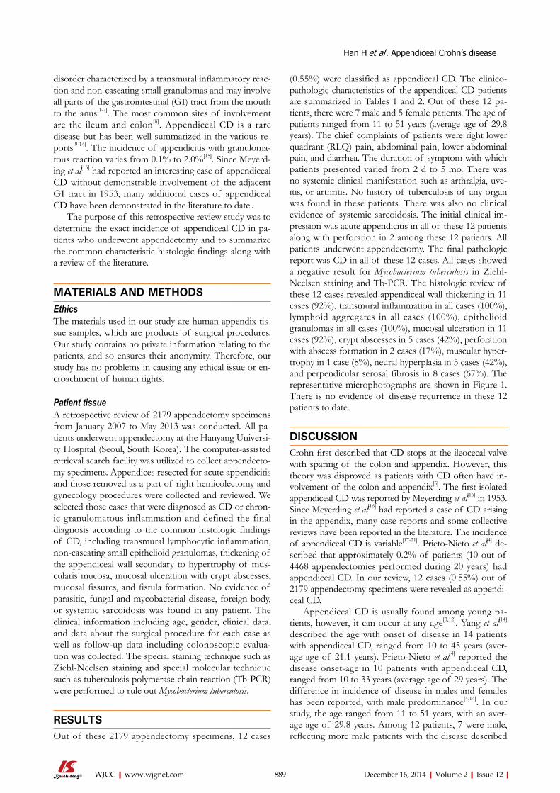
A B
C D
Figure 1 Appendiceal Crohn’s disease. A: The appendix with Crohn’s disease shows transmural inflammation with markedly thickened wall; B: There is a prominent lymphoid hyperplasia in the mucosa and serosa; C: The mucosa shows many small non-caseating granulomas; D: The serosa shows creeping fat with perpendicular thick fibrous bands.
Case No. Sex Age (yr) c/c SD Clinical impressions AFB Tb-PCR
1 F 30 RLQ pain None Acute appendicitis Negative Negative 2 M 38 RLQ pain 14 d Acute appendicitis Negative Negative 3 M 26 RLQ pain None Acute appendicitis Negative Negative 4 M 28 RLQ pain 7 d Acute appendicitis Negative Negative 5 F 25 RLQ pain 14 d Acute appendicitis Negative Negative 6 F 29 RLQ pain 7 d Acute appendicitis Negative Negative 7 M 51 RLQ pain 5 mo Acute appendicitis, perforation Negative Negative 8 F 49 RLQ pain 8 d Acute appendicitis Negative Negative 9 M 30 Abdominal pain 10 d Acute appendicitis, perforation Negative Negative 10 M 23 Lower abdominal pain 2 d Acute appendicitis Negative Negative 11 M 18 Lower abdominal pain, diarrhea 3 d Acute appendicitis Negative Negative 12 F 11 RLQ pain 7 d Acute appendicitis Negative Negative
Table 1 Summary of the patients with appendiceal Crohn’s disease
F: Female; M: Male; c/c: Chief complaint; RLQ: Right lower quadrant; SD: Symptom duration; AFB: Acid-fast bacillus; Tb-PCR: Tuberculosis polymerase chain reaction.
Han H et al . Appendiceal Crohn’s disease

previously described histologic characteristics. Interest-ingly, we found that appendiceal CD had the characteris-tic perpendicular serosal fibrous band formation in 8 out of 12 cases.
The differential diagnosis includes intestinal tubercu-losis, foreign body reaction, diverticulitis of the appendix, sarcoidosis, actinomycosis, and Yersinia infection[10,13,22,24,25]. Appendiceal tuberculosis results in the formation of epi-thelioid granulomas, however, the granulomas in tuber-culosis are larger with a central caseous necrosis and less discrete than those in Crohn’s disease[10,27-29]. If a foreign body is present, histologic examination should reveal the offending material and diverticular disease may be excluded via careful examination[14,27]. Intestinal sarcoid-osis is extremely rare and does not occur as an isolated finding[13,30]. Actinomycosis also results in a vague granu-lomatous tissue reaction, however, actinomycosis shows neutrophilic abscess formation with floating bacterial colonies (sulphur granules)[31-34]. Yersinia infection results in necrotizing granulomatous reaction in the appendiceal mucosa or submucosa and shows microabscess forma-tion[35,36].
The treatment of choice for appendiceal CD is ap-pendectomy[30]. Appendiceal CD shows lower recurrence rate compared with CD arising in other parts of the in-testine[25]. The prognosis of appendiceal CD seems to be much better than that of CD arising in the small or large bowel[14].
In conclusion, we described the incidence of appen-diceal CD in patients who underwent appendectomy and summarized the common characteristic histologic find-ings along with a review of the literature. Atypical and protracted clinical course, unusual gross features of the appendix and the characteristic features are a clue in the diagnosis of appendiceal CD.
COMMENTSBackgroundAppendiceal Crohn’s disease (CD) is a rare disease. Since Meyerding et al had reported an interesting case of appendiceal CD without demonstrable involve-ment of the adjacent gastrointestinal tract in 1953, many additional cases of ap-pendiceal CD have been demonstrated in the literature to date.Research frontiersThe incidence of appendicitis with granulomatous reaction varies from 0.1% to
2.0%. The incidence of appendiceal CD is variable. The purpose of this study was to determine the exact incidence of appendiceal CD in patients who un-derwent appendectomy and to summarize the common characteristic histologic findings along with a review of the literature. Innovations and breakthroughsThe histologic features are characterized by transmural chronic inflammation with marked fibrous thickening of the wall, lymphoid aggregates, small non-caseating granulomas, ulcerative mucosal change, crypt abscesses, muscular hypertrophy, and neural hyperplasia. In this study, the features were similar to the previously described histologic characteristics. However, the authors found that appendiceal CD had the characteristic perpendicular serosal fibrous band formation in 8 out of 12 cases.ApplicationsWith the characteristic clinical presentation and the typical pathologic findings, the clinicians and pathologists can consider the possibility of appendiceal CD. Atypical and protracted clinical course, unusual gross features of the appendix and the characteristic histologic features are a clue in the diagnosis of appendi-ceal CD.TerminologyCD is a chronic inflammatory bowel disorder characterized by a transmural inflammatory reaction and non-caseating small granulomas and may involve all parts of the gastrointestinal tract from the mouth to the anus. Peer reviewThe authors described appendiceal CD clinically presenting as acute appendici-tis. This is an interesting review and CD in appendix is a rare condition. When-ever it is encountered, the surgeon must know what to do and be aware of its prognosis. This paper will lead surgeons to this condition.
REFERENCES1 Stangl PC, Herbst F, Birner P, Oberhuber G. Crohn’s disease
of the appendix. Virchows Arch 2002; 440: 397-403 [PMID: 11956821 DOI: 10.1007/s004280100532]
2 Vanek VW, Spirtos G, Awad M, Badjatia N, Bernat D. Iso-lated Crohn’s disease of the appendix. Two case reports and a review of the literature. Arch Surg 1988; 123: 85-87 [PMID: 3276297 DOI: 10.1001/archsurg.1988.01400250095017]
3 Haddad M, Azim F, Koren A, Stelman E, Mor C, Zelikovski A. Crohn’s disease of the appendix. Eur J Surg 1993; 159: 191-192 [PMID: 8102900]
4 Prieto-Nieto I, Perez-Robledo JP, Hardisson D, Rodriguez-Montes JA, Larrauri-Martinez J, Garcia-Sancho-Martin L. Crohn’s disease limited to the appendix. Am J Surg 2001; 182: 531-533 [PMID: 11754865 DOI: 10.1016/S0002-9610(01)00811-X]
5 Crohn BB, Ginzburg L, Oppenheimer GD. Regional ileitis: a pathologic and clinical entity. 1932. Mt Sinai J Med 2000; 67: 263-268 [PMID: 10828911 DOI: 10.1001/jama.1932.02740680019005]
6 Kahn E, Markowitz J, Daum F. The appendix in inflamma-tory bowel disease in children. Mod Pathol 1992; 5: 380-383 [PMID: 1495944]
7 Kovalcik P, Simstein L, Weiss M, Mullen J. The dilemma of Crohn’s disease: Crohn’s disease and appendectomy. Dis Colon Rectum 1977; 20: 377-380 [PMID: 872706 DOI: 10.1007/BF02587363]
8 Nivatvongs S. Crohn’s disease of the appendix: report of a case and review of the literature. Dis Colon Rectum 1978; 21: 361-363 [PMID: 699727 DOI: 10.1007/BF02586668]
9 Zager JS, Gusani NJ, Derubertis BG, Shaw JP, Kaufman JP, DeNoto G. Laparoscopic appendectomy for Crohn’s disease of the appendix presenting as acute appendicitis. J Laparoen-dosc Adv Surg Tech A 2001; 11: 255-258 [PMID: 11569518 DOI: 10.1089/109264201750539808]
10 Lindhagen T, Ekelund G, Leandoer L, Hildell J, Lindström C, Wenckert A. Crohn’s disease confined to the appendix. Dis Colon Rectum 1982; 25: 805-808 [PMID: 6756829 DOI: 10.1007/BF02553319]
11 Agha FP, Ghahremani GG, Panella JS, Kaufman MW. Ap-pendicitis as the initial manifestation of Crohn’s disease:
December 16, 2014|Volume 2|Issue 12|WJCC|www.wjgnet.com 891
Histologic features Number of cases %
Wall thickening 11/12 92 Transmural inflammation 12/12 100 Lymphoid aggregates 12/12 100 Epithelioid granuolmas 12/12 100 Mucosal ulceration 11/12 92 Crypt abscess 5/12 42 Perforation 2/12 17 Muscular hypertrophy 1/12 8 Neural hyperplasia 5/12 42 Perpendicular serosal fibrosis 8/12 67
Table 2 Summary of histologic features of appendiceal Crohn’s disease
COMMENTS
Han H et al . Appendiceal Crohn’s disease

radiologic features and prognosis. AJR Am J Roentgenol 1987; 149: 515-518 [PMID: 3497535 DOI: 10.2214/ajr.149.3.515]
12 Ruiz V, Unger SW, Morgan J, Wallack MK. Crohn’s disease of the appendix. Surgery 1990; 107: 113-117 [PMID: 2404347]
13 McCue J, Coppen MJ, Rasbridge SA, Lock MR. Crohn’s dis-ease of the appendix. Ann R Coll Surg Engl 1988; 70: 300-303 [PMID: 3056207]
14 Yang SS, Gibson P, McCaughey RS, Arcari FA, Bernstein J. Primary Crohn’s disease of the appendix: report of 14 cases and review of the literature. Ann Surg 1979; 189: 334-339 [PMID: 426564 DOI: 10.1097/00000658-197903000-00014]
15 AbdullGaffar B. Granulomatous diseases and granulomas of the appendix. Int J Surg Pathol 2010; 18: 14-20 [PMID: 20106828 DOI: 10.1177/1066896909349246]
16 Meyerding EV, Bertram HF. Nonspecific granulomatous in-flammation (Crohn’s disease) of the appendix; a case report. Surgery 1953; 34: 891-894 [PMID: 13113513]
17 Larsen E, Axelsson C, Johansen A. The pathology of the ap-pendix in morbus Crohn and ulcerative colitis. Acta Pathol Microbiol Scand Suppl 1970; 212: Suppl 212: 161+ [PMID: 5267086]
18 Lennard-Jones JE, Morson BC. Changing concepts in Crohn’s disease. Dis Mon 1969; Aug: 1-37 [PMID: 4309015]
19 Lockhart-mummery HE, Morson BC. Crohn’s disease of the large intestine. Gut 1964; 5: 493-509 [PMID: 14244023 DOI: 10.1136/gut.5.6.493]
20 Rappaport H, Burgoyne FH, Smetana HF. The pathology of regional enteritis. Mil Surg 1951; 109: 463-502 [PMID: 14874971]
21 Warren S, Sommers SC. Cicatrizing enteritis as a pathologic entity; analysis of 120 cases. Am J Pathol 1948; 24: 475-501 [PMID: 18859355]
22 Cerdán FJ, Balsa T, Torres-Melero J, García MC, Remezal M, Balibrea JL. Appendiceal Crohn’s disease. Rev Esp Enferm Dig 1995; 87: 331-334 [PMID: 7794643]
23 Wettergren A, Munkholm P, Larsen LG, Meinecke B, Lang-holz E, Jess P, Binder V. Granulomas of the appendix: is it Crohn’s disease? Scand J Gastroenterol 1991; 26: 961-964 [PMID: 1947789 DOI: 10.3109/00365529108996249]
24 Kosakowski C, Thompson JE, Feinberg MJ. Coexistence of primary Crohn’s disease and carcinoid tumor isolated to the appendix. A case report. Acta Chir Scand 1986; 152: 233-236 [PMID: 3716745]
25 Ariel I, Vinograd I, Hershlag A, Olsha O, Argov S, Klaus-
ner JM, Rabau MY, Freund U, Rosenmann E. Crohn’s disease isolated to the appendix: truths and fallacies. Hum Pathol 1986; 17: 1116-1121 [PMID: 3770730 DOI: 10.1016/S0046-8177(86)80416-6]
26 Early CK, Kouri S. Granulomatous disease of the appen-dix manifesting as a cecal mass: report of a case. Dis Colon Rectum 1980; 23: 421-422 [PMID: 7418581 DOI: 10.1007/BF02586793]
27 Allen DC, Biggart JD. Granulomatous disease in the ver-miform appendix. J Clin Pathol 1983; 36: 632-638 [PMID: 6853730 DOI: 10.1136/jcp.36.6.632]
28 Wang TK, Tolnai G, Campbell JS, Sirois J, Liepa E. Crohn’s disease of the appendix. Can Med Assoc J 1972; 106: 233-236 [PMID: 5057957]
29 Weiss Y, Durst AL. Crohn’s disease of the appendix. Presen-tation of a case with review of the literature. Am J Gastroen-terol 1975; 63: 333-339 [PMID: 1130399]
30 Bak M, Andersen JC. Crohn’s disease limited to the vermi-form appendix. Acta Chir Scand 1987; 153: 441-446 [PMID: 3673453]
31 Liu V, Val S, Kang K, Velcek F. Case report: actinomycosis of the appendix--an unusual cause of acute appendicitis in children. J Pediatr Surg 2010; 45: 2050-2052 [PMID: 20920728 DOI: 10.1016/j.jpedsurg.2010.06.015]
32 Lee SY, Kwon HJ, Cho JH, Oh JY, Nam KJ, Lee JH, Yoon SK, Kang MJ, Jeong JS. Actinomycosis of the appendix mimicking appendiceal tumor: a case report. World J Gastroenterol 2010; 16: 395-397 [PMID: 20082489 DOI: 10.3748/wjg.v16.i3.395]
33 Altanis E. Actinomycosis of the appendix and pelvis. J Reprod Med 2009; 54: 411-412; author reply 412 [PMID: 19642234]
34 Yiğiter M, Kiyici H, Arda IS, Hiçsönmez A. Actinomycosis: a differential diagnosis for appendicitis. A case report and re-view of the literature. J Pediatr Surg 2007; 42: E23-E26 [PMID: 17560191 DOI: 10.1016/j.jpedsurg.2007.03.057]
35 Kojima M, Morita Y, Shimizu K, Yoshida T, Yamada I, Togo T, Johshita T. Immunohistological findings of suppurative granulomas of Yersinia enterocolitica appendicitis: a report of two cases. Pathol Res Pract 2007; 203: 115-119 [PMID: 17189675 DOI: 10.1016/j.prp.2006.10.004]
36 Bronner MP. Granulomatous appendicitis and the appen-dix in idiopathic inflammatory bowel disease. Semin Diagn Pathol 2004; 21: 98-107 [PMID: 15807470 DOI: 10.1053/j.semdp.2004.12.001]
P- Reviewer: Cetinkunar S, Matsumoto S, Sartelli M S- Editor: Tian YL L- Editor: A E- Editor: Wu HL
December 16, 2014|Volume 2|Issue 12|WJCC|www.wjgnet.com 892
Han H et al . Appendiceal Crohn’s disease

© 2014 Baishideng Publishing Group Inc. All rights reserved.
Published by Baishideng Publishing Group Inc8226 Regency Drive, Pleasanton, CA 94588, USA
Telephone: +1-925-223-8242Fax: +1-925-223-8243
E-mail: [email protected] Desk: http://www.wjgnet.com/esps/helpdesk.aspx
http://www.wjgnet.com
Related Documents