[Frontiers in Bioscience 12, 3140-3151, May 1, 2007] 3140 Apoptosis and endometriosis Tasuku Harada 1 , Fuminori Taniguchi 1 , Masao Izawa 2 , Yoko Ohama 1 , Yasuko Takenaka 1 , Yukiko Tagashira 1 , Ayako Ikeda 1 , Ayako Watanabe 1 , Tomio Iwabe 1 , Naoki Terakawa 1 1 Department of Obstetrics and Gynecology, Tottori University School of Medicine, Yonago, Japan, 2 Department of Biosignalling, Tottori University School of Medicine, Yonago, Japan TABLE OF CONTENTS 1. Abstract 2. Introduction 3. Molecular basis of apoptosis 3.1. Induction and execution of apoptosis 3.2. Hallmark of apoptotic cell structure 3.3. The mitochondria pathways through organelle morphogenesis 4. Apoptosis in the normal endometrium 4.1. Bcl-2 family and Fas/FasL system in normal endometrium 5. Apoptosis in endometriosis 5.1. Apoptosis of eutopic and ectopic endometrium in endometriosis 5.2. Bcl-2 family in endometriosis 5.3. Fas/FasL system in endometriosis 5.4. Apoptosis in peritoneal macrophages from patients with endometriosis 5.5. Apoptosis in pathophysiology of endometriosis 5.6. Apoptosis and treatment of endometriosis 6. Conclusions 7. Reference 1. ABSTRACT Apoptosis plays a critical role in maintaining tissue homeostasis and represents a normal function to eliminate excess or dysfunctional cells. Accumulated evidence suggest that apoptosis helps to maintain cellular homeostasis during the menstrual cycle by eliminating senescent cells from the functional layer of the uterine endometrium during the late secretory and menstrual phase of the cycle. BCL-2 family and Fas/FasL system have been extensively studied in human endometrium and endometriotic tissues. Eutopic endometrium from women with endometriosis reportedly has some fundamental differences compared with normal endometrium of women without endometriosis. The differences could contribute to the survival of regurgitating endometrial cells into the peritoneal cavity and the development of endometriosis. One mechanism that recently gained a lot of interest is the finding that apoptosis appeared in eutopic and ectopic endometrium of patients with endometriosis. This study is a current review of the literature focused on the physiological role of apoptosis in normal endometrium and the alterations in regulation of apoptosis in eutopic and ectopic endometrium from women with endometriosis. Finally, role of apoptosis in the treatment of endometriosis is reviewed to link the basic research findings into clinical applications. 2. INTRODUCTION Endometriosis is defined by the presence of endometrium-like glandular tissue and stroma outside the uterus. It is a common disease affecting 5% to 15% of women in the general population and 40% of women seeking infertility evaluation (1). Although the high incidence of endometriosis and the fact that eighty years have passed since the initial description of the disease by Sampson (2), our current understanding of the etiology and pathophysiology of endometriosis remains obscure. Several theories have been proposed, including development by metaplasia, development from Mullerian remnants, and after implantation and growth of endometrium following retrograde menstrual reflux. Nearly all women of reproductive age exhibit some degree of reflux of endometrial debris (3). Menstrual effluents retrogradely shed into the peritoneal cavity were observed to contain viable endometrial cells (4-8). These mechanisms are necessary but insufficient to explain why only some patients develop the disease. A couple of views have been proposed (9). The first theory is based on disorders of the endometrium in which it resists normal peritoneal means of cleaning. The second theory suggests that the disease is secondary to alterations of the cellular and humoral immunity that induce excessive receptivity of the peritoneal mesothelium,

Welcome message from author
This document is posted to help you gain knowledge. Please leave a comment to let me know what you think about it! Share it to your friends and learn new things together.
Transcript
[Frontiers in Bioscience 12, 3140-3151, May 1, 2007]
3140
Apoptosis and endometriosis Tasuku Harada1, Fuminori Taniguchi1, Masao Izawa2, Yoko Ohama1, Yasuko Takenaka1, Yukiko Tagashira1, Ayako Ikeda1, Ayako Watanabe1, Tomio Iwabe1, Naoki Terakawa1 1 Department of Obstetrics and Gynecology, Tottori University School of Medicine, Yonago, Japan, 2 Department of Biosignalling, Tottori University School of Medicine, Yonago, Japan TABLE OF CONTENTS 1. Abstract 2. Introduction 3. Molecular basis of apoptosis 3.1. Induction and execution of apoptosis 3.2. Hallmark of apoptotic cell structure
3.3. The mitochondria pathways through organelle morphogenesis 4. Apoptosis in the normal endometrium
4.1. Bcl-2 family and Fas/FasL system in normal endometrium 5. Apoptosis in endometriosis
5.1. Apoptosis of eutopic and ectopic endometrium in endometriosis 5.2. Bcl-2 family in endometriosis 5.3. Fas/FasL system in endometriosis 5.4. Apoptosis in peritoneal macrophages from patients with endometriosis 5.5. Apoptosis in pathophysiology of endometriosis 5.6. Apoptosis and treatment of endometriosis
6. Conclusions 7. Reference 1. ABSTRACT Apoptosis plays a critical role in maintaining tissue homeostasis and represents a normal function to eliminate excess or dysfunctional cells. Accumulated evidence suggest that apoptosis helps to maintain cellular homeostasis during the menstrual cycle by eliminating senescent cells from the functional layer of the uterine endometrium during the late secretory and menstrual phase of the cycle. BCL-2 family and Fas/FasL system have been extensively studied in human endometrium and endometriotic tissues. Eutopic endometrium from women with endometriosis reportedly has some fundamental differences compared with normal endometrium of women without endometriosis. The differences could contribute to the survival of regurgitating endometrial cells into the peritoneal cavity and the development of endometriosis. One mechanism that recently gained a lot of interest is the finding that apoptosis appeared in eutopic and ectopic endometrium of patients with endometriosis. This study is a current review of the literature focused on the physiological role of apoptosis in normal endometrium and the alterations in regulation of apoptosis in eutopic and ectopic endometrium from women with endometriosis. Finally, role of apoptosis in the treatment of endometriosis is reviewed to link the basic research findings into clinical applications.
2. INTRODUCTION
Endometriosis is defined by the presence of endometrium-like glandular tissue and stroma outside the uterus. It is a common disease affecting 5% to 15% of women in the general population and 40% of women seeking infertility evaluation (1). Although the high incidence of endometriosis and the fact that eighty years have passed since the initial description of the disease by Sampson (2), our current understanding of the etiology and pathophysiology of endometriosis remains obscure. Several theories have been proposed, including development by metaplasia, development from Mullerian remnants, and after implantation and growth of endometrium following retrograde menstrual reflux.
Nearly all women of reproductive age exhibit some degree of reflux of endometrial debris (3). Menstrual effluents retrogradely shed into the peritoneal cavity were observed to contain viable endometrial cells (4-8). These mechanisms are necessary but insufficient to explain why only some patients develop the disease. A couple of views have been proposed (9). The first theory is based on disorders of the endometrium in which it resists normal peritoneal means of cleaning. The second theory suggests that the disease is secondary to alterations of the cellular and humoral immunity that induce excessive receptivity of the peritoneal mesothelium,
Apoptosis in endometriosis
3141
hyperactivated macrophages, and abnormalities of NK cells. The peritoneal environment may alter a genetic predisposed endometrium, which then becomes favorable for invasion. It is also possible an excess of refluxing endometrium or altered endometrium has the potential to form a proinflammatory or hormonal environment favorable to establish the disease (9).
The fact that the eutopic endometrium of women with endometriosis shares changes with ectopic tissue and that these changes are not found in the eutopic endometrium of disease-free women has advanced the view that the primary defect in endometriosis is to be found in the eutopic endometrium (10). Cells and tissue elements, derived from such an altered eutopic endometrium and shed into the peritoneal cavity, have been proposed to have a higher potential for implantation and growth on peritoneal surfaces and development into endometriosis (11-13). On the other hand, the fact that many differences observed between eutopic endometrium and ectopic tissue of a patient with endometriosis can be explained as the direct influence of the different environment of peritoneal fluid (14-17). One of the endometrial alterations appearing in eutopic and ectopic endometrium from women with endometriosis refers to the regulation of apoptosis. 3. MOLECULAR BASIS OF APOPTOSIS Apoptosis is a distinctive form of programmed cell death that is defined by characteristic morphological and biochemical events that result in the efficient elimination of cells from tissue without eliciting an inflammatory response (18). Apoptosis is critical for tissue modeling during embryogenesis and cellular homeostasis, and also contributes to many facets of pathological processes in diseases including those in reproductive systems (19, 20). Here a current model of the death signaling pathways is summarized and topics on mitochondrial pathways and clearance mechanism of apoptotic cells by phagocytes are also described. 3.1. Induction and execution of apoptosis Apoptosis is a specific mode of cell death with a characteristic pattern of morphological, biochemical and molecular changes (21). Necrosis is associated with swelling of organelles, a disruption of plasma membranes and random DNA degradation. In contrast, apoptosis involves nuclear chromatin condensation, cytoplasmic blebbing, and internucleosomal DNA degradation. There are two major pathways for inducing apoptosis: one starts with binding of death receptors with cognate death ligands leading to the activation of caspase-8, and the other depends on stress, chemotherapy or irradiation, and involves the mitochondrial release of cytochrome c resulting in the activation of caspase-9. Activated caspase-8 also cleaves Bid to the truncated Bid leading to the mitochondrial pathway.
Fas and tumor necrosis factor receptor are members of the death receptor belonging to tumor necrosis factor/ nerve growth factor receptor superfamily. At variance with the structural homology among these receptors and their ligands, respective receptor systems
utilize distinct death signal pathways. Members of the receptor family (e.g., Fas and TNFR1) contain a cytoplasmic domain called the “death domain” (22). Upon receptor activation after the interaction with cognate death ligands (e.g., FasL and TNFα), the death domain undergoes interaction with a death domain in the adaptor protein (e.g., FADD) leading to the formation of death-inducing signaling complex (DISC), and initiate the execution process of apoptosis (23). Following their activation, caspase-8 and -9, the initiator caspases, cleave and activate effector caspases, caspase-3, -6 and/or -7. Although the same effector caspases are activated by different apoptotic stimuli, the upstream events leading to the activation of caspase-8 or caspase-9 exhibit diversity. Caspase-9 activation starts with the mitochondrial release of cytochrome c in response to apoptotic stimuli. The cytochrome c binds to Apaf-1 in the presence of dATP, and the Apaf-1/cytochrome c/dATP complex forms an oligomeric “Apoptosome” (24, 25), which recruits and activates the pro-caspase-9 leading to the activation of effector caspases such as caspase-3. Caspase-3 plays a central role in proteolytic cleavage of ICAD (DFF45) to activate CAD (DFF40) leading to DNA fragmentation into nucleosomal units.
Although caspase activity is regulated by the
zymogen activation as described above, direct inhibition of active caspase by the inhibitor of apoptotsis (IAP) is important. Among IAPs identified so far, XIAP, c-IAP1, and c-IAP2 are thought to directly inhibit caspase-3, caspase-7 and caspase-9 (26-29).
3.2. Hallmark of apoptotic cell structure
Apoptotic cells are characterized by many morphological and biochemical alterations. Morphologically, apoptotic cells present with condensed chromatin, multiple membrane-bound organelles (apoptotic bodies), and a shrunken appearance. Biochemically, apoptosis is characterized by 180- base pair (bp) units of genomic DNA fragmentation due to the digestion of double-stranded DNA at the linker region of nucleosomal structure through a caspase-3-dependent activation of CAD (DFF40) /ICAD (DFF45) complex. However, the absence of these DNA ladders does not always mean the absence of apoptosis. Cleavage of DNA into higher molecular weight structures (rosettes, 300 Kbp) and loops (50 Kbp) also become alternative apoptosis marker in some cells where CAD (DFF40)/ICAD (DFF45) system is defective (30). Another well-known characteristic of apoptotic cell structure is the exposure of phosphatidylserine (PS) onto the outer surface of the plasma membrane. This early event in apoptotic cells is now believed as an“eat-me flag” for phagocytes (31).
3.3. Mitochondria pathways through organelle morphogenesis
In mammalian cells, mitochondria have a central role in apoptosis that is regulated by members of the Bcl-2 family (30)). The mitochondria pathway integrates stress and some developmental apoptotic stimuli, and is triggered by the translocation of pro-apoptotic Bcl-2 family member such as Bax into the mitochondria. This event
Apoptosis in endometriosis
3142
results in the release of cytochrome c from mitochondria into the cytosol, which induces pro-caspase-9 activation and is sometimes accompanied by loss of mitochondrial membrane potential and destabilization of the outer mitochondrial membrane (32, 33). Although Bcl-2 family proteins are potent regulators of apoptosis, their intracellular localization to mitochondria has been observed, but function still remains unclear. Recently, Karbowski et al. showed that two members of the Bcl-2 family, Bax and Bak, change intracellular localization during the course of apoptotic process to concentrate into foci at sites of mitochondrial scission (34). The observation suggests that Bcl-2 family members may also regulate apoptosis through mitochondrial morphogenesis. 4. APOPTOSIS IN THE NORMAL ENDOMETRIUM
Historically, menstruation has been associated with ischemic necrosis of the functional layer of the endometrium caused by the contraction of the spiral arteries, with the process being dependent on sex hormone concentration (35, 36). The endometrial cycle in regularly menstruating women consists of three distinct phases (proliferative, secretory, and menstrual). Hopwood and Levison (37) reported the presence of apoptosis in human endometrium following advancement of the concept of apoptosis by Kerr and co-workers (18). Accumulated evidence suggests that apoptosis helps to maintain cellular homeostasis during the menstrual cycle by eliminating senescent cells from the functional layer of the uterine endometrium during the late secretory and menstrual phase of the cycle (37-39). This is followed by the proliferative phase of the cycle.
Apoptosis was detected in the glandular
epithelium of late secretory and menstruating endometrium, while very little apoptosis was detected during the proliferative phase or at the beginning of the secretory phase (40, 41). The proliferation of endometrial cells in the proliferative phase has been related generally to the action of estrogens, while progesterone is thought to direct the cells into the differentiation pathway, resulting in growth arrest. Considering the cyclical nature of apoptosis in normal endometrium, it seems likely that estrogen and progesterone can regulate the signals that result in apoptosis in this tissue. Vaskivuo et al. showed that the pattern of apoptosis negatively correlated to serum estradiol concentrations in the proliferative phase (41).
4.1. Bcl-2 family and Fas/FasL in normal endometrium The Bcl-2 (B cell lymphoma/ leukemia-2) gene is located on chromosome 18, which is important in determining whether a cell will be irreversibly committed to apoptosis (42). The BCL-2 protein is probably the best well characterized of apoptosis-related molecules, and data now equivocally support a role for the BCL-2 protein as a cell death repressor (43).
To date, the precise mechanism of apoptosis in the human endometrium is not yet fully clarified, although BCL-2 has been considered to inhibit apoptosis in the human endometrium during the proliferative phase (44).
BCL-2 cyclically expressed in endometrial glandular and stromal cells, peaks during the late proliferative phase, while decreased during the late secretory and menstruating phases (45-48). In contrast, myometrial smooth muscle cells showed consistent BCL-2 immunoreactivity throughout the menstrual cycle (47). In Bcl-2 deficient mice, many apoptotic cells and apoptotic bodies were often observed in the glands and the myometrium (49). Therefore, BCL-2 may be an essential gene product for the survival of both endometrial glandular cells and myometrial smooth muscle cells.
Rogers et al. demonstrated that the cyclical pattern
of the BCL-2 expression had no longer occurred after administration of levonogestrel, suggesting that a constant administration of steroid hormones can affect the expression of BCL-2 (50). Using immunohistochemical staining, the authors observed that BCL-2, Fas and caspase-3 showed different expression levels in the functional layer versus basal layer of normal endometrium (50). The anti-apoptotic protein BCL-2 presented with higher expression in the basal layer, whereas, death receptor Fas and caspase-3 were higher in the functional layer of the endometrium. These results fit well with the functional biology of endometrium. Since the basal layer remains relatively constant throughout the menstrual cycle, apoptosis is less common in this layer. In contrast, the functional layer that undergoes cyclical growth, differentiation and shedding, appears with increased level of apoptosis.
Some transcriptional molecules can regulate the expression of Bcl-2 in human endometrium. p53 immunoreactivity was not observed in either glandular cells or stromal cells throughout the cycle (47). In contrast, the staining pattern of c-jun and Sp-3 in glandular cells was similar to that of Bcl-2, in terms of pattern, intensity, and cellular distribution, throughout the menstrual cycle suggesting that c-jun and Sp-3 may have a role in regulating Bcl-2 expression (47).
Although expression of the protein product of
Bcl-2 gene in the human endomerium has been described (44, 51, 52), Bcl-2 is only one member of this multigene family, consisting of numerous proteins homologous to BCL-2 (53-55). Other members of the Bcl-2 gene family likely play important roles in controlling apoptosis by mechanisms that are independent of or complementary to the action of BCL-2. Members of the BCL-2 family interact through homodimeric and heterodimeric associations (53, 55), such that the susceptibility of any given cells to a potential apoptotic stimulus may be determined by the ratio of proapoptotic and antiapoptotic BCL-2 family members present in the cell at that time (53).
BAX is a BCL-2 family member that promotes
cell death susceptibility, possibly by countering the effect of BCL-2 on cellular survival through heterodimer interaction (53). Another member of this family of genes, Bcl-x, provides an interesting example of a single gene that, via alternative splicing mechanisms, encodes both a positive and a negative regulator of apoptosis (54). The long form BCL-X (BCL-Xlong) contains an open reading
Apoptosis in endometriosis
3143
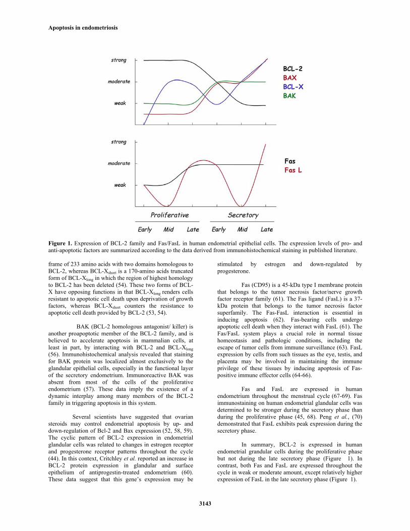
Figure 1. Expression of BCL-2 family and Fas/FasL in human endometrial epithelial cells. The expression levels of pro- and anti-apoptotic factors are summarized according to the data derived from immunohistochemical staining in published literature. frame of 233 amino acids with two domains homologous to BCL-2, whereas BCL-Xshort is a 170-amino acids truncated form of BCL-Xlong in which the region of highest homology to BCL-2 has been deleted (54). These two forms of BCL-X have opposing functions in that BCL-Xlong renders cells resistant to apoptotic cell death upon deprivation of growth factors, whereas BCL-Xshort counters the resistance to apoptotic cell death provided by BCL-2 (53, 54).
BAK (BCL-2 homologous antagonist/ killer) is
another proapoptotic member of the BCL-2 family, and is believed to accelerate apoptosis in mammalian cells, at least in part, by interacting with BCL-2 and BCL-Xlong (56). Immunohistochemical analysis revealed that staining for BAK protein was localized almost exclusively to the glandular epithelial cells, especially in the functional layer of the secretory endometrium. Immunoreactive BAK was absent from most of the cells of the proliferative endometrium (57). These data imply the existence of a dynamic interplay among many members of the BCL-2 family in triggering apoptosis in this system.
Several scientists have suggested that ovarian steroids may control endometrial apoptosis by up- and down-regulation of Bcl-2 and Bax expression (52, 58, 59). The cyclic pattern of BCL-2 expression in endometrial glandular cells was related to changes in estrogen receptor and progesterone receptor patterns throughout the cycle
(44). In this context, Critchley et al. reported an increase in BCL-2 protein expression in glandular and surface epithelium of antiprogestin-treated endometrium (60). These data suggest that this gene’s expression may be
stimulated by estrogen and down-regulated by progesterone. Fas (CD95) is a 45-kDa type I membrane protein that belongs to the tumor necrosis factor/nerve growth factor receptor family (61). The Fas ligand (FasL) is a 37-kDa protein that belongs to the tumor necrosis factor superfamily. The Fas-FasL interaction is essential in inducing apoptosis (62). Fas-bearing cells undergo apoptotic cell death when they interact with FasL (61). The Fas/FasL system plays a crucial role in normal tissue homeostasis and pathologic conditions, including the escape of tumor cells from immune surveillance (63). FasL expression by cells from such tissues as the eye, testis, and placenta may be involved in maintaining the immune privilege of these tissues by inducing apoptosis of Fas-positive immune effector cells (64-66).
Fas and FasL are expressed in human endometrium throughout the menstrual cycle (67-69). Fas immunostaining on human endometrial glandular cells was determined to be stronger during the secretory phase than during the proliferative phase (45, 68). Peng et al., (70) demonstrated that FasL exhibits peak expression during the secretory phase.
In summary, BCL-2 is expressed in human
endometrial grandular cells during the proliferative phase but not during the late secretory phase (Figure 1). In contrast, both Fas and FasL are expressed throughout the cycle in weak or moderate amount, except relatively higher expression of FasL in the late secretory phase (Figure 1).
Apoptosis in endometriosis
3144
5. APOPTOSIS IN ENDOMETRIOSIS 5.1. Apoptosis of eutopic and ectopic endometrium in endometriosis
Eutopic endometrium from women with endometriosis has some fundamental differences compared with normal endometrium of women without endometriosis. These include a variety of anomalies in structure, proliferation, immune components, adhesion molecules, proteolytic enzymes and their inhibitors, steroid and cytokine production and responsiveness, gene expression and protein production (10). These differences could contribute to the survival of the regurgitating endometrial cells into the peritoneal cavity and the development of endometriosis. One of the mechanisms that recently gained a lot of interest is the finding that apoptosis appears in eutopic and ectopic endometrium of patients with endometriosis.
Gebel et al. reported that the percentage of
apoptosis in sloughed endometrial cells was greatly reduced among women with endometriosis implying that the number of surviving cells that enter the peritoneal cavity is greater in women who develop endometriosis (71). Szymanowski recently described that apoptosis indices in the eutopic endometrium of women with endometriosis were lower compared to women without endometriosis (72). Dmowski et al. demonstrated that the apoptotic index in glandular epithelium was significantly lower in women with endometriosis than in controls (73). This difference was caused primarily by a significant decrease in apoptosis during the late secretory/menstrual and early proliferative phases in women with endometriosis. The cyclic variability of apoptosis may be lost in these women.
One can speculate that if the decrease in
apoptosis facilitates ectopic survival and implantation of the endometrial cells then there may be an inverse correlation between the level of apoptosis and the severity of the disease. To test this hypothesis Dmowski et al. analyzed the apoptotic index according to the stage of endometriosis and found that there was a trend toward decreased apoptosis with increasing stage of the disease, but the difference lacked statistical significance (73). On the other hand, Goumenou et al. showed that the apoptotic rate as well as Bcl-2 and Bax expression in ovarian endometriotic cells was not affected by the stage of endometriosis or the phase of the menstrual cycle (74).
5.2. Bcl-2 family in endometriosis
Watanabe et al. examined BCL-2 expression in eutopic and ectopic endometrium in patients with endometriosis, demonstrating that the expression of BCL-2 in endometrial glandular cells has a cyclic pattern in eutopic endometrium in patients with endometriosis, but that cyclic changes were not apparent in peritoneal and ovarian endometriotic tissues (45). In a study by Jones et al., the authors did not detect apoptosis in stromal cells from peritoneal endometriotic tissues (75). In accordance with these findings, BCL-2 is expressed to a greater extent in stromal cells from ectopic tissues (75). This
overexpression may be directly correlated to the increase in the number of estrogen receptors expressed by ectopic stroma (76).
An increased expression of BCL-2 protein was
found in proliferative eutopic endometrium from women with endometriosis when compared with normal endometrium from healthy women (77). In the same study BAX expression was absent in proliferative endometrium, whereas there was an increase in its expression in secretory endometrium from women with endometriosis and healthy women. The altered expression of BCL-2 in eutopic endometrium of women with endometriosis resulted to a decreased number of apoptotic cells and consequently to their abnormal survival in the ectopic locations (77).
In ovarian endometriosis, the weak BCL-2
expression, or the strong correlation between low BCL-2 and high BAX expression were shown by the immunohistochemical methods (74, 78). Recently, Dufournet et al. demonstrated that the Bcl-2 expression was lower in ovarian endometriosis than in peritoneal and colorectal endometriosis (79). Nezhat and Kalir reported that lower Bcl-2 expression in cystic than in non-cystic endometriotic lesions (80). Moreover, as observed by Beliard et al., the peritoneal endometriosis exhibited a high Bcl-2 expression, in addition, they showed that the peritoneal Bcl-2 expression was higher than those found in eutopic endometrium with or without endometriosis (81). The expression of Bcl-2 varied according to the location of endometriosis, suggesting the involvement of different apoptotic pathways and etipathologies. Recently, Braun et al. analyzed the expression of apotosis-regulating genes in eutopic endometrium from women with or without endometriosis. The ratio of transcript abundance levels for Bcl-xL : Bcl-xS (antiapoptotic : proapoptotic) was substantially higher in eutopic endometria from women with endometriosis compared to endometria from women without endometriosis (82). This result could be consistent with apoptotic resistance and enhanced survival of endometrial cells in endometriosis. 5.3. Fas/FasL system in endometriosis
Few studies have been published on the expression of Fas in endometriotic tissues. Moreover, according to our knowledge, there are no studies showing quantitative comparison of Fas expression between endometriotic tissues and endometrium from disease-free women. Harada et al. found that Fas is expressed randomly in both eutopic and ectopic endometrial tissues (83). The authors suggested that the expression of Fas antigen may be less involved in apoptosis of eutopic and ectopic endometrial tissues as an apoptosis-regulator. In accordance with this finding, Watanabe et al. also observed Fas expression in glandular cells of both ectopic and eutopic endometrium (45). In contrast, with the cyclic expression pattern of Bcl-2, Fas expression was constant in both tissues throughout the menstrual cycle. Differences in the expression of Fas were found between ovarian, cervical, and endometrial carcinoma tissues comparing with normal tissues. Tumor cells had significantly decreased levels of Fas (84). In addition, there was a higher Fas expression in ovarian endometriotic cells
Apoptosis in endometriosis
3145
comparing with benign ovarian tumors but the difference did not reach significance (85).
In contrast with Fas, there are many studies indicated that higher expression of FasL by endometriotic tissues contributes to their survival and the development of endometriosis. A recent study by Garcia-Velasco et al. suggests that levels of soluble/active FasL are higher in serum and peritoneal fluid in women with moderate to severe endometriosis than in women with early-stage disease or in disease-free women (86). Higher levels of soluble FasL in the peritoneal fluid of women with endometriosis may contribute to increased apoptosis of Fas-bearing immune cells in the peritoneal cavity, leading to their decreased scavenger activity (86). This may result in prolonged survival of endometrial cells into the peritoneal cavity.
The sources of the elevated levels of soluble
FasL in the peritoneal cavity were endometriotic lesions and peritoneal fluid leukocytes. Several authors have shown that endometrial glandular and stromal cells express FasL, at both the messenger RNA and protein levels. This membrane-bound FasL can be shed by matrilysin, producing an active, soluble form of the ligand (87). Peritoneal fluid leukocytes are another plausible source for high levels of soluble FasL in women with endometriosis, because human-activated peripheral blood mononuclear cells were shown to express FasL messenger RNA (62).
Macrophage-derived growth factors, such as
platelet-derived growth factor (PDGF) and transforming growth factor (TGF), are increased in the peritoneal fluid of women with endometriosis (88). Garcia-Velasco et al. showed that macrophage-conditioned media containing PDGF and TGF-β induced FasL expression by endometrial stromal cells, suggesting that peritoneal macrophages in endometriosis might stimulate a Fas-mediated apoptosis of immune cells (67). Expression of FasL by the endometriotic cells may protect them from attack by T-cells. Consequently, ectopic endometrial cells escaping from immune surveillance in the peritoneal cavity of women with endometriosis may contribute to the maintenance of the disease.
It is, therefore, possible that many endometriotic
cells not only become resistant to Fas-mediated apoptosis, but additionally they have acquired the ability to utilize this pathway to their advantage by launching a “Fas counterattack” against the host’s immune system. The increased expression of FasL by endometriotic cells coincides with their inherent resistance to Fas-mediated apoptosis which protects them from a “suicidal” death.
Up-regulation of FasL expression by endometriotic cells could be induced after the adhesion of these cells to the extracellular matrix proteins laminin, fibronectin, and collagen IV (89). FasL expressed on endometriotic cells may induce apoptosis of the local immune cells including activated T lymphocytes, thereby reducing attacks by host immune-surveillance and promoting the survival of endometrial stromal cells during
the initial attachment of endometrial implants. Early lesions of endometriosis reportedly invade the extracellular matrix of the peritoneum (90). FasL expression that occurs when endometrial stromal cells attach to the extracellular matrix may be one of the critical events in the development of endometriosis. Under these observations, we could speculate that the expression levels of soluble/active FasL may be enhanced in shedding endometrial cells presenting in the peritoneal environment and protect endometrial cells from the immune effector cells of the peritoneal cavity.
Interleukin-8, a chemokine for neutrophils and
a potent angiogenic agent, is elevated in the peritoneal fluid of women with endometriosis (91). IL-8 promotes proliferation of stromal cells derived from endometriotic tissues (91, 92), suggesting that it may facilitate growth of endometriotic implants. Selam et al., (93) examined whether IL-8 may up-regulate FasL expression in endometrial cells and may be relevant for the development of a relative local immunotolerance in endometriosis. They demonstrated a concentration-dependent increase in the protein expression of FasL by IL-8 in endometrial stromal cells. The authors speculated that elevated peritoneal fluid IL-8 levels, via stimulation of FasL-induced apoptosis in activated T lymphocytes, contribute to an immune-privileged environment around the endometriosis implants supporting their survival.
IL-8 exerts a chemotactic activity primarily on
neutrophils and inhibits their apoptosis even in the presence of Fas engagement (94). Kwak et al. investigated the effects of plasma and peritoneal fluid from patients with advanced endometriosis on apoptosis of neutrophils (95). Adding plasma and PF in neutrophil culture reduced spontaneous apoptosis. Neutralizing IL-8 antibody abrogated the delay of neutrophil apoptosis induced by peritoneal fluid, suggesting that IL-8 is one of the neutrophil survival factors in the PF of endometriosis patients. The impaired clearance of cells responsible for innate immunity in the peritoneal fluid of patients with endometriosis may be associated with the development of the disease (95).
5.4. Apoptosis in peritoneal macrophages from patients with endometriosis
The peritoneal cavity, the commonest site of endometriosis (96), contains fluid whose major cellular constituents are macrophages (97). In endometriosis, the number and secretory activity of these cells increase (3, 98) and recent evidence suggests that these cells play an important role in developing and maintaining of endometriosis (17, 99).
The function of macrophages is altered in
several aspects, as well. The cytotoxic power of peritoneal macrophages in endometriosis patients with respect to the endometrium is reduced (100). The diminution of cytotoxicity of peritoneal macrophages could be more significant than that of the circulating macrophages (101). The reduced capacity of peritoneal macrophages from women with endometriosis to mediate lysis of endometrial cells together with the increased resistance of ectopic endometrial cells to macrophage-mediated cytolysis may
Apoptosis in endometriosis
3146
promote survival of the endometrial cells in the peritoneal cavity of women with endometriosis (102).
McLaren et al. reported an increased percentage
of Bcl-2-positive macrophages in the peritoneal fluid of women with endometriosis compared with the non-endometriotic group, resulting in an increased number of cells surviving the process of activation and thus delaying apoptosis (78). This may explain the increased numbers of macrophages found in the peritoneal fluid of patients with endometriosis.
Immunohistochemical staining revealed a population of BCL-2-positive, BAX- negative tissue macrophages present only in ectopic tissue during both phases of the menstrual cycle (78). The expression of BCL-2 and the absence of BAX may confer on these macrophages a decreased susceptibility to apoptosis, given the known properties of BCL-2 and BAX, and may result in an extended life expectancy. The increased proportion of BCL-2 positive macrophages found in women with endometriosis may predispose these cells to resist apoptosis. 5.5. Apoptosis in pathophysiology of endometriosis Accumulating evidence suggest that the endometrial cells from women with and without endometriosis have fundamental differences. Endometrial cells from women with endometriosis has enhanced proliferation and increased ability to implant and survive in ectopic locations. Impaired sensitivity of endometrial tissue to spontaneous apoptosis contributes to the abnormal implantation and growth of endometrium at ectopic sites. The inability of endometrial cells to transmit a “death” signal or their ability to avoid cell death is associated with increased expression of anti-apoptotic factors (e.g., BCL-2) and decreased expression of pre-apoptotic factors (e.g., BAX) (77). It remains unclear whether the abnormal apoptosis in the eutopic endometrium from patients with endometriosis is primary in origin or secondary after establishment of pelvic endometriosis process. This could be attributed to the fact that at the time of clinical presentation and diagnosis most women have already established disease and therefore, it is very difficult to investigate the early developmental stages of the endometriosis. Reflux of endometrial fragments during menstruation into the peritoneal cavity is a common phenomenon. Under normal conditions, cells that do not adhere to their extracellular matrix enter apoptosis as they receive different signals from their adhesion receptors (103). However, in women with endometriosis these cells have the ability to adhere to mesothelial cells of peritoneum, to proliferate, and to produce neoangiogenesis resulting in the development of active endometriosis. The effect of MMPs on apoptotic factors and their regulation by steroid hormones may provide a link between endometrial turnover and the invasive process necessary for the development of endometriosis. Recently, cDNA microarray analysis has provided interesting insight for altered gene-expression
profiles in patients with endometriosis. Using this method Arimoto et al., (104) found 97 up-regulated and 337 down-regulated genes in women with endometriosis. Genes related to apoptosis (GADD34, GADD45A, GADD45B, PIG11) and the tumor suppressor TP53 gene, were down-regulated in endometriotic tissues. These findings are in consistent with the decreased spontaneous apoptosis observed in eutopic endometrium from women with endometriosis.
The survival of endometriotic cells in the ectopic site has also been suggested from the aspect of susceptibility of endometriotic cells to apoptosis (71, 20). Susceptibility to drug-induced apoptosis in endometriotic cells was attenuated compared to eutopic endometrial cells (105, 106), implying that endometriotic cells may have potential ability to survive and develop in the ectopic sites. 5.6. Apoptosis and treatment of endometriosis Endometriosis is an estrogen-dependent disease. Current therapeutic alternatives consist of various hormone treatments aimed at decreasing circulating estrogen to postmenopausal levels. Incubation with GnRH agonists increased the apoptotic rate in eutopic and ectopic endometrial cells from women with endometriosis (107-109). The increase in apoptotic rate may be due to alterations in the expression of apoptosis-related genes after GnRH agonists administration. Treatment with GnRH agonists was found to affect the expression of a diverse range of genes, including those that encode apoptotic factors (110). Sakamoto et al, (111) compared the gene and protein expression of IL-8, one of autocrine growth promoting factor, in endometriotic stromal cells of patients treated with GnRHa and those of patients without treatment before laparoscopic surgery. They showed that GnRHa treatment attenuated the expression of IL-8 by reducing TNF-α-induced NF-κB activation. A soluble inhibitor of NF-κB, BAY 11-7085, was used to examine the potential application for the treatment of endometriosis. It has been shown that BAY 11-7085 significantly inhibited the cell proliferation and induced apoptosis (112). Aromatase overexpression has recently been observed in endometriotic tissues (113, 114). Aromatase catalyze the conversion of androgens to estrogens. Local estrogen production by endometriotic implants may contribute to the progression of endometriosis in unfavorable condition. Aromatase inhibitors have been successfully used for the treatment of endometriosis (115, 116). Aromatase inhibitors, letrozole and anastrozole, produced a significant and positive effects on apoptosis and cell proliferation of epithelial endometrial cells from patients with endometriosis (117). The authors confirmed that enhanced apoptosis in endometrial cells by GnRH agonist was accompanied by an increase in expression of the pro-apoptotic proteins BAX and FasL and decrease in expression of the anti-apoptotic protein BCL-2 (118). Combined oral contraceptives (OC) can administer in women with endometriosis in order to maintain the status quo and to prevent progression or recurrence of the disease (119). In histological studies, there was an arrest in endometrial gland proliferation resulted in progressive
Apoptosis in endometriosis
3147
atrophy of the endometrium after long-term use of OC (52). Meresman and co-workers (120) have demonstrated that OC can enhance programmed cell death (decreased BCL-2/BAX expression ratio) in the eutopic endometrium of women with endometriosis. Another study has confirmed the inhibitory effects of progestogens on endometrial proliferation and the authors proposed that these compounds enhance apoptosis in the endometrium (60). Clinically, the use of progestins or OC was also suggested as efficacious treatment for endometriosis (121-123). Several new compounds have been investigated as new treatment modalities for endometriosis. Bufalin is a major digoxin-like immunoreactive component isolated from the skin and parotid venom glands of toad and is known as an apoptosis inducing agent. Adding bufalin in culture of ovarian endometriotic cyst stromal cells (ECSC) significantly inhibited the cell proliferation and DNA synthesis of the cells and induced apoptosis (124). Another apoptosis inducing agent, beta-hydroxy isoralerylshinkonin (beta-HIVS), was also examined using ECSCs. Beta-HIVS significantly inhibited the proliferation of the ECSCs and induced apoptosis (125). These agents may be potential therapeutic drugs for the treatment of endometriosis. Although recent medical management of endometriosis is almost exclusively accomplished through the use of GnRH agonists or steroidogenic compounds, these treatment are far away to consider ideal. There is definitely room for improvement of medical treatment of endometriosis with respect to the desire to avoid the adverse side effects associated with the hypoestrogenic environment induced by the current GnRH agonists therapies. Apoptosis of endometrial stromal cells in culture was elevated after exposure to κ-opioid agonists (126). κ-opioid caused a rapid but transient up-regulation of Fas protein, suggesting that its effect on apoptosis is mediated by activation of the Fas/FasL apoptotic pathway. 6. CONCLUSIONS
Apoptosis may play an important role in the development of endometriosis. Manipulation of cell death processes could be used to treat endometriosis. Advances in molecular biology and genetics will help us to understand these issues and may yield prevention and treatment modalities for the endometriosis in the near future. 7. REFERENCES 1. B. Eskenazi and M.L. Warner: Epidemiology of endometriosis. Obstet. Gynecol. Clin. North. Am. 24, 235-238 (1997) 2. J.A. Sampson: Peritoneal endometriosis due to menstrual dissemination of endometrial tissue into the peritoneal cavity. Am. J. Obstet. Gynecol. 14, 422-469 (1927) 3. J. Halme, M.G. Hammond, J.F. Hulka, S. Raj and L.M. Talbert: Retrograde menstruation in healthy women and in patients with endometriosis. Obstet. Gynecol. 64, 151-154 (1984) 4. W.C. Keettel and R.J. Stein: The viability of the cast-off menstrual endometrium. Am. J. Obstet. Gynecol. 61, 440 (1951)
5. P. Vercellini, O. De Giorgo, G. Aimi, S. Panazza, A. Uglietti and P.G. Crosignani: Menstrual characteristics in women with and without endometriosis. Obstet. Gynecol. 90, 264-268 (1997) 6. M. Nisolle, M. Berliere, B. Paindaveine, F. Casanas-Roux, A. Bourdon and J. Donnez: Histologic study of peritoneal endometriosis in infertile women. Fertil. Steril. 53, 984-988 (1990) 7. K. Arumugam and J.M. Lim: Menstrual characteristics associated with endometriosis. Br. J. Obstet. Gynecol. 104, 948-950 (1997) 8. R.F.P.M. Kruitwagen, L.G. Poels, W.N.P. Willemsen, I.J. de Ronde, P.H. Jap and R. Rolland: Endometrial epithelial cells in peritoneal fluid during the early follicular phase. Fertil. Steril. 55, 297-303 (1991) 9. D. Vinatier, G. Orazi, M. Cosson and P. Dufour: Theories of endometriosis. Eur. J. Obstet. Gynecol. Reprod. Biol. 96, 21-34 (2001) 10. K.L. Sharpe-Timms: Endometrial anomalies in women with endometriosis. Ann. N.Y. Acad. Sci. 943, 131-147 (2001) 11. C. Jolicoeur, M. Boutouil, R. Druin, I. Paradis, A. Lemay and A. Akoum: Increased expression of monocyte chemotactic protein-1 in the endometrium of women with endometriosis. Am. J. Pathol. 152, 125-133 (1998) 12. L.S. Noble, K. Takayama, K.M. Zeitun, J.M. Putman, D.A. Johns, M.M. Hinsshelwood, V.R. Agarwal, Y. Zhao, B.R. Carr and S.E. Bulun: Prostaglandins E2 stimulates aromatase expression in endometriosis-derived stromal cells. J. Clin. Endocrinol. Metabol. 82, 600-606 (1997) 13. G. Leyendecker, G. Kunz, M. Noe, M. Herbertz and G. Mall: Endometriosis: a dysfunction and disease of the archimetra. Hum. Reprod. Update. 4, 752-762 (1998) 14. P.R. Koninckx, S.H. Kennedy and D.H. Barlow: Endometriotic disease: the role of peritoneal fluid. Hum. Reprod. Update. 4, 741-751 (1998) 15. W. Kupker, A. Schulze-Mosgau and K. Dietrich: Paracrine changes in the peritoneal environment of women with endometriosis. Hum. Reprod. Update. 4, 719-723 (1998) 16. T. Harada, A. Enatsu, M. Mitsunari, Y. Nagano, M. Ito, T. Tsudo, F. Taniguchi, T. Iwabe, M. Tanikawa and N. Terakawa: Role of cytokines in progression of endometoriosis. Obstet. Gynecol. Invest. 47(suppl 1), 34-40 (1999) 17. T. Harada, T. Iwabe and N. Terakawa: Role of cytokines in endometriosis. Fertil. Steril. 76, 1-10 (2001) 18. J.F.R. Kerr, A.H. Wyllie and A.R. Currie: Apoptosis: a basic biological phenomenon with wide-ranging implications in tissue kinetics. Br. J. Cancer. 26, 239-257 (1972) 19. M. Izawa and J. Yeh: Apoptosis in human reproductive tissues:emerging concepts. Reprod. Med. Rev. 6, 23-36 (1997) 20. T. Harada , A. Kaponis, T. Iwabe, F. Taniguchi, G. Makrydimas, N. Sofikitis, M. Paschopoulos, E. Paraskevaidis and N. Terakawa : Apoptosis in endometrial and endometrotic tissues. Hum. Reprod. Update. 10:29-38 (2004) 21. J.F.R. Kerr, C.M. Winterford and B.V. Harmon: Apoptosis: its significance in cancer and cancer therapy. Cancer 73, 2013-2026, (1994)
Apoptosis in endometriosis
3148
22. L.A. Tartaglia, T.M. Ayres, G.H.W. Wong and D.V. Goeddel: A novel domain within the 55 kd TNF receptor signals cell death. Cell 74, 845-853 (1993) 23. L.A. O’Reilly and A. Strasser: Apoptosis and autoimmune disease. Inflamm. Res. 48, 5-21 (1999) 24. K. Cain, D.G. Brown, C. Langlais and G.M. Cohen: Caspase activation involves the formation of the apoptosome, a large caspase-activating complex. J. Biol. Chem. 274, 22686-22692, (1999) 25. K. Cain, S.B. Bratton, C. Langlais, G. Walker, D.G. Brown, X.M. Sun and G.M. Cohen: Apaf-1 oligomerizes into biological active approximately 700-Kda and inactive approximately 1.4-Mda apoptospme complex. J. Biol. Chem. 275, 6067-6070, (2000) 26. N. Roy, M.S. Mahadevan, M. McLean, G. Shutler, Z. Yaraghi, R. Farahani, S. Baird, A. Besner-Johnston, C. Lefebvre, X. Kang, et al. The gene for neuronal apoptosisinhibitory protein is partially deleted in individuals with spinal muscularatrophy. Cell 80, 167-178, (1995) 27. P. Liston, N. Roy, K. Tamai, C. Lefebvre, S. Baird, G. Cherton-Horvat, R. Farahani, M. McLean, J.E. Ikeda, A. MacKenzie and R.G. Korneluk: Suppression of apoptosis in mammalian cells by NAIP and a related family of IAP genes. Nature 379, 349-353, (1996) 28. N. Roy, Q.L. Deveraux, R. Takahashi, G.S. Salvesen and J.C. Reed: The c-IAP-1 and c-IAP-2 proteins are direct inhibitors of specific caspases. EMBO J. 16, 6914-6925, (1997) 29. Q.L. Deveraux and J.C. Reed: IAP family proteins-suppressor of apoptosis. Gene Dev. 13, 239-252, (1999) 30. J.A. Garcia-Velasco and A. Arici: Apoptosis and the pathogenesis of endometriosis. Sem. Reprod. Med., 21, 165-172 (2003) 31. J. Savill: Phagocytic docking without shocking. Nature, 392, 442-443 (1998) 32. I. Budihardjo, H. Oliver, M. Lutter, X. Luo and X. Wang: Biochemical pathways of caspase activation during apoptosis. Ann. Rev. Cell Dev. Biol. 15, 269-290, (1999) 33. M.O. Hengartner: The biochemistry of apoptosis. Nature 407, 770-776, (2000) 34. M. Karbowski, K. Norris, M.M. Cleland, S-Y. Jeong and R.J. Youle: Role of Bax and Bak in mitochondrial morphogenesis. Nature 443, 658662, (2006) 35. J.E. Markee: Menstruation in intraocular endometrial transplants in the rhesus monkey. Contrib. Embryol. Carnegie Inst. Wash. 28, 219-308, (1940) 36. G.W. Bartelmez: The form and functions of the uterine blood vessels in the rhesus monkey. Contrib. Embryol. Carnegie Inst. Wash. 36, 153-182 (1957) 37. D. Hopwood and D.A. Levison: Atrophy and apoptosis in the cyclical human endometrium. J. Pathol. 119, 159-166 (1976) 38. K. Kokawa, T. Shikone and R. Nakano: Apoptosis in the human uterine endometrium during the menstrual cycle. J. Clin. Endocrinol. Metabol. 81, 4144-4147 (1996) 39. T. Shikone, M. Yamoto, K. Kokawa, K. Yamashita, K. Nishimori and R. Nakano: Apoptosis of human corpora lutea during cyclic luteal regression and early pregnancy. J. Clin. Endocrinol. Metabol. 81, 2376-2380 (1996)
40. X-J. Tao, K.I. Tilly, D.V. Maravei, J.L. Shifren, S. Krajewski, J.C. Reed, J.T. Tilly and K.B. Isaacson: Differential expression of members of the bcl-2 gene family in proliferative and secretory human endometrium: Glandular epithelial cell apoptosis is associated with increased expression of bax. J. Clin. Endocrinol. Metabol. 82, 2738-2746 (1997) 41. T.E. Vaskivuo, F. Stenback, P. Karhumaa, J. Risteli, L. Dunkel and J.S. Tapanainen: Apoptosis and apoptosis-related proteins in human endometrium. Mol. Cell. Endocrinol. 165, 75-83 (2000) 42. J. Diebold, G. Barretton, M. Felchner, W. Meier, K. Dopfer, M. Schmidt and U. Lohrs: Bcl-2 expression, p53 accumulation and apoptosis in ovarian carcinoma cell lines. Am. J. Clin. Pathol. 105, 341-349 (1996) 43. J.C. Reed: Double identity for proteins of the Bcl-2 family. Nature, 382, 773-776 (1997) 44. Y. Otsuki, O. Misaki, O. Sugimoto, Y. Ito, Y. Tsujimoto and Y. Akao: Cyclic bcl-2 expression in human uterine endometrium during menstrual cycle. Lancet, 344, 28-29 (1994) 45. H. Watanabe, H. Kanzaki, S. Narukawa, T. Inoue, H. Katsuragawa, Y. Kaneko and T. Mori: Bcl-2 and Fas expression in eutopic and ectopic human endometrium during the menstrual cycle in relation to endometrial cell apoptosis. Am. J. Obstet. Gynecol. 176, 360-368 (1997) 46. R. Konno, H. Yamakawa, H. Utsunomiya, K. Ito, S. Sato and A. Yajima: Expression of survivin and Bcl-2 in the normal human endometrium. Mol. Hum. Reprod. 6, 529-534 (2000) 47. Y. Otsuki: Apoptosis in human endometrium: apoptotic detection methods and signaling. Med. Electron. Microsc. 34, 166-173 (2001) 48. M. Ulukus, H. Cakmak and A. Arici: The role of endomtrium in endometriosis. J. Soc. Gynecol. Investig. 13, 467-76, (2006) 49. E. Daikoku, Y. Ito and Y. Otsuki: The induction of apoptosis in ovaries and uteri of bcl-2 deficient mice. Med. Electron. Microsc. 31, 68-76 (1998) 50. P.A. Rogers, F. Lederman, D. Plunkett and B. Affandi: Bcl-2, Fas, and caspase-3 expression in endometrium from levonorgestrel implant users with and without breakthrough bleeding. Hum. Reprod. 15 (Suppl. 3), 152-161 (2000) 51. A. Gompel, J.C. Sabourin, A. Martin, H. Yaneva, J. Audouin, Y. Decroix and P. Poitout: bcl-2 expression in normal endometrium during the menstrual cycle. Am. J. Pathol. 144, 1195-1202 (1994) 52. E.T.A. Koh, P.J. Illingworth, W.C. Duncan and H.O. Critchley: Immunolocalization of Bcl-2 protein in human endometrium in the menstrual cycle and simulated early pregnancy. Hum. Reprod. 10, 1557-1562 (1995) 53. Z.N. Oltvai, C.L. Milliman and S.J. Korsmeyer: Bcl-2 heterodimerizes in vitro with a conserved homolog, Bax, the accerelated programmed cell death. Cell, 74, 609-619 (1993) 54. L.H. Boise, M. Gonzalez-Garcia, C.E. Postema, L. Ding, T. Lindsten, L.A. Turka, X. Mao, G. Nunez and G.B. Tompson: Bcl-x, a bcl-2 related gene that functions as dominant regulator of apoptotic cell death. Cell 74, 597-608 (1993) 55. M. Hanada, C. Aime-Sempe, T. Sato and J.C. Reed: Structure-function analysis of Bcl-2 protein. Identification
Apoptosis in endometriosis
3149
of conserved domains important for homodimerization with Bcl-2 and heterodimerization with Bax. J. Biol. Chem. 270, 11962-11969 (1995). 56. T. Chittenden, E.A. Harrington, R. O’Connor, C. Flemington, R.J. Lutz, G.I. Evan and B.C. Guild: Induction of apoptosis by the Bcl-2 homologue Bak. Nature 374, 731-733 (1995) 57. X-J. Tao, R.A. Sayegh, J.T. Tilly and K.B. Isaacson: Elevated expression of the proapoptotic BCL-2 family member, BAK, in the human endometrium coincident with apoptosis during the secretory phase of the cycle. Fertil. Steril. 70, 338-343 (1998) 58. R. Rotello, R.C. Lieberman, R.B. Lepoff and L.E. Gerschenson: Characterization of uterine epithelium apoptotic cell death kinetics and regulation by progesterone and RU 486. Am. J. Pathol. 140, 449-456 (1992) 59. S. Tabibzadeh: Signals and molecular pathways involved in apoptosis with special emphasis on human endometrium. Hum. Reprod. 1, 303-323 (1995) 60. H.O.D. Critchely, S. Tong, S.T. Cameron, T.A. Drudy, R.W. Kelly and D.T. Baird: Regulation of bcl-2 gene family members in human endometrium by antiprogestin administration in vivo. J. Reprod. Fert. 115, 389-395 (1999) 61. S. Nagata and P. Golstei: The Fas death factor. Science 267, 1449-1456, (1995) 62. T. Suda, T. Okazaki, Y. Naito, T. Yokota, N. Arai, S. Ozaki, K. Nakao and S. Nagata: Expression of the Fas ligand in T cell lineage. J. Immunol. 154, 3806-3813, (1995) 63. L. Lincz: Deciphering the apoptotic pathway: all roads lead to death. Immunol. Cell Biol. 76, 1-19, (1998) 64. T. Griffith, T. Brunner, S. Fletcher, D. Gree and T. Ferguson: Fas ligand induces apoptosis as a mechanism of immune privilege. Science 270, 1187-92, (1995) 65. D. Bellgrau, D. Gold, H. Selawry, J. Moore, A. Franzusoff and R. Duke: A role for CD95 ligand in preventing graft gejection. Science 377, 630-2, (1995) 66. G. Mor, L. Gutierrez, M. Eliza, F. Kahayaoglu and A. Arici: Fas- Fas ligand system-induced apoptosis in human palcenta and gestational trophoblastic disease. Am. J. Reprod. Immunol. 39, 89-94, (1998) 67. J. Garcia-Velasco, A. Arici, T. Zreick, F. Naftolin and G. Mor: Macrophage-derived growth factors regulate FasL expression in endometrial stromal cells: a role in endometriosis. Mol. Hum. Reprod. 5, 642-650, (1999) 68. H. Yamashita, Y. Otsuki, K. Matsumoto, K. Ueki and M. Ueki. Fas ligand, Fas antigen and Bcl-2 expression in human endometrium during the menstrual cycle. Mol. Hum. Reprod. 5, 358-364, (1999) 69. J. Song, T. Rutherford, F. Naftolin, S. Brown and G. Mor: Hormonal regulation of apoptosis and the Fas and Fas ligand system in human endometrial cells. Mol. Hum. Reprod. 8, 447-455, (2002) 70. X. Peng, T. Maruo, H. Matsuo, S. Takekida and J. Deguchi: Serum deprivation-induced apoptosis in cultures porcine granulosa cells is characterized by increased expression of p53 protein, Fas antigen and Fas ligand and by decreased expression of PCNA. Endocr. J. 45, 247-253, (1998) 71. H.M. Gebel, D.P. Braun, A. Tambur, D. Frame, N. Rana and D.P. Dmowski: Spontaneous apoptosis of
endometrial tissue is impaired in women with endometriosis. Fertil. Steril. 69, 1042-1047, (1998) 72. K. Szymanowski: Apoptosis pattern in human
endometrium in women with pelvic endometriosis. Eur. J. Obstet. Gynecol. Reprod. Biol. 12, (2006) in press.
73. W.P. Dmowski, J. Ding, J. Shen, N. Rana, B.B. Fernandez and D.P. Braun: Apoptosis in endometrial glandular and stromal cells in women with and without endometriosis. Hum. Reprod. 16, 1802-1808, (2001) 74. A.G. Goumenou, I.M. Matalliotakis, M. Tzardi, Y.G. Fragouli, N.G. Mahutte and A. Arici: Apoptosis and differential expression of apoptosis-related proteins in endometriotic glandular and stromal cells. J. Soc. Gynecol. Investig. 11,318-22, (2004) 75. R.K. Jones, R.F. Searle and J.N. Bulmer: Apoptosis and bcl-2 expression in normal human endometrium, endometriosis and adenomyosis. Hum. Reprod. 13, 3496-3502, (1998) 76. A. Fujishita, R.O. Chavez, P.K. Nakane, T. Yamabe, T. Koji, T. Ishimau and H. Masuzaki: Expression of estrogen and progesterone receptors in endometrium and peritoneal endometriosis: an immunohistochemical and in situ hybridization study. Fertil. Steril. 67, 856-864, (1997) 77. G.F. Meresman, S. Vighi, R.A. Buquet, O. Contreras-Ortiz, M. Tesone and L.S. Rumi: Apoptosis and expression of Bcl-2, Bax in eutopic endometrium from women with endometriosis. Fertil. Steril. 74, 760-766, (2000) 78. J. McLaren, A. Prentice, D.S. Charnock-Jones, A.M. Sharkey and S.A. Smith: Immunocolonization of the apoptosis regulating proteins Bcl-2 and Bax in human endometrium and isolated peritoneal fluid macrophages in endometriosis. Hum. Reprod. 12, 146-152, (1997) 79. C. Dufournet, C. Uzan, R. Fauvet, A. Cortez, J.P. Siffroi and E. Darai: Expression of apoptosis-related proteins in peritoneal, ovarian and colorectal endometriosis. J. Reprod. Immunol. 70, 151-62, (2006) 80. F.R. Nezhat and T. Kalir: Comparative immunohistochemical studies of endometriosis lesions and endometriotic cysts. Fertil. Steril. 78, 820-824, (2002) 81. A. Beliard, A. Noel and J.M. Foidart: Reduction of apoptosis and proliferation in endometriosis. Fertil. Steril. 82, 80-85, (2004) 82. D.P. Braun, J. Ding, F. Shaheen, J.C. Willey, N. Rana and W.P. Dmowski: Quantitative expression of apoptosis-regulating genes in endometrium from women with and without endometriosis. Fertil. Steril. (2007) in press 83. M. Harada, N. Suganuma, M. Furuchashi, T. Nagasaka, N. Nakashima, F. Kikkawa, Y. Tomoda and K. Furui: Detection of apoptosis in human endometriotic tissues. Mol. Hum. Reprod. 2, 307-315, (1996) 84. H. Das, T. Koizumi, T. Sugimoto, S. Chakraborty, T. Ichimura, K. Hasegawa and R. Nishimura: Quantitation of Fas and Fas ligand gene expression in human ovarian, cervical and endometrial carcinomas using real-time quantitative RT-PCR. Br. J. Cancer 82, 1682-1688, (2000) 85. R. Fauvet, C. Poncelet, D. Hugol, A. Lavaur, G. Feldmann and E. Darai: Expression of apoptosis-related proteins in endometriomas and benign and malignant ovarian tumours. Virchows Arch. 443, 38-43, (2003) 86. J.A. Garcia-Velasco, N. Mulayim, U.A. Kayisli and A. Arici: Elevated soluble Fas ligand levels may suggest a
Apoptosis in endometriosis
3150
role for apoptosis in women with endometriosis. Fertil. Steril. 78, 855-859, (2002) 87. W.C. Powell, B. Fingleton, C.L. Wilson, M. Boothby and L. Matrisian: The metalloproteinase matrilysin proteolytically generates active soluble Fas ligand and potentiates epithelial cell apoptosis. Curr. Biol. 9, 1441-1447, (1999) 88. D.J. Oosterlynck, C. Meuleman, M. Waer and P.R. Koninckx: Transforming growth factor beta activity is increased in peritoneal fluid from women with endometriosis. Obstet. Gynecol. 84, 287-292, (1994) 89. B. Selam, U.A. Kayisli, J.A. Garcia-Velasco and A. Arici: Extracellular matrix-dependent regulation of Fas ligand expression in human endometrial stromal cells. Biol. Reprod. 66, 1-5, (2002) 90. M.D. Spuijbroek, G.A. Dunselman, P.P. Menheere and J.L. Evers: Early endometriosis invades the extracellular matrix. Fertil. Steril. 58, 929-933, (1992) 91. T. Iwabe, T. Harada, T. Tsudo, M. Tanikawa, Y. Onohara and N. Terakawa. Pathogenetic significance of increased levels of interleukin-8 in the peritoneal fluid of patients with endometriosis. Fertil. Steril. 69, 924-930, (1998) 92. T. Iwabe, T. Harada, T. Tsudo, Y. Nagano, S. Yoshida, M. Tanikawa and N. Terakawa: Tumor necrosis factor-alpha promotes proliferation of endometriotic stromal cells by inducing interleukin-8 gene and protein expression. J. Clin. Endocrinol. Metab. 85, 824-829, (2000) 93. B. Selam, U.A. Kayisli, J.A. Garcia-Velasco, G.E. Akbas and A. Arici: Regulation of Fas Ligand expression by IL-8 in human endometrium. J. Clin. Endocrinol. Metabol. 8, 3921-3927, (2002) 94. S. Leuenroth, C. Lee, P. Grutkoski, H. Keeping and H.H. Simms: Interleukine-8-induced suppression of polymorphonuclear leukocyte apoptosis is mediated by suppressing CD95 (Fas/Apo-1) Fas-l interactions. Surgery 124, 409-417, (1998) 95. J.Y. Kwak, S.W. Park, K.H. Kim, Y.J. Na and K.S. Lee: Modulation of neutrophil apoptosis by plasma and peritoneal fluid from patients with advanced endometriosis. Hum. Reprod. 17, 595-600, (2002) 96. S. Jenkins, D.L. Olive and A.F. Haney: Endometriosis: pathogenetic implications to of the anatomic distribution. Obstet. Gynecol. 67, 335-338, (1986) 97. A. Eischen, B. Duclos, M. Schmit-Goguel, N. Rouyer, J.P. Bergerat, M. Hummel, R. Oskam and F. Oberling: Human resident peritoneal macrophages: phenotype and histology. Br. J. Haematol. 88, 712-722, (1994) 98. J. Halme, S. Becker, M.G. Hammond, M.H. Raj and S. Raj: Increased activation of pelvic macrophages in infertile women with endometriosis. Am. J. Obstet. Gynecol. 145, 333-337, (1983) 99. J.W. Ramsey: Peritoneal fluid: its relevance to the development of endometriosis. Fertil. Steril. 60, 1-14, (1993) 100. J. Halme, S. Becker and R. Wing: Accentuated cyclic activation of peritoneal macrophages in patients with endometriosis. Am. J. Obstet. Gynecol. 148, 85-90, (1984) 101. D.P. Braun, H. Gebel, C. Rotman, N. Rana and D.P. Dmowski: The development of cytotoxicity in peritoneal macrophages from women with endometriosis. Fertil. Steril. 57, 1203-1210, (1992)
102. D.P. Braun, H. Gebel, N. Rana and W.P. Dmowski: Cytolysis of eutopic and ectopic endometrial cells by peritoneal blood monocytes and peritoneal macrophages in women with endometriosis. Fertil. Steril. 69, 1103-1108, (1998) 103. A.E. Aplin, A. Howe, S.K. Alahari and R.L. Juliano: Signal transduction and signal modulation by cell adhesion receptors: the role of integrins, cadherins, immunoglobin-cell adhesion molecules, and selectins. Pharmacol. Rev. 50, 197-263, (1998) 104. T. Arimoto, T. Katagiri, K. Oda, T. Tsunoda, T. Yasugi, Y. Osuga, H. Yoshikawa, O. Nishii, T. Yano, Y. Taketani and Y. Nakamura: Genome-wide cDNA microarray analysis of gene-expression profiles involved in ovarian endometriosis. Int. J. Oncol. 22, 551-560, (2003) 105. M. Nishida, K. Nasu, T. Ueda, J. Fukuda, N. Takai and I. Miyakawa: Endometriotic cells are resistant to interferon-gamma-induced cell growth inhibition and apoptois: a possible mechanism involved in the pathogenesis of endometriosis. Mol. Hum. Reprod. 11, 29-34, (2005) 106. M. Izawa, T. Harada, I. Deura, F. Taniguchi, T. Iwabe and N. Tearakawa: Drug-induced apoptosis was markedly attenuated in endometriotic stromal cells. Hum. Reprod. 21, 600-4, (2006) 107. A. Imai, A. Takagi and T. Tamaya: Gonadotropin-releasing hormone analog repairs reduced endometrial cell apoptosis in endometriosis in vitro. Am. J. Obstet. Gynecol. 182, 1142-1146, (2000) 108. G.F. Meresman, M.A. Bilotas, E. Lombardi, M. Tesone, C. Sueldo and R. Baranao: Effect of GnRH analogues on apoptosis and release of interleukin-1β and vascular endothelial growth factor in endometrial cell cultures from patients with endometriosis. Hum. Reprod. 109. G.F. Meresman, M.A. Bilotas, R.A. Buquet, R. Baranao, C. Sueldo and M. Tesone: Gonadotoropin-releasing hormone agonist induces apoptosis and reduces cell proliferation in eutopic endometrial cultures from women with endometriosis. Fertil. Steril. 80, 702-707, (2003) 110. S.S. Kakar, S.J. Winters, W. Zacharias, D.M. Miller and S. Flynn: Identification of distinct gene expression profiles associated with treatment of LβT2 cells with gonadotropin-releasing hormone agonist using microarray analysis. Gene 308, 67-77, (2003) 111. Y. Sakamoto, T. Harada, S. Horie, Y. Iba, F. Taniguchi, S. Yoshida, T. Iwabe and N. Terakawa: Tumor necrosis factor-α-induced interleukin-8 (IL-8) expression in endometriotic stromal cells, probably through nuclear factor-κB activation: gonadotropin-releasing hormone agonist treatment reduced IL-8 expression. J. Clin. Endocrinol. Metabol. 88, 730-735, (2003) 112. K. Nasu, M. Nishida, T. Ueda, A. Yuge, N. Takai and H. Narahara: Application of the nuclear factor-κB inhibitor, BAY 11-7085, for the treatment of endometriosis: an in vitro study. Am. J. Physiol. Endocrinol. Metab. in press (2006) 113. L.S. Noble, E.R. Simpson, A. Johns and S.E. Bulun: Aromatase expression in endometriosis. J. Clin. Endocrinol. Metab. 81:174-9, (1996) 114. J. Kitawaki, T. Noguchi, T. Amatsu, K. Maeda, K. Tsukamoto, T. Yamamoto, S. Fushiki, Y. Osawa and H. Honjo: Expression of aromatase cytochrome P450 protein
Apoptosis in endometriosis
3151
and messenger ribonucleic acid in human endometriotic and adenomyotic tissues but not in normal endometrium. Biol. Reprod. 57:514-9, (1997) 115. K. Takayama, K. Zeitoun, R.T. Gunpy, H. Sasano, B.R. Carr and S.E. Bulun: Treatment of severe postmenopausal endometriosis with aromatase inhibitor. Fertil. Steril. 69, 709-13, (1998) 116. S. Soysal, M.E. Soysal, S. Ozer, N. Gul and T. Gezgin: The effects of post-surgical administration of goserelin plus anastrozole compared to goserelin alone in patients with severe endometriosis: prospective randomized trial. Hum. Reprod. 19, 160-7, (2004) 117. G.F. Meresman, M. Bliotas, V. Abello, R. Buquet, M. Tesone and C. Sueldo: Effects of aromatase inhibitors on proliferation and apoptosis in eutopic endometrial cell cultures from patients with endometriosis. Fertil. Steril. 84, 459-63, (2005) 118. M.A. Bilotas, R. Baranao, R. Buquet, C. Sueldo, M. Tesone and G.F. Meresman: Effect of GnRH analogues on apoptosis and expression of Bcl-2, Bax, Fas and FasL proteins in endometrial epithelial cell cultures from patients with endometriosis and controls. Hum. Reprod. in press (2006) 119. B.A. Lessey: Medical management of endometriosis and infertility. Fertil. Steril. 73, 1089-1096, (2000) 120. G.F. Meresman, L. Auge, R.I. Baranao, E. Lombardi, M. Tesone and C. Sueldo: Oral contraceptives suppress cell proliferation and enhance apoptosis of eutopic endometrial tissue from patients with endometriosis. Fertil. Steril. 77, 1141-1147, (2002) 121. L. Fedele, S. Bianch, G. Zanconato, A. Portuese and R. Raffaelli: Use of a levonorgestrel-releasing intrauterine device in the treatment of rectovaginal endometriosis. Fertil. Steril. 75, 485-488, (2001) 122. K.S. Moghissi: Treatment of endometriosis with estrogen-progestogen combination and progestogen alone. Clin. Obstet. Gynecol. 31, 823-828, (1988) 123. L.M. Kettel, A.A. Murphy, A.J. Morales and S.S.C. Yen: Preliminary report on the treatment of endometriosis with low-dose mifepristone (RU486). Am. J. Obstet. Gynecol. 178, 1151-1154, (1998) 124. K. Nasu, M. Nishida, T. Ueda, N. Takai, S. Bing, H. Narahara and I. Miyakawa: Bufalin induces apoptosis and the G0/G1 cell cycle arrest of endometriotic stromal cells: a promising agent for the treatment of endometriosis. Mol. Hum. Reprod. 11, 817-23, (2005) 125. M. Nishida, K. Nasu, T. Ueda, A. Yuge, N. Takai and H. Narahara: Beta-hydroxyisovalerylshikonin induces apoptosis and the G0/G1 cell cycle arrest of endometriotic stromal cells: a preliminary in vitro study. Hum. Reprod. 21, 2850-6, (2006) 126. E. Chatzaki, A. Makrigiannakis, A.N. Margioris, E. Kouimtzoglou and A. Gravanis: The Fas/FasL apoptotic pathway is involved in κ-opioid induced apoptosis of human endometrial stromal cells. Mol. Hum. Reprod. 7, 867-874, (2001) Key Words: Apoptosis, Bcl-2, Endometriosis, Fas, Fasl System, Review Send correspondence to: Dr. Tasuku Harada M.D., Ph.D., Dept. of OB/GYN, Tottori University School of Medicine, 36-1
Nishimachi, 683-8504, Yonago, Japan, Tel: 81-859-348127, Fax. 81-859-348089, E-mail: [email protected] http://www.bioscience.org/current/vol12.htm
Related Documents