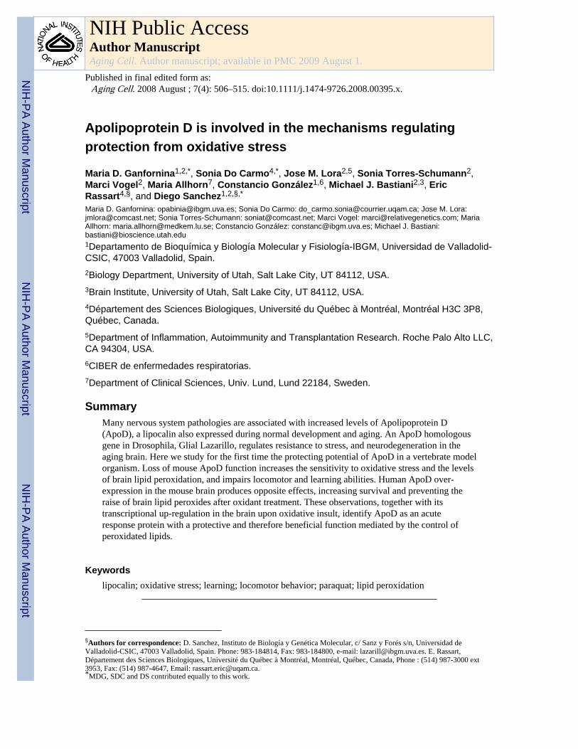
Apolipoprotein D is involved in the mechanisms regulating protection from oxidative stress Maria D. Ganfornina 1,2,* , Sonia Do Carmo 4,* , Jose M. Lora 2,5 , Sonia Torres-Schumann 2 , Marci Vogel 2 , Maria Allhorn 7 , Constancio González 1,6 , Michael J. Bastiani 2,3 , Eric Rassart 4,§ , and Diego Sanchez 1,2,§,* Maria D. Ganfornina: [email protected]; Sonia Do Carmo: [email protected]; Jose M. Lora: [email protected]; Sonia Torres-Schumann: [email protected]; Marci Vogel: [email protected]; Maria Allhorn: [email protected]; Constancio González: [email protected]; Michael J. Bastiani: [email protected] 1 Departamento de Bioquímica y Biología Molecular y Fisiología-IBGM, Universidad de Valladolid- CSIC, 47003 Valladolid, Spain. 2 Biology Department, University of Utah, Salt Lake City, UT 84112, USA. 3 Brain Institute, University of Utah, Salt Lake City, UT 84112, USA. 4 Département des Sciences Biologiques, Université du Québec à Montréal, Montréal H3C 3P8, Québec, Canada. 5 Department of Inflammation, Autoimmunity and Transplantation Research. Roche Palo Alto LLC, CA 94304, USA. 6 CIBER de enfermedades respiratorias. 7 Department of Clinical Sciences, Univ. Lund, Lund 22184, Sweden. Summary Many nervous system pathologies are associated with increased levels of Apolipoprotein D (ApoD), a lipocalin also expressed during normal development and aging. An ApoD homologous gene in Drosophila, Glial Lazarillo, regulates resistance to stress, and neurodegeneration in the aging brain. Here we study for the first time the protecting potential of ApoD in a vertebrate model organism. Loss of mouse ApoD function increases the sensitivity to oxidative stress and the levels of brain lipid peroxidation, and impairs locomotor and learning abilities. Human ApoD over- expression in the mouse brain produces opposite effects, increasing survival and preventing the raise of brain lipid peroxides after oxidant treatment. These observations, together with its transcriptional up-regulation in the brain upon oxidative insult, identify ApoD as an acute response protein with a protective and therefore beneficial function mediated by the control of peroxidated lipids. Keywords lipocalin; oxidative stress; learning; locomotor behavior; paraquat; lipid peroxidation § Authors for correspondence: D. Sanchez, Instituto de Biología y Genética Molecular, c/ Sanz y Forés s/n, Universidad de Valladolid-CSIC, 47003 Valladolid, Spain. Phone: 983-184814, Fax: 983-184800, e-mail: [email protected]. E. Rassart, Département des Sciences Biologiques, Université du Québec à Montréal, Montréal, Québec, Canada, Phone : (514) 987-3000 ext 3953, Fax: (514) 987-4647, Email: [email protected]. * MDG, SDC and DS contributed equally to this work. NIH Public Access Author Manuscript Aging Cell. Author manuscript; available in PMC 2009 August 1. Published in final edited form as: Aging Cell. 2008 August ; 7(4): 506–515. doi:10.1111/j.1474-9726.2008.00395.x. NIH-PA Author Manuscript NIH-PA Author Manuscript NIH-PA Author Manuscript

Welcome message from author
This document is posted to help you gain knowledge. Please leave a comment to let me know what you think about it! Share it to your friends and learn new things together.
Transcript
Apolipoprotein D is involved in the mechanisms regulatingprotection from oxidative stress
Maria D. Ganfornina1,2,*, Sonia Do Carmo4,*, Jose M. Lora2,5, Sonia Torres-Schumann2,Marci Vogel2, Maria Allhorn7, Constancio González1,6, Michael J. Bastiani2,3, EricRassart4,§, and Diego Sanchez1,2,§,*
Maria D. Ganfornina: [email protected]; Sonia Do Carmo: [email protected]; Jose M. Lora:[email protected]; Sonia Torres-Schumann: [email protected]; Marci Vogel: [email protected]; MariaAllhorn: [email protected]; Constancio González: [email protected]; Michael J. Bastiani:[email protected] de Bioquímica y Biología Molecular y Fisiología-IBGM, Universidad de Valladolid-CSIC, 47003 Valladolid, Spain.2Biology Department, University of Utah, Salt Lake City, UT 84112, USA.3Brain Institute, University of Utah, Salt Lake City, UT 84112, USA.4Département des Sciences Biologiques, Université du Québec à Montréal, Montréal H3C 3P8,Québec, Canada.5Department of Inflammation, Autoimmunity and Transplantation Research. Roche Palo Alto LLC,CA 94304, USA.6CIBER de enfermedades respiratorias.7Department of Clinical Sciences, Univ. Lund, Lund 22184, Sweden.
SummaryMany nervous system pathologies are associated with increased levels of Apolipoprotein D(ApoD), a lipocalin also expressed during normal development and aging. An ApoD homologousgene in Drosophila, Glial Lazarillo, regulates resistance to stress, and neurodegeneration in theaging brain. Here we study for the first time the protecting potential of ApoD in a vertebrate modelorganism. Loss of mouse ApoD function increases the sensitivity to oxidative stress and the levelsof brain lipid peroxidation, and impairs locomotor and learning abilities. Human ApoD over-expression in the mouse brain produces opposite effects, increasing survival and preventing theraise of brain lipid peroxides after oxidant treatment. These observations, together with itstranscriptional up-regulation in the brain upon oxidative insult, identify ApoD as an acuteresponse protein with a protective and therefore beneficial function mediated by the control ofperoxidated lipids.
Keywordslipocalin; oxidative stress; learning; locomotor behavior; paraquat; lipid peroxidation
§Authors for correspondence: D. Sanchez, Instituto de Biología y Genética Molecular, c/ Sanz y Forés s/n, Universidad deValladolid-CSIC, 47003 Valladolid, Spain. Phone: 983-184814, Fax: 983-184800, e-mail: [email protected]. E. Rassart,Département des Sciences Biologiques, Université du Québec à Montréal, Montréal, Québec, Canada, Phone : (514) 987-3000 ext3953, Fax: (514) 987-4647, Email: [email protected].*MDG, SDC and DS contributed equally to this work.
NIH Public AccessAuthor ManuscriptAging Cell. Author manuscript; available in PMC 2009 August 1.
Published in final edited form as:Aging Cell. 2008 August ; 7(4): 506–515. doi:10.1111/j.1474-9726.2008.00395.x.
NIH
-PA Author Manuscript
NIH
-PA Author Manuscript
NIH
-PA Author Manuscript
IntroductionApolipoprotein D (ApoD) is a Lipocalin, an ancient family of small proteins engaged in adiverse array of physiological processes, whose general molecular function is binding andtransporting hydrophobic ligands. ApoD is known to bind arachidonic acid, progesterone,retinol, cholesterol and other lipophilic ligands. Elevations in its expression level have beenassociated with a number of pathological and tissue damage conditions includingneurodegenerative diseases. Its levels increase as well during physiological aging (seeRassart et al. 2000; Van Dijk et al. 2006 for review). Although the correlation between thosestates and increased gene expression is consistent, the causal link with ApoD function is stillunclear. Whether ApoD is one of the factors contributing to the degenerative or agingprocesses, or its function is part of a defense mechanism providing protection to cells mustbe solved.
Our previous work on an ApoD homologue in insects, the Drosophila Glial Lazarillo(GLaz), shows that it regulates longevity, resistance to stress, locomotor behavior, anddegeneration in brain and other tissues upon aging (Sanchez et al. 2006; see also Walker etal. 2006). We therefore undertook a genetic analysis of the ApoD function in a vertebratemodel organism by generating knockout and transgenic over-expressing mice.
Results and DiscussionAn ApoD knockout mouse (ApoD-KO) was generated by standard homologousrecombination techniques. We replaced the wild-type ApoD gene with a copy interrupted bythe insertion of the neomycin resistance gene (Neo) in the opposite direction to ApoDtranscription (Fig. 1S A), which rendered a transcriptional null mutant (Fig. 1S B–D)without deleting any genomic fragment potentially containing regulatory regions. We alsogenerated transgenic mice over-expressing the human orthologue of ApoD (HApoD) underthe control of the neuronal human Thy-1 promoter (HApoD-Tg line; Fig. 2S A), thusexpressing HApoD in neurons in all regions of the nervous system (Fig. 2S B–E). Generalfeatures of ApoD-KO and HApoD-Tg mice are described in Supporting Informationavailable online.
Both ApoD-KO and HApoD-Tg mice are viable and breed normally. Three independentlines were obtained for the Thy-1 transgenic, but a single line was used in this work due totheir similar phenotypes based on general health, and biochemical and molecularcharacterization (Do Carmo & Rassart, unpublished observations). No apparent genotypicdifferences were observed during development when comparing sibling ApoD-KO and WTmice (see Supporting Online Information). We therefore turn to study ApoD function duringadulthood, both in normal conditions, and when exposing the organism to oxidative stresssituations mimicking the many pathological situations in which ApoD is up-regulated.
ApoD alters locomotor activity and learning abilities in young adult animalsOpen field analysis revealed a significant decrease in horizontal and vertical locomotoractivity in ApoD-KO mice (25% and 36% mean reduction in horizontal and vertical activityrespectively; Fig. 1A, left and right panels), while no differences were observed in anxietyrelated parameters (relative time spent in the center vs. periphery of the open field arena;Fig. 1A central panel). On the other hand, over-expression of human ApoD causes anincrease in vertical activity (number of rearings, Fig. 1B, left panel), while differences inhorizontal activity were not significant (Fig. 1B, right panel).
Visual ability, assessed by darkness preference and object recognition, was not significantlyaffected. Other behavioral tests exploring pain and touch sensitivity, gait, and muscular
Ganfornina et al. Page 2
Aging Cell. Author manuscript; available in PMC 2009 August 1.
NIH
-PA Author Manuscript
NIH
-PA Author Manuscript
NIH
-PA Author Manuscript
strength gave no significant differences between ApoD-KO mice and their wild-typelittermates. Similarly, the walking pole test exploring sensory-motor coordination gave nodifferences in ApoD-KO nor HApoD-Tg mice (see Supporting Online Material).
ApoD-KO mice and their wild-type control perform equally well in the Rotarod test (alsoexploring sensory-motor coordination) when first tested on the accelerated rod after a set oftraining sessions. However, a parameter that estimates the ability to learn a motor task(improvement of performance over consecutive trials) was much smaller in the ApoD-KOmice (Fig. 1C).
Hippocampal-dependent memory was assessed with the spatial version of the Barnes maze.Wild-type mice showed an average learning time of 64 days, while none of the ApoD-KOmice were able to meet the learning criteria (7 out of 8 sessions with 3 or less errors) for thetest period of 70 days (Fig. 1D).
Therefore, mice lacking ApoD expression reduce their locomotor and exploratory activity,while over-expression of HApoD in the brain renders the animals slightly more active. Inaddition, ApoD-KO mice show deficits in learning either a motor task or an orientation-based task (dependent on visual cues). Decreased locomotor performance was also shownfor Drosophila GLaz null mutants (Sanchez et al. 2006), illustrating interesting functionalconservation of these homologous genes throughout evolution.
Resistance to experimentally induced oxidative stress is undermined in the absence ofApoD and increased by the over-expression of human ApoD
The regulation of ApoD in physiological and pathological states where reactive oxygenspecies (ROS) are generated (reviewed in Rassart et al. 2000), and the higher sensitivity tooxidative stress experienced by the Drosophila GLaz mutants (Sanchez et al. 2006)motivated us to test the overall survival of ApoD-KO and HApoD-Tg mice exposed to anoxidative insult. Sensitivity to the ROS generator 1,1’-dimethyl-4,4’-bypiridinium (paraquat;PQ) was assayed using different paradigms of intraperitoneal injections (see Methods inSupporting Online Material). The ApoD null mutant mice show a higher sensitivity to PQ-induced chronic oxidative stress than wild-type controls (Fig. 2A). The survival curves showa reduction in both the median and the maximum lifespan (26% and 56% respectively, Fig.2A) reflecting an early onset of functional decline in the ApoD-KO mice. This effect isobserved independently of the genetic background (C57BL6/J or Balb/C), indicating that thereduction observed is originated by the lack of ApoD.
These results will predict that mice over-expressing ApoD would be more protected againstthe stress produced by PQ. Indeed, HApoD-Tg mice showed improved survival upon singlesystemic exposures to PQ at two different concentrations, with an increase in mediansurvival time of 41.6 and 27.5% respectively (Fig. 2B,C). Thus the human ApoD protein isable to exert a protective role when expressed in the mouse brain. This general effect onsurvival might be due to either the presence of the neuron-expressed ApoD in the systemiccirculation, or to low levels of non-neuronal ApoD expression in other tissues.
The protective effect of ApoD is also consistent with the results obtained in Drosophilaover-expressing the endogenous ApoD homologue GLaz (Walker et al. 2006) or the humanApoD gene (Muffat et al. personal communication). All these results strongly suggest thatthe protecting role in oxidative stress-compromised survival is a common factor in all ApoDhomologues, reflecting that it is probably part of the ancestral function of this lipocalin.Therefore, the analyses in model organisms can be very useful to understand the humanApoD function.
Ganfornina et al. Page 3
Aging Cell. Author manuscript; available in PMC 2009 August 1.
NIH
-PA Author Manuscript
NIH
-PA Author Manuscript
NIH
-PA Author Manuscript
ApoD expression is induced in the brain upon experimental oxidative stressThe up-regulation of ApoD in ROS-related pathological conditions predicts that anexperimentally produced oxidative stress would also regulate ApoD mRNA expression inthe mouse. Since ApoD is up-regulated in neurological pathologies, and our behavioralstudies in the mouse indicate that nervous system functionality is compromised in theabsence of ApoD, we assayed the transcription of ApoD in mouse brain after a singleexposure to PQ. An acute up-regulation of ApoD (1.86-fold induction) is detected 3 hoursafter exposure, and the expression returns to baseline by 24 hours (Fig. 3A). Similartransient effects of PQ on gene transcription have been shown in lung and kidney for severalgenes with antioxidant roles (Tomita et al. 2006a;Tomita et al. 2006b). We thereforeassayed the transcription of ApoD in several tissues 3 hours after PQ exposure, when theexpression peaks in the brain. No up-regulation is produced in the lung or the liver, whilethe brain tissue shows a strong ApoD induction upon PQ treatment (Fig. 3B). These resultssuggest a specific function of ApoD in the response of the nervous system to oxidativeinjury.
The higher sensitivity to oxidative stress of mice lacking ApoD parallels our results obtainedin the Drosophila GLaz null mutant (Sanchez et al. 2006) and in null mutants of the ApoDhomologue in plants (Frenette-Charron et al. personal communication) which are moresensitive to oxidative and other forms of stress. We can therefore predict that the regulationof these lipocalin genes must also be conserved (a transient up-regulation of GLaz alsooccurs in Drosophila, our unpublished observations also confirmed by J. Muffat et al.personal communication), and that the ApoD up-regulation in the mouse brain upon PQ-induced ROS generation is part of the normal protective response to stress.
The increased survival of the transgenic animals over-expressing human ApoD in thenervous system should be due to a higher and longer-lasting presence of ApoD in thetissues. Since the human transgene is placed in a wild-type genetic background (C57BL6/Jmice), we measured the expression level of the mouse endogenous ApoD gene in the brainof transgenic animals. Mouse ApoD transcript levels are not affected by the presence of thehuman transgene either under normal conditions or upon PQ treatment. As expected, thehuman transgene does not show a response by PQ (Fig. 3C).
ApoD controls the levels of brain lipid peroxidation under normal conditionsPQ treatment is known to generate protein and lipid peroxidation (Beal 2002). By evaluatingthe oxidation state of these macromolecules in the brain, both in the control situation and inthe presence of PQ-induced oxidative stress, we should gain insight into the biochemicalactions of ApoD in the brain tissue when it is exerting the protecting effects described aboveat the organism level.
We assayed lipid peroxidation levels upon the different paradigms used for PQ injection. Inagreement with McCormack et al. (McCormack et al. 2005), lipid peroxide adductsaccumulate in the wild type brain over time at a relatively slow pace (Fig. 3S A). As it is thecase for tissues like lung, kidney and liver (Sato et al. 1992), no increase in lipidperoxidation is observed in the brain 3 hours after a single high dose of PQ (acute PQtreatment, protocol B), while a low-dose chronic treatment (protocol C) does produce asignificant increase after two weeks. In contrast, protein carbonylation, a marker ofoxidative modification of proteins, is much faster, showing a patent increase by 3 hoursupon acute treatment (Fig. 3S B).
In control conditions (sham injection, Fig. 4A) we observe that the lack of ApoD results inincreased levels of lipid peroxides in the brain, but not in the lung, in accordance with thenormal tissue distribution of ApoD. Moreover, as expected for a lipid managing lipocalin,
Ganfornina et al. Page 4
Aging Cell. Author manuscript; available in PMC 2009 August 1.
NIH
-PA Author Manuscript
NIH
-PA Author Manuscript
NIH
-PA Author Manuscript
the effect is specific for lipids, since no alterations are seen in protein carbonylation. On theother hand, the amount of lipid peroxidation in the brain of HApoD-Tg mice is similar to thewild-type levels (Fig. 4C), indicating that an excess of human ApoD cannot reduce the basallevels of oxidation products related to normal cellular metabolic activity. As expected, lunglipid peroxidation levels are also unchanged by the human transgene (not shown).
An increased level of lipid peroxidation under basal conditions was also observed in theGLaz null mutant fruit flies (Sanchez et al. 2006). These results suggest that ApoD and itshomologues can condition the peroxidation state of lipids, even in normal non-stresssituations. The maintenance of cellular membrane integrity can be expected to becompromised in the ApoD-KO and, since ApoD absence is particularly noticed in the brain,it can result in neural functional alterations that we are able to detect by behavioral tests.
The data by Thomas and Yao (Thomas & Yao 2006) also support the above proposal. Theyperformed a brain lipid profile analysis in our ApoD-KO mice to understand the role ofApoD in the mechanisms of antipsychotic drug action. Their results in the control animals(without drug addition) show that ApoD-KO brains have increased levels of somepolyunsaturated fatty acids (dienes and hexanenes). Since polyunsaturated fatty acids aremore prone to peroxidation, the susceptibility of membranes to peroxidation (Hulbert 2005)must be increased in the ApoD-KO brains, and therefore membranes could be moresusceptible to the effects of normal oxidative metabolism.
ApoD is able to prevent the lipid peroxidation increase upon oxidative insultUpon oxidative insult (either acute or low-dose chronic PQ treatment) lipid peroxidationincreases (33% and 43% respectively) over the wild-type levels in the ApoD-KO brain (Fig.4B). This change can be correlated with the decrease in survival time observed in the ApoD-KO mice upon PQ treatment.
Again, no genotype-dependent differences in protein carbonylation were observed under PQtreatment (Fig. 4B), suggesting that the function of ApoD is confined to lipid management.
On the other hand, human ApoD over-expression abolishes the long-term accumulation oflipid peroxides after 2 weeks of PQ treatment, maintaining the levels at control values (Fig.4C).
These results show that an excess of human ApoD in the brain can counteract theexogenously-induced oxidative damage to lipids. Therefore, we can expect that asupplement of ApoD might be beneficial for human conditions where lipid oxidation is akey factor in the functional decline associated to physiological aging or neural pathologies.
Lipocalins as part of the oxidative stress response systemOur data support the notion that ApoD is part of the mechanisms regulating protection fromdiverse forms of stress, including oxidation.
We show in this report that ApoD specifically controls the levels of lipid peroxidation in anorgan as vulnerable as the brain. The structural property of ApoD shared with all otherlipocalins is the presence of a binding pocket that can bind diverse molecules, mostlyhydrophobic (Flower et al. 2000; Akerstrom et al.. Eds.) (2006), therefore it is probable thatits mechanism of action involves this lipid-binding property. The control of lipidperoxidation can be exerted directly, either a priori by preventing the oxidation of boundlipids, or a posteriori by removing peroxidated lipids from membranes, and thereforeavoiding the positive feedback loops leading to more oxidative damage. Alternatively, the
Ganfornina et al. Page 5
Aging Cell. Author manuscript; available in PMC 2009 August 1.
NIH
-PA Author Manuscript
NIH
-PA Author Manuscript
NIH
-PA Author Manuscript
control of lipid oxidation can be a consequence of the regulation of lipid composition inmembranes, which will determine the susceptibility of membranes to peroxidation.
Scavenging roles have been proposed for other lipocalins. Lcn-1 (tear lipocalin), expressedin secretory glands, has been shown to bind lipid peroxidation products in vitro and is alsoup-regulated upon oxidative stress in an epithelial cell line (Lechner et al. 2001). α-1microglobulin is able to reduce and then trap ABTS radicals by covalent binding (Akerstromet al. 2007).
However, can our data also be compatible with a direct antioxidant activity of ApoD?
The antioxidant defense system is currently known to comprise low molecular mass agents(e.g. glutathione or melatonin), iron or copper sequestration proteins (such as transferrin,ferritin, or albumin) and antioxidant enzymes (like superoxide dismutase or catalase). Withthe data available so far, ApoD belongs to none of these categories. While other lipocalinsare well known for their iron sequestration properties (Goetz et al. 2002; Flo et al. 2004),binding to toxic heme (Allhorn et al. 2002) or antioxidant catalytic activities (Allhorn et al.2005), neither metal sequestration properties nor enzymatic activity have been shown forApoD, or its orthologues in fruitflies or plants.
The lipocalin α-1 microglobulin (Allhorn et al. 2002; Allhorn et al. 2005; Akerstrom et al.2007) exerts reductase and dehydrogenase activities through a free cysteine group located ina flexible loop. ApoD has four cysteine residues that form two disulfide bonds at the core ofthe protein fold in all species studied so far. However, human ApoD has an extra cysteine(Cys116) known to bind covalently to Apolipoproteins A-II and B-100 (Blanco-Vaca et al.1992). Whether the thiol group of Cys116 confers additional antioxidant properties tohuman ApoD remains to be investigated. If this were the case, then this property would behuman-specific. In order to explain the results obtained in mouse (this study), fly (Sanchezet al. 2006) and plants (Frenette-Charron et al. personal communication), additionalantioxidant properties of ApoD that are conserved among these divergent species must beinvoked. Moreover, the fact that only lipid peroxidation, but not protein carbonylation, isaltered in the absence of ApoD argues against a direct antioxidant activity of mouse ApoD.
The oxidative modification of membrane lipids also triggers intracellular signaling cascadesthat direct the cell fate after an oxidative insult. Particularly, arachidonic acid (AA) is knownto be mobilized from membranes upon oxidative stress (Balboa & Balsinde 2006). ApoD isable to bind a variety of lipidic ligands (Rassart et al. 2000; Breustedt et al. 2006; Eichingeret al. 2007) of which the highest affinity is shown for AA. Data supporting the participationof ApoD in regulating the mobilization of AA has been shown in a cell culture system(Thomas et al. 2003b), and both ApoD and AA are regulated in patients with schizophreniain response to antipsychotic drug treatment (Thomas et al. 2001; Thomas et al. 2003a). Thelipid analysis of ApoD-KO brains upon treatment with clozapine (Thomas & Yao 2006) hasshown that the absence of ApoD alters the levels of AA upon drug treatment (even thoughlevels are not modified by the mutation in basal conditions). Thus by modulating importantsignaling lipids like AA, ApoD can influence the patient’s response to different treatments.
All data available so far point to a common biochemical function for ApoD, directly linkedto membrane physiology, which is essential for a proper nervous system function. Bycontrolling the levels of lipid peroxidation ApoD could influence parameters as important asneurotransmitter release, in turn affecting learning abilities or locomotor activity(Vajragupta et al. 2000) throughout life. Likewise, by performing the same biochemicalfunction, ApoD can control the rate of brain functional decline upon normal aging or inpathological conditions, although it is unlikely to be the only such factor.
Ganfornina et al. Page 6
Aging Cell. Author manuscript; available in PMC 2009 August 1.
NIH
-PA Author Manuscript
NIH
-PA Author Manuscript
NIH
-PA Author Manuscript
Functional significance of ApoD: its influence on normal and pathological nervous systemdecline upon aging
We have shown that ApoD expression in the mouse brain is boosted by experimentaloxidative stress, mimicking what happens in many human neurodegenerative and psychiatricdiseases, nervous system injuries, as well as during normal aging. The main known effect oflacking ApoD is to become more vulnerable to stress, while an ApoD overdose makesanimals less sensitive to the same stress. The fact that the human ApoD gene can produceeffects that oppose those observed in the ApoD-KO mouse strongly suggests a conservedfunction, and therefore the utility of the mouse model to test potential therapeutic uses ofApoD. This study is the first showing that an excess of ApoD results in seemingly beneficialeffects upon experimental oxidative stress in the mouse. Testing the effects of over-expressing ApoD in mouse models of disease should therefore become a priority.
Experimental proceduresAnimals
The experimental procedures were approved by the Animal Care and Use Committees of theUniversity of Utah, University of Valladolid and Université du Québec à Montréal, and werein accordance with the Guidelines for the Care and Use of Mammals in Neuroscience andBehavioral Research (2001). Mice were maintained in positive pressure-ventilated racks at25±1°C with a 12 h light/dark cycle, fed a standard rodent pellet diet (Global Diet 2014;Harlan), and allowed free access to filtered and UV-irradiated water. Experiments werecarried out with 3–5 months old animals of mixed sexes.
Targeted disruption of the mouse ApoD geneIn order to silence the mouse ApoD gene we constructed a targeting vector with theneomycin phosphotransferase (Neo) gene interrupting exon 6 of the ApoD gene (Fig. 1S A).G418r-GANCs 129/SvJ ES cells were transfected with the construct and tested for thepresence of the disrupted ApoD allele by BamHI digestion of their genomic DNA followedby Southern hybridization with an external probe comprising exon 4 (Fig. 1S A). GenomicDNA sequence of the region surrounding the targeting vector confirmed the properhomologous recombination of the positive lines injected into female mouse blastocysts atthe U. of Utah Transgenics Facility. Subsequent crosses with the chimeric mice showinggermline transformation of the targeted allele generated heterozygous ApoD+/− mice thatwere further confirmed by Southern blot with a 3’ probe (Fig. 1S A). Standard genotypingwas performed from tail DNA and PCR with Neo and ApoD-specific primers: ApoD-for: 5’CCA CCG GCA CCC TAC TGG ATC 3’; ApoD-rev: 5’ CGG GCA GTT CGC TTG ATCTGT 3’; and Neo-for: 5’ CGA TTG TCT GTT GTG CCC AGT 3’.
Phenotype stability was assessed by backcrossing and outcrossing the ApoD mutated alleleinto the C57BL6/J and Balb/C genetic backgrounds respectively. The mutant allele wasselected for ten generations of crosses with the corresponding wild type females. Anadditional generation was performed by crossing mutant females with wild type males tointroduce the Y chromosome of the wild type strain.
Generation of human ApoD transgenic mouseThe human ApoD transgenic mouse (HApoD-Tg) carries a construct (~ 4.5 kb) containingthe promoter, the first exon, the first intron and the 5’ non-coding region of the second exonof the human Thy-1 gene (generous gift from J. Silver, New York University MedicalCenter). This fragment was fused to the human ApoD coding sequence followed by thebovine growth hormone (BGH) polyadenylation signal (Fig. 2S A). The presence of thetransgene was verified by PCR using an ApoD-specific primer (5’ CCC AAT CCT CCG
Ganfornina et al. Page 7
Aging Cell. Author manuscript; available in PMC 2009 August 1.
NIH
-PA Author Manuscript
NIH
-PA Author Manuscript
NIH
-PA Author Manuscript
GTG CAG GAG AA 3’) and a BGH-specific primer (5’ GAA GGC ACA GTC GAG GCTGAT CAG 3’), producing a 0.6 kb fragment (Fig. 2S B). The HApoD-Tg mice used in thisstudy were backcrossed onto C57/BL6 wild-type mice as explained above.
RNA extraction and northern blot analysisAnimals were euthanized by inhalation of CO2/O2 (2:1). Tissues were collected, frozen indry ice, and kept at −80°C until extraction of total RNA using the Trizol reagent™(Invitrogen). Total RNA (10µg) was separated on 1.5% (wt/vol) agarose-formaldehyde gelsand blotted to a nylon membrane. The membranes were hybridized with [α-32P]dCTP-labeled mouse or human ApoD, or GAPDH cDNAs, exposed to Biorad Imaging screen Kand revealed with a PhosphorImager (Biorad Molecular Imager FX) and Quantity Onesoftware (Biorad). For each value, the optical density measured for each gene tested wasdivided by that of the GAPDH mRNA.
Protein extraction and western blot analysisFrozen tissues were thawed on ice and homogenized in lysis buffer (50 mM Tris-HCl pH7.3, 150 mM NaCl, 5 mM EDTA, 0.2% Triton X-100, and 10% Complete proteaseinhibitors (Roche)). After 30 min of incubation at 4°C, lysates were sonicated and cleared bycentrifugation. The protein concentration was determined using a protein assay reagent (Bio-Rad Laboratories). All extracts were stored at −80°C.
For western blot analysis, protein extracts (10µg) were separated on a 12% SDS-polyacrylamide gel and transferred to a polyvinylidene difluoride (PVDF) membrane. Aprestained size marker (Biorad prestained SDS-PAGE standard, low range) was included ineach run. Membranes were blocked in PBS containing 0.2% Tween-20 and 5% skim milkpowder before incubation with the primary antibodies diluted as follows: polyclonal anti-human ApoD (Caro2), 1:4000; anti-GAPDH, 1:4000. Subsequently, the blots wereincubated under gentle agitation at room temperature with a HRP-conjugated secondaryantibody diluted 1:5000 in blocking buffer. The blots were developed using the enhancedchemoluminescence method (Amersham-Pharmacia) with X-ray film.
Behavioral analysesOpen field tests in the ApoD-KO and their wild type controls were performed by videorecording in a 1m2 arena for a single session of 10 min. A total of 6 mice of each genotypeand sex were analyzed. The subsequent analysis explored horizontal (Number of squaresvisited) and vertical (Number of rearings) activities, as well as anxiety, measured as relativetime spent in the central area of the arena. Horizontal and vertical activity was measured inthe HApoD-Tg mice and control littermates in an open field arena measuring 30 × 44.5 cm.
A novel-object recognition test was used to assay for visual ability and short-term memoryrecognition. Mice habituated to the test arena are presented with two plastic objects ofdifferent shapes. The object recognition was evaluated by the ratio of time spent exploringthe novel vs. the familiar object 10 sec and 1 min after first exposure.
Hippocampal-dependent memory was explored in six WT and six ApoD-KO mice subjectedto the spatial version of Barnes maze test (Barnes 1979), where the mouse needs to learn andremember distal cues in the environment, and associate them with the location of an escapetunnel. The mice were tested daily for 70 days, and the number of holes searched,perseverations and time was recorded. The average for each genotype was recorded andgrouped into session blocks of 5 testing days.
Ganfornina et al. Page 8
Aging Cell. Author manuscript; available in PMC 2009 August 1.
NIH
-PA Author Manuscript
NIH
-PA Author Manuscript
NIH
-PA Author Manuscript
Rotarod analysis was performed in a Ugo Basile (Varese, Italy) apparatus with accelerationfrom 4 to 40 rpm over 5 minutes. Each mouse was subjected to two training sessionsseparated by a 15 min rest. Each session consisted of a 2 min period of exposure to the rodrotating at minimum speed (4 rpm). After another 15 min resting period a first test wasperformed, consisting of three runs with the rod accelerating from the starting 4 rpm speed.Time of permanence in the rod was recorded. A second test of three runs was performed 2hours later, after the mice had been injected intraperitoneally either with PBS or paraquat(see Protocol B below).
Other tests such as pain and touch sensitivity, gait, and muscular strength were performed asdetailed in (Crawley 2000).
Oxidative stress toxicity by 1,1’-dimethyl-4,4’-bypiridinium (paraquat)The following protocols of doses, administration patterns and analysis have been used:
Protocol A—Chronic paraquat (PQ) treatment. Mice were daily injected intraperitoneallywith a solution of PQ in phosphate buffered saline (PBS) at 15 mg/kg of body weight. Deathoccurrence was scored at least every 12 hours for the survival analysis. Morphologicalanalyses were performed on brains from mice euthanized on day 5 of treatment (when theApoD-KO mice survival is at the 50% level).
Protocol B—Acute PQ treatment. A single dose of either 30 or 50 mg/kg of PQ wasinjected as described above. Behavioral tests were performed just before injection (t =0), 1.5hours after injection, and 24 hours after injection. Mice were then euthanized either 3 hoursor 24 hours post-injection, and their brains extracted for biochemical andimmunohistochemical analyses.
Protocol C—Low-dose, neurotoxic chronic PQ treatment. Following Thyruchelvam et al.(Thiruchelvam et al. 2000), mice were injected twice a week, for a total of 2 weeks, with a15 mg/kg of PQ as described before. Animals were euthanized 3 hours after the lastinjection, and their brains extracted for biochemical and immunohistochemical analyses. Weconfirmed the absence of lung toxicity by histopathological analysis looking for alterationsin alveoli, respiratory ducts, and bronchioles (results not shown).
Lipid and protein oxidation assaysTBARS assay—The concentration of thiobarbituric acid (TBA) reactive species wasassayed to monitor the level of lipid peroxidation. Brain tissue was homogenized in PBS inthe presence of an antioxidant (butylated hydroxytoluene, BHT) to prevent new lipidperoxidation to occur during homogenization. A 12µl aliquot of each extract was incubatedwith 390µl of 0.2 M glycine-HCL, pH 3.6 and 250µl of fresh TBA reagent (0.5% TBA,0.5% SDS). After 15 min incubation at 90°C, samples were cooled on ice and transferred toa 96 well microplate for triplicate readings. Absorbance was monitored at 532 nm in aVersamax microplate reader (Molecular Devices). Values were normalized to the proteinconcentration of each sample, measured with the Micro BCA Protein Assay (Pierce).
Protein carbonylation-ELISA assay—Protein carbonylation was performed aspreviously reported (Buss et al. 1997). Briefly, triplicate protein samples were allowed toreact with dinitrophenylhydrazine (DNP) and then adsorbed to wells of an ELISA plate. Abiotinylated anti-DNP antibody (Molecular Probes Inc.) was used for detection of theprotein-bound DNP followed by an amplification step with streptavidin-biotinylatedhorseradish peroxidase (Amersham). Color reaction was performed by supplying o-phenylenediamine, hydrogen peroxide and citric acid in phosphate buffer. Absorbance at
Ganfornina et al. Page 9
Aging Cell. Author manuscript; available in PMC 2009 August 1.
NIH
-PA Author Manuscript
NIH
-PA Author Manuscript
NIH
-PA Author Manuscript
490 nm was measured in a Versamax microplate reader (Molecular Devices). Values werenormalized to the protein concentration of each sample.
Supplementary MaterialRefer to Web version on PubMed Central for supplementary material.
AcknowledgmentsWe thank J.R. Acebes, V. Pomies and S. Thompson for technical assistance. Polyclonal antibody anti-rat ApoD waskindly provided by Dr. S. Patel. This work was supported by start-up grants to D.S. and M.D.G. from the “Ramon yCajal Program”(MEC-Spain and FEDER-FSE); the SGFRH Foundation; NIH grant 2/R01/NS25387-10A1 toM.J.B.; CIHR grant MOP-15677 to E.R.; FRSQ and CRSNG studentships to S.d.C.; STINT fellowshipKU2003-4308 to M.A.; DGICyT grant BFU2004-06394 and JCyL grant VA011C05 to C.G.; and MEC grantBFU2005-00522 and JCyL grant VA049A05 to M.D.G. and D.S.
ReferencesAkerstrom, B.; Borregaard, N.; Flower, DR.; Salier, J-P., editors. Lipocalins. Georgetown, Texas:
Landes Biosciences; 2006.Akerstrom B, Maghzal G, Winterbourn CC, Kettle AJ. The lipocalin alpha 1-microglobulin has radical
scavenger activity. J Biol Chem. 2007; 282:31493–31503. [PubMed: 17766242]Allhorn M, Berggard T, Nordberg J, Olsson ML, Akerstrom B. Processing of the lipocalin alpha(1)-
microglobulin by hemoglobin induces heme-binding and heme-degradation properties. Blood. 2002;99:1894–1901. [PubMed: 11877257]
Allhorn M, Klapyta A, Akerstrom B. Redox properties of the lipocalin alpha1-microglobulin:reduction of cytochrome c, hemoglobin, and free iron. Free Radic Biol Med. 2005; 38:557–567.[PubMed: 15683711]
Balboa MA, Balsinde J. Oxidative stress and arachidonic acid mobilization. Biochim Biophys Acta.2006; 1761:385–391. [PubMed: 16651022]
Barnes CA. Memory deficits associated with senescence: a neurophysiological and behavioral study inthe rat. J Comp Physiol Psychol. 1979; 93:74–104. [PubMed: 221551]
Beal MF. Oxidatively modified proteins in aging and disease. Free Radic Biol Med. 2002; 32:797–803. [PubMed: 11978481]
Blanco-Vaca F, Via DP, Yang CY, Massey JB, Pownall HJ. Characterization of disulfide-linkedheterodimers containing apolipoprotein D in human plasma lipoproteins. J Lipid Res. 1992;33:1785–1796. [PubMed: 1479288]
Breustedt DA, Schonfeld DL, Skerra A. Comparative ligand-binding analysis of ten human lipocalins.Biochim Biophys Acta. 2006; 1764:161–173. [PubMed: 16461020]
Buss H, Chan TP, Sluis KB, Domigan NM, Winterbourn CC. Protein carbonyl measurement by asensitive ELISA method. Free Radic Biol Med. 1997; 23:361–366. [PubMed: 9214571]
Crawley, J. What's wrong with my mouse?: behavioral phenotyping of transgenic and knockout mice.New York: Wiley-Liss; 2000.
Eichinger A, Nasreen A, Kim HJ, Skerra A. Structural insight into the dual ligand specificity and modeof high density lipoprotein association of apolipoprotein D. J Biol Chem. 2007; 282:31068–31075.[PubMed: 17699160]
Flo TH, Smith KD, Sato S, Rodriguez DJ, Holmes MA, Strong RK, Akira S, Aderem A. Lipocalin 2mediates an innate immune response to bacterial infection by sequestrating iron. Nature. 2004;432:917–921. [PubMed: 15531878]
Flower DR, North ACT, Sansom CE. The lipocalin protein family: structural and sequence overview.Biochim Biophys Acta. 2000; 1482:9–24. [PubMed: 11058743]
Goetz DH, Holmes MA, Borregaard N, Bluhm ME, Raymond KN, Strong RK. The neutrophillipocalin NGAL is a bacteriostatic agent that interferes with siderophore-mediated iron acquisition.Mol Cell. 2002; 10:1033–1043. [PubMed: 12453412]
Ganfornina et al. Page 10
Aging Cell. Author manuscript; available in PMC 2009 August 1.
NIH
-PA Author Manuscript
NIH
-PA Author Manuscript
NIH
-PA Author Manuscript
Hulbert AJ. On the importance of fatty acid composition of membranes for aging. J Theor Biol. 2005;234:277–288. Epub 2005 Jan 2024. [PubMed: 15757684]
Lechner M, Wojnar P, Redl B. Human tear lipocalin acts as an oxidative-stress-induced scavenger ofpotentially harmful lipid peroxidation products in a cell culture system. Biochem J. 2001;356:129–135. [PubMed: 11336644]
McCormack AL, Atienza JG, Johnston LC, Andersen JK, Vu S, Di Monte DA. Role of oxidative stressin paraquat-induced dopaminergic cell degeneration. J Neurochem. 2005; 93:1030–1037.[PubMed: 15857406]
Rassart E, Bedirian A, Do Carmo S, Guinard O, Sirois J, Terrisse L, Milne R. Apolipoprotein D.Biochim Biophys Acta. 2000; 1482:185–198. [PubMed: 11058760]
Sanchez D, Lopez-Arias B, Torroja L, Canal I, Wang X, Bastiani MJ, Ganfornina MD, Walker DW,Muffat J, Rundel C, Benzer S. Loss of glial lazarillo, a homolog of apolipoprotein D, reduceslifespan and stress resistance in Drosophila. Curr Biol. 2006; 16:680–686. [PubMed: 16581513]
Sato N, Fujii K, Yuge O, Morio M. Changes in lipid peroxidation levels and lipid composition in thelungs, livers, kidneys and brains of mice treated with paraquat. J Appl Toxicol. 1992; 12:365–368.[PubMed: 1447483]
Thiruchelvam M, Richfield EK, Baggs RB, Tank AW, Cory-Slechta DA. The nigrostriataldopaminergic system as a preferential target of repeated exposures to combined paraquat andmaneb: implications for Parkinson's disease. J Neurosci. 2000; 20:9207–9214. [PubMed:11124998]
Thomas EA, Copolov DL, Sutcliffe JG. From pharmacotherapy to pathophysiology: emergingmechanisms of apolipoprotein D in psychiatric disorders. Curr Mol Med. 2003a; 3:408–418.[PubMed: 12942994]
Thomas EA, Dean B, Pavey G, Sutcliffe JG. Increased CNS levels of apolipoprotein D inschizophrenic and bipolar subjects: implications for the pathophysiology of psychiatric disorders.Proc Natl Acad Sci USA. 2001; 98:4066–4071. [PubMed: 11274430]
Thomas EA, George RC, Sutcliffe JG. Apolipoprotein D modulates arachidonic acid signaling incultured cells: implications for psychiatric disorders. Prostaglandins Leukot Essent Fatty Acids.2003b; 69:421–427. [PubMed: 14623496]
Thomas EA, Yao JK. Clozapine specifically alters the arachidonic acid pathway in mice lackingapolipoprotein D. Schizophr Res. 2006; 89:147–153. [PubMed: 17011169]
Tomita M, Okuyama T, Katsuyama H, Hidaka K, Otsuki T, Ishikawa T. Gene expression in rat lungsduring early response to paraquat-induced oxidative stress. Int J Mol Med. 2006a; 17:37–44.[PubMed: 16328009]
Tomita M, Okuyama T, Katsuyama H, Ishikawa T. Paraquat-induced gene expression in rat kidney.Arch Toxicol. 2006b; 80:687–693. [PubMed: 16555045]
Vajragupta O, Monthakantirat O, Wongkrajang Y, Watanabe H, Peungvicha P. Chroman amide 12Pinhibition of lipid peroxidation and protection against learning and memory impairment. Life Sci.2000; 67:1725–1734. [PubMed: 11021357]
Van Dijk, W.; Do Carmo, S.; Rassart, E.; Dalhlback, B.; Sodetz, J. The plasma Lipocalins α1-acidglycoprotein, apolipoprotein D, apolipoprotein M and complement C8γ. In: Akerstrom, B.;Borregaard, N.; Flower, D.; Salier, J., editors. Lipocalins. Georgetown, Texas: Landes Bioscience;2006. p. 140-166.
Walker DW, Muffat J, Rundel C, Benzer S. Overexpression of a Drosophila homolog ofapolipoprotein D leads to increased stress resistance and extended lifespan. Curr Biol. 2006;16:674–679. [PubMed: 16581512]
Ganfornina et al. Page 11
Aging Cell. Author manuscript; available in PMC 2009 August 1.
NIH
-PA Author Manuscript
NIH
-PA Author Manuscript
NIH
-PA Author Manuscript
Figure 1. Alterations in behavioral output in ApoD-KO and HApoD-Tg mice(A) Open field test on locomotor exploratory behavior in ApoD-KO mice. Both horizontal(number of squares visited) and vertical (number of rearings) activities are decreased in theApoD-KO mice, while an anxiety-related behavior (exploration of the center of the 1 m2
arena) is not altered. N= 12 mice/genotype (6th backcross generation onto C57BL/6background). Unpaired two-sided Student’s t-test, *p < 0.05, **p < 0.01.(B) Open field test in HApoD-Tg mice. Vertical activity increase in HApoD-Tg mice (leftpanel), while horizontal activity differences are not significant (right panel). A smaller arenawas used in this case, and the anxiety parameter was not measured. WT: N = 16; HApoD-Tg: N = 13 (11th backcross generation onto C57BL/6 background). Unpaired two-sidedStudent’s t-test, *p < 0.05.(C) Motor learning abilities were tested as the increment of the Rotarod test score over a 2hours interval, and is represented normalized to the score of the first test. ApoD-KO miceshow a lower learning ability under normal conditions. N= 14 mice/genotype (11thbackcross generation onto C57BL/6 background). Sign non-parametric test, *p < 0.05.(D) Barnes maze test on spatial learning. ApoD-KO mice fail to increase their rate ofsuccess in finding a safe escape hole, while wild type mice learn and remember using distalspatial clues. Mice were tested daily for 70 days. Results from each five consecutive dailysessions were combined in blocks. N= 6 mice/genotype (5th backcross generation ontoC57BL/6 background). ANOVA test, p < 0.0001.(A–D) Data are represented as mean ± SD.
Ganfornina et al. Page 12
Aging Cell. Author manuscript; available in PMC 2009 August 1.
NIH
-PA Author Manuscript
NIH
-PA Author Manuscript
NIH
-PA Author Manuscript
Figure 2. Oxidative stress-compromised survival is decreased in the absence of ApoD andincreased when human ApoD is over-expressed in the mouse brain(A) Survival analysis of mice treated daily with the ROS generator paraquat (15 mg/kg ofbody weight; Protocol A). KO and WT genotypes in C57BL/6 background (8th backcrossgeneration) are compared. N= 11 mice/genotype. The absence of ApoD shortens the micesurvival (Gehan-test, p = 0.026; Cox’s F-test, p = 0.028). A decrease of 26% in meanlifespan and 56% in maximum lifespan is observed. The survival phenotype is stable afterchanging the genetic background (Balb/C, 8th outcross generation) with 11% decrease inmean lifespan and 9% in maximum lifespan. N= 12 mice/genotype. Gehan-test, p = 0.031;Cox’s F-test, p = 0.043 (curve not shown).
Ganfornina et al. Page 13
Aging Cell. Author manuscript; available in PMC 2009 August 1.
NIH
-PA Author Manuscript
NIH
-PA Author Manuscript
NIH
-PA Author Manuscript
(B and C) Survival analysis of WT and transgenic (HApoD-Tg) mice treated with single PQdoses of 30 (B) or 50 (C) mg/kg of body weight (Protocol B). Death occurrence wasrecorded up to 10 days. Surviving animals were sacrificed at day 10. Data was fitted to aGompertz function. (B) Over-expression of ApoD increases the mice survival. WT: N = 16;HApoD-Tg: N = 13. Gehan-test, p = 0.029; Cox’s F-test, p = 0.043. (C) Over-expression ofApoD also protects mice at higher doses of paraquat. WT: N = 12; HApoD-Tg: N = 9.Gehan-test, p = 0.045; Cox’s F-test, p = 0.033. All transgenic mice are backcrossed on theC57Bl/6 background (11th generation).
Ganfornina et al. Page 14
Aging Cell. Author manuscript; available in PMC 2009 August 1.
NIH
-PA Author Manuscript
NIH
-PA Author Manuscript
NIH
-PA Author Manuscript
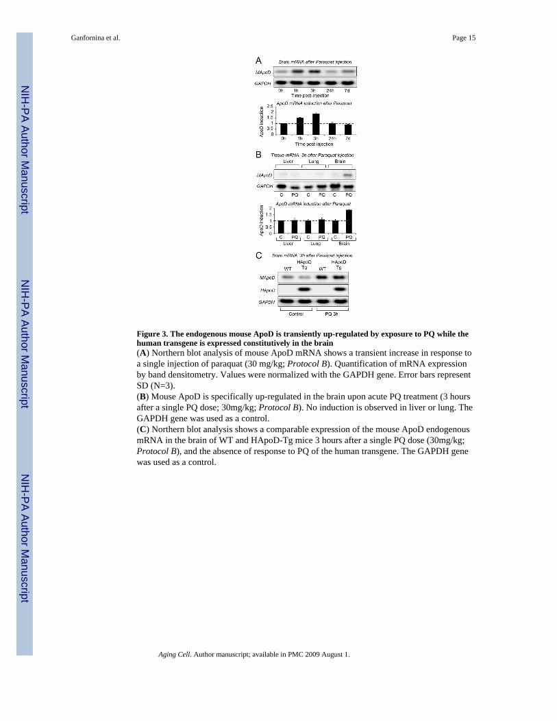
Figure 3. The endogenous mouse ApoD is transiently up-regulated by exposure to PQ while thehuman transgene is expressed constitutively in the brain(A) Northern blot analysis of mouse ApoD mRNA shows a transient increase in response toa single injection of paraquat (30 mg/kg; Protocol B). Quantification of mRNA expressionby band densitometry. Values were normalized with the GAPDH gene. Error bars representSD (N=3).(B) Mouse ApoD is specifically up-regulated in the brain upon acute PQ treatment (3 hoursafter a single PQ dose; 30mg/kg; Protocol B). No induction is observed in liver or lung. TheGAPDH gene was used as a control.(C) Northern blot analysis shows a comparable expression of the mouse ApoD endogenousmRNA in the brain of WT and HApoD-Tg mice 3 hours after a single PQ dose (30mg/kg;Protocol B), and the absence of response to PQ of the human transgene. The GAPDH genewas used as a control.
Ganfornina et al. Page 15
Aging Cell. Author manuscript; available in PMC 2009 August 1.
NIH
-PA Author Manuscript
NIH
-PA Author Manuscript
NIH
-PA Author Manuscript
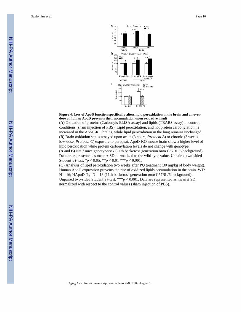
Figure 4. Loss of ApoD function specifically alters lipid peroxidation in the brain and an over-dose of human ApoD prevents their accumulation upon oxidative insult(A) Oxidation of proteins (Carbonyls-ELISA assay) and lipids (TBARS assay) in controlconditions (sham injection of PBS). Lipid peroxidation, and not protein carbonylation, isincreased in the ApoD-KO brains, while lipid peroxidation in the lung remains unchanged.(B) Brain oxidation status assayed upon acute (3 hours, Protocol B) or chronic (2 weekslow-dose, Protocol C) exposure to paraquat. ApoD-KO mouse brain show a higher level oflipid peroxidation while protein carbonylation levels do not change with genotype.(A and B) N= 7 mice/genotype/sex (11th backcross generation onto C57BL/6 background).Data are represented as mean ± SD normalized to the wild-type value. Unpaired two-sidedStudent’s t-test, *p < 0.05, **p < 0.01 ***p < 0.001.(C) Analysis of lipid peroxidation two weeks after PQ treatment (30 mg/kg of body weight).Human ApoD expression prevents the rise of oxidized lipids accumulation in the brain. WT:N = 16; HApoD-Tg: N = 13 (11th backcross generation onto C57BL/6 background).Unpaired two-sided Student’s t-test, ***p < 0.001. Data are represented as mean ± SDnormalized with respect to the control values (sham injection of PBS).
Ganfornina et al. Page 16
Aging Cell. Author manuscript; available in PMC 2009 August 1.
NIH
-PA Author Manuscript
NIH
-PA Author Manuscript
NIH
-PA Author Manuscript
Related Documents