APC & Antigen presentation

APC & Antigen presentation. antigen-presenting cell, APC Concept A group of immune cells, whose role is to take up, process and present antigenic peptides.
Dec 22, 2015
Welcome message from author
This document is posted to help you gain knowledge. Please leave a comment to let me know what you think about it! Share it to your friends and learn new things together.
Transcript
APC & Antigen presentation

antigen-presenting cell, APC
ConceptA group of immune cells, whose
role is to take up, process and present antigenic peptides to T cells.

• Professional APC– Macrophages, dendritic cells, and B cells, which
can express MHC class II molecules.• Non-professional APC
– Other cell type capable of expressing MHC class II molecules
eg. Endothelial cells, ECFibroblastsActivated T cell

1 Dendritic cell, DC• highly branched
morphology• can active naive
T cells• markers

1) Markers– CD1a, CD11c, CD83– Pathogen receptor, FcR– MHC II– co-stimulating factors (CD80,CD86)– Adhesion molecular CD40– CD54 (ICAM-1), etc.
• secreting cytokines– IL1, IL-6, IL-12, TNF-a, IFN-a, chemokin
es

2) Source, distribution and classification
• Source DC are bone marrow-derived
– Myeloid DC– Lymphoid DC
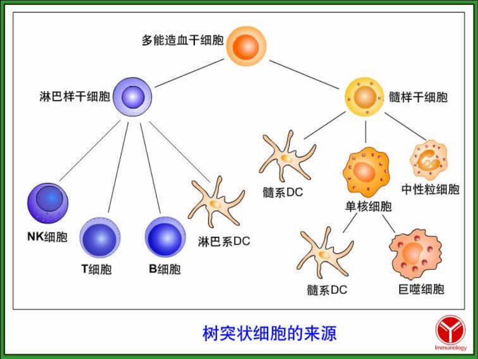

Bone marrow Blood Tissue
Blooddendritic cell
Monocyte
Indeterminatecell
Dendritic cell
Macrphage
Mycloid precursor
Pluripotent stem cell
?
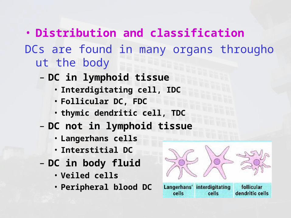
• Distribution and classificationDCs are found in many organs throughout the b
ody – DC in lymphoid tissue
• Interdigitating cell, IDC• Follicular DC, FDC• thymic dendritic cell, TDC
– DC not in lymphoid tissue• Langerhans cells • Interstitial DC
– DC in body fluid• Veiled cells• Peripheral blood DC

interdigitating DC, IDC
IDC express high levels of MHC molecules, and are more potent antigen-presenting cells than others.

follicular DC, FDC
B cells
FDC
FDC express high levels of membrane receptors for antibody and complement. By these, FDC actives the B cells in lymph nodes.

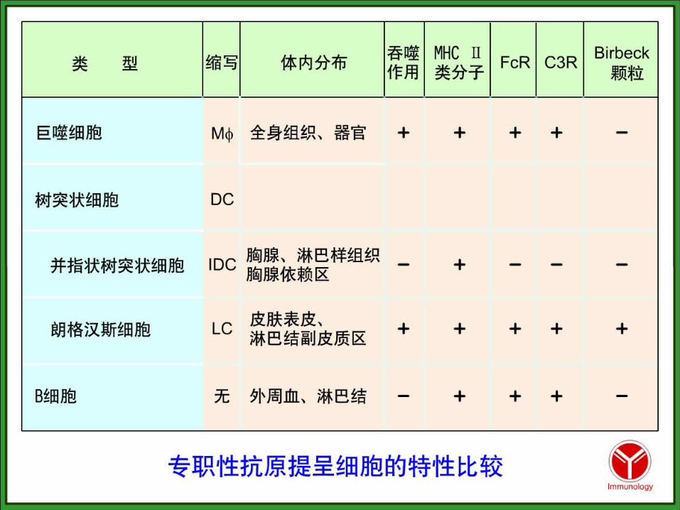
Langerhans cells found in the epidermis (skin) and mucous membranes (left), expressing high levels of FcR, receptor of complement, and MHC. Birbeck granule is the characteristic organelle. After capturing antigen in the tissues by phagocytosis or by endocytosis. DC migrate into the blood or lymph and circulate to lymphoid organs, become IDC( right)。
Langerhan’s cells, LC
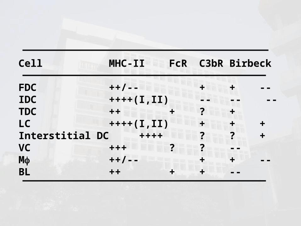
Cell MHC-II FcR C3bR Birbeck
FDC ++/-- + + --IDC ++++(I,II) -- -- --TDC ++ + ? +LC ++++(I,II) + + +Interstitial DC ++++ ? ? +VC +++ ? ? --M ++/-- + + --BL ++ + + --

3) Differentiation, development, maturing and migration
• Lymphoid DC– DC in lymph, negative selection of
T cells
• myeloid DC– Immature– mature

• Four phases– Pre-DC
• Monocyte, Mo– Immature DC
• Uptake antigen• Express MHC• Secrete chemokines
– Migration– Mature DC
• Express high levels of MHC I and II, CD80, CD86, CD40, CD54, HSP, etc.
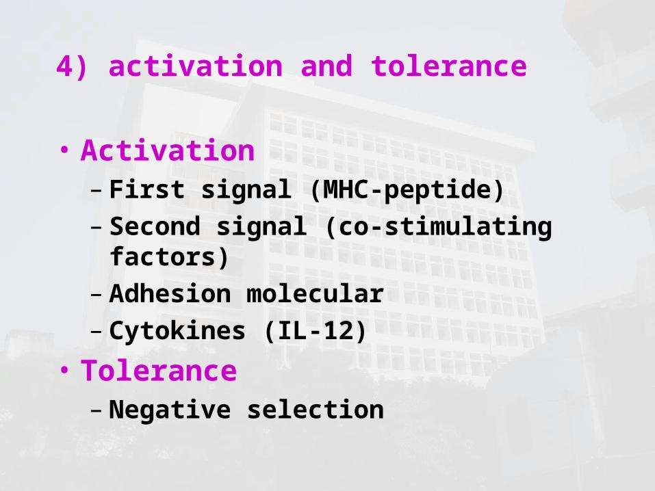
4) activation and tolerance
• Activation – First signal (MHC-peptide)– Second signal (co-stimulating
factors)– Adhesion molecular– Cytokines (IL-12)
• Tolerance – Negative selection

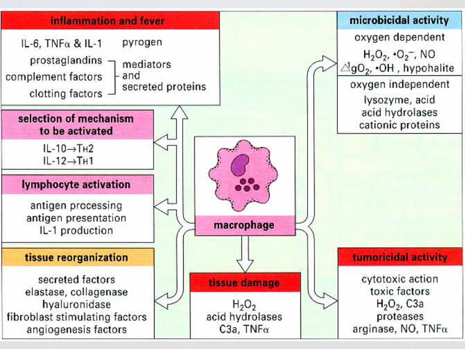
2 Mononuclear phagocyte system, MPS
Macrophages (M) are phagocytic cells of monocytic lineage residing within tissues and are particularly well equipped for effective antigen presentation.
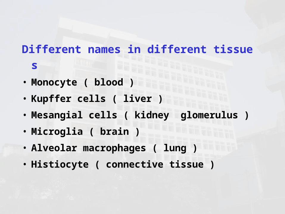
Different names in different tissues• Monocyte ( blood )• Kupffer cells ( liver )• Mesangial cells ( kidney glomerulus )• Microglia ( brain )• Alveolar macrophages ( lung )• Histiocyte ( connective tissue )

rested M responsive M stimulated M activated M
suppressor M
病原体
signal
LFA-1
MHC-II细胞增生趋化,杀菌
提呈 Ag ,激活 LC ,结合 TC ,
过度活化
适度活化
PGE
抑制免疫功能
杀瘤,杀菌
First signal:MAF/IFN-,MSF
second signal:LPS/IFN-,MSF,CK,
1 2
3The process of M activation

• markers– MHC II– CR1( CD35)– CR3( CD11b/CD18)– IgG Fc受体
• Functions– Receptors– Enzymes– Cytokines

扫描电镜显示,在感染早期, M伸出长长的伪足去捕获细菌


Biologic effects of M

3 B cellbone marrow-dependent
lymphocyteAbout 5-15% of the circulating lymphoid pool are
B cells difined by the presence of surface immunoglobulin.
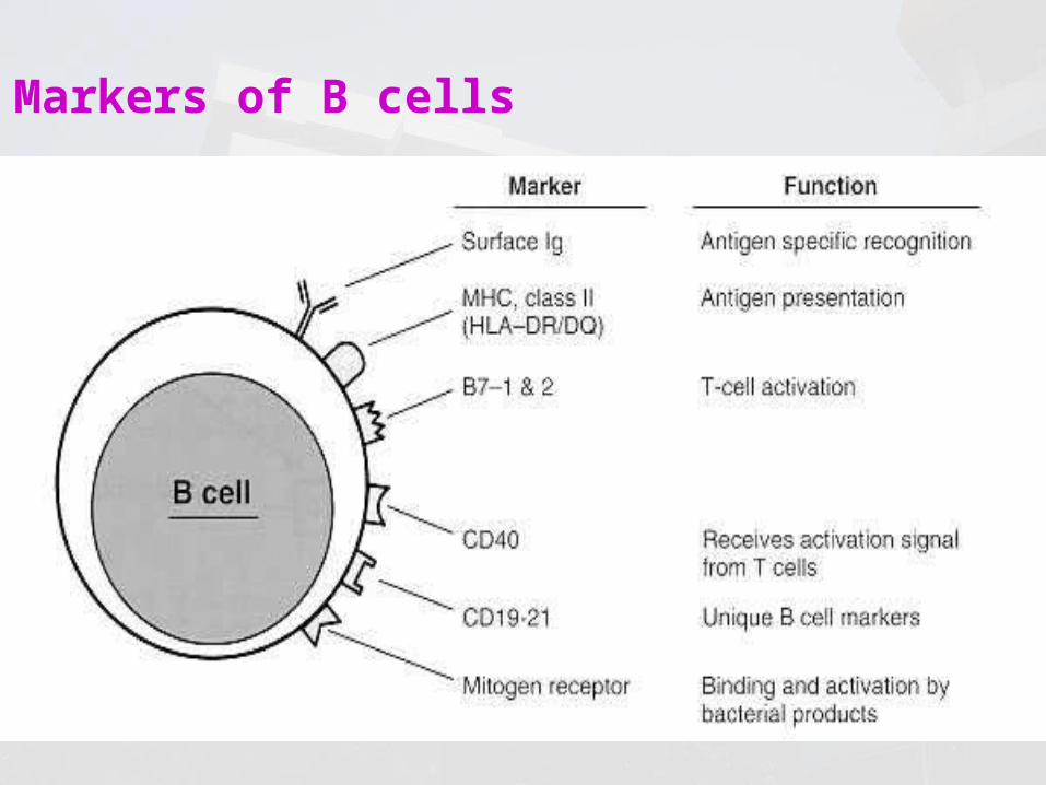
Markers of B cells

Characteristics of B cells– not actively phagocytic– Class II-positive – BCR

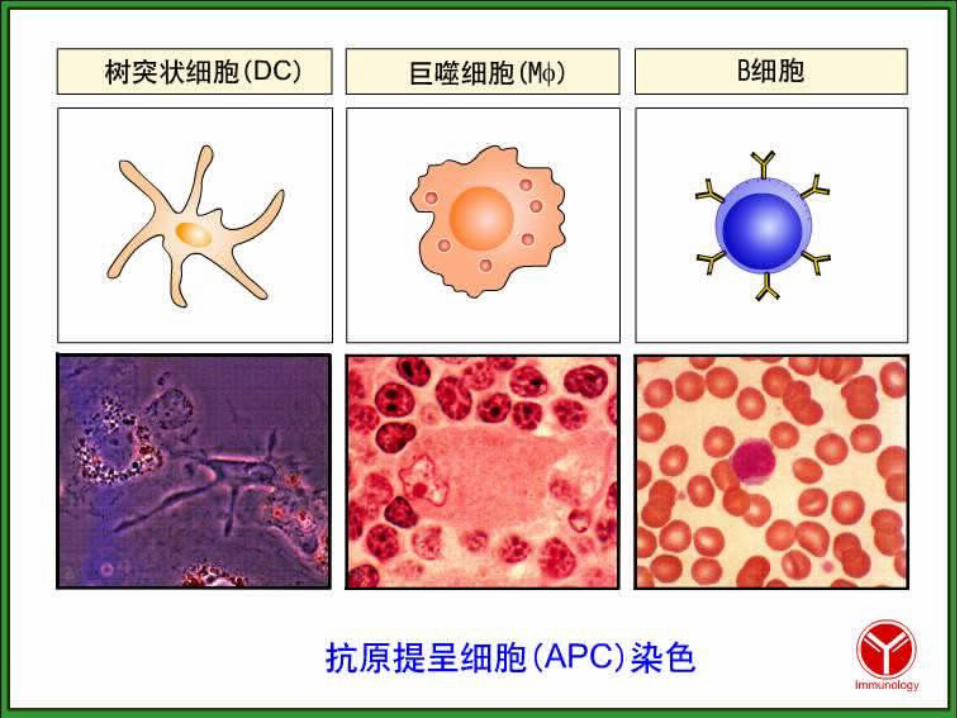
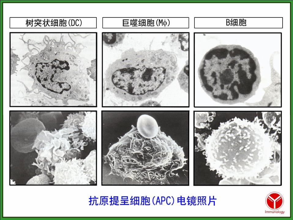
Antigen-presenting cells
APC Present to
Macrophage T cell via MHC antigen
Dendritic cells T cell via MHC antigen
B cells T cell via antigen captrue by surface antibody and MHC antigen
Activated T cells T cell via MHC antigen
ANTIGEN PROCESSING AND PRESENTATION
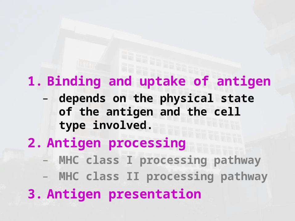
1. Binding and uptake of antigen– depends on the physical state of
the antigen and the cell type involved.
2. Antigen processing– MHC class I processing pathway– MHC class II processing pathway
3. Antigen presentation
1 Binding and uptake of antigen
• exogenous antigens– Bacteria, cells and soluble proteins– processed by APC
• endogenous antigens– Produced within the cells, Such as
viral proteins or tumor proteins– processed by host cell

Uptake antigen by immature DC
• Pinocytosis – Liquid or small granule
• Receptor-mediated endocytosis– effective– selective– saturated
• FCR, 甘露糖 R• Phagocytosis
– Large molecular or microbe

• Phagocytosis– Large solid or molecular complex, such
as bacteria, fragment of cells, etc.– Phagecyte (m, granulocyte)
• Pinocytosis– Receptor-mediated pinocytosis
• Endocytosis– Low levels of particulate or soluble ant
igens– exocytosis
Uptake antigen by MPC
• nonspecifically engulfed• BCR-mediated
Uptake antigen by B cells
2 Antigen processing
• Degradation of externally- or internally- derived antigen into short peptide sequences
• Association of the peptide with MHC molecules

Two antigen-processing pathways
MHC class I MHC class II
Major antigen sources
endogenous antigen
exogenous antigen
Processing machinery
proteasome lysosomal enzymes
Cell type where active
all nucleated cells
professional APCs
Site of antigen-MHC binding
endoplasmic reticulum
lysosome and endosome
MHC utilized MHC class I MHC class II
Presents to CD8+ T cell (Tc) CD4+ T cells (Th)

MHC class I processing pathway

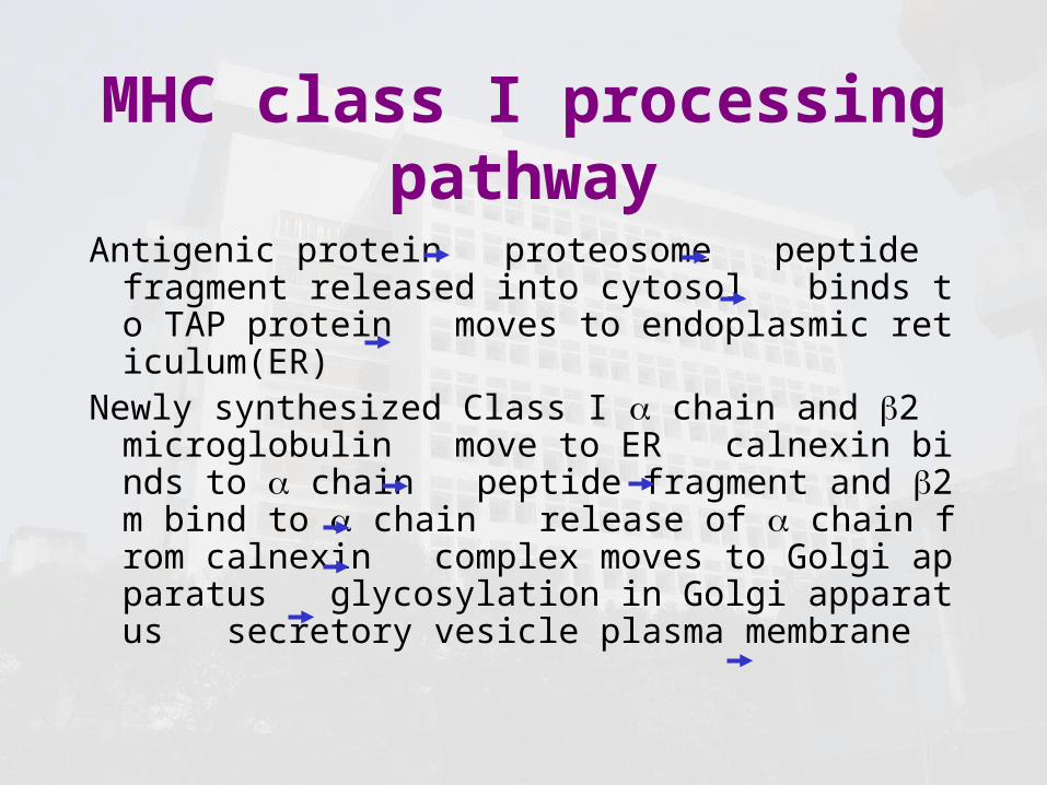
MHC class I processing pathway
Antigenic protein proteosome peptide fragment released into cytosol binds to TAP protein moves to endoplasmic reticulum(ER)
Newly synthesized Class I chain and 2 microglobulin move to ER calnexin binds to chain peptide fragment and 2m bind to chain release of chain from calnexin complex moves to Golgi apparatus glycosylation in Golgi apparatus secretory vesicle plasma membrane

Structure of MHC class I

proteasome•LMP, low molecular weight
polypeptide or large multifunctional protease
•Structure: – 20S 26S
•Function: – Degradation of protein
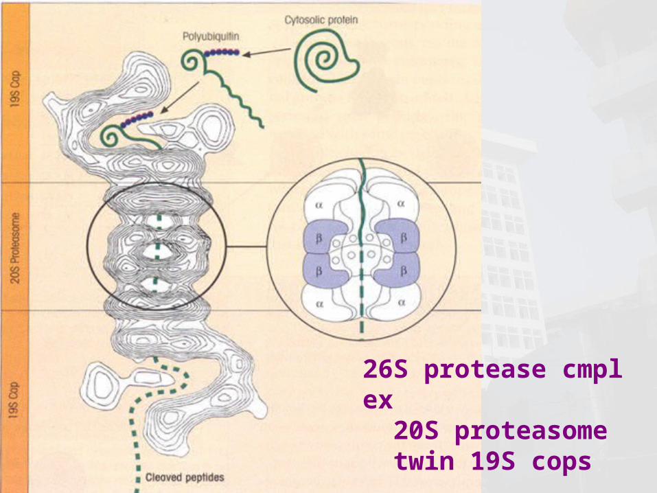
26S protease cmplex 20S proteasome twin 19S cops

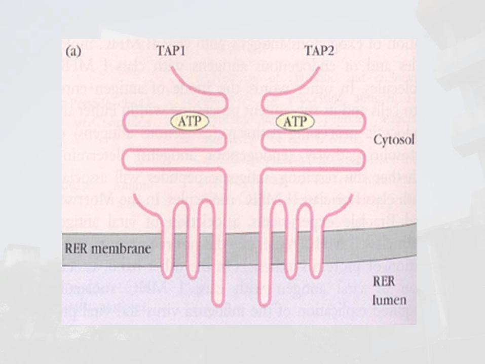
TAP, transporter associated with antigen
processing• structure:
– TAP-1 and TAP-2 • function:
– transports small peptides (8-13 aa) to the ER

calnexin• Structure
– 88kD integral ER membrane chaperone protein
• Function– Binds to a nascent MHC class I chain af
ter release from a ribosome into the ER lumen so that the chain will not leave the ER until it binds both a short peptide sequence and 2 microgobulin

Molecular chaperones: calnexin, calreticulin,tapasin
MHC class II processing pathway
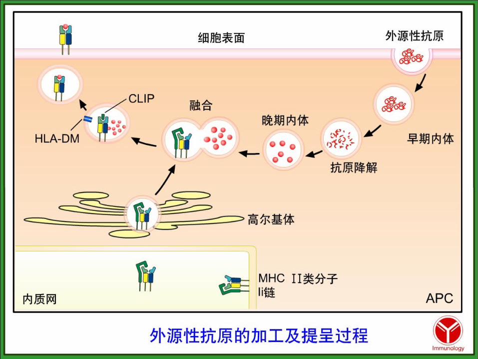

• Antigenic protein endosome/lysosome peptide fragment
• Newly synthesized class II molecules move to ER and associate with invariant chain protein molecule move to Golgi apparatus move to endosomes/lysosomes release of invariant chain from class II molecule class II binds antigenic peptide fragment transport to cell surface
MHC class II processing pathway

Structure of MHC class II

Endosome & lysosome• acidic protease & lysosome enzymes
• Function– Degrade protein into peptide fragment
s (10-30 aa)

invariant chain, Ii
• Function– Promote the formation of MHC II dimer– Directs the movement of newly synthesized
class II molecules into the Golgi and then the late endocytic compartment of the cell
– Prevent the binding of antigenic peptides to class II molecules, at least until the class II molecule reaches the late endocytic compartment

• CLIP, class II associated invariant chain peptides

3 Antigen presentation
• Antigen presentation– The activation of T cells via T cell
receptors, which specifically recognize antigenic peptide in association with either MHC class I or II molecules on the surface of APC.


Related Documents