Clinical Study Apathy in Parkinson’s Disease: An Electrophysiological Study Stéphane Mathis, Jean-Philippe Neau, Claudette Pluchon, Marie-Noëlle Fargeau, Stéphane Karolewicz, Anna Iljicsov, and Roger Gil Department of Neurology, CHU Poitiers, University of Poitiers, 2 rue de la Mil´ etrie, 86021 Poitiers, France Correspondence should be addressed to St´ ephane Mathis; [email protected] Received 3 October 2013; Accepted 28 January 2014; Published 7 April 2014 Academic Editor: Dirk Deleu Copyright © 2014 St´ ephane Mathis et al. is is an open access article distributed under the Creative Commons Attribution License, which permits unrestricted use, distribution, and reproduction in any medium, provided the original work is properly cited. In Parkinson’s disease (PD), apathy (or loss of motivation) is frequent. Nevertheless, the contribution of attentional disorders to its genesis is still not clearly known. We want to determine the relation existing between apathy and attentional disorders by using P300a (or novelty P3) as a marker of the attentional process. e study included 25 patients (13 women and 12 men) with PD for whom we have determined the relationship between automatic attention (represented by P300a) and motor status, apathy, executive dysfunction, mental flexibility, inhibitory control, and depression/anxiety. We have found a correlation between the apathy score and amplitude of novelty P300 during the ON period and also a correlation of the apathy score with a decrease in amplitude of P300 during the OFF period. In a linear regression model, changes in the P300a predicted the severity of apathy independently of any other variable. We concluded firstly that the reduction in amplitude of the P300a wave was a neurophysiological marker of apathy in PD and secondly that apathy led to both dopaminergic denervation (mesolimbic) and nondopaminergic (dorsolateral prefrontal-subcortical) dysfunction. 1. Introduction Parkinson’s disease (PD) is the most frequent neurodegener- ative disorder in Europe, with a prevalence of 1/1,000 in the general population and 1.5% in subjects more than 65 years of age [1]. It originates in destruction of the dopaminergic nigrostriatal circuit; and it is manifested in Parkinson’s syn- drome, which entails cognitive and psychic complications. In fact, depression is frequent in this disease, with an average prevalence of 40% [2]. Many other neuropsychiatric subcor- tical manifestations have been described in the literature [3– 5], but apathy is among the most frequent: current estimates of its prevalence in Parkinson’s disease vary between 16.5% and 42% [6, 7]. Apathy refers to a wide-ranging behavioural, emotional, and motivational constellation including reduced interest and participation in normal purposeful behaviour, lack of initiative with problems initiating or sustaining an activity to completion, lack of concern or indifference, and affective flattening [8, 9]. With that said, apathy syndrome may be partially sec- ondary, with regard to dysfunction of a fronto-subcortico- striato-thalamo-cortical loop [8, 10–12]. Furthermore, these circuits are in all likelihood the same as those involved in the motor and cognitive dysfunction typical of Parkinson’s disease [10, 13, 14]. In fact, apathy may be considered as a multicomposite entity consisting in dysfunction of associa- tive [4] and limbic loops and accompanied by emotional and motivational aspects [14]. In most cases of apathy, the emotional and motivational dimension arises from intricate links between the generally unconscious mobilization of attentional resources and their purposeful utilization, in which the automatic attentional process assumes a major role. Daffner et al. have underscored the basic role of the prefrontal cortex (particularly the dorsolateral prefrontal cortex) in attentional process [15]. eir studies were primarily based on analyses of patients with frontal lobe injury (with chronic infarction in the dorsolateral prefrontal cortex) in which subjects were made to view repetitive frequent (for voluntary attention) and infrequent (for automatic attention) back- ground stimuli: they demonstrated a correlation between decrease of attentional level, prefrontal lesions, and increase of apathy level (evaluated with Marin apathy scale) [16]. Another study showed a strong correlation between decrease of the electrophysiological attentional marker P300a (novelty Hindawi Publishing Corporation Neurology Research International Volume 2014, Article ID 290513, 9 pages http://dx.doi.org/10.1155/2014/290513

Welcome message from author
This document is posted to help you gain knowledge. Please leave a comment to let me know what you think about it! Share it to your friends and learn new things together.
Transcript
Clinical StudyApathy in Parkinson’s Disease: An Electrophysiological Study
Stéphane Mathis, Jean-Philippe Neau, Claudette Pluchon, Marie-Noëlle Fargeau,Stéphane Karolewicz, Anna Iljicsov, and Roger Gil
Department of Neurology, CHU Poitiers, University of Poitiers, 2 rue de la Miletrie, 86021 Poitiers, France
Correspondence should be addressed to Stephane Mathis; [email protected]
Received 3 October 2013; Accepted 28 January 2014; Published 7 April 2014
Academic Editor: Dirk Deleu
Copyright © 2014 Stephane Mathis et al. This is an open access article distributed under the Creative Commons AttributionLicense, which permits unrestricted use, distribution, and reproduction in any medium, provided the original work is properlycited.
In Parkinson’s disease (PD), apathy (or loss of motivation) is frequent. Nevertheless, the contribution of attentional disorders toits genesis is still not clearly known. We want to determine the relation existing between apathy and attentional disorders by usingP300a (or novelty P3) as a marker of the attentional process. The study included 25 patients (13 women and 12 men) with PD forwhomwe have determined the relationship between automatic attention (represented by P300a) andmotor status, apathy, executivedysfunction, mental flexibility, inhibitory control, and depression/anxiety. We have found a correlation between the apathy scoreand amplitude of novelty P300 during the ON period and also a correlation of the apathy score with a decrease in amplitude ofP300 during the OFF period. In a linear regression model, changes in the P300a predicted the severity of apathy independentlyof any other variable. We concluded firstly that the reduction in amplitude of the P300a wave was a neurophysiological marker ofapathy in PD and secondly that apathy led to both dopaminergic denervation (mesolimbic) and nondopaminergic (dorsolateralprefrontal-subcortical) dysfunction.
1. Introduction
Parkinson’s disease (PD) is the most frequent neurodegener-ative disorder in Europe, with a prevalence of 1/1,000 in thegeneral population and 1.5% in subjects more than 65 yearsof age [1]. It originates in destruction of the dopaminergicnigrostriatal circuit; and it is manifested in Parkinson’s syn-drome, which entails cognitive and psychic complications. Infact, depression is frequent in this disease, with an averageprevalence of 40% [2]. Many other neuropsychiatric subcor-tical manifestations have been described in the literature [3–5], but apathy is among the most frequent: current estimatesof its prevalence in Parkinson’s disease vary between 16.5%and 42% [6, 7]. Apathy refers to a wide-ranging behavioural,emotional, and motivational constellation including reducedinterest and participation in normal purposeful behaviour,lack of initiative with problems initiating or sustaining anactivity to completion, lack of concern or indifference, andaffective flattening [8, 9].
With that said, apathy syndrome may be partially sec-ondary, with regard to dysfunction of a fronto-subcortico-striato-thalamo-cortical loop [8, 10–12]. Furthermore, these
circuits are in all likelihood the same as those involved inthe motor and cognitive dysfunction typical of Parkinson’sdisease [10, 13, 14]. In fact, apathy may be considered as amulticomposite entity consisting in dysfunction of associa-tive [4] and limbic loops and accompanied by emotionaland motivational aspects [14]. In most cases of apathy, theemotional and motivational dimension arises from intricatelinks between the generally unconscious mobilization ofattentional resources and their purposeful utilization, inwhich the automatic attentional process assumes amajor role.Daffner et al. have underscored the basic role of the prefrontalcortex (particularly the dorsolateral prefrontal cortex) inattentional process [15]. Their studies were primarily basedon analyses of patients with frontal lobe injury (with chronicinfarction in the dorsolateral prefrontal cortex) in whichsubjects were made to view repetitive frequent (for voluntaryattention) and infrequent (for automatic attention) back-ground stimuli: they demonstrated a correlation betweendecrease of attentional level, prefrontal lesions, and increaseof apathy level (evaluated with Marin apathy scale) [16].Another study showed a strong correlation between decreaseof the electrophysiological attentional marker P300a (novelty
Hindawi Publishing CorporationNeurology Research InternationalVolume 2014, Article ID 290513, 9 pageshttp://dx.doi.org/10.1155/2014/290513
2 Neurology Research International
P3) and increase of apathy [17].The same correlation betweenP300a and apathy has been found in Alzheimer’s disease [18]and in cerebral trauma [17] with selective lesion of prefrontalcortex, as illustrated in functional cerebral imaging studies[19].
However, no comparable data on Parkinson’s disease arepresently available. We nonetheless wish to hypothesise that,in Parkinson’s disease, there exists a correlation betweenincrease of apathy level and decrease of P300a amplitude,which constitutes an electrophysiological marker of auto-matic attention.
2. Methods
2.1. Subjects. Twenty-five patients (13 women and 12 men),hospitalized in the Department of Neurology (CHU Poitiers)for evaluation of Parkinson’s disease, were includedwith theiragreement; written informed consent for research purposeswas obtained for each patient. The general characteristicsof this population were age = 64.1 ± 6.4 years; durationof PD = 11.5 ± 4.8 years; daily dose of dopa therapy =1567.8 ± 725.4mg; total UPDRS score during OFF period =25.8 ± 11.8; total UPDRS score during ON period = 9.0 ±7.1; Hoehn and Yahr score = 2.5 ± 0.4. Patients presentedwith the usual Parkinson’s disease criteria. The severity ofthe disease was evaluated with the Hoehn and Yahr score,which maps out stages from I through V [20]. Reactivity tolevodopa was less than 65%. The daily dose of dopa therapywas calculated by addition of a daily dose of levodopa andthe dopaminergic agonists transcribed as “dose-equivalentdopa” [21, 22]. Patients with dementia (MMSE < 24) or withserious depression, melancholy, or depression with delirium(according to the diagnostic and statistical manualcriteria)were excluded from the study.
Each subject included in the study was evaluated withcognitive, motor, psychiatric, and subsequent electrophysio-logical tests during OFF (without dopaminergic treatment)and ON (with dopaminergic treatment) periods. The studytook into account the fluctuating characteristics of PD overthe daytime. Each period (ON or OFF) was selected alterna-tively in order to have as many patients during the ON periodas during the OFF period (±1 subject). Only patients able towithstand the OFF period, or without important dyskinesiain the ON period, were included in the study. Patients wereanalyzed during the OFF period, before taking a levodopatest, patients stopped their treatments the previous evening,and the study began at 8:30 the following morning (prior tothe levodopa test). For each patient, the study took place over3 days (D0, D1, and D2); the order of tests was systematicallythe same for all patients. Duration of theOFF andONperiodswas likewise identical in every case.
2.2. Cognitive Study. The cognitive assessment included 4tests systematically taken in 30minutes in the following order(in both ON and OFF periods): verbal fluency test, Strooptest,Wisconsin sorting card test, and FAB (frontal assessmentbattery) test. In the verbal fluency test, patients were askedto name in one minute as many items as they could from
one semantic category (animals) and then as many wordsas they could beginning with the letter M (one minute);category switching (boys’ names and fruits) was then assessed(one minute). In an alternative form [23], the last one ofthese verbal fluency tests was used for evaluation of mentalflexibility [24]. The Stroop test was used for evaluation ofinhibitory control [25]. With the Wisconsin card sorting test(WCST), we collected data (“number of series,” “numberof mistakes,” and “perseveration percentage”) in order toevaluatemental flexibility [26]. Finally, the frontal assessmentbattery (FAB) was used for the purposes of general evaluationof executive functions [27].
2.3. Clinical Study. After day 0 (D0) assessment, we appliedthe UPDRS III scale [28] and then the Hoehn and Yahr score[20], for motor evaluation of PD [28].
In order to assess apathy, we used the Starkstein apathyscale [7]. As a variant of the “Marin apathy scale” [29], itincludes 14 items contributing to evaluation of degree ofapathy. Each item was rated from 0 to 3 so as to calculate anapathy score ranging from 0 to 42. The “cut-off score” was 14[7] with 66% sensitivity and 100% specificity. In our study,only patients with score >14 were considered apathetic.
For purposes of psychiatric evaluation, we used the DSM(diagnostic and statisticalmanual) criteria (D0) and theHADscale (D1 and D2). The HAD scale (Hospital Anxiety andDepression scale) was also used during the ON and OFFperiods in order to quantify the depression and anxiety levels[30]. We calculated a depression and anxiety score with cut-off >7 for each of two categories.
2.4. Electrophysiological Study. Each patient was subjectedto the recording of long-latency auditory evoked potentialsaccording to a previously described protocol [31–33], andaccording to the guidelines for event-related potentials (ERP)[34], 140 tone bursts (intensity: 80 dB; duration: 20ms; 0.9msrise-fall time) were presented binaurally through earphonesat a rate of 1 tone every 0.8 sec, including 100 low “frequency”sounds (1000Hz), 20 high “target” sounds (2000Hz), and20 randomized unexpected “novelty” stimulations (the word“airplane” in French, i.e., “avion”). A disc electrode wasaffixed to the midline site Cz and referred to linked mas-toids; responses to rare and frequent stimuli were averagedseparately. The EEG bandpass was 1–100Hz. The curves werenumerically filtrated (high-pass filter: 0.5Hz; low-pass filter:15Hz). The horizontal and vertical bipolar electrooculogram(EOG) was recorded during the task to monitor artefacts.Trials with artefacts were automatically excluded from theaverages: if any data point (beyond the first 2.5ms of thesweep) was greater than 96% full scale, the entire sweepwas rejected and was not added to the memory block. Twoconsecutive averages of artefact-free trials were obtained.Subjects were tested seated in a sound-attenuating chamber.All of them reported normal hearing function. The prere-corded stimulus sequencewas presentedwithout instructions(passive condition). After this first run, all subjects were ableto distinguish high-pitched from low-pitched tones and werethen instructed to keep a mental record of the rare tones
Neurology Research International 3
and to report their number at the end of the run (activecondition).The P300 wave (target P3) was clearly identifiableonly in the “active” condition in response to rare 2000Hztones; this condition was used in measurement of amplitudeand latency of N200 and P300 potentials. The P300a (noveltyP3) was clearly identifiable in response to the randomizedunexpected “novelty” stimulation (word “avion”). Latencyvalues were calculated from the intersection of extrapolatedlines from the ascending and descending slopes of eachpeak. Latencies of N200 and P300 were determined for eachsubject, as well as the peak-to-peak amplitude of N200-P300.Latency andmaximum amplitude of P300a were identified inan interval ranging from 250ms to 360ms after stimulation(on Fz, Cz, and Pz electrodes). In addition, average P300aamplitude was calculated in an interval varying from 250 to360ms after stimulation and was evaluated in comparisonwith the basal line (60ms before the stimulation).
2.5. Statistical Analysis. Correlations between apathy score(Starkstein apathy scale) and each feature (electrophysio-logical, cognitive, psychiatric, and motor) were carried outindependently (for data during ON and OFF periods) with 𝜌correlation index (nonparametric Spearman’s test). The rolesof dopaminergic innervation (reflected by the percentage ofimprovement after UPDRS III score) or of nondopaminer-gic lesions (reflected by the residual UPDRS III score inON period) on the different features (electrophysiological,cognitive, psychiatric, and motor) were determined with amatched nonparametric signed-rank Wilcoxon test (𝑧) withregard to averages during the ON and OFF periods for eachfeature. Differences for each feature (electrophysiological,cognitive, psychiatric, and motor) between “apathetic” and“nonapathetic” subjects (a “cut-off” score was establishedat 14) were determined by using a nonparametric Mann-Whitney 𝑈 test for quantitative data and a Chi-square test(corrected Chi-square test and exact Fisher’s test, accordingto the conditions of application) for qualitative data.
If a significant correlation between the apathy score andan electrophysiological data appeared, this electrophysiologi-cal variable is further dichotomized (median as cut-off value)for subsequent logistic regression to predict high or low valueof electrophysiological data, apathy score, and other potentialconfounding factors. After univariate data screening, anascending stepwise procedure for multivariate analysis wasused with significant 𝑃 values at 10% level to include it intologistic regression. All significance levels were set at 5% forthe logistic regression. Correlations between electrophysio-logical data and the other variables were calculated using theSpearman nonparametric correlation coefficient. The 𝛼 riskwas 5% and the statistical tests were carried out in bilateralsituations.The software we used for data capture and analysiswas Statview 5.0 (from SAS Institute©).
3. Results
3.1. Relations between Apathy and Electrophysiological, Cog-nitive, Psychiatric, and Motor Features (during ON and OFF
49𝜇V
EOG
F7Fz
F8
Cz
PzT6T5
−60 890
10𝜇V
Front
Rear
Fp2
A1 A2
Fz F8
T6T5
F7
PzCz
(ms)
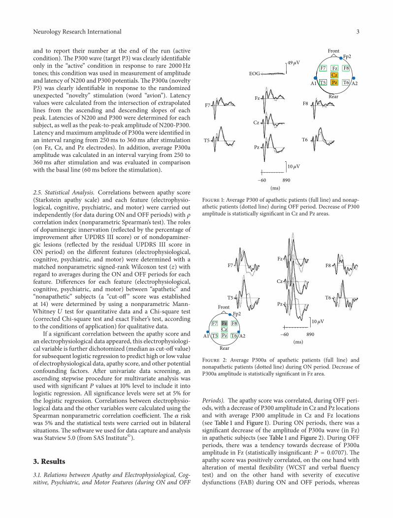
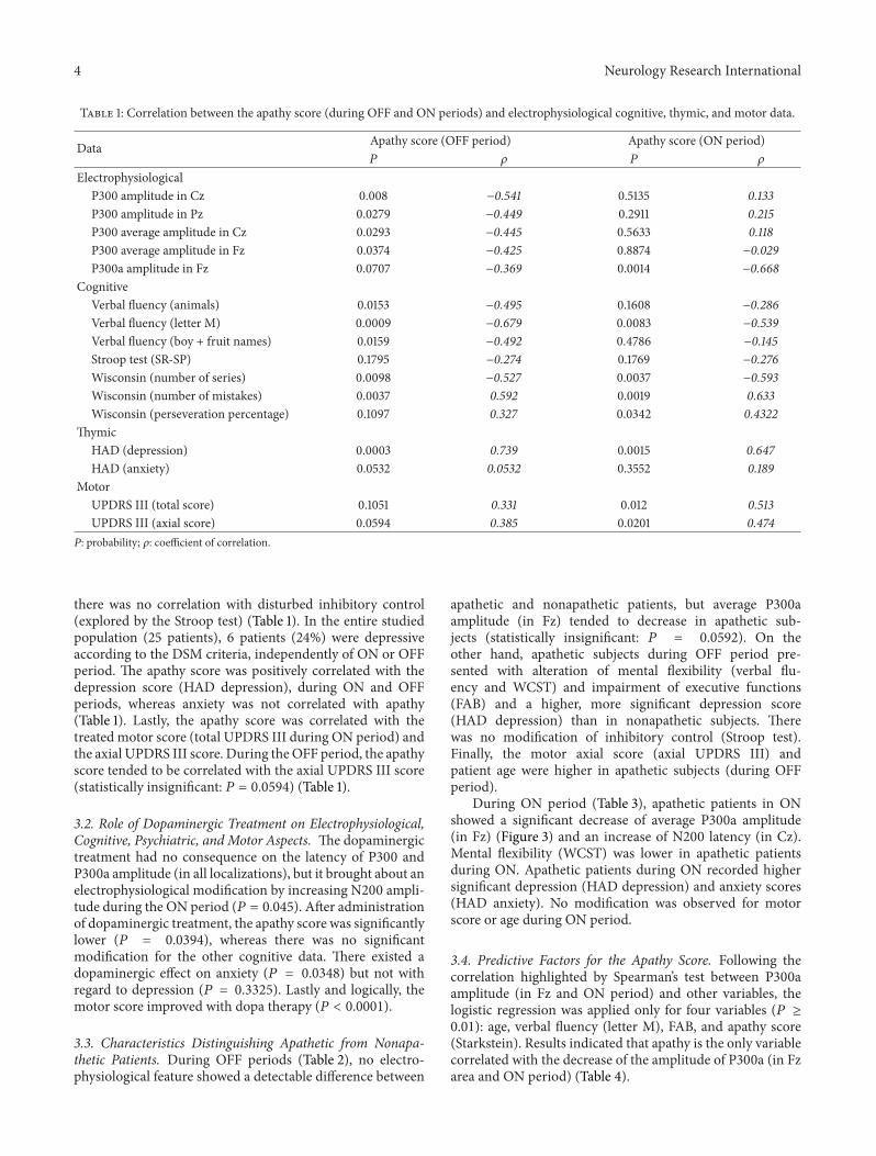
Figure 1: Average P300 of apathetic patients (full line) and nonap-athetic patients (dotted line) during OFF period. Decrease of P300amplitude is statistically significant in Cz and Pz areas.
F7Fz
F8
Cz
PzT6T5
−60 890
10𝜇V
Front
Rear
Fp2
A1 A2
Fz F8
T6T5
F7
PzCz
(ms)
Figure 2: Average P300a of apathetic patients (full line) andnonapathetic patients (dotted line) during ON period. Decrease ofP300a amplitude is statistically significant in Fz area.
Periods). The apathy score was correlated, during OFF peri-ods, with a decrease of P300 amplitude in Cz and Pz locationsand with average P300 amplitude in Cz and Fz locations(see Table 1 and Figure 1). During ON periods, there was asignificant decrease of the amplitude of P300a wave (in Fz)in apathetic subjects (see Table 1 and Figure 2). During OFFperiods, there was a tendency towards decrease of P300aamplitude in Fz (statistically insignificant: 𝑃 = 0.0707). Theapathy score was positively correlated, on the one hand withalteration of mental flexibility (WCST and verbal fluencytest) and on the other hand with severity of executivedysfunctions (FAB) during ON and OFF periods, whereas
4 Neurology Research International
Table 1: Correlation between the apathy score (during OFF and ON periods) and electrophysiological cognitive, thymic, and motor data.
Data Apathy score (OFF period) Apathy score (ON period)𝑃 𝜌 𝑃 𝜌
ElectrophysiologicalP300 amplitude in Cz 0.008 −0.541 0.5135 0.133P300 amplitude in Pz 0.0279 −0.449 0.2911 0.215P300 average amplitude in Cz 0.0293 −0.445 0.5633 0.118P300 average amplitude in Fz 0.0374 −0.425 0.8874 −0.029P300a amplitude in Fz 0.0707 −0.369 0.0014 −0.668
CognitiveVerbal fluency (animals) 0.0153 −0.495 0.1608 −0.286Verbal fluency (letter M) 0.0009 −0.679 0.0083 −0.539Verbal fluency (boy + fruit names) 0.0159 −0.492 0.4786 −0.145Stroop test (SR-SP) 0.1795 −0.274 0.1769 −0.276Wisconsin (number of series) 0.0098 −0.527 0.0037 −0.593Wisconsin (number of mistakes) 0.0037 0.592 0.0019 0.633Wisconsin (perseveration percentage) 0.1097 0.327 0.0342 0.4322
ThymicHAD (depression) 0.0003 0.739 0.0015 0.647HAD (anxiety) 0.0532 0.0532 0.3552 0.189
MotorUPDRS III (total score) 0.1051 0.331 0.012 0.513UPDRS III (axial score) 0.0594 0.385 0.0201 0.474𝑃: probability; 𝜌: coefficient of correlation.
there was no correlation with disturbed inhibitory control(explored by the Stroop test) (Table 1). In the entire studiedpopulation (25 patients), 6 patients (24%) were depressiveaccording to the DSM criteria, independently of ON or OFFperiod. The apathy score was positively correlated with thedepression score (HAD depression), during ON and OFFperiods, whereas anxiety was not correlated with apathy(Table 1). Lastly, the apathy score was correlated with thetreated motor score (total UPDRS III during ON period) andthe axial UPDRS III score. During theOFF period, the apathyscore tended to be correlated with the axial UPDRS III score(statistically insignificant: 𝑃 = 0.0594) (Table 1).
3.2. Role of Dopaminergic Treatment on Electrophysiological,Cognitive, Psychiatric, and Motor Aspects. The dopaminergictreatment had no consequence on the latency of P300 andP300a amplitude (in all localizations), but it brought about anelectrophysiological modification by increasing N200 ampli-tude during the ON period (𝑃 = 0.045). After administrationof dopaminergic treatment, the apathy score was significantlylower (𝑃 = 0.0394), whereas there was no significantmodification for the other cognitive data. There existed adopaminergic effect on anxiety (𝑃 = 0.0348) but not withregard to depression (𝑃 = 0.3325). Lastly and logically, themotor score improved with dopa therapy (𝑃 < 0.0001).
3.3. Characteristics Distinguishing Apathetic from Nonapa-thetic Patients. During OFF periods (Table 2), no electro-physiological feature showed a detectable difference between
apathetic and nonapathetic patients, but average P300aamplitude (in Fz) tended to decrease in apathetic sub-jects (statistically insignificant: 𝑃 = 0.0592). On theother hand, apathetic subjects during OFF period pre-sented with alteration of mental flexibility (verbal flu-ency and WCST) and impairment of executive functions(FAB) and a higher, more significant depression score(HAD depression) than in nonapathetic subjects. Therewas no modification of inhibitory control (Stroop test).Finally, the motor axial score (axial UPDRS III) andpatient age were higher in apathetic subjects (during OFFperiod).
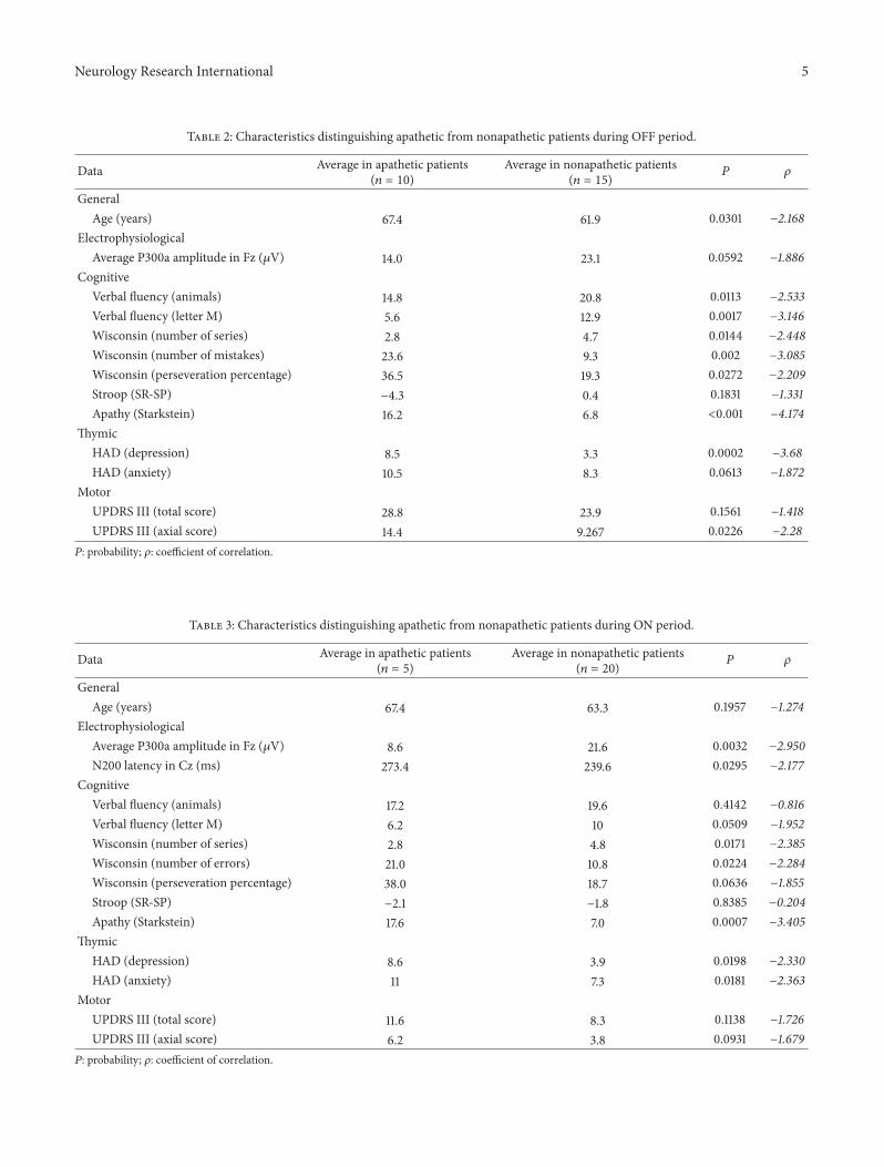
During ON period (Table 3), apathetic patients in ONshowed a significant decrease of average P300a amplitude(in Fz) (Figure 3) and an increase of N200 latency (in Cz).Mental flexibility (WCST) was lower in apathetic patientsduring ON. Apathetic patients during ON recorded highersignificant depression (HAD depression) and anxiety scores(HAD anxiety). No modification was observed for motorscore or age during ON period.
3.4. Predictive Factors for the Apathy Score. Following thecorrelation highlighted by Spearman’s test between P300aamplitude (in Fz and ON period) and other variables, thelogistic regression was applied only for four variables (𝑃 ≥0.01): age, verbal fluency (letter M), FAB, and apathy score(Starkstein). Results indicated that apathy is the only variablecorrelated with the decrease of the amplitude of P300a (in Fzarea and ON period) (Table 4).
Neurology Research International 5
Table 2: Characteristics distinguishing apathetic from nonapathetic patients during OFF period.
Data Average in apathetic patients(𝑛 = 10)
Average in nonapathetic patients(𝑛 = 15) 𝑃 𝜌
GeneralAge (years) 67.4 61.9 0.0301 −2.168
ElectrophysiologicalAverage P300a amplitude in Fz (𝜇V) 14.0 23.1 0.0592 −1.886
CognitiveVerbal fluency (animals) 14.8 20.8 0.0113 −2.533Verbal fluency (letter M) 5.6 12.9 0.0017 −3.146Wisconsin (number of series) 2.8 4.7 0.0144 −2.448Wisconsin (number of mistakes) 23.6 9.3 0.002 −3.085Wisconsin (perseveration percentage) 36.5 19.3 0.0272 −2.209Stroop (SR-SP) −4.3 0.4 0.1831 −1.331Apathy (Starkstein) 16.2 6.8 <0.001 −4.174
ThymicHAD (depression) 8.5 3.3 0.0002 −3.68HAD (anxiety) 10.5 8.3 0.0613 −1.872
MotorUPDRS III (total score) 28.8 23.9 0.1561 −1.418UPDRS III (axial score) 14.4 9.267 0.0226 −2.28𝑃: probability; 𝜌: coefficient of correlation.
Table 3: Characteristics distinguishing apathetic from nonapathetic patients during ON period.
Data Average in apathetic patients(𝑛 = 5)
Average in nonapathetic patients(𝑛 = 20) 𝑃 𝜌
GeneralAge (years) 67.4 63.3 0.1957 −1.274
ElectrophysiologicalAverage P300a amplitude in Fz (𝜇V) 8.6 21.6 0.0032 −2.950N200 latency in Cz (ms) 273.4 239.6 0.0295 −2.177
CognitiveVerbal fluency (animals) 17.2 19.6 0.4142 −0.816Verbal fluency (letter M) 6.2 10 0.0509 −1.952Wisconsin (number of series) 2.8 4.8 0.0171 −2.385Wisconsin (number of errors) 21.0 10.8 0.0224 −2.284Wisconsin (perseveration percentage) 38.0 18.7 0.0636 −1.855Stroop (SR-SP) −2.1 −1.8 0.8385 −0.204Apathy (Starkstein) 17.6 7.0 0.0007 −3.405
ThymicHAD (depression) 8.6 3.9 0.0198 −2.330HAD (anxiety) 11 7.3 0.0181 −2.363
MotorUPDRS III (total score) 11.6 8.3 0.1138 −1.726UPDRS III (axial score) 6.2 3.8 0.0931 −1.679𝑃: probability; 𝜌: coefficient of correlation.
6 Neurology Research International
Table 4: Logistic regression model of P300a amplitude (in Fz area and in ON period).
Variables Univariate analysis(𝑃 value)
Multivariate analysis(𝑃 value)
Adjustedodds ratio
95%confidence intervals
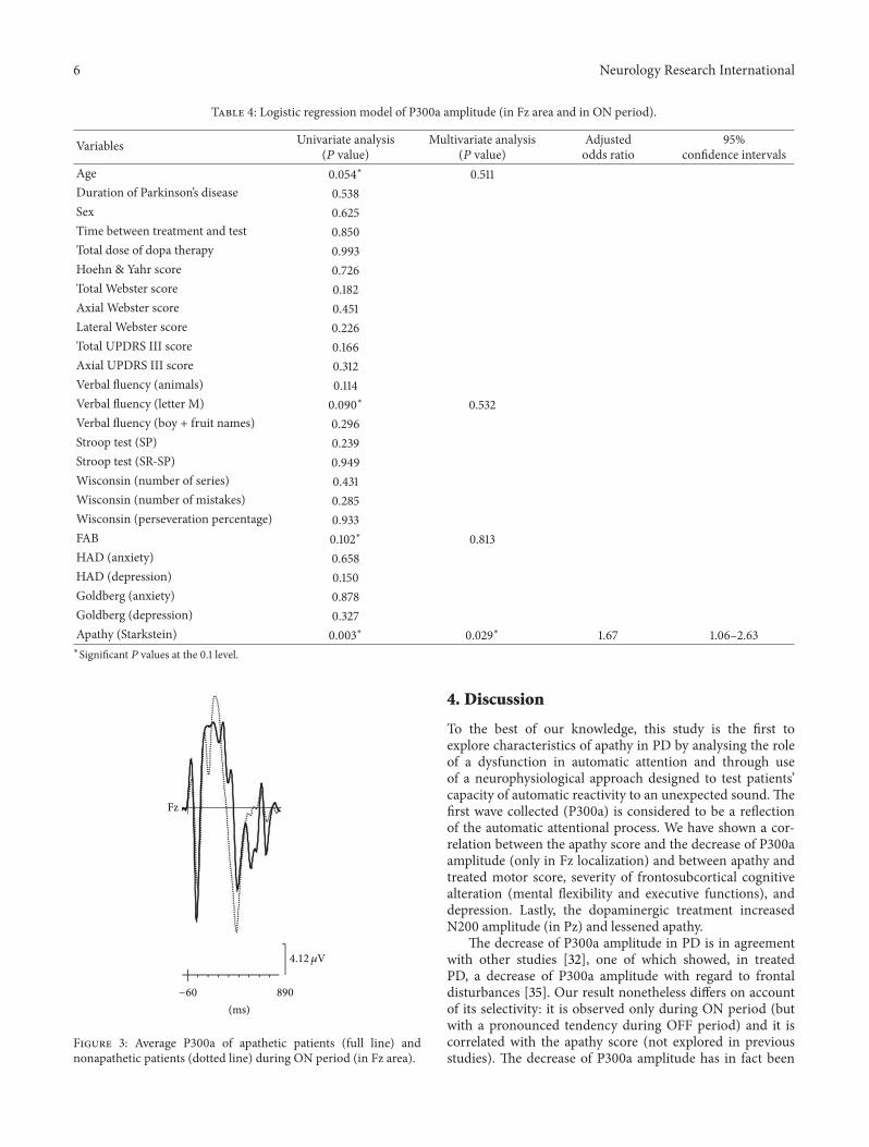
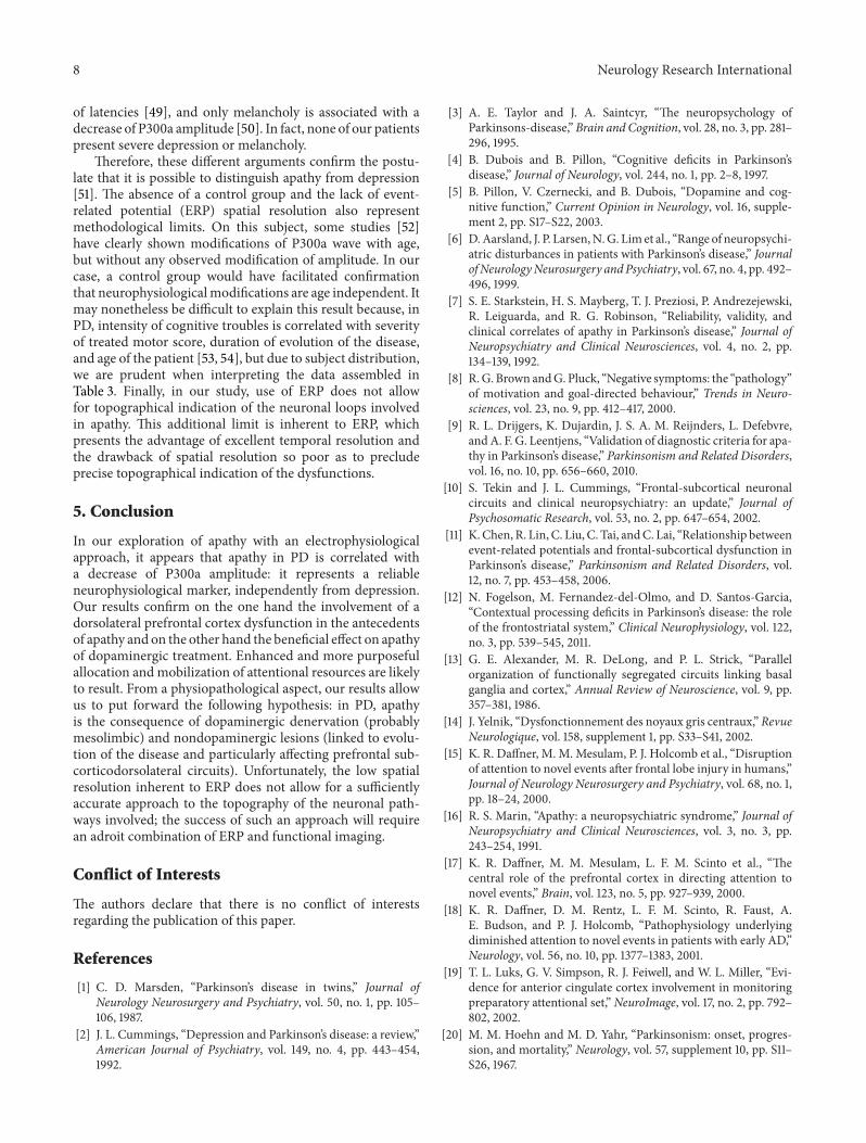
Age 0.054∗ 0.511Duration of Parkinson’s disease 0.538Sex 0.625Time between treatment and test 0.850Total dose of dopa therapy 0.993Hoehn & Yahr score 0.726Total Webster score 0.182Axial Webster score 0.451Lateral Webster score 0.226Total UPDRS III score 0.166Axial UPDRS III score 0.312Verbal fluency (animals) 0.114Verbal fluency (letter M) 0.090∗ 0.532Verbal fluency (boy + fruit names) 0.296Stroop test (SP) 0.239Stroop test (SR-SP) 0.949Wisconsin (number of series) 0.431Wisconsin (number of mistakes) 0.285Wisconsin (perseveration percentage) 0.933FAB 0.102∗ 0.813HAD (anxiety) 0.658HAD (depression) 0.150Goldberg (anxiety) 0.878Goldberg (depression) 0.327Apathy (Starkstein) 0.003∗ 0.029∗ 1.67 1.06–2.63∗Significant 𝑃 values at the 0.1 level.
Fz
−60 890
(ms)
4.12 𝜇V
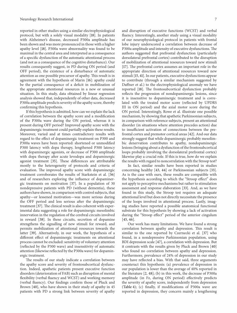
Figure 3: Average P300a of apathetic patients (full line) andnonapathetic patients (dotted line) during ON period (in Fz area).
4. Discussion
To the best of our knowledge, this study is the first toexplore characteristics of apathy in PD by analysing the roleof a dysfunction in automatic attention and through useof a neurophysiological approach designed to test patients’capacity of automatic reactivity to an unexpected sound. Thefirst wave collected (P300a) is considered to be a reflectionof the automatic attentional process. We have shown a cor-relation between the apathy score and the decrease of P300aamplitude (only in Fz localization) and between apathy andtreated motor score, severity of frontosubcortical cognitivealteration (mental flexibility and executive functions), anddepression. Lastly, the dopaminergic treatment increasedN200 amplitude (in Pz) and lessened apathy.
The decrease of P300a amplitude in PD is in agreementwith other studies [32], one of which showed, in treatedPD, a decrease of P300a amplitude with regard to frontaldisturbances [35]. Our result nonetheless differs on accountof its selectivity: it is observed only during ON period (butwith a pronounced tendency during OFF period) and it iscorrelated with the apathy score (not explored in previousstudies). The decrease of P300a amplitude has in fact been
Neurology Research International 7
reported in other studies using a similar electrophysiologicalprotocol, but with a solely visual modality [18]. In patientswith Alzheimer’s disease, decreased P300a amplitude hasbeen shown andwasmore pronounced in those with a higherapathy level [18]. P300a wave abnormality was found to bemaximal in the central area and interpreted as a consequenceof a specific dysfunction of the automatic attentional process(and not as a consequence of the cognitive disturbance). Ourresults consequently suggest, in PD during ON period (andOFF period), the existence of a disturbance of automaticattention as one possible precursor of apathy. This result is inagreement with the hypothesis of Marin [16]: apathy couldbe the partial consequence of a deficit in mobilization ofthe appropriate attentional resources in a new or unusualsituation. In this study, data obtained by linear regressionanalysis showed that, independently of other data, decreasedP300a amplitude predicts severity of the apathy score, therebyconfirming this hypothesis.
If this hypothesis is admitted, how canwe explain the lackof correlation between the apathy score and a modificationof the P300a wave during the ON period, whereas it ispresent during OFF period? Improved apathy score with thedopaminergic treatment could partially explain these results.Moreover, varied and at times contradictory results withregard to the effect of dopaminergic treatment on P300 andP300a waves have been reported: shortened or unmodifiedP300 latency with dopa therapy, lengthened P300 latencywith dopaminergic agonists, or decrease of P300 amplitudewith dopa therapy after acute levodopa and dopaminergicagonist treatment [35]. These differences are attributablemostly to the heterogeneity of protocols and criteria ofevaluation. The improved apathy score with dopaminergictreatment corroborates the results of Starkstein et al. [36]and of researchers exploring the influence of dopaminer-gic treatments on motivation [37]. In a population of 30nondepressive patients with PD (without dementia), theseauthors have shown, in comparisonwith control subjects, thatapathy—or lessened motivation—was more serious duringthe OFF period and less serious after the dopaminergictreatment [37].The clinical result is also coherent with exper-imental data suggesting a role for dopaminergic mesolimbicinnervation in the regulation of the cerebral circuits involvedin reward [38]. In these circuits, secretion of dopaminestrengthens the signalling of new stimuli for reward, andpermits mobilization of attentional resources towards thelatter [39]. Alternatively, in our work, the hypothesis of adifferent effect of dopaminergic treatments on attentionalprocess cannot be excluded: sensitivity of voluntary attention(reflected by the P300 wave) and insensitivity of automaticattention (likewise reflected by the P300a wave) for dopamin-ergic treatment.
The results of our study indicate a correlation betweenthe apathy score and severity of frontosubcortical dysfunc-tion. Indeed, apathetic patients present executive functiondisorders (deterioration of FAB) such as disruption of mentalflexibility (verbal fluency and WCST) and working memory(verbal fluency). Our findings confirm those of Pluck andBrown [40], who have shown in their study of apathy in 45patients with PD a strong link between severity of apathy
and disruption of executive functions (WCST) and verbalfluency. Interestingly, another study using a visual modalityof our neurophysiological protocol in patients with frontallobe injury underscored a correlation between decrease ofP300a amplitude and intensity of executive dysfunctions.Theauthors suggested that prefrontal dysfunction (particularlydorsolateral prefrontal cortex) contributed to the disruptionof mobilization of attentional resources toward new stimuli[17]. The prefrontal cortex assumes an important role in theappropriate direction of attentional resources toward newstimuli [15, 41]. In our patients, executive dysfunctions appearto contribute (through a similar mechanism suggested byDaffner et al.) to the electrophysiological anomaly we havereported [18]. The frontosubcortical dysfunction probablyreflects the progression of nondopaminergic lesions, sinceit is insensitive to dopaminergic treatment and is corre-lated with the treated motor score (reflected by UPDRSIII in ON period) and the axial motor score during theON period. Interestingly, Rowe et al. illustrate one possiblemechanism, by showing that apathetic Parkinsonian subjects,in comparison with reference subjects, present an attentionaldisorder (in situations where attention precedes action) dueto insufficient activation of connections between the pre-frontal cortex and premotor cortical areas [42]. And our datastrongly suggest that while dopaminergic probably mesolim-bic denervation contributes to apathy, nondopaminergiclesions (bringing about a dysfunction of the frontosubcorticalloops probably involving the dorsolateral prefrontal cortex)likewise play a crucial role. If this is true, how do we explainthe results with regard to noncorrelation with the Stroop test?
In fact, similar results have been reported in studiesconcerning healthy [43, 44] or Parkinsonian subjects [33].As is the case with ours, these results are compatible withthe hypothesis according to which the “Stroop effect” doesnot apply to perceptive mechanisms but rather to stimulationassessment and response elaboration [33]. And, as we havefound in this study, the Stroop test requires an adequateattentional level but does not directly necessitate the presenceof the loops involved in attentional process. Lastly, imag-ing studies have reported a possible anatomical functionalsubstrate for this hypothesis by showing a lack of activationduring the “Stroop effect” period of the anterior cingulum[45, 46].
Our work has many limitations. We have found a strongcorrelation between apathy and depression. This result issimilar to the one reported by Czernecki et al. [37] whofound, in a nondepressive Parkinsonian population, usingBDI depression scale [47], a correlation with depression. Butit contrasts with the results given by Pluck and Brown [40]who found no correlation between apathy and depression.Furthermore, prevalence of 24% of depression in our studymay have reflected a bias. With that said, three argumentscounteract this hypothesis: (a) prevalence of depression inour population is lower than the average of 40% reported inthe literature [2, 48]; (b) in this work, the decrease of P300aamplitude (in Fz, during ON period) effectively predictsthe severity of apathy score, independently from depression(Table 4); (c) finally, if modifications of P300a wave arereported in depression, they concern mainly a lengthening
8 Neurology Research International
of latencies [49], and only melancholy is associated with adecrease of P300a amplitude [50]. In fact, none of our patientspresent severe depression or melancholy.
Therefore, these different arguments confirm the postu-late that it is possible to distinguish apathy from depression[51]. The absence of a control group and the lack of event-related potential (ERP) spatial resolution also representmethodological limits. On this subject, some studies [52]have clearly shown modifications of P300a wave with age,but without any observed modification of amplitude. In ourcase, a control group would have facilitated confirmationthat neurophysiologicalmodifications are age independent. Itmay nonetheless be difficult to explain this result because, inPD, intensity of cognitive troubles is correlated with severityof treated motor score, duration of evolution of the disease,and age of the patient [53, 54], but due to subject distribution,we are prudent when interpreting the data assembled inTable 3. Finally, in our study, use of ERP does not allowfor topographical indication of the neuronal loops involvedin apathy. This additional limit is inherent to ERP, whichpresents the advantage of excellent temporal resolution andthe drawback of spatial resolution so poor as to precludeprecise topographical indication of the dysfunctions.
5. Conclusion
In our exploration of apathy with an electrophysiologicalapproach, it appears that apathy in PD is correlated witha decrease of P300a amplitude: it represents a reliableneurophysiological marker, independently from depression.Our results confirm on the one hand the involvement of adorsolateral prefrontal cortex dysfunction in the antecedentsof apathy and on the other hand the beneficial effect on apathyof dopaminergic treatment. Enhanced and more purposefulallocation andmobilization of attentional resources are likelyto result. From a physiopathological aspect, our results allowus to put forward the following hypothesis: in PD, apathyis the consequence of dopaminergic denervation (probablymesolimbic) and nondopaminergic lesions (linked to evolu-tion of the disease and particularly affecting prefrontal sub-corticodorsolateral circuits). Unfortunately, the low spatialresolution inherent to ERP does not allow for a sufficientlyaccurate approach to the topography of the neuronal path-ways involved; the success of such an approach will requirean adroit combination of ERP and functional imaging.
Conflict of Interests
The authors declare that there is no conflict of interestsregarding the publication of this paper.
References
[1] C. D. Marsden, “Parkinson’s disease in twins,” Journal ofNeurology Neurosurgery and Psychiatry, vol. 50, no. 1, pp. 105–106, 1987.
[2] J. L. Cummings, “Depression and Parkinson’s disease: a review,”American Journal of Psychiatry, vol. 149, no. 4, pp. 443–454,1992.
[3] A. E. Taylor and J. A. Saintcyr, “The neuropsychology ofParkinsons-disease,” Brain and Cognition, vol. 28, no. 3, pp. 281–296, 1995.
[4] B. Dubois and B. Pillon, “Cognitive deficits in Parkinson’sdisease,” Journal of Neurology, vol. 244, no. 1, pp. 2–8, 1997.
[5] B. Pillon, V. Czernecki, and B. Dubois, “Dopamine and cog-nitive function,” Current Opinion in Neurology, vol. 16, supple-ment 2, pp. S17–S22, 2003.
[6] D.Aarsland, J. P. Larsen,N.G. Limet al., “Range of neuropsychi-atric disturbances in patients with Parkinson’s disease,” Journalof NeurologyNeurosurgery and Psychiatry, vol. 67, no. 4, pp. 492–496, 1999.
[7] S. E. Starkstein, H. S. Mayberg, T. J. Preziosi, P. Andrezejewski,R. Leiguarda, and R. G. Robinson, “Reliability, validity, andclinical correlates of apathy in Parkinson’s disease,” Journal ofNeuropsychiatry and Clinical Neurosciences, vol. 4, no. 2, pp.134–139, 1992.
[8] R.G. Brown andG. Pluck, “Negative symptoms: the “pathology”of motivation and goal-directed behaviour,” Trends in Neuro-sciences, vol. 23, no. 9, pp. 412–417, 2000.
[9] R. L. Drijgers, K. Dujardin, J. S. A. M. Reijnders, L. Defebvre,andA. F. G. Leentjens, “Validation of diagnostic criteria for apa-thy in Parkinson’s disease,” Parkinsonism and Related Disorders,vol. 16, no. 10, pp. 656–660, 2010.
[10] S. Tekin and J. L. Cummings, “Frontal-subcortical neuronalcircuits and clinical neuropsychiatry: an update,” Journal ofPsychosomatic Research, vol. 53, no. 2, pp. 647–654, 2002.
[11] K. Chen, R. Lin, C. Liu, C. Tai, andC. Lai, “Relationship betweenevent-related potentials and frontal-subcortical dysfunction inParkinson’s disease,” Parkinsonism and Related Disorders, vol.12, no. 7, pp. 453–458, 2006.
[12] N. Fogelson, M. Fernandez-del-Olmo, and D. Santos-Garcia,“Contextual processing deficits in Parkinson’s disease: the roleof the frontostriatal system,” Clinical Neurophysiology, vol. 122,no. 3, pp. 539–545, 2011.
[13] G. E. Alexander, M. R. DeLong, and P. L. Strick, “Parallelorganization of functionally segregated circuits linking basalganglia and cortex,” Annual Review of Neuroscience, vol. 9, pp.357–381, 1986.
[14] J. Yelnik, “Dysfonctionnement des noyaux gris centraux,” RevueNeurologique, vol. 158, supplement 1, pp. S33–S41, 2002.
[15] K. R. Daffner, M. M. Mesulam, P. J. Holcomb et al., “Disruptionof attention to novel events after frontal lobe injury in humans,”Journal of Neurology Neurosurgery and Psychiatry, vol. 68, no. 1,pp. 18–24, 2000.
[16] R. S. Marin, “Apathy: a neuropsychiatric syndrome,” Journal ofNeuropsychiatry and Clinical Neurosciences, vol. 3, no. 3, pp.243–254, 1991.
[17] K. R. Daffner, M. M. Mesulam, L. F. M. Scinto et al., “Thecentral role of the prefrontal cortex in directing attention tonovel events,” Brain, vol. 123, no. 5, pp. 927–939, 2000.
[18] K. R. Daffner, D. M. Rentz, L. F. M. Scinto, R. Faust, A.E. Budson, and P. J. Holcomb, “Pathophysiology underlyingdiminished attention to novel events in patients with early AD,”Neurology, vol. 56, no. 10, pp. 1377–1383, 2001.
[19] T. L. Luks, G. V. Simpson, R. J. Feiwell, and W. L. Miller, “Evi-dence for anterior cingulate cortex involvement in monitoringpreparatory attentional set,” NeuroImage, vol. 17, no. 2, pp. 792–802, 2002.
[20] M. M. Hoehn and M. D. Yahr, “Parkinsonism: onset, progres-sion, and mortality,” Neurology, vol. 57, supplement 10, pp. S11–S26, 1967.
Neurology Research International 9
[21] P. Krack, P. Pollak, P. Limousin et al., “Subthalamic nucleus orinternal pallidal stimulation in young onset Parkinson’s disease,”Brain, vol. 121, no. 3, pp. 451–457, 1998.
[22] A. M. Lozano, A. E. Lang, N. Galvez-Jimenez et al., “Effect ofGPi pallidotomy on motor function in Parkinson’s disease,”TheLancet, vol. 346, no. 8987, pp. 1383–1387, 1995.
[23] M. Mitrushina, K. B. Boone, J. Razani, and L. F. D’Elia, Hand-book of Normative Data for Neuropsychological Assessment,OxfordUniversity Press,NewYork,NY,USA, 2nd edition, 2005.
[24] A. M. Ramier and H. Hecaen, “Role respectif des atteintesfrontales et de la lateralisation lesionnelle dans les deficits de la,‘fluence verbale’,” Revue Neurologique, vol. 123, no. 1, pp. 17–22,1970.
[25] J. R. Stroop, “Studies of interference in serial verbal reactions,”Journal of Experimental Psychology, vol. 18, no. 6, pp. 643–662,1935.
[26] H. E. Nelson, “A modified card sorting test sensitive to frontallobe defects,” Cortex, vol. 12, no. 4, pp. 313–324, 1976.
[27] B. Dubois, A. Slachevsky, I. Litvan, and B. Pillon, “The FAB: afrontal assessment battery at bedside,”Neurology, vol. 55, no. 11,pp. 1621–1626, 2000.
[28] Movement Disorder Society Task Force on Rating Scales forParkinson’s Disease, “The unified Parkinson’s Disease RatingScale (UPDRS): status and recommendations,” Movement Dis-orders, vol. 18, no. 7, pp. 738–750, 2003.
[29] R. S. Marin, “Differential diagnosis and classification of apathy,”American Journal of Psychiatry, vol. 147, no. 1, pp. 22–30, 1990.
[30] A. S. Zigmond and R. P. Snaith, “The hospital anxiety anddepression scale,” Acta Psychiatrica Scandinavica, vol. 67, no. 6,pp. 361–370, 1983.
[31] R. Gil, J.-P. Neau, M. Dary-Auriol, C. Agbo, A. M. Tantot,and P. Ingrand, “Event-related auditory evoked potentials andamyotrophic lateral sclerosis,”Archives of Neurology, vol. 52, no.9, pp. 890–896, 1995.
[32] R. Gil and M. P. Rosier, “Attention automatique et attentionvolontaire analysees par les potentiels evoques cognitifs dansla maladie de Parkinson,” in Les potentiels evoques cognitifs enneurologie et en neuropsychologie, pp. 103–108, Masson, Paris,France, 1997.
[33] R. Gil and M. P. Rosier, “Potentiels evoques cognitifs, effetStroop et maladie de Parkinson,” in Les potentiels evoquescognitifs en neurologie et en neuropsychologie, pp. 109–115,Masson, Paris, France, 1997.
[34] T. W. Picton, S. Bentin, P. Berg et al., “Guidelines for usinghuman event-related potentials to study cognition: recordingstandards and publication criteria,”Psychophysiology, vol. 37, no.2, pp. 127–152, 2000.
[35] R. Gil, Les potentiels evoques cognitifs en neurologie et enneuropsychologie, Masson, Paris, France, 1997.
[36] S. E. Starkstein, M. Esteguy, M. L. Berthier, H. Garcia, andR. Leiguarda, “Evoked potentials, reaction time and cognitiveperformance in on and off phases of Parkinson’s disease,”Journal of Neurology Neurosurgery and Psychiatry, vol. 52, no.3, pp. 338–340, 1989.
[37] V. Czernecki, B. Pillon, J. L. Houeto, J. B. Pochon, R. Levy, and B.Dubois, “Motivation, reward, and Parkinson’s disease: influenceof dopatherapy,” Neuropsychologia, vol. 40, no. 13, pp. 2257–2267, 2002.
[38] W. Schultz, L. Tremblay, and J. R. Hollerman, “Reward process-ing in primate orbitofrontal cortex and basal ganglia,” CerebralCortex, vol. 10, no. 3, pp. 272–284, 2000.
[39] W. Schultz, “Multiple reward signals in the brain,” NatureReviews Neuroscience, vol. 1, no. 3, pp. 199–207, 2000.
[40] G. C. Pluck and R. G. Brown, “Apathy in Parkinson’s disease,”Journal of Neurology Neurosurgery and Psychiatry, vol. 73, no. 6,pp. 636–642, 2002.
[41] F. Ortuno, N. Ojeda, J. Arbizu et al., “Sustained attentionin a counting task: normal performance and functional neu-roanatomy,” NeuroImage, vol. 17, no. 1, pp. 411–420, 2002.
[42] J. Rowe, K. E. Stephan, K. Friston, R. Frackowiak, A. Lees,and R. Passingham, “Attention to action in Parkinson’s disease:Impaired effective connectivity among frontal cortical regions,”Brain, vol. 125, no. 2, pp. 276–289, 2002.
[43] C. C. Duncan-Johnson and B. S. Kopell, “The Stroop effect:brain potentials localize the source of interference,” Science, vol.214, no. 4523, pp. 938–940, 1981.
[44] J. Grapperon, F. Vidal, and P. Leni, “Apport des potentielsevoques cognitifs a la connaissance des mecanismes du test deStroop,” Neuropsychologie Clinique, vol. 28, no. 3, pp. 207–220,1998.
[45] M. M. Botvinick, C. S. Carter, T. S. Braver, D. M. Barch,and J. D. Cohen, “Conflict monitoring and cognitive control,”Psychological Review, vol. 108, no. 3, pp. 624–652, 2001.
[46] A. Strobel, S. Debener, K. Anacker, J. Muller, K. Lesch, and B.Brocke, “Dopamine D4 receptor exon III genotype influence onthe auditory evoked novelty P3,”NeuroReport, vol. 15, no. 15, pp.2411–2415, 2004.
[47] A. T. Beck and R. A. Steer, Manual for the Beck DepressionInventory, The Psychological Corporation, San Antonio, Tex,USA, 1993.
[48] A. Anguenot, P. Y. Loll, J. P. Neau, P. Ingrand, and R. Gil,“Depression and Parkinson’s disease: a study of 135 parkinso-nian patients,” Canadian Journal of Neurological Sciences, vol.29, no. 2, pp. 139–146, 2002.
[49] F. Karaaslan, A. S. Gonul, A. Oguz, E. Erdinc, and E. Esel,“P300 changes in major depressive disorders with and withoutpsychotic features,” Journal of Affective Disorders, vol. 73, no. 3,pp. 283–287, 2003.
[50] M. Urretavizcaya, I. Moreno, L. Benlloch et al., “Auditory event-related potentials in 50 melancholic patients: Increased N100,N200 and P300 latencies and diminished P300 amplitude,”Journal of Affective Disorders, vol. 74, no. 3, pp. 293–297, 2003.
[51] M. L. Levy, J. L. Cummings, L. A. Fairbanks et al., “Apathyis not depression,” Journal of Neuropsychiatry and ClinicalNeurosciences, vol. 10, no. 3, pp. 314–319, 1998.
[52] D. S. Goodin, K. C. Squires, B. H. Henderson, and A. Starr,“Age-related variations in evoked potentials to auditory stimuliin normal human subjects,”Electroencephalography andClinicalNeurophysiology, vol. 44, no. 4, pp. 447–458, 1978.
[53] B. Pillon, B. Dubois, A.-M. Bonnet et al., “Cognitive slowing inParkinson’s disease fails to respond to levodopa treatment: the15-objects test,” Neurology, vol. 39, no. 6, pp. 762–768, 1989.
[54] B. Pillon, B. Dubois, G. Cusimano, A.-M. Bonnet, F. Lhermitte,and Y. Agid, “Does cognitive impairment in Parkinson’s diseaseresult from non-dopaminergic lesions?” Journal of NeurologyNeurosurgery and Psychiatry, vol. 52, no. 2, pp. 201–206, 1989.

Submit your manuscripts athttp://www.hindawi.com
Stem CellsInternational
Hindawi Publishing Corporationhttp://www.hindawi.com Volume 2014
Hindawi Publishing Corporationhttp://www.hindawi.com Volume 2014
MEDIATORSINFLAMMATION
of
Hindawi Publishing Corporationhttp://www.hindawi.com Volume 2014
Behavioural Neurology
EndocrinologyInternational Journal of
Hindawi Publishing Corporationhttp://www.hindawi.com Volume 2014
Hindawi Publishing Corporationhttp://www.hindawi.com Volume 2014
Disease Markers
Hindawi Publishing Corporationhttp://www.hindawi.com Volume 2014
BioMed Research International
OncologyJournal of
Hindawi Publishing Corporationhttp://www.hindawi.com Volume 2014
Hindawi Publishing Corporationhttp://www.hindawi.com Volume 2014
Oxidative Medicine and Cellular Longevity
Hindawi Publishing Corporationhttp://www.hindawi.com Volume 2014
PPAR Research
The Scientific World JournalHindawi Publishing Corporation http://www.hindawi.com Volume 2014
Immunology ResearchHindawi Publishing Corporationhttp://www.hindawi.com Volume 2014
Journal of
ObesityJournal of
Hindawi Publishing Corporationhttp://www.hindawi.com Volume 2014
Hindawi Publishing Corporationhttp://www.hindawi.com Volume 2014
Computational and Mathematical Methods in Medicine
OphthalmologyJournal of
Hindawi Publishing Corporationhttp://www.hindawi.com Volume 2014
Diabetes ResearchJournal of
Hindawi Publishing Corporationhttp://www.hindawi.com Volume 2014
Hindawi Publishing Corporationhttp://www.hindawi.com Volume 2014
Research and TreatmentAIDS
Hindawi Publishing Corporationhttp://www.hindawi.com Volume 2014
Gastroenterology Research and Practice
Hindawi Publishing Corporationhttp://www.hindawi.com Volume 2014
Parkinson’s Disease
Evidence-Based Complementary and Alternative Medicine
Volume 2014Hindawi Publishing Corporationhttp://www.hindawi.com
Related Documents