Citation: Ng, T.I.; Correia, I.; Seagal, J.; DeGoey, D.A.; Schrimpf, M.R.; Hardee, D.J.; Noey, E.L.; Kati, W.M. Antiviral Drug Discovery for the Treatment of COVID-19 Infections. Viruses 2022, 14, 961. https:// doi.org/10.3390/v14050961 Academic Editors: Charles Grose, Ravi Mahalingam and Joel Rovnak Received: 28 March 2022 Accepted: 29 April 2022 Published: 4 May 2022 Publisher’s Note: MDPI stays neutral with regard to jurisdictional claims in published maps and institutional affil- iations. Copyright: © 2022 by the authors. Licensee MDPI, Basel, Switzerland. This article is an open access article distributed under the terms and conditions of the Creative Commons Attribution (CC BY) license (https:// creativecommons.org/licenses/by/ 4.0/). viruses Review Antiviral Drug Discovery for the Treatment of COVID-19 Infections Teresa I. Ng 1, * , Ivan Correia 2 , Jane Seagal 3,† , David A. DeGoey 4 , Michael R. Schrimpf 4 , David J. Hardee 4 , Elizabeth L. Noey 5 and Warren M. Kati 1 1 Virology Drug Discovery, AbbVie Inc., North Chicago, IL 60064, USA; [email protected] 2 Department of Cell and Protein Sciences, Drug Discovery Science and Technology, AbbVie Inc., Worcester, MA 01605, USA; [email protected] 3 Department of Biologics Discovery, Drug Discovery Science and Technology, AbbVie Inc., Worcester, MA 01605, USA; [email protected] 4 Department of Centralized Medicinal Chemistry, Drug Discovery Science and Technology, AbbVie Inc., North Chicago, IL 60064, USA; [email protected] (D.A.D.); [email protected] (M.R.S.); [email protected] (D.J.H.) 5 Department of Structural Biology, Drug Discovery Science and Technology, AbbVie Inc., North Chicago, IL 60064, USA; [email protected] * Correspondence: [email protected] † Current address: AlivaMab Discovery Services, San Diego, CA 92121, USA. Abstract: The coronavirus disease 2019 (COVID-19) pandemic is caused by the severe acute respi- ratory syndrome coronavirus 2 (SARS-CoV-2), a recently emerged human coronavirus. COVID-19 vaccines have proven to be successful in protecting the vaccinated from infection, reducing the severity of disease, and deterring the transmission of infection. However, COVID-19 vaccination faces many challenges, such as the decline in vaccine-induced immunity over time, and the decrease in potency against some SARS-CoV-2 variants including the recently emerged Omicron variant, resulting in breakthrough infections. The challenges that COVID-19 vaccination is facing highlight the importance of the discovery of antivirals to serve as another means to tackle the pandemic. To date, neutralizing antibodies that block viral entry by targeting the viral spike protein make up the largest class of antivirals that has received US FDA emergency use authorization (EUA) for COVID-19 treatment. In addition to the spike protein, other key targets for the discovery of direct-acting antivi- rals include viral enzymes that are essential for SARS-CoV-2 replication, such as RNA-dependent RNA polymerase and proteases, as judged by US FDA approval for remdesivir, and EUA for Paxlovid (nirmatrelvir + ritonavir) for treating COVID-19 infections. This review presents an overview of the current status and future direction of antiviral drug discovery for treating SARS-CoV-2 infec- tions, covering important antiviral targets such as the viral spike protein, non-structural protein (nsp) 3 papain-like protease, nsp5 main protease, and the nsp12/nsp7/nsp8 RNA-dependent RNA polymerase complex. Keywords: antiviral; COVID-19; SARS-CoV-2; drug discovery; coronavirus; spike protein; Mpro; RdRp; PLpro 1. Introduction Severe acute respiratory syndrome coronavirus 2 (SARS-CoV-2), a novel human coro- navirus that emerged in late 2019, is the etiological agent of coronavirus disease 2019 (COVID-19) [1,2]. The COVID-19 pandemic has presented unprecedented challenges to health care, economics, and societies globally. At the time of writing (February of 2022), SARS-CoV-2 infection has exceeded 400 million cases, resulting in approximately 5.8 mil- lion deaths worldwide [3]. SARS-CoV-2 is a member of the betacoronavirus genus, the same genus as the two highly pathogenic human betacoronaviruses known as SARS-CoV Viruses 2022, 14, 961. https://doi.org/10.3390/v14050961 https://www.mdpi.com/journal/viruses

Welcome message from author
This document is posted to help you gain knowledge. Please leave a comment to let me know what you think about it! Share it to your friends and learn new things together.
Transcript

Citation: Ng, T.I.; Correia, I.; Seagal,
J.; DeGoey, D.A.; Schrimpf, M.R.;
Hardee, D.J.; Noey, E.L.; Kati, W.M.
Antiviral Drug Discovery for the
Treatment of COVID-19 Infections.
Viruses 2022, 14, 961. https://
doi.org/10.3390/v14050961
Academic Editors: Charles Grose,
Ravi Mahalingam and Joel Rovnak
Received: 28 March 2022
Accepted: 29 April 2022
Published: 4 May 2022
Publisher’s Note: MDPI stays neutral
with regard to jurisdictional claims in
published maps and institutional affil-
iations.
Copyright: © 2022 by the authors.
Licensee MDPI, Basel, Switzerland.
This article is an open access article
distributed under the terms and
conditions of the Creative Commons
Attribution (CC BY) license (https://
creativecommons.org/licenses/by/
4.0/).
viruses
Review
Antiviral Drug Discovery for the Treatment ofCOVID-19 InfectionsTeresa I. Ng 1,* , Ivan Correia 2, Jane Seagal 3,†, David A. DeGoey 4, Michael R. Schrimpf 4, David J. Hardee 4,Elizabeth L. Noey 5 and Warren M. Kati 1
1 Virology Drug Discovery, AbbVie Inc., North Chicago, IL 60064, USA; [email protected] Department of Cell and Protein Sciences, Drug Discovery Science and Technology, AbbVie Inc.,
Worcester, MA 01605, USA; [email protected] Department of Biologics Discovery, Drug Discovery Science and Technology, AbbVie Inc.,
Worcester, MA 01605, USA; [email protected] Department of Centralized Medicinal Chemistry, Drug Discovery Science and Technology, AbbVie Inc.,
North Chicago, IL 60064, USA; [email protected] (D.A.D.); [email protected] (M.R.S.);[email protected] (D.J.H.)
5 Department of Structural Biology, Drug Discovery Science and Technology, AbbVie Inc.,North Chicago, IL 60064, USA; [email protected]
* Correspondence: [email protected]† Current address: AlivaMab Discovery Services, San Diego, CA 92121, USA.
Abstract: The coronavirus disease 2019 (COVID-19) pandemic is caused by the severe acute respi-ratory syndrome coronavirus 2 (SARS-CoV-2), a recently emerged human coronavirus. COVID-19vaccines have proven to be successful in protecting the vaccinated from infection, reducing theseverity of disease, and deterring the transmission of infection. However, COVID-19 vaccinationfaces many challenges, such as the decline in vaccine-induced immunity over time, and the decreasein potency against some SARS-CoV-2 variants including the recently emerged Omicron variant,resulting in breakthrough infections. The challenges that COVID-19 vaccination is facing highlightthe importance of the discovery of antivirals to serve as another means to tackle the pandemic. Todate, neutralizing antibodies that block viral entry by targeting the viral spike protein make up thelargest class of antivirals that has received US FDA emergency use authorization (EUA) for COVID-19treatment. In addition to the spike protein, other key targets for the discovery of direct-acting antivi-rals include viral enzymes that are essential for SARS-CoV-2 replication, such as RNA-dependentRNA polymerase and proteases, as judged by US FDA approval for remdesivir, and EUA for Paxlovid(nirmatrelvir + ritonavir) for treating COVID-19 infections. This review presents an overview ofthe current status and future direction of antiviral drug discovery for treating SARS-CoV-2 infec-tions, covering important antiviral targets such as the viral spike protein, non-structural protein(nsp) 3 papain-like protease, nsp5 main protease, and the nsp12/nsp7/nsp8 RNA-dependent RNApolymerase complex.
Keywords: antiviral; COVID-19; SARS-CoV-2; drug discovery; coronavirus; spike protein; Mpro;RdRp; PLpro
1. Introduction
Severe acute respiratory syndrome coronavirus 2 (SARS-CoV-2), a novel human coro-navirus that emerged in late 2019, is the etiological agent of coronavirus disease 2019(COVID-19) [1,2]. The COVID-19 pandemic has presented unprecedented challenges tohealth care, economics, and societies globally. At the time of writing (February of 2022),SARS-CoV-2 infection has exceeded 400 million cases, resulting in approximately 5.8 mil-lion deaths worldwide [3]. SARS-CoV-2 is a member of the betacoronavirus genus, thesame genus as the two highly pathogenic human betacoronaviruses known as SARS-CoV
Viruses 2022, 14, 961. https://doi.org/10.3390/v14050961 https://www.mdpi.com/journal/viruses
Viruses 2022, 14, 961 2 of 27
and MERS-CoV, which were responsible for the deadly outbreaks in 2002 and 2012, respec-tively [4,5].
1.1. Medical Countermeasures for COVID-19
To combat the COVID-19 pandemic, there has been immense effort in the discoveryand development of medical countermeasures including vaccines and drug treatments forCOVID-19. COVID-19 vaccine development achieved scientific breakthroughs, deliveringseveral vaccine candidates that received the US Food and Drug Administration (FDA)emergency use authorization (EUA) in less than a year after the start of the COVID-19pandemic. COVID-19 vaccines have significantly reduced morbidity and mortality in thevaccinated population [6]. However, lessons learned from the use of COVID-19 vaccinessince the first vaccine approval in December 2020 indicate that COVID-19 vaccination alonecannot effectively address the current pandemic, and additional treatment options arerequired to end this global challenge. COVID-19 vaccination faces many hurdles to controlCOVID-19 infections worldwide: limited availability that results in inequality in globalaccess, logistic challenges to distribute vaccines requiring special storage conditions toremote areas, a requirement for 2 doses for some vaccines to establish satisfactory immune-protection [7], waning immunity in as little as 6 months after completion of the initialvaccine administration thus requiring boosting [8–10], and emergence of SARS-CoV-2variants resistant to the immunity induced by vaccines [11–14]. Breakthrough infections invaccinated populations with the recently emerged SARS-CoV-2 Omicron variant exposedsome of these limitations of COVID-19 vaccines and highlight the need for other medicaltreatments such as drug therapy, especially those that are broad spectrum and can beadministered orally, to complement the use of vaccines. The strategies for COVID-19drug discovery can be divided into two categories, targeting host factors or viral proteinsthat are important for the life cycle and/or pathogenesis of SARS-CoV-2 infections. Thisreview focuses on the discovery of COVID-19 drugs that directly act against viral proteins.Direct-acting antiviral therapeutics have a good track record for treating viral diseases,such as those caused by human immunodeficiency virus (HIV), hepatitis C virus (HCV),hepatitis B virus (HBV), herpesviruses, and influenza virus. In addition, some of therecently developed COVID-19 direct-acting antivirals have also demonstrated efficacy inclinical settings.
1.2. Representative Viral Targets for COVID-19 Antiviral Intervention
Several SARS-CoV-2-encoded proteins have been identified as promising moleculartargets for antiviral intervention due to their essential roles in the viral life cycle [15–17].The entry of SARS-CoV-2 is mediated by the binding of the viral spike (S) protein to the hostcell receptor angiotensin-converting enzyme 2 (ACE2) [18,19]. After entry, SARS-CoV-2viral RNA is translated by the host to produce two polyproteins from two overlappingopen reading frames (ORFs), ORF1a and ORF1b. The polyproteins are then proteolyti-cally cleaved by two virally encoded cysteine proteases, the non-structural protein (nsp)3 papain-like protease (PLpro) and the nsp5 main protease (Mpro, also known as 3CLpro)to yield 16 individual nsps [20]. A subset of these nsps associate to form a replication–transcription complex that mediates RNA synthesis, capping and proofreading. The nsp12RNA-dependent RNA polymerase (RdRp) is a key viral enzyme that mediates viral repli-cation and transcription. In short, the S protein, PLpro, Mpro, and RdRp represent primetargets for SARS-CoV-2 antiviral drug discovery. Not surprisingly, SARS-CoV-2 antiviralsthat have received US FDA formal approval or EUA for COVID-19 treatment encompassinhibitors targeting many of these viral proteins (Table 1).

Viruses 2022, 14, 961 3 of 27
Table 1. Antiviral drugs for the treatment of COVID-19 infections in the US.
COVID-19 Drug Viral Target Drug Modality Delivery ApprovalStatus Discovery Approach
Sotrovimab Spike Biologic IV EUA 1 Developed for SARS-CoV-2
Bebtelovimab Spike Biologic IV EUA 1 Developed for SARS-CoV-2
Tixagevimab + Cilgavimab Spike Biologic IM EUA 2 Developed for SARS-CoV-2
Bamlanivimab + Etesevimab Spike Biologic IV EUA 1,3 Developed for SARS-CoV-2
Casirivimab + Imdevimab Spike Biologic IV/SubQ EUA 1,3 Developed for SARS-CoV-2
Remdesivir RdRp Small molecule IV Approved Repurposed Ebola inhibitor
Molnupiravir RdRp Small molecule Oral EUA Repurposed VEEV inhibitor
Paxlovid(Nirmatrelvir + Ritonavir) Mpro Small molecule Oral EUA Nirmatrelvir designed for SARS-CoV-2;
ritonavir used as a PK enhancer1 For post-exposure treatment of COVID-19. 2 For pre-exposure prophylaxis of COVID-19 in special populations.3 Use limited by the FDA in January 2022 to treat COVID-19 due to the Omicron variant. IV: intravenous; EUA:emergency use authorization; IM: intramuscular; SubQ: subcutaneous; RdRp: RNA-dependent RNA polymerase;VEEV: Venezuelan equine encephalitis virus; Mpro: main protease; PK: pharmacokinetic.
1.3. Strategies of Antiviral Drug Discovery for COVID-19
Different strategies have been used in antiviral drug discovery for COVID-19, such asdrug repurposing of approved or investigational drugs beyond their original indications,high-throughput screening, computer-aided virtual screening, and structure-based drugdiscovery. At the onset of the COVID-19 pandemic, there was great interest in drugrepurposing as an expedited means to identify COVID-19 drugs [21,22]. There was indeedsome success in drug repurposing for COVID-19 treatment. Remdesivir (RDV), whichwas originally developed for the treatment of Ebola virus infection, was found to be activeagainst SARS-CoV-2 [23,24] and has successfully been developed into a COVID-19 drug [25].Molnupiravir was originally discovered for Venezuelan equine encephalitis virus (VEEV)infection, but was later found to have antiviral activity against a number of respiratoryviruses, including influenza and, most recently, SARS-CoV-2 [26,27]. When the COVID-19pandemic started, the development of molnupiravir was quickly switched from influenzato COVID-19, and it received US FDA EUA to treat COVID-19 infections in December2021 [26,27]. In addition to drug repurposing, novel molecules including a number ofanti-S neutralizing monoclonal antibodies (mAbs) and an Mpro inhibitor have also beendiscovered and developed as COVID-19 drugs (Table 1). Antiviral drugs used for thetreatment of COVID-19 infections provide protection from infection or improvement inrecovery, but all COVID-19 antivirals available to date have some limitations that maymake them not suitable for use in the general population. For example, molnupiravir is notrecommended for use in pregnancy because it may cause fetal harm [28]. Another exampleis Paxlovid (ritonavir-boosted nirmatrelvir), which has the potential for complex drug–druginteractions with concomitant medications because (1) ritonavir is a CYP3A inhibitor, andmay therefore increase plasma concentrations of drugs that are predominantly metabolizedby CYP3A, and (2) ritonavir and nirmatrelvir are substrates of CYP3A; drugs that induceCYP3A may thus decrease ritonavir and nirmatrelvir drug levels in plasma, reducingPaxlovid therapeutic potency [29,30]. Drug resistance of emerging SARS-CoV-2 variantsis also a concern as exemplified by the action of the FDA in January 2022 to limit the useof the anti-S mAb cocktail of bamlanivimab and etesevimab, as well as the mAb cocktailof casirivimab and imdevimab in COVID-19 patients, as both of these mAb cocktails arenot active against the Omicron variant, which was circulating in the US at a very highfrequency at that time [31]. Furthermore, patients receiving COVID-19 drugs that require IVadministration (e.g., most anti-S mAbs and RDV) need access to medical facilities capableof delivering these drugs. Due to these limitations, there is an urgent need to discover noveland improved antiviral drugs with convenience of administration, improved safety anddrug property profiles, and broad-spectrum coronavirus coverage to combat the currentCOVID-19 crisis as well as emerging coronaviruses with spillover and pandemic risk. This

Viruses 2022, 14, 961 4 of 27
review provides an up-to-date overview on the discovery of antiviral drugs for COVID-19,covering the functions of important antiviral targets such as the viral S protein, Mpro,RdRp, and PLpro, and the different inhibitors against these targets.
2. Spike Protein (S Protein)
Coronaviruses are enveloped viruses and their successful entry into the host targetcells requires completion of two steps [32–34]. The first is binding to a host cell receptorand the second is fusion of the viral envelope with the host cell membrane, which releasesviral genome into the cytoplasm, enabling viral replication. Both steps are mediated by theS protein, a heavily glycosylated class I fusion protein that is present on the envelope of thevirus as a trimer [35]. The importance of the viral S protein to host cell entry makes it anideal target for antibody (Ab)-based therapeutics [36–38].
2.1. Structural Organization of Spike
Each monomer in the S trimer consists of the receptor-binding S1 and a membrane-anchored S2 subunit that contains the membrane fusion machinery (Figure 1a) [19,32,34,39,40].The receptor-binding S1 subunit has an N-terminal domain (NTD) and a receptor-bindingdomain (RBD), also called the C-terminal binding domain (CTD). The RBD is composed ofa core region and a receptor-binding motif (RBM) that the virus uses to interact with thehost cell receptor. The RBM of the S protein is prone to mutations, while the core region ismore conserved [41]. The membrane-anchored S2 subunit has the fusion peptide (FP), adomain rich in hydrophobic residues, which is inserted into the host cell membrane. TheS2 domain also contains two heptad repeats, HR1 and HR2, that form a six-helix bundle tocomplete the fusion process and delivery of the viral genome into the cytoplasm [40]. Likemany other viral fusion proteins, the SARS-CoV-2 S protein is covered by a glycan shieldprotecting the S protein from host immune recognition. Both SARS-CoV and SARS-CoV-2S proteins present a different glycosylation pattern from that of the HIV-1 envelope protein,showing a larger presence of complex N-glycans relative to oligomannose type [35]. TheSARS-CoV-2 S protein has 22 predicted N-glycosylation sites per protomer plus at least twopredicted O-glycosylation sites. The RBD is less protected by glycans and is therefore moreimmunogenic. Natural Ab responses are mostly directed toward the RBD and, as describedfurther below, many mutations arise in this domain to escape Ab neutralization [42].
2.2. Interaction of Spike with Receptor and Mechanism of Viral Entry
SARS-CoV-2 and SARS-CoV share the same host cell receptor, ACE2, whereas forMERS-CoV, the receptor is dipeptidyl peptidase 4 (DPP4) [43–45]. The RBM has a hightolerance for mutations and both SARS-CoV and SARS-CoV-2 bind to the receptor in asimilar manner despite low sequence similarity. In addition to depending on ACE2 forhost cell entry, both SARS-CoV and SARS-CoV-2 depend on entry activation by host cellproteases (cathepsin L, TMPRSS2) acting at the S1/S2 boundary and an S2 site upstream ofthe fusion peptide called S2′ of the S protein (Figure 1a) [46]. The SARS-CoV-2 S proteinalso has a furin cleavable site that potentiates SARS-CoV-2 infectivity (Figure 1a) [47]. Ofinterest, although the interaction of the S protein with relevant host receptor plays a criticalrole in tissue tropism, other “background genes”, including nucleocapsid and replicase aswell as accessory genes, may also impact tissue tropism [48].
Cryo-EM and single-particle reconstruction have provided structural information onthe trimeric S protein [34,49]. In the closed pre-fusion conformation, all three RBDs lieflat (“down” state) and make the RBMs inaccessible for biological interactions, while inthe open pre-fusion conformation, one or more RBDs are lifted (“up” state) to expose thecorresponding RBMs, enabling S protein/ACE2 interactions (Figure 1b) [49].

Viruses 2022, 14, 961 5 of 27
Figure 1. (a) Domain architecture of the SARS-CoV-2 spike protein, comprising the N-terminaldomain (NTD), the receptor-binding domain (RBD), the receptor-binding motif (RBM), the furincleavage site (FCS), the S2′ cleavage site, the fusion peptide (FP), and heptad repeats 1 and 2 (HR1and HR2), as they relate to the S1 and S2 subunits, as well as the transmembrane domain (TM) andthe cytoplasmic tail (CT). Glycosylation sites are marked at the top of the figure. (b) Side view of thepre-fusion structure of the SARS-CoV-2 spike protein (PDB ID 6VYB [39]) with a single RBD in the“up” state and exposing the RBM. The two RBD “down” protomers are shown in gray and the RBD“up” protomer is shown in color corresponding to the schematic in (a).
As depicted in Figure 2, S protein binding to ACE2 destabilizes the pre-fusion trimer,resulting in shedding of the S1 subunit, and significant conformational change in themembrane-bound S2 subunit. The S2 subunit acquires an elongated shape, enabling theinsertion of the fusion peptide into the host cell membrane (intermediate conformation).The intermediate conformation is unstable and rapidly transitions to a stable post-fusionconformation in which HR1 and HR2 form a six-helix bundle. During this transition, viraland host-cell membranes are brought to a close proximity, resulting in the membranefusion [34,49,50].

Viruses 2022, 14, 961 6 of 27
Figure 2. Conformational changes in the SARS-CoV-2 spike ectodomain during membrane fusion.Pre-fusion conformation: Spike protein with two RBDs in the “down” state and one RBD in the “up”state with the RBM exposed and available for binding to the ACE2 receptor. The spike protein/ACE2interactions induce shedding of the S1 subunits. Intermediate conformation: The S2 subunits becomeelongated and reach out to the host cell membrane, enabling insertion of the fusion peptide. Post-fusion conformation: HR2 forms a six-helix bundle with HR1 inducing fusion of the viral membranewith the host cell membrane.
2.3. Anti-SARS-CoV-2 Antibodies Recognizing the Spike RBD
Monoclonal antibodies that target SARS-CoV-2 have been isolated from convalescentCOVID-19 patients, SARS-CoV patients, as well as immunized wild-type and transgenicmice. Three-dimensional structures are available for mAbs either in complex with the ECDor the RBD of the S protein, and most mAbs target immunodominant epitopes in the RBMthough several of them target the core RBD, the NTD and the S2 subunit [51–53].
Antibodies targeting the RBD of the S protein can be assigned to four functional classesbased on binding epitope determined by cryo-EM or high-resolution X-ray crystallogra-phy [54]. Class 1 and 2 Abs directly block ACE2, whereas class 3 and 4 do not; class 1 and 4only bind to the “up” RBDs, whereas class 2 and 3 bind to RBDs regardless of their “up“and “down“ states [33,55].
• Class 1 mAbs are ACE2 blocking and only bind to the “up” RBDs and prevent viralentry into the cell. Many are VH3-53 or VH3-66 Abs. Examples include etesevimab(LY-CoV016), casirivimab (REGN10933) and tixagevimab (AZD8895).
• Class 2 mAbs are ACE2 blocking and bind both the “up” and “down” RBDs andcontact adjacent RBDs. Shedding of S1 is reported when the RBD is captured in the“up” state and premature conversion to the post-fusion state which prevents fusion ofthe viral membrane with the host cell membrane. Examples include bamlanivimab(LY-CoV555) and cilgavimab (AZD1061).
• Class 3 mAbs do not block ACE2 and bind both the “up” and “down” RBDs. Contactwith adjacent RBDs limits movement and can lock the RBD in a closed conformation.Examples of Class 3 Abs include sotrovimab (VIR-7831), bebtelovimab (LY-CoV1404)and imdevimab (REGN10987).
• Class 4 mAbs do not block ACE2 and bind only the “up” RBDs. Shedding of S1 isreported when the RBD is captured in the “up” state. Examples include C1C-A3,CR3022 and S304.
There are multiple mechanisms for how Abs neutralize and clear viruses. Antibodiescan bind the RBD and block (directly or indirectly) binding to receptor thus preventingviral entry into the host cells [36]. Some Class 2 mAbs induce premature shedding of the S1domain [56], thus inducing the post-fusion state and preventing fusion of the host-viralmembranes. Bivalent crosslinking of the S proteins can result in steric hindrance or aggre-gate virions and neutralize viral entry. Finally, the Fc portion of Abs can interact with Fcgamma receptors found on myeloid and natural killer cells. These interactions are impor-tant for viral clearance brought about by engaging different receptors and inducing eitherAb-dependent cell-mediated phagocytosis (ADCP), Ab-dependent cellular cytotoxicity
Viruses 2022, 14, 961 7 of 27
(ADCC) or activation of the complement pathway [52]. For effective antiviral protection, acocktail of mAbs with more than one mechanism of action might be required.
2.4. Escape Mutations
RNA viruses replicating via an RdRp have high rates of mutation in nature, whichpresents challenges for designing effective vaccines or Ab therapeutics [57,58].
Multiple structures of the S protein in complex with ACE2 are available; both SARS-CoVand SARS-CoV-2 bind to the receptor in a similar manner despite low sequence similar-ity in their RBM, indicating a high tolerance for mutations [18,19,43,59–62]. SARS-CoV-2variants of concern (VOCs) have developed resistance to neutralizing Abs, including someclinical Abs [63]. The B.1.351 (Beta) VOC was found to have the largest magnitude ofimmune evasion upon acquiring the E484K and K417N mutations (Table 2) [64], whereasB.1.617.2 (Delta) quickly outcompeted all other circulating variants through acquisition ofmutations (L452R and T478K) that enhanced transmission and pathogenicity, as well aseroded neutralizing Ab responses. The recently emerged SARS-CoV-2 Omicron variantharbors 37 amino acid substitutions in the S protein, 15 of which are in the RBD and 10 inthe RBM [42,65]. Despite a significant number of mutations around the ACE2-binding site,Omicron binds to ACE2 with enhanced affinity relative to the Wuhan-Hu-1 strain and isthe dominant strain circulating around the globe at the time of writing (February of 2022).
Out of the eight mAbs currently authorized by the US FDA (Table 1), six (bam-lanivimab, etesevimab, casirivimab, imdevimab, cilgavimab, and tixagevimab) directlyblock binding of the S protein to ACE2 [42]. These mAbs are frequently used in combinationto bring about maximum coverage. The second class of mAbs, represented by sotrovimab,do not block ACE2 binding, but neutralize SARS-CoV-2 by targeting non-RBM epitopesshared across many sarbecoviruses, including SARS-CoV. Comparison of the in vitro neu-tralizing activity of therapeutic mAbs from these two groups against Wuhan-Hu-1 S andOmicron S proteins using VSV pseudoviruses revealed that the RBM-specific mAbs losttheir neutralizing activity except for the cilgavimab and tixagevimab cocktail, where therewas a ~200-fold reduction in potency [14,42]. Sotrovimab was found to have 3-fold re-duced potency against Omicron likely brought about by the G339D mutation (not shown inTable 2). Bebtelovimab was granted EUA by the US FDA in February 2022. Bebtelovimabbinds and potently neutralizes all currently known VOCs of SARS-CoV-2 including theOmicron variant [66]. As shown in Table 2, the binding epitope of bebtelovimab is verysimilar to imdevimab, and the structural location of the epitope is closer to the canonicalClass 3 binder, VIR-S309 [66].
2.5. Conclusions and Future Directions
The RBD is highly immunogenic but is prone to accumulate mutations. Under strongimmune selection, escape mutants rapidly arise as we observe with the Omicron vari-ant [14,42]. Further, as we prepare for emerging coronaviruses with “pandemic potential”,there is limited opportunity for having Abs with cross-reactivity to SARS-CoV-2 and othercoronaviruses due to the high diversity in RBD sequences. The membrane-anchored S2subunit, which contains the membrane fusion machinery exhibits a higher level of proteinsequence conservation across coronavirus S proteins. Several teams have identified a classof S2-targeting Abs with broad reactivity towards several human betacoronaviruses fromdistinct subgenera, and characterized their antiviral activity, epitope and in vivo protectiveefficacy [52,56,68–70]. S2-specific mAbs can prove to be very useful, and their binding couldinhibit conformational changes necessary for membrane fusion to occur. S2-specific mAbs,however, may not be sufficiently potent in viral neutralization, and enhanced Fc effectorfunction might be necessary to achieve in vivo efficacy [52]. These mAbs can be used incombination with clinically proven neutralizing anti-SARS-CoV-2 therapies to achieve abroad neutralization spectrum across all SARS-CoV-2 variants.

Viruses 2022, 14, 961 8 of 27
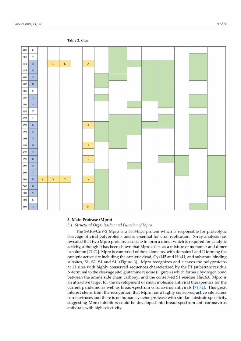
Table 2. Spike RBD sequence of SARS-CoV-2 Wuhan-Hu-1 with highlighted footprints of ACE2(pale blue), variants of concern Alpha, Beta, Gamma, Delta and Omicron with positions of mutations(peach), and 8 mAbs with binding epitopes on the RBM (pale green) [42,66,67]. (AA#: amino acidnumbering of the spike protein.).
Viruses 2022, 14, x 8 of 28
Table 2. Spike RBD sequence of SARS-CoV-2 Wuhan-Hu-1 with highlighted footprints of ACE2
(pale blue), variants of concern Alpha, Beta, Gamma, Delta and Omicron with positions of mutations
(peach), and 8 mAbs with binding epitopes on the RBM (pale green) [42,66,67]. (AA#: amino acid
numbering of the spike protein.).
AA# ACE2 Alpha Beta Gamma Delta (+) Omicron Etesevimab Bamlanivimab Cilgavimab Tixagevimab Casirivimab Imdevimab Regdanvimab Bebtelovimab
417 K
N T N N
438 S
439 N
440 N K
441 L
442 D
443 S
444 K
445 V
446 G S
447 G
448 N
449 Y
450 N
451 Y
452 L R
453 Y
454 R
455 L
456 F
457 R
458 K
459 S
460 N
461-469
470 T
471 E
472 I
473 Y
474 Q
475 A
476 G
477 S N
478 T K K
479 P
480 C
481 N
Viruses 2022, 14, 961 9 of 27
Table 2. Cont.
Viruses 2022, 14, x 9 of 28
482 G
483 V
484 E K K A
485 G
486 F
487 N
488 C
489 Y
490 F
491 P
492 L
493 Q K
494 S
495 Y
496 G S
497 F
498 Q R
499 P
500 T
501 N Y Y Y Y
502 G
503 V
504 G
505 Y H
2.5. Conclusions and Future Directions
The RBD is highly immunogenic but is prone to accumulate mutations. Under strong
immune selection, escape mutants rapidly arise as we observe with the Omicron variant
[14,42]. Further, as we prepare for emerging coronaviruses with “pandemic potential”,
there is limited opportunity for having Abs with cross-reactivity to SARS-CoV-2 and other
coronaviruses due to the high diversity in RBD sequences. The membrane-anchored S2
subunit, which contains the membrane fusion machinery exhibits a higher level of protein
sequence conservation across coronavirus S proteins. Several teams have identified a class
of S2-targeting Abs with broad reactivity towards several human betacoronaviruses from
distinct subgenera, and characterized their antiviral activity, epitope and in vivo protec-
tive efficacy [52,56,68–70]. S2-specific mAbs can prove to be very useful, and their binding
could inhibit conformational changes necessary for membrane fusion to occur. S2-specific
mAbs, however, may not be sufficiently potent in viral neutralization, and enhanced Fc
effector function might be necessary to achieve in vivo efficacy [52]. These mAbs can be
used in combination with clinically proven neutralizing anti-SARS-CoV-2 therapies to
achieve a broad neutralization spectrum across all SARS-CoV-2 variants.
3. Main Protease (Mpro)
3.1. Structural Organization and Function of Mpro
3. Main Protease (Mpro)3.1. Structural Organization and Function of Mpro
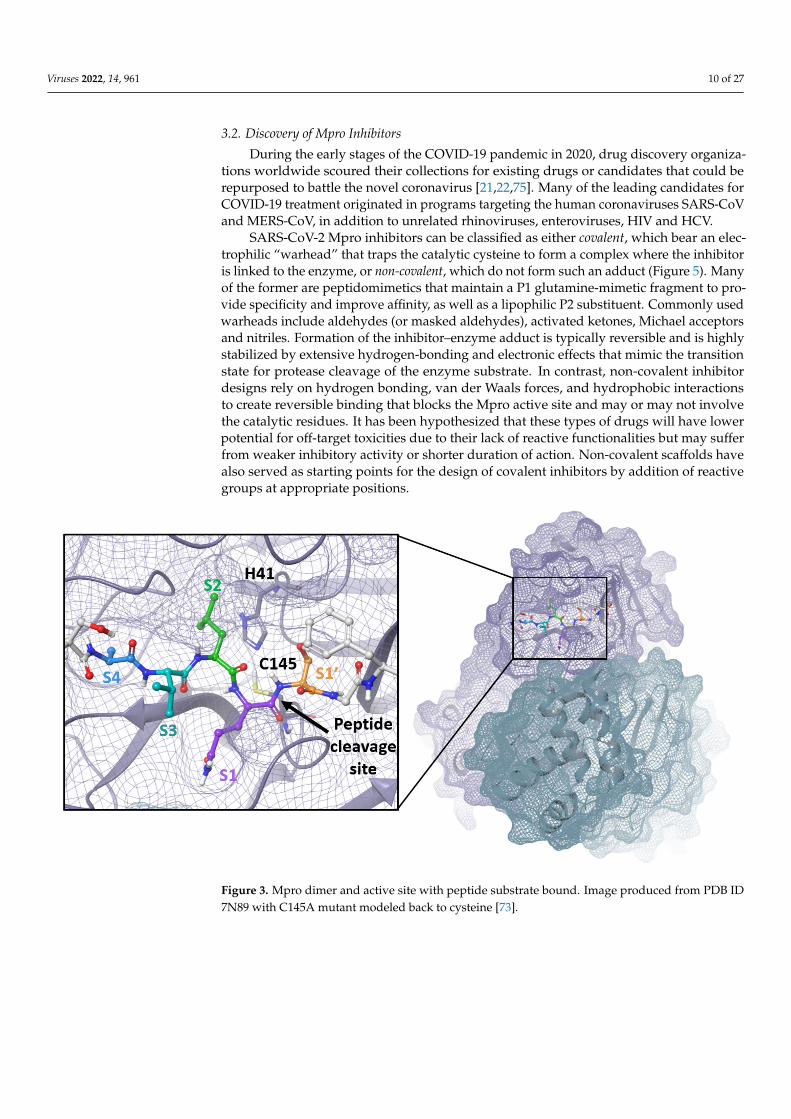
The SARS-CoV-2 Mpro is a 33.8-kDa protein which is responsible for proteolyticcleavage of viral polyproteins and is essential for viral replication. X-ray analysis hasrevealed that two Mpro proteins associate to form a dimer which is required for catalyticactivity, although it has been shown that Mpro exists as a mixture of monomer and dimerin solution [71,72]. Mpro is composed of three domains, with domains I and II forming thecatalytic active site including the catalytic dyad, Cys145 and His41, and substrate-bindingsubsites, S1, S2, S4 and S1′ (Figure 3). Mpro recognizes and cleaves the polyproteinsat 11 sites with highly conserved sequences characterized by the P1 (substrate residueN-terminal to the cleavage site) glutamine residue (Figure 4) which forms a hydrogen bondbetween the amide side chain carbonyl and the conserved S1 residue His163. Mpro isan attractive target for the development of small molecule antiviral therapeutics for thecurrent pandemic as well as broad-spectrum coronavirus antivirals [71,72]. This greatinterest stems from the recognition that Mpro has a highly conserved active site acrosscoronaviruses and there is no human cysteine protease with similar substrate specificity,suggesting Mpro inhibitors could be developed into broad-spectrum anti-coronavirusantivirals with high selectivity.
Viruses 2022, 14, 961 10 of 27
3.2. Discovery of Mpro Inhibitors
During the early stages of the COVID-19 pandemic in 2020, drug discovery organiza-tions worldwide scoured their collections for existing drugs or candidates that could berepurposed to battle the novel coronavirus [21,22,75]. Many of the leading candidates forCOVID-19 treatment originated in programs targeting the human coronaviruses SARS-CoVand MERS-CoV, in addition to unrelated rhinoviruses, enteroviruses, HIV and HCV.
SARS-CoV-2 Mpro inhibitors can be classified as either covalent, which bear an elec-trophilic “warhead” that traps the catalytic cysteine to form a complex where the inhibitoris linked to the enzyme, or non-covalent, which do not form such an adduct (Figure 5). Manyof the former are peptidomimetics that maintain a P1 glutamine-mimetic fragment to pro-vide specificity and improve affinity, as well as a lipophilic P2 substituent. Commonly usedwarheads include aldehydes (or masked aldehydes), activated ketones, Michael acceptorsand nitriles. Formation of the inhibitor–enzyme adduct is typically reversible and is highlystabilized by extensive hydrogen-bonding and electronic effects that mimic the transitionstate for protease cleavage of the enzyme substrate. In contrast, non-covalent inhibitordesigns rely on hydrogen bonding, van der Waals forces, and hydrophobic interactionsto create reversible binding that blocks the Mpro active site and may or may not involvethe catalytic residues. It has been hypothesized that these types of drugs will have lowerpotential for off-target toxicities due to their lack of reactive functionalities but may sufferfrom weaker inhibitory activity or shorter duration of action. Non-covalent scaffolds havealso served as starting points for the design of covalent inhibitors by addition of reactivegroups at appropriate positions.
Figure 3. Mpro dimer and active site with peptide substrate bound. Image produced from PDB ID7N89 with C145A mutant modeled back to cysteine [73].

Viruses 2022, 14, 961 11 of 27
Figure 4. The consensus recognition sequence cleaved by SARS-CoV-2 (Uniprot code P0DTD1) Mpro.The cleavage site is marked by the blue arrow. Image generated by WebLogo [74].
The Ugi class of non-covalent inhibitors originated in a high-throughput screen of theNIH sample library in search of novel SARS-CoV Mpro inhibitors in the early 2000s. Thesimple dipeptide structure is easily assembled using the Ugi multicomponent couplingmethodology, which allowed for rapid SAR exploration. The lead compound ML 188 (1)was later shown to have similar and perhaps improved activity against SARS-CoV-2 Mpro(IC50 = 2.5 µM). The benzotriazole class was also discovered by screens targeting SARS-CoV [76,77]. In response to the COVID-19 pandemic, work on this series was refocusedon SARS-CoV-2, leading to improved analogs such as CCF981 (2) that exhibited severalorders of magnitude improvement in Mpro potency (IC50 = 68 nM) with submicromolarantiviral activity in infected Vero E6 cells (CPE EC50 = 497 nM). Perampanel, an anti-epileptic agent, was found to be a weak Mpro inhibitor through a repurposing effort thatevaluated approximately 2000 known drugs [78]. Structure-guided optimization afforded aperampanel analog (3), which has a SARS-CoV-2 Mpro inhibitory potency of IC50 = 0.17 µMand antiviral EC50 = ~1 µM in a plaque assay in Vero E6 cells with low cytotoxicity. Theisoquinoline-containing Mpro inhibitor (4) is a lead compound discovered through a uniquecrowd-sourced initiative called the COVID-19 Moonshot program. Scientists worldwidewere able to contribute designs through a web portal, and donations enabled the synthesisand testing of compounds [79]. Data and x-ray structures provided by partners includingthe Diamond Light Source and the Weizmann Institute were made publicly available inreal time. Lead compound 4 is a potent inhibitor of SARS-CoV-2 Mpro (IC50 = 37 nM) anddemonstrates antiviral activity against several circulating variants in HeLa-ACE2 cells [80].The consortium is hoping to advance this compound or an analog to clinical development.Shionogi has reported on S-217622 (5), an orally delivered Mpro inhibitor with activityagainst SARS-CoV-2 variants (EC50 = 23.9–61.7 nM, 293T-ACE2-TMPRSS2 cells), that hascompleted Phase 2a studies [81,82].
GC376 (6) is the prodrug form of a peptidomimetic drug GC373 (7), which wasoriginally developed for feline coronavirus (feline infectious peritonitis virus) infections [83].Anivive, a biotech company focused on animal healthcare, is reportedly working withpartners to expand use of the compound to treat COVID-19 infections in humans [84].The chemical structure of GC376 features a gamma-lactam as a P1 glutamine bioisostereand an aldehyde-bisulfite adduct as a latent covalent warhead. X-ray structures establishthat the isobutyl sidechain occupies the lipophilic S2 site and that key hydrogen-bondinginteractions are made between the inhibitor and backbone amides of Glu166, His164and Gln189. The active form 7 exhibits moderate potency against SARS-CoV-2 Mpro(IC50 = 0.40 µM), similar to its level of activity against FIPV Mpro (IC50 = 0.49 µM) butweaker than observed with SARS-CoV (IC50 = 0.070 µM). Plaque assays in infected VeroE6 cells demonstrate that both compounds 6 and 7 have antiviral activity (EC50 = 0.92and 1.5 µM, respectively). In a virus yield reduction assay, both compounds decreasedSARS-CoV-2 titers by approximately three orders of magnitude [83].
Viruses 2022, 14, 961 12 of 27
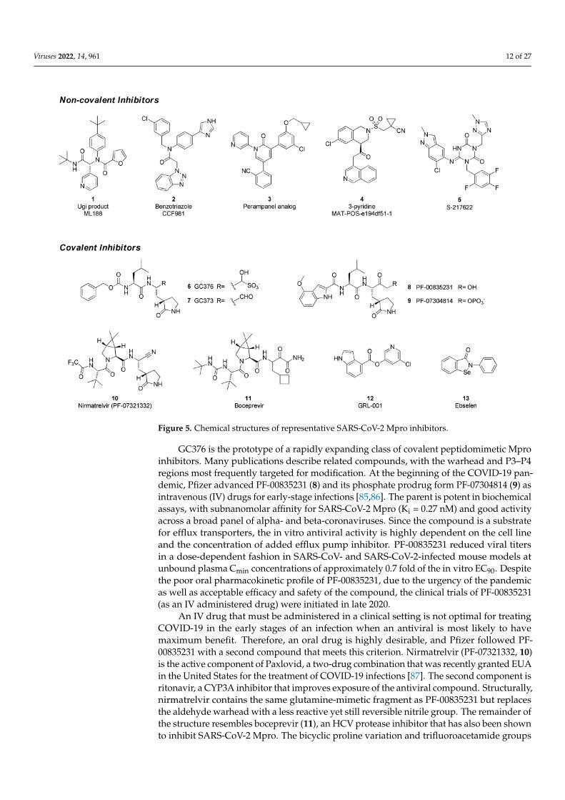
Figure 5. Chemical structures of representative SARS-CoV-2 Mpro inhibitors.
GC376 is the prototype of a rapidly expanding class of covalent peptidomimetic Mproinhibitors. Many publications describe related compounds, with the warhead and P3–P4regions most frequently targeted for modification. At the beginning of the COVID-19 pan-demic, Pfizer advanced PF-00835231 (8) and its phosphate prodrug form PF-07304814 (9) asintravenous (IV) drugs for early-stage infections [85,86]. The parent is potent in biochemicalassays, with subnanomolar affinity for SARS-CoV-2 Mpro (Ki = 0.27 nM) and good activityacross a broad panel of alpha- and beta-coronaviruses. Since the compound is a substratefor efflux transporters, the in vitro antiviral activity is highly dependent on the cell lineand the concentration of added efflux pump inhibitor. PF-00835231 reduced viral titersin a dose-dependent fashion in SARS-CoV- and SARS-CoV-2-infected mouse models atunbound plasma Cmin concentrations of approximately 0.7 fold of the in vitro EC90. Despitethe poor oral pharmacokinetic profile of PF-00835231, due to the urgency of the pandemicas well as acceptable efficacy and safety of the compound, the clinical trials of PF-00835231(as an IV administered drug) were initiated in late 2020.
An IV drug that must be administered in a clinical setting is not optimal for treatingCOVID-19 in the early stages of an infection when an antiviral is most likely to havemaximum benefit. Therefore, an oral drug is highly desirable, and Pfizer followed PF-00835231 with a second compound that meets this criterion. Nirmatrelvir (PF-07321332, 10)is the active component of Paxlovid, a two-drug combination that was recently granted EUAin the United States for the treatment of COVID-19 infections [87]. The second component isritonavir, a CYP3A inhibitor that improves exposure of the antiviral compound. Structurally,nirmatrelvir contains the same glutamine-mimetic fragment as PF-00835231 but replacesthe aldehyde warhead with a less reactive yet still reversible nitrile group. The remainder ofthe structure resembles boceprevir (11), an HCV protease inhibitor that has also been shownto inhibit SARS-CoV-2 Mpro. The bicyclic proline variation and trifluoroacetamide groups

Viruses 2022, 14, 961 13 of 27
were necessary to improve permeability and oral absorption. Nirmatrelvir has slightlylower affinity for SARS-CoV-2 Mpro (Ki = 3.11 nM) than the earlier IV drug candidate 8 butmaintains similar antiviral activity, with an EC50 = 78 nM in A549-ACE2 cells. Clinical datarevealed high efficacy (up to 89% reduction in risk of hospitalization or death) in at-riskpatients who began treatment within 5 days of infection [88]. Whereas GC376, nirmatrelvirand related compounds are reversible covalent inhibitors, GRL-001 (12) and ebselen (13) arerepresentatives of a category of non-peptidic small molecules that react with the catalyticcysteine in an irreversible and time-dependent manner to produce an inactivated enzyme.
Several additional SARS-CoV-2 Mpro inhibitors have been developed for which clini-cal trials have been initiated or preclinical data have been presented, but their chemicalstructures have not been disclosed yet. Enanta has released preclinical data for EDP-235,an oral Mpro inhibitor with a biochemical IC50 of 5.8 nM and antiviral activity in primaryhuman airway epithelial cells with an EC90 of 33 nM [89]. Cocrystal Pharma has announcedplans to advance two Mpro inhibitors, CDI-988 and CDI-873, to clinical trials in 2022 [90].Aligos has reported preclinical data for ALG-097111, which demonstrated a biochemicalIC50 of 7 nM and antiviral EC50 of 200 nM (A549-ACE2 cells) [91].
3.3. Conclusions and Future Directions
The discovery and advancement of the first-generation SARS-CoV-2 Mpro inhibitorsto clinical trials and the approval of Paxlovid for the treatment of COVID-19 infectionsare unprecedented in their speed, with Paxlovid demonstrating significant clinical benefitin reducing severe illness, hospitalization, and death. In the future, Mpro inhibitorsthat do not require pharmacokinetic boosting may enable broader utilization of theseinhibitors with less risk for drug–drug interactions and use for pre- and post-exposureprophylaxis for high-risk patient populations. Previous experience with direct-actingantivirals against other viruses such as HIV and HCV suggests that combinations of drugswith different mechanisms of action may provide higher efficacy and prevent the emergenceof resistance, an area of research that will become clearer as the first Mpro inhibitor seesmore widespread use.
4. RNA-Dependent RNA Polymerase (RdRp)4.1. Structural Organization and Function of RdRp
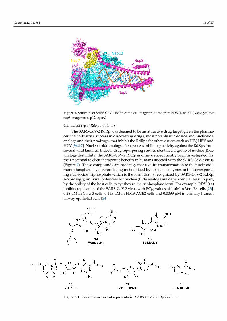
Faithful replication of the coronavirus genome is a complicated process that includesRNA synthesis, proofreading, template switching and 5′-capping, resulting in the formationof genome-length (+)-strand RNA for incorporation into newly formed virions as well as avariety of subgenomic (+)-strand RNAs that are translated into structural and accessoryproteins [92]. The core SARS-CoV-2 RdRp consists of the 934 amino acid nsp12 protein,the 85 amino acid nsp7 protein and the 200 amino acid nsp8 protein. RNA synthesisoccurs within the nsp12 protein but only when the nsp7 and nsp8 accessory proteins arecomplexed with it in a 1:1:2 stoichiometry [93,94]. Cryo-electron microscopy studies haveshown that the polymerase domain of nsp12 resembles a human “right hand” consisting offingers, thumb and palm subdomains. The two copies of nsp8 bind to nsp12 on opposingsides of the RNA-binding cleft. The C-terminal domain of one copy of nsp8 also binds tonsp7, whereas the C-terminal domain of the second nsp8 copy adopts a different fold andinteracts with nsp12 directly. When long duplex RNA is present, the N-terminal domains ofboth nsp8 copies form long helical extensions that contact the RNA duplex product. Theseextensions likely act as sliding poles to enhance the processivity of the RdRp core complexand enable it to efficiently replicate the long genome of the coronaviruses (Figure 6) [94,95].Additional proteins are thought to associate with the core RdRp complex to perform thehelicase and proofreading functions.
Viruses 2022, 14, 961 14 of 27
Figure 6. Structure of SARS-CoV-2 RdRp complex. Image produced from PDB ID 6YYT. (Nsp7: yellow;nsp8: magenta; nsp12: cyan.)
4.2. Discovery of RdRp Inhibitors
The SARS-CoV-2 RdRp was deemed to be an attractive drug target given the pharma-ceutical industry’s success in discovering drugs, most notably nucleoside and nucleotideanalogs and their prodrugs, that inhibit the RdRps for other viruses such as HIV, HBV andHCV [96,97]. Nucleos(t)ide analogs often possess inhibitory activity against the RdRps fromseveral viral families. Indeed, drug repurposing studies identified a group of nucleos(t)ideanalogs that inhibit the SARS-CoV-2 RdRp and have subsequently been investigated fortheir potential to elicit therapeutic benefits in humans infected with the SARS-CoV-2 virus(Figure 7). These compounds are prodrugs that require transformation to the nucleotidemonophosphate level before being metabolized by host cell enzymes to the correspond-ing nucleotide triphosphate which is the form that is recognized by SARS-CoV-2 RdRp.Accordingly, antiviral potencies for nucleos(t)ide analogs are dependent, at least in part,by the ability of the host cells to synthesize the triphosphate form. For example, RDV (14)inhibits replication of the SARS-CoV-2 virus with EC50 values of 1 µM in Vero E6 cells [23],0.28 µM in Calu-3 cells, 0.115 µM in H549-ACE2 cells and 0.0099 µM in primary humanairway epithelial cells [24].
Figure 7. Chemical structures of representative SARS-CoV-2 RdRp inhibitors.
Viruses 2022, 14, 961 15 of 27
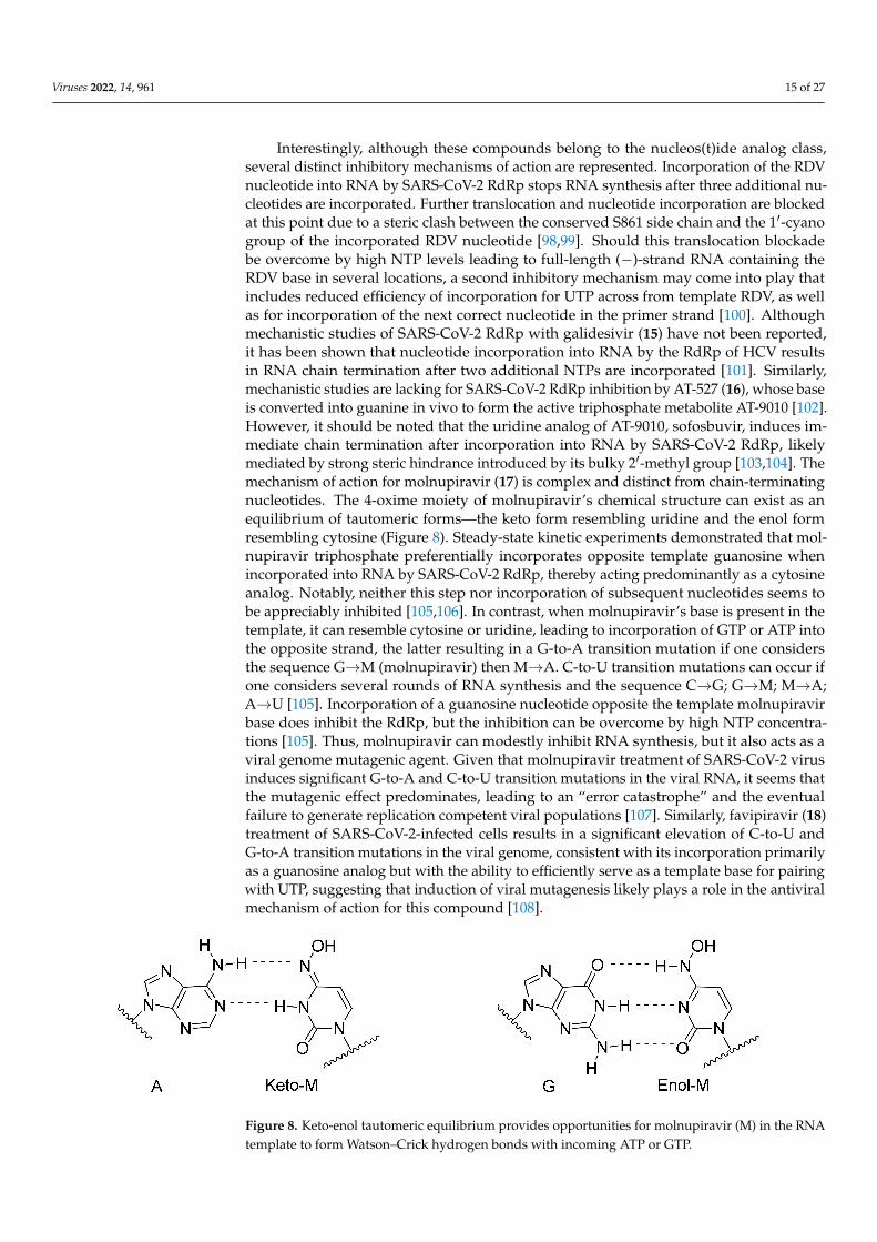
Interestingly, although these compounds belong to the nucleos(t)ide analog class,several distinct inhibitory mechanisms of action are represented. Incorporation of the RDVnucleotide into RNA by SARS-CoV-2 RdRp stops RNA synthesis after three additional nu-cleotides are incorporated. Further translocation and nucleotide incorporation are blockedat this point due to a steric clash between the conserved S861 side chain and the 1′-cyanogroup of the incorporated RDV nucleotide [98,99]. Should this translocation blockadebe overcome by high NTP levels leading to full-length (−)-strand RNA containing theRDV base in several locations, a second inhibitory mechanism may come into play thatincludes reduced efficiency of incorporation for UTP across from template RDV, as wellas for incorporation of the next correct nucleotide in the primer strand [100]. Althoughmechanistic studies of SARS-CoV-2 RdRp with galidesivir (15) have not been reported,it has been shown that nucleotide incorporation into RNA by the RdRp of HCV resultsin RNA chain termination after two additional NTPs are incorporated [101]. Similarly,mechanistic studies are lacking for SARS-CoV-2 RdRp inhibition by AT-527 (16), whose baseis converted into guanine in vivo to form the active triphosphate metabolite AT-9010 [102].However, it should be noted that the uridine analog of AT-9010, sofosbuvir, induces im-mediate chain termination after incorporation into RNA by SARS-CoV-2 RdRp, likelymediated by strong steric hindrance introduced by its bulky 2′-methyl group [103,104]. Themechanism of action for molnupiravir (17) is complex and distinct from chain-terminatingnucleotides. The 4-oxime moiety of molnupiravir’s chemical structure can exist as anequilibrium of tautomeric forms—the keto form resembling uridine and the enol formresembling cytosine (Figure 8). Steady-state kinetic experiments demonstrated that mol-nupiravir triphosphate preferentially incorporates opposite template guanosine whenincorporated into RNA by SARS-CoV-2 RdRp, thereby acting predominantly as a cytosineanalog. Notably, neither this step nor incorporation of subsequent nucleotides seems tobe appreciably inhibited [105,106]. In contrast, when molnupiravir’s base is present in thetemplate, it can resemble cytosine or uridine, leading to incorporation of GTP or ATP intothe opposite strand, the latter resulting in a G-to-A transition mutation if one considersthe sequence G→M (molnupiravir) then M→A. C-to-U transition mutations can occur ifone considers several rounds of RNA synthesis and the sequence C→G; G→M; M→A;A→U [105]. Incorporation of a guanosine nucleotide opposite the template molnupiravirbase does inhibit the RdRp, but the inhibition can be overcome by high NTP concentra-tions [105]. Thus, molnupiravir can modestly inhibit RNA synthesis, but it also acts as aviral genome mutagenic agent. Given that molnupiravir treatment of SARS-CoV-2 virusinduces significant G-to-A and C-to-U transition mutations in the viral RNA, it seems thatthe mutagenic effect predominates, leading to an “error catastrophe” and the eventualfailure to generate replication competent viral populations [107]. Similarly, favipiravir (18)treatment of SARS-CoV-2-infected cells results in a significant elevation of C-to-U andG-to-A transition mutations in the viral genome, consistent with its incorporation primarilyas a guanosine analog but with the ability to efficiently serve as a template base for pairingwith UTP, suggesting that induction of viral mutagenesis likely plays a role in the antiviralmechanism of action for this compound [108].
Figure 8. Keto-enol tautomeric equilibrium provides opportunities for molnupiravir (M) in the RNAtemplate to form Watson–Crick hydrogen bonds with incoming ATP or GTP.
Viruses 2022, 14, 961 16 of 27
Remdesivir was approved by the US FDA in October, 2020 for the treatment ofCOVID-19 infections in hospitalized subjects. The approval of this intravenously ad-ministered drug was based on three clinical studies, the ACTT1 study [109] and two studiessponsored by Gilead [110,111], but omitted results from a study conducted in China [112].Shortly after approval, the results from the much larger Solidarity trial were published [113].A meta-analysis of four of these randomized controlled trials with 7334 patients concludedthat subjects treated with RDV were more likely to demonstrate recovery and were associ-ated with higher rates of hospital discharge, but there was no significant reduction in meantime to clinical improvement or mortality [114].
Molnupiravir exhibits an in vitro antiviral EC50 value of 0.3 µM against SARS-CoV-2virus when tested in Vero cells [107] as well as broad-spectrum antiviral activity againstseasonal coronaviruses [115]. In preclinical models, molnupiravir inhibits SARS-CoV-2replication in the Syrian hamster model [116,117] and mice [107] and blocks SARS-CoV-2transmission in ferrets [118]. Given its mechanism of action as a viral mutagenesis agent,molnuprivar has been closely scrutinized for its potential to elicit DNA mutagenesis inhost cells and tissues. One lab has found that molnupiravir displays host mutationalactivity in an animal cell culture assay [119] although molnupiravir was negative in a28 day transgenic rodent mutagenicity study [28]. In addition, this orally administeredcompound was safe and well tolerated in Phase 1 studies conducted in healthy humanvolunteers [27]. A Phase 3 clinical study of 1408 unvaccinated participants demonstratedthat molnupiravir treatment for 5 days reduced the risk of hospitalization and death by30% [120]. Molnupiravir received EUA by the US FDA in December, 2021 for treatment ofmild-to-moderate COVID-19 infections. Molnupiravir is not authorized for use in patientsyounger than 18 years of age because it may affect bone growth and cartilage formationand it is also important to recognize the drug may cause fetal harm when administered topregnant individuals [28].
Galidesivir inhibits replication of SARS-CoV and MERS-CoV viruses in Vero E6 cellswith EC50 values of 57.7 and 68.4 µM, respectively [101]. Early administration of galidesivirin a COVID-19 animal model reduced the viral burden and pathology in lung tissue [121].A small Phase I study in COVID-19 patients demonstrated that galidesivir was safe and gen-erally well tolerated, but it did not show signs of significant clinical benefit. Consequently,the sponsor has discontinued plans to develop galidesivir for treatment of COVID-19 [121].AT-527 was initially developed as a drug to treat HCV infections but its EC90 value of0.47 µM in a virus yield reduction assay against SARS-CoV-2 in primary human airwayepithelial cells suggested potential utility for treating COVID-19 [102]. Unfortunately,AT-527 failed to meet its primary goal of reducing SARS-CoV-2 RNA at various intervals ina Phase 2 clinical trial in subjects with mild or moderate COVID-19 in the outpatient setting,leading the sponsor to update its clinical development strategy [122]. The orally admin-istered drug favipiravir, approved to treat novel influenza in Japan, inhibits SARS-CoV-2replication and the generation of cytopathic effects in Vero E6 cells with EC50 values of 207and 118 µM, respectively [108]. Russia has approved favipiravir for treating COVID-19infections and several other countries such as Mexico, India and Malaysia have grantedEUA for this indication. A meta-analysis on clinical studies that evaluated the efficacy andsafety of favipiravir as a treatment for COVID-19 found that there was a significant clinicaland radiological improvement following treatment with favipiravir in comparison to thestandard of care but with no significant differences on viral clearance, oxygen supportrequirement and side effect profiles [123].
4.3. Conclusions and Future Directions
Drug repurposing studies initiated at the beginning of the COVID-19 pandemic iden-tified at least two compounds from the nucleos(t)ide analog inhibitor class, remdesivirand molnupiravir, with sufficient antiviral activity against the SARS-CoV-2 virus to meritapproval or EUA by the US FDA for treating COVID-19 infections in select populations.However, both drugs have their limitations and so research is currently underway to
Viruses 2022, 14, 961 17 of 27
discover new RdRp inhibitors including those from the non-nucleoside inhibitor classwith improved safety and efficacy properties. Non-nucleosides are compounds that binddirectly to the viral RdRp without the need for chemical transformation to the nucleotidetriphosphate form and are represented in drug therapies to treat HIV and cure HCV in-fections [124,125]. Non-nucleosides often possess high selectivity for their viral RdRpsand so it is understandable that no potent non-nucleoside inhibitors of SARS-CoV-2 RdRpemerged from early drug repurposing studies. Discovering non-nucleoside inhibitors ofSARS-CoV-2 RdRp will require screening and medicinal chemistry optimization—activitiesat the very early stages of the drug discovery process. Fortunately, recent advances in DNAencoded libraries [126] and virtual screening [127,128] offer the ability to rapidly screenbillions of molecules and should facilitate that first step on the road towards the discoveryof highly selective non-nucleoside SARS-CoV-2 RdRp inhibitors.
5. Papain-Like Protease (PLpro)5.1. Structural Organization and Functions of PLpro
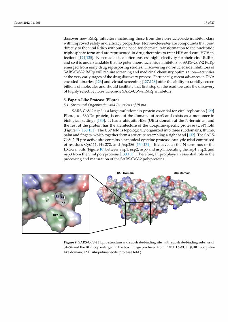
SARS-CoV-2 nsp3 is a large multidomain protein essential for viral replication [129].PLpro, a ~36 kDa protein, is one of the domains of nsp3 and exists as a monomer inbiological settings [130]. It has a ubiquitin-like (UBL) domain at the N-terminus, andthe rest of the protein has the architecture of the ubiquitin-specific protease (USP) fold(Figure 9) [130,131]. The USP fold is topologically organized into three subdomains, thumb,palm and fingers, which together form a structure resembling a right hand [132]. The SARS-CoV-2 PLpro active site contains a canonical cysteine protease catalytic triad comprisedof residues Cys111, His272, and Asp286 [130,131]. It cleaves at the N terminus of theLXGG motifs (Figure 10) between nsp1, nsp2, nsp3 and nsp4, liberating the nsp1, nsp2, andnsp3 from the viral polyproteins [130,133]. Therefore, PLpro plays an essential role in theprocessing and maturation of the SARS-CoV-2 polyproteins.
Figure 9. SARS-CoV-2 PLpro structure and substrate-binding site, with substrate-binding subsites ofS1–S4 and the BL2 loop enlarged in the box. Image produced from PDB ID 6WUU. (UBL: ubiquitin-like domain; USP: ubiquitin-specific protease fold.)
Viruses 2022, 14, 961 18 of 27
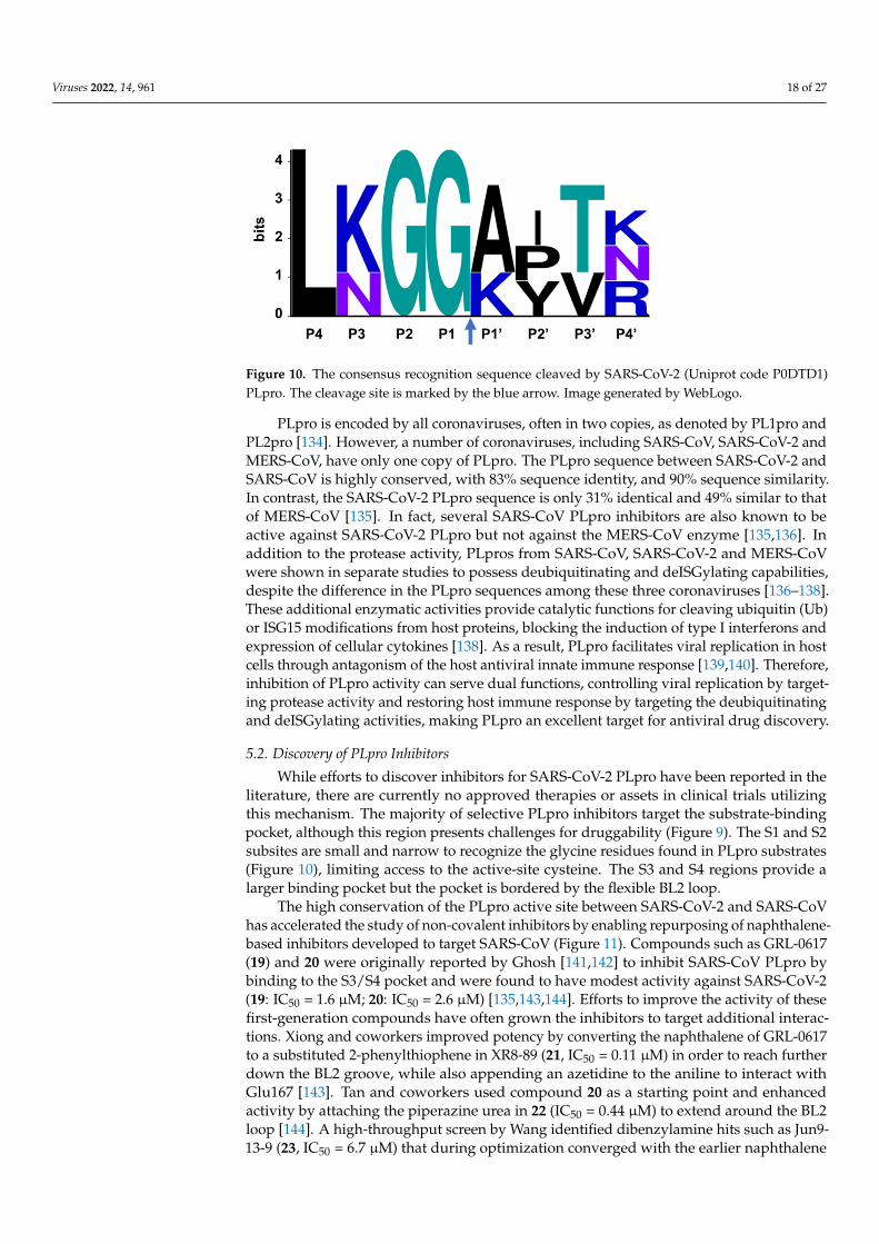
Figure 10. The consensus recognition sequence cleaved by SARS-CoV-2 (Uniprot code P0DTD1)PLpro. The cleavage site is marked by the blue arrow. Image generated by WebLogo.
PLpro is encoded by all coronaviruses, often in two copies, as denoted by PL1pro andPL2pro [134]. However, a number of coronaviruses, including SARS-CoV, SARS-CoV-2 andMERS-CoV, have only one copy of PLpro. The PLpro sequence between SARS-CoV-2 andSARS-CoV is highly conserved, with 83% sequence identity, and 90% sequence similarity.In contrast, the SARS-CoV-2 PLpro sequence is only 31% identical and 49% similar to thatof MERS-CoV [135]. In fact, several SARS-CoV PLpro inhibitors are also known to beactive against SARS-CoV-2 PLpro but not against the MERS-CoV enzyme [135,136]. Inaddition to the protease activity, PLpros from SARS-CoV, SARS-CoV-2 and MERS-CoVwere shown in separate studies to possess deubiquitinating and deISGylating capabilities,despite the difference in the PLpro sequences among these three coronaviruses [136–138].These additional enzymatic activities provide catalytic functions for cleaving ubiquitin (Ub)or ISG15 modifications from host proteins, blocking the induction of type I interferons andexpression of cellular cytokines [138]. As a result, PLpro facilitates viral replication in hostcells through antagonism of the host antiviral innate immune response [139,140]. Therefore,inhibition of PLpro activity can serve dual functions, controlling viral replication by target-ing protease activity and restoring host immune response by targeting the deubiquitinatingand deISGylating activities, making PLpro an excellent target for antiviral drug discovery.
5.2. Discovery of PLpro Inhibitors
While efforts to discover inhibitors for SARS-CoV-2 PLpro have been reported in theliterature, there are currently no approved therapies or assets in clinical trials utilizingthis mechanism. The majority of selective PLpro inhibitors target the substrate-bindingpocket, although this region presents challenges for druggability (Figure 9). The S1 and S2subsites are small and narrow to recognize the glycine residues found in PLpro substrates(Figure 10), limiting access to the active-site cysteine. The S3 and S4 regions provide alarger binding pocket but the pocket is bordered by the flexible BL2 loop.
The high conservation of the PLpro active site between SARS-CoV-2 and SARS-CoVhas accelerated the study of non-covalent inhibitors by enabling repurposing of naphthalene-based inhibitors developed to target SARS-CoV (Figure 11). Compounds such as GRL-0617(19) and 20 were originally reported by Ghosh [141,142] to inhibit SARS-CoV PLpro bybinding to the S3/S4 pocket and were found to have modest activity against SARS-CoV-2(19: IC50 = 1.6 µM; 20: IC50 = 2.6 µM) [135,143,144]. Efforts to improve the activity of thesefirst-generation compounds have often grown the inhibitors to target additional interac-tions. Xiong and coworkers improved potency by converting the naphthalene of GRL-0617to a substituted 2-phenylthiophene in XR8-89 (21, IC50 = 0.11 µM) in order to reach furtherdown the BL2 groove, while also appending an azetidine to the aniline to interact withGlu167 [143]. Tan and coworkers used compound 20 as a starting point and enhancedactivity by attaching the piperazine urea in 22 (IC50 = 0.44 µM) to extend around the BL2loop [144]. A high-throughput screen by Wang identified dibenzylamine hits such as Jun9-13-9 (23, IC50 = 6.7 µM) that during optimization converged with the earlier naphthalene

Viruses 2022, 14, 961 19 of 27
inhibitors to yield more potent compounds such as Jun9-75-4 (24, IC50 = 0.62 µM) [145].While activity gains have recently been made in this class of non-covalent PLpro inhibitors,further improvements in antiviral potency will be required to generate candidates withclinical efficacy.
Figure 11. Chemical structures of representative SARS-CoV-2 PLpro inhibitors.
Efforts to produce covalent SARS-CoV-2 PLpro inhibitors have also been described.Olsen and coworkers used a combinatorial library of fluorogenic tetrapeptide substratesto identify the optimal peptide sequences for PLpro reactivity [146]. Replacement of thechromophore with an α,β-unsaturated ester warhead provided inhibitors VIR250 (25) andVIR251 (26). Co-crystal structures highlighted the key interactions with the protease andverified the covalent inhibition mechanism. Li and coworkers used 19 as the startingpoint for their covalent inhibitor and appended a sulfonium-tethered peptide to generate apeptide drug conjugate [147]. Both strategies incorporated the glycine residues of the PLprosubstrates to navigate the S1 and S2 sites. Further advancements in covalent strategieswill need to overcome the modest activity and challenging peptidic properties of theseearly compounds.
High-throughput screens of known bioactive compounds have led to reports thata variety of other classes of compounds inhibit SARS-CoV-2 PLpro. Organoseleniumcompounds such as Ebselen (13) were reported to inhibit PLpro [148], but others have foundthese act as non-selective modifiers of cysteine, including the cysteine at the active site ofPLpro [149]. Validation studies of reported quinone and nucleoside hits also suggest theseclasses of compounds are non-specific inhibitors [150]. The limited antiviral activity andpoor selectivity of these hits are significant challenges to advancing them into useful toolsand highlight the importance of confirming PLpro screening results across multiple assays.
5.3. Conclusions and Future Directions
SARS-CoV-2 PLpro is a multifunctional enzyme with protease, deubiquitinase, anddeISGylating activities, with the latter two activities involved in blocking the expression oftype I interferons in the infected cells. In principle, targeting the activities of PLpro maynot only inhibit viral replication but also block the viral-mediated evasion of host innateimmunity. One recent report presented data that support this hypothesis: GRL-0617 couldinhibit SARS-CoV-2 replication as well as maintain the antiviral interferon signaling in

Viruses 2022, 14, 961 20 of 27
the infected cells [136]. The discovery of potent PLpro inhibitors has been challengingdue to the apparent lack of well-defined binding pockets at its substrate-binding pocket.However, the growing number of structural studies conducted with PLpro to identifypotential binding pockets and the increasing interest in optimizing known PLpro inhibitorsbased on structural data would certainly facilitate the understanding of the druggability ofPLpro and advance the drug discovery for this antiviral target.
6. Summary
In this review, we discuss the current status and future strategies for the discovery ofsmall- and large-molecule antiviral therapeutics for COVID-19 and, potentially, emergingcoronaviruses. There is an increasing number of inhibitors reported to have anti-SARS-CoV-2 activity in vitro and in vivo. Several of these inhibitors [e.g., some Mpro or RdRpinhibitors, and at least two examples of anti-S mAbs (sotrovimab and bebtelovimab)] havealso been shown to be efficacious for a reasonably wide spectrum of coronaviruses and/orCOVID-19 variants [87,98,99,151]. This is not surprising due to the high protein sequenceconservation among coronaviruses and variants at the catalytic sites and substrate-bindingsites of Mpro and RdRp, as well as parts of the S protein that are important for viralentry. PLpro protein sequence is less conserved among coronaviruses when compared tothose of Mpro and RdRp. The breadth of the antiviral spectrum of an optimized PLproinhibitor remains to be determined. This review has focused on small molecules and mAbsthat directly target viral proteins. In addition to these drug modalities, other therapeuticapproaches (e.g., CRISPR, RNAi, or antisense oligonucleotide) may also prove to be usefulfor COVID-19 therapies.
Resistance generation during antiviral therapy can occur during treatment for chronicviral infections such as HIV and HCV. With the short duration of COVID-19 antiviraltherapy, development of drug resistance to small molecule inhibitors may not be an issue.Should drug resistance become a concern, the availability of SARS-CoV-2 inhibitors tar-geting different viral proteins will allow combination therapy to minimize the generationof resistance.
Drug discovery is progressing at lightning speed to address the urgent need of theCOVID-19 crisis. We bear witness to unprecedented partnerships among industry, aca-demics and/or government to join forces in the discovery and development of COVID-19drugs. In addition, the basic research, drug discovery and clinical development frame-works established by decades of antiviral research on human pathogenic viruses enableinvestigators to quickly apply prior knowledge and cutting-edge technology to discovertreatments for COVID-19 infections. Recognizing the importance of antivirals, the USgovernment has pledged to support a new initiative called the Antiviral Program for Pan-demics by committing $3.2 billion in funding for research on COVID-19 antiviral drugdevelopment, as well as new drugs for viruses that could cause future pandemics [152].With the availability of several validated and druggable targets for SARS-CoV-2, as wellas the unprecedented partnerships and resources dedicated to COVID-19 drug discovery,we expect that a rich pipeline of COVID-19 drugs will emerge that will become importantweapons to tackle COVID-19 and future coronaviruses with pandemic risk.
Author Contributions: All authors wrote, reviewed, and edited the manuscript. All authors haveread and agreed to the published version of the manuscript.
Funding: The financial support for this research was provided by AbbVie.
Acknowledgments: We would like to thank Bernhard Sielaff for assistance in generating the figure forthe domain architecture of the SARS-CoV-2 S protein, Charles Hutchins for assistance in generatingthe figure for the structure of the RdRp complex, and Morteza Khabiri for assistance in generatingthe figure for the consensus recognition sequence of PLpro.
Conflicts of Interest: The authors declare no conflict of interest.
Viruses 2022, 14, 961 21 of 27
Disclosures: T.I.N., I.C., D.A.D., M.R.S., D.J.H., E.L.N. and W.M.K. are employees of AbbVie. J.S.was an employee of AbbVie at the time of writing. AbbVie participated in the interpretation of data,review, and approval of the publication.
References1. Wu, F.; Zhao, S.; Yu, B.; Chen, Y.-M.; Wang, W.; Song, Z.-G.; Hu, Y.; Tao, Z.-W.; Tian, J.-H.; Pei, Y.-Y.; et al. A new coronavirus
associated with human respiratory disease in China. Nature 2020, 579, 265–269. [CrossRef] [PubMed]2. Zhu, N.; Zhang, D.; Wang, W.; Li, X.; Yang, B.; Song, J.; Zhao, X.; Huang, B.; Shi, W.; Lu, R.; et al. A Novel Coronavirus from
Patients with Pneumonia in China, 2019. N. Engl. J. Med. 2020, 382, 727–733. [CrossRef] [PubMed]3. COVID-19 Dashboard by the Center for Systems Science and Engineering at Johns Hopkins University. Available online:
https://coronavirus.jhu.edu/map.html (accessed on 21 March 2022).4. Lee, N.; Hui, D.; Wu, A.; Chan, P.; Cameron, P.; Joynt, G.M.; Ahuja, A.; Yung, M.Y.; Leung, C.; To, K.; et al. A Major Outbreak of
Severe Acute Respiratory Syndrome in Hong Kong. N. Engl. J. Med. 2003, 348, 1986–1994. [CrossRef] [PubMed]5. Zaki, A.M.; Van Boheemen, S.; Bestebroer, T.M.; Osterhaus, A.D.M.E.; Fouchier, R.A.M. Isolation of a Novel Coronavirus from a
Man with Pneumonia in Saudi Arabia. N. Engl. J. Med. 2012, 367, 1814–1820. [CrossRef] [PubMed]6. Tregoning, J.S.; Flight, K.E.; Higham, S.L.; Wang, Z.; Pierce, B.F. Progress of the COVID-19 vaccine effort: Viruses, vaccines and
variants versus efficacy, effectiveness and escape. Nat. Rev. Immunol. 2021, 21, 626–636. [CrossRef]7. Arunachalam, P.S.; Scott, M.K.D.; Hagan, T.; Li, C.; Feng, Y.; Wimmers, F.; Grigoryan, L.; Trisal, M.; Edara, V.V.; Lai, L.; et al.
Systems vaccinology of the BNT162b2 mRNA vaccine in humans. Nature 2021, 596, 410–416. [CrossRef]8. Thomas, S.J.; Moreira, E.D., Jr.; Kitchin, N.; Absalon, J.; Gurtman, A.; Lockhart, S.; Perez, J.L.; Pérez Marc, G.; Polack, F.P.;
Zerbini, C.; et al. Safety and Efficacy of the BNT162b2 mRNA COVID-19 Vaccine through 6 Months. N. Engl. J. Med. 2021, 385,1761–1773. [CrossRef]
9. Goldberg, Y.; Mandel, M.; Bar-On, Y.M.; Bodenheimer, O.; Freedman, L.; Haas, E.J.; Milo, R.; Alroy-Preis, S.; Ash, N.; Huppert, A.Waning Immunity after the BNT162b2 Vaccine in Israel. N. Engl. J. Med. 2021, 385, e85. [CrossRef]
10. Pegu, A.; O’Connell, S.E.; Schmidt, S.D.; O’Dell, S.; Talana, C.A.; Lai, L.; Albert, J.; Anderson, E.; Bennett, H.; Corbett, K.S.; et al.Durability of mRNA-1273 vaccine–induced antibodies against SARS-CoV-2 variants. Science 2021, 373, 1372–1377. [CrossRef]
11. Accorsi, E.K.; Britton, A.; Fleming-Dutra, K.E.; Smith, Z.R.; Shang, N.; Derado, G.; Miller, J.; Schrag, S.J.; Verani, J.R. Associationbetween 3 Doses of mRNA COVID-19 Vaccine and Symptomatic Infection Caused by the SARS-CoV-2 Omicron and DeltaVariants. JAMA 2022, 327, 639. [CrossRef]
12. Ai, J.; Zhang, H.; Zhang, Y.; Lin, K.; Zhang, Y.; Wu, J.; Wan, Y.; Huang, Y.; Song, J.; Zhangfan, F.; et al. Omicron variant showedlower neutralizing sensitivity than other SARS-CoV-2 variants to immune sera elicited by vaccines after boost. Emerg. MicrobesInfect. 2022, 1, 337–343. [CrossRef] [PubMed]
13. Lusvarghi, S.; Pollett, S.D.; Neerukonda, S.N.; Wang, W.; Wang, R.; Vassell, R.; Epsi, N.J.; Fries, A.C.; Agan, B.K.; Lindholm, D.A.;et al. SARS-CoV-2 Omicron neutralization by therapeutic antibodies, convalescent sera, and post-mRNA vaccine booster. bioRxiv2021. [CrossRef]
14. Cao, Y.; Wang, J.; Jian, F.; Xiao, T.; Song, W.; Yisimayi, A.; Huang, W.; Li, Q.; Wang, P.; An, R.; et al. Omicron escapes the majorityof existing SARS-CoV-2 neutralizing antibodies. Nature 2021, 602, 657–663. [CrossRef]
15. Poduri, R.; Joshi, G.; Jagadeesh, G. Drugs targeting various stages of the SARS-CoV-2 life cycle: Exploring promising drugs forthe treatment of COVID-19. Cell. Signal. 2020, 74, 109721. [CrossRef]
16. Mei, M.; Tan, X. Current Strategies of Antiviral Drug Discovery for COVID-19. Front. Mol. Biosci. 2021, 8, 671263. [CrossRef]17. Gil, C.; Ginex, T.; Maestro, I.; Nozal, V.; Barrado-Gil, L.; Cuesta-Geijo, M.; Urquiza, J.; Ramírez, D.; Alonso, C.; Campillo, N.E.;
et al. COVID-19: Drug Targets and Potential Treatments. J. Med. Chem. 2020, 63, 12359–12386. [CrossRef] [PubMed]18. Shang, J.; Ye, G.; Shi, K.; Wan, Y.; Luo, C.; Aihara, H.; Geng, Q.; Auerbach, A.; Li, F. Structural basis of receptor recognition by
SARS-CoV-2. Nature 2020, 581, 221–224. [CrossRef]19. Lan, J.; Ge, J.; Yu, J.; Shan, S.; Zhou, H.; Fan, S.; Zhang, Q.; Shi, X.; Wang, Q.; Zhang, L.; et al. Structure of the SARS-CoV-2 spike
receptor-binding domain bound to the ACE2 receptor. Nature 2020, 581, 215–220. [CrossRef]20. Naqvi, A.A.T.; Fatima, K.; Mohammad, T.; Fatima, U.; Singh, I.K.; Singh, A.; Atif, S.M.; Hariprasad, G.; Hasan, G.M.; Hassan, I.
Insights into SARS-CoV-2 genome, structure, evolution, pathogenesis and therapies: Structural genomics approach. Biochim. etBiophys. Acta (BBA)–Mol. Basis Dis. 2020, 1866, 165878. [CrossRef]
21. Ng, Y.L.; Salim, C.K.; Chu, J.J.H. Drug repurposing for COVID-19: Approaches, challenges and promising candidates. Pharmacol.Ther. 2021, 228, 107930. [CrossRef]
22. Bellera, C.L.; Llanos, M.; Gantner, M.E.; Rodriguez, S.; Gavernet, L.; Comini, M.; Talevi, A. Can drug repurposing strategies be thesolution to the COVID-19 crisis? Expert Opin. Drug Discov. 2021, 16, 605–612. [CrossRef] [PubMed]
23. Riva, L.; Yuan, S.; Yin, X.; Martin-Sancho, L.; Matsunaga, N.; Pache, L.; Burgstaller-Muehlbacher, S.; De Jesus, P.D.; Teriete, P.;Hull, M.V.; et al. Discovery of SARS-CoV-2 antiviral drugs through large-scale compound repurposing. Nature 2020, 586, 113–119.[CrossRef] [PubMed]
24. Prescribing Information for Veklurytm (Remdesivir). Available online: https://www.gilead.com/-/media/files/pdfs/medicines/COVID-19/veklury/veklury_pi.pdf (accessed on 21 March 2022).
Viruses 2022, 14, 961 22 of 27
25. Pardo, J.; Shukla, A.M.; Chamarthi, G.; Gupte, A. The journey of remdesivir: From Ebola to COVID-19. Drugs Context 2020, 9, 1–9.[CrossRef] [PubMed]
26. Painter, G.R.; Natchus, M.G.; Cohen, O.; Holman, W.; Painter, W.P. Developing A Direct Acting, Orally Available AntiviralAgent in a Pandemic: The Evolution of Molnupiravir as a Potential Treatment for COVID-19. Curr. Opin. Virol. 2021, 50, 17–22.[CrossRef]
27. Painter, W.P.; Holman, W.; Bush, J.A.; Almazedi, F.; Malik, H.; Eraut, N.C.J.E.; Morin, M.J.; Szewczyk, L.J.; Painter, G.R. HumanSafety, Tolerability, and Pharmacokinetics of Molnupiravir, a Novel Broad-Spectrum Oral Antiviral Agent with Activity againstSARS-CoV-2. Antimicrob. Agents Chemother. 2021, 65, e02428-20. [CrossRef]
28. Fact Sheet for Healthcare Providers: Emergency Use Authorization for Molnupiravir. Available online: https://www.fda.gov/media/155054/download (accessed on 21 March 2022).
29. Fact Sheet for Healthcare Providers: Emergency Use Authorization for Paxlovid. Available online: https://www.covid19oralrx-patient.com/files/Final-Emergency-Use-Full-Prescribing-Info-HCP-Fact-Sheet-COVID-19-Oral-Antiviral.pdf (accessedon 21 March 2022).
30. NIH COVID-19 Treatment Guidelines. Ritonavir-Boosted Nirmatrelvir (Paxlovid). Available online: https://www.covid19treatmentguidelines.nih.gov/therapies/antiviral-therapy/ritonavir-boosted-nirmatrelvir--paxlovid-/ (accessed on 21 March 2022).
31. FDA. Coronavirus (COVID-19) Update: FDA Limits Use of Certain Monoclonal Antibodies to Treat COVID-19 Due to the OmicronVariant. Press Announcements. Available online: https://www.fda.gov/news-events/press-announcements/coronavirus-covid-19-update-fda-limits-use-certain-monoclonal-antibodies-treat-covid-19-due-omicron (accessed on 21 March 2022).
32. Tortorici, M.A.; Veesler, D. Structural insights into coronavirus entry. Adv. Virus Res. 2019, 105, 93–116. [CrossRef]33. Finkelstein, M.; Mermelstein, A.; Miller, E.; Seth, P.; Stancofski, E.-S.; Fera, D. Structural Analysis of Neutralizing Epitopes of the
SARS-CoV-2 Spike to Guide Therapy and Vaccine Design Strategies. Viruses 2021, 13, 134. [CrossRef]34. Wrapp, D.; Wang, N.; Corbett, K.S.; Goldsmith, J.A.; Hsieh, C.-L.; Abiona, O.; Graham, B.S.; McLellan, J.S. Cryo-EM structure of
the 2019-nCoV spike in the prefusion conformation. Science 2020, 367, 1260–1263. [CrossRef]35. Casalino, L.; Gaieb, Z.; Goldsmith, J.A.; Hjorth, C.K.; Dommer, A.C.; Harbison, A.M.; Fogarty, C.A.; Barros, E.P.; Taylor, B.C.;
McLellan, J.S.; et al. Beyond Shielding: The Roles of Glycans in the SARS-CoV-2 Spike Protein. ACS Cent. Sci. 2020, 6, 1722–1734.[CrossRef]
36. Baum, A.; Fulton, B.O.; Wloga, E.; Copin, R.; Pascal, K.E.; Russo, V.; Giordano, S.; Lanza, K.; Negron, N.; Ni, M.; et al. Antibodycocktail to SARS-CoV-2 spike protein prevents rapid mutational escape seen with individual antibodies. Science 2020, 369,1014–1018. [CrossRef]
37. Hansen, J.; Baum, A.; Pascal, K.E.; Russo, V.; Giordano, S.; Wloga, E.; Fulton, B.O.; Yan, Y.; Koon, K.; Patel, K.; et al. Studiesin humanized mice and convalescent humans yield a SARS-CoV-2 antibody cocktail. Science 2020, 369, 1010–1014. [CrossRef][PubMed]
38. Rappazzo, C.G.; Tse, L.V.; Kaku, C.I.; Wrapp, D.; Sakharkar, M.; Huang, D.; Deveau, L.M.; Yockachonis, T.J.; Herbert, A.S.;Battles, M.B.; et al. Broad and potent activity against SARS-like viruses by an engineered human monoclonal antibody. Science2021, 371, 823–829. [CrossRef] [PubMed]
39. Walls, A.C.; Park, Y.-J.; Tortorici, M.A.; Wall, A.; McGuire, A.T.; Veesler, D. Structure, Function, and Antigenicity of the SARS-CoV-2Spike Glycoprotein. Cell 2020, 181, 281–292.e6. [CrossRef] [PubMed]
40. Cai, Y.; Zhang, J.; Xiao, T.; Peng, H.; Sterling, S.M.; Walsh, R.M.; Rawson, S.; Rits-Volloch, S.; Chen, B. Distinct conformationalstates of SARS-CoV-2 spike protein. Science 2020, 369, 1586–1592. [CrossRef]
41. Starr, T.N.; Greaney, A.J.; Hilton, S.K.; Ellis, D.; Crawford, K.H.D.; Dingens, A.S.; Navarro, M.J.; Bowen, J.E.; Tortorici, M.A.;Walls, A.C.; et al. Deep mutational scanning of SARS-CoV-2 receptor binding domain reveals constraints on folding and ACE2binding. Cell 2020, 182, 1295–1310.e1220. [CrossRef]
42. Cameroni, E.; Saliba, C.; Bowen, J.E.; Rosen, L.E.; Culap, K.; Pinto, D.; De Marco, A.; Zepeda, S.K.; di Iulio, J.; Zatta, F.; et al.Broadly neutralizing antibodies overcome SARS-CoV-2 Omicron antigenic shift. Nature 2021, 602, 664–670. [CrossRef]
43. Li, F.; Li, W.; Farzan, M.; Harrison, S.C. Structure of SARS Coronavirus Spike Receptor-Binding Domain Complexed with Receptor.Science 2005, 309, 1864–1868. [CrossRef]
44. Wang, N.; Shi, X.; Jiang, L.; Zhang, S.; Wang, D.; Tong, P.; Guo, D.; Fu, L.; Cui, Y.; Liu, X.; et al. Structure of MERS-CoV spikereceptor-binding domain complexed with human receptor DPP4. Cell Res. 2013, 23, 986–993. [CrossRef]
45. Li, W.; Moore, M.J.; Vasilieva, N.; Sui, J.; Wong, S.K.; Berne, M.A.; Somasundaran, M.; Sullivan, J.L.; Luzuriaga, K.;Greenough, T.C.; et al. Angiotensin-converting enzyme 2 is a functional receptor for the SARS coronavirus. Nature 2003, 426,450–454. [CrossRef]
46. Millet, J.K.; Whittaker, G.R. Host cell proteases: Critical determinants of coronavirus tropism and pathogenesis. Virus Res. 2015,202, 120–134. [CrossRef]
47. Peacock, T.P.; Goldhill, D.H.; Zhou, J.; Baillon, L.; Frise, R.; Swann, O.C.; Kugathasan, R.; Penn, R.; Brown, J.C.; Sanchez-David, R.Y.; et al.The furin cleavage site in the SARS-CoV-2 spike protein is required for transmission in ferrets. Nat. Microbiol. 2021, 6, 899–909. [CrossRef][PubMed]
48. Weiss, S.R. Forty years with coronaviruses. J. Exp. Med. 2020, 217, e20200537. [CrossRef] [PubMed]49. Ke, Z.; Oton, J.; Qu, K.; Cortese, M.; Zila, V.; McKeane, L.; Nakane, T.; Zivanov, J.; Neufeldt, C.J.; Cerikan, B.; et al. Structures and
distributions of SARS-CoV-2 spike proteins on intact virions. Nature 2020, 588, 498–502. [CrossRef] [PubMed]
Viruses 2022, 14, 961 23 of 27
50. Lai, A.L.; Millet, J.K.; Daniel, S.; Freed, J.H.; Whittaker, G.R. The SARS-CoV Fusion Peptide Forms an Extended Bipartite FusionPlatform that Perturbs Membrane Order in a Calcium-Dependent Manner. J. Mol. Biol. 2017, 429, 3875–3892. [CrossRef]
51. Chi, X.; Yan, R.; Zhang, J.; Zhang, G.; Zhang, Y.; Hao, M.; Zhang, Z.; Fan, P.; Dong, Y.; Yang, Y.; et al. A neutralizing humanantibody binds to the N-terminal domain of the Spike protein of SARS-CoV-2. Science 2020, 369, 650–655. [CrossRef]
52. Pinto, D.; Park, Y.-J.; Beltramello, M.; Walls, A.C.; Tortorici, M.A.; Bianchi, S.; Jaconi, S.; Culap, K.; Zatta, F.; De Marco, A.; et al.Cross-neutralization of SARS-CoV-2 by a human monoclonal SARS-CoV antibody. Nature 2020, 583, 290–295. [CrossRef]
53. Wang, C.; van Haperen, R.; Gutiérrez-Álvarez, J.; Li, W.; Okba, N.M.A.; Albulescu, I.; Widjaja, I.; van Dieren, B.; Fernandez-Delgado, R.; Sola, I.; et al. A conserved immunogenic and vulnerable site on the coronavirus spike protein delineated bycross-reactive monoclonal antibodies. Nat. Commun. 2021, 12, 1715. [CrossRef]
54. Barnes, C.O.; Jette, C.A.; Abernathy, M.E.; Dam, K.M.A.; Esswein, S.R.; Gristick, H.B.; Malyutin, A.G.; Sharaf, N.G.;Huey-Tubman, K.E.; Lee, Y.E.; et al. Structural classification of neutralizing antibodies against the SARS-CoV-2 spikereceptor-binding domain suggests vaccine and therapeutic strategies. bioRxiv 2020. [CrossRef]
55. Nabel, K.G.; Clark, S.A.; Shankar, S.; Pan, J.; Clark, L.E.; Yang, P.; Coscia, A.; McKay, L.G.A.; Varnum, H.H.; Brusic, V.; et al.Structural basis for continued antibody evasion by the SARS-CoV-2 receptor binding domain. Science 2022, 375, eabl6251.[CrossRef]
56. Li, W.; Chen, Y.; Prévost, J.; Ullah, I.; Lu, M.; Gong, S.Y.; Tauzin, A.; Gasser, R.; Vézina, D.; Anand, S.P.; et al. Structural basisand mode of action for two broadly neutralizing antibodies against SARS-CoV-2 emerging variants of concern. Cell Rep. 2022,38, 110210. [CrossRef]
57. Guo, H.; Hu, B.-J.; Yang, X.-L.; Zeng, L.-P.; Li, B.; Ouyang, S.; Shi, Z.-L. Evolutionary Arms Race between Virus and Host DrivesGenetic Diversity in Bat Severe Acute Respiratory Syndrome-Related Coronavirus Spike Genes. J. Virol. 2020, 94, e00902-20.[CrossRef] [PubMed]
58. Dolan, P.T.; Whitfield, Z.J.; Andino, R. Mechanisms and Concepts in RNA Virus Population Dynamics and Evolution. Annu. Rev.Virol. 2018, 5, 69–92. [CrossRef] [PubMed]
59. Lu, R.; Zhao, X.; Li, J.; Niu, P.; Yang, B.; Wu, H.; Wang, W.; Song, H.; Huang, B.; Zhu, N.; et al. Genomic characterisation andepidemiology of 2019 novel coronavirus: Implications for virus origins and receptor binding. Lancet 2020, 395, 565–574. [CrossRef]
60. Wang, Q.; Zhang, Y.; Wu, L.; Niu, S.; Song, C.; Zhang, Z.; Lu, G.; Qiao, C.; Hu, Y.; Yuen, K.Y.; et al. Structural and Functional Basisof SARS-CoV-2 Entry by Using Human ACE2. Cell 2020, 181, 894–904.e889. [CrossRef]
61. Yan, R.; Zhang, Y.; Li, Y.; Xia, L.; Guo, Y.; Zhou, Q. Structural basis for the recognition of SARS-CoV-2 by full-length human ACE2.Science 2020, 367, 1444–1448. [CrossRef]
62. Yi, C.; Sun, X.; Ye, J.; Ding, L.; Liu, M.; Yang, Z.; Lu, X.; Zhang, Y.; Ma, L.; Gu, W.; et al. Key residues of the receptor binding motifin the spike protein of SARS-CoV-2 that interact with ACE2 and neutralizing antibodies. Cell. Mol. Immunol. 2020, 17, 621–630.[CrossRef]
63. Thomson, E.C.; Rosen, L.E.; Shepherd, J.G.; Spreafico, R.; Filipe, A.D.S.; Wojcechowskyj, J.A.; Davis, C.; Piccoli, L.; Pascall, D.J.;Dillen, J.; et al. Circulating SARS-CoV-2 spike N439K variants maintain fitness while evading antibody-mediated immunity. Cell2021, 184, 1171–1187.e20. [CrossRef]
64. Tegally, H.; Wilkinson, E.; Giovanetti, M.; Iranzadeh, A.; Fonseca, V.; Giandhari, J.; Doolabh, D.; Pillay, S.; San, E.J.; Msomi, N.; et al.Emergence and rapid spread of a new severe acute respiratory syndrome-related coronavirus 2 (SARS-CoV-2) lineage withmultiple spike mutations in South Africa. medRxiv 2020. [CrossRef]
65. Sheward, D.J.; Pushparaj, P.; Das, H.; Kim, C.; Kim, S.; Hanke, L.; Dyrdak, R.; McInerney, G.M.; Albert, J.; Murrell, B.; et al.Structural basis of Omicron neutralization by affinity-matured public antibodies. bioRxiv 2022. [CrossRef]
66. Westendorf, K.; Žentelis, S.; Foster, D.; Vaillancourt, P.; Wiggin, M.; Lovett, E.; van der Lee, R.; Hendle, J.; Pustilnik, A.;Sauder, J.M.; et al. LY-CoV1404 (bebtelovimab) potently neutralizes SARS-CoV-2 variants. bioRxiv 2022. [CrossRef]
67. Jawad, B.; Adhikari, P.; Podgornik, R.; Ching, W.-Y. Key Interacting Residues between RBD of SARS-CoV-2 and ACE2 Receptor:Combination of Molecular Dynamics Simulation and Density Functional Calculation. J. Chem. Inf. Model. 2021, 61, 4425–4441.[CrossRef] [PubMed]
68. Hsieh, C.-L.; Werner, A.P.; Leist, S.R.; Stevens, L.J.; Falconer, E.; Goldsmith, J.A.; Chou, C.-W.; Abiona, O.M.; West, A.;Westendorf, K.; et al. Stabilized coronavirus spike stem elicits a broadly protective antibody. Cell Rep. 2021, 37, 109929. [CrossRef][PubMed]
69. Zhou, P.; Yuan, M.; Song, G.; Beutler, N.; Shaabani, N.; Huang, D.; He, W.-T.; Zhu, X.; Callaghan, S.; Yong, P.; et al. A humanantibody reveals a conserved site on beta-coronavirus spike proteins and confers protection against SARS-CoV-2 infection. Sci.Transl. Med. 2022, 14, eabi9215. [CrossRef] [PubMed]
70. Sauer, M.M.; Tortorici, M.A.; Park, Y.-J.; Walls, A.C.; Homad, L.; Acton, O.J.; Bowen, J.E.; Wang, C.; Xiong, X.; de van der Schueren, W.; et al.Structural basis for broad coronavirus neutralization. Nat. Struct. Mol. Biol. 2021, 28, 478–486. [CrossRef]
71. Zhang, L.; Lin, D.; Sun, X.; Curth, U.; Drosten, C.; Sauerhering, L.; Becker, S.; Rox, K.; Hilgenfeld, R. Crystal structure ofSARS-CoV-2 main protease provides a basis for design of improved α-ketoamide inhibitors. Science 2020, 368, 409–412. [CrossRef]
72. Jin, Z.; Du, X.; Xu, Y.; Deng, Y.; Liu, M.; Zhao, Y.; Zhang, B.; Li, X.; Zhang, L.; Peng, C.; et al. Structure of Mpro from SARS-CoV-2and discovery of its inhibitors. Nature 2020, 582, 289–293. [CrossRef]
73. Kneller, D.W.; Zhang, Q.; Coates, L.; Louis, J.M.; Kovalevsky, A. Michaelis-like complex of SARS-CoV-2 main protease visualizedby room-temperature X-ray crystallography. IUCrJ 2021, 8, 973–979. [CrossRef]
Viruses 2022, 14, 961 24 of 27
74. Crooks, G.E.; Hon, G.; Chandonia, J.-M.; Brenner, S.E. WebLogo: A Sequence Logo Generator. Genome Res. 2004, 14, 1188–1190.[CrossRef]
75. Jang, W.D.; Jeon, S.; Kim, S.; Lee, S.Y. Drugs repurposed for COVID-19 by virtual screening of 6218 drugs and cell-based assay.Proc. Natl. Acad. Sci. USA 2021, 118, e2024302118. [CrossRef]
76. Han, S.H.; Goins, C.M.; Arya, T.; Shin, W.-J.; Maw, J.; Hooper, A.; Sonawane, D.P.; Porter, M.R.; Bannister, B.E.; Crouch, R.D.; et al.Structure-Based Optimization of ML300-Derived, Noncovalent Inhibitors Targeting the Severe Acute Respiratory SyndromeCoronavirus 3CL Protease (SARS-CoV-2 3CLpro). J. Med. Chem. 2021, 65, 2880–2904. [CrossRef]
77. Jacobs, J.; Grum-Tokars, V.; Zhou, Y.; Turlington, M.; Saldanha, S.A.; Chase, P.; Eggler, A.; Dawson, E.S.; Baez-Santos, Y.M.;Tomar, S.; et al. Discovery, Synthesis, And Structure-Based Optimization of a Series of N-(tert-Butyl)-2-(N-arylamido)-2-(pyridin-3-yl) Acetamides (ML188) as Potent Noncovalent Small Molecule Inhibitors of the Severe Acute Respiratory Syndrome Coronavirus(SARS-CoV) 3CL Protease. J. Med. Chem. 2012, 56, 534–546. [CrossRef]
78. Zhang, C.-H.; Stone, E.A.; Deshmukh, M.; Ippolito, J.A.; Ghahremanpour, M.M.; Tirado-Rives, J.; Spasov, K.A.; Zhang, S.;Takeo, Y.; Kudalkar, S.N.; et al. Potent Noncovalent Inhibitors of the Main Protease of SARS-CoV-2 from Molecular Sculpting ofthe Drug Perampanel Guided by Free Energy Perturbation Calculations. ACS Central Sci. 2021, 7, 467–475. [CrossRef] [PubMed]
79. von Delft, F.; Calmiano, M.; Chodera, J.; Griffen, E.; Lee, A.; London, N.; Matviuk, T.; Perry, B.; Robinson, M.; von Delft, A. Awhite-knuckle ride of open COVID drug discovery. Nature 2021, 594, 330–332. [CrossRef] [PubMed]
80. Achdout, H.; Aimon, A.; Bar-David, E.; Barr, H.; Ben-Shmuel, A.; Bennett, J.; Boby, M.L.; Borden, B.; Bowman, G.R.; Brun, J.; et al.Open Science Discovery of Oral Non-Covalent SARS-CoV-2 Main Protease Inhibitor Therapeutics. bioRxiv 2021. [CrossRef]
81. Shionogi. Shionogi R&D Day 2021. Available online: https://www.shionogi.com/content/dam/shionogi/global/investors/ir-library/presentation/2021/e_20210929_4.pdf (accessed on 21 March 2022).
82. Sasaki, M.; Tabata, K.; Kishimoto, M.; Itakura, Y.; Kobayashi, H.; Ariizumi, T.; Uemura, K.; Toba, S.; Kusakabe, S.; Maruyama, Y.;et al. Oral administration of S-217622, a SARS-CoV-2 main protease inhibitor, decreases viral load and accelerates recovery fromclinical aspects of COVID-19. bioRxiv 2022. [CrossRef]
83. Vuong, W.; Khan, M.B.; Fischer, C.; Arutyunova, E.; Lamer, T.; Shields, J.; Saffran, H.A.; McKay, R.T.; van Belkum, M.J.;Joyce, M.A.; et al. Feline coronavirus drug inhibits the main protease of SARS-CoV-2 and blocks virus replication. Nat. Commun.2020, 11, 4282. [CrossRef]
84. Anivive. Anivive Repurposes Veterinary Drug GC376 for COVID-19 and Submits Pre-IND to FDA. Available online:https://www.prnewswire.com/news-releases/anivive-repurposes-veterinary-drug-gc376-for-covid-19-and-submits-pre-ind-to-fda-301065619.html (accessed on 21 March 2022).
85. Hoffman, R.L.; Kania, R.S.; Brothers, M.A.; Davies, J.F.; Ferre, R.A.; Gajiwala, K.S.; He, M.; Hogan, R.J.; Kozminski, K.; Li, L.Y.;et al. Discovery of Ketone-Based Covalent Inhibitors of Coronavirus 3CL Proteases for the Potential Therapeutic Treatment ofCOVID-19. J. Med. Chem. 2020, 63, 12725–12747. [CrossRef]
86. Boras, B.; Jones, R.M.; Anson, B.J.; Arenson, D.; Aschenbrenner, L.; Bakowski, M.A.; Beutler, N.; Binder, J.; Chen, E.; Eng, H.; et al.Preclinical characterization of an intravenous coronavirus 3CL protease inhibitor for the potential treatment of COVID19. Nat.Commun. 2021, 12, 6055. [CrossRef]
87. Owen, D.R.; Allerton, C.M.N.; Anderson, A.S.; Aschenbrenner, L.; Avery, M.; Berritt, S.; Boras, B.; Cardin, R.D.; Carlo, A.;Coffman, K.J.; et al. An oral SARS-CoV-2 M pro inhibitor clinical candidate for the treatment of COVID-19. Science 2021, 374,1586–1593. [CrossRef]
88. Pfizer. Pfizer’s Novel COVID-19 Oral Antiviral Treatment Candidate Reduced Risk of Hospitalization or Death by 89% in InterimAnalysis of Phase 2/3 EPIC-HR Study. Available online: https://www.pfizer.com/news/press-release/press-release-detail/pfizers-novel-covid-19-oral-antiviral-treatment-candidate (accessed on 21 March 2022).
89. Enanta. Enanta Pharmaceuticals Presents New Data for Edp-235, Its Lead Oral Protease Inhibitor Designed for the Treatment ofCOVID-19, at The ISIRV–Who Virtual Conference 2021. Available online: https://www.enanta.com/investors/news-releases/press-release/2021/Enanta-Pharmaceuticals-Presents-New-Data-for-EDP-235-its-Lead-Oral-Protease-Inhibitor-Designed-for-the-Treatment-of-COVID-19-at-the-ISIRVWHO-Virtual-Conference-2021/default.aspx (accessed on 21 March 2022).
90. Cocrystal Pharam. Cocrystal Pharma Selects Two Lead Antiviral Drug Candidates for Its COVID-19 Oral Drug Program.Available online: https://www.cocrystalpharma.com/news/press-releases/detail/139/cocrystal-pharma-selects-two-lead-antiviral-drug-candidates (accessed on 21 March 2022).
91. Vandyck, K.; Abdelnabi, R.; Gupta, K.; Jochmans, D.; Jekle, A.; Deval, J.; Misner, D.; Bardiot, D.; Foo, C.S.; Liu, C.; et al.ALG-097111, a potent and selective SARS-CoV-2 3-chymotrypsin-like cysteine protease inhibitor exhibits in vivo efficacy in aSyrian Hamster model. Biochem. Biophys. Res. Commun. 2021, 555, 134–139. [CrossRef]
92. Snijder, E.J.; Decroly, E.; Ziebuhr, J. The Nonstructural Proteins Directing Coronavirus RNA Synthesis and Processing. Adv. VirusRes. 2016, 96, 59–126. [CrossRef] [PubMed]
93. Subissi, L.; Posthuma, C.C.; Collet, A.; Zevenhoven-Dobbe, J.C.; Gorbalenya, A.E.; Decroly, E.; Snijder, E.J.; Canard, B.; Imbert, I.One severe acute respiratory syndrome coronavirus protein complex integrates processive RNA polymerase and exonucleaseactivities. Proc. Natl. Acad. Sci. USA 2014, 111, E3900–E3909. [CrossRef] [PubMed]
94. Hillen, H.S. Structure and function of SARS-CoV-2 polymerase. Curr. Opin. Virol. 2021, 48, 82–90. [CrossRef]95. Hillen, H.S.; Kokic, G.; Farnung, L.; Dienemann, C.; Tegunov, D.; Cramer, P. Structure of replicating SARS-CoV-2 polymerase.
Nature 2020, 584, 154–156. [CrossRef] [PubMed]
Viruses 2022, 14, 961 25 of 27
96. Geraghty, R.; Aliota, M.; Bonnac, L. Broad-Spectrum Antiviral Strategies and Nucleoside Analogues. Viruses 2021, 13, 667.[CrossRef]
97. Sofia, M.J. Nucleosides and Nucleotides for the Treatment of Viral Diseases. Annual Reports in Medicinal Chemistry; Academic Press:Cambridge, MA, USA, 2014; Volume 49, pp. 221–247. ISSN 0065-7743. ISBN 9780128001677. [CrossRef]
98. Gordon, C.J.; Tchesnokov, E.P.; Feng, J.Y.; Porter, D.P.; Götte, M. The antiviral compound remdesivir potently inhibits RNA-dependent RNA polymerase from Middle East respiratory syndrome coronavirus. J. Biol. Chem. 2020, 295, 4773–4779. [CrossRef]
99. Gordon, C.J.; Tchesnokov, E.P.; Woolner, E.; Perry, J.K.; Feng, J.Y.; Porter, D.P.; Götte, M. Remdesivir is a direct-acting antiviralthat inhibits RNA-dependent RNA polymerase from severe acute respiratory syndrome coronavirus 2 with high potency. J. Biol.Chem. 2020, 295, 6785–6797. [CrossRef]
100. Tchesnokov, E.P.; Gordon, C.J.; Woolner, E.; Kocinkova, D.; Perry, J.K.; Feng, J.Y.; Porter, D.P.; Götte, M. Template-dependentinhibition of coronavirus RNA-dependent RNA polymerase by remdesivir reveals a second mechanism of action. J. Biol. Chem.2020, 295, 16156–16165. [CrossRef]
101. Warren, T.K.; Wells, J.; Panchal, R.G.; Stuthman, K.S.; Garza, N.L.; Van Tongeren, S.A.; Dong, L.; Retterer, C.J.; Eaton, B.P.;Pegoraro, G.; et al. Protection against filovirus diseases by a novel broad-spectrum nucleoside analogue BCX4430. Nature 2014,508, 402–405. [CrossRef]
102. Good, S.S.; Westover, J.; Jung, K.H.; Zhou, X.-J.; Moussa, A.; La Colla, P.; Collu, G.; Canard, B.; Sommadossi, J.-P. AT-527, a DoubleProdrug of a Guanosine Nucleotide Analog, Is a Potent Inhibitor of SARS-CoV-2 In Vitro and a Promising Oral Antiviral forTreatment of COVID-19. Antimicrob. Agents Chemother. 2021, 65, e02479-20. [CrossRef]
103. Jockusch, S.; Tao, C.; Li, X.; Chien, M.; Kumar, S.; Morozova, I.; Kalachikov, S.; Russo, J.J.; Ju, J. Sofosbuvir terminated RNA ismore resistant to SARS-CoV-2 proofreader than RNA terminated by Remdesivir. Sci. Rep. 2020, 10, 16577. [CrossRef] [PubMed]
104. Yuan, C.; Goonetilleke, E.C.; Unarta, I.C.; Huang, X. Incorporation efficiency and inhibition mechanism of 2′-substitutednucleotide analogs against SARS-CoV-2 RNA-dependent RNA polymerase. Phys. Chem. Chem. Phys. 2021, 23, 20117–20128.[CrossRef] [PubMed]
105. Gordon, C.J.; Tchesnokov, E.P.; Schinazi, R.F.; Götte, M. Molnupiravir promotes SARS-CoV-2 mutagenesis via the RNA template.J. Biol. Chem. 2021, 297, 100770. [CrossRef]
106. Kabinger, F.; Stiller, C.; Schmitzová, J.; Dienemann, C.; Kokic, G.; Hillen, H.S.; Höbartner, C.; Cramer, P. Mechanism ofmolnupiravir-induced SARS-CoV-2 mutagenesis. Nat. Struct. Mol. Biol. 2021, 28, 740–746. [CrossRef] [PubMed]
107. Sheahan, T.P.; Sims, A.C.; Zhou, S.; Graham, R.L.; Pruijssers, A.J.; Agostini, M.L.; Leist, S.R.; Schäfer, A.; Dinnon, K.H., 3rd;Stevens, L.J.; et al. An orally bioavailable broad-spectrum antiviral inhibits SARS-CoV-2 in human airway epithelial cell culturesand multiple coronaviruses in mice. Sci. Transl. Med. 2020, 12, eabb5883. [CrossRef] [PubMed]
108. Shannon, A.; Selisko, B.; Le, N.-T.-T.; Huchting, J.; Touret, F.; Piorkowski, G.; Fattorini, V.; Ferron, F.; Decroly, E.; Meier, C.; et al.Rapid incorporation of Favipiravir by the fast and permissive viral RNA polymerase complex results in SARS-CoV-2 lethalmutagenesis. Nat. Commun. 2020, 11, 4682. [CrossRef] [PubMed]
109. Beigel, J.H.; Tomashek, K.M.; Dodd, L.E.; Mehta, A.K.; Zingman, B.S.; Kalil, A.C.; Hohmann, E.; Chu, H.Y.; Luetkemeyer, A.;Kline, S.; et al. Remdesivir for the Treatment of COVID-19—preliminary report. N. Engl. J. Med. 2020, 383, 1813–1826. [CrossRef]
110. Spinner, C.D.; Gottlieb, R.L.; Criner, G.J.; López, J.R.A.; Cattelan, A.M.; Viladomiu, A.S.; Ogbuagu, O.; Malhotra, P.; Mullane, K.M.;Castagna, A.; et al. Effect of Remdesivir vs Standard Care on Clinical Status at 11 Days in Patients with Moderate COVID-19: ARandomized Clinical Trial. JAMA 2020, 324, 1048–1057. [CrossRef]
111. Goldman, J.D.; Lye, D.C.; Hui, D.S.; Marks, K.M.; Bruno, R.; Montejano, R.; Spinner, C.D.; Galli, M.; Ahn, M.-Y.; Nahass, R.G.;et al. Remdesivir for 5 or 10 Days in Patients with Severe COVID-19. N. Engl. J. Med. 2020, 383, 1827–1837. [CrossRef]
112. Wang, Y.; Zhang, D.; Du, G.; Du, R.; Zhao, J.; Jin, Y.; Fu, S.; Gao, L.; Cheng, Z.; Lu, Q.; et al. Remdesivir in adults with severeCOVID-19: A randomised, double-blind, placebo-controlled, multicentre trial. Lancet 2020, 395, 1569–1578. [CrossRef]
113. WHO Solidarity Trial Consortium. Repurposed Antiviral Drugs for COVID-19—Interim WHO Solidarity Trial Results. N. Engl. J.Med. 2021, 384, 497–511. [CrossRef] [PubMed]
114. Al-Abdouh, A.; Bizanti, A.; Barbarawi, M.; Jabri, A.; Kumar, A.; Fashanu, O.E.; Khan, S.U.; Zhao, D.; Antar, A.A.; Michos, E.D.Remdesivir for the treatment of COVID-19: A systematic review and meta-analysis of randomized controlled trials. Contemp.Clin. Trials 2021, 101, 106272. [CrossRef] [PubMed]
115. Wang, Y.; Li, P.; Solanki, K.; Li, Y.; Ma, Z.; Peppelenbosch, M.P.; Baig, M.S.; Pan, Q. Viral polymerase binding and broad-spectrumantiviral activity of molnupiravir against human seasonal coronaviruses. Virology 2021, 564, 33–38. [CrossRef] [PubMed]
116. Rosenke, K.; Hansen, F.; Schwarz, B.; Feldmann, F.; Haddock, E.; Rosenke, R.; Barbian, K.; Meade-White, K.; Okumura, A.;Leventhal, S.; et al. Orally delivered MK-4482 inhibits SARS-CoV-2 replication in the Syrian hamster model. Nat. Commun. 2021,12, 2295. [CrossRef]
117. Abdelnabi, R.; Foo, C.S.; De Jonghe, S.; Maes, P.; Weynand, B.; Neyts, J. Molnupiravir Inhibits Replication of the EmergingSARS-CoV-2 Variants of Concern in a Hamster Infection Model. J. Infect. Dis. 2021, 224, 749–753. [CrossRef]
118. Cox, R.M.; Wolf, J.D.; Plemper, R.K. Therapeutically administered ribonucleoside analogue MK-4482/EIDD-2801 blocks SARS-CoV-2transmission in ferrets. Nat. Microbiol. 2021, 6, 11–18. [CrossRef]
119. Zhou, S.; Hill, C.S.; Sarkar, S.; Tse, L.V.; Woodburn, B.M.D.; Schinazi, R.F.; Sheahan, T.P.; Baric, R.S.; Heise, M.T.; Swanstrom, R.β-d-N 4-hydroxycytidine inhibits SARS-CoV-2 through lethal mutagenesis but is also mutagenic to mammalian cells. J. Infect.Dis. 2021, 224, 415–419. [CrossRef]

Viruses 2022, 14, 961 26 of 27
120. Jayk Bernal, A.; Gomes da Silva, M.M.; Musungaie, D.B.; Kovalchuk, E.; Gonzalez, A.; Delos Reyes, V.; Martín-Quirós, A.;Caraco, Y.; Williams-Diaz, A.; Brown, M.L.; et al. Molnupiravir for Oral Treatment of COVID-19 in Nonhospitalized Patients. N.Engl. J. Med. 2022, 386, 509–520. [CrossRef]
121. BioCryst. BioCryst Provides Update on Galidesivir Program. Available online: https://ir.biocryst.com/news-releases/news-release-details/biocryst-provides-update-galidesivir-program (accessed on 21 March 2022).
122. Atea. Atea Pharmaceuticals Provides Update and Topline Results for Phase 2 MOONSONG Trial Evaluating AT-527 in theOutpatient Setting. Available online: https://ir.ateapharma.com/news-releases/news-release-details/atea-pharmaceuticals-provides-update-and-topline-results-phase-2 (accessed on 21 March 2022).
123. Shrestha, D.B.; Budhathoki, P.; Khadka, S.; Shah, P.B.; Pokharel, N.; Rashmi, P. Favipiravir versus other antiviral or standard ofcare for COVID-19 treatment: A rapid systematic review and meta-analysis. Virol. J. 2020, 17, 141. [CrossRef]
124. Sluis-Cremer, N. Future of nonnucleoside reverse transcriptase inhibitors. Proc. Natl. Acad. Sci. USA 2018, 115, 637–638. [CrossRef]125. Kati, W.; Koev, G.; Irvin, M.; Beyer, J.; Liu, Y.; Krishnan, P.; Reisch, T.; Mondal, R.; Wagner, R.; Molla, A.; et al. In Vitro Activity and
Resistance Profile of Dasabuvir, a Nonnucleoside Hepatitis C Virus Polymerase Inhibitor. Antimicrob. Agents Chemother. 2015, 59,1505–1511. [CrossRef] [PubMed]
126. Gironda-Martínez, A.; Donckele, E.J.; Samain, F.; Neri, D. DNA-Encoded Chemical Libraries: A Comprehensive Review withSuccesful Stories and Future Challenges. ACS Pharmacol. Transl. Sci. 2021, 4, 1265–1279. [CrossRef] [PubMed]
127. Gorgulla, C.; Boeszoermenyi, A.; Wang, Z.-F.; Fischer, P.D.; Coote, P.W.; Das, K.M.P.; Malets, Y.S.; Radchenko, D.S.; Moroz, Y.S.;Scott, D.A.; et al. An open-source drug discovery platform enables ultra-large virtual screens. Nature 2020, 580, 663–668. [CrossRef][PubMed]
128. Gorgulla, C.; Das, K.M.P.; Leigh, K.E.; Cespugli, M.; Fischer, P.D.; Wang, Z.-F.; Tesseyre, G.; Pandita, S.; Shnapir, A.; Calderaio, A.;et al. A multi-pronged approach targeting SARS-CoV-2 proteins using ultra-large virtual screening. iScience 2021, 24, 102021.[CrossRef]
129. Lei, J.; Kusov, Y.; Hilgenfeld, R. Nsp3 of coronaviruses: Structures and functions of a large multi-domain protein. Antivir. Res.2018, 149, 58–74. [CrossRef]
130. Báez-Santos, Y.M.; St. John, S.E.; Mesecar, A.D. The SARS-coronavirus papain-like protease: Structure, function and inhibition bydesigned antiviral compounds. Antiviral. Res. 2015, 115, 21–38. [CrossRef]
131. Gao, X.; Qin, B.; Chen, P.; Zhu, K.; Hou, P.; Wojdyla, J.A.; Wang, M.; Cui, S. Crystal structure of SARS-CoV-2 papain-like protease.Acta Pharm. Sin. B 2021, 11, 237–245. [CrossRef]
132. Ye, Y.; Akutsu, M.; Reyes-Turcu, F.; I Enchev, R.; Wilkinson, K.D.; Komander, D. Polyubiquitin binding and cross-reactivity in theUSP domain deubiquitinase USP21. EMBO Rep. 2011, 12, 350–357. [CrossRef]
133. Harcourt, B.H.; Jukneliene, D.; Kanjanahaluethai, A.; Bechill, J.; Severson, K.M.; Smith, C.M.; Rota, P.A.; Baker, S.C. Identificationof Severe Acute Respiratory Syndrome Coronavirus Replicase Products and Characterization of Papain-Like Protease Activity. J.Virol. 2004, 78, 13600–13612. [CrossRef]
134. Woo, P.C.Y.; Huang, Y.; Lau, S.K.P.; Yuen, K.-Y. Coronavirus Genomics and Bioinformatics Analysis. Viruses 2010, 2, 1804–1820.[CrossRef]
135. Osipiuk, J.; Azizi, S.-A.; Dvorkin, S.; Endres, M.; Jedrzejczak, R.; Jones, K.A.; Kang, S.; Kathayat, R.S.; Kim, Y.; Lisnyak, V.G.; et al.Structure of papain-like protease from SARS-CoV-2 and its complexes with non-covalent inhibitors. Nat. Commun. 2021, 12, 743.[CrossRef] [PubMed]
136. Shin, D.; Mukherjee, R.; Grewe, D.; Bojkova, D.; Baek, K.; Bhattacharya, A.; Schulz, L.; Widera, M.; Mehdipour, A.R.; Tascher, G.; et al.Papain-like protease regulates SARS-CoV-2 viral spread and innate immunity. Nature 2020, 575, 210–216. [CrossRef] [PubMed]
137. Lindner, H.A.; Fotouhi-Ardakani, N.; Lytvyn, V.; Lachance, P.; Sulea, T.; Meénard, R. The Papain-Like Protease from the SevereAcute Respiratory Syndrome Coronavirus Is a Deubiquitinating Enzyme. J. Virol. 2005, 79, 15199–15208. [CrossRef] [PubMed]
138. Mielech, A.M.; Kilianski, A.; Baez-Santos, Y.M.; Mesecar, A.D.; Baker, S.C. MERS-CoV papain-like protease has deISGylating anddeubiquitinating activities. Virology 2014, 450-451, 64–70. [CrossRef]
139. Devaraj, S.G.; Wang, N.; Chen, Z.; Chen, Z.; Tseng, M.; Barretto, N.; Lin, R.; Peters, C.J.; Tseng, C.-T.K.; Baker, S.C.; et al. Regulationof IRF-3-dependent Innate Immunity by the Papain-like Protease Domain of the Severe Acute Respiratory Syndrome Coronavirus.J. Biol. Chem. 2007, 282, 32208–32221. [CrossRef]
140. Békés, M.; van der Heden van Noort, G.J.; Ekkebus, R.; Ovaa, H.; Huang, T.T.; Lima, C.D. Recognition of Lys48-Linked Di-ubiquitin and Deubiquitinating Activities of the SARS Coronavirus Papain-like Protease. Mol. Cell 2016, 62, 572–585. [CrossRef]
141. Ghosh, A.K.; Takayama, J.; Aubin, Y.; Ratia, K.; Chaudhuri, R.; Baez, Y.; Sleeman, K.; Coughlin, M.; Nichols, D.B.; Mulhearn, D.C.;et al. Structure-Based Design, Synthesis, and Biological Evaluation of a Series of Novel and Reversible Inhibitors for the SevereAcute Respiratory Syndrome—Coronavirus Papain-Like Protease. J. Med. Chem. 2009, 52, 5228–5240. [CrossRef]
142. Ghosh, A.K.; Takayama, J.; Rao, K.V.; Ratia, K.; Chaudhuri, R.; Mulhearn, D.C.; Lee, H.; Nichols, D.B.; Baliji, S.; Baker, S.C.; et al.Severe Acute Respiratory Syndrome Coronavirus Papain-like Novel Protease Inhibitors: Design, Synthesis, Protein−LigandX-ray Structure and Biological Evaluation. J. Med. Chem. 2010, 53, 4968–4979. [CrossRef]
143. Shen, Z.; Ratia, K.; Cooper, L.; Kong, D.; Lee, H.; Kwon, Y.; Li, Y.; Alqarni, S.; Huang, F.; Dubrovskyi, O.; et al. Design ofSARS-CoV-2 PLpro Inhibitors for COVID-19 Antiviral Therapy Leveraging Binding Cooperativity. J. Med. Chem. 2021, 65,2940–2955. [CrossRef]
Viruses 2022, 14, 961 27 of 27
144. Shan, H.; Liu, J.; Shen, J.; Dai, J.; Xu, G.; Lu, K.; Han, C.; Wang, Y.; Xu, X.; Tong, Y.; et al. Development of potent and selectiveinhibitors targeting the papain-like protease of SARS-CoV-2. Cell Chem. Biol. 2021, 28, 855–865.e9. [CrossRef]
145. Ma, C.; Sacco, M.D.; Xia, Z.; Lambrinidis, G.; Townsend, J.A.; Hu, Y.; Meng, X.; Szeto, T.; Ba, M.; Zhang, X.; et al. Discoveryof SARS-CoV-2 Papain-like Protease Inhibitors through a Combination of High-Throughput Screening and a FlipGFP-BasedReporter Assay. ACS Central Sci. 2021, 7, 1245–1260. [CrossRef] [PubMed]
146. Rut, W.; Lv, Z.; Zmudzinski, M.; Patchett, S.; Nayak, D.; Snipas, S.J.; El Oualid, F.; Huang, T.T.; Bekes, M.; Drag, M.; et al. Activityprofiling and crystal structures of inhibitor-bound SARS-CoV-2 papain-like protease: A framework for anti–COVID-19 drugdesign. Sci. Adv. 2020, 6, eabd4596. [CrossRef] [PubMed]
147. Liu, N.; Zhang, Y.; Lei, Y.; Wang, R.; Zhan, M.; Liu, J.; An, Y.; Zhou, Y.; Zhan, J.; Yin, F.; et al. Design and Evaluation of a NovelPeptide–Drug Conjugate Covalently Targeting SARS-CoV-2 Papain-like Protease. J. Med. Chem. 2022, 65, 876–884. [CrossRef][PubMed]
148. Weglarz-Tomczak, E.; Tomczak, J.M.; Talma, M.; Burda-Grabowska, M.; Giurg, M.; Brul, S. Identification of ebselen and itsanalogues as potent covalent inhibitors of papain-like protease from SARS-CoV-2. Sci. Rep. 2021, 11, 3640. [CrossRef]
149. Ma, C.; Hu, Y.; Townsend, J.A.; Lagarias, P.I.; Marty, M.T.; Kolocouris, A.; Wang, J. Ebselen, Disulfiram, Carmofur, PX-12,Tideglusib, and Shikonin Are Nonspecific Promiscuous SARS-CoV-2 Main Protease Inhibitors. ACS Pharmacol. Transl. Sci. 2020, 3,1265–1277. [CrossRef]
150. Ma, C.; Wang, J. Validation and Invalidation of SARS-CoV-2 Papain-like Protease Inhibitors. ACS Pharmacol. Transl. Sci. 2022, 5,102–109. [CrossRef]
151. Cathcart, A.L.; Havenar-Daughton, C.; Lempp, F.A.; Ma, D.; Schmid, M.A.; Agostini, M.L.; Guarino, B.; Rosen, L.E.; Tucker, H.;Dillen, J.; et al. The dual function monoclonal antibodies VIR-7831 and VIR-7832 demonstrate potent in vitro and in vivo activityagainst SARS-CoV-2. bioRxiv 2021. [CrossRef]
152. NIH. Biden Administration to Invest $3 Billion from American Rescue Plan as Part of COVID-19 Antiviral Development Strategy.Available online: https://www.niaid.nih.gov/news-events/biden-administration-invest-3-billion-american-rescue-plan-part-covid-19-antiviral (accessed on 21 March 2022).
Related Documents