Antioxidants 2013, 2, 23-36; doi:10.3390/antiox2010023 antioxidants ISSN 2076-3921 www.mdpi.com/journal/antioxidants Article Antioxidant and Anti-Hepatitis C Viral Activities of Commercial Milk Thistle Food Supplements Kevin Anthony 1 , Gitanjali Subramanya 2,† , Susan Uprichard 2,† , Faiza Hammouda 3 and Mahmoud Saleh 1, * 1 Department of Chemistry, Texas Southern University, Houston, TX 77004, USA; E-Mail: [email protected] 2 Department of Medicine, University of Illinois Chicago, Chicago, IL 60612, USA; E-Mails: [email protected] (G.S.); [email protected] (S.U.) 3 Department of Phytochemistry, National Research Center, Dokki 12311, Cairo, Egypt; E-Mail: [email protected] † Current Address: Department of Medicine, Loyola University Medical Center, Maywood, IL 60153, USA. * Author to whom correspondence should be addressed; E-Mail: [email protected]; Tel.: +1-713-313-1912; Fax: +1-713-313-7824. Received: 6 November 2012; in revised form: 27 December 2012 / Accepted: 25 January 2013 / Published: 6 February 2013 Abstract: Milk thistle dietary supplements that contain silymarin are widely marketed and used in the USA and other countries for liver enhancement and recovery. More recently, silymarin has also been identified as a possible antiviral for the treatment of hepatitis C virus (HCV) infection. To assess different brands of commercially sold silymarin, 45 products were collected from local stores and analyzed for their silymarin content, antioxidant activities, and antiviral activity against HCV. Antioxidant activity was measured as radical scavenging activity using DPPH and by estimating their antioxidant capacity as trolox equivalent. Anti-HCV activity was measured in an HCV genotype 1b replication inhibition assay. Samples were found to vary widely in their silymarin content, with some samples having none or very low concentrations while silymarin represented higher than 80% of other samples. Both antioxidant and anti-HCV activity correlated with the overall level of silymarin. Keywords: Silybum marianum; radical scavenger; food supplement; over the counter drugs; hepatitis C virus OPEN ACCESS

Welcome message from author
This document is posted to help you gain knowledge. Please leave a comment to let me know what you think about it! Share it to your friends and learn new things together.
Transcript
Antioxidants 2013, 2, 23-36; doi:10.3390/antiox2010023
antioxidants ISSN 2076-3921
www.mdpi.com/journal/antioxidants
Article
Antioxidant and Anti-Hepatitis C Viral Activities
of Commercial Milk Thistle Food Supplements
Kevin Anthony 1, Gitanjali Subramanya
2,†, Susan Uprichard
2,†, Faiza Hammouda
3 and
Mahmoud Saleh 1,
*
1 Department of Chemistry, Texas Southern University, Houston, TX 77004, USA;
E-Mail: [email protected] 2 Department of Medicine, University of Illinois Chicago, Chicago, IL 60612, USA;
E-Mails: [email protected] (G.S.); [email protected] (S.U.) 3 Department of Phytochemistry, National Research Center, Dokki 12311, Cairo, Egypt;
E-Mail: [email protected]
† Current Address: Department of Medicine, Loyola University Medical Center, Maywood,
IL 60153, USA.
* Author to whom correspondence should be addressed; E-Mail: [email protected];
Tel.: +1-713-313-1912; Fax: +1-713-313-7824.
Received: 6 November 2012; in revised form: 27 December 2012 / Accepted: 25 January 2013 /
Published: 6 February 2013
Abstract: Milk thistle dietary supplements that contain silymarin are widely marketed and
used in the USA and other countries for liver enhancement and recovery. More recently,
silymarin has also been identified as a possible antiviral for the treatment of hepatitis C
virus (HCV) infection. To assess different brands of commercially sold silymarin,
45 products were collected from local stores and analyzed for their silymarin content,
antioxidant activities, and antiviral activity against HCV. Antioxidant activity was
measured as radical scavenging activity using DPPH and by estimating their antioxidant
capacity as trolox equivalent. Anti-HCV activity was measured in an HCV genotype 1b
replication inhibition assay. Samples were found to vary widely in their silymarin content,
with some samples having none or very low concentrations while silymarin represented higher
than 80% of other samples. Both antioxidant and anti-HCV activity correlated with the overall
level of silymarin.
Keywords: Silybum marianum; radical scavenger; food supplement; over the counter
drugs; hepatitis C virus
OPEN ACCESS

Antioxidants 2013, 2 24
1. Introduction
Over-the-counter nutritional or dietary supplements are becoming extremely popular in the United
States, Europe and many other countries. As defined by the USA Food and Drug Administration
(FDA), a dietary supplement is a product taken by mouth that contains a “dietary ingredient,” which
can be vitamin, mineral, herb, amino acid, enzyme, or metabolite. Traditional medicines, including
medicinal herbs and their preparations, are used as part of the primary health care for 70%–95% of the
population in the developing world, while over 70% of the population in developed nations use some
form of complementary/alternative medicines [1]. Nearly 50% of older adults regularly use dietary
aids [2]. As a result, one recent estimate of the global market for traditional medicines was $83 billion
annually with the expectation that this will grow considerably in the coming years [3].
One of the products that has gained popularity in recent years is milk thistle seed extract, also
known as silymarin, which is sold under many different brand names. Silymarin is isolated from the
milk thistle plant Silybum marianum of the family Asteraceae. The product is advertised as a
hepatoprotective, antioxidant, antiradical, and free radical scavenging food supplement and has been
used widely for centuries for the protection of the liver from toxic substances, treating liver damage
and for the therapy of hepatitis and cirrhosis [4–7]. In addition to its antioxidant properties, it has been
reported to have high anti-tumor promoting activity [8] and has been linked to the prevention of skin
carcinogenesis [9]. Recent studies have also reported that silymarin is an effective antiviral treatment
for hepatitis C virus (HCV) [10–17]. Silymarin is a mixture of seven major compounds: taxifolin,
silychristin, silydianin, silybin A, silybin B, isosilybin A and isosilybin B [18,19]. The chemical
structures of the seven main active constituents of silymarin are shown in Figure 1.
Figure 1. Chemical structure of the major constituents of silymarin.
Antioxidants 2013, 2 25
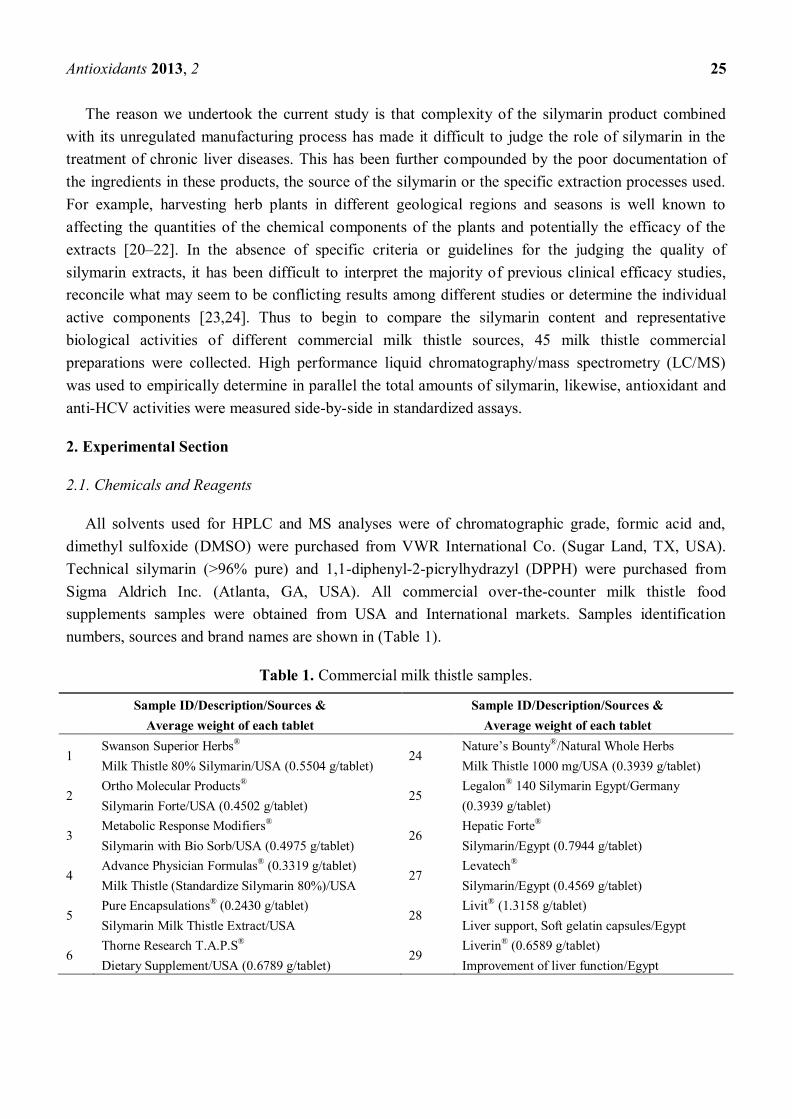
The reason we undertook the current study is that complexity of the silymarin product combined
with its unregulated manufacturing process has made it difficult to judge the role of silymarin in the
treatment of chronic liver diseases. This has been further compounded by the poor documentation of
the ingredients in these products, the source of the silymarin or the specific extraction processes used.
For example, harvesting herb plants in different geological regions and seasons is well known to
affecting the quantities of the chemical components of the plants and potentially the efficacy of the
extracts [20–22]. In the absence of specific criteria or guidelines for the judging the quality of
silymarin extracts, it has been difficult to interpret the majority of previous clinical efficacy studies,
reconcile what may seem to be conflicting results among different studies or determine the individual
active components [23,24]. Thus to begin to compare the silymarin content and representative
biological activities of different commercial milk thistle sources, 45 milk thistle commercial
preparations were collected. High performance liquid chromatography/mass spectrometry (LC/MS)
was used to empirically determine in parallel the total amounts of silymarin, likewise, antioxidant and
anti-HCV activities were measured side-by-side in standardized assays.
2. Experimental Section
2.1. Chemicals and Reagents
All solvents used for HPLC and MS analyses were of chromatographic grade, formic acid and,
dimethyl sulfoxide (DMSO) were purchased from VWR International Co. (Sugar Land, TX, USA).
Technical silymarin (>96% pure) and 1,1-diphenyl-2-picrylhydrazyl (DPPH) were purchased from
Sigma Aldrich Inc. (Atlanta, GA, USA). All commercial over-the-counter milk thistle food
supplements samples were obtained from USA and International markets. Samples identification
numbers, sources and brand names are shown in (Table 1).
Table 1. Commercial milk thistle samples.
Sample ID/Description/Sources &
Average weight of each tablet
Sample ID/Description/Sources &
Average weight of each tablet
1 Swanson Superior Herbs®
Milk Thistle 80% Silymarin/USA (0.5504 g/tablet) 24
Nature’s Bounty®/Natural Whole Herbs
Milk Thistle 1000 mg/USA (0.3939 g/tablet)
2 Ortho Molecular Products®
Silymarin Forte/USA (0.4502 g/tablet) 25
Legalon® 140 Silymarin Egypt/Germany
(0.3939 g/tablet)
3 Metabolic Response Modifiers®
Silymarin with Bio Sorb/USA (0.4975 g/tablet) 26
Hepatic Forte®
Silymarin/Egypt (0.7944 g/tablet)
4 Advance Physician Formulas® (0.3319 g/tablet)
Milk Thistle (Standardize Silymarin 80%)/USA 27
Levatech®
Silymarin/Egypt (0.4569 g/tablet)
5 Pure Encapsulations® (0.2430 g/tablet)
Silymarin Milk Thistle Extract/USA 28
Livit® (1.3158 g/tablet)
Liver support, Soft gelatin capsules/Egypt
6 Thorne Research T.A.P.S®
Dietary Supplement/USA (0.6789 g/tablet) 29
Liverin® (0.6589 g/tablet)
Improvement of liver function/Egypt
Antioxidants 2013, 2 26
Table 1. Cont.
7 Metagenics
®
Silymarin 80/USA (0.2450 g/tablet) 30
Levatone®
Food Supplement/Egypt (1.2484 g/tablet)
8 Himalaya Liver Care®
Liv.52/USA (0.3768 g/tablet) 31
Liver Albumin Plus® (0.9610 g/tablet)
Dietary Supplement/Egypt
9 Jarrow Formulas®/USA (0.2786 g/tablet)
Milk Thistle (Standardize Silymarin Extract 30:1) 32
Hipamax Plus® (1.5340 g/tablet)
Dietary Supplement/Egypt
10 Metabolic Maintenance®
Silymarin/USA (0.5696 g/tablet) 33
SEDICO® (13597 g/tablet)
Silymarin Plus, Dietary Supplement/Egypt
11 Life Extension® (0.9040 g/tablet)
Mega Silymarin with isosilybin B/USA 34
Hepaticum® (0.4228 g/tablet)
Cyclodextrin enhanced formula/Egypt
12 Purintin’s Pride®
Silymarin Milk Thistle/USA (0.3466 g/tablet) 35
Silipex® (0.3351 g/tablet)
Dietary Supplement /Egypt
13 Natural Wellness®/USA (0.5063 g/tablet) Maximum
Milk Thistle, Silybin Phytosome 240 mg 36
MEPACURE® (0.3907 g/tablet)
Liver support /Egypt
14 Enzymatic Therapy®
Super Milk Thistle/USA (0.3262 g/tablet) 37
Hepanox® Cap. (1.3371 g/tablet)
Napha food support/Egypt
15 Advanced Beta Glucon Therapy® (0.4867 g/tablet)
Bio-Silymarin, Aloha Medicinal Inc./USA 38
SELECTIVAL® (1.2695 g/tablet)
Dietary Supplement/Egypt
16 Futurebiotics®
Silymarin Plus/USA (1.0514 g/tablet) 39
Ursoplus® MINAPHARM
Silymarin 70%/Egypt (0.5625 g/tablet)
17 Planetary Herbals® (0.7060 g/tablet)
Full Spectrum Silymarin 80™/USA 40
Leaglon® 70 Silymarin/Egypt/Germany
(0.4259 g/tablet)
18 Wonder Laboratories® Advanced B-12
Sublingual/USA (0.3443 g/tablet) 41
Hepamarin® 140mg
Hepatoprotective/Egypt (0.3124 g/tablet)
19 21st Century® (0.4508 g/tablet)
200 count Milk Thistle Extract/USA 42
Trade Mark® (0.0518 g/tablet)
Biphenyldicarboxylate/China/Egypt
20 Source Naturals®
Silymarin Plus/USA (0.9847 g/tablet) 43
MEPASIL® (0.5075 g/tablet)
Silymarin, Liver support/Egypt
21 Now®
Silymarin 100 V caps/USA (0.5090 g/tablet) 44
MARIAGON® (0.4775 g/tablet)
Hepatoprotective/Egypt
22 Good’N Natural® Milk Thistle Extract
250 mg/USA(0.6765 g/tablet) 45
Hepato-Forte® (1.2624 g/tablet)
Liver Supplement/Egypt
23 TwinLab®
Silymarin/USA (0.1610 g/tablet)
2.2. Cells
The Clone B HCV genotype 1b sub-genomic (sg1b) replicon Huh7 cells, which constitutively
replicate a subgenomic HCV RNA in their cytoplasm, were obtained from C.M. Rice (Rockefeller
University, NY, USA) through the NIH AIDS Research and Reference Reagent Program and have
been described previously [25]. Cells were cultured in complete Dulbecco’s modified Eagle’s medium
(cDMEM) (Hyclone, Logan, UT, USA) supplemented with 10% fetal bovine serum (FBS) (Hyclone),
100 units/mL penicillin, 100 mg/mL streptomycin, and 2 mM L-glutamine (Gibco Invitrogen,
Carlsbad, CA, USA) as previously described [26].
Antioxidants 2013, 2 27
2.3. Preparation of Samples
Ten tablets of each commercial sample were randomly taken, crushed and homogenized. Weight of
each 10 tablets was recorded and is presented for each brand sample (Table 1). For chromatography
and mass spectrometry analysis: 20 mg of each crushed product were extracted in 5 mL of methanol
(3 replicate each). For all bioassay evaluations a second batch of 20 mg of each product were
separately extracted in 5 mL of DMSO (3 replicate). Extractions were performed in 10 mL sealed
tubes at room temperature rotated constantly using a Labnet Labroller II (Optics Planet Inc., 3150
Commercial Avenue Northbrook, IL, USA), at maximum speed for 24 h. Extracts were then filtered
and stored in the refrigerator. External calibrated standards were made under the same condition for
technical silymarin (Sigma Products).
2.4. Determination of Total Silymarin
High Performance Liquid chromatography and Mass Detection HPLC/MS were used to determine
the chemical composition of each the commercial products as previously described by us [27]. HPLC
of silymarin and commercial samples was performed on Agilent 1100 HPLC/MSD VL using
Phenomenex Kinetic 2.6 μ C18 100 A 100 × 4.16 mm column with electrospray (ES) ionization.
Methanol, water, and formic acid (90:10:1) was used as mobile phase A and 0.1% formic acid for
mobile phase B at a gradient flow rate of 0.5 mL/min. Solvent A = 55% 0.1 formic acid, solvent B = 45%
90:10:1 MeOH:H2O:Formic acid. Starting at time 0, 45% B, at 15 min, increase to 65% B, at 15.5 min
decrease to 45% B and hold at 45% B for 5 min run end 20.5 min and diode array detection at 288 nm.
Mass Spectroscopy was performed using single ion monitoring in the positive ESI mode for ions of
m/z 327 (M + Na) for taxifolin and m/z 505 (M + Na) for all other isomers. Mass detection conditions
were: quasi molecular ions dwell time of 294 ms, nitrogen was used both as drying gas and nebulizing
gas at flow rates of 12 L/min and 35 (psig). The temperature of the drying gas was set to 350 °C. Data
collection was handled using Chemstation V. B.04.02. All samples (45 × 3 replicate) were analyzed by
injecting 5 μL of sample in methanol and the analysis was repeated three times to calculate the average and
standard deviations.
2.5. Free Radical-Scavenging Activity: DPPH Test
Free radical-scavenging activity of each commercial sample was carried out using the DPPH
scavenging method [28]. The antioxidant activity was carried out using Perkin Elmer Victor 4X micro
plate reader performed in a 96 well plate using a total volume of 200 μL methanol containing 0.004 μg
DPPH and samples aliquots at a series of concentrations of 1, 10, 20, 40, 60, 80, 200, 400, 800 and
2000 μg/mL. The test was repeated at all concentration of each sample in triplicate. DPPH solutions at
the same concentration without the tested samples were used as control. Each sample, as well as each
control was analyzed in triplicates. After filling the well plates, they were incubated in the dark with
continuous shaking for 30 min followed by reading the absorbance at 520 nm. The free radical
scavenging activity of each solution was then calculated as percent inhibition according to the
following equation:
Antioxidants 2013, 2 28
% inhibition = 100 × (Ablank − Asample)/Ablank (1)
where Asample is the absorbance of the sample and Ablank is the absorbance of the blank. Inhibition %
was plotted against concentration and the EC50 was calculated graphically.
2.6. Trolox-Equivalent Antioxidant Capacity Assay
Trolox-equivalent antioxidant capacity (TEAC) of the commercial silymarin samples was carried
out using the procedure from Antioxidant Assay Kit item No. 709001 from Cayman Chemical
Company1180 E. Ellsworth Rd. Ann Arbor, MI, USA. The 45 commercial silymarin samples were
prepared by removing 100 μL of the stock preparation (20mg commercial silymarin/5 mL of DMSO)
and adding it to 400 μL HPLC grade water. On a 96 well plate, 10 μL of this preparation was removed
and added to 10 μL of metmyoglobin, 150 μL of chromogen and 40 μL of hydrogen peroxide mixture
for a total of 210 μL in each well. The plate was covered and place on a shaker for five minutes and
read at 750 nm on a Perkin Elmer Victor X4 2030 Multilabel Reader (710 Bridgeport Avenue Shelton,
CT, USA). The absorbance was plotted as a function of the final Trolox concentration (mM) according
to the assay.
Antioxidant (mM) = Sample absorbance − (Y − intercept)/Slope × Dilution (2)
2.7. Anti HCV Bioassay
Clone B sg1b cells were seeded in 96-well BIOCoat culture plates (BD Biosciences) at a density of
8 × 10³ cells/well in cDMEM. Upon reaching 90%–95% confluence, media was replaced with 200 μL
cDMEM supplemented with 1% DMSO (Sigma) and cells were cultured for an additional 20 days,
replacing medium every 2 days as previously described [29–31]. After these 20 days of culturing,
testing of the silymarin samples was initiated in parallel cultures of Clone B replicon cells. Cells were
treated with the individual silymarin samples at the indicated doses or diluents (DMSO) control. On
days 2, 4 and 6 post-silymarin treatment initiation medium was collected from culture plates and stored
for cytotoxicity analysis as described below. On day 6 post-silymarin treatment, cells were lysed in
200 μL 1X Nucleic Acid Purification lysis solution (Applied Biosystems, Foster City, CA, USA) and
immediately frozen (−80 °C). Real-time quantitative PCR (RTqPCR) analysis was performed as
described below to quantify intracellular HCV RNA levels.
2.8. Cytotoxicity Assay
Silymarin-mediated cellular toxicity was determined using the Toxilight Bioassay Kit (Lonza,
Walkersville, MD, USA), a bioluminescence-based assay which measures adenylate kinase (AK)
released from damaged cells, as per the manufacturer’s instructions.
2.9. RNA Isolation and RTqPCR Analysis
Total cellular RNA was isolated using a 1X Nucleic Acid Purification Lysis Solution (Applied
Biosystems, Foster City, CA, USA) and purified using an ABI PRISM™ 6100 Nucleic Acid
Antioxidants 2013, 2 29
PrepStation (Applied Biosystems), as per the manufacturer’s instructions. One μg of purified RNA was
used for cDNA synthesis TaqMan reverse transcription reagents (Applied Biosystems) and FastStart
Universal SYBR Green master mix (Roche Applied Sciences, Indianapolis, IN, USA), using an
Applied Biosystems 7300 real-time thermocycler (Applied Biosystems). Thermal cycling consisted of
an initial 10 min denaturation step at 95 °C followed by 40 cycles of denaturation (15 s at 95 °C) and
annealing/extension (1 min at 60 °C). HCV JFH-1 and GAPDH transcript levels were determined
relative to a standard curve comprised of serial dilutions of plasmid containing the JFH-1 HCV cDNA
or the human GAPDH gene, respectively. The PCR primers used to detect GAPDH and HCV were:
human GAPDH5′-GAAGGTGAAGGTCGGAGTC-3′ (sense) and 5′-AAGATGGTGATGGGATTTC-3′
(anti-sense) and JFH-1 HCV 5′-TCTGCGGAACCGGTGAGTA-3′ (sense) and 5′-TCAGGCAGTA
CCACAAGGC-3′ (anti-sense).
2.10. Statistical Analysis
Data was entered in SPSS Statistics [32] and analyzed using Pearson and Spearman nonparametric
correlation analysis with two-tailed significance determined.
3. Results and Discussion
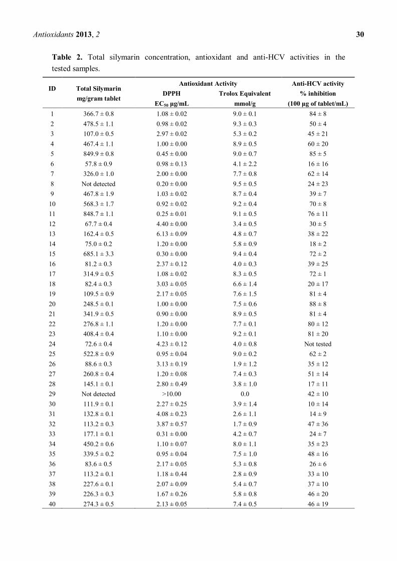
3.1. Determination of Total Silymarin
Because the harvesting, extraction, manufacturing, and quantification techniques used for
generating commercially available for the treatment of chronic liver diseases is unregulated, it is
difficult to directly compare different commercial sources of milk thistle and interpret the many studies
that have reported various levels of biological activities. Hence, we sought to compare the actual
content of silymarin among 45 different milk thistle products. Quantitative analysis of total silymarin
in all of the selected commercial samples was performed with 3 replicate extracts and three analytical
measurements. Total silymarin was calculated based on the sum of all of the major silymarin
constituents as shown in Figure 2. Average concentrations and standard deviation of each sample are
shown in Table 2 as mg per gram of tablet.
Figure 2. HPLC chromatograms of silymarin.
Antioxidants 2013, 2 30
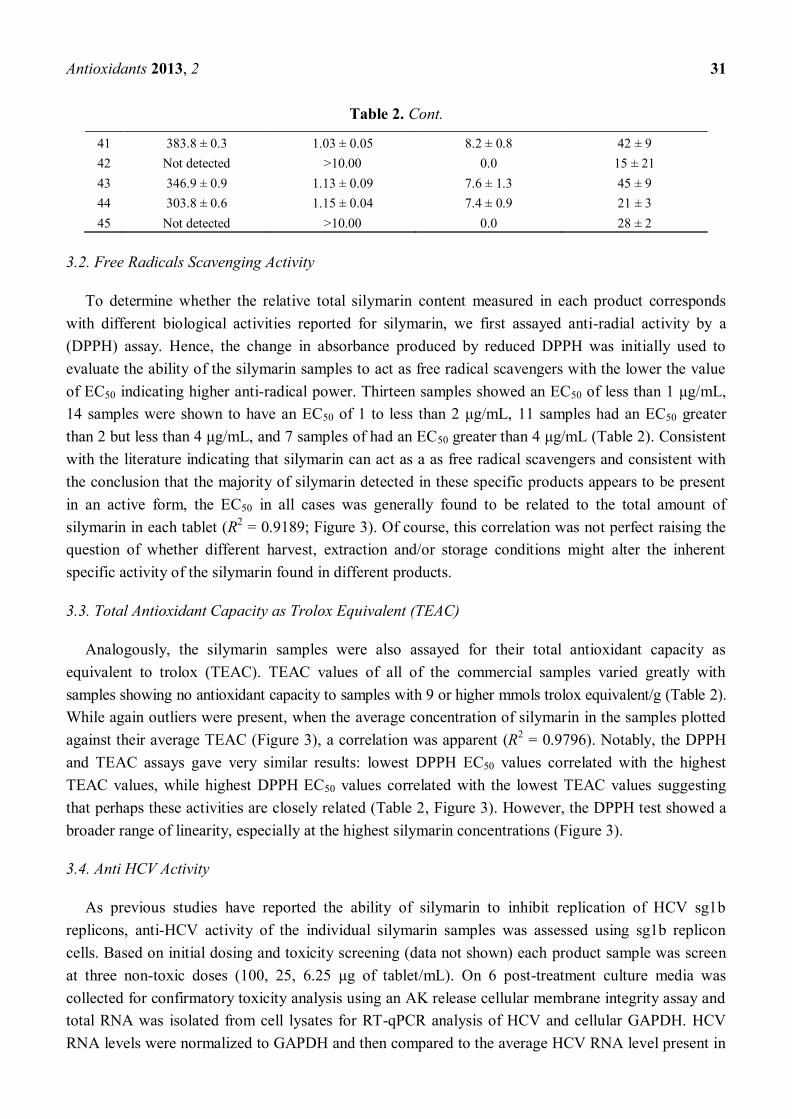
Table 2. Total silymarin concentration, antioxidant and anti-HCV activities in the
tested samples.
ID
Total Silymarin
mg/gram tablet
Antioxidant Activity Anti-HCV activity
DPPH
EC50 μg/mL
Trolox Equivalent
mmol/g
% inhibition
(100 μg of tablet/mL)
1 366.7 ± 0.8 1.08 ± 0.02 9.0 ± 0.1 84 ± 8
2 478.5 ± 1.1 0.98 ± 0.02 9.3 ± 0.3 50 ± 4
3 107.0 ± 0.5 2.97 ± 0.02 5.3 ± 0.2 45 ± 21
4 467.4 ± 1.1 1.00 ± 0.00 8.9 ± 0.5 60 ± 20
5 849.9 ± 0.8 0.45 ± 0.00 9.0 ± 0.7 85 ± 5
6 57.8 ± 0.9 0.98 ± 0.13 4.1 ± 2.2 16 ± 16
7 326.0 ± 1.0 2.00 ± 0.00 7.7 ± 0.8 62 ± 14
8 Not detected 0.20 ± 0.00 9.5 ± 0.5 24 ± 23
9 467.8 ± 1.9 1.03 ± 0.02 8.7 ± 0.4 39 ± 7
10 568.3 ± 1.7 0.92 ± 0.02 9.2 ± 0.4 70 ± 8
11 848.7 ± 1.1 0.25 ± 0.01 9.1 ± 0.5 76 ± 11
12 67.7 ± 0.4 4.40 ± 0.00 3.4 ± 0.5 30 ± 5
13 162.4 ± 0.5 6.13 ± 0.09 4.8 ± 0.7 38 ± 22
14 75.0 ± 0.2 1.20 ± 0.00 5.8 ± 0.9 18 ± 2
15 685.1 ± 3.3 0.30 ± 0.00 9.4 ± 0.4 72 ± 2
16 81.2 ± 0.3 2.37 ± 0.12 4.0 ± 0.3 39 ± 25
17 314.9 ± 0.5 1.08 ± 0.02 8.3 ± 0.5 72 ± 1
18 82.4 ± 0.3 3.03 ± 0.05 6.6 ± 1.4 20 ± 17
19 109.5 ± 0.9 2.17 ± 0.05 7.6 ± 1.5 81 ± 4
20 248.5 ± 0.1 1.00 ± 0.00 7.5 ± 0.6 88 ± 8
21 341.9 ± 0.5 0.90 ± 0.00 8.9 ± 0.5 81 ± 4
22 276.8 ± 1.1 1.20 ± 0.00 7.7 ± 0.1 80 ± 12
23 408.4 ± 0.4 1.10 ± 0.00 9.2 ± 0.1 81 ± 20
24 72.6 ± 0.4 4.23 ± 0.12 4.0 ± 0.8 Not tested
25 522.8 ± 0.9 0.95 ± 0.04 9.0 ± 0.2 62 ± 2
26 88.6 ± 0.3 3.13 ± 0.19 1.9 ± 1.2 35 ± 12
27 260.8 ± 0.4 1.20 ± 0.08 7.4 ± 0.3 51 ± 14
28 145.1 ± 0.1 2.80 ± 0.49 3.8 ± 1.0 17 ± 11
29 Not detected >10.00 0.0 42 ± 10
30 111.9 ± 0.1 2.27 ± 0.25 3.9 ± 1.4 10 ± 14
31 132.8 ± 0.1 4.08 ± 0.23 2.6 ± 1.1 14 ± 9
32 113.2 ± 0.3 3.87 ± 0.57 1.7 ± 0.9 47 ± 36
33 177.1 ± 0.1 0.31 ± 0.00 4.2 ± 0.7 24 ± 7
34 450.2 ± 0.6 1.10 ± 0.07 8.0 ± 1.1 35 ± 23
35 339.5 ± 0.2 0.95 ± 0.04 7.5 ± 1.0 48 ± 16
36 83.6 ± 0.5 2.17 ± 0.05 5.3 ± 0.8 26 ± 6
37 113.2 ± 0.1 1.18 ± 0.44 2.8 ± 0.9 33 ± 10
38 227.6 ± 0.1 2.07 ± 0.09 5.4 ± 0.7 37 ± 10
39 226.3 ± 0.3 1.67 ± 0.26 5.8 ± 0.8 46 ± 20
40 274.3 ± 0.5 2.13 ± 0.05 7.4 ± 0.5 46 ± 19
Antioxidants 2013, 2 31
Table 2. Cont.
41 383.8 ± 0.3 1.03 ± 0.05 8.2 ± 0.8 42 ± 9
42 Not detected >10.00 0.0 15 ± 21
43 346.9 ± 0.9 1.13 ± 0.09 7.6 ± 1.3 45 ± 9
44 303.8 ± 0.6 1.15 ± 0.04 7.4 ± 0.9 21 ± 3
45 Not detected >10.00 0.0 28 ± 2
3.2. Free Radicals Scavenging Activity
To determine whether the relative total silymarin content measured in each product corresponds
with different biological activities reported for silymarin, we first assayed anti-radial activity by a
(DPPH) assay. Hence, the change in absorbance produced by reduced DPPH was initially used to
evaluate the ability of the silymarin samples to act as free radical scavengers with the lower the value
of EC50 indicating higher anti-radical power. Thirteen samples showed an EC50 of less than 1 μg/mL,
14 samples were shown to have an EC50 of 1 to less than 2 μg/mL, 11 samples had an EC50 greater
than 2 but less than 4 μg/mL, and 7 samples of had an EC50 greater than 4 μg/mL (Table 2). Consistent
with the literature indicating that silymarin can act as a as free radical scavengers and consistent with
the conclusion that the majority of silymarin detected in these specific products appears to be present
in an active form, the EC50 in all cases was generally found to be related to the total amount of
silymarin in each tablet (R2 = 0.9189; Figure 3). Of course, this correlation was not perfect raising the
question of whether different harvest, extraction and/or storage conditions might alter the inherent
specific activity of the silymarin found in different products.
3.3. Total Antioxidant Capacity as Trolox Equivalent (TEAC)
Analogously, the silymarin samples were also assayed for their total antioxidant capacity as
equivalent to trolox (TEAC). TEAC values of all of the commercial samples varied greatly with
samples showing no antioxidant capacity to samples with 9 or higher mmols trolox equivalent/g (Table 2).
While again outliers were present, when the average concentration of silymarin in the samples plotted
against their average TEAC (Figure 3), a correlation was apparent (R2 = 0.9796). Notably, the DPPH
and TEAC assays gave very similar results: lowest DPPH EC50 values correlated with the highest
TEAC values, while highest DPPH EC50 values correlated with the lowest TEAC values suggesting
that perhaps these activities are closely related (Table 2, Figure 3). However, the DPPH test showed a
broader range of linearity, especially at the highest silymarin concentrations (Figure 3).
3.4. Anti HCV Activity
As previous studies have reported the ability of silymarin to inhibit replication of HCV sg1b
replicons, anti-HCV activity of the individual silymarin samples was assessed using sg1b replicon
cells. Based on initial dosing and toxicity screening (data not shown) each product sample was screen
at three non-toxic doses (100, 25, 6.25 μg of tablet/mL). On 6 post-treatment culture media was
collected for confirmatory toxicity analysis using an AK release cellular membrane integrity assay and
total RNA was isolated from cell lysates for RT-qPCR analysis of HCV and cellular GAPDH. HCV
RNA levels were normalized to GAPDH and then compared to the average HCV RNA level present in
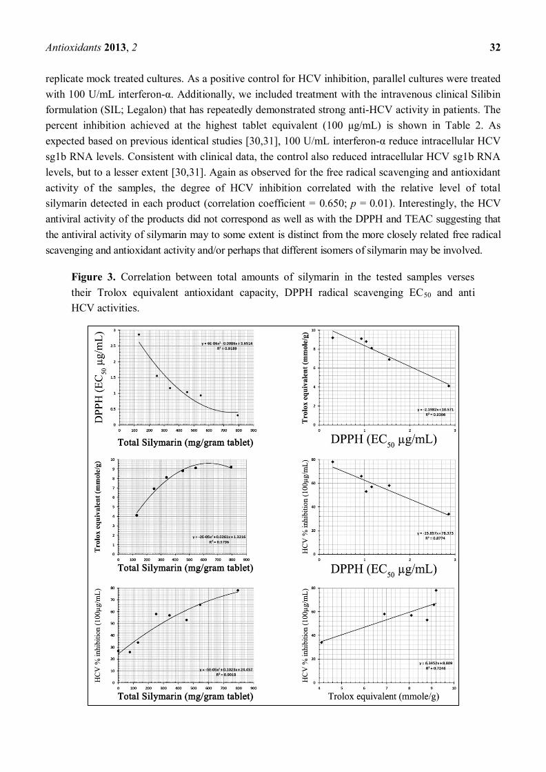
Antioxidants 2013, 2 32
replicate mock treated cultures. As a positive control for HCV inhibition, parallel cultures were treated
with 100 U/mL interferon-α. Additionally, we included treatment with the intravenous clinical Silibin
formulation (SIL; Legalon) that has repeatedly demonstrated strong anti-HCV activity in patients. The
percent inhibition achieved at the highest tablet equivalent (100 μg/mL) is shown in Table 2. As
expected based on previous identical studies [30,31], 100 U/mL interferon-α reduce intracellular HCV
sg1b RNA levels. Consistent with clinical data, the control also reduced intracellular HCV sg1b RNA
levels, but to a lesser extent [30,31]. Again as observed for the free radical scavenging and antioxidant
activity of the samples, the degree of HCV inhibition correlated with the relative level of total
silymarin detected in each product (correlation coefficient = 0.650; p = 0.01). Interestingly, the HCV
antiviral activity of the products did not correspond as well as with the DPPH and TEAC suggesting that
the antiviral activity of silymarin may to some extent is distinct from the more closely related free radical
scavenging and antioxidant activity and/or perhaps that different isomers of silymarin may be involved.
Figure 3. Correlation between total amounts of silymarin in the tested samples verses
their Trolox equivalent antioxidant capacity, DPPH radical scavenging EC50 and anti
HCV activities.
Antioxidants 2013, 2 33
4. Conclusions
Our findings on different commercial preparations of silymarin are significant in light of the fact
that oxidative stress is a secondary effect of many human diseases [33] and chronic HCV infection is a
leading cause of liver disease worldwide. As such, consumption of antioxidant-containing foods can
potentially reduce oxidative damage to cells and specifically reduce HCV-associated disease. Notably,
because malnutrition depresses cellular immune function, consumption of these antioxidant-containing
plants could have the general effect of protecting the immune system of malnourished individuals. This
is relevant not only to HCV infection [34–36], but available data indicate that antioxidants are
deficient in HIV infected populations due to increased utilization of antioxidant micro-nutrients [36]
and observational studies suggest that increased intake of antioxidants may delay progression of HIV
infection to AIDS [37,38].
Importantly however, our study documents that the different commercial sources tested varied
greatly in overall silymarin While both anti-oxidant and anti-HCV activity exhibited some degree of
correlation with silymarin levels suggesting that in the majority of products the silymarin present
exhibited comparable levels of biological activity, we did note outliers for which this correlation was
less apparent. There are several possible, non-mutually exclusive, explanation for these outliers: (1)
Many of these products consist of a mixture of multiple extracts and/vitamins that also may contribute
some biological activity in our assays. Thus, the absence of high levels of silymarin may not mean a
sample is void of liver-related protective activities. (2) The potency of silymarin can vary from
season-to-season, from one region to another, and/or be affected by specific extraction and storage
conditions. (3) The ratio of the seven different silymarin isomers often can vary in different extracts
depending on extraction methods and the plants used. Notably the ratio of the different silymarin
isomers did vary among the various products, but in general the level of the different isomers was
highly correlative making it difficult to determine if particular individual isomers were responsible to
the various activities assayed in this limited sample set. Although preliminarily it appears that
measurement of taxifolin concentrations in silymarin products may be an effective way of measuring
the antioxidant potency of products from different suppliers, further analysis is required to conclusive
determined which specific isomer(s) are responsible for the different therapeutic activities reported.
Such future efforts are warranted as such insight could potentially, aid in the development of more
potent silymarin formulations and inform regarding if specific isomer quantification could be used to
standardize silymarin product activities.
Acknowledgments
This work is supported by the National Institutes of Health (NIH) Public Health Service
Grants RCMI-5G12RR003045-21 (MAS), R01-AI070827 (SLU), R01-AI078881 (SLU), and
NASA-CBER-URC Grant #NNX108Q16A (MAS) and RDP 04 07 01 “Development of a Natural
Product for the treatment or/and control of Hepatitis C Virus (HCV) infection”.
Antioxidants 2013, 2 34
References
1. Radimer, K.; Bindewald, B.; Hughes, J.; Ervin, B.; Swanson, C.; Picciano, M.F. Dietary
supplement use by US adults: Data from the National Health and Nutrition Examination Survey,
1999–2000. Am. J. Epidemiol. 2004, 160, 339–349.
2. Qato, D.; Alexander, C.; Conti, R.; Johnson, M.; Schumm, P.; Lindau, S. Use of prescription and
over-the-counter medications and dietary supplements among older adults in the United States.
JAMA 2008, 300, 2867–2878.
3. World Health Organization. The World Medicines Situation 2011—Traditional Medicines: Global
Situation, Issues and Challenges; World Health Organization: Geneva, Switzerland, 2011.
4. Flora, K.; Hahn, M.; Rosen, H.; Benner, K. Milk thistle (Silybum marianum) for the therapy of
liver disease. Am. J. Gastroenterol. 1998, 93, 139–143.
5. Mayer, K.; Myers, P.; Lee, S. Silymarin treatment of viral hepatitis: A systematic review. J. Viral
Hepat. 2005, 12, 559–567.
6. Wellington, K.; Jarvis, B. Silymarin: A review of its clinical properties in the management of
hepatic disorders. BioDrugs 2001, 15, 465–489.
7. Saller, R.; Meier, R.; Brignoli, R. The use of silymarin in the treatment of liver diseases. Drugs
2001, 61, 2035–2063.
8. Roy, S.; Gagan, G.; Agarwal, R. Silibinin prevents ultraviolet B radiation-induced epidermal
damages in JB6 cells and mouse skin in a p53-GADD45α-dependent manner. Carcinogenesis
2012, 33, 629–636.
9. Chilampalli, S.; Zhang, X.; Fahmy, H.; Kaushik, R.; Zeman, D.; Hildreth, M.; Dwivedi, C.
Chemopreventive effects of honokiol on UVB-induced skin. Anticancer Res. 2010, 30, 777–783.
10. Bárcena, R.; Moreno, A.; Rodríguez-Gandía, M.; Albillos, A.; Arocena, C.; Blesa, C.; García-Hoz, F.;
Graus, J.; Nuño, J.; López-Hervás, P.; et al. Safety and anti-HCV effect of prolonged intravenous
silibinin in HCV-genotype 1 subjects in the immediate liver transplant period.
J. Hepatol. 2012, in press.
11. Mariño, Z.; Crespo, G.; D’Amato, M.; Brambilla, N.; Giacovelli, G.; Rovati, L.; Costa, J.;
Navasa, M.; Forns, X. Intravenous silibinin monotherapy shows significant antiviral activity in
HCV-infected patients in the peri-transplantation period. J. Hepatol. 2012, in press.
12. Biermer, M.; Schlosser, B.; Fülöp, B.; van Bömmel, F.; Brodzinski, A.; Heyne, R.; Keller, K.;
Sarrazin, C.; Berg T. High-dose silibinin rescue treatment for HCV-infected patients showing
suboptimal virologic response to standard combination therapy. J. Viral Hepat. 2012, 19,
547–553.
13. Rutter, K.; Scherzer, T.; Beinhardt, S.; Kerschner, H.; Stättermayer, A.; Hofer, H.; Popow-Kraupp, T.;
Steindl-Munda, P.; Ferenci, P. Intravenous silibinin as “rescue treatment” for on-treatment
non-responders to pegylated interferon/ribavirin combination therapy. Antivir. Ther. 2011, 16,
1327–1333.
14. Payer, B.; Reiberger, T.; Rutter, K.; Beinhardt, S.; Staettermayer, A.; Peck-Radosavljevic, M.;
Ferenci, P. Successful HCV eradication and inhibition of HIV replication by intravenous silibinin
in an HIV-HCV coinfected patient. J. Clin. Virol. 2010, 49, 131–133.
Antioxidants 2013, 2 35
15. Neumann, U.; Biermer, M.; Eurich, D.; Neuhaus, P.; Berg, T. Successful prevention of hepatitis C
virus (HCV) liver graft reinfection by silibinin mono-therapy. J. Hepatol. 2010, 52, 951–952.
16. Ferenci, P.; Scherzer, T.; Kerschner, H.; Rutter, K.; Beinhardt, S.; Hofer, H.; Schöniger-Hekele, M.;
Holzmann, H.; Steindl-Munda, P. Silibinin is a potent antiviral agent in patients with chronic
hepatitis C not responding to pegylated interferon/ribavirin therapy. Gastroenterology 2008, 135,
1561–1567.
17. Wagoner, J.; Negash, A.; Kane, O.; Martinez, L.; Nahmias, Y.; Bourne, N.; Owen, D; Grove, J.;
Brimacombe, C.; McKeating, J.; et al. Multiple effects of silymarin on the hepatitis C virus
lifecycle. Hepatology 2010, 51, 1912–1921.
18. Sy-Cordero, A.; Graf, T.; Nakanishi, Y.; Wani, M.; Agarwal, R.; Kroll, D.; Oberlies, N. Large
scale isolation of flavonolignans from Silybum marianum (milk thistle) extract affords new minor
constituents and preliminary structure-activity relationships. Planta Med. 2010, 76, 644–647.
19. Daniela, D.; Gažák, R.; Marhol, P.; Biedermann, D.; Purchartová, K.; Fedrigo, M.; Riva, S.; Křen, V.
Enzymatic kinetic resolution of silybin diastereoisomers. J. Nat. Prod. 2010, 73, 613–619.
20. Křen, V.; Kubisch, J.; Sedmera, P.; Halada, P.; Přikrylová, V.; Jegorov, A.; Cvak, L.; Gebhardt, R.;
Ulrichová, J.; Šimánek, V. Glycosylation of silybin. J. Chem. Soc. 1997, 2467–2474,
doi:10.1039/A703283H.
21. Liu, J.; Manheimer, E.; Tsutani, K.; Gluud, C. Medicinal herbs for hepatitis C virus infection: A
Cochrane hepato-biliary systematic review of randomized trials. Am. J. Gastroenterol. 2003, 98,
538–544.
22. Hammouda, F.; Ismail, S.; Hassan, N.; Zaki, A.; Kamel, A.; Rimpler, H. Evaluation of the
silymarin content in Silybum marianum (L.) Gaertn. Cultivated under different agricultural
conditions. Phytother. Res. 1993, 7, 90–91.
23. Simanek, V.; Kren, V.; Ulrichova, J.; Vicar, J.; Cvak, L. Silymarin: What is in the name...? An
appeal for a change of editorial policy. Hepatology 2000, 32, 442–444.
24. Farghaly, T.; Abdel Hafez, N.; Ragab, E.; Awad, H.; Abdalla, M. Synthesis, anti-HCV,
antioxidant and peroxynitrite inhibitory activity of fused benzosuberone derivatives. Eur. J. Med.
Chem. 2010, 45, 492–500.
25. Blight, K.; Kolykhalov, A.; Rice, C. Efficient initiation of HCV RNA replication in cell culture.
Science 2000, 290, 1972–1974.
26. Zhong, J.; Gastaminza, P.; Cheng, G.; Kapadia, S.; Kato, T.; Burton, D.R.; Wieland, S.;
Uprichard, S.; Wakita, T.; Chisari, F. Robust hepatitis C virus infection in vitro. Proc. Natl. Acad.
Sci. USA 2005, 102, 9294–9299.
27. Anthony, K.; Saleh, M. Chemical profiling and antioxidant activity of commercial milk thistle
food supplements. J. Chem. Pharm. Res. 2012, 4, 4440–4450.
28. Brand-Williams, W.; Cuvelier, M.; Berset, C. Use of free radical method to evaluate antioxidant
activity. Lebensm. Wiss. Technol. 1995, 28, 25–30.
29. Choi, S.; Sainz, J.; Corcoran, P.; Uprichard, S.; Jeong, H. Characterization of increased drug
metabolism activity in dimethyl sulfoxide (DMSO)-treated Huh7 hepatoma cells. Xenobiotica
2009, 39, 205–217.
Antioxidants 2013, 2 36
30. Yu, X.; Sainz, B.; Uprichard, S. Development of a cell-based hepatitis C virus infection
fluorescent resonance energy transfer assay for high-throughput antiviral compound screening.
Antimicrob. Agents Chemother. 2009, 53, 4311–4319.
31. Yu, X.; Uprichard, S. Cell-based hepatitis C virus infection fluorescence resonance energy
transfer (FRET) assay for antiviral compound screening. Curr. Protoc. Microbiol. 2010, 17,
17–25.
32. Statistics SPSS Software, version 20; IBM Corporation: New York, NY, USA, 2011.
33. Gutteridge, J. Free radicals in disease processes: A compilation of cause and consequence. Free
Radic. Res. Commun. 1993, 19, 141–158.
34. Rivas-Estilla, A.; Marrugo, O.; Trujillo-Murillo, K.; Pérez-Ibave, D.; Charles-Niño, C.;
Pedroza-Roldan, C.; Ríos-Ibarra, C.; Ramírez-Valles, E.; Ortiz-López, R.; Islas-Carbajal, M.; et al.
Cu/Zn superoxide dismutase (sod 1) induction is implicated in the antioxidative and antiviral
activity of acetylsalicylic acid in HCV-expressing cells. Am. J. Physiol. Gastrointest. 2012, 22,
in press.
35. Farawela, H.; Khorshied, M.; Shaheen, I.; Gouda, H.; Nasef, A.; Abulata, N.; Mahmoud, H.;
Zawam, H.; Mousa, S. The association between hepatitis C virus infection, genetic
polymorphisms of oxidative stress genes and B-cell non-Hodgkin’s lymphoma risk in Egypt
infection. Infect. Genetics Evol. 2012, 12, 1189–1194.
36. Agrawal, L.; Louboutina, J.; Reyes, B.; Bockstaele, E.; Strayer, D. HIV-1 Tat neurotoxicity: A
model of acute and chronic exposure, and neuroprotection by gene delivery of antioxidant
enzymes. Neurobiol. Dis. 2012, 45, 657–670.
37. Dworkin, B.; Wormser, G.; Axelrod, F.; Pierre, N.; Schwarz, E.; Seaton, T. Dietary intake in
patients with acquired immunodeficiency syndrome (AIDS), patients with AIDS-related complex,
and serologically positive human immunodeficiency virus patients: Correlations with nutritional
status. J. Parenter. Enter. Nutr. 1990, 14, 605–609.
38. Abrams, B.; Duncan, D.; Hertz-Picciotto, I. A prospective study of dietary intake and acquired
immune deficiency syndrome in HIV-seropositive homosexual men. J. Acquir. Immune Defic.
Syndr. 1993, 6, 949–958.
© 2013 by the authors; licensee MDPI, Basel, Switzerland. This article is an open access article
distributed under the terms and conditions of the Creative Commons Attribution license
(http://creativecommons.org/licenses/by/3.0/).
Related Documents


![14] Antioxidants](https://static.cupdf.com/doc/110x72/577ccfa61a28ab9e78904327/14-antioxidants.jpg)
