Antioxidant Efficacy and Adhesion Rescue by a Recombinant Mussel Foot Protein-6 S. C. T. Nicklisch a,* , Saurabh Das b , N. R. Martinez Rodriguez a , J. N. Israelachvili b , and J. H. Waite a a Department of Molecular, Cellular and Developmental Biology, University of California, Santa Barbara, CA 93106 USA b Department of Chemical Engineering, University of California, Santa Barbara, CA 93106 USA Abstract Mytilus foot protein type 6 (mfp-6) is crucial for maintaining the reducing conditions needed for optimal wet adhesion in marine mussels. In this report we describe the expression and production of a recombinant Mytilus californianus foot protein type 6 variant 1 (rmfp-6.1) fused with a hexa- histidine affinity tag in Escherichia coli and its purification by affinity chromatography. Recombinant mfp-6 showed high purification yields of 5–6 mg/L cell culture and excellent solubility in low pH buffers that retard oxidation of its many thiol groups. Purified rmfp-6.1 protein showed high DPPH radical scavenging activity as compared to Vitamin C. Using the highly sensitive surface force apparatus (SFA) technique to measure interfacial surface forces in the nanoNewton range we show that rmfp-6.1 is also able to rescue the oxidation-dependent adhesion loss of mussel foot protein 3 (mfp-3) at pH 3. The adhesion rescue is related to a reduction of dopaquinone back to DOPA in mfp-3 which is the reverse reaction observed during the detrimental enzymatic browning process in fruits and vegetables. Broadly viewed, rmfp-6.1 has potential as a versatile antioxidant for applications ranging from personal products to anti- spoilants for perishable foods during processing and storage. Keywords Adhesion rescue; antioxidant; DOPA oxidation; recombinant mussel protein; thiol 1 Introduction Strong underwater adhesion is an evolutionary adaptation that developed in parallel among many different sessile organisms, yet only scarce information is available regarding the underlying molecular mechanisms involved. Recent investigations of the adhesive plaques of Mytilus californianus have provided deeper insights into the reactivity and organization of some of these proteins [1]. Among these proteins, five are unique to the plaque (mfp-2, -3, -4, -5, and -6) and all contain the tyrosine derivative 3,4-dihydroxyphenyl-L-alanine (DOPA) that by itself is known to mediate strong adhesion on polar surfaces [2]. However, at the high pH of seawater (~ 8.2) DOPA containing protein readily undergoes auto- * Current address: Sascha C.T. Nicklisch, University of California, San Diego, Scripps Institution of Oceanography, 8750 Biological Grade Rd., 3155 Hubbs Hall, La Jolla, CA-92037, [email protected], Phone: (858) 8224303, Fax: (858) 534-7313 Contributions: S.C.T.N constructed overexpressed and purified rmfp-6 proteins, designed and performed the DPPH assay and performed MALDI mass spectrometry and thrombin cleavage experiments; S.D. designed and performed the SFA experiments; N.R.M.R performed the SFA experiments; J.H.W. designed and co-supervised the whole project with J.N.I., who helped analyze the results. NIH Public Access Author Manuscript Biotechnol Prog. Author manuscript; available in PMC 2014 November 01. Published in final edited form as: Biotechnol Prog. 2013 November ; 29(6): . doi:10.1002/btpr.1810. NIH-PA Author Manuscript NIH-PA Author Manuscript NIH-PA Author Manuscript

Welcome message from author
This document is posted to help you gain knowledge. Please leave a comment to let me know what you think about it! Share it to your friends and learn new things together.
Transcript
Antioxidant Efficacy and Adhesion Rescue by a RecombinantMussel Foot Protein-6
S. C. T. Nicklischa,*, Saurabh Dasb, N. R. Martinez Rodrigueza, J. N. Israelachvilib, and J. H.Waitea
aDepartment of Molecular, Cellular and Developmental Biology, University of California, SantaBarbara, CA 93106 USAbDepartment of Chemical Engineering, University of California, Santa Barbara, CA 93106 USA
AbstractMytilus foot protein type 6 (mfp-6) is crucial for maintaining the reducing conditions needed foroptimal wet adhesion in marine mussels. In this report we describe the expression and productionof a recombinant Mytilus californianus foot protein type 6 variant 1 (rmfp-6.1) fused with a hexa-histidine affinity tag in Escherichia coli and its purification by affinity chromatography.Recombinant mfp-6 showed high purification yields of 5–6 mg/L cell culture and excellentsolubility in low pH buffers that retard oxidation of its many thiol groups. Purified rmfp-6.1protein showed high DPPH radical scavenging activity as compared to Vitamin C. Using thehighly sensitive surface force apparatus (SFA) technique to measure interfacial surface forces inthe nanoNewton range we show that rmfp-6.1 is also able to rescue the oxidation-dependentadhesion loss of mussel foot protein 3 (mfp-3) at pH 3. The adhesion rescue is related to areduction of dopaquinone back to DOPA in mfp-3 which is the reverse reaction observed duringthe detrimental enzymatic browning process in fruits and vegetables. Broadly viewed, rmfp-6.1has potential as a versatile antioxidant for applications ranging from personal products to anti-spoilants for perishable foods during processing and storage.
KeywordsAdhesion rescue; antioxidant; DOPA oxidation; recombinant mussel protein; thiol
1 IntroductionStrong underwater adhesion is an evolutionary adaptation that developed in parallel amongmany different sessile organisms, yet only scarce information is available regarding theunderlying molecular mechanisms involved. Recent investigations of the adhesive plaquesof Mytilus californianus have provided deeper insights into the reactivity and organizationof some of these proteins [1]. Among these proteins, five are unique to the plaque (mfp-2,-3, -4, -5, and -6) and all contain the tyrosine derivative 3,4-dihydroxyphenyl-L-alanine(DOPA) that by itself is known to mediate strong adhesion on polar surfaces [2]. However,at the high pH of seawater (~ 8.2) DOPA containing protein readily undergoes auto-
*Current address: Sascha C.T. Nicklisch, University of California, San Diego, Scripps Institution of Oceanography, 8750 BiologicalGrade Rd., 3155 Hubbs Hall, La Jolla, CA-92037, [email protected], Phone: (858) 8224303, Fax: (858) 534-7313
Contributions: S.C.T.N constructed overexpressed and purified rmfp-6 proteins, designed and performed the DPPH assay andperformed MALDI mass spectrometry and thrombin cleavage experiments; S.D. designed and performed the SFA experiments;N.R.M.R performed the SFA experiments; J.H.W. designed and co-supervised the whole project with J.N.I., who helped analyze theresults.
NIH Public AccessAuthor ManuscriptBiotechnol Prog. Author manuscript; available in PMC 2014 November 01.
Published in final edited form as:Biotechnol Prog. 2013 November ; 29(6): . doi:10.1002/btpr.1810.
NIH
-PA Author Manuscript
NIH
-PA Author Manuscript
NIH
-PA Author Manuscript

oxidation to dopaquinone (DQ) resulting in a considerable loss of adhesion [3]. In contrastto the other four proteins, mfp-6 shows weak adhesion and contains small amounts of DOPA(< 5 mol %) but significant levels of cysteine (11 mol %, [4]). Interestingly, recentbiochemical studies on mfp-6 supported the hypothesis of mfp-6 as a highly potentproteinogenic antioxidant for the DOPA-containing adhesives [3]. The five mfp-6 isoformsannotated to date have a highly conserved amino acid composition and sequence homology(Table S1) assuming that their respective antioxidant activities are similar. Here we reportthe construction and characterization of Mytilus californianus recombinant foot protein type6 variant 1 (rmfp-6.1) fused with a hexahistidine affinity tag in Escherichia coli. The His6-tagged rmfp-6.1 protein showed high antioxidant activity that was determined using anoptimized 1,1-diphenyl-2-picryl-hydrazyl (DPPH) assay as described previously [5]. Rescueof oxidation-dependent adhesion loss due to rmfp-6.1 mediated reduction of dopaquinoneback to DOPA in the mussel foot protein mfp-3 was tested using a Surface Forces Apparatus[6].
2 Materials and MethodsChemicals
2,2-Diphenyl-1-picrylhydrazyl (DPPH•), L-Ascorbic acid (> 99 % purity), and Lysozyme(Chicken Egg White) were obtained from Sigma Aldrich (St. Louis, USA).Spectrophotometric grade of methanol and the nonionic detergent Triton X-100 werepurchased from Fisher Scientific (Pittsburgh, USA). Citric acid monohydrate (> 99 %purity) and sodium phosphate monobasic (> 98 % purity) were purchased from EMDMillipore (San Diego, USA). Double distilled water (EMD Millipore, San Diego, USA) wasused throughout the experiments.
Strains and Plasmid ConstructionE. coli One Shot TOP10 chemically competent cells [F- mcrA Δ(mrr-hsdRMS-mcrBC)ϕ80lacZΔM15 Δlacχ74 recA1 araD139 Δ(ara-leu) 7697 galU galK rpsL (StrR) endA1 nupGλ-] (Life Technologies Grand Island, NY) were used for recombinant plasmid construction.E. coli Rosetta 2 (DE3) cells [F− ompT hsdSB(rB
− mB−) gal dcm (DE3) pRARE (CamR)]
were used as a host strain for expressing recombinant rmfp-6.1. We used the Rosetta 2(DE3) cells to compensate for rarely used tRNAs in E. coli and thus enhance theheterologous expression of the eukaryotic mussel protein. Previous studies on theheterologous expression of recombinant mussel adhesive proteins frequently led to lowyields or failed to express functional protein in E. coli BL21 or JM109 strains (personalcommunication, Dr. Dong-Soo Hwang). Gene sequence information for native mfp6-v1 wasobtained from GenBank (DQ351537.1; http://www.ncbi.nlm.nih.gov/genbank/). The mfp6-v1 sequence (without the signal sequence) was cloned out of a mixture of Mytiluscalifornianus foot cDNA library ([4], Table S1) using combined gradient and touchdownpolymerase chain reaction (PCR). Specific codon-optimized primers for PCR amplificationwere synthesized (Table S2). The forward primer was: v1-NdeI-FW, 5′-GGG CAT ATGGGT GGG GGA AAC TAC AGA GG – 3′ (29 nt, containing a NdeI recognition site), andreverse primer was: v1-HindIII-Rev, 5′-GGG AAG CTT AGT AAC CAC TAC GAAGAC AAC - 3′ (30 nt, containing a HindIII recognition site). The bolded parts of primersequences are complementary to the nucleotide sequences of the mfp6-v1 gene, whereas 5′overhanging ends of primers contain recognition sites for restriction endonucleases(underlined), and are designed to facilitate cloning. Designed primers and 2.5 units of TaqDNA polymerase (Fisher Scientific, Pittsburgh, PA) were used in a touchdown PCRreaction for 10 cycles with a temperature profile of 30s at 95°C, 45s at 70°C, and 1min at72°C as well as another 20 cycles with a temperature profile of 30s at 95°C, 45s at 60°C,and 1min at 72°C in an Eppendorf Mastercycler Gradient (Eppendorf, Hauppauge, NY). The
Nicklisch et al. Page 2
Biotechnol Prog. Author manuscript; available in PMC 2014 November 01.
NIH
-PA Author Manuscript
NIH
-PA Author Manuscript
NIH
-PA Author Manuscript

amplification products were analyzed by electrophoresis on a 1% agarose gels stained withethidium bromide. An approximately 300bp-specific rmfp-6.1 PCR product was insertedinto a pCR 2.1-TOPO vector for sequencing using the TOPO TA Cloning Kit (Invitrogen,Carlsbad, CA). E. coli One Shot TOP10 cells were transformed with the vector constructand three independently derived clones containing the rmfp-6.1 gene were selected andsequenced using a commercial service (Genewiz, South Plainfield, NJ). The sequenced geneconstruct was digested with NdeI and HindIII, purified and ligated into the Nde I and HindIII sites of the pET-28a(+) vector (EMD Chemicals, Philadelphia, PA). The vector containsa hexahistidine (His6) tag at the N-terminus to simplify protein purification and a T7promoter that is inducible by isopropyl-β-D-1-thiogalactopyranoside (IPTG). E. coli Rosetta2 (DE3) cells were transformed with the ligation mixture and used for the expression andpurification procedure.
Media and Cell CultureFor strain construction and protein expression, E. coli cells were grown in enriched 2x TYmedium containing 16 g/L tryptone, 10 g/L yeast extract, and 5 g/L NaCl. The constructedtransformant harboring the plasmid was stored at −80 °C. Culture experiments wereperformed with 2L of 2xTY medium supplemented with 50 μg/mL kanamycin and 50 μg/mL chloramphenicol (Sigma-Aldrich, St. Louis, MO) at 37 °C and shaking at 250 rpm. Cellgrowth was monitored by measuring the optical density at 600 nm (OD600) using aNanodrop 2000c UV-vis spectrophotometer (Thermo Scientific, Wilmington, DE). Whencultures reached an OD600 of 0.7–0.8, 10 μM (final concentration) IPTG was added to theculture broth for induction of recombinant mfp-6.1. It has to be noted that commonly usedIPTG concentrations to initiate protein expression range from 0.5 – 4 mM finalconcentration. However, inducing the plasmid containing E. coli cells with IPTGconcentrations higher than 50 μM led to cell death (Figure 1A). The induced cells weregrown for 5h, harvested and centrifuged at a 5,000 g for 20 min at 4 °C, and the pellets werestored at −80 °C for further analyses.
Purification of Recombinant mfp-6-v1The cell pellets were dissolved in lysis buffer (1x PBS (137mM NaCl, 2.7mM KCl, 10mMNa2HPO4, 2mM KH2PO4) pH 7.4, 1 μg/ml pepstatin, 1 μg/ml leupeptin, 5 mg/ml lysozyme,1 U/ml DNAse) and disrupted using a Q700 sonicator at 4°C (Qsonica, Newtown, CT) andin pulse mode (5 s pulse, 10 s break, 10 min total, 40 % amplitude). Cell debris andinclusion bodies (IB) were spun down at 10,000 g and 4°C for 30min and washed twice withwashing buffer (1x PBS pH 7.4, 1 μg/μl Pepstatin, 1 μg/μl Leupeptin, and 1 % v/v TritonX-100). Recombinant mfp-6.1 was extracted from the pellet with extraction buffer (5% v/vacetic acid, 50mM TCEP, 8M urea) and the supernatant was dialyzed overnight against 5%v/v acetic acid in a total volume ratio of 1:1200 and in 1 kDa-cut off dialysis tubing(Spectrum Laboratories, Rancho Dominguez, CA). Immobilized metal affinitychromatography (IMAC) purification was performed using the Akta MFPLC PurificationSystem at room temperature with 1 mL/min and a pre-packed 1ml HisTrap FF Crudecolumn (GE Healthcare, Pittsburgh, PA) as affinity purification resin. Affinity purificationwas performed under denaturing conditions. Dialyzed samples were freeze-dried andresuspended in washing buffer (50 mM Na2HPO4, 8 M Urea, 500 mM NaCl, 10 mMImidazole, pH 7.4). The column was equilibrated with 5 resin volumes of washing bufferand then loaded with 2 ml of the resuspended denatured samples. Target recombinantmpf6.1 was eluted with elution buffer (50 mM Na2HPO4, 8 M Urea, 100 mM NaCl, 250mM Imidazole, pH 7.4). Eluted rmfp-6.1 was dialyzed in 5 % v/v acetic acid overnight at 4°C, concentrated by freeze-drying, and finally resolved in 5 % v/v acetic acid. A finalpurification was performed using reverse phase C8 HPLC with a 260×7 mm RP-300Aquapore (Applied Biosystems, San Jose, CA) column and a linear gradient of aqueous
Nicklisch et al. Page 3
Biotechnol Prog. Author manuscript; available in PMC 2014 November 01.
NIH
-PA Author Manuscript
NIH
-PA Author Manuscript
NIH
-PA Author Manuscript

acetonitrile acidified with 0.1 % v/v TFA at 1 ml/min. Acid-urea or SDS polyacrylamide gelelectrophoresis (PAGE) with Coomassie blue staining were performed to assess the purity ofprotein samples. To calculate the purified rmfp-6.1 concentration the protein’s absorbance at280 nm was measured on a Nanodrop 2000C spectrophotometer (Thermo Scientific,Wilmington, DE) or quantified by a standard Bradford assay (Life Science Research,Hercules, CA). We used GelQuant.NET software provided by biochemlabsolutions.com toquantify relative protein yields in the PAGE gels.
MALDI-TOF Mass Spectrometry AnalysisMatrix-assisted laser desorption ionization (MALDI) mass spectrometry analysis with time-of-flight (TOF) was performed on a Voyager Mass Spectrometer LBT2 (AppliedBiosystems, San Jose, CA) with 1.2 meter ion path in the positive ion linear mode. Asmatrix solution α-cyano-4-hydroxy-cinnamic acid (CHCA) in 50 % acetonitrile and 0.1 %trifluoroacetic acid (TFA) was used. Samples were diluted 1:20 with matrix solution and 1μL was spotted onto the MALDI sample target plates and evaporated using a vacuum pump.Spectra were obtained in the mass range between 500 and 30,000 Da with 300 laser shotsper spectrum. Accelerating voltage was 25.000 V with a grid voltage of 93 % and a guidewire 0 percentage of 0.3 %. Spectra were recorded in delayed extraction with a delay time of300 ns. Internal calibration was performed using chicken egg white lysozyme (Sigma-Aldrich, St. Louis, MO) with a calculated molecular mass of 14.3 kDa. All data wasanalyzed using Voyager Data Explorer 4.0.0.0 (Applied Biosystems) and plotted usingOrigin 7.0 (Originlab).
DPPH coupled antioxidant assayA well-established protocol for the determination of antioxidant activity in food and otherbiological samples is the 2,2-Diphenyl-1-picrylhydrazyl (DPPH) assay [7]. In its radicalform DPPH absorbs at 515nm (ε = 11,240 M−1 cm−1 in MeOH), but upon reduction by anantioxidant or radical species, the absorption disappears and the color of the solutionchanges from violet to pale yellow [8]. We used an optimized protocol as describedpreviously [5] to test for the antioxidant activity of a protein like rmfp-6.1. Briefly, a mildnon-ionic detergent (0.3 % v/v Triton X-100) is used to keep both the hydrophobic hydrazylradical and the basic recombinant rmfp-6.1 protein soluble. To start the reaction, 5 μM ofpurified rmfp-6.1 or L-ascorbic acid (control antioxidant) was added to 100 μM freshlyprepared DPPH in 0.1 M citrate phosphate buffer, pH 3.0 [9]. The decrease in absorbance at515 nm is followed over 60 min to monitor the reduction of free radical DPPH to DPPH2.The absorbance reduction at 515 nm was normalized against 100 mM DPPH without theaddition of antioxidant as control. Antioxidant activity is described as relative DPPHreduction in percentage. The absorbance measurements were carried out in standard 1.5 mlmicro-cuvettes on a Nanodrop 2000C Dual-mode UV-Vis Spectrophotometer at RT(Thermo Fisher Scientific Inc., Waltham, USA). When needed, the buffer pH was readjustedusing glacial acetic acid (17.4 N) or sodium hydroxide solution (10 N) purchased fromFisher Scientific (Pittsburgh, USA) and followed with a digital pH meter (Radiometer modelPHM 210, Radiometer Analytical SAS, France). The data was plotted using Origin 7.0(Originlab) and fitted to a biphasic exponential decay function (y = y0 + A1*exp(-x/t1) +A2*exp(-x/t2)).
The Surface Forces Apparatus (SFA)The normal force, F, normalized by the surface radius of curvature, R, was measured as afunction of the mica-mica separation distance, D, using a SFA and has been describedelsewhere [6]. Freshly cleaved mica surfaces glued onto a silica disc were incubated in asolution of 20 μL mfp-3 or rmfp-6.1 (20 μg/mL) for 15 minutes. The surfaces were then
Nicklisch et al. Page 4
Biotechnol Prog. Author manuscript; available in PMC 2014 November 01.
NIH
-PA Author Manuscript
NIH
-PA Author Manuscript
NIH
-PA Author Manuscript

rinsed with the pH 3.0 buffer and always kept wet under the buffer solution withoutexposure to the atmosphere. The surfaces were then mounted in a sealed SFA for measuringthe interfacial energy between the surfaces. The interaction (adhesion) energy of micacoated with a protein film (mfp-3 or rmfp-6.1) against an opposing mica surface(asymmetric) was measured with the SFA. In another experiment, both mica surfaces werecoated with the protein (mfp-3 or rmfp-6.1) to measure the cohesive forces (symmetric) ofinteraction between the respective protein surfaces.
To test adhesion loss recovery about 100 pmol rmfp-6.1 were injected into the gap solutionbetween the two mfp-3 (symmetric configuration) coated mica surfaces. In a differentexperiment, adhesion loss recovery was gauged by measuring the adhesive force ofinteraction between mfp-3 and the opposing mica surface (asymmetric configuration) afterincubating the mfp-3 covered mica surface in 100 pmol rmfp-6.1, pH 3.0 buffer solution.The pH of the solution was adjusted using the following buffer solutions: 5 % acetic acid(EMD Chemicals, Gibbstown, NJ) (pH 3.0) and 0.016 M potassium phosphate monobasic(Mallinckrodt, Hazelwood, MO) and 0.084 M potassium phosphate dibasic (EMDChemicals, Gibbstown, NJ) (pH 7.5). All glassware was cleaned in piranha solution andrinsed thoroughly in Milli-Q water (Millipore, Bedford, MA). All the solutions wereprepared in Milli-Q water as well.
3 Results and DiscussionExpression of Recombinant mfp-6.1
In this work, the cDNA for M. californianus foot protein-6 variant-1 (mfp-6.1) wassuccessfully amplified from a cDNA library kindly obtained by Dr. Dong-Soo Hwang andthen inserted into a cloning vector. The nucleotide sequence was determined by DNAsequencing analysis and found to be identical to that in the GenBank database(DQ351537.1). We cloned the rmfp-6.1 cDNA into an expression vector containing an N-terminal His6-tag sequence and an IPTG-inducible T7 promoter to express recombinantmfp-6.1 in an E. coli expression system. Using this recombinant vector, we were able tosuccessfully express recombinant rmfp-6.1 in E. coli cultures. SDS-gel densitometry usingGelQuant.NET confirmed that about 86 % of the overexpressed rmfp-6.1 was found in theinsoluble fraction of the cell lysate (Figure 1B and Figure S7). The apparent molecular massof the recombinant mfp-6.1 according to SDS-PAGE analysis was about the same as fornative mcmfp6 that we used as an internal control (Figure 1B). This was unexpected sincethe mix of native mfp-6 variants extracted from mussel feet has a calculated mass of 11.6 –12.1 kDa [4] whereas the calculated mass for rmfp-6.1 fused to the N-terminal His6-tag andThrombin cleavage site is about 13.6 kDa (Table S1). The mismatch between the apparentand actual masses of native and recombinant mfp-6.1 might result from the proteins beingboth basic but to a different extent (Table S1). For example, the calculated pI value forrmfp-6.1 is 9.0 whereas the calculated pI for the native mfp-6 variants is about 9.3. Proteinswith higher pI values tend to bind more SDS molecules, thus increasing their electrophoreticmobility or even precipitating (e.g. mfp-1 in [10]) due to the high proportion of basic aminoacid that can bind more SDS relative to the increase in overall charge [11]. Most of theexpressed rmfp-6.1 protein was found in the insoluble fraction of the disrupted E. coli cells,indicating the formation of inclusion bodies typical for cysteine-rich proteins (Figure 1B andFigure S7, [12, 13]). Interestingly, cells induced with commonly used concentrations ofIPTG (50–100 μM final concentration) failed to express the protein and exhibited adecreased cell growth upon induction. Only at very low concentrations of IPTG (≤ 10 μM),did the cells show growth profiles similar to the un-induced controls thereby expressingrmfp-6.1 in inclusion bodies (Figure 1A and B). This indicates that a slow expression of thethiol-rich protein might favor the formation of inclusion bodies that are more compatiblewith the cell metabolism of E. coli [14].
Nicklisch et al. Page 5
Biotechnol Prog. Author manuscript; available in PMC 2014 November 01.
NIH
-PA Author Manuscript
NIH
-PA Author Manuscript
NIH
-PA Author Manuscript

In addition, we constructed all three mfp6 variants with and without His6-tags using thecodon-optimized primers listed in table S2. The expression and growth profile of theuntagged rmfp-6.3 was similar to rmfp-6.1 (Figure S8) with a slightly lower yield of 4–5 mgpurified protein per liter of cell culture. Purification of this untagged protein was morechallenging and had to go through additional steps including dialysis, desalting, RP-HPLC,and SEC to reach the level of purity of His-tagged rmfp-6.1 after affinity purification(Figure S10). The thrombin cleavage efficiency of purified His6-rmfp-6.1 was explored toinsure that His6-tag could be removed for future studies requiring a non-tagged recombinantantioxidant protein (Figure S11). Because the presence of the His6-tag in rmfp-6.1 did notalter its high antioxidant activity, this variant was used for all studies undertaken here.
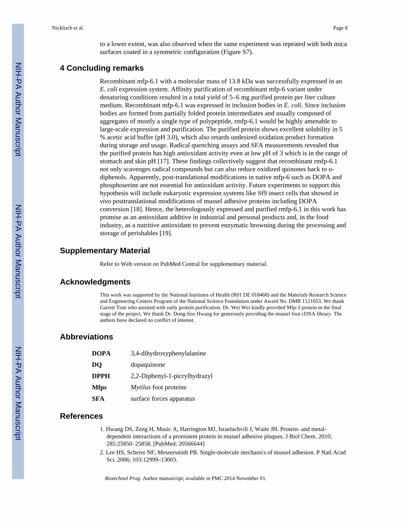
Purification of Recombinant mfp-6.1The expressed recombinant rmfp-6.1 had a His6 tag, which enabled affinity purification.Protein purity was assessed using SDS- (Figure 2A) or AU-PAGE (Figure S5, lower rightpanel). Denaturing conditions were used during purification to keep the basic and thiol-richrmfp-6.1 protein soluble at pH 7.4 in the washing and elution buffers. Cells were lysed with8M urea yielding 30 mL of cell lysate per liter of culture volume containing approximately5–6 mg rmfp-6.1. Lysates were loaded into IMAC columns and purified by affinityinteraction between nickel and the N-terminal His6 residues (Figure 2A). For furtherpurification we first dialyzed out the urea from the samples (1:1200) and freeze-dried thesoluble fraction prior to reconstitution in 5 % acetic acid buffer and application to a RP-HPLC column. This further increased the purity of the samples and resulted in monomericrmfp-6.1 as shown by MALDI mass spectrometry (Figure 2B). Occasionally, the thiol-richrmfp-6.1 became oxidized to dimers or higher oligomers during the purification procedureand size exclusion chromatography (SEC) were used to separate the monomeric rmfp-6.1form for all further experiments. MALDI-TOF MS analysis the purified sample showed thatthe actual molecular mass of recombinant rmfp-6.1 was almost identical to the calculatedmass of 13.6 kDa and confirmed that recombinant rmfp-6.1 had been successfully purifiedto a high level (Figure 2B).
Antioxidant activity of purified rmfp-6.1We used an optimized DPPH radical quenching assay to determine the relative antioxidantactivity of purified rmfp-6.1 as described previously [5]. In short, the DPPH radical has anabsorption maximum at 515–520 nm. Upon addition of antioxidants to the reaction bufferthe DPPH radical gets reduced to DPPH2, which can be followed by a decrease inabsorbance at λ515.
Like the native mfp-6, rmfp-6.1 shows highest solubility at pH 3 and starts precipitatingabove pH 5. Thus, we tested the antioxidant activity of rmfp-6.1 and ascorbic acid as controlantioxidant at pH 3. Adding 5 μM of rmfp-6.1 to 100 μM DPPH in the reaction buffer showsa continuous exponential decay of the relative DPPH absorbance over the time course with amaximum reduction down to 70 % after 60 min (Figure 3). The addition of the molarequivalent of ascorbic acid (5 μM) as control antioxidant decreases the initial DPPH radicalconcentration by about 5 % after 5 min and goes into equilibrium over the whole timecourse of 60 min. This shows an antioxidant activity of rmfp-6.1 that is 6 times higher thanthat of ascorbic acid at a low pH of 3. The curves were fitted to a biphasic exponential decayfunction using the equation: y = y0 + A1*exp(-x/t1) + A2*exp(-x/t2) and with regressioncoefficient, r2 values of 0.994 for ascorbic acid and 0.999 for rmfp-6.1. The analysis of therespective rate kinetics reveals that for ascorbic acid we can calculate only one fast timeconstant with t1 = t2 = 2.4 ± 1.2 s−1 indicative of a single population of electron donatinggroups involved in the reduction of DPPH. With pKas of 4.2 and 11.6 for the two enolic OHgroups of ascorbic acid we expect both reactive groups of ascorbic acid to be essentially
Nicklisch et al. Page 6
Biotechnol Prog. Author manuscript; available in PMC 2014 November 01.
NIH
-PA Author Manuscript
NIH
-PA Author Manuscript
NIH
-PA Author Manuscript

protonated at pH 3 and responsible for the DPPH reduction. For rmfp-6.1 we get two timeconstants, a fast t1 = 3.4 ± 1.4 s−1 and as slow t2 = 26.1 ± 9.7 s−1. Two different electrondonating populations exist in this protein and are responsible for DPPH reduction.Interestingly, the fast contribution (t1) is similar to that of ascorbic acid and might indicatethat mainly protonated enol or phenol groups in rmfp-6.1 are reducing the DPPH radical.With respect to this, the phenol groups of the 20 tyrosines (pKa ~ 10) could serve as protonand electron reservoirs for DPPH reduction by rmfp-6.1. However, we recently showed thatalso the cysteine thiol moiety (pKa ~ 8–9) in native mfp-6 is contributing to the proteinsantioxidant activity [3, 5]. In future experiments it needs to be determined which chemicalmoiety in the fp-6 proteins is the major contributor to the overall antioxidant activity. Forthis, a direct comparison of rmfp-6.1 and the DOPA-containing native mfp-6 antioxidantactivity as well as a comparison to other antioxidant compounds could further elucidate thecomparative antioxidant capacity of this recombinant antioxidant protein.
The Surface Forces Apparatus (SFA)A critical test of function in rmfp-6.1 is to demonstrate that the protein is capable ofmaintaining the adhesion of Mfps from the mussel plaque of Mytilus californianus bykeeping them in the reduced form ([3], Figure 5). Please refer to the supplemental“Materials and Methods” part in [1] for a more detailed description and illustration of usingthe SFA technique to identify mussel foot protein interactions.
The normalized force vs distance (F/R vs. D) profile of rmfp-6.1 coated mica surface againstthe opposing mica surface (with no protein on it) did not show any adhesion at pH 3.0 or7.5, and the measured forces were purely repulsive during both approach and separation(Figure 4A). Mfp-3 coated on only one mica sheet (asymmetric configuration) showed astrong adhesion with an adhesion energy of Wad = 3.2±0.4 mJ/m2 at pH 3.0 (Figure 4B).This is a combined effect of hydrogen bonding and hydrophobic interactions. The –OHgroup in DOPA forms hydrogen bonds with the O atoms of the polysiloxane groups in mica.In addition, the polysiloxane surface of mica exhibits some hydrophobic tendencies at lowpH (unpublished data, J. Israelachvili), hence the high adhesion energy could also beattributed to hydrophobic interactions between the mica surface and DOPA or otherhydrophobic residues in mfp-3 (i.e. tryptophan and tyrosine). Increasing the pH to 7.5oxidizes DOPA to dopaquinone but also makes mica more negatively charged andhydrophilic, resulting in an adhesion loss between the surfaces. The increase in hardwallthickness from ~ 2 nm to 10 nm was already reported by [15] and indicates reversibletautomerization of DOPA to Δ-DOPA which stiffens the backbone thereby expanding mfp-3at higher pHs. Lowering the pH back to 3.0 did not recover the adhesion by substantialamount (Wad = 0.6 mJ/m2). That dopaquinone is not reversibly reduced back to DOPA onpH reversal reflects a kinetic rather than thermodynamic barrier. However, injectingrmfp-6.1 between the mica surfaces recovered about 72 % of the initial adhesion energy(Initial Wad = 3.2±0.4 mJ/m2) of mfp-3 (Figure 4B) with a measured adhesion energy of Wad= 2.3±0.2 mJ/m2.
It is noteworthy that rmfp-6.1 shows the same hardwall thickness of about 10 nm at pH 3.0and at pH 7.5 where it undergoes precipitation (Figure 4A). Once rmfp-6.1 is injected intothe gap solution it recovers the adhesion of mfp-3 and only increases the hardwall thicknessfrom about 2 nm up to 5 nm. This slight increase in hardwall thickness upon adhesion lossrecovery was previously observed by [3] in the symmetrical setup. This might indicate thatupon oxidation both mfp-6 and rmfp-6.1 adapt a more compact structure with a smallerhydrodynamic radius and may partially intercalate into the mfp-3 layer instead of toppingoff the mfp-3 protein. A decrease in the hydrodynamic radius upon cysteine oxidation isknown to occur in human serum albumin [16]. Similar adhesion loss recovery of mfp-3, but
Nicklisch et al. Page 7
Biotechnol Prog. Author manuscript; available in PMC 2014 November 01.
NIH
-PA Author Manuscript
NIH
-PA Author Manuscript
NIH
-PA Author Manuscript
to a lower extent, was also observed when the same experiment was repeated with both micasurfaces coated in a symmetric configuration (Figure S7).
4 Concluding remarksRecombinant mfp-6.1 with a molecular mass of 13.8 kDa was successfully expressed in anE. coli expression system. Affinity purification of recombinant mfp-6 variant underdenaturing conditions resulted in a total yield of 5–6 mg purified protein per liter culturemedium. Recombinant mfp-6.1 was expressed in inclusion bodies in E. coli. Since inclusionbodies are formed from partially folded protein intermediates and usually composed ofaggregates of mostly a single type of polypeptide, rmfp-6.1 would be highly amenable tolarge-scale expression and purification. The purified protein shows excellent solubility in 5% acetic acid buffer (pH 3.0), which also retards undesired oxidation product formationduring storage and usage. Radical quenching assays and SFA measurements revealed thatthe purified protein has high antioxidant activity even at low pH of 3 which is in the range ofstomach and skin pH [17]. These findings collectively suggest that recombinant rmfp-6.1not only scavenges radical compounds but can also reduce oxidized quinones back to o-diphenols. Apparently, post-translational modifications in native mfp-6 such as DOPA andphosphoserine are not essential for antioxidant activity. Future experiments to support thishypothesis will include eukaryotic expression systems like Sf9 insect cells that showed invivo posttranslational modifications of mussel adhesive proteins including DOPAconversion [18]. Hence, the heterologously expressed and purified rmfp-6.1 in this work haspromise as an antioxidant additive in industrial and personal products and, in the foodindustry, as a nutritive antioxidant to prevent enzymatic browning during the processing andstorage of perishables [19].
Supplementary MaterialRefer to Web version on PubMed Central for supplementary material.
AcknowledgmentsThis work was supported by the National Institutes of Health (R01 DE 018468) and the Materials Research Scienceand Engineering Centers Program of the National Science Foundation under Award No. DMR 1121053. We thankGarrett Tom who assisted with early protein purification. Dr. Wei Wei kindly provided Mfp-3 protein in the finalstage of the project. We thank Dr. Dong-Soo Hwang for generously providing the mussel foot cDNA library. Theauthors have declared no conflict of interest.
Abbreviations
DOPA 3,4-dihydroxyphenylalanine
DQ dopaquinone
DPPH 2,2-Diphenyl-1-picrylhydrazyl
Mfps Mytilus foot proteins
SFA surface forces apparatus
References1. Hwang DS, Zeng H, Masic A, Harrington MJ, Israelachvili J, Waite JH. Protein- and metal-
dependent interactions of a prominent protein in mussel adhesive plaques. J Biol Chem. 2010;285:25850–25858. [PubMed: 20566644]
2. Lee HS, Scherer NF, Messersmith PB. Single-molecule mechanics of mussel adhesion. P Natl AcadSci. 2006; 103:12999–13003.
Nicklisch et al. Page 8
Biotechnol Prog. Author manuscript; available in PMC 2014 November 01.
NIH
-PA Author Manuscript
NIH
-PA Author Manuscript
NIH
-PA Author Manuscript

3. Yu J, Wei W, Danner E, Ashley RK, Israelachvili JN, Waite JH. Mussel protein adhesion dependson interprotein thiol-mediated redox modulation. Nature Chem Biol. 2011b; 7:588–590. [PubMed:21804534]
4. Zhao H, Waite JH. Linking adhesive and structural proteins in the attachment plaque of Mytiluscalifornianus. J Biol Chem. 2006; 281:26150–26158. [PubMed: 16844688]
5. Nicklisch SCT, Waite JH. Mini-review: The role of redox in DOPA-mediated marine adhesion.Biofouling. 2012; 28(8):865–877. [PubMed: 22924420]
6. Israelachvil JN, Min Y, Akbulut YM, Alig A, et al. Recent advances in the surface forces apparatus(SFA) technique. Rep Prog Phys. 2010; 73:036601.
7. Blois MS. Antioxidant Determinations by the Use of a Stable Free Radical. Nature. 1958;181:1199–1200.
8. Brand-Williams W, Cuvelier M, Berset C. Use of a free radical method to evaluate antioxidantactivity. LWT-Food. 1995; 8:25–30.
9. McIlvaine TC. A buffer solution for colorimetric comparison. J Biol Chem. 1921; 49:183–186.
10. Waite JH, Tanzer ML. Polyphenolic Substance of Mytilus edulis: Novel Adhesive Containing L-Dopa and Hydroxyproline. Science. 1981; 212:1038–1040. [PubMed: 17779975]
11. Shi, Q.; Jackowski, G. Gel electrophoresis of proteins: A practical approach. 3. Hames, BD.,editor. Oxford University Press; New York: 1998. p. 33
12. DeCollo TV, Lees WJ. Effects of aromatic thiols on thiol-disulfide interchange reactions that occurduring protein folding. J Org Chem. 2001; 66:4244–4249. [PubMed: 11397160]
13. Vallejo LF, Rinas U. Strategies for the recovery of active proteins through refolding of bacterialinclusion body proteins. Microb Cell Fact. 2004; 3:11. [PubMed: 15345063]
14. Sriubolmas N, Panbangred W, Sriurairatana S, Meevootisom V. Localization and characterizationof inclusion bodies in recombinant Escherichia coli cells overproducing penicillin G acylase. ApplMicrobiol Biotechnol. 1997; 47(4):373–8. [PubMed: 9163951]
15. Yu J, Wei W, Danner E, Israelachvili JN, Waite JH. Effects of interfacial redox in mussel adhesiveprotein films on mica. Adv Mater. 2011a; 23:2362–2366. [PubMed: 21520458]
16. Lee JY, Hirose M. Partially Folded State of the Disulfide-reduced Form of Human Serum Albuminas an Intermediate for Reversible Denaturation. J Biol Chem. 1992; 267(21):14753–14758.[PubMed: 1634518]
17. Lambers H, Piessens S, Bloem A, Pronk H, Finkel P. Natural skin surface pH is on average below5, which is beneficial for its resident flora. Int J Cosmetic Sci. 2006; 28:359–370.
18. Lim S, Kim KR, Choi YS, Kim DK, Hwang D, Cha HJ. In vivo post-translational modifications ofrecombinant mussel adhesive protein in insect cells. Biotechnol Prog. 2011; 27(5):1390–6.[PubMed: 21732552]
19. Whitaker, JR.; Lee, CY. Enzymatic browning and its prevention, Recent advances in chemistry ofenzymatic browning. In: Lee, CY.; Whitaker, JR., editors. Amer Chem Soc Symp Ser. Vol. 600.Washington DC: 1995. p. 2-7.
Nicklisch et al. Page 9
Biotechnol Prog. Author manuscript; available in PMC 2014 November 01.
NIH
-PA Author Manuscript
NIH
-PA Author Manuscript
NIH
-PA Author Manuscript

Figure 1. Expression of Recombinant mfp-6.1(A) Growth profiles of E. coli Rosetta 2 cells expressing rmfp-6.1 with and N-terminal His6-tag (V1DH). Cells were cultured in 2xTY medium at 37 °C and 250 rpm. The OD600 wasdetermined in duplicate with two independent samples out of each culture and the meanvalue was plotted against the cultivation time. Induction with different amounts of IPTG wascarried out when cells reached an OD600 of 0.7-0.8. Un-induced cells were grown as acontrol. Protein yields were 5-6 mg/L cell culture when induced with 10μM IPTG. (B)Effect of different inductor concentrations (IPTG) on the yield of rmfp-6.1 as shown byPAGE. 15% SDS-PAGE with Coomassie blue staining of the insoluble cell debris fractionwith rmfp-6.1 expressed in inclusion bodies. Recombinant cells were cultured in LBmedium at 37 °C and 250 rpm. Protein expression was induced at an OD600 of 0.7-0.8 usingdifferent amounts of IPTG (10, 50, 100μM). Lanes: M, protein molecular weight marker;mfp-6, native mfp-6 (11.6-12.2kDa) from M. californianus feet.
Nicklisch et al. Page 10
Biotechnol Prog. Author manuscript; available in PMC 2014 November 01.
NIH
-PA Author Manuscript
NIH
-PA Author Manuscript
NIH
-PA Author Manuscript
Figure 2. Purification of Recombinant mfp-6.1Purification of recombinant mfp-6.1. (A) 15% SDS-PAGE with Coomassie blue staining ofthe His6-tag purification of rmfp-6.1 using fast protein liquid chromatography and (B)MALDI TOF mass spectrum of the MFPLC-purified rmfp-6.1. Matrix=α-cyano-4-hydroxycinnamic acid, Accelerating Voltage=25000 V, Grid Voltage=93%, Guide WireVoltage = 0.3, Delay Time=300ms, CE = crude extract, El2-4 = flow through, El5-8 =elution, M = marker. Please refer to the FPLC chromatogram in Figure S9 for more details.
Nicklisch et al. Page 11
Biotechnol Prog. Author manuscript; available in PMC 2014 November 01.
NIH
-PA Author Manuscript
NIH
-PA Author Manuscript
NIH
-PA Author Manuscript

Figure 3. DPPH coupled assay: Antioxidant activity of rmfp-6.1Free radical reporter of antioxidant activity 1,1-diphenyl-2-picrylhydrazyl (DPPH). Timecourse of DPPH reduction using the antioxidant L-ascorbic acid and rmfp-6.1 protein at pH3. 1 ml of a 0.1 M citrate phosphate reaction buffer contained 0.3% (v/v) Triton X-100 and100 μM DPPH alone or supplemented with 5 μM of L-ascorbic acid or rmfp-6.1. Thereaction was started by the addition of the antioxidant and the decrease in absorbance wasmonitored at 515 nm. The absorbance of DPPH without antioxidant addition was stable over60 min (dashed line). For clarity, the control absorbance of 100 mM DPPH is displayed as100% DPPH radical.
Nicklisch et al. Page 12
Biotechnol Prog. Author manuscript; available in PMC 2014 November 01.
NIH
-PA Author Manuscript
NIH
-PA Author Manuscript
NIH
-PA Author Manuscript

Figure 4. SFA: mfp-3 adhesion recovery with rmfp-6.1(A) Interaction of rmfp-6.1 deposited on one of the mica surfaces with the opposing micasurface at pH 3.0 and 7.5 showing no adhesion between rmfp-6.1 and the mica surface. (B)Interaction of mfp-3 deposited on one of the mica surfaces with the opposing mica surface atpH 3.0 and 7.5 is shown. Strong adhesion (Wad = 3.2±0.4 mJ/m2) between mfp-3 and themica surface was observed at pH 3.0 (black circles). There was a significant loss in theadhesion on increasing the pH to 7.5 (green circles). Decreasing the pH back to 3.0 did notrecover the loss adhesion (black triangles). Incubating the mfp-3 surface in 100 pmolrmfp-6.1 recovered almost 72 % of the initial adhesion (Wad = 2.3±0.2 mJ/m2) and is shownin red circles. It should be noted that we reported the adhesion energy using the JKR theory(Wad = Fad/1.5πR) and not Derjaguin approximation (Wad = Fad/2πR) since the surfaces goto a flat contact. Forces measured on approach are shown by solid circles and on separationby open circles.
Nicklisch et al. Page 13
Biotechnol Prog. Author manuscript; available in PMC 2014 November 01.
NIH
-PA Author Manuscript
NIH
-PA Author Manuscript
NIH
-PA Author Manuscript

Figure 5. SFA discussionThiol-mediated adhesion loss recovery of rmfp-6.1. In the reduced state, the DOPA sidechains of mfp-3 contribute to H-bonding on mica surface (H-bond acceptor). At high pH(>5) auto-oxidation of DOPA to dopaquinone (DQ) prevents H-bonding and thus adhesionto mica. Oxidation of the thiol groups of rmfp-6.1 provides electrons to fully reduce DQback to DOPA.
Nicklisch et al. Page 14
Biotechnol Prog. Author manuscript; available in PMC 2014 November 01.
NIH
-PA Author Manuscript
NIH
-PA Author Manuscript
NIH
-PA Author Manuscript
Related Documents