388 AYU | Jul-Sep 2011 | Vol 32 | Issue 3 Introduction Prevention is undeniably the sensible maneuver towards the ultimate goal of cancer control. Several methods exist for the treatment of cancer in modern medicine. These include chemotherapy, radiotherapy, and surgery. Chemotherapy is now considered as the most effective method of cancer treatment. Intervention with chemopreventive agents at the early stage in carcinogenesis is theoretically more rational than attempting to eradicate fully developed tumors with chemotherapeutic drugs. However, most cancer chemotherapeutants severely affect the host normal cells. Hence the use of natural products now has been contemplated as of exceptional value in the control of cancer and its eradication program. [1,2] Plant-derived natural Address for correspondence: Mr. Shaktikumar C. Shivhare, Sadar Bazar, B/H Ram Mandir, Paratwada, Amaravati – 444 805, Maharashtra, India. E-mail: [email protected] AYU Access this article online Website: www.ayujournal.org DOI: 10.4103/0974-8520.93921 Quick Response Code: Pharmacological Research Antioxidant and anticancer evaluation of Scindapsus officinalis (Roxb.) Schott fruits Shaktikumar C. Shivhare 1 , Arjun O. Patidar 2 , K. G. Malviya 3 , K. K. Shivhare-Malviya 4 1 Asst. Professor in Pharmaceutics, Department of Pharmaceutical Biotechnology, Mahatma Jyoti Rao Phoole College of Health Care and Allied Sciences, MJRP University, Jaipur, Rajasthan, 2 Beharulal Mangilal College of Pharmaceutical Education and Research, Indore, Madhya Pradesh, 3 Asst. Professor, Department of Pharmacognosy, Mahatma Jyoti Rao Phoole College of Health Care and Allied Sciences, MJRP University, Jaipur, Rajasthan, 4 Medical Officer, Rani Dhullaiya Memorial Ayurvedic College and Hospital, Bhopal, Madhya Pradesh, India Abstract Several methods exist for the treatment of cancer in modern medicine. These include chemotherapy, radiotherapy, and surgery; most cancer chemotherapeutants severely affect the host normal cells. Hence the use of natural products now has been contemplated of exceptional value in the control of cancer. Plant-derived natural products such as flavonoids, terpenes, alkaloids, etc., have received considerable attention in recent years due to their diverse pharmacological properties including cytotoxic and cancer chemopreventive effects. Looking into this, the antioxidant and anticancer evaluation of Scindapsus officinalis (Roxb.) Schott fruits has been attempted to investigate its antitumor activity. The collection and authentication of the plant material mainly fruits and their various extractions was done. Identification of plant's active constituents by preliminary phytochemical screening was carried out. An in‑vitro cytotoxic assay using the brine shrimp lethality assay with brine shrimp eggs (Artemia salina) at a dose of 1–10 µg/ml with the fruit extract was performed by the method described by Mayer et al. Cell viability using the Trypan blue dye exclusion test at a dose of 20, 40, 80, 120, and 160 µg/ml dissolved in DMSO (final concentration 0.1%), and cytotoxicity using the MTT assay where viable cells convert MTT into a formazan salt were performed. All pharmacological screening for acute toxicity and anti tumour studies using EAC 1 x 10 6 cells/ mouse treated Swiss albino mice at a dose of 100 and 200 mg/kg/day orally was carried out. Biochemical and antioxidants predictions from various parameters like hematological, RBC, WBC count, PVC, total protein, Tissue Lipid Peroxidation, SOD, CATALASE, GPx, GST levels and anti tumour activity of Scindapsus officinalis were observed. The data was statistically analyzed by one- way ANOVA followed by Dunnett’s and Tukey’s multiple comparison test. The antitumor effect of the extract is evident from the increase in mean survival time (MST) lifespan, reduction in the solid tumor volume, and also the reversal of altered hematological parameters almost equal to normal. The methanolic extract (100–200 mg/kg/day orally) was found to be cytotoxic on human cancer cell lines. In addition, the methanolic extract had an antioxidant effect as reflected by a decrease in LPO, GST, and GPx (oxidant enzymes), and an increase in SOD and catalase. Key words: Antioxidant, Ehrlich’s ascites carcinoma, hematological parameter, mean survival time, solid tumor volume, Scindapsus officinalis [Downloaded free from http://www.ayujournal.org on Monday, December 9, 2019, IP: 157.46.78.202]

Welcome message from author
This document is posted to help you gain knowledge. Please leave a comment to let me know what you think about it! Share it to your friends and learn new things together.
Transcript
388 AYU | Jul-Sep 2011 | Vol 32 | Issue 3
Introduction
Prevention is undeniably the sensible maneuver towards the ultimate goal of cancer control. Several methods exist for
the treatment of cancer in modern medicine. These include chemotherapy, radiotherapy, and surgery. Chemotherapy is now considered as the most effective method of cancer treatment. Intervention with chemopreventive agents at the early stage in carcinogenesis is theoretically more rational than attempting to eradicate fully developed tumors with chemotherapeutic drugs. However, most cancer chemotherapeutants severely affect the host normal cells. Hence the use of natural products now has been contemplated as of exceptional value in the control of cancer and its eradication program.[1,2] Plant-derived natural
Address for correspondence: Mr. Shaktikumar C. Shivhare, Sadar Bazar, B/H Ram Mandir, Paratwada, Amaravati – 444 805, Maharashtra, India. E-mail: [email protected]
AYU Access this article online
Website: www.ayujournal.org
DOI: 10.4103/0974-8520.93921
Quick Response Code:Pharmacological ResearchAntioxidant and anticancer evaluation of Scindapsus officinalis (Roxb.) Schott fruitsShaktikumar C. Shivhare1, Arjun O. Patidar2, K. G. Malviya3, K. K. Shivhare-Malviya4
1Asst. Professor in Pharmaceutics, Department of Pharmaceutical Biotechnology, Mahatma Jyoti Rao Phoole College of Health Care and Allied Sciences, MJRP University, Jaipur, Rajasthan, 2Beharulal Mangilal College of Pharmaceutical Education and Research, Indore, Madhya Pradesh, 3Asst. Professor, Department of Pharmacognosy, Mahatma Jyoti Rao Phoole College of Health Care and Allied Sciences, MJRP University, Jaipur, Rajasthan, 4Medical Officer, Rani Dhullaiya Memorial Ayurvedic College and Hospital, Bhopal, Madhya Pradesh, India
Abstract
Several methods exist for the treatment of cancer in modern medicine. These include chemotherapy, radiotherapy, and surgery; most cancer chemotherapeutants severely affect the host normal cells. Hence the use of natural products now has been contemplated of exceptional value in the control of cancer. Plant-derived natural products such as flavonoids, terpenes, alkaloids, etc., have received considerable attention in recent years due to their diverse pharmacological properties including cytotoxic and cancer chemopreventive effects. Looking into this, the antioxidant and anticancer evaluation of Scindapsus officinalis (Roxb.) Schott fruits has been attempted to investigate its antitumor activity. The collection and authentication of the plant material mainly fruits and their various extractions was done. Identification of plant's active constituents by preliminary phytochemical screening was carried out. An in‑vitro cytotoxic assay using the brine shrimp lethality assay with brine shrimp eggs (Artemia salina) at a dose of 1–10 µg/ml with the fruit extract was performed by the method described by Mayer et al. Cell viability using the Trypan blue dye exclusion test at a dose of 20, 40, 80, 120, and 160 µg/ml dissolved in DMSO (final concentration 0.1%), and cytotoxicity using the MTT assay where viable cells convert MTT into a formazan salt were performed. All pharmacological screening for acute toxicity and anti tumour studies using EAC 1 x 10⁶ cells/mouse treated Swiss albino mice at a dose of 100 and 200 mg/kg/day orally was carried out. Biochemical and antioxidants predictions from various parameters like hematological, RBC, WBC count, PVC, total protein, Tissue Lipid Peroxidation, SOD, CATALASE, GPx, GST levels and anti tumour activity of Scindapsus officinalis were observed. The data was statistically analyzed by one-way ANOVA followed by Dunnett’s and Tukey’s multiple comparison test. The antitumor effect of the extract is evident from the increase in mean survival time (MST) lifespan, reduction in the solid tumor volume, and also the reversal of altered hematological parameters almost equal to normal. The methanolic extract (100–200 mg/kg/day orally) was found to be cytotoxic on human cancer cell lines. In addition, the methanolic extract had an antioxidant effect as reflected by a decrease in LPO, GST, and GPx (oxidant enzymes), and an increase in SOD and catalase.Key words: Antioxidant, Ehrlich’s ascites carcinoma, hematological parameter, mean survival time, solid tumor volume, Scindapsus officinalis
[Downloaded free from http://www.ayujournal.org on Monday, December 9, 2019, IP: 157.46.78.202]
AYU | Jul-Sep 2011 | Vol 32 | Issue 3 389
Shivhare, et al.: Anticancerous herb
products such as flavonoids, terpenes, alkaloids, etc., have received considerable attention in recent years due to their diverse pharmacological properties including cytotoxic and cancer chemopreventive effects. Only few of them were scientifically explored. Scindapsus officinalis is a folklore medicinal plant used against diseases such as skin diseases and asthma; it causes flatulence, is good for curing ulcers, leprosy, nocturnal emissions, diabetes, and throat troubles, ophthalmia, tumors, and dysentery. It is alexetric, anthelmintic, and astringent.[3-7]
Hence, the antioxidant and anticancer evaluation of S. officinalis (Roxb.) Schott fruits is an attempt to investigate the antitumor activity against Ehrlich’s ascites carcinoma in mice.
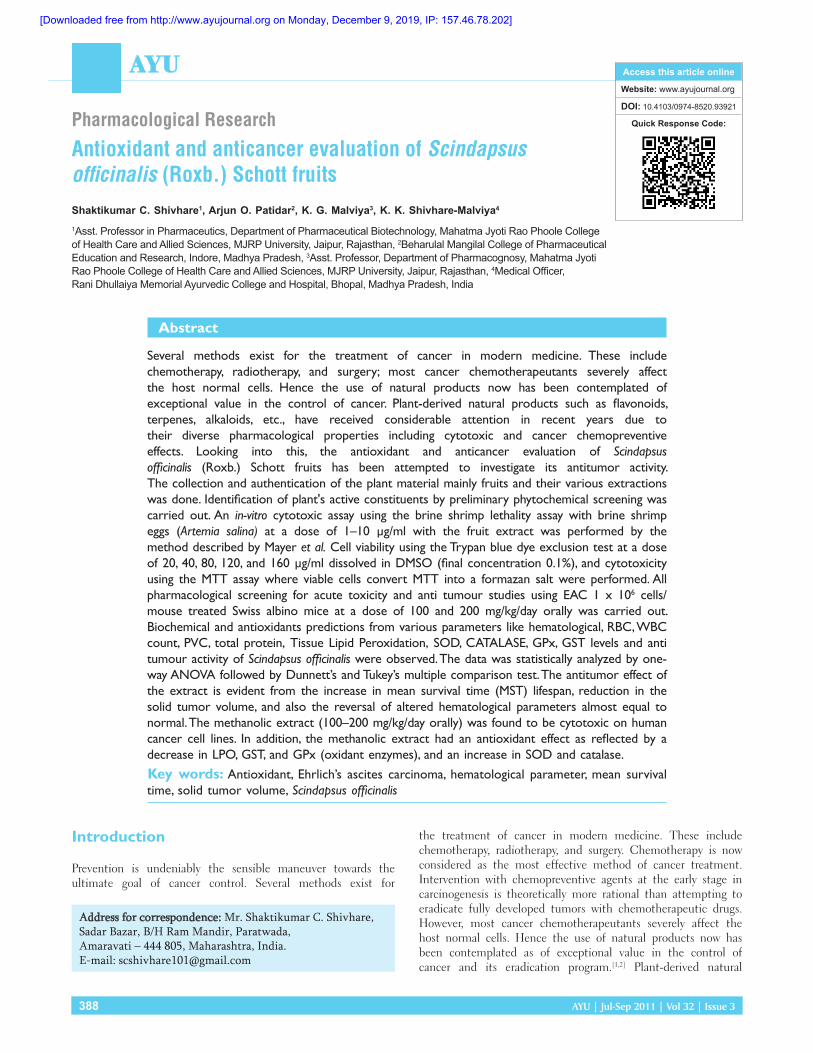
Scindapsus officinalis (Roxb.) SchottPlant name:[Figure 1a] Scindapsus officinalis, Class: Liliopsida, Subclass: Aridae Superorde: Aranae, Order: Alismatales, Family: Araceae
Vernacular names: Gaj‑pipali, Gajapipal, Atti‑tippili, Enugutippali etc.
Synonyms: Monstera officinalis (Roxb.) Schott, Pothos officinalis Roxb., Scindapsus annamicus Gagnep. Latin: Pothos officinalis Roxb.
Botanical name: Scindapsus officinalis (Roxb.) Schott[4-9]
Part useds: Fruit, Dried mature inflorescence, Shoots, Roots and Leaves.
Fruits: [Figure 1b] Fruits are the most important part of S. officinalis (Roxb.) Schott (Gaj‑pipali) and accepted in both Unani and Ayurveda for its known properties. Fruits are pungent in taste, used to sharp hearing, regulating the bowels, appetizer, galactagogue, stimulant, carminative, diaphoretic, anthelmintic, antiprotozoal, antidiarrheal, intestinal colic in horse, tuberculosis, azoena, cancer, cholera, pneumonia, scabies, syphilis and bronchitis, hypoglycemic. Fruit decoction is used in asthma, bronchitis, diarrhea, and also as an expectorant. Fruit pulp is applied externally in rheumatism. The Ayurvedic Pharmacopoeia of India recommends dried pieces of mature female spadix in dyspnea. It is also considered as atonic and diuretic. The fruit of Gaj‑pipali is an ingredient of preparation
“Chandraprabha” tablets and “Ma’jun Jograj Gugul churna" which is used as a Nervine tonic and anticatarrhal. It is also useful in facial paralysis and general paralysis. The fruit of Gaj‑pipali is an ingredient of the Ayurvedic preparation which is prescribed for Vatavikara, obesity (Medoroga and allied complaints), and obstinate urinary disorders including diabetes. It is useful in vitiated conditions of vata and kapha, diarrhea, cough, bronchitis, asthma, worm infestations, rheumatism, pharyngopathy, and helminthiasis.[4-15]
Materials and Methods
Collection and authentication of the plant material S. officinalis fruits were collected from Chennai, Tamil Nadu, India. They were identified and authenticated by a field botanist from Plant Anatomy Research Centre (PARC) (Tambaram, Chennai). The voucher specimen has been deposited at the herbarium unit of the Department of Pharmacognosy, Vel’s College of Pharmacy, Pallavaram, Chennai.
Extraction of S. officinalisThe dried fruits were used for the preparation of the extract. The fruits were dried under shade and coarsely powdered and extracted with petroleum ether, chloroform, acetone, methanol, and water.
Identification of plant active constituents by preliminary phytochemical studies [Table 1]. All the extracts of dried fruits of S. officinalis (Roxb.) Schott were subjected for the identification of various active constituents, such as carbohydrates, glycosides. alkaloids, fixed oils and fats, proteins and free amino acids, saponins, phenolic compounds and tannins, gums and mucilages, flavonoids, and phytosterol.
In vitro cytotoxicity assay using brine shrimp and human cancer cell linesBrine shrimp lethality and cytotoxicity assayThis assay uses brine shrimp, Artemia salina, which is used to determine the toxicity of the plant extract. Brine shrimp eggs of A. salina were hatched in artificial sea water (ASW; aqueous solution of NaCl, 3.8%w/v) and incubated at 25°C. The starting pH of the ASW was 8–8.5. After 48 h of hatching,
Figure 1: Scindapsus officinalis (a) plant and (b) fruits
a
b
Table 1: Preliminary phytochemical studiesExtracts PESO PESO CESO ACESO MESO AESOAlkaloids ‑ ‑ ‑ ‑ ‑s ‑Coumarin ‑ ‑ ‑ ‑ ‑ ‑Flavonoids ‑ ‑ ‑ + + +Glycosides ‑ ‑ ‑ ‑ + +Mucilages + + + + + ‑Tannins ‑ ‑ ‑ ‑ + ‑Phytosterols ‑ ‑ ‑ ‑ ‑ ‑Quinones ‑ ‑ ‑ ‑ ‑ ‑Saponins ‑ ‑ ‑ ‑ ‑ ‑Triterpenoid ‑ ‑ ‑ ‑ ‑ ‑+ = Present; - = Absent; PESO = Petroleum ether extract of fruits of Scindapsus officinalis; CESO = Chloroform extract of fruits S. officinalis; ACESO = Acetone extract of dried fruits of S. officinalis; MESO = Methanolic extract of dried fruits of S. officinalis; AESO = Aqueous extract of dried fruits of S. officinalis.
[Downloaded free from http://www.ayujournal.org on Monday, December 9, 2019, IP: 157.46.78.202]
390 AYU | Jul-Sep 2011 | Vol 32 | Issue 3
Shivhare, et al.: Anticancerous herb
the larvae (nauplii) were collected and used for brine shrimp lethality (BSL) bioassay.[16] The BSL assay of the successive leaf extract of plant materials was carried out by the method described by Mayer et al.[17-21] All the extracts and fractions were tested at concentration levels of 1–10 µg/ml. Each test was done in six replicates. A suspension of 10 nauplli (100 µl) was added into each well of a 24-well microplate and covered. The microplate was incubated for 24 h at room temperature. After this period, the number of dead nauplii in each well was counted using a binocular microscope. Potassium permanganate (100 µg/ml) was used as a standard (positive control) and a control reaction was carried out without the sample (negative control). The results were calculated statistically against their respective controls. The statistical method of probit analysis was used to calculate the concentration of the extract or fraction that would kill 50% of brine shrimps within the 24-h exposure, i.e., at the LC50 value with the 95% confidence intervals. The extracts were considered bioactive when the LC50 value was 1000 µg/ml or less [Table 2].
Cell culturesFour human cancer cell lines were used for the present investigation. Acute myeloblastic leukemia (HL-60) and chronic myelogenic leukemia (K562) cells were maintained in RPMI1640 supplemented with the 15% heat inactivated fetal bovine serum and gentamycin (40 µg/ml), penicillin (100 units/ml), and streptomycin (10µg/ml). Breast adenocarcinoma (MCF7) and cervical epithelial carcinoma (HeLa) cells were maintained in MEM supplemented with similar concentrations of serum and antibiotics as stated above. Cells were grown at 37°C in a humidified atmosphere of 5% CO2/95% air.
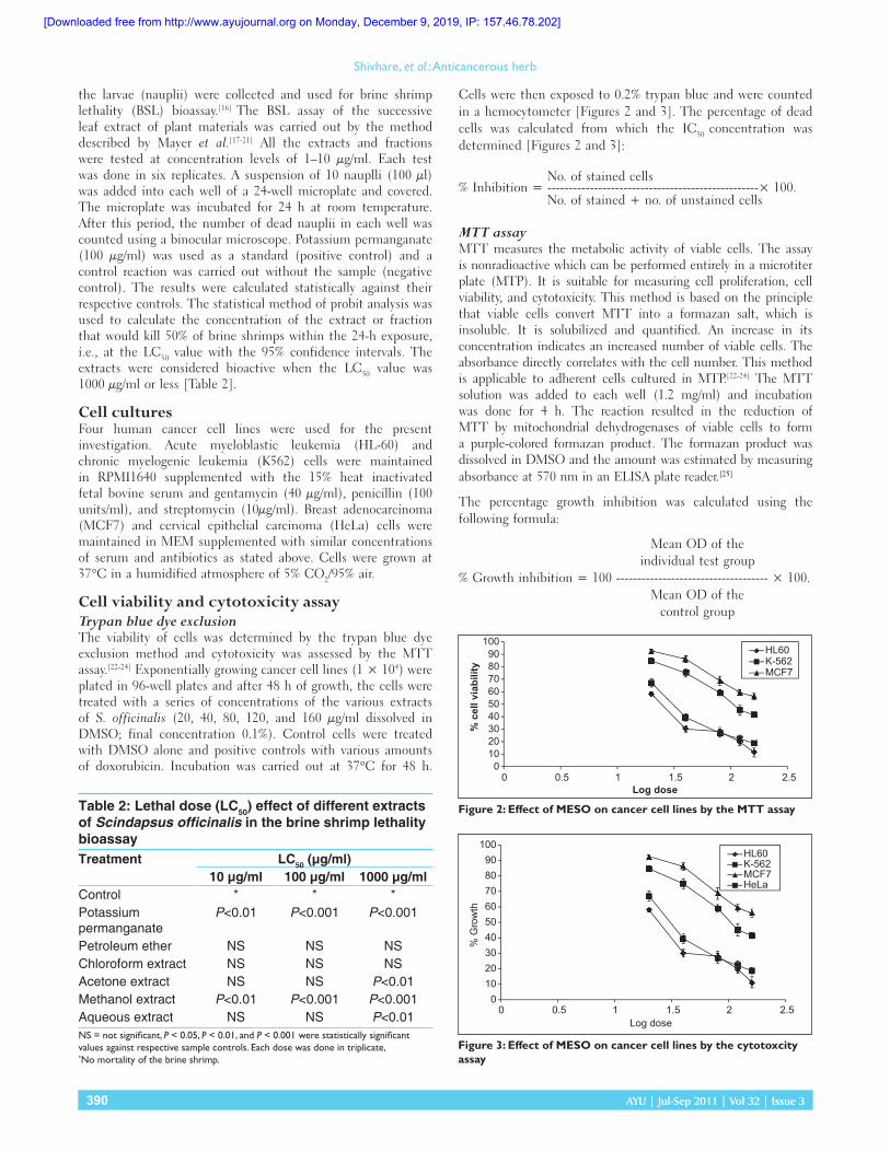
Cell viability and cytotoxicity assayTrypan blue dye exclusionThe viability of cells was determined by the trypan blue dye exclusion method and cytotoxicity was assessed by the MTT assay.[22-24] Exponentially growing cancer cell lines (1 × 10⁴) were plated in 96-well plates and after 48 h of growth, the cells were treated with a series of concentrations of the various extracts of S. officinalis (20, 40, 80, 120, and 160 µg/ml dissolved in DMSO; final concentration 0.1%). Control cells were treated with DMSO alone and positive controls with various amounts of doxorubicin. Incubation was carried out at 37°C for 48 h.
Cells were then exposed to 0.2% trypan blue and were counted in a hemocytometer [Figures 2 and 3]. The percentage of dead cells was calculated from which the IC50 concentration was determined [Figures 2 and 3]:
No. of stained cells% Inhibition = --------------------------------------------------× 100. No. of stained + no. of unstained cells
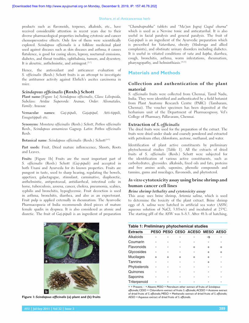
MTT assayMTT measures the metabolic activity of viable cells. The assay is nonradioactive which can be performed entirely in a microtiter plate (MTP). It is suitable for measuring cell proliferation, cell viability, and cytotoxicity. This method is based on the principle that viable cells convert MTT into a formazan salt, which is insoluble. It is solubilized and quantified. An increase in its concentration indicates an increased number of viable cells. The absorbance directly correlates with the cell number. This method is applicable to adherent cells cultured in MTP.[22-24] The MTT solution was added to each well (1.2 mg/ml) and incubation was done for 4 h. The reaction resulted in the reduction of MTT by mitochondrial dehydrogenases of viable cells to form a purple-colored formazan product. The formazan product was dissolved in DMSO and the amount was estimated by measuring absorbance at 570 nm in an ELISA plate reader.[25]
The percentage growth inhibition was calculated using the following formula:
Mean OD of the individual test group
% Growth inhibition = 100 ------------------------------------ × 100.Mean OD of the
control group
Table 2: Lethal dose (LC50) effect of different extracts of Scindapsus officinalis in the brine shrimp lethality bioassayTreatment LC50 (µg/ml)
10 µg/ml 100 µg/ml 1000 µg/mlControl * * *Potassium permanganate
P<0.01 P<0.001 P<0.001
Petroleum ether NS NS NSChloroform extract NS NS NSAcetone extract NS NS P<0.01Methanol extract P<0.01 P<0.001 P<0.001Aqueous extract NS NS P<0.01NS = not significant, P < 0.05, P < 0.01, and P < 0.001 were statistically significant values against respective sample controls. Each dose was done in triplicate, *No mortality of the brine shrimp.
Figure 3: Effect of MESO on cancer cell lines by the cytotoxcity assay
0102030405060708090
100
0 0.5 1 1.5 2 2.5
% G
row
th
Log dose
HL60K-562MCF7HeLa
Figure 2: Effect of MESO on cancer cell lines by the MTT assay
0102030405060708090
100
0 0.5 1 1.5 2 2.5
% c
ell v
iabi
lity
Log dose
HL60K-562MCF7
[Downloaded free from http://www.ayujournal.org on Monday, December 9, 2019, IP: 157.46.78.202]
AYU | Jul-Sep 2011 | Vol 32 | Issue 3 391
Shivhare, et al.: Anticancerous herb
Pharmacological screeningAcute toxicity studyIn the acute toxicity study, the toxicity effect of the drug can be evaluated and the LD50 and ED50 values and the therapeutic index were determined for the drug under investigation.
ProcedureThe up-and-down procedure was carried out as per the guidelines set by Organization for Economic Co-operation and Development (OECD). The chronic oral toxicity study was done according to OECD guideline 423.[26-27] In this experiment, two groups of Wistar rats (n = 6) were used. Animals were kept on fast overnight with water ad libitum and food was withheld for 3–4 h after the oral administration of the extracts. One group of animals was treated with a starting dose of 1000 mg/kg b.wt. orally and the maximum dose of 2000 mg/kg b.wt. was administered to rats. Another group was treated with normal saline. The observation included mortality and clinical signs, which included changes in skin fur, eyes, and mucous membranes. The gross behaviors like body positions, locomotion, rearing, tremors, gait, passivity, grip strength, pain response, stereotypy, vocalization, righting reflex, body weight, and water intake were observed. From acute toxicity studies, it was observed that the administration of the methanol extract of S. officinalis to rats did not induce drug-related toxicity and mortality in the animals. The rats treated with the methanol extract of S. officinalis were well tolerated and exhibited normal behavior up to a dose of 2 g/kg orally. All animals were alert with normal grooming, touch response, and pain response and there was no sign of passivity, stereotypy, and vocalization. Their motor activity and secretary signs were also normal.
Anti-tumor activity of the S. officinalis methanolic extractAnimals usedSwiss albino mice (20–25 g) were used throughout the study after the clearance from the animal ethical committee (IX/290/CPCSEA/PHARMCOL/I-1.15/06). They were housed in standard microloan boxes and were given standard laboratory diet and water ad libitum.
Tumor cell linesEhrlich’s ascites carcinoma (EAC) cells were obtained through the courtesy of Amala Cancer Research Centre (Thrissur, Kerala, India). EAC cells were maintained by weekly interaperitoneal (i.p.) inoculation of 1 × 10⁶ cells/mouse.
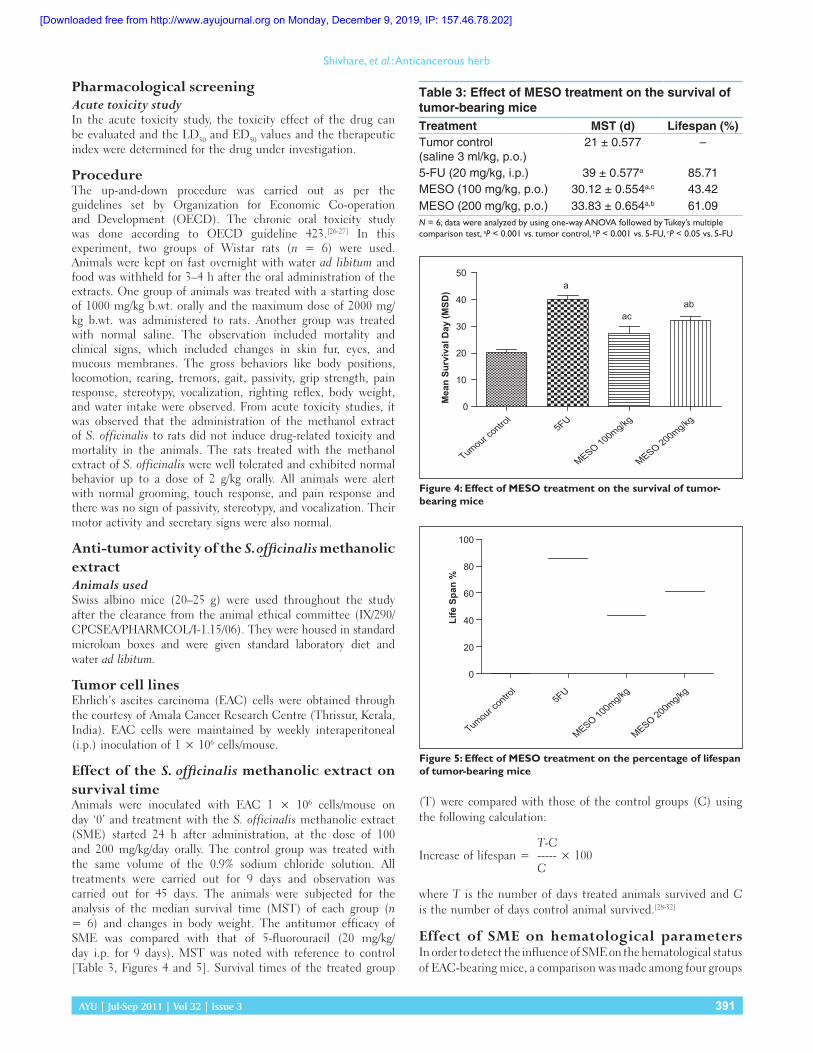
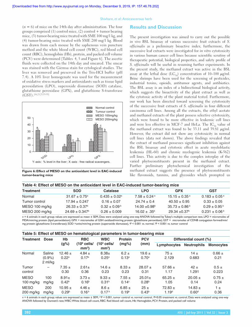
Effect of the S. officinalis methanolic extract on survival timeAnimals were inoculated with EAC 1 × 10⁶ cells/mouse on day ‘0’ and treatment with the S. officinalis methanolic extract (SME) started 24 h after administration, at the dose of 100 and 200 mg/kg/day orally. The control group was treated with the same volume of the 0.9% sodium chloride solution. All treatments were carried out for 9 days and observation was carried out for 45 days. The animals were subjected for the analysis of the median survival time (MST) of each group (n = 6) and changes in body weight. The antitumor efficacy of SME was compared with that of 5-fluorouracil (20 mg/kg/day i.p. for 9 days). MST was noted with reference to control [Table 3, Figures 4 and 5]. Survival times of the treated group
(T) were compared with those of the control groups (C) using the following calculation:
T-CIncrease of lifespan = ----- × 100 C
where T is the number of days treated animals survived and C is the number of days control animal survived.[28-32]
Effect of SME on hematological parameters In order to detect the influence of SME on the hematological status of EAC-bearing mice, a comparison was made among four groups
Table 3: Effect of MESO treatment on the survival of tumor‑bearing miceTreatment MST (d) Lifespan (%)Tumor control (saline 3 ml/kg, p.o.)
21 ± 0.577 –
5‑FU (20 mg/kg, i.p.) 39 ± 0.577a 85.71MESO (100 mg/kg, p.o.) 30.12 ± 0.554a,c 43.42MESO (200 mg/kg, p.o.) 33.83 ± 0.654a,b 61.09N = 6; data were analyzed by using one-way ANOVA followed by Tukey’s multiple comparison test, aP < 0.001 vs. tumor control, bP < 0.001 vs. 5-FU, cP < 0.05 vs. 5-FU
Figure 5: Effect of MESO treatment on the percentage of lifespan of tumor-bearing mice
Tumour contro
l5FU
MESO 100mg/kg
MESO 200mg/kg
0
20
40
60
80
100
Life
Spa
n %
Figure 4: Effect of MESO treatment on the survival of tumor-bearing mice
Tumour contro
l5FU
MESO 100mg/kg
MESO 200mg/kg0
10
20
30
40
50a
abac
Mea
n Su
rviv
al D
ay (M
SD)
[Downloaded free from http://www.ayujournal.org on Monday, December 9, 2019, IP: 157.46.78.202]
392 AYU | Jul-Sep 2011 | Vol 32 | Issue 3
Shivhare, et al.: Anticancerous herb
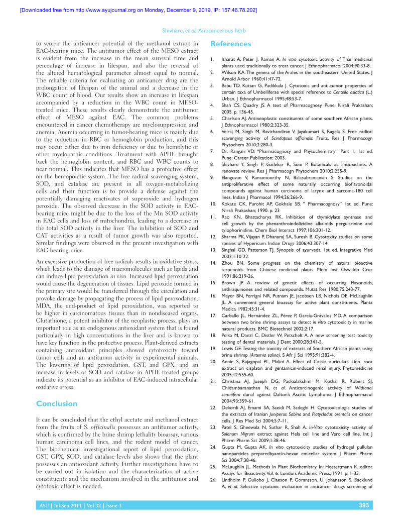
(n = 6) of mice on the 14th day after administration. The four groups comprised (1) control mice, (2) control + tumor bearing mice, (3) tumor-bearing mice treated with SME 100 mg/1 kg, and (4) tumor-bearing mice treated with SME 200 mg/1 kg. Blood was drawn from each mouse by the saphenous vein puncture method and the white blood cell count (WBC), red blood cell count (RBC), hemoglobin (Hb), protein, and packed cell volume (PCV) were determined [Tables 4, 5 and Figure 6]. The ascetic fluids were collected on the 14th day and smeared. The smear was stained with the Giemsa stain for cytological studies. The liver was removed and preserved in the Tris-HCI buffer (pH 7.4). A 10% liver homogenate was used for the measurement of oxidative stress markers and antioxidants such as tissue lipid peroxidation (LPO), superoxide dismutase (SOD) catalase, glutathione peroxidase (GPX), and glutathione S-transferase (GST).[16,21,25,33-35]
Results and Discussion
The present investigation was aimed to carry out the possible in vivo BSL bioassay of various successive fruit extracts of S. officinalis as a preliminary bioactive index; furthermore, the successive leaf extracts were investigated for in vitro cytotoxicity in various human cancer cell lines because scientific scrutiny of therapeutic potential, biological properties, and safety profile of S. officinalis will be useful in resuming further experiments. In the present study, the methanol extract was active in the BSL assay at the lethal dose (LC50) concentration of 10–100 µg/ml. Brine shrimps have been used for the screening of pesticides, industrial toxins, opioids, antitumor agents, and antibiotics. The BSL assay is an index of a bidirectional biological activity, which suggests the bioactivity of the plant extract as well as the cytotoxic activity of the plant material tested. Furthermore, our work has been directed toward screening the cytotoxicity of the successive fruit extracts of S. officinalis in four different carcinoma cell lines. Among all the extracts, the ethyl acetate and methanol extracts of the plant possess selective cytotoxicity, which were found to be more effective in leukemic cell lines and were less effective in MCF-7 and HeLa. The IC50 value of the methanol extract was found to be 33.11 and 39.81 µg/ml. However, the extract did not show any cytotoxicity in normal cell lines (data not shown). The above findings revealed that the extract of methanol possesses significant inhibition against the BSL bioassay and cytotoxic effect in acute myeloblastic leukemia (HL-60) and chronic myelogenic leukemia (K-562) cell lines. This activity is due to the complex interplay of the varied phytoconstituents present in the methanol extract. Further preliminary phytochemical investigation of the methanol extract suggests the presence of phytoconstituents like flavonoids, tannins, and glycosides which prompted us
Table 4: Effect of MESO on the antioxidant level in EAC‑induced tumor‑bearing miceTreatment SOD Catalase LPO GPX GSTNormal 31.67 ± 0.75a 0.435 ± 0.02a 7.58 ± 0.24 a 15.15 ± 0.35 a 0.183 ± 0.05 a
Tumor control 17.94 ± 0.247 0.16 ± 0.07 24.74 ± 0.4 40.50 ± 0.95 0.33 ± 0.05MESO 100 mg/kg 26.33 ± 0.37b 0.32 ± 0.09 b 14.00 ±0.98b 35.73 ± 0.86 b 0.29 ± 0.95 b
MESO 200 mg/kg 24.69 ± 0.347b 0.26 ± 0.009 16.02 ± .35b 29.34 ±0.37b 0.231 ± 0.06 b
n = 6 animals in each group; values are expressed as mean ± SEM, Data were analyzed using one-way ANOVA followed by Tukey’s multiple comparison test, LPO = micromoles of MDA/min/mg protein (lipid peroxidation); GPX = micromoles of GSH oxidized/min/mg protein (glutathione peroxidase); GST = micromoles of CDNB conjugation formed/min/mg protein (glutathione S-transferase); SOD = units/min/mg protein (superoxide dismutase), aP < 0.001 vs. normal, bP < 0.001 vs. tumor control
Table 5: Effect of MESO on hematological parameters in tumor‑bearing miceTreatment Dose Hb
(g%)RBC
(106 cells/mm3)
WBC (103 cells/
mm3)
Protein (mg%)
PCV (mm)
Differential count (%)Lymphocytes Neutrophils Monocytes
Normal Saline(0.9%)2 ml/kg
12.46 ± 0.22a
4.84 ± 0.17a
8.38± 0.25a
6.2 ± 0.13a
19.6 ± 0.70a
75 ± 2.129
14 ± 0.683
0.66 ± 0.21
Tumor control
– 7.35 ± 0.30
2.61± 0.36
14.6 ± 0.23
8.33 ± 0.23
28.07 ± 0.31
57.66 ± 1.17
42 ± 1.291
0.5 ± 0.223
MESO100 mg/kg
100 mg/kg
8.91± 0.42b
3.73 ± 0.16b
9.33 ± 0.31b
7.55 ± 0.14c
25.01± 0.28b
65.25 ± 1.05
20.05 ± 0.14
0.75 ± 0.24
MESO200 mg/kg
200mg/kg
10.95 ± 0.29b
4.46 ± 0.16 b
8.6 ± 0.17 b
6.85 ± 0.19b
25 ± 0.43b
72.83 ± 1.19d
14.83 ± 0.60d
1 ± 0.36
n = 6 animals in each group; values are expressed as mean ± SEM, a,bP < 0.001, tumor control vs. normal control, cP<0.05 treatment vs. control, Data were analyzed using one-way ANOVA followed by Dunnett’s test, WBC: White blood cell count, RBC: Red blood cell count, Hb: Hemoglobin, PCV: Protein, and packed cell volume
Figure 6: Effect of MESO on the antioxidant level in EAC-induced tumor-bearing mice
SODLPO
GPX0
10
20
30
40
50
Normal controlTumour controlMESO 100mg/kgMESO 200mg/kg
a
bb
c b
a
a
bc
Y-axis : % level in the liver ; X-axis : free radical scavengers.
[Downloaded free from http://www.ayujournal.org on Monday, December 9, 2019, IP: 157.46.78.202]
AYU | Jul-Sep 2011 | Vol 32 | Issue 3 393
Shivhare, et al.: Anticancerous herb
to screen the anticancer potential of the methanol extract in EAC-bearing mice. The antitumor effect of the MESO extract is evident from the increase in the mean survival time and percentage of increase in lifespan, and also the reversal of the altered hematological parameter almost equal to normal. The reliable criteria for evaluating an anticancer drug are the prolongation of lifespan of the animal and a decrease in the WBC count of blood. Our results show an increase in lifespan accompanied by a reduction in the WBC count in MESO-treated mice. These results clearly demonstrate the antitumor effect of MESO against EAC. The common problems encountered in cancer chemotherapy are myelosuppression and anemia. Anemia occurring in tumor-bearing mice is mainly due to the reduction in RBC or hemoglobin production, and this may occur either due to iron deficiency or due to hemolytic or other myelopathic conditions. Treatment with APHE brought back the hemoglobin content, and RBC and WBC counts to near normal. This indicates that MESO has a protective effect on the hemopoietic system. The free radical scavenging system, SOD, and catalase are present in all oxygen-metabolizing cells and their function is to provide a defense against the potentially damaging reactivates of superoxide and hydrogen peroxide. The observed decrease in the SOD activity in EAC-bearing mice might be due to the loss of the Mn SOD activity in EAC cells and loss of mitochondria, leading to a decrease in the total SOD activity in the liver. The inhibition of SOD and CAT activities as a result of tumor growth was also reported. Similar findings were observed in the present investigation with EAC-bearing mice.
An excessive production of free radicals results in oxidative stress, which leads to the damage of macromolecules such as lipids and can induce lipid peroxidation in vivo. Increased lipid peroxidation would cause the degeneration of tissues. Lipid peroxide formed in the primary site would be transferred through the circulation and provoke damage by propagating the process of lipid peroxodation. MDA, the end-product of lipid peroxidation, was reported to be higher in carcinomatous tissues than in nondiseased organs. Glutathione, a potent inhibitor of the neoplastic process, plays an important role as an endogenous antioxidant system that is found particularly in high concentrations in the liver and is known to have key function in the protective process. Plant-derived extracts containing antioxidant principles showed cytotoxicity toward tumor cells and an antitumor activity in experimental animals. The lowering of lipid peroxidation, GST, and GPX, and an increase in levels of SOD and catalase in APHE-treated groups indicate its potential as an inhibitor of EAC-induced intracellular oxidative stress.
Conclusion
It can be concluded that the ethyl acetate and methanol extract from the fruits of S. officinalis possesses an antitumor activity, which is confirmed by the brine shrimp lethality bioassay, various human carcinoma cell lines, and the rodent model of cancer. The biochemical investigational report of lipid peroxidation, GST, GPX, SOD, and catalase levels also shows that the plant possesses an antioxidant activity. Further investigations have to be carried out in isolation and the characterization of active constituents and the mechanism involved in the antitumor and cytotoxic effect is needed.
References
1. Itharat A, Peter J. Raman A. In vitro cytotoxic activity of Thai medicinal plants used traditionally to treat cancer. J Ethnopharmacol 2004;90:33-8.
2. Wilson KA. The genera of the Arales in the southeastern United States. J Arnold Arbor 1960;41:47-72.
3. Babu TD, Kuttan G, Padikkala J. Cytotoxic and anti-tumor properties of certain taxa of Umbelliferae with special reference to Centella asiatica (L.) Urban. J Ethnopharmacol 1995;48:53-7.
4. Shah CS, Quadry JS. A text of Pharmacognosy. Pune: Nirali Prakashan; 2005. p. 136-45.
5. Charlson AJ. Antineoplastic constituents of some southern African plants. J Ethnopharmacol 1980;2:323-35.
6. Velraj M, Singh M, Ravichandiran V, Jayakumari S, Ragela S. Free radical scavenging activity of Scindapsus officinalis Fruits. Res J Pharmacogn Phytochem 2010;2:280-3.
7. Dr. Rangari VD. “Pharmacognosy and Phytochemistry” Part 1, 1st ed. Pune: Career Publication; 2003.
8. Shivhare Y, Singh P, Gadekar R, Soni P. Botanicals as antioxidants: A renovate review. Res J Pharmacogn Phytochem 2010;2:255-9.
9. Elangovan V, Ramamoorthy N, Balasubramanian S. Studies on the antiproliferative effect of some naturally occurring bioflavonoidal compounds against human carcinoma of larynx and sarcoma-180 cell lines. Indian J Pharmacol 1994;26:266-9.
10. Kokate CK, Purohit AP, Gokhale SB. “ Pharmacognosy” 1st ed. Pune: Nirali Prakashan; 1990. p. 23
11. Rao KN, Bhattacharya RK. Inhibition of thymidylate synthase and cell growth by the phenanthroindolizidine alkaloids pergularinine and tylophorinidine. Chem Biol Interact 1997;106:201-12.
12. Sharma PK, Vijayan P, Dhanaraj SA, Suresh B. Cytotoxity studies on some spesies of Hypericum. Indian Drugs 2006;43:307-14.
13. Singhal GD, Patterson TJ. Synopsis of ayurveda. 1st ed. Integrative Med 2002;1:10-22.
14. Zhou BN. Some progress on the chemistry of natural bioactive terpenoids from Chinese medicinal plants. Mem Inst Oswaldo Cruz 1991;86:219-26.
15. Brown JP. A rewiew of genetic effects of occurring Flavonoids, anthraquinones and related compounds. Mutat Res 1980;75:243-77.
16. Meyer BN, Ferrigni NR, Putnam JE, Jacobsen LB, Nichols DE, McLaughlin JL. A convenient general bioassay for active plant constituents. Planta Medica 1982;45:31-4.
17. Carballo JL, Hernández ZL, Pérez P, García-Grávalos MD. A comparison between two brine shrimp assays to detect in vitro cytotoxicity in marine natural products. BMC Biotechnol 2002;2:17.
18. Pelka M, Danzl C, Distler W, Petschelt A. A new screening test toxicity testing of dental materials. J Dent 2000;28:341-5.
19. Lewis GE. Testing the toxicity of extracts of Southern African plants using brine shrimp (Artemia salina). S Afr J Sci 1995;91:382-4.
20. Annie S, Rajagopal PL, Malini A. Effect of Cassia auriculata Linn. root extract on cisplatin and gentamicin-induced renal injury. Phytomedicine 2005;12:555-60.
21. Christina AJ, Joseph DG, Packialakshmi M, Kothai R, Robert SJ, Chidambaranathan N, et al. Anticarcinogenic activity of Withanai somnifera dunal against Dalton’s Ascitic Lymphoma. J Ethnopharmacol 2004;93:359-61.
22. Dekordi AJ, Emami SA, Saeidi M, Sadeghi H. Cytotoxicologic studies of the extracts of Iranian Juniperus Sabina and Platycladus orentalis on cancer cells. J Res Med Sci 2004;5:7-11.
23. Patel S, Gheewala N, Suthar R, Shah A. In‑Vitro cytotoxicity activity of Solanum Nigrum extract against Hela cell line and Vero cell line. Int J Pharm Pharm Sci 2009;1:38-46.
24. Gupta M, Gupta AK. In vitro cytotoxicity studies of hydrogel pullulan nanoparticles preparedbyaot/n-hexan emicellar system. J Pharm Pharm Sci 2004;7:38-46.
25. McLaughlin JL. Methods in Plant Biochemistry. In: Hostettmann K, editor. Assays for Bioactivity. Vol. 6. London: Academic Press; 1991. p. 1-33.
26. Lindholm P, Gullobo J, Claeson P, Goransson. U, Johansson S, Backlund A, et al. Selective cytotoxic evaluation in anticancer drugs screening of
[Downloaded free from http://www.ayujournal.org on Monday, December 9, 2019, IP: 157.46.78.202]
394 AYU | Jul-Sep 2011 | Vol 32 | Issue 3
Shivhare, et al.: Anticancerous herb
fractionated plant extracts J Biomol Screen 2002;7:333-40.27. Croce CM. "Oncogenes and cancer". N Engl J Med 2008;358:502-11.28. Banerjee S, Prashar R, Kumar A, Rao AR. Modulatory influence of alcoholic
extract of Ocimum leaves on Carcinogen- metabolizing enzyme activities and reduced glutathione levels in mouse. Nutr Cancer 1996;25:205-17.
29. Jageti GC, Rao SK. Evaluation of the antineoplastic activity of Guduchi (Tinospora cordifolia) in Ehrlich ascites carcinoma in bearing mice. J Biol Pharm Bull 2006;29:460-6.
30. Knanam JA, Bag SP. Antineoplastic activity of copper benzohydroxamic acid complex against Erhlich Ascitic carcinoma in mice. Indian J Pharmacol 1997;29:157-61.
31. Mazumdar UK, Gupta M, Maiti S, Mukherjee D. Antitumor activity of
Hygrophila spinosa on Ehrlich ascites carcinoma and sarcoma-180 induced mice. Indian J Exp Biol 1997;35:473-7.
32. Rosangki G, Prasad SB. Antitumour activity of some plants from Meghalaya against murine Ehrlich Ascites Carcinoma. Indian J Exp Biol 2004;42:981-8.
33. Denizot F, Lang R. Rapid colorimetric assay for cell growth and survival: Modifi cations to the tetrazolium dye procedure giving improved sensitivity and reliability. J Immunol Methods 1986;89:271-7.
34. Miyake Y, Yamamoto K, Tsujihara N, Osawa T. Protective effects of lemon flavonoids on oxidative stress in diabetic rats Lipids 1998;33:689-95.
35. Leeuwenburgh C, Ji LL. Glutathione depletion in rested and exercised mice: Biochemical consequence and adaptation. Arch Biochem Biophys 1995;316:941-9.
<Y^Z] ∆√}√|_
«©…]…∑ wv√ wvw÷v }√≤« I≤* G˙fifiA
_<ØvwtvI√} ∆]. <_ËY}≤, G©t÷A G√≤. …√æ>]Z√}, w≤v. ©]. I√∑Ë]fi, w≤v. w≤v. <_ËY}≤ I√∑Ë]fi >
w§*v∆} fi√ wvw÷v }√≤« w≤v @…Î√} Y≤ot ∆√I√^fio# ™wvI√≤<E}≤…], }≤™s>fi√≤pI]÷ <ΙwvM∆√ GEË√ _Ofi <ΙwvM∆√ wv√ @…fi√≤« ™wvfi√ ©√o√ Y§ ©√≤ ™wv ∆√I√^fi wv√≤<_wv√G√≤* wv√≤ µ] Y√<A …Y|¯Î√o] Y§ $ fi<Z }√≤«] G√fitË≤÷<Zwv G√§B<pfi√≤* (fiE√ «©…]…∑) wv√ @…fi√≤« wv}≤ o√≤ wvw÷v ©§∆≤ G∆√˙fi }√≤«√≤* wv√≤ µ] <lA√ ™wv∆] Z¯~…™}J√I√≤* w≤v <Afi|<⁄o ™wvfi√ ©√ ∆wvo√ Y§ $ …L¢oto ∑≤x I≤* x√≤©wvo√÷G√≤* A≤ «©…]…∑ wv√ ÎÍY√≤* …} …Lfi√≤« wv}w≤v wvw÷v }√≤« ∆◊l|<po Û∆ G√§B<p w≤v «tJ√≤* w≤v l√}≤ I≤* ©√Awv√}] Z≤A≤ wv√ …Lfi√∆ ™wvfi√ Y§ $ …Lfi√≤«√≤* G√§} o‘fi√≤* w≤v G√p√} …} fiY wvY√ ©√ ∆wvo√ Y§ ™wv fiY G√§B<p wvw÷v }√≤« <Afi|⁄J wv}A≤ I≤* wv√}«} <∆Ù Y√≤ ∆wvo] Y§ $
[Downloaded free from http://www.ayujournal.org on Monday, December 9, 2019, IP: 157.46.78.202]
Related Documents