Adv. Biomed. Pharma. 2:2 (2015) 56-67 Original Paper Antioxidant activity of aqueous extract of Piliostigma thonningii Leaf following Indomethacin induced gastric mucosa onslaught in male wistar Albino Rats. Dasofunjo K 1 , Okwari O.O 2 , Jeje S.O 2 , Alagwu E.A 3 , Emerole .C.G 4 and Ogar N.B 1 1 Department of Medical Biochemistry, Cross River University of Technology, Okuku, Nigeria. 2 Department of Physiology, Cross River University of Technology, Okuku, Nigeria. 3 Department of Physiology, College of Medicine and Health Sciences .Imo State University, Owerri, Nigeria. 4 Optometry Unit Eye Clinic, Federal Medical Centre, Owerri, Nigeria. *Corresponding Author; Dasofunjo Kayode: Email ID: [email protected] .Tel:+2348032370325 Running Title: Antioxidant Activity of Aqueous Extract of Piliostigma thonningii Received: 19 February 2015; Revised 25 March 2015; Accepted: 16 April 2015 Abstract Antioxidant activity of P. thonningii extract following indomethacin induced gastric mucosa onslaught in male Wistar albino rats was carried out on 36 male rats that were divided into six (6) groups of 6 rats each. Group one (1) served as control and was given 0.5ml of normal saline (vehicle). Group two (2) was treated with 100mg/kg body weight of the drug (Cimetidine). Group three (3), five (5) and six (6) were given 100, 100 and 200mg/kg body weight of the extract respectively. The vehicle and extract were administered orally while the drug was administered intra-muscularly for 12days. After 12days of administration ,all rats were fasted for 24 hours, gastric ulceration was then induced using 40mg/kg body weight of indomethacin orally only to group 2, 4, 5 and 6 respectively. Twelve (12) hours after indomethacin administration all rats were sacrificed after been anaesthetised with chloroform, the abdomen of each rats was opened to remove the stomach, Liver, Kidney and Testes respectively for the determination of antioxidant activity and tissue protein concentration. The result shows significant (P<0.05) increase in tissue protein, SOD, CAT but significant (P<0.05) decrease in MDA in groups treated with the extract compared with the control .Similar pattern was also exhibited with Cimetidine treated group. The untreated group showed a significant (P<0.05) decrease in tissue protein, SOD, CAT with a significant increase (P<0.05) in MDA when compared with the control. The biochemical and physiological alterations from this result are indications that the extract has a dose dependent protective effect in indomethacin-mediated gastric mucosa onslaught, which can be attributed to its antioxidant potential or activity. Keywords: Antioxidant activity, Indomethacin, Piliostigma thonningii, Ulcer, Mortality, Morbidity Introduction An ulcer is basically an inflamed break in the skin or mucus membrane lining the alimentary tract. Ulceration occurs when there is a disturbance of the normal equilibrium caused by either enhanced aggression or diminished mucosal resistance [1]. About 19 out of 20 peptic ulcers are duodenal while gastric ulcers found in the stomach wall are less common [2]. The gastric mucosa is continuously exposed to potentially injurious agents such as acids, pepsin, bile acids, food ingredients, bacterial products (Helicobacter pylori) and drugs [3]. These agents have been implicated in the pathogenesis of gastric ulcer, including enhanced gastric acid and pepsin secretion, inhibition of prostaglandin synthesis and cell proliferation growth, diminished gastric blood flow and gastric motility [3]. Symptoms of ulcer include epigastria pain of a burning or gnawing nature (postprandial pain and pain relieved by food or antacids), nausea, vomiting, belching and bloating. Advances in Biomedicine and Pharmacy (An International Journal of Biomedicine, Natural Products and Pharmacy) 56

Welcome message from author
This document is posted to help you gain knowledge. Please leave a comment to let me know what you think about it! Share it to your friends and learn new things together.
Transcript

Adv. Biomed. Pharma. 2:2 (2015) 56-67
Original Paper
Antioxidant activity of aqueous extract of Piliostigma thonningii Leaf
following Indomethacin induced gastric mucosa onslaught in male
wistar Albino Rats.
Dasofunjo K1, Okwari O.O2, Jeje S.O2, Alagwu E.A3, Emerole .C.G4 and Ogar N.B1
1 Department of Medical Biochemistry, Cross River University of Technology, Okuku, Nigeria.
2 Department of Physiology, Cross River University of Technology, Okuku, Nigeria.
3 Department of Physiology, College of Medicine and Health Sciences .Imo State University, Owerri, Nigeria.
4 Optometry Unit Eye Clinic, Federal Medical Centre, Owerri, Nigeria.
*Corresponding Author; Dasofunjo Kayode: Email ID: [email protected] .Tel:+2348032370325
Running Title: Antioxidant Activity of Aqueous Extract of Piliostigma thonningii
Received: 19 February 2015; Revised 25 March 2015; Accepted: 16 April 2015
Abstract
Antioxidant activity of P. thonningii extract following indomethacin induced gastric mucosa onslaught in male Wistar albino
rats was carried out on 36 male rats that were divided into six (6) groups of 6 rats each. Group one (1) served as control and
was given 0.5ml of normal saline (vehicle). Group two (2) was treated with 100mg/kg body weight of the drug (Cimetidine).
Group three (3), five (5) and six (6) were given 100, 100 and 200mg/kg body weight of the extract respectively. The vehicle
and extract were administered orally while the drug was administered intra-muscularly for 12days. After 12days of
administration ,all rats were fasted for 24 hours, gastric ulceration was then induced using 40mg/kg body weight of
indomethacin orally only to group 2, 4, 5 and 6 respectively. Twelve (12) hours after indomethacin administration all rats were
sacrificed after been anaesthetised with chloroform, the abdomen of each rats was opened to remove the stomach, Liver,
Kidney and Testes respectively for the determination of antioxidant activity and tissue protein concentration. The result shows
significant (P<0.05) increase in tissue protein, SOD, CAT but significant (P<0.05) decrease in MDA in groups treated with the
extract compared with the control .Similar pattern was also exhibited with Cimetidine treated group. The untreated group
showed a significant (P<0.05) decrease in tissue protein, SOD, CAT with a significant increase (P<0.05) in MDA when
compared with the control. The biochemical and physiological alterations from this result are indications that the extract has a
dose dependent protective effect in indomethacin-mediated gastric mucosa onslaught, which can be attributed to its antioxidant
potential or activity.
Keywords: Antioxidant activity, Indomethacin, Piliostigma thonningii, Ulcer, Mortality, Morbidity
Introduction
An ulcer is basically an inflamed break in the skin or
mucus membrane lining the alimentary tract. Ulceration
occurs when there is a disturbance of the normal
equilibrium caused by either enhanced aggression or
diminished mucosal resistance [1]. About 19 out of 20
peptic ulcers are duodenal while gastric ulcers found in the
stomach wall are less common [2]. The gastric mucosa is
continuously exposed to potentially injurious agents such
as acids, pepsin, bile acids, food ingredients, bacterial
products (Helicobacter pylori) and drugs [3]. These agents
have been implicated in the pathogenesis of gastric ulcer,
including enhanced gastric acid and pepsin secretion,
inhibition of prostaglandin synthesis and cell proliferation
growth, diminished gastric blood flow and gastric motility
[3]. Symptoms of ulcer include epigastria pain of a burning
or gnawing nature (postprandial pain and pain relieved by
food or antacids), nausea, vomiting, belching and bloating.
Advances in Biomedicine and Pharmacy (An International Journal of Biomedicine, Natural Products and Pharmacy)
56

Advances in Biomedicine and Pharmacy Vol. 2 (2) 2015 Dasofunjo K et al.
Reactive oxygen species (ROS) are generated through
numerous normal metabolic processes and are needed for
normal functioning of the organism. Various antioxidant
enzymes like superoxide dismutase (SOD), catalase (CAT)
and glutathione peroxidase (GPX) control their
accumulation [4]. Any imbalance in the activity of these
enzymes normally leads to faulty disposal of free radical
and its accumulation. These ROS are responsible for
oxidation of tissues leading to lipid peroxidation and tissue
damage. They are also responsible for oxidation of bases
in cellular DNA making them mutagenic, cytotoxic and
cross linking agents, which in turn causes uncontrolled
expression of certain genes causing increased
multiplication of cells leading to cancer [5]. Antioxidants
seemed to have protective role in gastric ulcers
[6].Antioxidant agents are compounds that have the
potentials to scavenge reactive oxygen species of free
radicals. These free radicals play important roles in energy
production, synthesis of some biomolecules, phagocytosis
and cell growth. . It is well known that antioxidant activity
in higher plants has often been associated with phenolic
compounds, which have been demonstrated to be present
in both Piliostigma species [7]. Generation of free radicals
in the body beyond its antioxidant capacity leads to
oxidative stress which has been implicated in diseases like
cancer, diabetes, hypertension, inflammation and AIDS
[8, 9]
Piliostigma thonningii is an underexplored leguminous
plant that belongs to family of Caesalpiniacea is
commonly known in African and across other sub-saharan
countries as follows: Carmel’s foot (English) Kameel
spoor (Africans), Mukolokote (Venda); Mokogoropo
(North Sotho .In Nigeria it is known locally as abefe in
Yoruba, kalgo in Hausa, okpoatu in Igbo, nyihar in Tiv,
omepa in Igede, ejei-jei in Igala, obepa in Yala and
Kidakpam in Obudu languages [8-10].
Ulcers are deep lesions penetrating through the entire
thickness of the gastrointestinal tract (GIT) mucosa and
muscularis mucosa. H. Pylori is the main cause of stomach
ulcers. H. Pylori is a gram negative bacillus, motile,
microaerophilic flagellated and spiral shaped bacteria [11].
Type 1 strian of H. Pyloripossess a pathogenic activity,
which encodes, gene A (CagA). Gastric acid is established
as one of the major ulcerogenic factor for the induction of
gastric ulcer disease. It has been reported that about 50%
of gastric ulcer patients are pepsin and acid hypersecretors.
But on the other hand, gastric acid plays a stringent role in
gastric defenses to prevent bacterial colonization and
reduced the ability to entrance in the mucosal layer [12].
Acid secretion is suggested to be stimulated by three
principle secretion secretagogues: histamine, acetylcholine
and gastin receptor on the surface of parietal cells, receptor
that are sensitive to muscarinic effect of acetylcholine
released from the vagus nerve and probably receptor
responsive endogenous circulating gastrin [13].Gastrin
stimulates acid secretion either by direct stimulation of
parietal cells or by the release of histamine from
Extracellular cells.
Helicobacter pylori (H. Pylori) is etiologically linked to
several major gastro duodenal diseases, the mechanism of
its action has not been fully explained. However, it has
been suggested that free radicals are closely related with
gastric ulcer and gastritis [14]. Oxygen free radicals are
detrimental to the integrity of biological tissues and
mediate their injury. The mechanism of damage involves
lipid peroxidation, which destroys cell membranes with the
release of intracellular components, such as lysosome
enzymes, leading to further tissue damage. The radicals
also promote mucosal damage by causing degradation of
the epithelial basement membrane components, complete
alteration of the cell metabolism and DNA damage [15].
The generation of the superoxide anion as a mechanism of
damage is well established in different models of acute and
chronic injury, but it has not been clarified whether this
radical is involved in gastric mucosal damage [16].
Reactive oxygen species (ROS) are generated through
numerous normal metabolic processes and are needed for
normal functioning of the organism. Various antioxidant
enzymes like superoxide dismutase (SOD), catalase (CAT)
and glutathione peroxidase (GPX) control their
accumulation [6]. Any imbalance in the activity of these
enzymes normally leads to faulty disposal of free radicals
and its accumulation. These ROS are responsible for
oxidation of tissues leading to lipid peroxidation and tissue
damage. They are also responsible for oxidation of bases
in cellular DNA making them mutagenic, cytotoxic and
cross linking agents, which in turn causes uncontrolled
expression of certain genes causing increased
multiplication of cells leading to cancer [16]. Antioxidants
seemed to have protective role in gastric ulcers. Therefore,
this study entails the effect of aqueous leaf extract of
Piliostigma. thonningii on some antioxidant enzymes
following indomethacin induced gastric ulcer in albino
wistar rats.
Plant material
Fresh leaves of P. thonningii were collected from Igoli
Road, Cross River University of Technology, Cross River
State, Nigeria. The leaves were taken to Federal College of
Forestry (FCOFJ) Jos in Plateau State, Department
57
Antioxidant Activity of Aqueous Extract of Piliostigma thonningii Advances in Biomedicine and Pharmacy Vol. 2 (2) 2015
Herbarium for identification and authentication. The
Voucher number #25 has been deposited for future
reference at the department’s (FCOF J) Herbarium.
Preparation of plant material
Fresh leaves of P.thonnigii were air-dried at room
temperature for twenty (20) days, macerated and
pulverized into powdery form using the blender and then
sieved.
Aqueous extraction
Three hundred (300) g of powdered P.thonningii, leaves
were dissolved with 1200mls of distilled water for 24
hours in a refrigerator. Thereafter, it was filtered with
muslin cloth and filtered using Whatman filter No1.The
filtrate was evaporated to dryness and the percentage yield
was calculated reconstituted into dosage and administered
into rats.
Experimental animal
Thirty-Six (36) wistar albino rats (120-200) g were
obtained from the Animal Holding Unit of the Department
of Medical Biochemistry, Cross River University of
Technology Cross River State, Nigeria. The animals were
allowed to undergo acclimatization period for seven (7)
days before the commencement of the research. Each rat
was housed in a plastic cage. The animal room was
ventilated and kept at room temperature and relative
humidity 29± 20C and 70% with 12 hours natural light
dark cycle and were allowed free access to standard feed
and water, Good hygiene was maintained by constant
cleaning and removal of faeces and spilled feeds from
cages daily.
Anti-ulcer activity
The experiment was carried out on 36 male rats that were
divided into six (6) groups of 6 rats each. Group one (1)
served as control and was given 0.5ml of normal saline
(vehicle). Group three (3), five (5) and six (6) were given
100, 100 and 200mg/kg body weight of the extract
respectively while group two (2) was treated with
100mg/kg body weight of the drug (cimetidine). The
vehicle and extract were administered orally while the
drug was administered intra-muscularly for 12days. After
12days of administration ,all rats were fasted for 24 hours,
gastric ulceration was then induced by the administration
of 40mg/kg body weight of indomethacin orally only to
group 2, 4, 5 and 6 respectively. Twelve(12) hours after
indomethacin administration all rats were sacrificed after
been anaesthetised with chloroform, the abdomen of each
rats was opened to remove the stomach, Liver, Kidney and
Testes for the determination of their superoxide dismutase
(SOD), catalase, lipid peroxidation (MDA) activity and
tissue protein concentration.
Protein determination by folin-ciocalteau (lowry)
method
Protein was determined according to the method of [17]
using Standard protein solution: 0.2 mg per ml of bovine
serum albumin (BSA).
PRINCIPLE:
The Folin-Ciocalteau reagent was used in the
quantification of proteins by [17]. In its simplest form the
reagent detects tyrosine residues due to their phenolic
nature. The reaction of a protein in solution with the Folin
reagent occurs in two stages:
(1) Reaction with Cu++ in alkaline medium.
Cu++ + protein Cu++ - protein
(2) Reduction of the phosphomolybdic-
phosphotungstic reagent by the Cu++ - protein
complex.
The reduced complex gives a blue solution with an
absorption in the red portion of the visible spectrum (600-
800 nm).
Preparation of reagents for protein determination
Reagent A: 2% Sodium Carbonate (Na2Co3), in 0.1N
Sodium hydroxide NaOH
Reagent B: 0.5 % Copper Sulphate (CuSO4. 5H2O) in 1%
Potassium Sodium tartate
Reagent C: Prepare freshly, alkaline Copper Solution:
Mix 50ml of A and 1ml of B,
prior to use.
Reagent D: Folin- Ciocalteau reagent.
Procedure
Nine (9) ml of distilled water was pippeted into 1 ml of
sample (serum) to make 10 times dilution. Then 2 ml of
reagent C (working solution) was added to each tube and
were mixed thoroughly and allowed the mixture to stand
for about 10 minutes. 0.2 ml of Folin-Ciocalteau reagent
was thereafter mixed with it and the mixture was mixed
thoroughly and the tubes were then kept in a dark
cupboard for about 30 minutes. Absorbance was read at
660nm for all the tubes.
Determination of standard protein curve
Take another 6 clean test tubes. Pipette 0.0, 0.2, 0.4, 0.6,
0.8, 1.0 ml of the standard protein solution, (100ug/ml to
tubes 1-6 respectively). Appropriate amount of distilled
water was added to make a volume of 1ml. 2ml of reagent
58
Advances in Biomedicine and Pharmacy Vol. 2 (2) 2015 Dasofunjo K et al.
c was added to each tube mixed thoroughly and allowed to
stand for 10 mins. Then 0.2ml of Folin-Ciocalteau reagent
was added and mixed thoroughly and then the tubes were
kept in a dark cupboard for about 30min. The absorbance
at 660nm of all the tubes against the blank was determined.
Determination of superoxide dismutase (SOD) activity
The levels of total SOD activity in the tissues were
determined by the method of [18]
Principle
The ability of superoxide dismutase to inhibit the auto
oxidation of adrenaline at pH 10.2 makes this reaction a
basis for the SOD assay. Superoxide anion (O2) generated
by the xanthine oxidase reaction is known to cause the
oxidation of adrenaline to adrenochrome. The yield of
adrenochrome produced per superoxide anion increased
with increasing pH and also with increasing concentration
of adrenaline. These led to the proposal that auto oxidation
of adrenaline proceeds by at least two distinct pathways,
one of which is a free radical chain reaction involving
superoxide radical and hence could be inhibited by SOD.
Reagents
0.3 mM Epinephrine
0.01 of epinephrine (Sigma Chemical) was dissolved in
17ml of distilled water.
0.05 M Carbonate buffer (pH 10.2)
14.32g of Na2Co3.10H2O and 4.20 g of NaHCO3 were
dissolved in distilled water and made up to 1000 ml with
distilled water and the pH adjusted to 10.2.
Procedure
An aliquot of 0.2 ml of each of the tissue homogenates was
added to 2.5 ml of 0.05 carbonate buffer (pH 10.2) to
equilibrate in the spectrophotometer and the reaction was
started by the addition of 0.3 ml of freshly prepared 0.3
mM epinephrine to the mixture. The absorbance of the
sample was measured at 450 nm using spectrophotometer.
Change in ab/ min = A5-A1/2.5
% Inhibition = Increase in abs of sample
---------------------------------------------X 100%
Increase in abs of blank
I unit SOD== amount that cause 50% inhibition.
Determination of catalase activity
Catalase activity was determined according to the method
of [19].
Principle
This method is based on the fact that dichromate in acetic
acid is reduced to chromic acetate when heated in the
presence of H2O2 with the formation of perchromic acid as
an unstable intermediate. The chromate acetate then
produced is measured colorimetrically at 570-610nm.
Since dichromate has no absorbance in this region, the
presence of the compound in the assay mixture does not
interfere at all with the colorimetric determination of
chromic acetate. The catalase preparation is allowed to
split H2O2 for different periods of time. The reaction us
stopped at a particular time by the addition of dichromate
acetic acid mixture and the remaining H2O2 is determined
by measuring chromic acetate colorimetrically after
heating the reaction mixture.
Reagents
(a) 5% K2Cr2O7
5g of potassium heptaoxodichromate (VI) was dissolved in
some distilled
Water and the solution were made up to 100ml with the
same.
(b) 0.2M Hydrogen Peroxide (H2O2)
11.50ml of 30% (w/w) H2O2 was diluted with distilled
water in a volumetric flask and the solution was made up
to 500ml.
(c) Dichromate/acetic acid solution
This reagent was prepared by mixing 5% solution of K2C
r2O7 with glacial acetic (1:3 by volume)
(d) 0.01M phosphate buffer, pH 7.0
3.58g of Na2HPO4 12H2O and 1.19g and of
NaH2PO42H2O were dissolved in 900ml of distilled
water. The pH was adjusted to 7.0 and distilled water was
then added to make up to 1 litre.
Colorimetric determination of H2O2 standard curve
Different amounts of H2O2 ranging from 10 to 100µmoles
were pipette into test tubes and 2ml of dichromate/acetate
was added to each. Addition of the reagent instantaneously
produced an unstable blue precipitate of perchromic acid.
Subsequent heating for 10mins in a boiling water-bath
changed the colour of the solution to stable green due to
formation of chromic acetate. After cooling at room
temperature, the volume of the reaction mixture was made
up to 3ml and the optical density measured with a
spectrophotometer at 570nm. The concentrations of the
standard were plotted against absorbance.
59
Antioxidant Activity of Aqueous Extract of Piliostigma thonningii Advances in Biomedicine and Pharmacy Vol. 2 (2) 2015
Determination of catalase activity in samples
1ml of the supernatant fraction of the tissue homogenate
was mixed with 19ml distilled water to give a 1:20
dilution. The assay mixture contained 4ml of solutionH2O2
(800 µmoles) and 5ml of phosphate buffer, pH 7.0 in a
10ml flat bottom flask. 1ml of properly diluted sample was
rapidly mixed with the reaction mixture by a gentle
swirling motion at room temperature. 1ml portion of the
reaction mixture was withdrawn and blown into 2ml
dichromate/acetic acid reagent at 60 seconds interval. The
hydrogen peroxide contents of the withdrawn sample were
determined by the method described above.
The monomolecular velocity constant K for the
decomposition of H2O2by catalase was determined by
using the equation for a first-order reaction.
K=1/t log So/S
Where so=initial concentration of H2O2 and S =
concentration of H2O2at 1min interval. The values of K
were plotted against time in minutes and the velocity
constant of catalase K (O) at 0 minute was determined by
extrapolation.
The catalase content of enzyme preparation was expressed
in terms of catalase feiahigkeit or “Kat f” (which is
equivalent to micromole H2O2 consumed per min mg
protein) according to Von Euler and Josephson (1927):
Kat f = KO
Mg protein/ml
Assessment of lipid peroxidation
A breakdown product of lipid peroxidation thiobarbituric
acid reactive substances (TBARS) was measured by the
method of [19]
Principle
Malondialdehyde, formed from the breakdown of
polyunsaturated fatty acids, serves as a convenient index
for determining the extent of the peroxidation reaction.
Malondialdehyde (MDA) has been identified as the
product of lipid peroxidation that reacts with thiobarbituric
acid to give a red species absorbing at 535nm.
Reagents
Hydrochloric acid (0.25N)
250ml of 1N HCl was diluted with distilled water and
made up to 100ml.
TCA-TBA-HCl-Stock
The stock solution contained equal volumes of
trichloroacetic acid (TCA) 15% (w/v) (Sigma chemical
Co, London) in 0.25N hydrochloric acid and 2-
thiobarbituric acid (TBA) 0.375% (w/v) (Sigma Chemical
Co, London) in 0.25N hydrochloric acid. Dissolution of
TBA was aided by shaking in a boiling water bath.
Procedure
One volume of the test sample and two volume of stock
reagent were mixed in a cooked test tube and heated for 15
minutes on a boiling water bath. After cooling at room
temperature, the precipitate was removed by centrifugation
at 1000 x g for 10 minutes and the absorbance of the
supernatant was measured at 532nm against blank
containing all the reagents except test sample.
Calculation
The malondialdehyde (MDA) concentration of the sample
was calculated from the absorbance using an extinction
coefficient of 1.56 x 10⁵ M-1 cm-1 according to the
method of [20]
MDA (moles / g tissue) = absorbance x T.V
-------------------------
E532 x Vs x g tissue
Where E532 = Molar extinction coefficient for MDA
T.V = total volume of reaction mixture
Vs = Volume of Sample
g = gram
Statistical analysis
Data obtained from the experiment were expressed as
mean + SEM. Since there were more than two groups
ANOVA was used and differences between the control and
the treatment were tested for significance by student’s t-
test, p-values less than 0.05 were certified significant
statistically.
60
Advances in Biomedicine and Pharmacy Vol. 2 (2) 2015 Dasofunjo K et al.
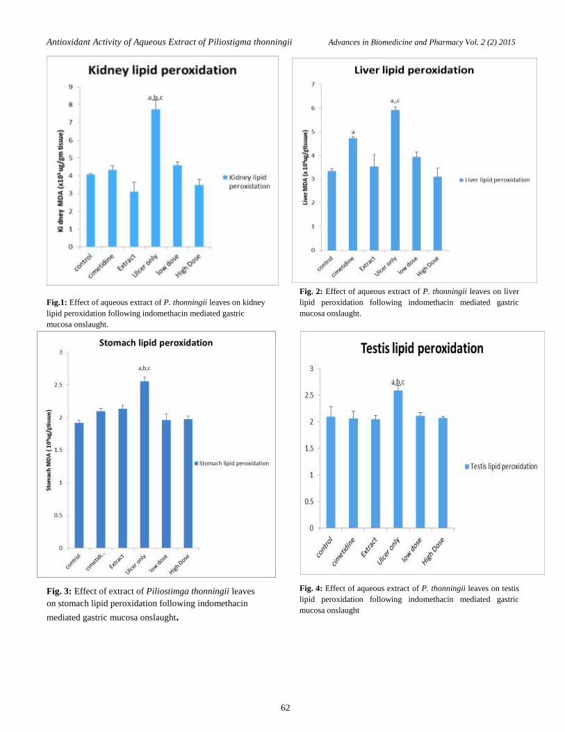
Results
The effect of the extract of P. thonningii leaf on kidney
lipid peroxidation (MDA) produce a significant reduction
(p<0.05) in all the treated groups when compared with the
untreated group (Fig.1). Furthermore, the effect of extract
of P.thonningii leaf on liver lipid peroxidation following
indomethacin mediated gastric mucosa onslaught showed a
significant increase (P<0.05) in groups treated with
cimetidine (standard drug), indomethacin without
treatment group (negative control) and the treated with
100mg/kg body weight of the extract while other treated
groups shows no significant difference when compared
with the control (Fig. 2). The effect of extract of P.
thonningii leaf on stomach lipid peroxidation following
indomethacin induced gastric ulceration shows a
significant increase (P<0.05) in the indomethacin without
treatment group whereas other treated groups shows no
significant difference when compared with the control
(Fig .3). Similar pattern were shown in the testes lipid
peroxidation (Fig .4).
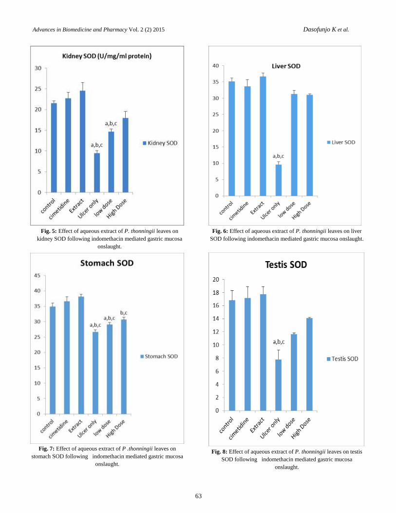
The effect of P. thonningii leaf on kidney SOD following
indomethacin induced gastric ulceration shows a
significant increase (P<0.05) when compared with the
control while the groups treated with 100 and 200mg /kg
body weight of the extract produced a significant decrease
(P<0.05) when compared with the control, likewise more
significant decrease were observed in the ulcerated group
without treatment when compared with the control (Fig 5).
The effect of extract of P. thonningii leaf in liver SOD
following indomethacin induced ulceration shows
significant decrease (P<0.05) in all extract treated group
and Cimetidine group (standard drug) with an exception of
extract only group which shows a significant increase
(P<0.05) (Fig 6). The effect of extract of P. thonningii leaf
on stomach SOD following indomethacin mediated gastric
mucosa onslaught shows a significant increase (P<0.05) in
extract only and cimetidine treated groups while there was
significant decrease (P<0.05) in the groups treated with
100 and 200mg/kg body weight of extract and
indomethacin without treatment group (negative control)
compared to the control (Fig 7). Similar pattern was
observed in the testes SOD (Fig 8).
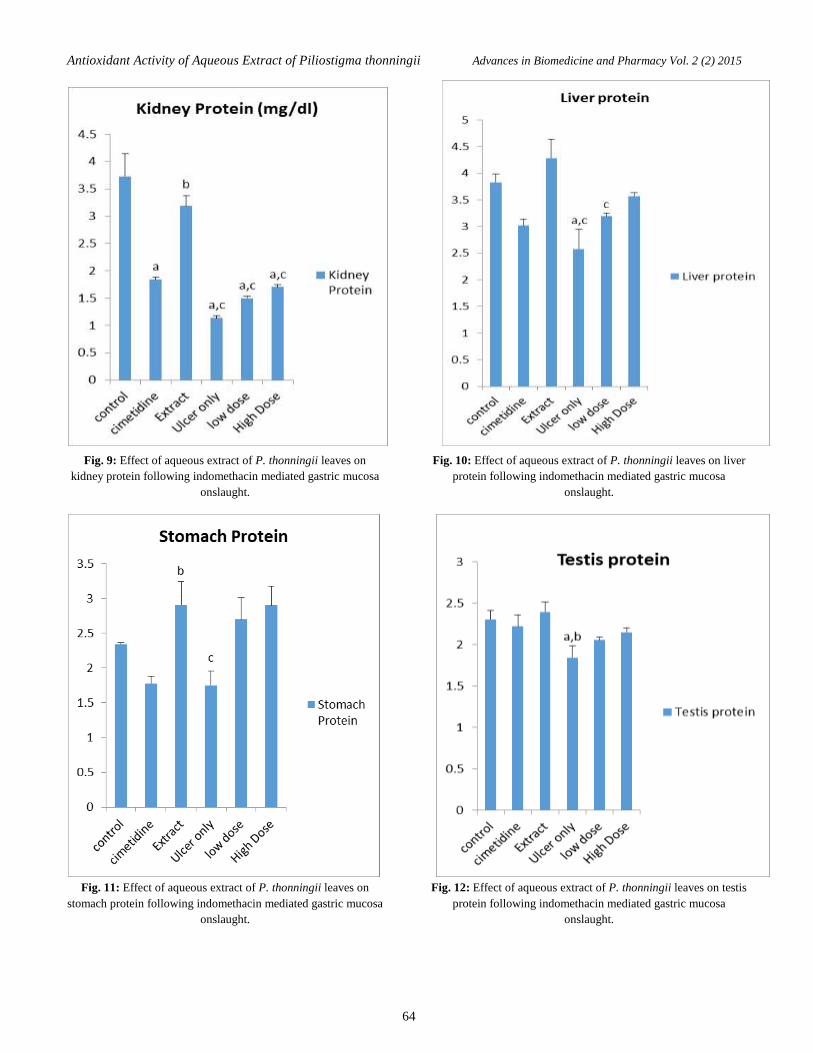
The effect of extract of P. thonningii on kidney protein
following indomethacin induced gastric ulceration shows a
significant decrease (P< 0.05) in all treated groups with
compared to the control group (Fig. 9). The effect of
extract of P.thonningii on liver protein following
indomethacin induced gastric ulceration shows a
significant decrease (P< 0.05) in all treated groups with an
exception in the extract only treated group which shows a
significant increase (P<0.05) when compared with the
control group (Fig. 10).The effect of extract of
P.thonningii on stomach protein following indomethacin
induced gastric ulceration show a significant decrease
(P<0.05) in the cimetidine (standard drug) and
indomethacin treated group (negative control) while other
treated groups shows a significant increase (P<0.05)
compared to the control group (Fig.11). The effect of the
extract of P.thonningii on testes protein following
indomethacin induced gastric ulcer shows no significant
different in group treated with extract only compared with
control group while other treated groups shows significant
decrease (P<0.05) when compared with control group
(Fig.12).
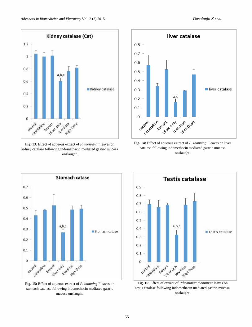
The effect of extract of P.thonningii leaf on kidney
catalase activity following indomethacin mediated gastric
mucosa onslaught reveals a significant decrease (P<0.05)
in indomethacin induced untreated group, low dose and
high dose compared with the control whereas groups
treated with extract only and cimetidine had no significant
difference when compared with the control group (Fig.
13).Likewise, Fig 14 reveals the effect of extract of
P.thonningii leaf on liver catalase activity following
indomethacin mediated gastric mucosa onslaught. All
treated groups produced a significant decrease (P<0.05)
when compared with the control group. The effect of
P.thonningii leaf on stomach catalase activity following
indomethacin mediated gastric mucosa onslaught shows a
significant decrease (P<0.05) in all treated group with
exception of the group treated with extract only which
shows a significant increase (P<0.05) compared to the
control (Fig.15).The effect of extract of P.thonningii leaf
on testes catalase following indomethacin mediated gastric
mucosa onslaught shows no significant difference different
(P<0.05) in all the treated group with an exception in the
indomethacin induced untreated group (negative control)
when compared with the control group (Fig. 16)
61
Antioxidant Activity of Aqueous Extract of Piliostigma thonningii Advances in Biomedicine and Pharmacy Vol. 2 (2) 2015
Fig.1: Effect of aqueous extract of P. thonningii leaves on kidney
lipid peroxidation following indomethacin mediated gastric
mucosa onslaught.
Fig. 3: Effect of extract of Piliostimga thonningii leaves
on stomach lipid peroxidation following indomethacin
mediated gastric mucosa onslaught.
Fig. 2: Effect of aqueous extract of P. thonningii leaves on liver
lipid peroxidation following indomethacin mediated gastric
mucosa onslaught.
Fig. 4: Effect of aqueous extract of P. thonningii leaves on testis
lipid peroxidation following indomethacin mediated gastric
mucosa onslaught
62
Advances in Biomedicine and Pharmacy Vol. 2 (2) 2015 Dasofunjo K et al.
Fig. 5: Effect of aqueous extract of P. thonningii leaves on
kidney SOD following indomethacin mediated gastric mucosa
onslaught.
Fig. 7: Effect of aqueous extract of P .thonningii leaves on
stomach SOD following indomethacin mediated gastric mucosa
onslaught.
Fig. 6: Effect of aqueous extract of P. thonningii leaves on liver
SOD following indomethacin mediated gastric mucosa onslaught.
Fig. 8: Effect of aqueous extract of P. thonningii leaves on testis
SOD following indomethacin mediated gastric mucosa
onslaught.
63
Antioxidant Activity of Aqueous Extract of Piliostigma thonningii Advances in Biomedicine and Pharmacy Vol. 2 (2) 2015
Fig. 9: Effect of aqueous extract of P. thonningii leaves on
kidney protein following indomethacin mediated gastric mucosa
onslaught.
Fig. 11: Effect of aqueous extract of P. thonningii leaves on
stomach protein following indomethacin mediated gastric mucosa
onslaught.
Fig. 10: Effect of aqueous extract of P. thonningii leaves on liver
protein following indomethacin mediated gastric mucosa
onslaught.
Fig. 12: Effect of aqueous extract of P. thonningii leaves on testis
protein following indomethacin mediated gastric mucosa
onslaught.
64
Advances in Biomedicine and Pharmacy Vol. 2 (2) 2015 Dasofunjo K et al.
Fig. 13: Effect of aqueous extract of P. thonningii leaves on
kidney catalase following indomethacin mediated gastric mucosa
onslaught.
Fig. 15: Effect of aqueous extract of P. thonningii leaves on
stomach catalase following indomethacin mediated gastric
mucosa onslaught.
Fig. 14: Effect of aqueous extract of P. thonningii leaves on liver
catalase following indomethacin mediated gastric mucosa
onslaught.
Fig. 16: Effect of extract of Piliostimga thonningii leaves on
testis catalase following indomethacin mediated gastric mucosa
onslaught.
65
Antioxidant Activity of Aqueous Extract of Piliostigma thonningii Advances in Biomedicine and Pharmacy Vol. 2 (2) 2015
Discussion
Gastric ulcer is one of the diseases responsible for high
mortality and morbidity among the less privileged in
Africa and beyond, possibly due to wide- spread or usage
of NSAIDs recently or due to poor understanding of the
pathophysiology of this disease [21]. Studies investigating
new active compounds are needed. As well, various
pharmaceutical products currently used for treatment of
gastric ulcers are not completely efficient and cause many
adverse side effects. Consequently, it is necessary to
develop more effective agents that are also less toxic, with
medicinal plants being an attractive source for the
development of new drugs because of their wide array of
active ingredients [22].
In this study we used indomethacin to induce gastric
mucosa ulcer. Indomethacin is known to induce the
reactive oxygen metabolites in animal models, which may
contribute to mucosal injury. These free radicals also
damage the cellular antioxidant enzymes such as CAT,
SOD among others, acting as the first line of cellular
defense against oxidative injury, this might lead to
aggravated tissue damage during stomach ulceration.
Indomethacin- induced stomach ulceration can triggers
severe oxidative stress in gastric tissue causing damage to
key bio molecules such as lipids. This was apparent from
the stimulated lipid oxidation leading to increased
accumulation of MDA as well as reduction in the gastric
activity of CAT As a matter of fact indomethacin being a
NSAID is widely used in clinical practices due to its
efficacy and various therapeutic effects, on the other hand
acute gastrointestinal lesions are the most serious and
frequent side effects of NSAIDs, making them the most
common cause of gastro duodenal ulcers in Western
countries [21,23].Ulcer formation induced by
Indomethacin, is known to be co-related with inhibition of
cyclooxygenase (COX1 and COX2), that prevents
prostaglandin biosynthesis [24] which in turn inhibits the
release of mucus a defensive factor against
gastrointestinal damage.
Recently, much attention has been focused on oxygen
derived free radicals which play an important role in the
pathogenesis of gastric ulcer apart from the interactive
processes like many other tissue degeneration situations.
Oxygen derived free radicals cause tissue injury through
lipid peroxidation. Oxygen handling cells have different
systems, e.g. superoxide dismutase (SOD), peroxidases
and catalases which are able to protect them against the
toxic effects of oxygen derived free radicals. As shown in
this present results, treatment with extract of P.thonningii
leaf significantly reverted the indomethacin-induced
changes or alterations in MDA and CAT. This significant
reduction in MDA levels along with significant increase in
SOD and CAT level suggest decreased lipid peroxidation
and cytoprotective or antioxidant activity of extract of
P.thonningii. Cimetidine also provided a marked
suppression of oxidative damage due to its excellent
radical scavenging capacity; it brought MDA level closer
to normal levels, but less than observed in the extract with
concomitant increase in CAT level and possibly with the
release of Nitric oxide. Nitric oxide (NO) is an endogenous
defensive factor for gastric cells and exhibits gastro
protective properties against different types of aggressive
agents [25]. It is also involved in the maintenance of
mucosal integrity through the regulation of mucus and
alkaline secretion, gastric motility and microcirculation
[26]. NO is known to modulate acid levels, gastric mucus
secretion, and blood flow in gastric tissues, prevention
membrane lipid peroxidation ,protection against NSAID
damage by promotion of prostaglandin synthesis [27].
In the present study, it appears that P.thonningii has
bioactive compounds which have synergistic relationship
with NO synthesis. Indomethacin also significantly
reduced total protein for negative control group but
significantly increased in groups treated with high and low
doses of P.thonningii suggest that the Plant might contain
a bioactive compound which might aid in protein synthesis
or antibody or free radical scavenging property hence
aiding its antioxidant activity by preventing the formation
of free radicals or by scavenging superoxide anions.
Our results show that treatment with extract of
P.thonningii at the dose of 100 and 200mg/kg body weight
significantly decreased the level of lipid peroxidation
product (MDA) when compared to untreated ulcerated
rats. The activities of both SOD and Catalase were
decreased in ulcerated untreated groups and maintained to
near normalcy in treated group with the exception of the
extract treated group which shows a significant increase
compared to the control group.
Conclusion
The present results of biochemical and physiological
alterations are indications that the extract of P.thonningii
leaf has a dose dependent cytoprotective effect against
indomethacin-mediated gastric mucosa onslaught, which
can be attributed to its antioxidant activity.
Conflict of interest
The authors declare that there is no conflict of interest to
reveal.
66
Advances in Biomedicine and Pharmacy Vol. 2 (2) 2015 Dasofunjo K et al.
References
[1] Sravani P, Jayasri PS, Ershad Khan P et al., “Review on natural antiulcer agents”, Inter. J. pharm. and Ind. Res., 1:
67 – 70,2011.
[2] Gadekar R, Singour PK, Chaurasiya PK et al., “A potential of some medicinal plants as an antiulcer agent”,
Pharmcogn. Rev., 4: 136 – 140, 2010.
[3] Grossman M., “Peptic ulcer: A guide for practicing physicians”, Am. J. Pharm. Toxicol., 4: 79, 89 – 93, 2009.
[4] Fridovich I., “Biological effects of superoxide radical”, Arch Biochem Biophy., 247: 1–11, 1986.
[5] Freidovich I., “Fundamental aspects of reactive oxygen species, or what’s the matter with oxygen”, Ann NY Acad
Sci., 893:13 -23, 1999.
[6] Ito N, Hirose M and Imaida K., “Antioxidants: Carcinogenic and chemo preventive properties”, In: Bertino JR.
Encyclopedia of Cancer, 1: 51–63, 1996.
[7] Aderogba MA, Okoh EK, Okeke IN et al., “Antimicrobial and anti-inflammatory effects of Piliostigmareticulatum
leaf extract”, Int. J. Pharmacol., 2: 70–74, 2006.
[8] Thabrew MI, Hughes RD and McFarlane IG., “Antioxidant activity of Osbeckiaaspera”, Phytother. Res., 12: 288-290,
1998.
[9] Burits M and Bucar F., “Antioxidant Activity of Nigetta sativa essential oil”, Phytother. Res., 14, 2000.
[10] Dasofunjo K, Asuk AA, Ezugwu H C et al., “Aphrodisiac effect of ethanol extract of Piliostigma thonningiileaf on
male Albino Wistar Rats”, J. Appl. Pharm. Sci., 3:130-135,2013.
[11] Dasofunjo K, Nwodo OFC , Ipav SS et al., “Effect of the ethanolic extract of Piliostigma thonningiion haematological
parameters of male albino wistar rats”, J. Nat. Prod. Plant Resour., 2:670-674, 2012.
[12] Aihara T, Nakamamura E, Amagasa K et al., “Pharmacological control of gastric acid secretion for the treatment of
acid-related peptic disease: past and present and future”, Pharmacol. Ther., 98:109-127, 2003.
[13] Tasman-Jones C., “Pathogenesis of peptic ulcer disease and gastristis importance of aggressive and cytoprotective
factor”, Scand. J. Gastroenterol., 21: 1-5, 1986.
[14] Ludovico B, Eric S and Annemarthe G., “Helicobacter pylori cytotoxin associated gene A subverts the apoptosis-
stimulating protein of P53 ASPP2turmor suppressor pathway of host”, Proc. Natl. Acad. Sci., 108: 9238-9243, 2011.
[15] Krishnaswami K., “Indian functional food: role in prevention of cancer”, Nutr. Rev., 54:127-31, 1996.
[16] Ito N, Hirose M and Imaida K., “Antioxidants: Carcinogenic and chemopreventive properties”, In: Bertino JR, editor.
Encyclopedia of Cancer, California, Academic Press., 51–63, 1996.
[17] Plumer DT., “An introduction to practical Biochemistry”, 2ndEdition M.C. Graw-hill London pp 144-145, 1978.
[18] Martin P, Dailey M and Sugarman E., “Negative and positive assays of superoxide dismutase based on heamatoxylin
autoxidation”, Arch. Biochem. Biophys., 255:329-336, 1987.
[19] Sinha AK., “Colorimetric assay of catalase”, Anal. Biochem., 47:389-394, 1972.
[20] Buege JA and Aust SD., “Microsomal lipid peroxidation Method”, Enzymol., 52: 302-310, 1978.
[21] Scarpingnato C and Hunt RH., “Non-steroidal anti-inflammatory drug- related injury to gastrointestinal tract: clinical
picture, pathogenesis and prevention”, Gastroenterol. Clinc. North Am., 39:433-464.2010.
[22] Akram M, Ahmed A, Usmanghani K et al., “Peptic ulcer and Helicobacter pylori eradication”, A review article. Int. J.
Med. Sci., 2:370-375, 2010.
[23] Griffin MR, Piper JM and Daugherty JR., “Non -steroidal ant- inflammatory drug use and increased risk for peptic
ulcer disease in elderly persons”, Ann. Intern. Med., 114: 257-263, 1991.
[24] Whittle BJ., “Gastrointestinal “effects of non-steroidal anti-inflammatory drugs”, Fundam. Clinc Pharmcol., 17:
301-313, 2002.
[25] Sairam K, RaoCh V and Dorababu M., “Prophylactic and curative effects of Bacopamonniera in gastric ulcer
models”, Phytomedicine, 8: 423–430, 2001.
[26] Ignarro LJ, Cirino G, Casini A et al., “Nitric oxide as a signalling molecule in the vascular system: an overview”, J.
Cardiovasc. Pharmacol., 34:879, 1999.
[27] Turrens JF, Crapo JD and Freeman BA., “Protection against oxygen toxicity by intravenous injection of liposome-
entrapped catalase and superoxide dis- mutase” J. Clin. Invest., 73: 87–95, 1984.
ABP
© 2015 Reproduction is free for scientific studies
67
Related Documents


