JPP 2007, 59: 1721–1728 © 2007 The Authors Received March 29, 2007 Accepted July 17, 2007 DOI 10.1211/jpp.59.12.0015 ISSN 0022-3573 1721 Antioxidant activity of 4-methylcoumarins Jens Z. Pedersen, Cristina Oliveira, Sandra Incerpi, Vineet Kumar, Anna Maria Fiore, Paolo De Vito, Ashok K. Prasad, Sanjay V. Malhotra, Virinder S. Parmar and Luciano Saso Abstract Polyphenolic coumarins are known to act as antioxidants in biological systems, but it is difficult to distinguish their antioxidant activity from the many other effects they produce in cells. We have determined the radical scavenging capacity of 22 structurally related natural and synthetic 4-meth- ylcoumarins, by measuring their reaction with radicals, galvinoxyl and 2,2-diphenyl-1-picrylhydrazyl, using electron paramagnetic resonance spectroscopy. Efficient antioxidant activity of 4-methylcou- marins in cells was verified using the DCF fluorescent probe assay for determination of intracellular reactive oxygen species levels. As expected, the o-dihydroxysubstituted coumarins were found to be excellent radical scavengers and better than the m-dihydroxysubstituted or monohydroxysubsti- tuted analogues, but surprisingly the corresponding o-diacetoxy derivatives also turned out to be good scavengers, even in the absence of an esterase. Another unexpected result was that the anti- oxidant efficiency of 4-methylcoumarins could be modulated by introducing an ethoxycarbonyl- ethyl substituent at the C-3 position; this effect cannot be explained by simple electron donating/ withdrawing properties. Coumarin concentrations of 10 mM or less were used in all experiments, cor- responding to the levels relevant for therapeutic purposes. Considering that 4-methylcoumarins, in contrast to many other coumarins, are not metabolized to toxic epoxide intermediates, these results indicate promising new strategies for the design of non-toxic antioxidant coumarin-based drugs. Coumarins are a group of compounds that show a surprising variety of biological effects. They occur naturally in many plants, fungi and bacteria, and have found applications for centuries as spices and in traditional medicine. Several natural polyphenolic coumarins show anti-inflammatory, antimicrobial, antiviral, anti-carcinogenic, anticoagulant and anti- oxidant activity; but generally the reason for these effects and the precise nature of their actions are not known (Borges et al 2005). Many different types of antioxidant activity of coumarins have been reported for a variety of biological systems, but it has not yet been possible to correlate the effects observed to the chemical structures of the coumarins studied (Fylaktakidou et al 2004; Borges et al 2005; Kostova 2006). Hydroxycoumarins are believed to behave like classic phenol- or quinol-based antioxidants, in which a hydroxy group on an aromatic ring structure can carry out the single-electron reduction of a free rad- ical. The resulting phenoxyl radical or semiquinone can either be stabilized through the presence of bulky or electron-withdrawing groups on the ring system or be oxidized further through the consecutive single-electron reduction by a second hydroxy group to produce a quinone-type end product. This is the mechanism behind both natural and synthetic antioxi- dants, like vitamin E and butylated hydroxytoluene; however, sometimes it turns out that coumarins do not work in this way (Fylaktakidou et al 2004; Kostova 2006). One problem with coumarin compounds has been the tendency to form 3,4-coumarin epoxides during metabolic degradation; these intermediates are believed to be mutagenic and probably also have other toxic effects. To prevent this problem a series of 4-methylcou- marins have been synthesized; in these compounds 3,4-coumarin epoxide formation is no longer possible as 4-methylcoumarins are not substrates for the liver P-450 monoxygenases that epoxidize coumarins lacking the C-4 methyl group. Several 4-methylcoumarins have shown promising antioxidant effects, such as inhibition of lipid peroxidation and scavenging Introduction Department of Biology, University of Rome ‘Tor Vergata’, Italy Jens Z. Pedersen, Paolo De Vito Department of Biology, University of Rome ‘Roma Tre’, Italy Sandra Incerpi, Anna Maria Fiore Department of Chemistry, University of Delhi, India Vineet Kumar, Ashok K. Prasad, Virinder S. Parmar Department of Chemistry and Environmental Science, New Jersey Institute of Technology, Newark, USA Vineet Kumar, Sanjay V. Malhotra Department of Human Physiology and Pharmacology, Sapienza University of Rome, Italy Luciano Saso, Cristina Oliveira Correspondence: J. Z. Pedersen, Department of Biology, University of Rome ‘Tor Vergata’, Via della Ricerca Scientifica 1, 00133 Roma, Italy. E-mail: [email protected] Acknowledgement: The financial support from the Italian Ministry for University and Research, General Management for Strategies and Development of Internationalization of Scientific and Technological Research is gratefully acknowledged. JPP59(12).book Page 1721 Wednesday, November 7, 2007 3:48 PM

Welcome message from author
This document is posted to help you gain knowledge. Please leave a comment to let me know what you think about it! Share it to your friends and learn new things together.
Transcript
JPP 2007, 59: 1721–1728© 2007 The AuthorsReceived March 29, 2007Accepted July 17, 2007DOI 10.1211/jpp.59.12.0015ISSN 0022-3573
1721
Antioxidant activity of 4-methylcoumarins
Jens Z. Pedersen, Cristina Oliveira, Sandra Incerpi, Vineet Kumar,
Anna Maria Fiore, Paolo De Vito, Ashok K. Prasad, Sanjay V. Malhotra,
Virinder S. Parmar and Luciano Saso
Abstract
Polyphenolic coumarins are known to act as antioxidants in biological systems, but it is difficult todistinguish their antioxidant activity from the many other effects they produce in cells. We havedetermined the radical scavenging capacity of 22 structurally related natural and synthetic 4-meth-ylcoumarins, by measuring their reaction with radicals, galvinoxyl and 2,2-diphenyl-1-picrylhydrazyl,using electron paramagnetic resonance spectroscopy. Efficient antioxidant activity of 4-methylcou-marins in cells was verified using the DCF fluorescent probe assay for determination of intracellularreactive oxygen species levels. As expected, the o-dihydroxysubstituted coumarins were found to beexcellent radical scavengers and better than the m-dihydroxysubstituted or monohydroxysubsti-tuted analogues, but surprisingly the corresponding o-diacetoxy derivatives also turned out to begood scavengers, even in the absence of an esterase. Another unexpected result was that the anti-oxidant efficiency of 4-methylcoumarins could be modulated by introducing an ethoxycarbonyl-ethyl substituent at the C-3 position; this effect cannot be explained by simple electron donating/withdrawing properties. Coumarin concentrations of 10 mM or less were used in all experiments, cor-responding to the levels relevant for therapeutic purposes. Considering that 4-methylcoumarins, incontrast to many other coumarins, are not metabolized to toxic epoxide intermediates, these resultsindicate promising new strategies for the design of non-toxic antioxidant coumarin-based drugs.
Coumarins are a group of compounds that show a surprising variety of biological effects.They occur naturally in many plants, fungi and bacteria, and have found applications forcenturies as spices and in traditional medicine. Several natural polyphenolic coumarinsshow anti-inflammatory, antimicrobial, antiviral, anti-carcinogenic, anticoagulant and anti-oxidant activity; but generally the reason for these effects and the precise nature of theiractions are not known (Borges et al 2005). Many different types of antioxidant activity ofcoumarins have been reported for a variety of biological systems, but it has not yet beenpossible to correlate the effects observed to the chemical structures of the coumarins studied(Fylaktakidou et al 2004; Borges et al 2005; Kostova 2006). Hydroxycoumarins arebelieved to behave like classic phenol- or quinol-based antioxidants, in which a hydroxygroup on an aromatic ring structure can carry out the single-electron reduction of a free rad-ical. The resulting phenoxyl radical or semiquinone can either be stabilized through thepresence of bulky or electron-withdrawing groups on the ring system or be oxidized furtherthrough the consecutive single-electron reduction by a second hydroxy group to produce aquinone-type end product. This is the mechanism behind both natural and synthetic antioxi-dants, like vitamin E and butylated hydroxytoluene; however, sometimes it turns out thatcoumarins do not work in this way (Fylaktakidou et al 2004; Kostova 2006).
One problem with coumarin compounds has been the tendency to form 3,4-coumarinepoxides during metabolic degradation; these intermediates are believed to be mutagenicand probably also have other toxic effects. To prevent this problem a series of 4-methylcou-marins have been synthesized; in these compounds 3,4-coumarin epoxide formation is nolonger possible as 4-methylcoumarins are not substrates for the liver P-450 monoxygenasesthat epoxidize coumarins lacking the C-4 methyl group. Several 4-methylcoumarins haveshown promising antioxidant effects, such as inhibition of lipid peroxidation and scavenging
Introduction
Department of Biology, University of Rome ‘Tor Vergata’, Italy
Jens Z. Pedersen, Paolo De Vito
Department of Biology, University of Rome ‘Roma Tre’, Italy
Sandra Incerpi, Anna Maria Fiore
Department of Chemistry, University of Delhi, India
Vineet Kumar, Ashok K. Prasad, Virinder S. Parmar
Department of Chemistry and Environmental Science, New Jersey Institute of Technology, Newark, USA
Vineet Kumar, Sanjay V. Malhotra
Department of Human Physiology and Pharmacology, Sapienza University of Rome, Italy
Luciano Saso, Cristina Oliveira
Correspondence: J. Z. Pedersen, Department of Biology, University of Rome ‘Tor Vergata’, Via della Ricerca Scientifica 1, 00133 Roma, Italy. E-mail: [email protected]
Acknowledgement: The financial support from the Italian Ministry for University and Research, General Management for Strategies and Development of Internationalization of Scientific and Technological Research is gratefully acknowledged.
JPP59(12).book Page 1721 Wednesday, November 7, 2007 3:48 PM
1722 Jens Z. Pedersen et al
of superoxide anions (Raj et al 1998a, b, c; Kumar et al2005a). We here present a study of the radical scavengingcapacity of a group of natural and synthetic coumarins exam-ined under normalized conditions. Using electron paramag-netic resonance (EPR) spectroscopy we have tested 22structurally related 4-methylcoumarins (Figure 1, Table 1),measuring the kinetics of their reaction with the standard rad-icals galvinoxyl and 2,2-diphenyl-1-picrylhydrazyl (DPPH).Very low concentrations have been used for these measure-ments (10 mM or less), corresponding to the levels that wouldbe considered for therapeutic purposes. The antioxidant effi-ciency of 4-methylcoumarins in cells was verified using astandard fluorescent assay for determination of intracellularreactive oxygen species (ROS) levels. The results showedthat ortho-dihydroxysubstituted coumarins were more effi-cient radical scavengers than meta-dihydroxysubstituted ormonohydroxysubstituted molecules, but surprisingly the
corresponding ortho-diacetoxy derivatives also turned out tobe good scavengers, even in the absence of an esterase. Theantioxidant efficiency of 4-methylcoumarins could be modi-fied by changing the type of substituent at the C-3 position.These results indicate new possible strategies for the designof non-toxic coumarin antioxidants.
Reagents
All 4-methylcoumarins and 4-methylthionocoumarins weresynthesized and characterized at the Department of Chemis-try of the University of Delhi as previously described (Parmaret al 1996; Raj et al 1996; Singh et al 2002; Kumar et al2005a). Galvinoxyl, 2,2-diphenyl-1-picrylhydrazyl (DPPH)
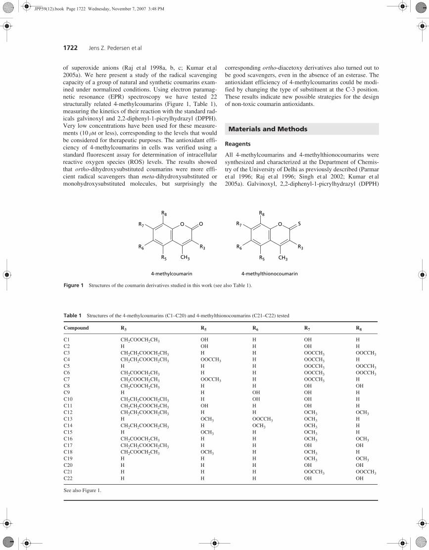
Figure 1 Structures of the coumarin derivatives studied in this work (see also Table 1).
O
CH3
OR7
R3
R5
R6
R8
4-methylcoumarin
O
CH3
SR7
R3
R5
R6
R8
4-methylthionocoumarin
Table 1 Structures of the 4-methylcoumarins (C1–C20) and 4-methylthionocoumarins (C21–C22) tested
See also Figure 1.
Compound R3 R5 R6 R7 R8
C1 CH2COOCH2CH3 OH H OH H C2 H OH H OH H C3 CH2CH2COOCH2CH3 H H OOCCH3 OOCCH3 C4 CH2CH2COOCH2CH3 OOCCH3 H OOCCH3 H C5 H H H OOCCH3 OOCCH3 C6 CH2COOCH2CH3 H H OOCCH3 OOCCH3 C7 CH2COOCH2CH3 OOCCH3 H OOCCH3 H C8 CH2COOCH2CH3 H H OH OH C9 H H OH OH H C10 CH2CH2COOCH2CH3 H OH OH H C11 CH2CH2COOCH2CH3 OH H OH H C12 CH2CH2COOCH2CH3 H H OCH3 OCH3 C13 H OCH3 OOCCH3 OCH3 H C14 CH2CH2COOCH2CH3 H OCH3 OCH3 H C15 H OCH3 H OCH3 H C16 CH2COOCH2CH3 H H OCH3 OCH3 C17 CH2CH2COOCH2CH3 H H OH OH C18 CH2COOCH2CH3 OCH3 H OCH3 H C19 H H H OCH3 OCH3 C20 H H H OH OH C21 H H H OOCCH3 OOCCH3 C22 H H H OH OH
Materials and Methods
JPP59(12).book Page 1722 Wednesday, November 7, 2007 3:48 PM
Antioxidant activity of 4-methylcoumarins 1723
and cumene hydroperoxide were purchased from Sigma-Aldrich (St Louis, MO); 2′,7′-dichlorodihydrofluorescein dia-cetate (DCFH2-DA) was obtained from Molecular Probes(Eugene, OR). Dulbecco’s modified Eagle’s medium(DMEM), antibiotics and sterile plasticware for cell culturewere from Flow Laboratory (Irvine, UK). Fetal bovine serumwas from GIBCO (Grand Island, NY).
Electron paramagnetic resonance spectroscopy
Stock solutions of all 4-methylcoumarin compounds wereprepared in ethanol 95% at a concentration of 5 mM. Agalvinoxyl stock solution (5 mM in ethanol 95%) was freshlyprepared immediately before the experiments. Systematicscreening of all compounds was made with a final concentra-tion of 10 mM in the presence of galvinoxyl at the same con-centration. For some experiments, DPPH (50 mM) was used asan alternative to galvinoxyl. The solutions were drawn intoglass capillaries, sealed and measured using an ESP300instrument (Bruker Spectrospin, Karlsruhe, Germany)equipped with a high sensitivity TM110 X-band cavity. Radi-cal spectra were recorded at room temperature, using 0.6 Gmodulation, 1 mW microwave power and a scan time of 42 sfor a 30 G spectrum. Normally, four spectra were accumulatedfor each measurement to obtain a suitable signal-to-noiseratio. The kinetics of the reaction was followed for 3 h atroom temperature or until the radical signal had disappeared.
Cell culture
L-6 cells from rat skeletal muscle were obtained from theAmerican Type Culture Collection (Rockville, MD). Cellswere seeded in 75-cm2 flasks for tissue culture and grown inDMEM supplemented with 10% fetal bovine serum, 100 mgmL−1 streptomycin and 100 U mL−1 penicillin, in an atmos-phere of 5% CO2 at 37°C. The cells reached confluency after5 days (about 6 × 10⁶ cells) and were kept in culture as myo-blasts by continuous passages at pre-confluent stages, as pre-viously reported (D’Arezzo et al 2004).
Cytotoxicity assay
Toxicity of the coumarins during the experiments on intracel-lular ROS measurements was determined by the method ofHansen et al (1989) with some modifications. Briefly, cellviability was quantified by the conversion of yellow MTT (3-(4,5-dimethylthiazol-2-yl)-2,5-diphenyltetrazolium bromide;Sigma) to purple MTT formazan by the mitochondrial dehy-drogenases of living cells. The experiment was carried out insix-well plates using 1 × 10⁶ cells at the confluent state perwell. Control and treated cells were incubated with MTT at afinal concentration of 1 mg mL−1 for 3 h at 37°C. Thereaftercells were scraped off and centrifuged at 1200 rev min−1 for5min; the pellet was re-suspended in 300mL phosphate-bufferedsaline (PBS) and sonicated on ice for 15 s with an UltrasonicW-225R, at setting 4, and then centrifuged in a microfuge at13 000 rev min−1 for 10 min. The supernatant was discardedand the final pellet re-suspended in 200 mL dimethyl sulfoxide(DMSO); after appropriate dilutions MTT formazan forma-tion was measured with a spectrophotometer at 560 nm.
Results are given as the mean ± standard deviation (s.d.) of 3similar experiments carried out in triplicate.
Intracellular ROS determination
At the time of the experiment the DMEM was discarded andcells were washed twice with 5 mL PBS containing 5.0 mM
glucose at 37°C. Cells were gently scraped off with 5 mLPBS plus glucose at 37°C, the cell suspension was transferredto a centrifuge tube and the buffer was added up to 8–10 mL.Cells were centrifuged at 1200 rev min−1 for 5 min (about100 g), the supernatant was discarded and the pellet re-suspended with a plastic Pasteur pipette in 5 mL PBS. Incuba-tion with the probe DCFH2-DA at a final concentration of10 mM (from a stock solution of 10 mM in DMSO) was carriedout for 30 min in the dark at 37°C, as previously reported(Pallottini et al 2005). The cells were gently re-suspendedevery 10 min; at the end of incubation cells were centrifugedat 1200 rev min−1 for 5 min, the supernatant was discardedand the cell pellet was re-suspended in 5 mL of PBS plus glu-cose and centrifuged again. The final supernatant was dis-carded and the cell pellet re-suspended in 2 mL PBS at a finalconcentration of 3 × 10⁶ cells/mL. Before starting the experi-ment, a recovery was carried out at room temperature for45 min in the dark.
Intracellular fluorescence was measured under continuousgentle magnetic stirring at 37°C in a Perkin–Elmer (Norwalk,CT) LS 50B luminescence spectrometer. Excitation and emis-sion wavelengths were set at 498 nm and 530 nm, respec-tively, using 5 and 10 nm slits for the two light paths. Theassay was carried out in 3 mL final buffer containing 200 mLcell suspension. Cumene hydroperoxide diluted 1:100 inDMSO was used as radical generator (final concentration300 mM); DMSO at the concentrations used did not affect thefluorescence signal. The antioxidant potency of a coumarinwas determined by the decrease in the intracellular DCF fluo-rescence, reported as ΔF/10 min, and was calculated relativeto the fluorescence change induced by 300 mM cumenehydroperoxide alone (100%). Cells were incubated with cou-marins for 10 min at 37°C before the addition of cumenehydroperoxide; none of the coumarins tested gave rise tofluorescence on their own.
Statistical analysis
All assays were performed at least in triplicate and data wereexpressed as mean ± s.d. Statistical analyses were made usingthe GraphPad Prism 4.0 software; the type of test used isspecified for each table and figure.
Radical scavenging activity
The 22 methylcoumarins studied here (Table 1) can bearranged into different groups according to their structure.Essentially, there are 9 dihydroxycoumarins, 7 diacetoxycou-marins and 6 dimethoxycoumarins; these groups can be sub-divided further according to the positions of the substituents
Results and Discussion
JPP59(12).book Page 1723 Wednesday, November 7, 2007 3:48 PM
1724 Jens Z. Pedersen et al
(7,8-ortho, 6,7-ortho or 5,7-meta) and the presence of anadditional ester at the C-3 position. In addition, two of thecompounds are thionocoumarins. The ability to scavenge freeradicals was tested with a very simple assay, mixing a cou-marin with a radical and following the reaction by EPR spec-troscopy (Iuliano et al 1999; Shi et al 2001; McPhail et al2003). A systematic screening of all compounds was made,with a final concentration of 10 mM in ethanol in the presenceof galvinoxyl at the same concentration. Alternatively, somemeasurements were made with lower concentrations, or usinganother radical, DPPH, at a concentration of 50 mM.
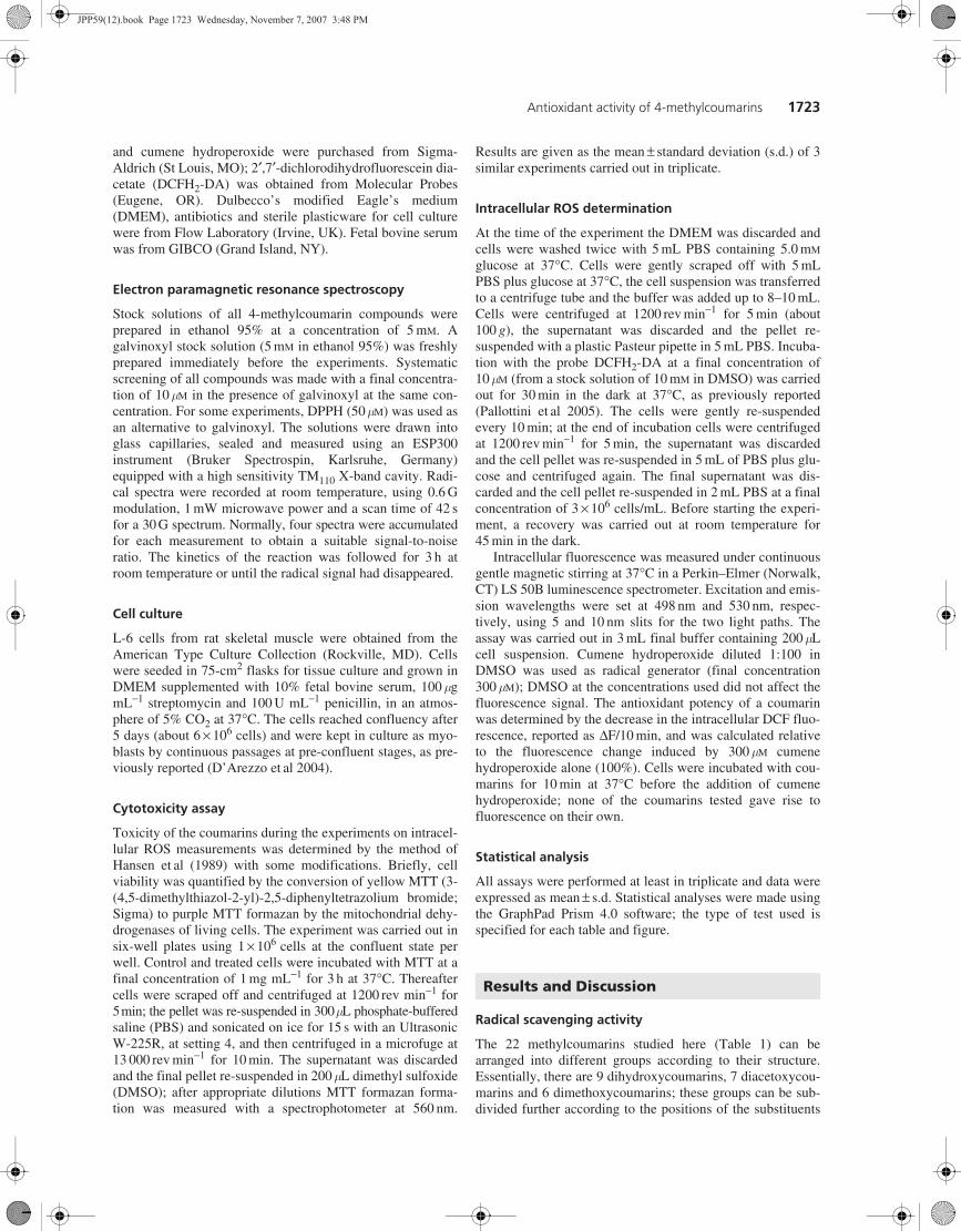
The EPR spectra of galvinoxyl and DPPH are shown inFigure 2. At the concentrations used here, the height of theEPR signal is directly proportional to the concentration of theradical; when this radical is reduced by an antioxidant, thespectrum disappears. For some of the coumarins it was pos-sible to detect the spectrum of the corresponding coumarylradical superimposed on the galvinoxyl or DPPH spectrumimmediately after mixing the sample; all the coumaryl radi-cals were too reactive to allow characterization and disap-peared within seconds or minutes (data not shown).
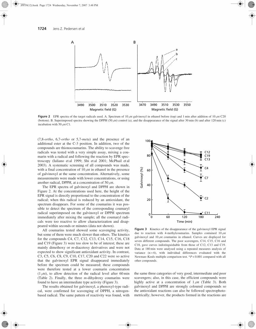
All coumarins tested showed some scavenging activity,but some of them were much slower than others. The kineticsfor the compounds C4, C7, C12, C13, C14, C15, C16, C18and C19 (Figure 3) were too slow to be of interest; these aremainly dimethoxy or m-diacetoxy derivatives and were notexpected to show significant antioxidant activity. In contrast,C3, C5, C6, C8, C9, C10, C17, C20 and C22 were so activethat the galvinoxyl EPR signal disappeared immediatelybefore the spectrum could be measured; these compoundswere therefore tested at a lower coumarin concentration(1 mM), to allow detection of the radical level after 60 min(Table 2). Finally, the three m-dihydroxy coumarins werefound to have an intermediate type activity (Figure 3).
The results obtained for galvinoxyl, a phenoxyl-type radi-cal, were confirmed for scavenging of DPPH, a nitrogen-based radical. The same pattern of reactivity was found, with
the same three categories of very good, intermediate and poorscavengers; also, in this case, the efficient compounds werehighly active at a concentration of 1 mM (Table 3). Bothgalvinoxyl and DPPH are strongly coloured compounds sothe antioxidant reactions can also be followed spectrophoto-metrically; however, the products formed in the reactions are
Figure 2 EPR spectra of the target radicals used. A. Spectrum of 10 mM galvinoxyl in ethanol before (top) and 1 min after addition of 10 mM C20(bottom). B. Superimposed spectra showing the DPPH (50 mM) control (a), and the disappearance of the signal after 30 min (b) and after 120 min (c)incubation with 50 mM C1.
A
Magnetic field (G) Magnetic field (G)3490 3500 3510 3520 3530 3470 3490 3510 3530 3550
B
ab
c
Figure 3 Kinetics of the disappearance of the galvinoxyl EPR signaldue to reaction with 4-methylcoumarins. Samples contained 10 mMgalvinoxyl and 10 mM coumarins in ethanol. Curves are displayed forseven different compounds. The poor scavengers, C14, C15, C16 andC18, gave curves indistinguishable from those of C12, C13 and C19.Data at 180 min were analysed using a repeated measures analysis ofvariance (n = 6), with individual differences evaluated with theNewman–Keuls multiple comparison test. *P < 0.001 compared with allother compounds.
0 60 120 180 2400
20
40
60
80
100
C11
C2
C7
C4
C12C19C13
Time (min)
Gal
vin
oxy
l (%
)
∗
∗
∗
∗
JPP59(12).book Page 1724 Wednesday, November 7, 2007 3:48 PM
Antioxidant activity of 4-methylcoumarins 1725
strongly coloured too and unfortunately absorb in the samespectral region, making it complicated to determine the kinet-ics (results not shown). The particular advantages of the EPRassay are that only radicals are observed, and they can bedetected at very low concentrations (Rossi et al 1996).
Antioxidant activity of esters
The results obtained with different assays consistently con-firm that ortho-dihydroxy-4-methylcoumarins are very goodradical scavengers; there did not appear to be any differencesbetween 7,8- and 6,7-substituted coumarins, at least in thesein-vitro experiments (Table 2). Sharma et al (2005) alsoreported that the ortho position is more favourable than themeta position for dihydroxy-4-methylcoumarins, but in theirantioxidant assay the 7,8-arrangement was better than the 6,7-substitution. 7,8-Dihydroxy-4-methylcoumarin (C20, 4-meth-yldaphnetin), which is a natural compound, was previouslyfound to be an excellent antioxidant and particularly efficientagainst lipid peroxidation (Raj et al 1998a; Liu et al 1999),although this effect seems to be connected with its metal che-lation properties (Raj et al 1998c). This compound has alsobeen shown to protect against DNA damage induced by H2O2(Liu & Zheng 2002). It is possible that all these effects areconnected to the capacity of C20 to selectively inhibit the pro-inflammatory 5-lipoxygenase, as reported for rat leucocytes byHoult & Paya (1996). Interestingly, another ortho-dihydroxy-coumarin, C9 (4-methylesculetin), inhibited 5-lipoxygenase
with the same selectivity, whereas the meta-dihydroxycou-marin C2 showed a higher inhibitory effect on the leucocytecyclooxygenase (Hoult & Paya 1996).
A more surprising result was the strong scavenger activityexerted by the ortho-diacetoxy-4-methylcoumarins, whichwere less efficient than the corresponding dihydroxy parentstructures, but much better antioxidants than the meta-dihy-droxy-4-methylcoumarins (Table 2). This behaviour has beennoticed before and attributed to the formation of deacetylatedcompounds through the activity of esterases in biological sys-tems, or to the interaction with lipid ketene structures (Rajet al 1998a). However, in the EPR experiments presented herethe samples contained only the diacetoxy compound, the tar-get radical and ethanol, leaving little doubt that the reactionproceeds via one-electron reduction of the radical, excludingeven the possibility of a hydrogen-atom-transfer mechanism.
Although the diacetyl esters of dihydroxy-4-methylcou-marins are less active as radical scavengers, they display awide range of biological effects that seem to be independentof the antioxidant activity. Diacetoxy-4-methylcoumarinsinhibit binding of aflatoxin B1 to DNA in-vitro and thus havepotential anti-mutagenic properties; the diesters were foundto have much higher activity than the corresponding dihy-droxy or dimethoxy compounds (Raj et al 1996, 1998a, c,2001). The diacetoxy-4-methylcoumarins are also involved inprotein acetylation catalysed by transacetylases (Kohli et al2002; Singh et al 2002; Kumar et al 2005b; Raj et al 2005).The specificity for the transacetylase substrates with respectto the number and position of acetoxy groups on the coumarinring structure has been examined, and again the most activecompounds are those with acetoxy groups in the ortho posi-tion, particularly 7,8-diacetoxy-4-methylcoumarin (C5). Thedihydroxy derivatives are not substrates for these enzymesbut are actually the products; at least in this case there is aclear separation of enzymatic and antioxidant effects. In othercases it is not easy to evaluate the antioxidant component of abiological response; both C5 and C20 show good inhibitoryactivity against ICAM-1 expression (Kumar et al 2005a), andC5 has demonstrated activity as an anti-invasive agent againstMCF-7/6 solid tumour cancer cell lines (Parmar et al 2003).All this diverse activity may involve both antioxidant andnon-antioxidant mechanisms. Another interesting result is theimportance of the substitution at the C-3 position (i.e., on the2-oxopyran ring), as seen from the data reported in Table 2.
Table 2 Effect of ethoxycarbonylmethyl or ethoxycarbonylethyl substitution at the C-3 position
Expressed as percentage of the galvinoxyl EPR signal remaining after incubation of 10 mM galvinoxyl with 4-methylcoumarinsfor 60 min at room temperature. Coumarin numbers are shown in brackets. Note that not all data are directly comparable, dueto the different coumarin concentrations employed. *P < 0.001, with respect to R3 = H in the same row (Student’s t-test).aInitial coumarin concentration 1 mM; binitial coumarin concentration 10 mM; cnot determined, compound not available.
4-Methylcoumarin R3 =H R3 =CH2COOCH2CH3 R3 =CH2CH2COOCH2CH3
7,8-Dihydroxy-a 25 ± 2 (C20) 23 ± 4 (C8) 11 ± 2 (C17)* 6,7-Dihydroxy-a 21 ± 3 (C9) n.d.c 13 ± 4 (C10)* 5,7-Dihydroxy-b 44 ± 4 (C2) 30 ± 3 (C1) 20 ± 2 (C11)* 7,8-Diacetoxy-a 64 ± 3 (C5) 76 ± 5 (C6) 72 ± 5 (C3) 5,7-Diacetoxy-b n.d. 70 ± 4 (C7) 73 ± 3 (C4)
Table 3 Decay of the DPPH radical EPR signal during the reactionwith 7,8-dihydroxy-4-methylcoumarin (C20) or with the correspondingdiacetyl ester (C5)
Data are shown as the percentage of radical remaining; 100% corre-sponds to 50 mM DPPH. Data shown are the mean ± s.d. of 3–6 individualexperiments.
Incubation time = 1 min Incubation time= 60 min
Compound 1 mM 10 mM 50 mM 1 mM 10 mM 50 mM
C5 99 ± 2 97 ± 4 70 ± 4 96 ± 4 84 ± 3 18 ± 6C20 95 ± 5 63 ± 1 11 ± 6 86 ± 8 43 ± 2 0
JPP59(12).book Page 1725 Wednesday, November 7, 2007 3:48 PM
1726 Jens Z. Pedersen et al
The introduction of the ethoxycarbonylethyl moiety in thisposition enhanced the radical scavenging capacity of bothmeta- and ortho-dihydroxy coumarins, whereas it did nothave any effect on the diacetoxy compounds. It is not obviouswhy a medium-length ester in this position should affect thereductive potential; direct electron-donating or -withdrawingcontributions are not likely, but the ester side chain may besufficiently long to interact with the oxopyran oxygens abovethe plane of the coumarin ring. This phenomenon could beexploited for the design of improved coumarin antioxidants.
Thionocoumarins may represent a different way of modi-fying coumarin properties to increase their pharmacologicalpotentials (Kumar et al 2005a). The two 4-methylthionocou-marins tested here did not show much improved radical scav-enger activity; the 7,8-dihydroxythionocoumarin (C22) wasapproximately 10% more active than the corresponding cou-marin C20, while the activity of the 7,8-diacetoxythionocou-marin (C21) was not significantly different from its C5analogue (data not shown). This is in agreement with thefinding that thionocoumarins were not more efficient thancoumarins in inhibiting microsomal lipid peroxidation(Kumar et al 2005a).
Intracellular antioxidant activity of 4-methylcoumarins
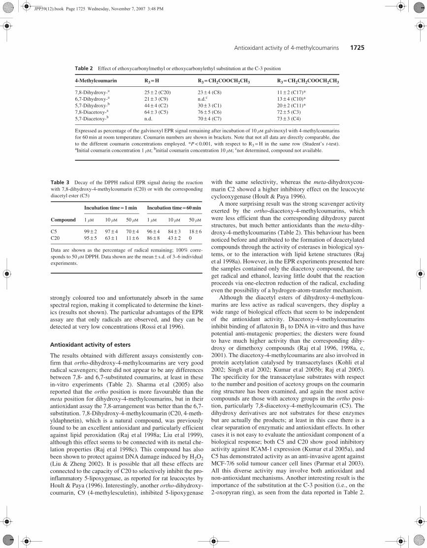
Further experiments were made to verify whether the anti-oxidant effects of 4-methylcoumarins could be observeddirectly in cells. The method used was a standard assaybased on a fluorescent probe (Pallottini et al 2005). Afterloading the precursor DCFH2, the cells are exposed to anoxidative stress resulting in the formation of H2O2; intracel-lular peroxidases then use this H2O2 to oxidize DCFH2 tothe fluorescent DCF. Antioxidants that intercept superoxideor other peroxide-forming radicals prevent the increase inDCF fluorescence.
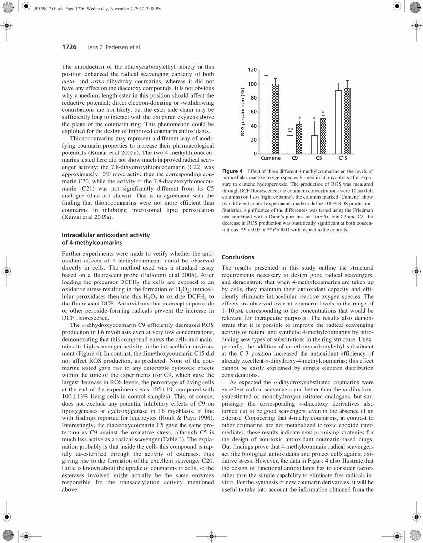
The o-dihydroxycoumarin C9 efficiently decreased ROSproduction in L6 myoblasts even at very low concentrations,demonstrating that this compound enters the cells and main-tains its high scavenger activity in the intracellular environ-ment (Figure 4). In contrast, the dimethoxycoumarin C15 didnot affect ROS production, as predicted. None of the cou-marins tested gave rise to any detectable cytotoxic effectswithin the time of the experiments (for C9, which gave thelargest decrease in ROS levels, the percentage of living cellsat the end of the experiments was 105 ± 19, compared with100 ± 13% living cells in control samples). This, of course,does not exclude any potential inhibitory effects of C9 onlipoxygenases or cyclooxygenase in L6 myoblasts, in linewith findings reported for leucocytes (Hoult & Paya 1996).Interestingly, the diacetoxycoumarin C5 gave the same pro-tection as C9 against the oxidative stress, although C5 ismuch less active as a radical scavenger (Table 2). The expla-nation probably is that inside the cells this compound is rap-idly de-esterified through the activity of esterases, thusgiving rise to the formation of the excellent scavenger C20.Little is known about the uptake of coumarins in cells, so theesterases involved might actually be the same enzymesresponsible for the transacetylation activity mentionedabove.
Conclusions
The results presented in this study outline the structuralrequirements necessary to design good radical scavengers,and demonstrate that when 4-methylcoumarins are taken upby cells, they maintain their antioxidant capacity and effi-ciently eliminate intracellular reactive oxygen species. Theeffects are observed even at coumarin levels in the range of1–10 mM, corresponding to the concentrations that would berelevant for therapeutic purposes. The results also demon-strate that it is possible to improve the radical scavengingactivity of natural and synthetic 4-methylcoumarins by intro-ducing new types of substitutions in the ring structure. Unex-pectedly, the addition of an ethoxycarbonylethyl substituentat the C-3 position increased the antioxidant efficiency ofalready excellent o-dihydroxy-4-methylcoumarins; this effectcannot be easily explained by simple electron distributionconsiderations.
As expected the o-dihydroxysubstituted coumarins wereexcellent radical scavengers and better than the m-dihydrox-ysubstituted or monohydroxysubstituted analogues, but sur-prisingly the corresponding o-diacetoxy derivatives alsoturned out to be good scavengers, even in the absence of anesterase. Considering that 4-methylcoumarins, in contrast toother coumarins, are not metabolized to toxic epoxide inter-mediates, these results indicate new promising strategies forthe design of non-toxic antioxidant coumarin-based drugs.Our findings prove that 4-methylcoumarin radical scavengersact like biological antioxidants and protect cells against oxi-dative stress. However, the data in Figure 4 also illustrate thatthe design of functional antioxidants has to consider factorsother than the simple capability to eliminate free radicals in-vitro. For the synthesis of new coumarin derivatives, it will beuseful to take into account the information obtained from the
Figure 4 Effect of three different 4-methylcoumarins on the levels ofintracellular reactive oxygen species formed in L6 myoblasts after expo-sure to cumene hydroperoxide. The production of ROS was measuredthrough DCF fluorescence; the coumarin concentrations were 10 mM (leftcolumns) or 1 mM (right columns), the columns marked ‘Cumene’ showtwo different control experiments made to define 100% ROS production.Statistical significance of the differences was tested using the Friedmantest combined with a Dunn’s post-hoc test (n = 5). For C9 and C5, thedecrease in ROS production was statistically significant at both concen-trations; *P < 0.05 or **P < 0.01 with respect to the controls.
Cumene C9 C5 C150
20
40
60
80
100
120
RO
S p
rod
uct
ion
(%)
∗∗
∗∗∗
JPP59(12).book Page 1726 Wednesday, November 7, 2007 3:48 PM
Antioxidant activity of 4-methylcoumarins 1727
numerous studies on flavonoid molecules (Firuzi et al 2005).Also in flavonoids the substituents influence the antioxidantactivity — the greater the number of hydroxyl substitutions,the greater the scavenging activity, with the C-3 hydroxy sub-stitution seeming to be particularly important (Firuzi et al2004). However, the many contrasting findings concerningthe biological functions of natural flavonoid compoundsagain point to the problem of separating the antioxidant activ-ity of such molecules from all their other possible biologicaleffects.
Borges, F., Roleira, F., Milhazes, N., Santana, L., Uriarte, E. (2005)Simple coumarins and analogous in medicinal chemistry: occur-rence, synthesis and biological activity. Curr. Med. Chem. 12:887–916
D’Arezzo, S., Incerpi, S., Davis, F. B., Acconcia, F., Marino, M.,Farías, R. N., Davis, P. J. (2004) Rapid nongenomic effects of3,5,3′-triiodo-L-thyronine on the intracellular pH of L-6 myo-blasts are mediated by intracellular calcium mobilization andkinase pathways. Endocrinology 145: 5694–5703
Firuzi, O., Mladenka, P., Petrucci, R., Marrosu, G., Saso, L. (2004)Hypochlorite scavenging activity of flavonoids. J. Pharm.Pharmacol. 56: 801–807
Firuzi, O., Lacanna, A., Petrucci, R., Marrosu, G., Saso, L. (2005)Evaluation of the antioxidant activity of flavonoids by ferricreducing antioxidant power assay and cyclic voltammetry. Bio-chim. Biophys. Acta 1721: 174–184
Fylaktakidou, K. C., Hadjipavlou-Litina, D. J., Litinas, K. E.,Nicolaides, D. N. (2004) Natural and synthetic coumarin deriva-tives with anti-inflammatory antioxidant activities. Curr. Pharm-aceut. Design 10: 3813–3833
Hansen, M. B., Nielsen, S. E., Berg, K. (1989) Re-examination andfurther development of a precise and rapid dye method for meas-uring cell growth/cell kill. J. Immunol. Methods 119: 203–210
Hoult, J. R. S., Paya, M. (1996) Pharmacological and biochemicalactions of simple coumarins: natural products with therapeuticpotential. Gen. Pharmacol. 27: 713–722
Iuliano, L., Pedersen, J. Z., Camastra, C., Bello, V., Ceccarelli, S.and Violi F. (1999) Protection of low density lipoprotein oxidationby the antioxidant agent IRFI005, a new synthetic hydrophilicvitamin E analogue. Free Radic. Biol. Med. 26: 858–868
Kohli, E., Gaspari, M., Raj, H. G., Parmar, V. S., van der Greef, J.,Gupta, G., Kumari, R., Prasad, A. K., Goel, S., Pal, G., Tyagi, Y.K., Jain, S. C., Ahmad, N., Watterson, A. C., Olsen, C. E. (2002)Establishment of the enzymatic protein acetylation independent ofacetyl CoA: recombinant glutathione S-transferase 3-3 isacetylated by a novel membrane-bound transacetylase using 7,8-diacetoxy-4-methylcoumarin as the acetyl donor. FEBS Lett. 530:139–142
Kostova, I. (2006) Synthetic and natural coumarins as antioxidants.Mini Rev. Med. Chem. 6: 365–374
Kumar, S., Singh, B. K., Kalra, N., Kumar, V., Kumar, A., Prasad,A. K., Raj, H. G., Parmar, V. S., Ghosh, B. (2005a) Novel thio-coumarins as inhibitors of TNF-alpha induced ICAM-1 expressionon human umbilical vein endothelial cells (HUVECs) and micro-somal lipid peroxidation. Bioorg. Med. Chem. 13: 1605–1613
Kumar, A., Singh, B. K., Tyagi, R., Jain, S. K., Sharma, S. K.,Prasad, A. K., Raj, H. G., Rastogi, R. C., Watterson, A. C.,Parmar, V. S. (2005b) Mechanism of biochemical action of substi-tuted 4-methylcoumarins. Part 11: comparison of the specificitiesof acetoxy derivatives of 4-methylcoumarin and 4-phenylcoumarin
to acetoxycoumarins:protein transacetylase. Bioorg. Med. Chem.13: 4300–4305
Liu, G. A., Zheng, R. I. (2002) Protection against damaged DNA inthe single cell by polyphenols. Pharmazie 57: 852–854
Liu, Z.-Q., Yu, W., Liu, Z.-L. (1999) Antioxidative and prooxidativeeffects of coumarin derivatives on the free radical initiated andphotosensitized peroxidation of human low-density lipoprotein.Chem. Phys. Lipids 103: 125–135
McPhail, D. B., Hartley, R. C., Gardner, P. T., Duthie, G. G. (2003)Kinetic and stoichiometric assessment of the antioxidant activityof flavonoids by electron spin resonance spectroscopy. J. Agric.Food Chem. 51: 1684–1690
Pallottini, V., Martini, C., Pascolini, A., Cavallini, G., Gori, Z.,Bergamini, E., Incerpi, S., Trentalance, A. (2005) 3-Hydroxy-3-methylglutaryl coenzyme A reductase deregulation and agerelated hypercholesterolemia: a new role for ROS. Mech. AgeingDevelop. 126: 845–851
Parmar, V. S., Bisht, K. S., Jain, R., Singh, S., Sharma, S. K., Gupta, S.,Malhotra, S., Tyagi, O. D., Vardhan, A., Pati, H. N., vandenBerghe, D., Vlietinck, A. J. (1996) Synthesis, antimicrobial andantiviral activities of novel polyphenolic compounds. Indian J.Chem. 35B: 220–232
Parmar, V. S., Sharma, N. K., Husain, M., Watterson, A. C., Kumar, J.,Samuelson, L. A., Cholli, A. L., Prasad, A. K., Kumar, A.,Malhotra, S., Kumar, N., Jha, A., Singh, A., Singh, I., Himanshu,Vats, A., Shakil, N. A., Trikha, S., Mukherjee, S., Sharma, S. K.,Singh, S. K., Kumar, A., Jha, H. N., Olsen, C. E., Stove, C. P.,Bracke, M. E., Mareel, M. M. (2003) Synthesis, characterizationand in vitro anti-invasive activity screening of polyphenolic andheterocyclic compounds. Bioorg. Med. Chem. 11: 913–929
Raj, H. G., Gupta, S., Biswas, G., Singh, S., Singh, A., Jha, A.,Bisht, K. S., Sharma, S. K., Jain, S. C., Parmar, V. S. (1996)Chemoprevention of carcinogen-DNA binding: the relative roleof different oxygenated substituents on 4-methylcoumarins inthe inhibition of aflatoxin B1-DNA binding in vitro. Bioorg.Med. Chem. 4: 2225–2228
Raj, H. G., Parmar, V. S., Jain, S. C., Goel, S., Poonam, Himanshu,Malhotra, S., Singh, A., Olsen, C. E., Wengel, J. (1998a) Mecha-nism of biochemical action of substituted 4-methylbenzopyran-2-ones. Part I: dioxygenated 4-methyl coumarins as superbanti-oxidant and radical scavenging agents. Bioorg. Med. Chem.6: 833–839
Raj, H. G., Parmar, V. S., Jain, S. C., Goel, S., Singh, A., Gupta, K.,Rohil, V., Tyagi, Y. K., Jha, H. N., Olsen, C. E., Wengel, J.(1998b) Mechanism of biochemical action of substituted 4-meth-ylbenzopyran-2-ones. Part II: Mechanism-based inhibition of ratliver microsome-mediated aflatoxin B1-DNA binding by the can-didate antimutagen 7,8-diacetoxy-4-methylcoumarin. Bioorg.Med. Chem. 6: 1895–1904
Raj, H. G., Sharma, R. K., Garg, B. S., Parmar, V. S., Jain, S. C.,Goel, S., Tyagi, Y. K., Singh, A., Olsen, C. E., Wengel, J.(1998c) Mechanism of biochemical action of substituted 4-methylbezopyran-2-ones. Part 3: a novel mechanism for theinhibition of biological membrane lipid peroxidation by dioxy-genated 4-methylcoumarins mediated by the formation of a sta-ble ADP-Fe-inhibitor mixed ligand complex. Bioorg. Med.Chem. 6: 2205–2212
Raj, H. G., Kohli, E., Rohil, V., Dwarakanath, B. S., Parmar, V. S.,Malik, S., Adhikari, J. S., Tyagi, Y. K., Goel, S., Gupta, K.,Bose, M., Olsen, C. E. (2001) Acetoxy-4-methylcoumarinsconfer differential protection from aflatoxin B1-induced micro-nuclei and apoptosis in lung and bone marrow cells. Mutat.Res. 494: 31–40
Raj, H. G., Singh, B. K., Kohil, E., Dwarkanath, B. S., Jain, S. C.,Rastogi, R. C., Kumar, A., Adhikari, J. S., Watterson, A. C.,Olsen, C. E., Parmar, V. S. (2005) Acetoxy drug:protein
References
JPP59(12).book Page 1727 Wednesday, November 7, 2007 3:48 PM
1728 Jens Z. Pedersen et al
transacetylase: a novel enzyme-mediating protein acetylation bypolyphenolic peracetates. Pure Appl. Chem. 77: 245–250
Rossi, L., De Angelis, I., Pedersen, J. Z., Marchese, E., Stammati, A.,Rotilio, G. and Zucco, F. (1996) N-[5-Nitro-2-furfurylidene]-3-amino-2-oxazolidinone activation by the human intestinal cell lineCaco-2 monitored through noninvasive electron spin resonancespectroscopy. Mol. Pharmacol. 49: 547–555
Sharma, S. D, Rajor, H. K., Chopra, S., Sharma, R. K. (2005) Studieson structure activity relationship of some dihydroxy-4-methylcou-marin antioxidants based on their interaction with Fe(III) andADP. Biometals 18: 143–154
Shi, H., Noguchi, N., Niki E. (2001) Galvinoxyl method for stand-ardizing electron and proton donation activity. Meth. Enzymol.335: 157–166
Singh, I., Kohli, E., Raj, H. G., Gyanda, K., Jain, S. K., Tyagi, Y. K,Gupta, G., Kumari, R., Kumar, A., Pal, G., Prasad, A. K., Rastogi,R. C., Olsen, C. E., Jain, S. C., Parmar, V. S. (2002) Mechanismof biochemical action of substituted 4-methylbenzopyran-2-ones.Part 9: Comparison of acetoxy 4-methylcoumarins and otherpolyphenolic acetates reveal the specificity to acetoxy drug:pro-tein transacetylase for pyran carbonyl group in proximity to theoxygen heteroatom. Bioorg. Med. Chem. 10: 4103–4111
JPP59(12).book Page 1728 Wednesday, November 7, 2007 3:48 PM
Related Documents