ANTIMICROBIAL, ANTIOXIDANT AND PROTECTIVE EFFICACY OF FLOWER AND LEAF EXTRACTS OF CALOTROPIS PROCERA AGAINST FREE RADICAL DAMAGE By ABID ALI DEPARTMENT OF BIOCHEMISTRY UNIVERSITY OF KARACHI KARACHI 2015

Welcome message from author
This document is posted to help you gain knowledge. Please leave a comment to let me know what you think about it! Share it to your friends and learn new things together.
Transcript

ANTIMICROBIAL, ANTIOXIDANT AND
PROTECTIVE EFFICACY OF FLOWER AND
LEAF EXTRACTS OF CALOTROPIS PROCERA
AGAINST FREE RADICAL DAMAGE
By
ABID ALI
DEPARTMENT OF BIOCHEMISTRY
UNIVERSITY OF KARACHI
KARACHI
2015

ANTIMICROBIAL, ANTIOXIDANT AND
PROTECTIVE EFFICACY OF FLOWER AND LEAF
EXTRACTS OF CALOTROPIS PROCERA AGAINST
FREE RADICAL DAMAGE
BY
ABID ALI
THESIS SUBMITTED
TO THE FACULTY OF SCIENCE, UNIVERSITY OF
KARACHI, IN FULFILLMENT OF THE REQUIREMENT
FOR THE DEGREE OF DOCTOR OF PHILOSOPHY
DEPARTMENT OF BIOCHEMISTRY
UNIVERSITY OF KARACHI
KARACHI-75270.
2015

ANTIMICROBIAL, ANTIOXIDANT AND
PROTECTIVE EFFICACY OF FLOWER AND LEAF
EXTRACTS OF CALOTROPIS PROCERA AGAINST
FREE RADICAL DAMAGE
THESIS APPROVAL SHEET
SUPERVISOR_______________________________
DR. TABASSUM MAHBOOB
MERITORIUS PROFESSOR
EXTERNAL EXAMINER______________________

DECLARATION
It is declared that this research work entitled: “Antimicrobial, antioxidant and protective
efficacy of flower and leaf extracts of Calotropis procera against free radical damage”
is my own work and has not been submitted in any form for another degree at any
university. Information derived from the published or unpublished literature of other
workers is acknowledged in the text and a list of references.
ABID ALI

DEDICATION
This thesis is dedicated to
My beloved Parents


ACKNOWLEDGEMENTS
First and foremost, I am thankful to Almighty ALLAH who blessed and
gave me the power to complete this research work.
I am greatly indebted to my supervisor Dr. Tabassum Mahboob,
Meritorious Professor, Department of Biochemistry, University of
Karachi for her expert advice, generous guidance and encouragement
throughout the period of research work.
I owe a great debt of gratitude to Dr. Shah Ali Ul Qader, Associate
Professor, Khan Institute of Biotechnology and Genetic Engineering
(KIBGE), University of Karachi for his guidance, cooperation and
providing laboratory facilities during this research.
I am also thankful to Prof. Dr. Majid Mumtaz and Dr. Sumayya Saied
Department of Chemistry, University of Karachi for their kind help in
connection with the preparation of various extracts.
Special thanks are also for Mr. Qader, Mr. Safaraz and Mr. Aijaz for their
cooperation in various ways.
I cannot express enough thanks to all of my family members, especially
my wife for encouragement and moral support throughout the period of
this research work.

CONTENTS
Title Page No.
ABSTRACT
i. English…………………………………………………………….1-3
ii. Urdu ………………………………………………………………4-5
CHAPTER # 1
GENERAL INTRODUCTION………………………………………………….6-23
CHAPTER # 2
GENERAL MATERIAL AND METHODS …………………………………..24-64
CHAPTER # 3
PHYTOCHEMICAL STUDIES OF CALOTROPIS PROCERA………………...65-70
CHAPTER # 4
ANTIMICROBIAL EFFECT OF CALOTROPIS PROCERA………………….71-85
CHAPTER # 5
EFFECT OF CALOTROPIS PROCERA ON ENZYMES ACTIVITY………....86-97
CHAPTER # 6
IN VITRO ANTIOXIDANT PROPERTIES OF CALOTROPIS PROCERA…98-117
CHAPTER # 7
EFFECT OF CALOTROPIS PROCERA ON IBUPROFEN TREATED
RATS………………………………………………………………………….118-144
CHAPTER # 8
GENERAL DISCUSSION…………………………………………………...145-155
CONCLUSION…………………………………………………………………....156
REFERENCES………………………………………………………………..157-184
PUBLISHED RESEARCH PAPER…………………………………………..185-190

EXPANDED CONTENT
Title Page No.
ABSTRACT
i. English…………………………………………………………….1-3
ii. Urdu ………………………………………………………………4-5
CHAPTER # 1
GENERAL INTRODUCTION………………………………………………….6-23
CHAPTER # 2
GENERAL MATERIAL AND METHODS ………………………………..…24-64
2.1. Collection of plant material……………………………………………………24
2.2. Preparation of extract………………………………………………………….24
2.3. Preparation of fractions………………………………………………………..24
2.4. General chemicals and materials………………………………………………28
2.5. Estimation of total protein……………………………………………………..28
2.6. Estimation of carbohydrates…………………………………………………...31
2.7. Estimation of total reducing sugars……………………………………………34
2.8. Estimation of total non-reducing sugars……………………………………….37
2.9. Estimation of total amino acids………………………………………………..37
2.10. Estimation of amino acids by paper chromatography….…………………….39
2.11. Estimation of phenolic compounds…………………………………………..40
2.12. Detection of phenolic compounds by chromatography…….………………...43
2.13. Method for protein free filtrate……………………………………………….44
2.13.1. Plasma urea estimation……………………………………………………..44
2.13.2. Methodology for plasma urea estimation…………………………………..45
2.13.3. Plasma creatinine estimation……………………………………………….45
2.13.4. Kidney homogenate………………………………………...........................48
2.13.5. Malonyldialdehyde estimation……………………………………………..48
2.13.6. 4 hydroxyl 2-nonenal estimation…………………………...........................50
2.13.7. Catalase estimation…………………………………………………............52
2.13.8. Superoxide dismutase estimation…………………………………………..54
2.13.9. Glutathione estimation………………………………………......................55
2.14. Lipid peroxidation inhibition method……………………………………….56
2.14.1. Preparation of tissue homogenate…………………………………………56
2.14.2. Procedure for lipid peroxidation inhibition………………………………..56
2.14.3. DPPH free radical scavenging method……………………………………57
2.14.4. Method for determining reducing power assay…………………………...58
2.15.1. Estimation of glucoamylase……………………………………………….59
2.15.1.1. Glucoamylase activity assay…………………………………………….59
2.15.1.2. GOD – PAP method…………………………………………………….59
2.15.1.3. Reducing sugar estimation for alpha amylase activity………………….60
2.15.1.4. Alpha amylase enzyme activity assay..………………………………….62
2.15.1.5. Urease activity…………………………………………………………...63
2.16. Statistical analysis…………………………………………………………....64
CHAPTER # 3
PHYTOCHEMICAL STUDIES OF CALOTROPIS PROCERA…………….....65-70
3.1. Introduction…………………………………………………………….….….65
3.2. Material and methods………………………………………………….….…..65
3.2.1. Total proteins estimation………………………………………….….……..65
3.2.2. Carbohydrates estimation………………………………………….………..66
3.2.3. Total reducing sugars………………………………………………….……66
3.2.4. Total non reducing sugars……………………………………………….….66
3.2.5. Total amino acids estimation………………………………………….….....66
3.2.6. Amino acid estimation by paper chromatography…………………….…….66
3.2.7. Phenolic compounds estimation…………………………………………….66
3.2.8. Estimation of phenolic compounds by chromatography…………………...66
3.3. Results………………………………………………………………………...67
3.4. Discussion…………………………………………………………………….69
CHAPTER # 4
EFFECT OF CALOTROPIS PROCERA AS ANTIMICROBIAL AGENT…..71-85
4. Introduction…………………………………………………………………….71
4.1. Material and methods………………………………………………………...75
4.2. Preparation of extract………………………………………………………...75
4.3. Preparation of fractions………………………………………………………75
4.4. Experimental microbes……………………………………………………….75
4.5. Culture media………………………………………………………………...75
4.6. Procedure for antimicrobial activity………………………………………….76
4.7. Results………………………………………………………………………...83
4.8. Discussion………………………………………………………………….…84
CHAPTER # 5
EFFECT OF CALOTROPIS PROCERA ON ENZYMES ACTIVITY……….86-97
5. Introduction……………………………………………………………………..86
5.1. Material and methods…………………………………………………………88
5.1.1. Preparation of aqueous extract……………………………………………...88
5.1.2. Enzyme activity for glucoamylase, alpha amylase and urease……………...88
5.1.3. Estimation of glucoamylase…………………………………………………88
5.1.4. Glucoamylase activity assay………………………………………………...88
5.1.5. Estimation of reducing sugar for alpha-amylase………………………….....88
5.1.6. Estimation of urease activity………………………………………………...89
5.1.7. Alpha amylase activity assay……………………………………………….89
5.2. Results and discussion………………………………………………………...96
CHAPTER # 6
IN VITRO ANTIOXIDANT PROPERTIES OF CALOTROPIS PROCERA..98-117
6. Introduction……………………………………………………………………...98
6.1. Material and methods………………………………………………………...100
6.1.1. Preparation of extract………………………………………………………100
6.1.2. Preparation of fractions…………………………………………………….100
6.1.3. Preparation of tissue homogenate………………………………………….100
6.2. Lipid peroxidation inhibition ………………………………………………..101
6.3. 1,1-diphenyl -2-picrylhydrazyl (DPPH) radical scavenging assay………....101
6.4. Reducing power assay………………………………………………………..101
6.5. Results………………………………………………………………………..114
6.6. Discussion…………………………………………………………………….115
CHAPTER # 7
EFFECT OF CALOTROPIS PROCERA ON IBUPROFEN TREATED
RATS…………………………………………………………………………118-144
7. Introduction…………………………………………………………………….118
7.1. Nephrotoxic agents…………………………………………………………...119
7.2. Nsaids………………………………………………………………………...119
7.3. Ibuprofen…………………………………………………………………......120
7.4. Mechanism of action of Ibuprofen…………………………………………...120
7.5. Material and methods………………………………………………………...121
7.5.1. Preparation of extract………………………………………………………121

7.5.2. Preparation of fractions…………………………………………………..121
7.5.3. Experimental animals and diet……………………………………….…..121
7.6. Proper recommendations…………………………………………………...122
7.7. Preparation of drug……….………………………………………………...122
7.8. Study protocol and drug administration plan…………………………….....122
7.9. Collection of samples…………………………………………………….....123
7.9.1. Blood sample……………………………………………………………...123
7.9.2. Kidney sample………………………………………………………….....123
7.10. Analytical methods……………………………………………………......124
7.10.1. Preparation of protein free filtrate……………………………………….124
7.10.2. Estimation of plasma urea……………………………………………….124
7.10.3. Estimation of plasma creatinine…………………………………………124
7.10.4. Preparation of kidney homogenate……………………………………...124
7.10.5. Estimation of malonyldialdehyde (MDA)………………………………125
7.10.6. Estimation of 4-hydroxyl-2-nonenal (4-HNE) ………………………….125
7.10.7. Estimation of catalase …………………………………………………...125
7.10.8. Estimation of superoxide dismutase (SOD) ……………………………125
7.10.9. Estimation of glutathione (GSH) ……………………………………….125
7.11. Results……………………………………………………………………..140
7.12. Discussion………………………………………………………………….142
CHAPTER # 8
8. GENERAL DISCUSSION……………………………………………....145-155
CONCLUSION……………………………………………………………..…...156
REFERENCES……………………………………………………………...157-184
LIST OF FIGURES
Title Page No.
Fig. 1.1. Causes of oxidative stress………………………………………….........14
Fig. 1.2. Showing free radical and oxidative stress……………………………….15
Fig. 1.3. Showing structure of Ibuprofen………………………………………....16
Fig. 1.4. Showing mechanism of inhibition by NSAID………………………….17
Fig. 1.5. Graphical representation of Lipid damage cycle………………………..19
Fig. 1.6. Showing antioxidant defense - enzymes………………………………..20
Fig. 2.1. Flow chart showing preparation of extract from Calotropis procera…..26
Fig. 2.2. Method for preparation of fractions of C. procera in different
solvents……….…………………………………………………………27
Fig. 2.3. Standard curve for total protein…………………………………………30
Fig. 2.4. Standard curve for total carbohydrates………………………………….33
Fig. 2.5. Standard curve for reducing sugar………………………………………36
Fig. 2.6. Standard curve for amino acids…………………………………………38
Fig. 2.7. Standard curve for phenol……………………………………………….42
Fig. 2.8. Standard curve for plasma urea………………………………………….46
Fig. 2.9. Standard curve for plasma creatinine……………………………………47
Fig. 2.10. Standard curve for MDA………………………………………………49
Fig. 2.11. Standard curve for 4-HNE……………………………………………..51
Fig. 2.12. Standard curve for catalase…………………………………………….53
Fig. 2.13. Standard curve for reducing sugar for amylase………………………..61
Fig. 4.1. Antimicrobial activity of flower extracts……………………………….78
Fig. 4.2. Antimicrobial activity of leaf extracts………….………………………80
Fig. 4.3. Showing zone of inhibition of C. procera flower extract……………...…81
Fig. 4.4. Showing zone of inhibition of C. procera leaf extract……………...……82
Fig. 5.1. Representing starch structure showing α-1,4-linkage
where α-amylase targets………………..…………………………………87
Fig. 5.2. Showing the effect of aqueous extract of C. procera
on glucoamylase activity………………………………………..………...93
Fig. 5.3. Showing the effect of aqueous extract of C. procera
on alpha amylase activity………………………………………..……….95
Fig. 5.4. Showing the effect of aqueous extract of C. procera
on urease activity………………………………………………...………97
Fig. 6.1. Effect of different (flower) extracts of C. procera
on Lipid peroxidation inhibition (%)………………………………...…108
Fig. 6.2. Effect of different (leaves) extracts of C. procera
on Lipid peroxidation inhibition (%)………………………………...…109
Fig. 6.3. DPPH radical scavenging activity of(flower) extracts of
C. procera…………………………………..…………………………..110
Fig. 6.4. DPPH radical scavanging activity of (leaves) extracts of
C. procera………………………………………………..……………..111
Fig. 6.5. Reducing power assay of(flower) extracts of C. procera……….…….112
Fig. 6.6. Reducing power assay of (leaves) extracts of C. procera……………113
Fig. 7.1. Effect on Body weight of rats in Control, Ibuprofen, Hexane
and Ibuprofen + Hexane treated groups…….………………………...130
Fig .7.2. Effect on Kidney weight of rats in Control, Ibuprofen, Hexane
and Ibuprofen + Hexane treated groups……………………………....131
Fig. 7.3. Effect on Plasma Urea level of rats in Control, Ibuprofen,
Hexane and Ibuprofen + Hexane treated groups…………………….132
Fig. 7.4. Effect on Plasma Creatinine level of rats in Control, Ibuprofen,
Hexane and Ibuprofen + Hexane treated groups……………………..133
Fig. 7.5. Effect on Tissue SOD level of rats in Control, Ibuprofen,
Hexane and Ibuprofen + Hexane treated groups……………………134

Fig. 7.6. Effect on Tissue Catalase level of rats in Control, Ibuprofen,
Hexane and Ibuprofen + Hexane treated groups…………………….135
Fig. 7.7. Effect on Plasma MDA level of rats in Control, Ibuprofen,
Hexane and Ibuprofen + Hexane treated groups……………………136
Fig. 7.8. Effect on Tissue MDA level of rats in Control, Ibuprofen,
Hexane and Ibuprofen + Hexane treated groups…………………….137
Fig. 7.9. Effect on Tissue 4HNE level of rats in Control, Ibuprofen,
Hexane and Ibuprofen + Hexane treated groups……………………138
Fig. 7.10. Effect on Tissue GSH level of rats in Control, Ibuprofen,
Hexane and Ibuprofen + Hexane treated groups……………………139
Fig. 8.1. Proposed inhibition of MDA formation and metabolism………..……...148
Fig. 8.2. Proposed inhibition of 4HNE production and metabolism…………..…149
Fig. 8.3. Proposed inhibition process of lipid peroxidation by C. procera extract
……………………………………………………………………….....150
LIST OF TABLES
Title Page No.
Table 3.1. Phytochemical Estimation of Calotropis procera……………………...67
Table 3.2. Detected Amino Acids in Calotropis procera…………………………67
Table 3.3. Phenolic constituents of Calotropis procera…..………………………68
Table 4.1. Antiicrobial activity of flower extracts against different
pathogenic strains…………………………………………………….....77
Table 4.2. Antimicrobial activity of leaf extracts against different
pathogenic strains…………………………………………………….....79
Table 5.1. Effect of aqueous extract of C. procera on glucoamylase activity……..90
Table 5.2. Effect of aqueous extract of C. procera on alpha amylase activity….....92
Table 5.3. Effect of aqueous extract of C. procera on urease activity……………..94
Table 6.1. Lipid peroxidation inhibition activity of C. procera flowers
extracts…………………………………………………………………102
Table 6.2. Lipid peroxidation inhibition activity (%) of C. procera leaves
extracts…………………………………………………………………103
Table 6.3. DPPH radical scavenging activity of C. procera flowers
extracts…………………………………………………………….…...104
Table 6.4. DPPH radical scavenging activity (%) of C. procera leaves
extracts………………………………………………………………....105
Table 6.5. Reducing power assay of C. procera flowers
extracts…………………………………………………………..……..106
Table 6.6. Reducing power assay of C. procera leaves
extracts…………………………………………………………………107
Table 7.1. Effect on Body and Kidney weight in control, ibuprofen, hexane,
and ibuprofen+hexane treated rats……………………………………..126
Table 7.2. Effect on renal function in control, Ibuprofen, hexane and
Ibu+hexane pretreated rats……………………………………………..127

Table 7.3. Tissue SOD and Catalase in Control, Ibuprofen, hexane and
Ibu+Hexane……………………………………………………………128
Table 7.4. Plasma MDA, Tissue MDA, 4HNE, and Glutathione levels…………129
1
ABSTRACT
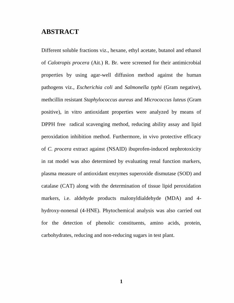
Different soluble fractions viz., hexane, ethyl acetate, butanol and ethanol
of Calotropis procera (Ait.) R. Br. were screened for their antimicrobial
properties by using agar-well diffusion method against the human
pathogens viz., Escherichia coli and Salmonella typhi (Gram negative),
methcillin resistant Staphylococcus aureus and Micrococcus luteus (Gram
positive), in vitro antioxidant properties were analyzed by means of
DPPH free radical scavenging method, reducing ability assay and lipid
peroxidation inhibition method. Furthermore, in vivo protective efficacy
of C. procera extract against (NSAID) ibuprofen-induced nephrotoxicity
in rat model was also determined by evaluating renal function markers,
plasma measure of antioxidant enzymes superoxide dismutase (SOD) and
catalase (CAT) along with the determination of tissue lipid peroxidation
markers, i.e. aldehyde products malonyldialdehyde (MDA) and 4-
hydroxy-nonenal (4-HNE). Phytochemical analysis was also carried out
for the detection of phenolic constituents, amino acids, protein,
carbohydrates, reducing and non-reducing sugars in test plant.
2
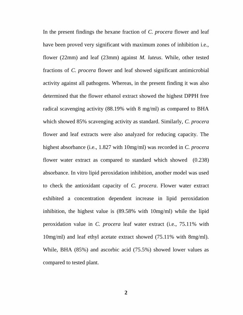
In the present findings the hexane fraction of C. procera flower and leaf
have been proved very significant with maximum zones of inhibition i.e.,
flower (22mm) and leaf (23mm) against M. luteus. While, other tested
fractions of C. procera flower and leaf showed significant antimicrobial
activity against all pathogens. Whereas, in the present finding it was also
determined that the flower ethanol extract showed the highest DPPH free
radical scavenging activity (88.19% with 8 mg/ml) as compared to BHA
which showed 85% scavenging activity as standard. Similarly, C. procera
flower and leaf extracts were also analyzed for reducing capacity. The
highest absorbance (i.e., 1.827 with 10mg/ml) was recorded in C. procera
flower water extract as compared to standard which showed (0.238)
absorbance. In vitro lipid peroxidation inhibition, another model was used
to check the antioxidant capacity of C. procera. Flower water extract
exhibited a concentration dependent increase in lipid peroxidation
inhibition, the highest value is (89.58% with 10mg/ml) while the lipid
peroxidation value in C. procera leaf water extract (i.e., 75.11% with
10mg/ml) and leaf ethyl acetate extract showed (75.11% with 8mg/ml).
While, BHA (85%) and ascorbic acid (75.5%) showed lower values as
compared to tested plant.

3
However, body weight loss was successfully restored by the co-
administration of Ibuprofen with C. procera hexane extract. While,
increased level of renal function markers (urea, creatinine) was
normalized by the administration of C. procera hexane with ibuprofen
treatment. The imbalance in oxidative status was determined by
evaluating decreased level of catalase, superoxide dismutase and
glutathione along with increased levels of malonyldialdehyde and 4-
hydroxynonenal, which was counteracted by the co-administration of C.
procera hexane extract with ibuprofen which maintained cell
sustainability and indicated nephro-protective activity of C. procera.
Besides the above results C. procera leaf and flower aqueous extract were
also used to check enzymatic activities of glucoamylase, α-amylase and
urease enzymes. The flower extract is found proved to be a good enhancer
of glucoamylase, α-amylase and urease activity as compared to leaf
extract. A number of phytoconstituents were also detected. The presence
of phytochemicals in C. procera may indicate a good correlation with that
of antibacterial, antioxidant potential and protective role for in vivo model
which also proved as a good enhancer of enzyme activities. Thus due to
aforementioned activities, Calotropis procera may serve as a better and a
protective therapeutic agent than any other synthetic drug.
4

5

6
1.GENERAL INTRODUCTION
Plants are a major source of traditional medicine even modern medicine system
depends on pharmacologically active agents from plant to obtain useful drugs.
Calotropis procera (Aiton) R. Br. is one of the famous medicinal plant having
bioactive molecules which may serve against various ailments. C. procera is
commonly known as “Aak” and belongs to the family Asclepiadaceae, Plant is wide
spread in Arab, Africa and Asian countries (Mabberley, 2008). C. procera is
characterized by the presence of opposite and decussate leaves. Flowers in terminal or
axillary umbelloid cyme, consists of five deeply lobed and dirty white sepals with
purple tips and white base petals, corona of five fleshy laterally compressed lobes
surrounding the pentagonal stigma (Ali, 1983).
In the past C. procera was also used for epilepsy, mental stress, diarrhoea, earache,
sprain, toothache, anxiety, pain (Kew, 1985; Kareem et al., 2008; Ahmad et al., 2011).
Sometimes, leaves and stem are inhaled or smoked after burning to cure the fever,
swellings, paralysis and arthralgia. Similarly, leaves are also taken for the treatment of
various heart diseases and chest cold (Agharkar, 1991; Hemalatha et al., 2011). The
crude extracts, and dilutions in potencies of C. procera are being used in modern,
homeopathy, unani, as well as in veterinary practices (Dewan et al., 2000; Alencar et
al., 2004; Kareem et al., 2008; Johnson et al., 2011). Flowers decoction having
laxative and anthelmintic properties (Meena et al., 2011) and also cures jaundice
(Sharma et al., 2011). Plant is also used as antidiabetic (Roy et al., 2005; Jaiswal et
al., 2014; Shankar et al., 2014), and relief stomach pain and also acts as an
expectorant (Goyal and Mathur, 2011; Quazi et al., 2013), possesses analgesic,

7
antitumor, antioxidant, anticonvulsant, antidiarrhoeal, antimalarial (Oloumi, 2014) and
oestrogenic activity (Rahimi, 2015).
Biologically active compounds derived from plants are usually categorized in
secondary and primary metabolites. Whereas, last one is the part of metabolic
pathways and secondary metabolites are waste products or byproducts of metabolic
pathways. Regarding to the medicinal uses of Calotropis procera secondary
metabolites namely, phenolic compounds tannins, terpenoids and saponins have
received an immense attention (Vaya & others, 1997; Sengul & others, 2009; Patel &
others, 2010), such as Hassan & others (2006) evaluated roots, leaves and stem bark of
C. procera with aqueous, hexane, petroleum ether extracts for the detection of
phytochemicals where leaves and root extracts showed the presence of glycosides,
saponins, triterpenoids, steroids, alkaloids, tannins and flavonoids while stem bark
showed flavonoids, triterpenoids and saponins. Similarly, Qureshi & others (2007)
observed that the flower ethanol extract of C. procera having strong antioxidant
potential due to its Quercetin related flavonoids. Alam and Ali (2009) investigated
roots of C. procera and yield two phytochemical compounds, namely procerur acetate
and proceranol along with other known compounds. However, the studies of Kawo et
al. (2009) revealed that water extracts of leaf and milky sap of C. procera showed the
presence of flavonoids, saponins, steroids, and tannins. Leaf showed stronger
antibacterial potential while latex have no antibacterial activity. Similarly, methanolic
extract of C. procera leaf has been proved to contain alkaloids, flavonoids, phenolic
acids and tannins as significant constituents for drug development (Yadav et al.,
2010). Moustafa et al. (2010) evaluated that C. procera has cardenolides, flavonoids
8
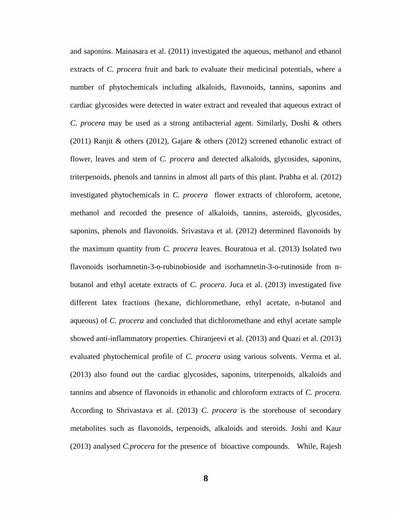
and saponins. Mainasara et al. (2011) investigated the aqueous, methanol and ethanol
extracts of C. procera fruit and bark to evaluate their medicinal potentials, where a
number of phytochemicals including alkaloids, flavonoids, tannins, saponins and
cardiac glycosides were detected in water extract and revealed that aqueous extract of
C. procera may be used as a strong antibacterial agent. Similarly, Doshi & others
(2011) Ranjit & others (2012), Gajare & others (2012) screened ethanolic extract of
flower, leaves and stem of C. procera and detected alkaloids, glycosides, saponins,
triterpenoids, phenols and tannins in almost all parts of this plant. Prabha et al. (2012)
investigated phytochemicals in C. procera flower extracts of chloroform, acetone,
methanol and recorded the presence of alkaloids, tannins, asteroids, glycosides,
saponins, phenols and flavonoids. Srivastava et al. (2012) determined flavonoids by
the maximum quantity from C. procera leaves. Bouratoua et al. (2013) Isolated two
flavonoids isorhamnetin-3-o-rubinobioside and isorhamnetin-3-o-rutinoside from n-
butanol and ethyl acetate extracts of C. procera. Juca et al. (2013) investigated five
different latex fractions (hexane, dichloromethane, ethyl acetate, n-butanol and
aqueous) of C. procera and concluded that dichloromethane and ethyl acetate sample
showed anti-inflammatory properties. Chiranjeevi et al. (2013) and Quazi et al. (2013)
evaluated phytochemical profile of C. procera using various solvents. Verma et al.
(2013) also found out the cardiac glycosides, saponins, triterpenoids, alkaloids and
tannins and absence of flavonoids in ethanolic and chloroform extracts of C. procera.
According to Shrivastava et al. (2013) C. procera is the storehouse of secondary
metabolites such as flavonoids, terpenoids, alkaloids and steroids. Joshi and Kaur
(2013) analysed C.procera for the presence of bioactive compounds. While, Rajesh

9
et al. (2014) studied stem powder of C. procera with different extracts of hexane,
chloroform, methanol and sterile water for detection of saponins, flavonoids, sterols,
tannins and alkaloids. However, studies of Tiwari et al. (2014) evaluated the
phytochemicals of petroleum ether and methanol leaf extracts of C. procera and
determined the presence of glycosides, protein, triterpenoids, steroids and flavonoids
and suggested that these chemicals may be a significant indicator for the medicinal
importance of the plant. Moreover, Gholamshahi et al. (2014) also observed the leaf,
flower and fruit extracts of C. procera with the detection of phenolic constituents and
proved it as a strong antioxidant plant, which could be utilized in food and drug
industry. While, Al Snafi (2015) suggested that C. procera exhibited many
pharmacological aspects due to the presence of biologically active constituents.
Similarly, Shetty et al. (2015) studied the phytochemicals of C. procera leaves and
made a positive correlation between phytochemicals and antibacterial activity.
Beside the medicinal reports, C. procera also proved to be the antibacterial agent
against various human disease producing bacteria, which includes the bacteria with
violet stain and bacteria without violet stain. Akhtar et al. (1992) isolated a
cardenolide, proceragenin from C. procera and observed its strong antibacterial
activity against non-violet stain and violet stain bacteria. Ali et al. (2001) investigated
ethanolic extracts of 20 different plants, including C. procera against pathogenic
bacterial strains and concluded that C. procera affords antibacterial potential.
Similarly, acetone, methanol, ethanol, hexane, chloroform and ethyl acetate fractions
were used against S. aureus, S. epidermidis, Bacillus cereus, Pseudomonas
aeroginosa, Kleibsiella pneumonia, Serratia marcenes, Bacillus subtilis, and M.

10
luteus. (Parabia et al., 2008). It was reported that water, methanol, ethyl acetate flower
extracts are potent against fungal pathogens viz. Fusarium and T. vesiculatum (Devi et
al., 2008). Kareem et al. (2008) observed ethanol extract of the leaf and sap of C.
procera which showed widest zone of inhibition. Similarly, Yesmin et al. (2008) also
demonstrated the antibacterial activity through leaf watery & methanolic extracts of
C.procera and it was found that both extracts were active against bacterium with
violet stained and non-violet stained bacteria at low concentrations. Similarly, Kawo et
al. (2009) studied the antimicrobial potential of watery and ethanol leaf extracts and
sap of C. procera against the different bacterial strains where it was revealed that
aqueous extract did not show antibacterial activity while, leaf ethanol and latex have
significant antibacterial potential. Moreover, Mohanraj et al. (2010) observed that the
ethyl acetate extract of C.procera leaf and roots were effective against tested bacteria.
Antibacterial activities were performed from the flower extract with different organic
solvents viz., hexane, chloroform and methanol against Alternaria alternata,
Aspergillus flavus, Aspergillus niger, Bipolaris bicolour, Curvularia lunata, Penicillin
expansum, Pseudomonas marginalis and Rhizoctonia solani (Vadlapudi and Naido,
2010). While ethanol flower extract (Doshi et al., 2011) was used against the larvae of
A. stephansi. However, acetone and methanol flower extracts were further used
against Bacillus pumilis, E.coli, A. niger, Fusarium oxysporum, (David et al., 2011).
Amin and Khan (2011) proved antibacterial efficacy of methanolic fraction of leaf for
Enterobacter, Pseudomonas, S. aureus and Micrococcus. Similarly, Doshi et al.
(2011) utilized flower, young buds, mature leaves and stems of C. procera for
determination of antibacterial activity. While, mature leaves were found strong, potent

11
antimicrobial agent against all micro flora included during the test. Gajare et al. (2012)
evaluated ethanol root extract of C. procera, which was proved as potent antibacterial
agent. While, Vadlapudi et al. (2012) examined C. procera organic extract of aerial
parts against Pseudomonas marginalis and S. mutans. Prabha and Vasantha (2012)
screened C. procera flower extract of chloroform, acetone and ethanol against various
pathogens and maximum antibacterial activity was recorded against B. subtilis and S.
aureus. Velmurugan and his co-workers (2012) studied C. procera leaf extract of
hexane, ethyl acetate and methanol against aquatic micro pathogens from shrimp
fishes. It was noted that ethyl acetate extract effectively suppressed bacterial strains.
Mako et al. (2012) evaluated the antimicrobial activity of aqueous and ethanol root
and leaves extract of C. procera where ethanol extract showed more significant
potential than aqueous extract. Ranjit et al. (2012) observed ethanol flower extract of
C. procera and proved it as a strong inhibitory agent against human pathogenic
bacterias. Neenah (2013) studied the antimicrobial potential of solvent extract and
phenolic compounds of C. procera, using the agar well diffusion method. The crude
flavonoid fraction of methanolic extract was found to possess highest antimicrobial
activity and Gram positive bacteria were more vulnerable than the non-violet stained
bacteria and the yeast species were more vulnerable than filamentous fungi. Joshi and
Kaur (2013) determined the antimicrobial potential of ethanol, methanol and watery
extract of C. procera and found that ethanolic extract have strong antimicrobial
activity against Pseudomonas aeroginosa. Parabia et al. (2008) reported antibacterial
activity of aqueous elixir of twig and milky sap of C. procera. Both the samples
exhibited greater inhibition zone on S. aureus bacterial strain. Muzammal (2014)

12
determined antibacterial activity of aqueous, ethanol and methanol concentrate of C.
procera. While, ethanol concentrate of leaves and flower showed significant potential
against S. typhi and E. coli. Salem et al. (2014) demonstrated the antibacterial activity
through leaf and latex chloroform, ethanolic and methanolic concentrates of C.
procera where it was noted that watery and ethanolic leaf extracts showed maximum
potential against Gram negative and Gram-positive pathogenic bacteria. Kazemipour
et al. (2014) investigated antimicrobial activity of ethanol, chloroform and water
extracts of C. procera flowers and leaves. All extracts showed high potential against
Klebsiella pneumonia and S. epidermidis. Javadian et al. (2014) evaluated
antimicrobial activity of ethanol extract of C. procera and it found effective against E.
coli isolates. Ahmed et al. (2014) also proved the antibacterial activity of C. procera
latex against E.coli and Salmonella. Similarly, Ali et al. (2014) determined
antibacterial potential of different fractions including ethyl acetate, butanol and
aqueous flower extracts, where it was concluded that flower of C. procera have a
significant potential against many bacterial strains. Shetty et al. (2015) studied the
antibacterial effect of methanol, ethyl acetate, ethanol, acetone and aqueous extracts of
C. procera leaves against human pathogenic bacteria. It was revealed that leaves of C.
procera have significant antibacterial potential. Pandey et al. (2015) assessed
antibacterial activity of C. procera methanol, acetone, petroleum ether and ethyl
acetate extracts where methanol leaves extract and stem ethyl acetate extract showed
highest range of inhibition against E.coli and S. aureus.
However, toxicity is the degree to which a substance is able to damage an organism.
Toxicity can affect the whole organism or substructure of the organism and it may be

13
occur due to certain biological, physical or chemical effects. Drug induced toxicity can
damage any tissue depending on dosage such as acute dosage of a drug can produce
the toxicity for nervous system and its chronic exposure may cause the serious injuries
to the other organs. Toxicity can also be produced by the medicines which are
normally be used for curative purposes. Sometimes, the use of over the counter
medicines and long term use of overdoses of drugs may also cause toxicity to certain
specific organs. The Process of oxidation continuously takes place in all aerobic living
bodies, due to this ROS (reactive oxygen species) including O2 anion, H2O2 hydrogen
peroxide, -OH hydroxyl radical and nitric oxide/peroxinitrates (NO/NOO
-) are
constantly formed within the cells. The over production of these substances may cause
oxidative load in the cells. This oxidative stress produces deleterious effects to cells of
DNA, proteins and lipids. Lipid are specifically more damaged due to the formation of
lipid peroxidation products.
The toxicity of different metals, pollution, pesticides radiations, use of alcohols,
smoking, fast food, lack of good nutrition, stress, inadequate intake of fruits and
vegetables contaminants, excessive exercise and inadequate physical activity are the
reasons of free radical formation (Davies 1991; Halliwell and Aruoma 1993; Langseth
1995).

14
Figure 1.1. Causes of oxidative stress
(Adapted from http://currentscienceperspectives.com/)

15
Figure 1.2. Showing free radical and antioxidant in cell (Adapted from http://currentscienceperspectives.com/)
There are various reports available on toxicity producing substances like NSAIDS
(Derle & others, 2006; Fackovcova & others, 2000; Kocaoglu & coworkers, 1997),
Phenacetin (Murray and Brater, 1993), Mefenamic acid (Robertson & coworkers,
1980; Somchit & others, 2004), Caffeine (De Crespigny & coworkers, 1980),
Paracetamol (Younes & others, 1988), Acetaminophen (Tarloff & coworkers, 1990;
Trumper & others, 1992), Diclofenac (Hickey & coworkers, 2001; Yasmeen & others,
2007), Tenofovir (Morelle & others, 2009), Tacrolimus and NSAIDS (Soubhia &
others, 2005), Diclofenac (Kim & coworkers, 1999) and Metals including arsenic,
cadmium, lead and mercury (Nicholson, 1985; Fowler, 1992).
While, Cholestyramine was utilized against the Paracetamol induced toxicity in rats
and that was evident by a reduction in plasma enzyme activity and creatinine levels
(Siegers and Moller, 1989). On the other hand, Cadmium was used to prevent the
Acetaminophen induced toxicity in female rats (Bernard et al., 1988). Moreover,

16
Ibuprofen and Diclofenac were found useful protective drugs against Gentamicin
toxicity (Farag et al., 1996). While, Sharma et al. (2007) suggested that the algal
supplementation of Spirulina fusiformis can play a significant role against Mercuric
Chloride induced toxicity.
There are several substances which can initiate toxicity to the kidneys, called
nephrotoxic agents. These substances include antibiotics, anticancer drugs, heavy
metals, herbicides, pesticides, excess amount of uric acid and long term use and high
doses of analgesics may also cause nephrotoxicity these analgesics usually include
aspirin and ibuprofen.
Likewise, non-steroidal anti-inflammatory drugs or NSAID are the commonly used
over the counter drugs. They are pain relievers, help in reducing inflammation and
lower fever. They also prevent blood from clotting.
Ibuprofen is selected for present experimental studies. It is a derivative of propionic
acid its chemical name is Isobutylphenylpropionic acid, the structure containing a
benzene ring conjugated to a propionic acid. It was first derived during 1950-1960s at
the research laboratories of Boots group and discovered by the scientist Andrew RM
Dunlop with his co-researchers Stewart Adams, John Nicholson, Jeff Wilson and
Colin Burrows (Robertson, 2014).
Figure 1.3. showing structure of Ibuprofen
17
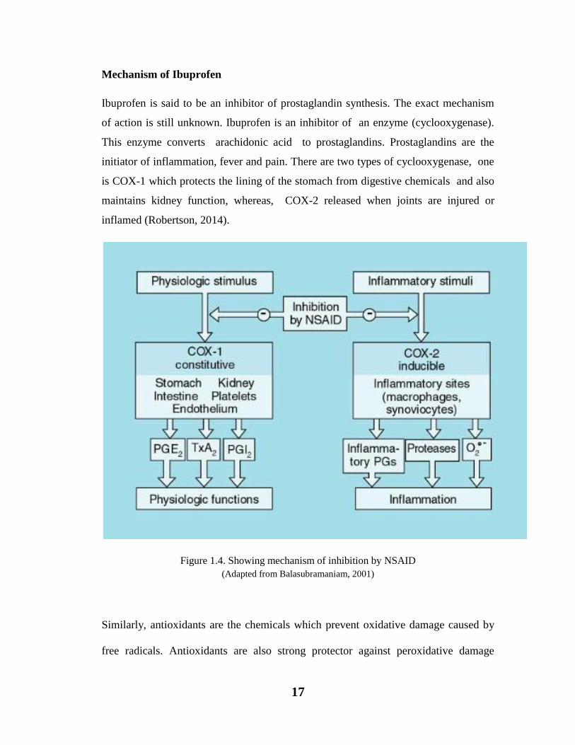
Mechanism of Ibuprofen
Ibuprofen is said to be an inhibitor of prostaglandin synthesis. The exact mechanism
of action is still unknown. Ibuprofen is an inhibitor of an enzyme (cyclooxygenase).
This enzyme converts arachidonic acid to prostaglandins. Prostaglandins are the
initiator of inflammation, fever and pain. There are two types of cyclooxygenase, one
is COX-1 which protects the lining of the stomach from digestive chemicals and also
maintains kidney function, whereas, COX-2 released when joints are injured or
inflamed (Robertson, 2014).
Figure 1.4. Showing mechanism of inhibition by NSAID
(Adapted from Balasubramaniam, 2001)
Similarly, antioxidants are the chemicals which prevent oxidative damage caused by
free radicals. Antioxidants are also strong protector against peroxidative damage
18
through their metal ion chelating and radical scavenging activities. A considerable
attention has been paid to the antioxidant activity of C. procera in respect to phenolic
constituents such as, Patel et al. (2014) studied the comparative antioxidant activity by
DPPH (1,1-Diphenyl-2-picryl hydrazyl) of C. procera and C. gigentia by using their
methanolic extract and it was reported that C. procera possesses high antioxidant
properties due to more phenols and flavonoids as compared to C. gigantea. Similarly,
Srividya et al. (2013) also proved antioxidant activity of ethanolic fruit extract of C.
procera by DPPH method.
Pooja et al. (2014) found out the antioxidant potential of ethyl acetate and acetone
fractions of C. procera by utilizing two different assays namely, Ferric reducing
antioxidant power (FRAP) and 2,2,Azino-bis-(3-ethyl) benzo thiazoline-6-sulfonic
acid (ABTS).
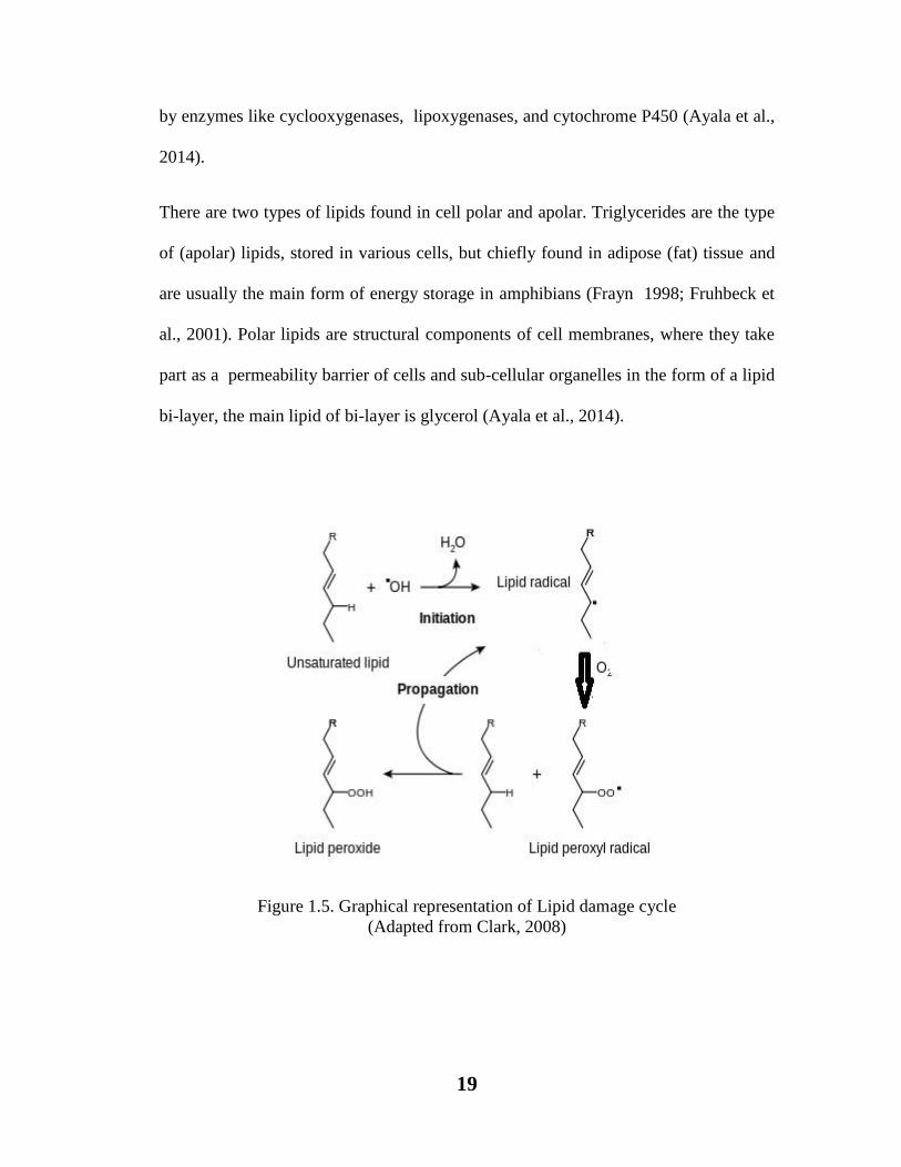
Lipid peroxidation is a process of oxidative degradation of lipids, in which a free
radical like hydroxyl group (OH) extract electrons from the unsaturated lipids present
in cell membranes. This result in the formation of a water molecule and lipid/fatty acid
radical. This radical again reacts with oxygen to form lipid peroxyl radical. Lipid
radical and lipid peroxyl radical both are unstable species, therefore, lipid radical
reacts with oxygen and convert into lipid peroxyl radical and lipid peroxyl radical
again react with other unsaturated lipid, this cycle continues and new lipid radical
reacts with same way and finally lipid peroxide is formed, that may cause cellular
damage. Cholesterol, Glycolipids, phospholipids, are also well-known targets of lipid
damaging and potentially cause fatal peroxidative change. Lipids also can be oxidized
19
by enzymes like cyclooxygenases, lipoxygenases, and cytochrome P450 (Ayala et al.,
2014).
There are two types of lipids found in cell polar and apolar. Triglycerides are the type
of (apolar) lipids, stored in various cells, but chiefly found in adipose (fat) tissue and
are usually the main form of energy storage in amphibians (Frayn 1998; Fruhbeck et
al., 2001). Polar lipids are structural components of cell membranes, where they take
part as a permeability barrier of cells and sub-cellular organelles in the form of a lipid
bi-layer, the main lipid of bi-layer is glycerol (Ayala et al., 2014).
Figure 1.5. Graphical representation of Lipid damage cycle
(Adapted from Clark, 2008)
20
The consequence of lipid peroxidation is the formation of reactive aldehyde
malonyldialdehyde (MDA) and 4 –hydroxy-nonenal (4-HNE). 4-HNE is a major
marker of lipid peroxidation.
Reducing power is another property of antioxidants, with this power an antioxidant
may reduce ferric Fe3+
ion into ferrous Fe2+
(Pohanka et al., 2009). So in this way
antioxidant may be beneficial for the living body.
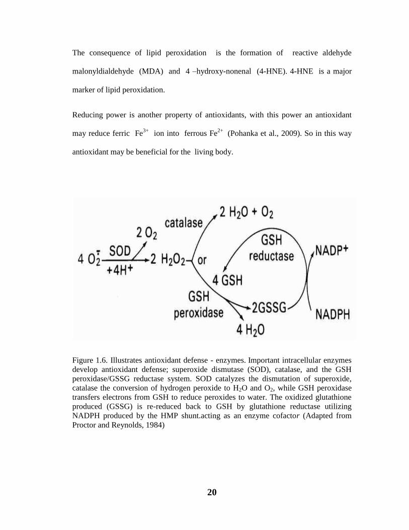
Figure 1.6. Illustrates antioxidant defense - enzymes. Important intracellular enzymes
develop antioxidant defense; superoxide dismutase (SOD), catalase, and the GSH
peroxidase/GSSG reductase system. SOD catalyzes the dismutation of superoxide,
catalase the conversion of hydrogen peroxide to H2O and O2, while GSH peroxidase
transfers electrons from GSH to reduce peroxides to water. The oxidized glutathione
produced (GSSG) is re-reduced back to GSH by glutathione reductase utilizing
NADPH produced by the HMP shunt.acting as an enzyme cofactor (Adapted from
Proctor and Reynolds, 1984)

21
A considerable attention has been paid to the antioxidant, lipid peroxidation, and
reducing power of C. procera such as Roy et al. (2005) performed in vivo
experiments for determination of lipid peroxidation and antioxidant ability of dried
latex of C. procera which showed potential by increasing the levels of SOD,
catalase, and GSH. Similarly, Setty et al. (2007) evaluated lipid peroxidation and
antioxidant ability of C. procera ethanol flower extract which exhibited a marked
increase in tissue GSH level. Qureshi et al. (2007) determined the strong antioxidant
potential of flowers ethanol extract of C. procera. Yesmin et al. (2008) performed
DPPH method to determine the antioxidant activity of methanol leaves extract of C.
procera which shows strong antioxidant activity. Chavda et al. (2010) evaluated
hexane, ethyl acetate and chloroform root extract of C. procera, the fractions showed
significant lipid peroxidation inhibition activity and exhibited significant potential to
normalized the levels of tissue SOD, Catalase and GSH. Parihar et al. (2011)
elucidated a positive change in lipid peroxidation by increasing GSH contents in tissue
after administration of ethanol root extract of C. procera. Bouratoua et al. (2013)
determined a moderate antioxidant activity of butanol and ethyl acetate fractions of
aerial parts of C. procera. Srividya et al. (2013) also proved antioxidant activity of
ethanol fruit extracts of C. procera by DPPH method. Ahmed et al. (2014) determined
in vitro antioxidant activity of methanol extract of C. procera latex, which exhibited
positive activity to scavenge free radicals. Pooja et al. (2014) investigated the
antioxidant potential of ethyl acetate and acetone fractions of flowers of Alastonia
scholaris, Cassia auriculata, Catharanthus roseas and Calotropis procera. Amongst
all flowers, C. procera showed lower antioxidant activity.

22
While, the antioxidant capacity of extract may also be determined by reduction of the
ferricyanide Fe3+
complex in the ferrous Fe2+
form in the presence of plant extract
(Oyaizu, 1986; Kumar et al., 2013).
Enzymes are biological molecules that speed up the biological chemical reactions
(Brayer et al., 1995). The ability of an enzyme to catalyze a specific reaction could be
measured in terms of enzyme activity. There are various enzymes like, Glucoamylase,
also known as glucan 1,4-alpha-glucosidase, (EC 3.2. 1 .3). It is a type of digestive
enzyme which cleaves one glucose unit from a non reducing end of starch (amylose
and amylopectin). Most of the glucoamylases are also able to hydrolyze the 1,6-a
linkages in branch points of starch molecules.
Alpha amylase is a digestive enzyme. The formal title of alpha amylase is 1,4 α-D-
glucanohydrolase; EC 3.2.1.1. The enzyme alpha amylase aids in the hydrolysis of α–
1,4 glycosidic bond in the conversion of starch to maltose (Brayer et al., 1995). In
humans, it is found in both saliva and pancrease. Amylases are also used in various
industries like paper, food and textile industries (Windish et al., 1965; Gupta et al.,
2003).
The enzyme urease (EC 3.5.1.5) catalyzes the breakdown of urea into carbon
dioxide and ammonia. The reaction occurs as follows (Zimmer, 2000).
(NH2)2CO + H2O → CO2 + 2NH3
In view of the previous studies the present study was carried out to find out the in vitro
antioxidant potential of Calotropis procera which was further confirmed by in vivo
effects in rats against Ibuprofen induced nephrotoxicity.

23
In vitro lipid peroxidation, reducing power and DPPH free radical scavenging activity
was also determined to ensure the degree of protective efficacy of naturally growing
Calotropis procera as a therapeutic agent.

24
2. GENERAL MATERIAL AND METHODS
2.1. Collection of Plant Material
Healthy and fresh flower and leaf of Calotropis procera were collected from different
population occurring in the Karachi. Collection were made in the year 2013-2014.
Sample specimen were deposited to the Herbarium, University of Karachi. General
herbarium = 86455.
2.2. Extraction methodology
After collection flower and leaf materials (c. 8-10 kg) were properly washed and air
dried for about 30 days. Then their powders were prepared by using grinder. The dried
powder matrial was soacked in 80% ethanol and left for one week. Then extract was
filtered with the help of filter paper. After filtration these extracts were concentrated
by rotary evaporator and kept for further use.
2.3. Fractioning of extract
Fractions of extract were prepared with the help of separating funnel. Various solvents
viz. butanol, hexane, ethyl acetate were used for fractioning. These fractions were

25
further concentrated. The obtained extracts were dried till converted into solid form.
This form can be used for further analyses.

26
Sorting of Flowers and
Leaves
Shade drying of
Flowers and Leaves
Grinding of
Flowers and Leaves
Collection of Plant
Calotropis procera
Extraction of Flowers and Leaves with
hexane, ethyl acetate, ethanol & butanol
Collection of
fractions from
each solvent
Storage of fractions
In vitro and In vivo experiments
Soaking of Flowers and Leaves
with Solvent (ethanol)
After 10 days soaked material was
filtered in a new bottle in form of
crude extract.
Crude Extract was concentrated
with the help of rotary evaporator
Figure 2.1. Flow Chart For Preparation of Extract From Calotropis procera

27
Ethanolic Crude Extract was concentrated with the help
of rotary evaporator
Concentrated Ethanolic Extract was mixed
with Hexane Solvent in Separating Funnel.
Let the mixture for few minutes to be separated
in two parts. A separation layer was observed.
Lower portion of the mixture contains
Aqueous Solution and Upper portion of the
mixture was taken as Hexane soluble part.
Hexane soluble Fraction was
collected Aqueous mixture was again treated with hexane
solvent until a clear upper part was obtained.
Aqueous portion was mixed with Ethylacetate
in separating funnel
Lower portion of the mixture contains
Aqueous Solution and Upper portion of the
mixture was taken as Ethylacetate soluble
part.
Ethylacetate soluble Fraction
was collected
Hexane soluble Fraction was
concentrated with the help of
rotary evaporator
Aqueous portion was mixed with Butanol in
separating funnel
Aqueous mixture was again treated with
Ethylacetate solvent until a clear upper part
was obtained.
Ethyacetate soluble Fraction
was concentrated with the
help of rotary evaporator
Lower portion of the mixture contains Aqueous
Solution and Upper portion of the mixture
was taken as Butanol soluble part.
Butanol soluble Fraction was
collected
Butanol soluble Fraction was
concentrated with the help of
rotary evaporator
Aqueous mixture was again treated with
Butanol until a clear upper part was obtained.
Figure 2.2. Method For Preparation of Fractions In Different Solvents

28
2.4. GENERAL CHEMICALS AND MATERIALS
Formalin, ethanol, ethyleacetate, methanol, hexane, Butanol, acetonitrile, pyridine,
sulphuric acid, eosin, EDTA, BHT, Na-tungstate from Fluka AG, phosphoric acid,
oxidized glutathione Amresco, disodium hydrogen phosphate from Merck, diacetyl
monoxime from Riedel de Haen, sodium hydroxide, potassium chloride, -NADPH,
formaldehyde, thiobarbituric acid, 2,4 dinitrophenyl hydrazine from Fisher Scientific,
disodium carbonate, nitro blue, paraffin, hydrogen peroxide, tetrazolium,
hydroxylamine hydrochloride, triton X-100, hematoxylin, 1,14,4 diethoxypropane,
sodium dihydrogen phosphate, picric acid and acetic acid.
PHYTOCHEMISTRY
2.5. Estimation of total protein (Bradford, 1976)
REAGENTS:
2.5.1. Coomassie Reagent
Coomassie stain (100gm) was mixed in 50ml of methanol and filtered. The solution
was added into 100ml of 85% phosphoric acid and volume was made up to 200ml.
The reagent was prepared by adding 1ml of dye stock with 4ml of water.
2.5.2. Tris HCl buffer (pH = 6.8)
Solution of 100mM Tris base was prepared and pH was maintained by the addition
of 0.2M HCl.
29
2.5.3 . Extraction
0.5 g fresh leaf and flowers were taken and macerated in 5ml of Tris HCl buffer and
centrifuged for 20 minutes at 2500rpm. Supernatant was collected in separate test
tubes for estimation.
2.5.4. Procedure for estimation of protein
0.04ml of leaf and flower extracts was added in test tubes. Then 2ml of assay reagent
was added in each tube. Test tubes were kept for incubation at room temperature for
30 minutes and finally the optical density was determined at 595nm.
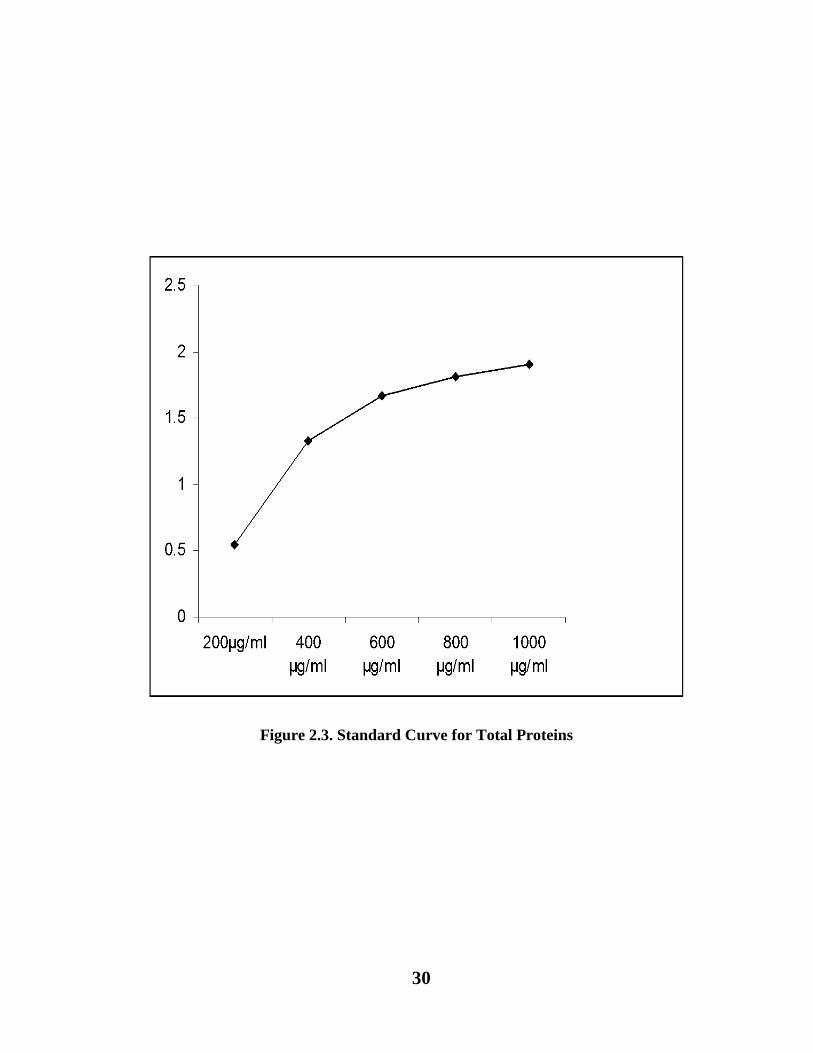
2.5.5. Calibration of standard curve
Bovine serum albumin (BSA) was used to prepare standard curve.
2.5.6. Stock solution
Stock was processed by adding 0.1gram of BSA in 10ml of Tris HCl buffer.
2.5.7. Working standard solution and colour development
Dilutions of BSA stock solution were prepared as 200µg/ml, 400µg/ml, 600µg/ml, 800µg/ml
and 1000µg/ml. 0.04ml was taken from each dilution in separate test tubes. 2.0 ml of
reagent was also poured. All test tubes were kept at room temperature for 30 minutes.
Optical density was noted at 595nm.
30
Figure 2.3. Standard Curve for Total Proteins
31
2.6. Estimation of Carbohydrates (Yemm and Willis, 1954)
Reagents:
2.6.1. Anthrone Reagent
Anthrone (0.4ml) was added in 200 ml of concentrated sulpfuric acid with continuous
shaking. The acid solution was then cooled and 15 ml of 95% ethanol and 60ml of
distilled water were taken in a dark coloured flask which was placed in an ice bar.
Then acid solution was transferred drop by drop in a dark coloured flask with constant
shaking. Fresh Anthrone reagent was prepared each time.
2.6.2. Method for 100 mM Tris HCL
Solution A: 12.1gm of Tris base was added in 100ml of distilled water and the
volume was made up to 1000 ml.
Solution B: 1.21 ml of concentrated HCl was added in 100 ml distilled water and the
volume was made up to 1000 ml.
500ml of solution A added in to 200 ml of solution B. The pH of solution was
maintained up to 6.8. When pH was basic, solution B was added, and for acidic pH
solution A was added.
2.6.3. Extraction
1 gm fresh leaves and flowers were crushed in a mortar with 5 ml Tris HCl. Crushed
material was centrifuged at 2500 rpm for 20 minutes. The supernatant was separated
in test tubes.

32
2.6.4. Procedure for estimation of carbohydrates
To 1ml extract, 4ml distilled water was added in 10 ml of anthrone reagent and
shaked well. Content was kept for 16 minutes in a water bath. Test tubes were left for
cooling. Finally, optical density was observed at 660 nm.
2.6.5. Calibration of standard curve
To prepare standard curve sucrose was used.
Stock Solution
1000μgm/ml prepared by adding 0.1 gm of sucrose in 100 ml distilled water.
2.6.6. Working standard solution and colour development
An aliquot of 200 μgm/ml, 400 μgm/ml, 600 μgm/ml, 800 μgm/ml and 1000 μgm/ml
dilution of the sucrose stock solution was made 1 ml of each dilution in separate test
tubes. Distilled water for reagent blank was taken. 4.0 ml distilled water was added.
10 ml of anthrone reagent was poured drop by drop. Test tubes were kept in boiling
water bath for 16 minutes. Test tubes were then cooled and optical density was noted
at 660nm.

33
Figure 2.4. Standard Curve for Total Carbohydrates
0
0.1
0.2
0.3
0.4
0.5
0.6
0.7
0.8
0.9
0 200 400 600 800 1000 1200
Ab
sorb
an
ce a
t 6
60
nm
Concentration of Sucrose

34
2.7. Estimation of total reducing sugars (Miller, 1959)
Reagents
2.7.1. DNS Reagent 3, 5 di-nitro salicylic acid (100mg) was mixed in 20 ml of 2N
sodium hydroxide and dissolved in 50ml distilled water. 30 g of Sodium Potassium
tartrate was also added, then volume was made up to 100 ml and stored at 4oC in
refrigerator.
2.7.2. Preparation of 100 mM tris HCl
Solution A: Tris base (12.1gm) was mixed in 100ml of water and volume was made
up to 1000 ml.
Solution B: Concentrated HCl (1.21ml) was added in 100 ml water and made the
volume up to 1000 ml. Took 500 ml of solution A and added in to 200 ml of solution
B. PH of solution was maintained at 6.8. If the pH was basic then solution B was
added, if it was more acidic then solution A was added.
2.7.3. Extraction
1 gm fresh leaf and flower samples were crushed separately in a mortar with 5 ml tris
HCl. Crushed material were centrifuged at 4000 rpm for 15 minutes. Supernatent was
then collected in test tubes.
2.7.4. Procedure for estimation of reducing sugars
2 ml DNS was added in 1 ml extract. Then tubes were kept in boiling water bath for 2 to 3
minutes then cooled. Optical density was noted at 540 nm.

35
2.7.5. Preparation of standard curve
For the preparation of standard curve glucose was used.
2.7.6. Stock standard
Glucose (2.5 mg) was added in distilled water and volume was made up to 50 ml.
2.7.7. Working and colour standard
Different concentrations 0.2,0.4,0.6,0.8 and 1 ml of stock standard was taken into test
tubes. Each working standard was diluted up to 1ml with distilled water except last
one so that each test tube contains 50, 100, 150, 200 and 250 μgm/ml. However, for
the reagent blank only 1 ml distilled water was poured in test tube, then 1 ml of DNS
reagent was added, boiled it for 2-3 minutes. Then cooled and the optical density was
noted at 540 nm.

36
Figure 2.5. Standard Curve for Reducing Sugar
0
0.5
1
1.5
2
2.5
0 50 100 150 200 250 300
Ab
sorb
an
ce
Concentration of DNS

37
2.8. Estimation of total non-reducing sugars
Non reducing sugar was calculated by the following formula:
Total Non Reducing Sugars=Total Sugars-Total Reducing Sugars
2.9. Estimation of total amino acids (Spices, 1957).
2.9.1. Extraction
Leaf and flower were crushed in distilled water and these water extracts were
centrifuged twice at 3000rpm for 5 minutes.
2.9.2. Reagents and preparation of solution
Citrate buffer: For preparing citrate buffer citric acid monohydrate (21gm) mixed
with 200ml 1N sodium hydroxide and volume was made up to 50ml by adding water.
Dilution solvent: For making dilution solvent water and n-propanol were equally
added.
Acid ninhydrine mixture:
Ninhydrin 1.20gm was mixed in 200ml of methyl cellulose with pH 5. Then Tin
chloride 2.08gm was mixed in 500ml citrate buffer. Then a mixture was made by
adding ninhydrin and tin chloride and kept for further use.

38
2.9.3. Development of coloured complex
Took 1ml of flower and leaf extracts in test tubes and 1ml of ninhydrine mixture was
added to these test tubes, and covered with aluminum foil then these tubes were placed
in a water bath for 20 minutes at 50- 70oC. Diluted solvent (0.5ml) was mixed in test
tubes and kept these tubes at room temperature for 15 minutes. After the appearance of
purple coloration absorbance was noted at 570nm by using Schimadzu
spectrophotometer.
2.9.4. Preparation of standard calibration curve
Amino acid standards were prepared by adding 250µg amino acid in 1ml absolute
ethanol.
Figure 2.6. Standard Curve for Amino Acids
0
0.05
0.1
0.15
0.2
0.25
0.3
0.35
0 10 20 30 40 50 60 70 80
Ab
sorb
ance
nm
Lucine ug/ml

39
2.10. Estimation of amino acids by paper chromatography
2.10.1. Method of extraction
Leaf and flower material (1.0 gm) of Calotropis procera was kept in a flask containing 10ml
ethanol (80%). Then these extracts were boiled for 10 minutes and left for 24 hours. The
samples were centrifuged at 4000 rpm. About 1 ml of supernatant was collected and volume
was made up to 2 ml by adding 1 ml of 50 % ethanol.
2.10.2. Methodology for amino acids estimation
Extracts of leaf and flower were further applied on a chromatogram. These
chromatograms were allowed to run in ascending tank by using BAW as a solvent.
Then these chromatograms were kept for air drying. After drying chromatograms were
sprayed with 0.2% ninhydrin in acetone. Then these chromatograms were kept in a
heater at 80oC for about 10 minutes. Appearance of coloured spots indicated the
presence of amino acids. These chromatograms were observed under UV lamp for the
detection of amino acids (Harborne, 1984). Rf values were calculated with the help of
following formula:
Rf= Distance travelled by solute front/ Distance travelled by solvent front
The calculated Rf values were compared with known values for the identification of
amino acids.
40
2.11. Phenolic compounds
Phenolic contents were detected following the method of Swain and Hillis (1959).
2.11.1. Details of reagents
Reagent-I: 1N HCl (Conc. HCl 82.8ml (37%) was mixed with deionized water,
Allowed to cool and the volume was made up to 10ml with water.
Reagent-II: Pure ethanol
Reagent-III: Folin-ciocalteu reagent was prepared by mixing folin ciocalteu solution
with distilled water in the ratio of 1:9 in respective manner.
Reagent IV: Saturated NaHCO3 solution.
2.11.2. Extraction
1.0gm leaf and flower of C. procera were soaked in hot HCl (1N) for softening the
tissues. Further the tissues were crushed by adding HCl up to 10 ml. Material was
boiled for about 30 minutes and centrifuged for 5 minutes at 1000rpm. The
supernatant was extracted and dried by heating.
2.11.3. Methodology to estimate total phenolic contents
Dried extract was dissolved in 0.5ml ethanol and from this 0.1ml of mixture was
transferred separately in a test tube. Then 5ml distilled water and 0.2ml of folin
reagent were added to this tube. The tube was shaked well and then 1 ml sodium
bicarbonate was also added. Tube was again shaked and allowed to incubate for 30

41
minutes at 26oC. Absorbance was noted against reagent blank at 660nm. With the help
of standard curve total phenolic contents (µg/ml) were calculated.
2.11.4. Preparation of standard curve
To prepare the standard curve gallic acid was used.
2.11.5. Stock solution
For the preparation of stock solution 2 mg of gallic acid was dissolved in 1ml of pure
methanol.
2.11.6. Working standard
For working standard 1.0ml of stock solution was added into 10ml of pure methanol to
get 200µg/ml gallic acid.
2.11.7. Colour development
A series of 0.1–0.5ml of working standard were taken in test tubes each having 20, 40,
60, 80, 100µg of gallic acid in respective manner.Volume of tubes was maintained up
to 1.0ml by adding pure methanol. In these tubes 5 ml of reagent III was added and
shaked well. These tubes were left for 3 minutes. 1 ml of reagent IV was also added in
tubes. The tubes were allowed to incubate for 30 minutes at 26oC. The optical density
was observed at 660nm. The curve was plotted between micrograms of gallic acid and
optical density.
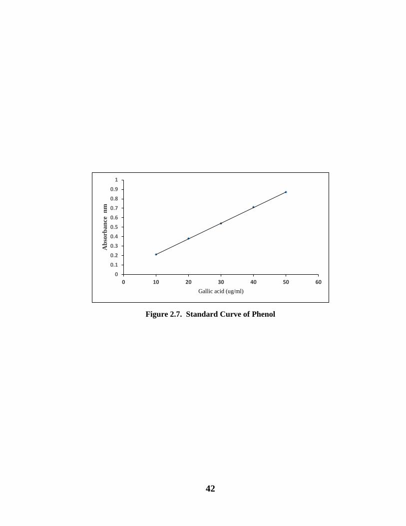
42
Figure 2.7. Standard Curve of Phenol
0
0.1
0.2
0.3
0.4
0.5
0.6
0.7
0.8
0.9
1
0 10 20 30 40 50 60
Ab
sorb
an
ce
nm
Gallic acid (ug/ml)

43
2.12. Detection of phenolic compounds by chromatography
2.12.1. Preparation of ethanolic extract
1 gm of C. procera leaves and flowers were extracted separately in 70% ethanol
overnight at room temperature. Extracts were filtered and concentrated.
2.12.2. Preparation of extract by hydrolysis method (Harborne, 1984)
Dried leaves and flowers (1 gm each) were dipped separately in 2M HCl and heated
for 30-40 minutes at 100oC. Then cooled extract was filtered and extracted with small
amount of ethyl acetate. The ethyl acetate layer was concentrated to dryness then it
was dissolved in a few drops of ethanol. The aqueous extract was further heated to
remove the last traces of ethyl acetate and re-extracted with small volume of amyl
alcohol. The amyl alcohol extract was concentrated to dryness then it was dissolved in
1% methanol HCl.
2.12.3. Chromatography
Whatman no. 1 paper was used to load the extracts. Quercetin was used as a marker,
and chromatographed 2-dimentionally using two different solvents i.e. (acetic acid:n-
butanol:water=1:4:5) and 15% acetic acid. Phenolic compounds were identified by
comparing the Rf values and colour in ultraviolet light before and after ammonia
fumigation.
44
IN-VIVO ANTIOXIDANT STUDIES
2.13. Method for protein free filtrate preparation
1.0 ml plasma was added in a test tube containing 3 ml deionized water and mixed
well. Then sodium tungstate 10% (0.3ml) and 2/3N H2SO4 (0.3ml) were also mixed in
test tube. The constituents were kept at room temperature for 5 minutes and samples
were centrifuged at 3000rpm for 5 minutes. The resulting supernatant was collected as
protein free filtrate.
2.13.1. Plasma urea estimation
Plasma urea was estimated by the standard procedure of Butler et al. (1981).
Reagent-I: (DAM) Diacetyl monoxime solution (2% glacial acetic acid was added in
2% diacetyl monoxime solution).
Reagent-II: Mixture of phosphoric and sulphuric acids (50ml Conc. H2SO4 + 150ml
85% phosphoric acid) and 140ml deionized water was also added to this mixture
Reagent-III: In blank test tube 2ml deionized water, 0.4ml DAM, 1.6ml of
Phosphoric acid-sulphuric acid mixture was also added.
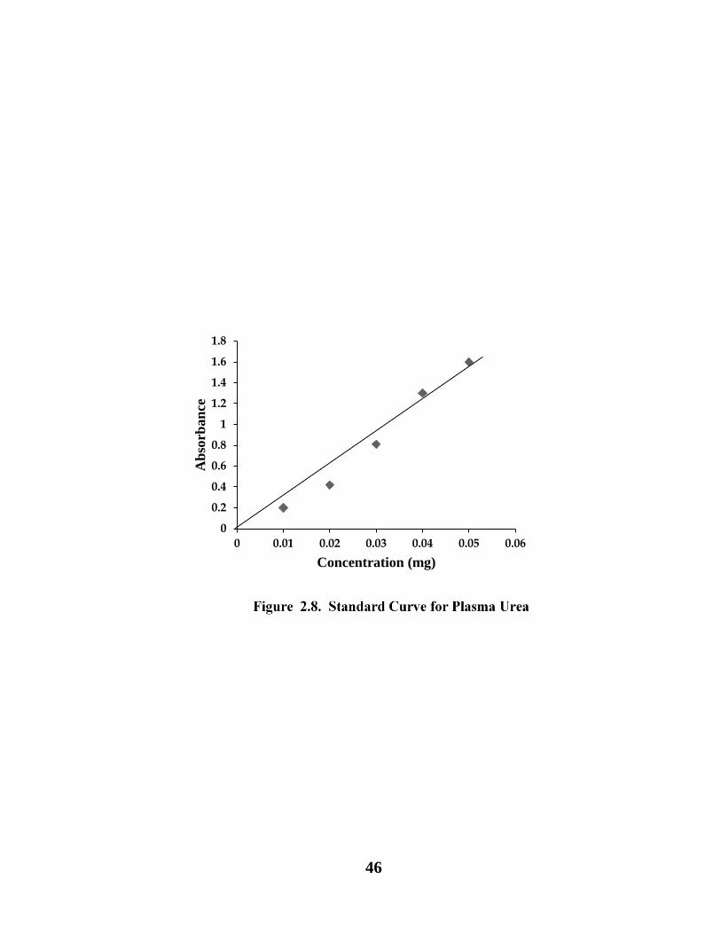
Standard Curve: The standard curve was plotted by using a series of standards (0.01-
0.05mg) from calibration solution which was 0.025mg/ml (Fig. 2.8).

45
2.13.2. Methodology for plasma urea estimation
1 ml of protein free filtrate was poured in test tubes and 1ml of deionized water was
added into tubes. Then 0.4ml of DAM solution and 1.6 ml mixture of phosphoric and
sulfuric acid was also added in tubes. After shaking tubes were kept for half an hour in
boiling water bath. Stand for cooling, and optical density was noted at 480nm against
reagent blank. The contents of urea in plasma were estimated in mg/dl.
2.13.3. Plasma creatinine estimation
Plasma creatinine was estimated by following the procedure of Spierto et al. (1979).
Blank: 0.5ml of sodium hydroxide mixed with 1.5 ml of picric acid in 3ml of
deionized water.
Standard Curve: The standard curve was prepared by using a series of standard
values (0.005-0.035mg) from main standard solutions (0.015mg/ml) (Fig. 2.9).
2.13.3.1. Methodology for plasma creatinine
Took 3ml protein free filtrate in test tubes and 0.5 ml (4N) sodium hydroxide was
added. Then 1.5 ml (0.04M) picric acid was also mixed and the tubes were kept at
room temperature for 15 minutes. The optical density was noted at 530nm against
reagent blank. The quantity of creatinine in plasma was estimated in mg/dl.
46
0
0.2
0.4
0.6
0.8
1
1.2
1.4
1.6
1.8
0 0.01 0.02 0.03 0.04 0.05 0.06
Ab
sorb
an
ce
Concentration (mg)
Series1

47

48
2.13.4. Kidney homogenate
Kidney homogenate was prepared at 4oC by using Homogenizer model (Ultra
Taurax).
Tissues were divided into pieces. The homogenate 1:10w/v was made by adding
0.3mM EDTA and buffer of potassium chloride 100mM, at pH=7.0 was also added to
homogenate. Then centrifuged at 4oC for 60 minutes at 600nm and the supernatant
was collected for various analyses.
2.13.5. Malonyldialdehyde estimation
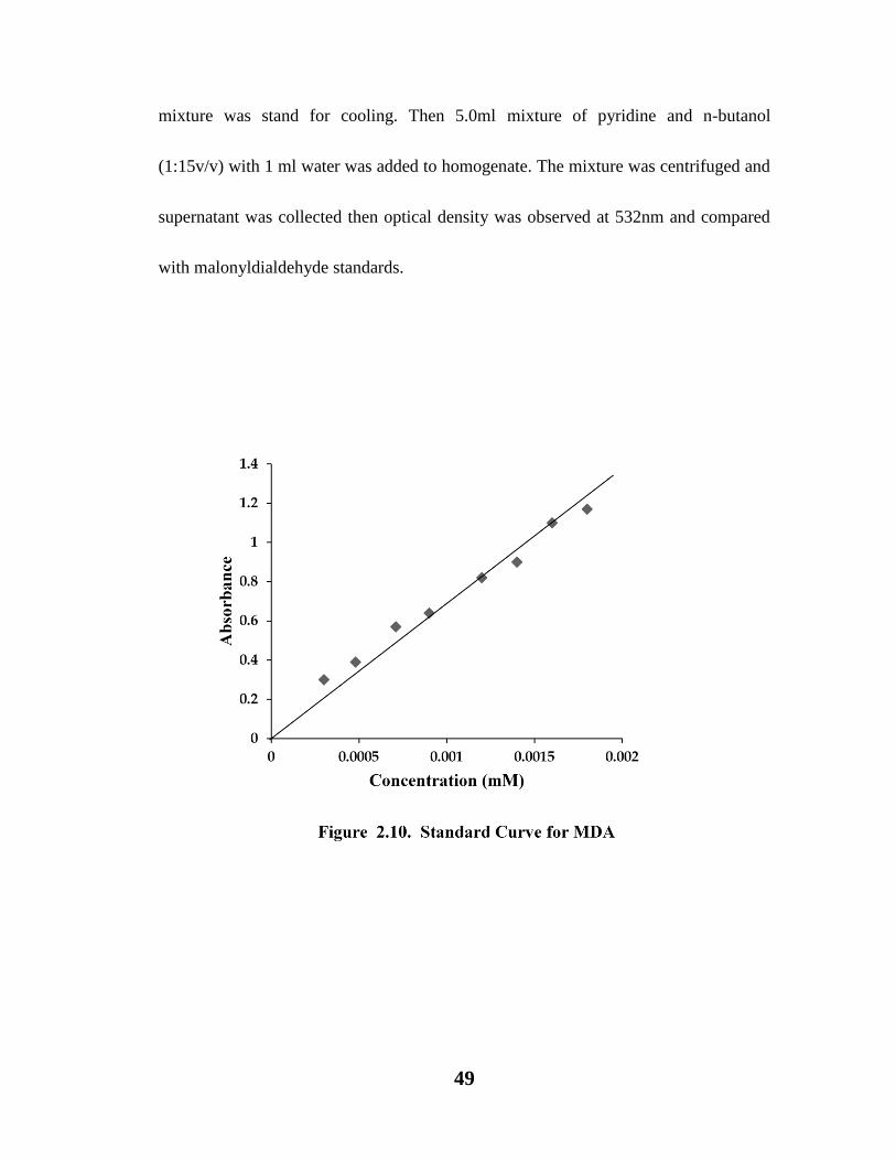
Standard Curve: Values of malonyldialdehyde were estimated by comparing with
the standard absorption values in nM/g of tissue. Whereas, the standard curve was
prepared in a range between 0.0002-0.00176mM from main mixture (0.02mM/L (Fig.
2.10).
Malonyldialdehyde was estimated by following the procedure of Okhawa et al. (1979).
The mixture for reaction contained 1.5ml of 20% acetic acid with 0.2ml of 8.1%
sodium dodecyl sulphate and pH was maintained at 3.5 by using NaOH. Then 1.5ml of
thiobarbituric acid diluted in water (0.8%) was also mixed to the homogenate. The
volume was made up to 4.0 ml by water and heated for 60 minutes at 95oC. The
49
mixture was stand for cooling. Then 5.0ml mixture of pyridine and n-butanol
(1:15v/v) with 1 ml water was added to homogenate. The mixture was centrifuged and
supernatant was collected then optical density was observed at 532nm and compared
with malonyldialdehyde standards.

50
2.13.6. 4-hydroxyl-2-nonenal estimation
The 4 hydroxyl-2-nonenal contents were calculated by following the procedure of
Kinter et al. (1996).
Standard curve: The calibration curve was obtained by using a series of standard
solutions (concentration range 0.0189-0.11378 mM) from main calibration solution
(0.006 mM/L) (Fig. 2.11.).
Methodology
In a clean glass tube, 2 ml of filtrate was taken and added 1 ml of 2,4 Dinitrophenyl
hydrazine and kept for 1 hour at room temperature. The Sample was then extracted
with hexane three times and the extract was evaporated at 40oC. The sample was then
cooled and reconstituted with 2 ml of methanol, the absorbance was then measured at
350 nm against methanol blank on Schimadzu-spectrophotometer UV 120- 01. The
concentration values were calculated from absorption measurement as standard
absorption in nM/g tissue.

51

52
2.13.7. Catalase estimation
For the determination of catalase level the methodology of Sinha et al. (1972) was
adopted.
Dichromate acetic acid Reagent: 150 ml of glacial acetic acid was added with 50ml
5% dichromic acid.
Phosphate buffer: 0.01M, pH=7.0
Hydrogen peroxide: 0.2M
Standard curve: The concentration values were calculated by measuring the optical
density of standards in mM/gram of tissue. The standard curve was obtained by using
a series of standard solutions (concentration range from 0.05-0.3mM) from main
calibrating solution (0.2mM/ml) (Fig. 2.12).
Methodology
1ml of hydrogen peroxide and 1.96ml of phosphate buffer was poured in a test tube
then 0.04 ml of 10% homogenate was also added. Then 2ml of dichromate acetic acid
reagent was mixed with 1 ml of test tube contents. The mixture was boiled for 10
minutes, stand for cooling and the optical density was noted at 570nm with the help of
Schimadzu spectrophotometer (UV 120-01).

53

54
2.13.8. Superoxide dismutase estimation
For the estimation of superoxide dismutase level the methodology of Kono (1978)
was adopted.
Reagents:
Reagent I: 0.1mM EDTA with 50mM sodium carbonate at pH=10.0.
Reagent II: 90µM nitro blue tetrazolium dye.
Reagent III: 0.6% Triton X-100 in reagent I.
Reagent IV: 20mM Hydroxylamine hydrochloride, pH=6.0.
Reference test tube: 0.1 ml of supernatant was added to the test and reference
cuvette.
Methodology
1.3ml of reagent I, 0.5ml of reagent II, 0.1ml of reagent III and 0.1 ml of reagent IV
were mixed with homogenate. The rate of nitro blue tetrazolium reduction was noted
for one minute at 560nm with the help of Schimadzu spectrophotometer (UV 120-01).
The activity was calculated by using the percent inhibition in gram of tissue and
expressed in U/gram of tissue.
Inhibition (%) = Abs of test – Abs of reference/ Abs of test – Abs of blank X 100
U/ml: % inhibition / gram of tissue

55
2.13.9. Glutathione estimation
For the determination of glutathione level the methodology of Carlberg and
Mannervik, (1985) was adopted.
Methodology
In a test tube 0.35 ml of 0.8 mM βNADPH was added then 0.3 ml of 10% BSA was
taken, after that 1.5 ml of 50 mM potassium phosphate buffer (pH = 7.6) was added
then 0.1 ml of 30 mM oxidized glutathione and 0.1 ml of homogenate was also
poured to test tube. After shaking, the absorbance was noted at 340nm for 5 minutes at
25oC temperature on Kinetic spectrophotometer PRIM 500 (Germany with automatic
aspiration and thermostat). The activity was calculated by using the molar coefficient
for NADPH of 6.22 µmol-1 x cm-1 and expressed in the Unit/g of tissue.
Activity of GSH
Activity U/L = µM / L = (340/min / 0.00622 x (Total Volume/Sample Volume in µl)
56
IN-VITRO ANTIOXIDANT STUDIES
2.14. Lipid peroxidation inhibition method (Halliwell and Gutteridge,
1999)
Effect of C. procera on inhibition of lipid peroxidation activity was studied in vitro,
according to the guidelines of Halliwell and Gutteridge (1999).
2.14.1. Preparation of tissue homogenate:
Fresh tissue of a normal albino rat was sliced into small pieces and transferred in a test
tube. Then phosphate buffer saline pH 7.4 was added. The homogenate was
centrifuged at 3000 rpm for 15 minutes, clear upper layer was collected for anti lipid
peroxidation assay.
2.14.2. Procedure for lipid peroxidation inhibition
C. procera leaf and flower extracts were taken with (1, 2, 4, 6, 8, 10 mg/ml) concentrations
from stock solution of (10mg/ml) i.e. 0.01, 0.02, 0.04, 0.06, 0.08 and 0.1 ml was added in test
tubes containing distilled water in 0.09, 0.08, 0.06, 0.04, 0.02, and 0.0 ml respectively. Further
test tubes were standing till dryness. To these dried test tubes 1 ml of 0.15M Potassium
chloride solution was added and then tissue homogenate (0.5ml) was added in each tube. To
start the lipid peroxidation process (0.1ml) of 0.2mM ferric chloride (FeCl2) was added. Then
all test tubes were incubated for 30 minutes at 37oC. The reaction was terminated by addition
of (2 ml) of Hydrochloric acid (0.125N) having 1.68 gms of 15% Tricarboxylic acid with
41.60mg Thiobarbituric acid (0.38%) and 0.5% BHT in ethanol was also added. Again
mixture was incubated at 80oC for 1 hour.

57
After cooling samples were centrifuged, after the appearance of pink layer, absorbance
was measured at 532nm. For comparison purpose BHA was used as a control. A
similar test was performed without the presence of the extract and standard to
determine the amount of lipid served as control. All tests were carried out in triplicate
and the results were expressed as mean ± SD. Lipid peroxidation percent of inhibition
was calculated by following formula:
LPOI (%) = [(A1-A
2)/A
s] x100
Where A1 is the absorbance of control and A
2 is the absorbance of the standard
/sample
2.14.3. 1,1-diphenyl -2-picrylhydrazyl radical scavenging method
The in vitro antioxidant power of C. procera extracts was determined by the method
of Kumar et al. (2013). Six different concentrations of test extracts were prepared from
(1-10mg/ml). 3.0 ml of 0.1mM DPPH solution was mixed to each test tube. The
contents were allowed to stand at room temperature for thirty minutes in dark. The
absorbance was determined at 517nm. Ascorbic acid was used as standard. The
percent inhibition was calculated by using following formula:
% I = [(AC-AS)/AC] x 100
58
Where I = inhibition, AC and AS = Absorbance values of the control and the sample
respectively. Each sample was used in triplicate and results were expressed as mean ±
SD.
2.14.4. Method for determining Reducing power
The reducing ability of C. procera flower and leaf extracts were evaluated by the
method of Kumar & others (2013), Oyaizu (1986) and Mishra & coworkers (2013).
1ml of each sample of four different solvents (water, ethanol, hexane and ethyl
acetate) extracts was taken in test tubes, each in different concentrations (1, 2, 4, 6, 8
and10mg/ml). To each test tube 2.5ml of 1% potassium hexacyanoferrate and 2.5ml of
phosphate buffer (0.2M, pH 6.6) were mixed. All tubes were incubated for 20 minutes
at 50OC temperature in a water bath. The reaction was terminated by mixing 2.5ml of
10% trichloroacetic acid and then centrifuged at 4000rpm for 10min. 1ml of the upper
layer was mixed with 0.5ml of ferric chloride solution (0.1%, w/v) and 1ml of distilled
water and tubes were stand for two minutes at room temperature. The optical density
was noted at 700 nm. The butylated hydroxyanisole was used as standard for
comparison. Higher reducing ability was noted in terms of higher optical density
reading. All the tests were run in triplicate. Results are reported as mean ± SD.
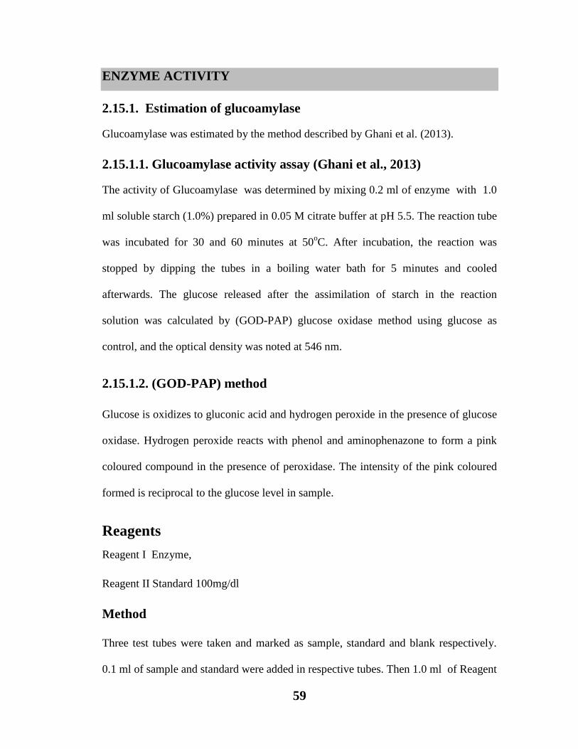
59
ENZYME ACTIVITY
2.15.1. Estimation of glucoamylase
Glucoamylase was estimated by the method described by Ghani et al. (2013).
2.15.1.1. Glucoamylase activity assay (Ghani et al., 2013)
The activity of Glucoamylase was determined by mixing 0.2 ml of enzyme with 1.0
ml soluble starch (1.0%) prepared in 0.05 M citrate buffer at pH 5.5. The reaction tube
was incubated for 30 and 60 minutes at 50oC. After incubation, the reaction was
stopped by dipping the tubes in a boiling water bath for 5 minutes and cooled
afterwards. The glucose released after the assimilation of starch in the reaction
solution was calculated by (GOD-PAP) glucose oxidase method using glucose as
control, and the optical density was noted at 546 nm.
2.15.1.2. (GOD-PAP) method
Glucose is oxidizes to gluconic acid and hydrogen peroxide in the presence of glucose
oxidase. Hydrogen peroxide reacts with phenol and aminophenazone to form a pink
coloured compound in the presence of peroxidase. The intensity of the pink coloured
formed is reciprocal to the glucose level in sample.
Reagents
Reagent I Enzyme,
Reagent II Standard 100mg/dl
Method
Three test tubes were taken and marked as sample, standard and blank respectively.
0.1 ml of sample and standard were added in respective tubes. Then 1.0 ml of Reagent
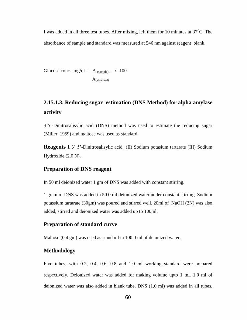
60
I was added in all three test tubes. After mixing, left them for 10 minutes at 37oC. The
absorbance of sample and standard was measured at 546 nm against reagent blank.
Glucose conc. mg/dl = A (sample) x 100
A(standard)
2.15.1.3. Reducing sugar estimation (DNS Method) for alpha amylase
activity
3’5’-Dinitrosalisylic acid (DNS) method was used to estimate the reducing sugar
(Miller, 1959) and maltose was used as standard.
Reagents I 3’ 5’-Dinitrosalisylic acid (II) Sodium potasium tartarate (III) Sodium
Hydroxide (2.0 N).
Preparation of DNS reagent
In 50 ml deionized water 1 gm of DNS was added with constant stirring.
1 gram of DNS was added in 50.0 ml deionized water under constant stirring. Sodium
potassium tartarate (30gm) was poured and stirred well. 20ml of NaOH (2N) was also
added, stirred and deionized water was added up to 100ml.
Preparation of standard curve
Maltose (0.4 gm) was used as standard in 100.0 ml of deionized water.
Methodology
Five tubes, with 0.2, 0.4, 0.6, 0.8 and 1.0 ml working standard were prepared
respectively. Deionized water was added for making volume upto 1 ml. 1.0 ml of
deionized water was also added in blank tube. DNS (1.0 ml) was added in all tubes.

61
Test tubes were placed in a boiling water bath for 5.0 minutes. After boiling, deionized
water (9.0 ml) was added in all test tubes including blank and mixed well. Absorbance
of all the standard tubes was measured against blank at 546 nm.
Figure 2.13. Standard Curve for Reducing Sugar for α-amylase
0
0.5
1
1.5
2
2.5
0 50 100 150 200 250 300
Ab
sorb
an
ce
Concentration of DNS

62
2.15.1.4. Alpha amylase enzyme activity assay
1. Buffer: 50mM Tris –HCl buffer (pH 7.00)
2. Substrate: Starch (2.00gm/dl) in Tris-HCl buffer was used as substrate.
3. Enzyme: Partially purified alpha amylase (0.10 ml)
Method
Two test tubes were marked as Control (C), and two test tubes were taken as Test (T).
In test tube (T) 0.1 ml enzyme having flower and leaf extract was mixed with 0.9 ml
substrate while pure enzyme 0.1 ml was added in control tube and mixed with 0.9 ml
of substrate. 1 ml deionized water was used in blank tube. Test tubes were incubated
at 60oC for 5 minutes and 1.0 ml DNS was added in all tubes (Test, Control and
Blank) to stop the enzyme–substrate reaction. All tubes were placed in boiling water
bath for 5.0 minutes and allowed to cool. After cooling 9 ml deionized water was
added in each tube. The absorbance of test and control was observed at 546 nm
against blank.
Calculation
mg of reducing sugar/ml/minute=
OD of test – OD of control x Conc. of standard x 1 x
1
OD of standard volume of sample
time (min)
1 µmol of maltose = 0.00036 mg/ml = 1 Unit
Therefore,

63
U/ml/minute = mg reducing sugar/ml/minute
0.00036 mg/ml
2.15.1.5. Estimation of urease activity
Urease activity was determined by kit method (Modified Berthelot’s Method, 1859)
(Fawcett and Scott (1960).
Reagents
Reagent I (Enzyme): Urease 5000 U/L,
Reagent II (Colour reagent): Phosphate Buffer pH 7.0 = 60.00 mmol/L,
Sodium Nitroprusside = 2.50 mmol/L, Sodium Salicylate = 31.25 mmol/L, EDTA
0.74 mmol/L
Standard: Urea = 50mg/dl
Preparation of mono-reagent
One ml of Reagent-I was mixed with 20 ml of Reagent-II
Method
Three test tubes were taken and marked as Sample, Standard and Blank. 0.1 ml of
sample (extract, Standard (Urea) and blank (deionized water were added in respective
tubes. Then 1.0 ml of mono-reagent was added in each of the tubes. After mixing,
tubes were incubated for 5 minutes at 37oC. Then 1.0 ml of reagent-III was added to
each of the tubes. After mixing, the tubes were again incubated for 5 minutes at 37oC.
Absorbance was recorded at 580 nm against reagent blank.
Urea = A(sample) x concentration of standard
A(standard)

64
2.16. Statistical analysis
Data were analyzed statistically by using mean values ± SE at P<0.05 through One-
way ANOVA and Duncan’s multiple comparison test (DMCT) with the help of a
computer software, statistical package for social sciences (SPSS) for windows version
14.0.0 (SPSS, 2005).
65
PHYTOCHEMICAL STUDIES OF CALOTROPIS
PROCERA
3.1. Introduction
Biologically active compounds derived from plants are usually classified as primary
and secondary metabolites. Whereas, primary metabolites are the part of metabolic
pathways and secondary metabolites are waste products or byproducts of metabolic
pathways. Regarding to the medicinal uses of Calotropis procera secondary
metabolites namely, phenolic compounds tannins, terpenoids and saponins have
received an immense attention (Vaya et al., 1997; Sengul et al., 2009; Patel et al.,
2010).
Therefore, C. procera was previously investigated for its phytochemicals in relation
to medicinal importance. Present studies are carried out to investigate phytochemical
properties of Calotropis procera and to substantiate the previous findings.
3.2. Material and Methods
3.2.0. Extract Methodology
Detailed method is described in Chapter 2 (2.2).
3.2.1. Total proteins estimation
Total protein in extract was evaluated by the procedure of Bradford (1976) as
described in Chapter 2 (2.5).

66
3.2.2. Carbohydrates estimation
Total carbohydrates were calculated by the guidlines of Yemm and Willis (1957) as
described in Chapter 2 (2.6).
3.2.3. Total reducing sugars
Total reducing sugars were calculated by the technique of Miller (1959) as explained
in Chapter 2 (2.7).
3.2.4. Total non-reducing sugars
Total non reducing sugars were calculated by the formula as mentioned in Chapter 2
(2.8).
3.2.5. Total amino acids estimation
Total amino acids were evaluated by the technique of Spices (1957) as mentioned in
Chapter 2 (2.9).
3.2.6. Amino acid detection by paper chromatography
Amino acid detection was performed by the technique described in Chapter 2 (2.10).
3.2.7. Phenolic compounds estimation
Total phenolic compounds were evaluated by the technique of Swain and Hillis (1959)
as mentioned in Chapter 2 (2.11).
3.2.8. Phenolic compounds detection by chromatography
Detection of phenolic compounds was performed by the technique of Harborne (1984)
as mentioned in Chapter 2 (2.12).
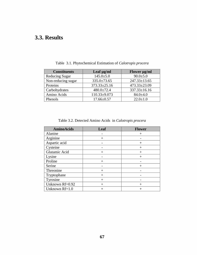
67
3.3. Results
Table 3.1. Phytochemical Estimation of Calotropis procera
Constituents Leaf µg/ml Flower µg/ml
Reducing Sugar 145.0±5.0 90.0±5.0
Non-reducing sugar 335.0±73.65 247.33±13.65
Proteins 373.33±25.16 473.33±23.09
Carbohydrates 480.0±72.4 337.33±16.16
Amino Acids 110.33±9.073 84.0±4.0
Phenols 17.66±0.57 22.0±1.0
Table 3.2. Detected Amino Acids in Calotropis procera
AminoAcids Leaf Flower
Alanine - +
Arginine + -
Aspartic acid - +
Cysteine - +
Glutamic Acid + +
Lysine - +
Proline + -
Serine - +
Threonine + -
Tryptophane + -
Tyrosine + -
Unknown Rf=0.92 + +
Unknown Rf=1.0 + +
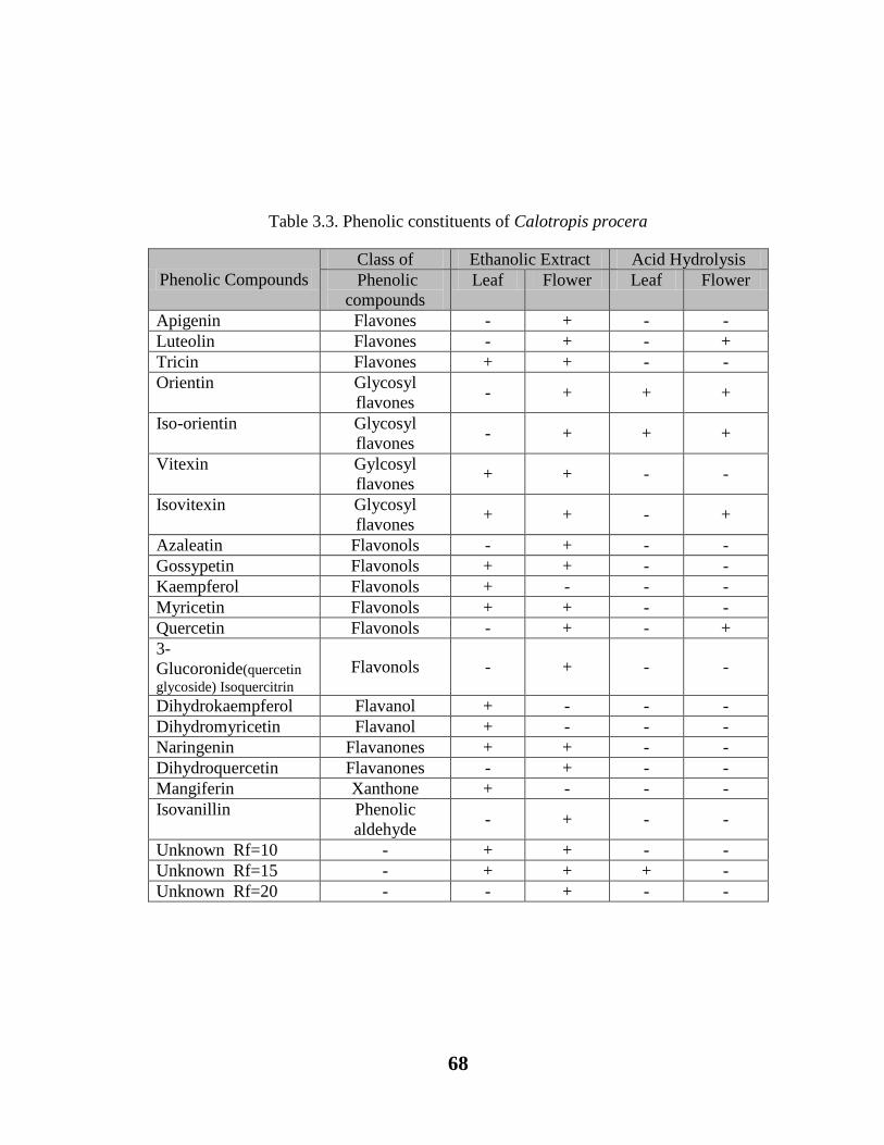
68
Table 3.3. Phenolic constituents of Calotropis procera
Phenolic Compounds
Class of Ethanolic Extract Acid Hydrolysis
Phenolic
compounds
Leaf Flower Leaf Flower
Apigenin Flavones - + - -
Luteolin Flavones - + - +
Tricin Flavones + + - -
Orientin Glycosyl
flavones - + + +
Iso-orientin Glycosyl
flavones - + + +
Vitexin Gylcosyl
flavones + + - -
Isovitexin Glycosyl
flavones + + - +
Azaleatin Flavonols - + - -
Gossypetin Flavonols + + - -
Kaempferol Flavonols + - - -
Myricetin Flavonols + + - -
Quercetin Flavonols - + - +
3-
Glucoronide(quercetin
glycoside) Isoquercitrin Flavonols - + - -
Dihydrokaempferol Flavanol + - - -
Dihydromyricetin Flavanol + - - -
Naringenin Flavanones + + - -
Dihydroquercetin Flavanones - + - -
Mangiferin Xanthone + - - -
Isovanillin Phenolic
aldehyde - + - -
Unknown Rf=10 - + + - -
Unknown Rf=15 - + + + -
Unknown Rf=20 - - + - -

69
3.4. Discussion
In C. procera a large number of chemical constituents like phenolic compounds,
including phenolic acids and flavonoids, carbohydrates, proteins and amino acids are
detected (Table 3.1, 3.2, 3.3). Amongst the primary metabolites, value of amino acids
is higher in leaf (i.e. 110.33 µg/ml) than flower (84.0 µg/ml) these quantitative values
could also be supported by the qualitative detection as leaves having greater amino
acids in relation to flowers (Table 3.1). Regarding to the amount of carbohydrates,
reducing and non-reducing sugars, leaf showing greater value as compared to flower.
While, greater value of protein is recorded in flowers (460.7µg/ml) in comparison with
leaf (Table 3.1).
Flower extract showed 21 µg/ml total phenols and leaf exhibited 17.5 µg/ml phenols.
Similarly, in the qualitative determination of phenolic constituents more compounds
are observed in flower as compared to leaf (Table 3.3).
Amongst the detected phenolic compounds, especially flavonols and flavones of the
class flavonoids have been received considerable attention to prove their medicinal
importance (Ikken & others, 1999; Gao & others, 2000; Patel & coworker, 2010;
Gholamshahi & coworkers, 2014; Pooja & others, 2014; Shetty & coworkers, 2015),
such as quercetin, luteolin and kaempferol are most commonly consumable and
natural flavonoids and reported to have antibacterial and antioxidant properties (Bentz,
2009; Calderon-Montana et al., 2011; Majewska et al., 2011). Another flavonol
Gossypetin also showed antibacterial/antiseptic potential ( Mounnisamy et al., 2002).
Similarly Mangiferin is a natural phenolic compound exhibited antimicrobial and

70
antioxidant activities (Stoilova et al., 2005). Tricin is another O-methylated flavones
known to minimize the risk of intestinal cancer (Lutskii et al., 1971). The
dihydromyricetin belongs to flavonol and used as anti-inflammatory agent (Satoshi,
2007; Shihui et al., 2015). Isoorientin is a flavones, showing hypoglycaemic activity
(Lim et al., 2007). Besides these above information it has been observed that the
extract of C. procera flower exhibited comparatively large amount of phenolic
compounds as compared to leaf extract. While, on the other hand flower also showed
high scavenging potential by DPPH and more effective for lipid peroxidation
inhibition. So there seems a good correlation between phenolic compounds,
antioxidant and antibacterial potential.
71
EFFECT OF FLOWERS AND LEAVES EXTRACTS OF
CALOTROPIS PROCERA AS ANTIMICROBIAL AGENT.
4. Introduction
Infectious diseases are usually controlled by commercial antimicrobial drugs that
largely produce toxicity in the human body which intimated the attention of workers to
trace out any other alternate source for the prevention of infectious disease with less or
zero toxicity. In the last few decades, several new natural anti-microbial compounds
were discovered for the control of severe infections. A discovery of new antibacterial
agent against multidrug resistant organisms is still in full swing due to the
development of continuous resistance developed by microbes. The multidrug resistant
organisms have received great clinical attention because of increasing reported cases
around the globe. Along with this, there is an increase in consumer demand for those
drugs, which are isolated or derived from natural sources. The Threat posed to general
public health by various multidrug resistant organisms and pathogens can be resolved
by the discovery of natural antibacterial compounds having effective broad spectrum
inhibition against pathogens prevalent in the local community.
Calotropis procera is reported to have therapeutic properties such as flowers show
anti-inflammatory activity (Basu and Chaudhry, 1991; Neenah and Ahmed, 2011) for
curing cholera , wound, piles, asthma (Mohanraj et al., 2010) and also used as an
appetizer and tonic (Sharma et al., 2011). Sometimes, leaves and stem are inhaled or
smoked after burning to cure the fever, swellings, paralysis and arthralgia. Similarly,
72
leaves are also taken for the treatment of various heart diseases and chest cold
(Agharkar, 1991; Hemalatha et al., 2011). The leaves also have anthelmintic, laxative,
healing, and bitter properties. It acts as an expectorant, relieves stomach pain and
cures ulcers (Goyal and Mathur, 2011). Chopped leaves showed properties as a
nematicide (Kristova and Tissot, 1995; Kawo, 2009) and as a cure for jaundice
(Sharma et al., 2011). Besides this flower and leaves are also used as a source of anti-
bacterial agent against gram positive and gram negative bacteria (Moscolo et al., 1988;
Larhsini, et al., 1999; Dewan et al., 2000; Alencer et al., 2004; Parabia et al., 2008;
Vadlapudi and Naidu, 2009; Amin et al., 2011; David et al., 2011; Doshi et al.,
2011; Ahmad et al., 2011; Sharma et al., 2011; Johnson et al, 2011; Prabha and
Vasantha, 2012; Muzammal, 2014; Kazemipour et al., 2014) ).
It is also evident by various reports that various works gave attention only to the
flower extract to check its antibacterial activity such as Doshi et al. (2011) used
ethanolic flower and other parts extracts against larvae of A. stephansi, there was no
antibacterial activity observed in flower and stem extracts of C. procera against S.
aureus, Bacillus cereus, Bacillus subtilis, and Micrococcus luteus. Whereas, David et
al. (2011) acetone and methanol flower extracts were further used against Bacillus
pumilis, E.coli, A. niger, Fusarium oxysporum. Similarly, Prabha and Vasantha (2012)
screened C. procera flower extract of chloroform, acetone and ethanol against various
pathogens and maximum antibacterial activity was recorded against S. aureus and B.
subtilis. Ranjit et al., (2012) also used ethanol fraction against S. aureus, Bacillus
subtilis, Bacilis pumilis, Micrococcus luteus, E. coli, Pseudomonas Aeroginosa and
Proteus vulgaris. Observations revealed a significant inhibitory action against test
73
bacteria. Joshi and Kaur (2013) determined the antimicrobial potential of ethanol,
methanol and aqueous extract of C. procera and found that ethanol extract have strong
antimicrobial activity against P aeroginosa. While, Javadian et al. (2014) evaluated
the antimicrobial activity of ethanol extract of C. procera and found effective against
E. coli isolates.
The antibacterial efficacy of C. procera leaves against human disease producing
bacteria was already studies by various scientists. Yesmin et al. (2008) demonstrated
the antibacterial activity through the leaves aqueous and methanol extracts of C.
procera and it was revealed that both extracts were active against Gram positive and
Gram negative bacteria at low concentrations. Studies of Kareem et al. (2008)
revealed positive activity of ethanol and chloroform fractions against Candida,
Miocrosporum, Aspergillus niger, S. pneumonia, S. pyogens, E. coli and S. aureus.
Similarly, Kawo et al. (2009) studied the antimicrobial efficacy of water and ethanol
leaves extract of C. procera and it was revealed that the aqueous sample did not
showed any activity while, ethanol extract have significant antibacterial potential.
Whereas, Goyal and Mathur (2011) applied fractions of petroleum ether, ethanol and
butanol against Candida para, Candida albican, Enterococci, Pseudomonas
aeroginosa, Staphylococci and E. coli which exhibited significant antimicrobial
activity. In another study (Hemalatha et al., 2011) reported leaf extract of acetone,
ethyl acetate, methanol and water against Shigella, S. typhi, Vibrio cholera,
Pseudomonas aeroginosa, Lactobacillus, B. cireus, S. aureus and B. subtilis, all of the
solvent extracts showed inhibitory activity against bacterial samples. Similarly,
Johnson et al., (2011) determined that aqueous and alcoholic extract showed strong

74
antibacterial efficacy against Aspergillus, E.coli and S. aureus. While, Velmurugan et
al., (2012) studied C. procera leaf extract of hexane, ethyl acetate and methanol
against aquatic micro pathogens from shrimp fish and It was noted that ethyl acetate
extract effectively suppressed bacterial strains. On the other hand Mako et al. (2012)
evaluated the antimicrobial activity of aqueous and ethanol root and leaves extract of
C. procera where ethanol extract showed more significant potential than aqueous
extract. Salem et al. (2014) demonstrated the antibacterial activity through leaf and
latex chloroform, ethanol, methanol extracts of the leaf where it was observed that
aqueous and ethanol extracts of leaves showed maximum potential against Gram
negative and Gram-positive pathogenic bacteria. However, Shetty et al. (2015) studied
the antibacterial effect of methanol, ethyl acetate, ethanol, acetone and aqueous
extracts of C. procera leaves against human pathogenic bacteria and it was found that
leaves extract showed significant antibacterial activity against Micrococcus aureus in
all test solvents. Pandey et al. (2015) assessed the antibacterial activity of C. procera
methanol, acetone, petroleum ether and ethyl acetate extracts, amongst them
methanol leaves extract showed highest range of inhibition against E.coli and S.
aureus.
In view of the above mentioned studies the present studies were carried out to evaluate
the antimicrobial efficacy of C. procera from Karachi (Pakistan) using different
solvent fractions of flowers and leaves with butanol, hexane, ethyl acetate and aqueous
extracts against various disease producing bacteria to substantiate the earlier findings
for its significant use.

75
4.1. Material and Methods
Detail of collection of plant samples already mentioned in Chapter 2 (2.1)
4.2. Preparation of Extract
Detailed method is mentioned in Chapter 2 (2.2).
4.3. Preparation of Fraction
Detailed method is mentioned in Chapter 2 (2.3).
4.4. Experimental Microbes
The antimicrobial activity of C. procera flowers and leaf extracts was evaluated
against pathogenic microorganism types viz. Gram negative organisms includes
Escherichia coli and Salmonella typhi were separated from a dirty water sample.
While, Gram positive organisms includes Micrococcus luteus (JQ 250612) and
methicillin resistant Staphylococcus aureus were isolated from soil sample and clinical
specimen respectively.
4.5. Culture Media
Nutrient broth was used to restore experimental strains for 24 hours at 37 C with the
shaking of 135 rpm. Strains were maintained on nutrient agar slants at 4 C for further
studies.
76
4.6. Procedure for Antimicrobial Activity
To evaluate the antimicrobial potential of leaf and flower extracts of Calotropis procera
against pathogenic bacterial species. Agar well diffusion method was adopted by Tagg and
McGiven (1971) and Ali (2014). For the revival of bacterial species sterilized petri plates were
filled with with nutrient agar and inoculated 100µl of bacterial species having 108 cfu/ml
(Iqbal, 1998; Ali et al., 2014) compared with the 0.5 McFarland turbidity index. Then 100µl of
concentrated leaf and flower extracts were introduced in the wells of petri plate and plates
were incubated for 24 hours at 37oC temperature. While, for control plates no extracts were
introduced. To determine the potential against pathogenic bacterial species, zone of inhibition
was calculated. All experiments were performed in triplicate.

77
Table 4.1. Antimicrobial activity of flower extracts against different
pathogenic strains.
Key: MRSA= Methicillin resistant Staphylococcus aureus, Significant zone= > 11 mm,
-ve = No activity detected.
S.No Extracts Zones of inhibition (mm)
S. typhi Control E. coli Control MRSA Control M. luteus Control
1 Butanol -ve -ve -ve -ve -ve -ve 30 -ve
2 Ethyl
acetate -ve -ve 15 -ve 18 -ve 25 -ve
3 Aqua -ve -ve -ve -ve -ve -ve 30 -ve
4 Hexane 13 -ve 12 -ve 15 -ve 22 -ve

78
Figure 4.1: Antimicrobial activity of flowers extracts (aqueous, butanol,
hexane, ethyl acetate) against A= Micrococcus luteus, B= Salmonella
typhi, C= Escherichia coli, D= (Methicillin resistant Staphylococcus
aureus ) MRSA

79
Table 4.2. Antimicrobial activity of leaves extracts against different pathogenic
strains.
Key: MRSA= Methicillin resistant Staphylococcus aureus, Significant zone = > 11 mm,
-ve = No activity detected.
S.# Extracts Zones of inhibition (mm)
S. typhi Control E. coli Control MRSA Control M. luteus Control
1 Butanol -ve -ve -ve -ve 7 -ve -ve -ve
2 Ethyl
acetate -ve -ve 12 -ve 15 -ve -ve -ve
3 Aqua -ve -ve -ve -ve -ve -ve 19 -ve
4 Hexane 15 -ve 18 -ve 12 -ve 23 -ve
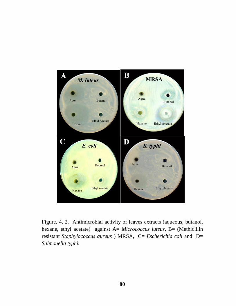
80
Figure. 4. 2. Antimicrobial activity of leaves extracts (aqueous, butanol,
hexane, ethyl acetate) against A= Micrococcus luteus, B= (Methicillin
resistant Staphylococcus aureus ) MRSA, C= Escherichia coli and D=
Salmonella typhi.

81
Figure 4.3. Zone of inhibition of C. procera flowers extracts in different
solvents (butanol, ethyl acetate, aqueous and hexane).
0
5
10
15
20
25
30
35
S. typhi E. coli MRSA M. luteus
Zo
ne
of
inh
ibit
ion
(m
m)
Experimental Bacterial Strains
Butanol
Ethyl acetate
Aqua
Hexane

82
Figure 4.4. Zone of inhibition of C. procera leaves extracts in different
solvents (butanol, ethyl acetate, aqueous and hexane).
0
5
10
15
20
25
S. typhi E. coli MRSA M. luteus
Zo
ne
of
inh
ibit
ion
(m
m)
Experimental Bacterial Strains
Butanol
Ethyl acetate
Aqua
Hexane

83
4.7. RESULTS
To explore the antimicrobial potential of different fractions (hexane, ethyl acetate,
butanol and aqueous) of Calotropis procera flower and leaf were used.
Flowers: All solvents of flower extracts of C. procera showed dissimilar results of
inhibition against M. luteus, methicillin resistant Staphylococus aureus, E. coli, and S.
typhi (Table 4.1., Fig. 4.1. A-D & 4.3). Amongst four extracts, hexane fraction has
been proved very significant as an antimicrobial agent against all the examined
bacterial strains. A maximum zone of inhibition (22mm) was observed against M.
luteus. E. coli, Butanol and Aqueous fractions also exhibited inhibitory efficacy
against M. luteus, whereas, another bacterial strain were resistant to both fractions.
Fraction of ethyl acetate showed inhibitory activity not only against M. luteus (25mm)
but also against E. coli (15mm) and methicillin resistant Staphylococus aureus
(18mm).
Leaf: Different solvents of leaf extracts of C. procera exhibit variation in the
inhibition spectrum against E. coli, methicillin resistant Staphylococcus aureus,
Micrococcus luteus and Salmonella typhi (Table 4.2., Fig. 4.2. A-D). Amongst four
extracts, hexane fraction has been found highly significant antibacterial factor against
all bacterial strains. A maximum zone of inhibition (23mm) was observed against
Micrococcus luteus. Ethyl acetate also showed significant inhibitory activity against
MRSA (15mm) and E. coli (12mm). While, Butanol and hexane extract did not
produce any significant inhibitory activity against MRSA. Similarly, no activity was
observed in E. coli against butanol and aqua, M. luteus did not show any significant
activity in butanol and ethyl acetate. While, butanol and ethyl acetate samples were
proved insignificant against the activity of S. typhi.

84
4.8. DISCUSSION
Resistance to different broad-spectrum antibiotic has now become a global concern
due to emerging cases of drug resistance (Mohanraj et al., 2010). Due to these
emerging cases and also due to the increase consumer demand towards natural
antibacterial agents there is a need of screening of natural anti-microbial compounds
effective against different drug resistant pathogens. In the last few decades, several
new natural anti-microbial compounds were discovered for the control of severe
infections. Keeping this in view, the present study was designed to explore the anti-
bacterial potential of medicinally important flower of C. procera. Different soluble
flower extracts of C. procera showed dissimilar inhibition pattern against tested
disease producing microorganisms. Amongst four extracts, hexane fraction has been
proved very significant as an antibacterial agent against studied pathogens. A
maximum zone of inhibition was observed against Micrococcus luteus which can
cause infections in immune-compromised individuals (Seifert et al., 1995; Ali et al.,
2014). It is also noteworthy that present findings are in contrast to the earlier findings
of Parabia et al. (2008) where hexane fraction of stem of C. procera showed least
antibacterial activity (7mm) against Micrococcus luteus. Similarly, fraction of ethyl
acetate showed significant inhibitory activity not only against Micrococcus luteus but
also against E.coli and methicillin resistant Staphylococcus aureus (except S. typhi)
and these findings could be well supported by the studies of Patil and Saini (2012)
where they also found significant role of ethyl acetate against various pathogens. E.
coli is a toxin producing human pathogen. E. coli is an enteric hemorrhagic strain and
cause severe diarrhoea leads to kidney failure through food. However, Staphylococcus
85
aureus (methicillin resistant) is also a disease producing bacteria, commonly resistant
against β-lactam antibiotics (Iqbal, 1998; Que and Moreillon, 2009; Ali et al., 2014).
Presently ethyl acetate and hexane extracts of C. procera significantly inhibited the
growth of this multidrug resistant organism.
These findings are also in agreement with the previous reports of Kazemipour et al.
(2014) and Javadian et al. (2014). However, flower aqueous and butanol extracts
showed significant inhibitory activity only against M. luteus.
Similarly, leaf extracts of C. procera also showed various spectrums of inhibition
against E. coli, methicillin resistant S. aureus (MRSA), S. typhi and M. luteus (Table
4.2., Fig. 4.2. (A-D). Amongst all of the extracts, the C. procera hexane fraction has
been proved very significant as antibacterial agent against all of the studied pathogens.
A maximum significant zone of inhibition of hexane extract (23mm) was observed
against M. luteus as compared to butanol, and ethyl acetate fractions which did not
show any activity against M. luteus and S. typhi. However, present finding is in
contrast to that of the findings of (Doshi et al., 2011) where an inhibition zone of C.
procera leaf extract for M. luteus was observed at 9mm. Similar to the findings of
Joshi and Kaur (2013) hexane soluble leaf extract also shows significant activity
against E. coli. While, ethyl acetate fraction of leaf also showed significant zone of
inhibition (12mm) against E. coli and MRSA (15mm) . These significant readings are
in contrast with the observation of Doshi et al. (2011) where 7mm inhibition zone was
observed. Whereas, butanol soluble extract shows in significant (7mm) zone of
inhibition. Thus, flower and leaf extracts of C. procera found to be strong
antimicrobial agent.
86
EFFECT OF CALOTROPIS PROCERA AQUEOUS
EXTRACT ON GLUCOAMYLASE, ALPHA-
AMYLASE AND UREASE ACTIVITY.
5. Introduction
Enzymes are biological molecules that speed up the biological chemical reactions
(Brayer et al., 1995). Generally enzymes are classified based on the type of reaction
that they catalyze such as glucoamylase, also known as glucan 1,4-alpha-glucosidase,
(EC 3.2. 1 .3). It is a type of digestive enzyme which cleaves one glucose unit from a
non reducing end of starch (amylose and amylopectin). Most of the glucoamylases are
also able to hydrolyze the 1,6-a linkage in branch points of starch molecules.
Alpha amylase is a digestive enzyme. The formal title of alpha amylase is 1,4 α-D-
glucanohydrolase; EC 3.2.1.1. The enzyme alpha amylase aids in the hydrolysis of α–
1,4 glycosidic bond in the conversion of starch to maltose (Brayer et al., 1995). In
humans, it is found in both saliva and pancreas. Amylases are also used in various
industries like paper, food and textile industries (Windish et al., 1965; Gupta et al.,
2003).
The enzyme urease (EC 3.5.1.5) catalyzes the breakdown of urea into carbon
dioxide and ammonia. The reaction occurs as follows (Zimmer, 2000).
(NH2)2CO + H2O → CO2 + 2NH3

87
Figure 5.1. Representing starch structure showing α-1,4-linkage where α-amylase
targets. (Adapted from El-Fallal et al., 2012).

88
5.1. Material and Methods
5.1.1. Preparation of aqueous extract.
Leaves and flowers of Calotropis procera from two different collections, already
mentioned in Chapter 2(2.2) were powdered and mixed with deionized water to
extract its compounds. The extract was kept at 4oC for 24 hours and then it was
centrifuged at 12000 rpm for 10 minutes at 4oC.
5.1.2. Enzyme activity
Glucoamylase (100U/mg), Amylase (50U/mg) and Urease (5U/mg) were mixed
separately with leaf and flower extracts at a concentration of 50mg/ml for 30 and 60
minutes. Control was used for comparison, in which 50mM phosphate buffer (pH=7)
was added. After the incubation, enzyme activity was performed.
5.1.3. Estimation of glucoamylase
Glucoamylase was estimated by the method of Ghani et al. (2013) as described in
Chapter 2 (2.15.1).
5.1.4. Glucoamylase activity assay
Glucoamylase activity was determined by the method of Ghani et al. (2013) as
described in Chapter 2 (2.15.1.1).
5.1.5. Estimation of reducing sugar for alpha-amylase
3’5’-Dinitrosalisylic acid (DNS) method was used to estimate the reducing sugar
(Miller, 1959) as described in chapter 2 (2.15.1.3).

89
5.1.6. Estimation of urease activity
Urease activity was determined by modified Berthelot’s method (Fawcett and Scott,
1960) as described in Chapter 2 (2.15.1.5).
5.1.7. Alpha amylase enzyme activity assay
Detailed method is mentioned in Chapter 2 (2.15.1.4).

90
Table 5.1. Effect of aqueous extract of C. procera on glucoamylase activity
S. No. Samples
Glucoamylase activity
after 30 minutes
(%)
Glucoamylase activity
after 60 minutes
(%)
1 Control 100e±5.23 100
c±4.25
2 Leaf Extract 1 ***134d±3.35 100
c±3.62
3 Leaf Extract 2 ***148b±4.56 ***137
a±5.67
4 Flower Extract 1 ***168a±6.24 100
c±6.55
5 Flower Extract 2 ***141c±7.25 ***131
b±5.84
Extract concentration = 50mg/ml
n=6, mean values ± SE, ***= P<0.05, a-d= ranks of mean values, sharing of same letter do not
differ significantly.

91
Figure 5.2. Effect of aqueous extract of C. procera on glucoamylase activity.
n=6, mean values ± SE, ***= P<0.05, a-d= ranks of mean values, sharing of same letter do not
differ significantly.
***e
***d ***b
***a ***c
c c
***a
c
***b
0
50
100
150
200
Control Leaf Extract 1 Leaf Extract 2 Flower Extract 1
Flower Extract 2
Re
lati
ve
En
zy
me
Act
ivit
y (
%)
Extract conc. 50mg/ml
Glucoamylase activity
Incubation at 30 minutes Incubation at 60 minutes
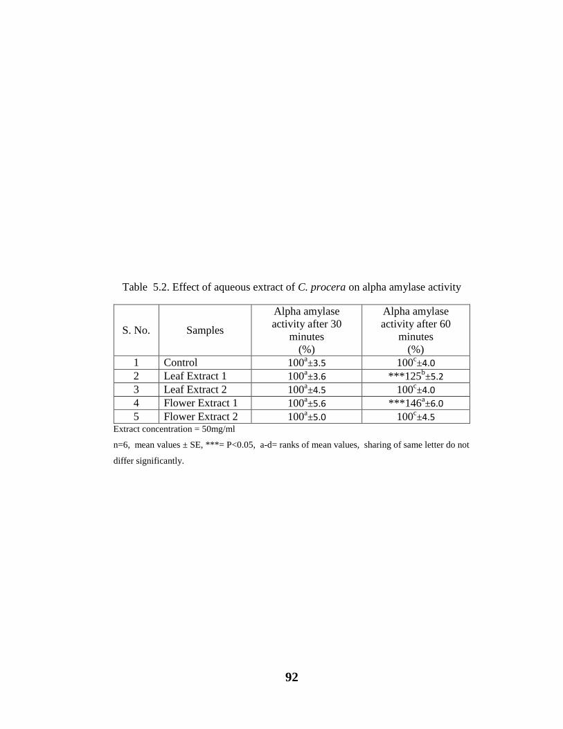
92
Table 5.2. Effect of aqueous extract of C. procera on alpha amylase activity
S. No. Samples
Alpha amylase
activity after 30
minutes
(%)
Alpha amylase
activity after 60
minutes
(%)
1 Control 100a±3.5 100
c±4.0
2 Leaf Extract 1 100a±3.6 ***125
b±5.2
3 Leaf Extract 2 100a±4.5 100
c±4.0
4 Flower Extract 1 100a±5.6 ***146
a±6.0
5 Flower Extract 2 100a±5.0 100
c±4.5
Extract concentration = 50mg/ml
n=6, mean values ± SE, ***= P<0.05, a-d= ranks of mean values, sharing of same letter do not
differ significantly.

93
Figure 5.3. Effect of aqueous extract of C. procera on alpha amylase activity.
n=3, mean values ± SE, ***= P<0.05, a-d= ranks of mean values, sharing of same letter do not
differ significantly.
a a a a a c ***b
c
***a
c
0
50
100
150
200
Control Leaf Extract 1 Leaf Extract 2 Flower Extract 1
Flower Extract 2
Re
lati
ve
En
zy
me
Act
ivit
y (
%)
Extract conc. 50mg/ml
Alpha-amylase activity
Incubation at 30 minutes Incubation at 60 minutes

94
Table 5.3. Effect of aqueous extract of C. procera on urease activity.
S. No. Samples
Urease activity
after 30
minutes
(%)
Urease
activity after
60 minutes
(%)
1 Control 100d±5.2 100
a±4.25
2 Leaf Extract 1 ***111b±6.45 100
a±5.26
3 Leaf Extract 2 100d±7.58 100
a±4.32
4 Flower Extract 1 ***125a±4.25 100
a±3.59
5 Flower Extract 2 ***107c±5.12 100
a±5.26
Extract concentration = 50mg/ml
n=3, mean values ± SE, ***= P<0.05, a-d= ranks of mean values, sharing of
same letter do not differ significantly.

95
Figure 5.4. Effect of aqueous extract of C. procera on urease activity.
n=3, mean values ± SE, ***= P<0.05, a-d= ranks of mean values, sharing of
same letter do not differ significantly.
d
***b
d
***a
***c a a a a a
0
20
40
60
80
100
120
140
Control Leaf Extract 1 Leaf Extract 2 Flower Extract 1
Flower Extract 2
Re
lati
ve
En
zy
me
Act
ivit
y (
%)
Extract conc. 50mg/ml
Urease Activity Incubation at 30 minutes Incubation at 60 minutes

96
5.2. Results and Discussion
According to one way Anova values of glucoamylase activity in leaf and flower
extracts differ significantly (P<0.05) (Table 5.1, Fig. 5.2). While, Duncan’s multiple
comparison test showed an increase in glucoamylase activity in leaf and flower extract
after 30 minutes incubation period (Table 5.1, Fig. 5.2).
Flower extracts with concentration of 50mg/ml show comparatively greater
glucoamylase activity as compared to leaves extracts (P<0.05) and after increasing the
incubation time from 30-60 minutes, a marked decrease in glucoamylase activity was
observed (Table 5.1, Fig. 5.2).
One way Anova for alpha amylase activity showed insignificant difference (P>0.05)
after the 30 minutes incubation period in both leaf and flower extracts of C. procera
as compared to control (P>0.05) (Table 5.2, Fig. 5.3). However, a significant
difference was observed in alpha amylase activity with leaf and flower extract after the
60 minutes incubation period (P<0.05). On the other hand, Duncan’s multiple
comparison test showed that the leaves and flower extract after 30 minutes of
incubation did not cause any effect on α-amylase activity and results were remained
insignificant (P>0.05) (Table 5.2, Fig. 5.3). However, an increase in α-amylase
activity was observed (P<0.05) when the enzyme was mixed with leaf and flower
extract for 60 minutes (Table 5.2, Fig. 5.3).

97
According to one way Anova there was a significant difference for urease activity
after 30 minutes incubation period (P<0.05). While, after 60 minutes no significant
difference was found amongst samples and control (P>0.05) (Table 5.3, Fig. 5.4).
The Duncan’s multiple comparison test showed the highest urease activity in flower
extract 1 than leaf extract 1 and flower extract 2 respectively (P<0.05).
Thus, urease activity was increased when extracts were incubated for 30 minutes.
While, no effect on urease activity was observed after 60 minutes. Hence, flower
extract of C. procera is proven to be a good enhancer of urease as compared to
leaves of C. procera (Table 5.3, Fig. 5.4).
In general, it is concluded that flower of C. procera proves to be best suited to
enhance the enzymatic activities like α-amylase, glucoamylase and urease as
compared to leaves and the activities of these enzymes could be well correlated with
the change of incubation time and concentration of the extract.

98
IN VITRO ANTIOXIDANT PROPERTIES OF
CALOTROPIS PROCERA
6. Introduction
Antioxidants are the substance that reduces oxidative damage caused by free radicals.
While, free radical are part of metabolic pathways but over production of the free
radicals may injured the tissues which may lead to various diseases of heart by
damaging lipid, cancer by damaging DNA other age related diseases by damaging
proteins (Sen et al., 2010).
Antioxidant potential may be determined by various ways like reduction of
ferricyanide complex into ferrous or metal ions chelating, reducing ability, free radical
scavenging activity or lipid peroxidation.
Lipid peroxidation is a process of oxidative degradation of lipids, in which a free
radical like hydroxyl group (OH) extract electrons from the unsaturated lipids present
in cell membranes. This result in the formation of a water molecule and lipid/fatty
acid radical. This radical again reacts with oxygen to form lipid peroxyl radical.
Lipid radical and lipid peroxyl radical both are unstable species, therefore, lipid
radical reacts with oxygen and convert into lipid peroxyl radical and lipid peroxyl
radical again react with another unsaturated lipid, this cycle continues and new lipid
radical reacts with same way and finally lipid peroxide is formed, that may cause
cellular damage. Cholesterol, glycolipids, phospholipids, are also well-known targets
of lipid damaging and potentially cause fatal peroxidative change. Lipids also can be
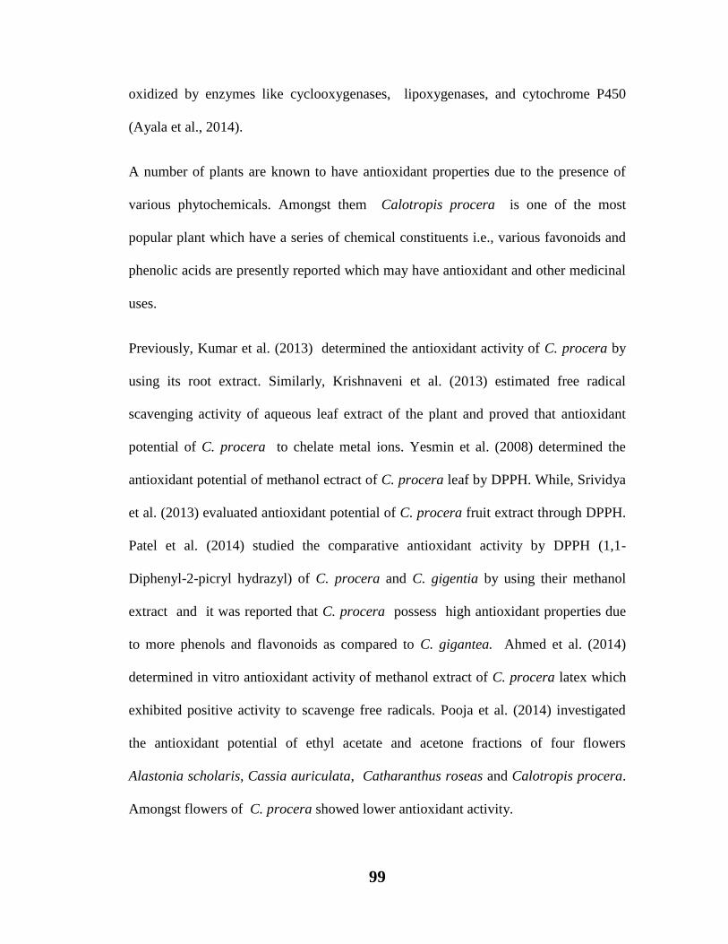
99
oxidized by enzymes like cyclooxygenases, lipoxygenases, and cytochrome P450
(Ayala et al., 2014).
A number of plants are known to have antioxidant properties due to the presence of
various phytochemicals. Amongst them Calotropis procera is one of the most
popular plant which have a series of chemical constituents i.e., various favonoids and
phenolic acids are presently reported which may have antioxidant and other medicinal
uses.
Previously, Kumar et al. (2013) determined the antioxidant activity of C. procera by
using its root extract. Similarly, Krishnaveni et al. (2013) estimated free radical
scavenging activity of aqueous leaf extract of the plant and proved that antioxidant
potential of C. procera to chelate metal ions. Yesmin et al. (2008) determined the
antioxidant potential of methanol ectract of C. procera leaf by DPPH. While, Srividya
et al. (2013) evaluated antioxidant potential of C. procera fruit extract through DPPH.
Patel et al. (2014) studied the comparative antioxidant activity by DPPH (1,1-
Diphenyl-2-picryl hydrazyl) of C. procera and C. gigentia by using their methanol
extract and it was reported that C. procera possess high antioxidant properties due
to more phenols and flavonoids as compared to C. gigantea. Ahmed et al. (2014)
determined in vitro antioxidant activity of methanol extract of C. procera latex which
exhibited positive activity to scavenge free radicals. Pooja et al. (2014) investigated
the antioxidant potential of ethyl acetate and acetone fractions of four flowers
Alastonia scholaris, Cassia auriculata, Catharanthus roseas and Calotropis procera.
Amongst flowers of C. procera showed lower antioxidant activity.

100
Presently, leaf and flower extracts of C. procera are investigated to determine the
antioxidant properties of the plant through techniques like DPPH, Lipid peroxidation
and reducing power ability.
6.1. Material and Methods
Detail of collection of plant samples already mentioned in Chapter 2 (2.1).
6.1.1. Preparation of extract
Detailed method is mentioned in Chapter 2 (2.2).
6.1.2. Preparation of fraction
Detailed method is explained in Chapter 2 (2.3).
6.1.3. Preparation of tissue homogenate
Fresh tissue of a normal albino rat was taken, the tissue was sliced into small pieces
and phosphate buffer saline pH 7.4 was added. The homogenate was centrifuged at
3000 rpm for 15 minutes, clear supernatant was collected for anti lipid peroxidation
assay.

101
6.2. Lipid peroxidation inhibition
Effect of C. procera on inhibition of lipid peroxidation activity was studied in vitro
according to the guidelines of Halliwell and Gutteridge (1999) as described in Chapter
2 (2.14).
6.3. 1,1-diphenyl -2-picrylhydrazyl (DPPH) radical scavenging assay
The in vitro antioxidant power of C. procera extracts were determined as described by
Kumar et al. (2013) in Chapter 2 ( 2.14.3).
6.4. Reducing power assay
The reducing power of C. procera flowers and leaves extracts were determined by
the methods of Oyaizu (1986) and Mishra et al. (2013) as described in Chapter 2
(2.14.4).

102
Table 6.1
Lipid peroxidation inhibition activity of C. procera flowers
extracts
Extract Conc. Water Hexane Ethanol Ethyl Acetate
mg/ml % % % %
1 21.36 27.48 56.87 28.43
2 26.07 38.62 75.11 42.83
4 43.13 43.83 78.54 46.6
6 55.69 46.68 88.38 56.87
8 70.86 53.79 88.62 65.23
10 89.58 54.99 89.33 63.12
BHA 85
Ascorbic Acid 75.5
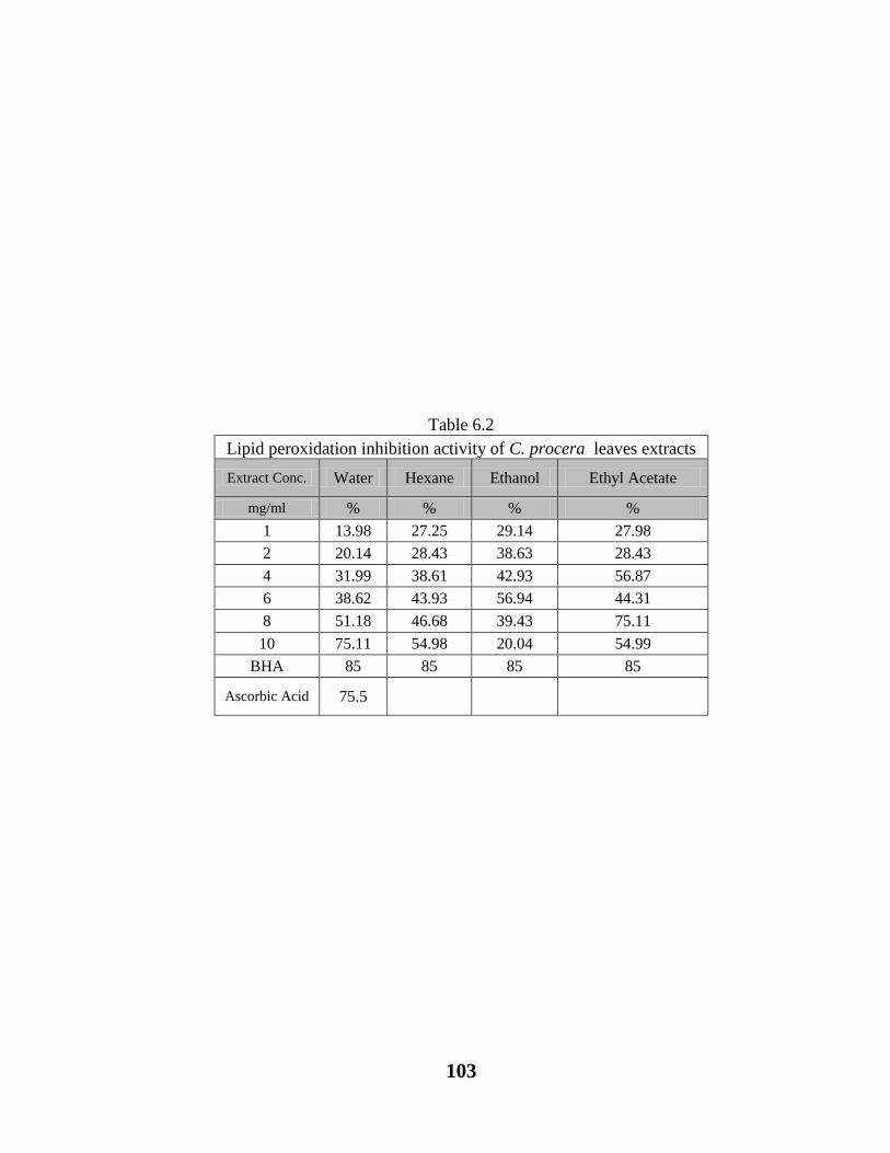
103
Table 6.2
Lipid peroxidation inhibition activity of C. procera leaves extracts
Extract Conc. Water Hexane Ethanol Ethyl Acetate
mg/ml % % % %
1 13.98 27.25 29.14 27.98
2 20.14 28.43 38.63 28.43
4 31.99 38.61 42.93 56.87
6 38.62 43.93 56.94 44.31
8 51.18 46.68 39.43 75.11
10 75.11 54.98 20.04 54.99
BHA 85 85 85 85
Ascorbic Acid 75.5

104
Table 6.3
DPPH radical scavenging activity of C. procera flowers extracts
Extract
Conc.
mg/ml
Water Hexane Ethanol Ethyl Acetate
1 65.81 36.12 48.45 56.98
2 69.6 66.9 62.52 61.7
4 71.51 42.91 76.18 51.1
6 77.82 53.79 81.72 70.84
8 80.9 71.45 88.19 66.22
10 83.05 76.38 86.13 77.2
Ascorbic
Acid 75.5 75.5 75.5 75.5
BHA 85

105
Table 6.4
DPPH radical scavenging activity (%) of C. procera leaves
extracts
Extract
Conc.
mg/ml
Water Hexane Ethanol Ethyl Acetate
1 70.1 27.25 29.14 27.98
2 75.97 28.43 38.63 28.43
4 55.8 38.61 42.93 56.87
6 41 43.93 56.94 44.31
8 33.6 46.68 39.43 75.11
10 7.9 54.98 20.04 54.99
Ascorbic
Acid 75.56 75.56 75.56 75.56
BHA 85

106
Table 6.5
Reducing power assay of C. procera flowers extracts
Extract
Conc.
mg/ml
Water Hexane Ethanol Ethyl Acetate
1 0.673 0.296 0.484 0.049
2 1.287 0.427 0.403 0.132
4 1.167 0.864 0.287 0.137
6 1.403 0.613 0.236 0.193
8 1.746 0.849 0.487 0.033
10 1.827 1.469 0.572 0.145
Ascorbic
acid 0.238 0.238 0.238 0.238
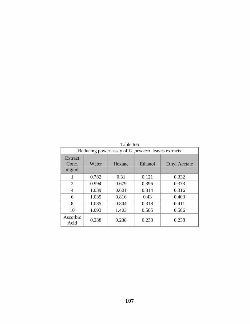
107
Table 6.6
Reducing power assay of C. procera leaves extracts
Extract
Conc.
mg/ml
Water Hexane Ethanol Ethyl Acetate
1 0.782 0.31 0.121 0.332
2 0.994 0.679 0.396 0.373
4 1.039 0.601 0.314 0.316
6 1.035 0.816 0.43 0.403
8 1.085 0.804 0.318 0.411
10 1.093 1.403 0.585 0.586
Ascorbic
Acid 0.238 0.238 0.238 0.238

108
Figure 6.1. Effect of different flower extracts of C. procera on Lipid
peroxidation inhibition (%).
0
10
20
30
40
50
60
70
80
90
100
Water Hexane Ethanol Ethyl Acetate
(%)
Inh
ibit
ion
of
lip
id p
erox
idati
on
C. procera flower extracts in different solvents
1mg/ml
2mg/ml
4mg/ml
6mg/ml
8mg/ml
10mg/ml
BHA 2mg/ml

109
Figure 6.2. Effect of different leaves extracts of C. procera on Lipid
peroxidation inhibition (%).
0
10
20
30
40
50
60
70
80
90
Water Hexane Ethanol Ethyl Acetate
(%)
Lip
id p
erox
idati
on
in
hib
itio
n
C. procera leaves extracts in different solvents
1mg/ml
2mg/ml
4mg/ml
6mg/ml
8mg/ml
10mg/ml
BHA 2mg/ml Linear (BHA 2mg/ml)

110
Figure 6.3. DPPH radical scavenging activity of flower extracts of C. procera
0
10
20
30
40
50
60
70
80
90
100
Water Hexane Ethanol Ethyl Acetate
(%)
DP
PH
rad
ical
scaven
gin
g a
ctiv
ity
C. procera flower extracts in different solvents
1mg/ml
2mg/ml
4mg/ml
6mg/ml
8mg/ml

111
Figure 6.4. DPPH radical scavanging activity of leaves extracts of C. procera
0
10
20
30
40
50
60
70
80
Water Hexane Ethanol Ethyl Acetate
(%)
DP
PH
rad
ical
scaven
gin
g a
ctiv
ity
C. procera leaves extracts in different solvents
1mg/ml
2mg/ml
4mg/ml
6mg/ml
8mg/ml
10mg/ml
Ascorbic acid

112
Figure 6.5. Reducing power of flower extracts of C. procera
0
0.2
0.4
0.6
0.8
1
1.2
1.4
1.6
1.8
2
Water Hexane Ethanol Ethyl Acetate
Ab
sorb
an
ce a
t 7
00
nm
C. procera flower extracts in different solvents
1mg/ml
2mg/ml
4mg/ml
6mg/ml
8mg/ml
10mg/ml
Ascorbic acid
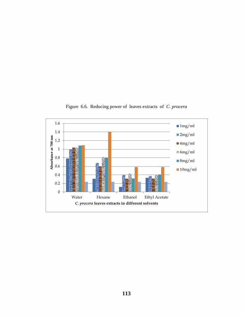
113
Figure 6.6. Reducing power of leaves extracts of C. procera
0
0.2
0.4
0.6
0.8
1
1.2
1.4
1.6
Water Hexane Ethanol Ethyl Acetate
Ab
sorb
an
ce a
t 7
00
nm
C. procera leaves extracts in different solvents
1mg/ml
2mg/ml
4mg/ml
6mg/ml
8mg/ml
10mg/ml

114
6.5. Results
Results showed that highest lipid peroxidation inhibition activity was found in flower
water and ethanol extracts i.e. about 89.5%. While, leaf water and ethyl acetate
extracts exhibited highest LPOI activity i.e. about 75%. These values of LPOI were
more or less similar to the standard value of BHA and ascorbic acid (Table 6.1, Fig.
6.1).
Flower showed highest value of DPPH radical scavenging activity (88.19%) in
ethanol. While, leaf water extract showed highest value of DPPH radical scavenging
activity i.e. 75.95% and the value of standard are more or less similar to that of DPPH
scavenging values of plant extracts (Table 6.2, Fig. 6.2).
Highest reducing power value was about more than 1.0 in both flower and leaf water
extracts. Whereas, reducing power value of tested extracts are significantly higher than
the standard value of ascorbic acid that was 0.238.

115
6.6. Discussion
Plants are the main source of the natural antioxidants against free radicals and their
reactive derivatives (ROS), which are known to induce various diseases of heart by
damaging lipids, cancer by DNA damaging and ageing by the damage of protein
(Sen et al., 2014). In the present study to counter act this action C. procera leaf and
flower extracts are utilized in vitro as an antioxidant agent by analyzing DPPH
scavenging activity, reducing power and lipid peroxidation inhibition activities. While,
C. procera was earlier found to be beneficent to inhibit or reduce the lipid
peroxidation chain reaction (Kumar et al., 2015), free radical scavenging activity and
reducing power ( Yesmin et al., 2008).
Flower and leaf extracts of C. procera with various solvents possess effective lipid
peroxidation inhibition (LPOI) activity. Flower extract exhibited highest lipid
peroxidation inhibition value as compared to leaf extract. Lipid peroxidation
inhibition of flower extract showed concentration dependent increase in water, hexane
and ethanol extract except ethyl acetate extract. Highest LPOI activity is found in
water extract i.e. (89.58%), ethyl acetate (65.23%) and hexane (54.99%) respectively.
However, BHA and Ascorbic acid exhibited lower values (85 and 75%) as compared
to water extract value (Table 6.1, Fig. 6.1).
There is a concentration dependent LPOI activity in water and hexane. Whereas,
ethanol and ethyl acetate LPOI activity was independent to their concentrations. The
highest activity of LPOI in terms of percentage was observed in water (i.e., 75.11%
with 10gm/ml) and ethyl acetate ( 75.11% with 8mg/ml) as compared to ethanol

116
(56.94% with 6mg/ml) and hexane (54.98% with 10mg/ml) in respective manner.
Similarly BHA exhibited 85% activity as compared to ascorbic acid showing 75%
lipid peroxidation inhibition activity.
Similarly, leaf extracts of C. procera with various solvents possess effective DPPH
scavenging activity in all concentrations.
Flower extract exhibited highest scavenging activity as compared to leaf. The
antioxidant activity of flower extract usually increases with the increase of
concentration and highest activity is found in ethanol extract (i.e., 88.19%) followed
by water extract (83.05%), ethyl acetate (77.2%) and hexane (76.38) respectively.
However, BHA showed highest activity (85%) as compared to ascorbic acid (75.5%)
(Table 6.3, Fig. 6.3).
Leaf extract with various solvents showed different values of scavenging activity
irrespective of their concentration. Amongst all of the solvents water extract exhibited
highest activity (i.e., 75.95% with 2mg/ml) following ethyl acetate (75.11 with
8mg/ml), ethanol (56.94% with 6mg/ml) activity and 10mg/ml hexane showing
(54.98%) scavenging activity respectively. While BHA and ascorbic acid were used
as standard showing 85% and 75.56% activity as compared to test extracts (Table 6.4,
Fig. 6.4).
Different solvent extracts of C. procera flower and leaf were also analyzed to
determine the reducing power. Flower showed highest reducing power as compared
to leaf. Amongst all of the test extracts, flower extract showed different absorbance
pattern of reducing power irrespective of their concentrations. While, flower water
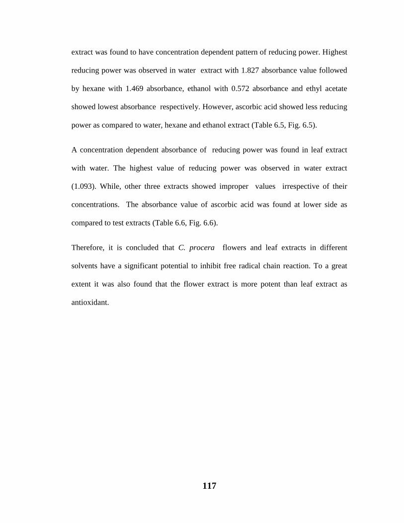
117
extract was found to have concentration dependent pattern of reducing power. Highest
reducing power was observed in water extract with 1.827 absorbance value followed
by hexane with 1.469 absorbance, ethanol with 0.572 absorbance and ethyl acetate
showed lowest absorbance respectively. However, ascorbic acid showed less reducing
power as compared to water, hexane and ethanol extract (Table 6.5, Fig. 6.5).
A concentration dependent absorbance of reducing power was found in leaf extract
with water. The highest value of reducing power was observed in water extract
(1.093). While, other three extracts showed improper values irrespective of their
concentrations. The absorbance value of ascorbic acid was found at lower side as
compared to test extracts (Table 6.6, Fig. 6.6).
Therefore, it is concluded that C. procera flowers and leaf extracts in different
solvents have a significant potential to inhibit free radical chain reaction. To a great
extent it was also found that the flower extract is more potent than leaf extract as
antioxidant.

118
EFFECT OF CALOTROPIS PROCERA LEAF
HEXANE SOLUBLE EXTRACT ON NSAID
(IBUPROFEN) TREATED RATS.
7. Introduction
The ability of a substance to damage any living body is called toxicity. Toxicity can
affect the whole organism or substructure of the organism and it may be occur due to
certain biological, physical or chemical effects. Drug induced toxicity can damage any
tissue depending on dosage such as acute dosage of a drug can produce the toxicity for
nervous system and its chronic exposure may cause the serious injuries to the other
organs. Toxicity can also be produced by the medicines which are normally be used
for curative purposes. Sometimes, the use of over the counter medicines and long term
use of overdoses of drugs may also cause toxicity to certain specific organs. The
Process of oxidation continuously takes place in all aerobic living bodies, due to this
ROS (reactive oxygen species) including O2 anion, H2O2 hydrogen peroxide -OH
hydroxyl radical and nitric oxide/peroxinitrates (NO/NOO-) are constantly formed
within the cells. The over production of these substances may cause oxidative load in
the cells. This oxidative stress produces deleterious effects to cells of DNA, proteins
and lipids. Lipid are specifically more damaged due to the formation of lipid
peroxidation products.
There are various reports available on toxicity producing substances like the NSAIDS.
Particularly, Paracetamol (Younes et al., 1988), Acetaminophen (Tarloff et al., 1990;
Trumper et al., 1992), Diclofenac (Hickey et al., 2001; Yasmeen et al., 2007),
Phenacetin (Murray and Brater, 1993; Kocaoglu et al., 1997; Fackovcova et al., 2000;

119
Soubhia et al., 2005; Derle et al., 2006), Mefenamic acid (Somchit et al., 2004),
Tenofovir (Morelle et al., 2009), Paracetamol (Somanawat et al., 2013) and metals
including arsenic, cadmium, lead and mercury (Nicholson et al., 1985; Fowler, 1992).
However, Cholestyramine was utilized against the Paracetamol induced toxicity in rats
and that was evident by a reduction in plasma enzyme activity and creatinine levels.
(Siegers and Moller, 1989). On the other hand Cadmium was used to prevent the
Acetaminophen induced toxicity in female rats (Bernard et al., 1988). Moreover,
Ibuprofen and Diclofenac were found useful protective drugs against Gentamicin
toxicity (Farag et al., 1996). While, Sharma et al. (2007) suggested that the
supplementation of Spirulina fusiformis can play a significant role against mercuric
chloride induced toxicity.
7.1. Nephrotoxic Agents
There are several substances which can initiate toxicity to the kidneys. These
substances include antibiotics, anticancer drugs, heavy metals, herbicides, pesticides,
excess amount of uric acid and long term use and high doses of analgesics may also
cause nephrotoxicity these analgesics usually include aspirin and ibuprofen
(Robertson, 2014).
7.2. NSAIDs
Nonsteroidal anti-inflammatory drugs or NSAID are the commonly used over the
counter drugs. They are pain relievers, help in reducing inflammation and lower
fever. They also prevent blood from clotting (Robertson, 2014).

120
7.3. Ibuprofen
Ibuprofen is selected for present experimental studies. It is a derivative of propionic
acid its chemical name is Isobutylphenylpropionic acid, the structure containing a
benzene ring conjugated to a propionic acid. It was first derived during 1950-1960s at
the research laboratories of Boots group and discovered by the scientist Andrew RM
Dunlop with his co-researchers Stewart Adams, John Nicholson, Jeff Wilson and
Colin Burrows (Robertson, 2014).
7.4. Mechanism of action of Ibuprofen
Ibuprofen is said to be an inhibitor of prostaglandin synthesis. The exact mechanism
of action is still unknown. Ibuprofen is an inhibitor of an enzyme (cyclooxygenase).
This enzyme converts arachidonic acid to prostaglandins. Prostaglandins are the
initiator of inflammation, fever and pain. There are two types of cyclooxygenase,
one is COX-1 which protects the lining of the stomach from digestive chemicals and
also maintains kidney function whereas, COX-2 released when joints are injured or
inflamed (Robertson, 2014).
A number of medicinal plants have been reported as antioxidants such as fruit extract
of Berberis vulgaris was used as an antioxidant (Motalleb et al., 2005; Hanachi et al.,
2008). Zingiber officinalis was reported to be used against ROS induced oxidative
stress (Ajith, 2010). Similarly the species of Gemelia, Kigelia, Hibiscus,Parthenium
(Patel et al., 2010) and Calotropis procera (Ahmed et al., 2014) have been reported
for their high radical scavenging activity.

121
In the present study Calotropis procera is selected as anti-oxidant against the NSAID
(Ibuprofen) induced toxicity. As the species has already been reported to have various
medicinal properties (Goyal and Mathur, 2011; Johnson et al., 2011; Doshi et al.,
2011; Prabha and Vasantha, 2012).
7.5. Material and Methods
Detail of collection of plant samples already mentioned in Chapter 2 (2.1).
7.5.1. Preparation of extract
Detailed method is described in Chapter 2 (2.2).
7.5.2. Preparation of fractions
Detailed method is described in Chapter 2 (2.3).
7.5.3. Experimental animals and diet
Wistar white male rats (180-250g b.w.), bought from the animal house of Dow
University of Health Sciences, Karachi, Pakistan. Before starting experiments rats
were adjusted to the laboratory atmosphere and accommodated separately in a
artificially maintained temperature (22-26oC). Water and diet were provided to rats
Diet preparation is explained in the following table:

122
Rat’s Diet Preparation Table
INGREDIENTS QUANTITY
Wheat Flour 5 Kg
Barley Flour 2.5 Kg
Corn Flour 1.25 Kg
Cooking Oil 1.5Litre
7.6. Proper Recommendations
The scientific procedures were conducted in compliance with the ethical
recommendations of Institutional ERB (Ethical Review Board) and internationally
accepted ethics for laboratory use and care in animal research (Health Research
Extension Act, 1985).
7.7. Drug: Ibuprofen was purchased from market.
7.8. Study Protocol and Drug Administration Plan
Animals were divided into 3 different groups (n= 6)
Each group consists of six rats and treated as follows:
The Group I consists of healthy animals, untreated rats and termed as control.
Rat’s weight was recorded between 11:00 -12:00 hr for 10 alternate days.
Group II treated with prepared Ibuprofen suspension orally at a dose of 2ml /200gm
b.w. for 15 days. Termed as Ibuprofen Treated +ve Control. They were weighed
before administration of oral dose daily.

123
Group III treated with only hexane suspension orally at a dose of 2ml/200gm b.w.
Group IV treated with Ibuprofen + Hexane extract treated group, received hexane
extract orally according to the recommended doses i.e., they were weighed before
hexane extract treatment. Hexane extract was given 30 minutes prior to ibuprofen
administration.
7.9. Collection of Samples
7.9.1. Blood Samples
After 24 hours of last dose, rats were killed by chop off their heads and blood was
collected in the lithium heparin coated tubes. Then these tubes were shaked well.
After shaking, the tubes were centrifuged at 2000 rpm for 20 minutes. The plasma was
separated and collected in disposable eppendorff tubes and stored at -70oC for further
processing.
7.9.2. Kidney Sample
Kidneys were taken and blood was cleaned by passing through saline, Then dried
under filter paper and weighed. The kidneys were stored in the freezer at -70oC for
biochemical analysis.

124
7.10. Analytical methods
7.10.1. Preparation of protein free filtrate (PFF)
Detailed method of preparation for protein free filtrate is mentioned in Chapter 2
(2.13).
7.10.2. Estimation of plasma urea
Plasma urea was estimated by using Diacetyl monoxime method (Butler et al., 1981)
as mentioned in chapter 2 (2.13.1 and 2).
7.10.3. Estimation of plasma creatinine
Plasma creatinine was estimated by Modified Jeff’s method (Spierto et al., 1979) is
explained in Chapter 2 (2.13.3).
7.10.4. Preparation of kidney homogenate
Kidney homogenate was prepared by the procedure of Ricardo et al. (2005) as
mentioned in Chapter 2 (2.13.4).
7.10.5. Estimation of malonyldialdehyde (MDA)
MDA was estimated following the method of Okhawa et al. (1979) is explained in
Chapter 2 (2.13.5).

125
7.10.6. Estimation of 4-hydroxyl-2-nonenal (4-HNE)
4-HNE was measured by the method of Kinter et al. (1996) as mentioned in Chapter 2
(2.13.6).
7.10.7. Estimation of catalase
Method of Sinha (1972) was used to estimate catalase is explained in Chapter 2
(2.13.7).
7.10.8. Estimation of superoxide dismutase (SOD)
SOD was estimated by the method of Kono (1978) is explained in Chapter 2 (2.13.8).
7.10.9. Estimation of glutathione (GSH)
GSH was estimated by the method of Carlberg and Mannervik (1985) as mentioned in
Chapter 2 (2.13.9).

126
Table 7.1. Effect on body and kidney weight in control, Ibuprofen, hexane, and
Ibuprofen+hexane treated rats.
Parameters Control Ibuprofen
200mg/kg
Hexane
200mg/kg
Ibu+Hex.
200mg/kg
Mean Body
Weight (gms) ***261.66
a±12.88 ***238.1
d±12.35 ***245.49
c±10.66 ***259.2
b±11.61
Mean Kidney
Weight
(gms)
***0.75c±0.05 ***0.85
b±0.12 ***0.89
a±0.06 ***0.88
a±0.05
n=6, mean values ± SE, ***= P<0.05, a-d= ranks of mean values, sharing of same letter do not differ
significantly.

127
Table 7.2. Effect on renal function in control, Ibuprofen, hexane and
Ibuprofen+hexane pretreated rats.
Parameters Control Ibuprofen
200mg/kg
Hexane
200mg/kg
Ibu+Hex.
200mg/kg
Plasma Urea
(mg%) ***59.1
b±0.493 ***68.5
a±0.401 ***56.9
c±0.596 ***54.4
d±3.961
Plasma
Creatinine
(mg%)
***0.54c±0.046 ***1.8
a±0.692 ***0.67
b±0.448 ***0.43
d±0.443
n=6, mean values ± SE, ***= P<0.05, a-d= ranks of mean values, sharing of same letter do not differ
significantly.

128
Table 7.3. Effect on tissue SOD and catalase in control, Ibuprofen, hexane and
Ibuprofen+hexane pretreated rats.
Parameters Control Ibuprofen
200mg/kg
Hexane
200mg/kg
Ibu+Hex.
200mg/kg
SOD
(u/g tissue) ***17.19
b±1.01 ***13.35
d±0.98 ***14.11
c±2.49 ***19.69
a±1.12
Catalase
(mM/g tissue)
***3.11b±0.008 ***1.99
d±0.63 ***2.99
c±0.68 ***3.50
a±0.17
n=6, mean values ± SE, ***= P<0.05, a-d= ranks of mean values, sharing of same letter do not differ
significantly.

129
Table 7.4. Effect on plasma MDA, tissue MDA, 4HNE, and glutathione levels in
control, Ibuprofen, hexane and Ibuprofen+Hexane pretreated rats.
Parameters Control Ibuprofen
200mg/kg
Hexane
200mg/kg
Ibu+Hex.
200mg/kg
Plasma MDA
(nM/ml)
***3.31b±0.5
3 ***4.01
a±0.06 ***2.10
c±0.79 ***1.87
d±0.09
Tissue MDA
(nM/gm)
***0.55c±0.0
4 ***0.73
a±0.05 ***0.58
b±0.13 ***0.48
d±0.08
Tissue 4-
HNE (nM/g)
***159.61b±1
0.48
***210.14a±5.
23
***130.15c±11.
12
***143.15d±21.
25
Tissue GSH
(U/g tissue)
***3.56b±0.4
01 ***1.79
d±0.56 ***2.80
c±0.27 ***5.93
a±0.81
n=6, mean values ± SE, ***= P<0.05, a-d= ranks of mean values, sharing of same letter do not differ
significantly.

130
Figure 7.1. Effect on Body weight of rats in Control, Ibuprofen, Hexane
and Ibuprofen + Hexane treated groups.
***a ***d ***c
***b
0
50
100
150
200
250
300
Control Ibuprofen Hexane Ibu+Hex
Me
an
Bo
dy
We
igh
t
(gm
)
Experimental Groups (200mg/kg body weight)

131
Figure 7.2. Effect on Kidney weight of rats in Control, Ibuprofen, Hexane
and Ibuprofen + Hexane treated groups.
***c
***b ***a ***a
0
0.2
0.4
0.6
0.8
1
1.2
Control Ibuprofen Hexane Ibu+Hex
Me
an
Kd
ne
y W
eig
ht
(gm
)
Experimental Groups (200mg/kg body weight)

132
Figure 7.3. Effect on Plasma Urea level of rats in Control, Ibuprofen,
Hexane and Ibuprofen + Hexane treated groups.
***b
***a
***c ***d
0
10
20
30
40
50
60
70
80
Control Ibuprofen Hexane Ibu+Hex
Pla
sma
Ure
a
(mg
/dl)
Experimental Groups (200mg/kg. body weight)

133
Figure 7.4. Effect on Plasma Creatinine level of rats in Control, Ibuprofen,
Hexane and Ibuprofen + Hexane treated groups.
***c
***a
***b
***d
-0.5
0
0.5
1
1.5
2
2.5
3
Control Ibuprofen Hexane Ibu+Hex
Pla
sma
Cre
ati
nin
e (
mg
/dl)
Experimental Groups (200mg/kg body weight)

134
Figure 7.5. Effect on Tissue SOD level of rats in Control, Ibuprofen,
Hexane and Ibuprofen + Hexane treated groups.
***b
***d ***c
***a
0
5
10
15
20
25
Control Ibuprofen Hexane Ibu+Hex
Tis
sue
SO
D l
ev
el
(U
/gm
tis
sue
)
Experimental Groups (200 mg/kg body weight)

135
Figure 7.6. Effect on Tissue Catalase level of rats in Control, Ibuprofen,
Hexane and Ibuprofen + Hexane treated groups.
***b
***d
***c
***a
0
0.5
1
1.5
2
2.5
3
3.5
4
Control Ibuprofen Hexane Ibu+Hex
Tis
sue
Ca
tala
se l
ev
el
(m
mo
l/g
m
tiss
ue
)
Experimental Groups (200mg/kg body weight)

136
Figure 7.7. Effect on Plasma MDA level of rats in Control, Ibuprofen,
Hexane and Ibuprofen + Hexane treated groups.
***b
***a
***c ***d
0
0.5
1
1.5
2
2.5
3
3.5
4
4.5
Control Ibuprofen Hexane Ibu+Hex
Pla
sma
MD
A l
ev
el
(n
mo
l/m
l)
Experimental Groups (200mg/kg body weight)
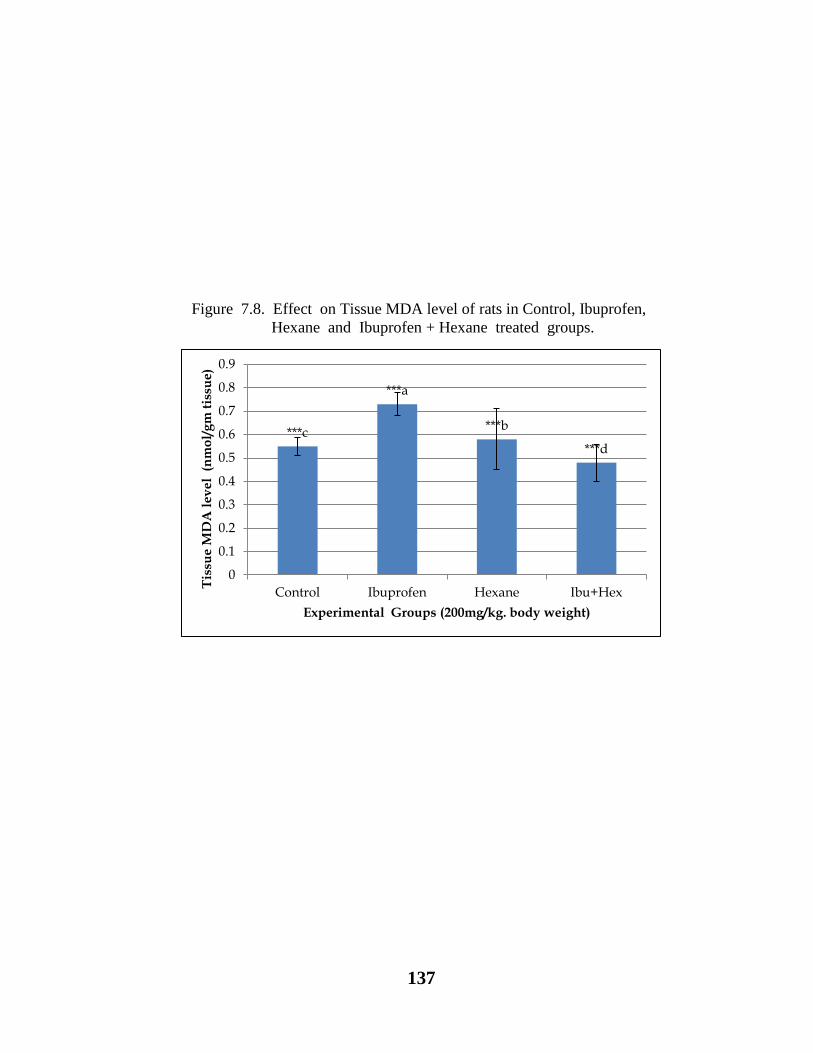
137
Figure 7.8. Effect on Tissue MDA level of rats in Control, Ibuprofen,
Hexane and Ibuprofen + Hexane treated groups.
***c
***a
***b
***d
0
0.1
0.2
0.3
0.4
0.5
0.6
0.7
0.8
0.9
Control Ibuprofen Hexane Ibu+Hex
Tis
sue
MD
A l
ev
el
(n
mo
l/g
m t
issu
e)
Experimental Groups (200mg/kg. body weight)

138
Figure 7.9. Effect on Tissue 4HNE level of rats in Control, Ibuprofen,
Hexane and Ibuprofen + Hexane treated groups.
***b
***a
***c ***d
0
50
100
150
200
250
Control Ibuprofen Hexane Ibu+Hex
Pla
sma
4H
NE
le
ve
l
(mg
/L)
Experimental Groups (200mg/kg body weight)

139
Figure 7.10. Effect on Tissue GSH level of rats in Control, Ibuprofen,
Hexane and Ibuprofen + Hexane treated groups.
***b
***d
***c
***a
0
1
2
3
4
5
6
7
8
Control Ibuprofen Hexane Ibu+Hex
Tis
sue
GS
H l
ev
el
(U
/gm
tis
sue
)
Experimental Groups (200mg/kg. body weight)

140
7.11. Results
According to one way Anova values of body and kidney weight differ significantly
(P<0.05) (Table 7.1, Fig. 7.1). Duncan’s multiple comparison test (DMCT) showed a
significant decrease in rat body weight in treated rats as compared to control (P<0.05).
Similarly marked decreased was observed in Ibu+hex and Ibuprofen respectively
(P<0.05) (Table 7.1, Fig. 7.1). According to DMCT kidney weight of treated rats was
significantly higher as compared to control (P<0.05). A marked increase in kidney
weight in hexane, Ibu+hex and Ibuprofen was observed respectively (P<0.05).
However, values of kidney weight were insignificant between hexane and Ibu+hex
(P<0.05) (Table 7.1, Fig. 7.2).
One way Anova showed, the values of plasma urea and plasma creatinine differ
significantly (P<0.05) (Table 7.2, Fig. 7.3). According to Duncan’s multiple
comparison test, Ibuprofen treated rats showed a marked increase in plasma urea level
as compared to control, hexane and Ibu+hex respectively (P<0.05). Similarly, plasma
creatinine level was significantly increased in Ibuprofen treated rats as compared to
hexane and control (P<0.05). While, a significant decrease was observed in Ibu+hex
than all the other treated and control rats (P<0.05) (Table 7.2, Fig. 7.4).
According to one way Anova values of SOD and catalase differ significantly (P<0.05)
(Table 7.3, Figs. 7.5, 7.6).While, Duncan’s multiple comparison test showed
significant increase in SOD level in Ibu+hex treated rats as compared to control,
hexane and Ibuprofen respectively (P<0.05) (Table 7.3, Fig. 7.5). However, increased

141
level of catalase was observed in Ibu+hex treated rats as compared to control, hexane
and Ibuprofen treated rats respectively (P<0.05) (Table 7.3, Fig. 7.6).
One way Anova showed a significant difference in plasma MDA, tissue MDA, tissue
4HNE and GSH levels (P<0.05). (Table 7.4, Figs. 7.7 – 10). Duncan’s multiple
comparison test showed a significantly increased level of plasma MDA in Ibuprofen
treated rats (P<0.05). Whereas, level of plasma MDA was significantly decreased in
control followed by hexane and Ibu+hex treated rats (P<0.05).
Similarly, tissue MDA level in Ibuprofen treated rats increased significantly as
compared to hexane, control and Ibu+hex respectively (P<0.05) (Table 7.4, Fig. 7.8).
The value of tissue 4HNE showed a significant increase in Ibuprofen treated rats
(P<0.05). While, the level of tissue 4HNe gradually decreased in control, hexane and
Ibu+hex treated rats respectively (P<0.05) (Table 7.4, Fig. 7.9).
On the other hand level of tissue GSH was significantly higher in Ibu+hex treated rats
as compared to control, hexane and Ibuprofen treated rats (P<0.05) (Table 7.4, Fig.
7.10).
142
7.12. Discussion
Nonsteroidal anti-inflammatory drugs (NSAIDS) are one of the sources for pre-renal
failure (Hoitsma et al., 1991). Ibuprofen (NSAID) is used to induce nephrotoxicity in
rats with pretreatment of C. procera leaf hexane extract. The antioxidant activity of
endogenous enzymes (SOD and Catalase) is evaluated. Presently increase in
antioxidant enzyme levels (SOD and Catalase) was observed after Ibuprofen+hexane
administration (P<0.05) (Table 7.3). While, SOD converts superoxide radicals into
H2O2 (Hydrogenperoxide) and serve as a first line of defense against ROS (Sen et al.,
2010).
Previously, it was concluded that accumulation of urea and creatinine is the indicator
of improper renal function (Javed et al., 2015). In the present study, serum urea and
creatinine levels (Table 5.2) were significantly increased (P<0.05) after the
administration of Ibuprofen and showing renal disorder, this result is also in
accordance with the previous findings of Mahalakshmi et al. (2010) where ibuprofen
was used as NSAID. The significant decrease in urea and creatinine values after
treatment of Ibuprofen with C. procera hexane extract also provide the evidence of
positive role to inhibit the toxicity in rats produced by Ibuprofen (P<0.05) (Table 7.2).
The reactive oxygen species (ROS) initiates the contraction of masangial cells which
change the filtration surface area and alter the ultrafiltration coefficient factor that
decreases the rate of glomerular filtration (Leena and Alaraman, 2005).
C. procera hexane extract prevented Ibuprofen induced decline in glutathione (GSH)
activity in the renal mitochondria of rats (P<0.05) (Table 7.4) which is also supported

143
by the report of Setty et al. (2007) where C. procera flower extract increased the
depleted concentration of GSH. Due to Ibuprofen administration different complexes
formed are taken up by renal cells and stabilized by intracellular GSH. GSH
Peroxidase present in the cytoplasm of the cells, removes H2O2 by coupling its
reduction to H2O with oxidation of GSH. In case of intracellular depletion the
complexes undergo the rapid transformation to receive metabolites, this depletion
seems to be the main factor that impairs antioxidant enzyme (Ozen et al., 2004; Ban et
al., 1994). It is also noteworthy that the presence of flavonoids in C. procera revealed
the correlation of antioxidant properties (Hesham et al., 2002; Javed et al., 2015)
which may ultimately useful for the treatment of kidney damage.
It is also shown that Ibuprofen administration is associated with increased formation
of free radicals, and with heavy oxidative stress (Chen et al., 1994; Setty et al., 2007;
Mahalakshmi et al., 2010; Javed et al., 2015).This will lead to oxidative damage of
cell components, like proteins and nucleic acids (Boya et al., 1999).
The high concentration of manonyldialdehyde (MDA) in kidney tissues may cause its
malfunction. Present findings (Table 7.4) showed decreased value of MDA in C.
procera treated tissue and plasma (P<0.05). These results are also supported with
previous findings of Roy et al. (2005) where, decrease in MDA level, treated with C.
procera latex was observed to correlate antioxidant acivity.
4-hydroxynonenal (4HNE) an unsaturated aldehyde is considerably more toxic for
cell in vivo, than MDA, it is very important to measure 4HNE levels (Ong et al.,
2000). In present study treatment by C. procera with Ibuprofen decreased the level
of 4HNE (P<0.05) (Table 7.4).
144
Thus, it is concluded that Ibuprofen induced nephrotoxicity in rats is the result of over
production of hydrogen peroxide and hydroxyl radical that may finally cause renal
oxidative stress. However, with the supplementation of C. procera oxidative stress
was significantly decreased by regularizing the levels of superoxide dismutase,
catalase, 4HNE and MDA.
145
8. GENERAL DISCUSSION
Phytochemically leaf and flower of C. procera are investigated and a large number of
chemical constituents are reported which exhibit diversity in quantitative and
qualitative values of carbohydrates, reducing and non reducing sugars, proteins, amino
acids and phenolic compounds.
At one side plant is considered as a tonic (Gholamshahi et al., 2014) due to the
presence of enormous amount of protein, amino acids, carbohydrates, reducing and
non reducing sugars. While on the other hand, plant usually serves against infectious
diseases caused by bacteria or fungi and other diseases related to free radical induced
oxidative stress (Patel et al., 2010; Moteriya et al., 2015). These properties of plant
may be correlated with the detected amount of phenolic acids and flavonoids, as
22ug/ml and 17.66ug/ml total phenol are detected from flower and leaf extracts
respectively. While, a large number of flavonoids (Table 3.3) were also detected from
flower and leaf.
Similarly, use of NSAIDs is very common in the treatment of rheumatism, pain, fever,
inflammation and cardiovascular diseases but over the counter (OTC) use for a long
period of time, is the beginning of the production of free radicals which may result in
the gastric or duodenal ulceration and severe complications such as perforation and
gastrointestinal hemorrhage as well as kidney failure (Hoitsma et al., 1991;Kamboj,
2000). A marked decrease was observed in body weight of Ibuprofen treated rats as
compared to control (P<0.05) (Table 7.1, Fig.7.1). This weight loss was restored by
146
the administration of pretreatment of C. procera leaf hexane with Ibuprofen (P<0.05)
(Table 7.1, Fig.7.1). Previously it was reported that Ibuprofen may cause
gastrointestinal disturbance due to damaging effect in gastrointestinal mucosa that
results in reduced ingestion of food (Mahalakshmi et al., 2010). This is the
consequence of inhibition of cyclooxygenase -1 enzyme system which protect
gastrointestinal lining from digestive chemicals. In the present study Ibuprofen treated
rats show increase in kidney weight as compared to control (P<0.05) (Table 7.1, Fig.
7.1). Similarly, a significant increase is also observed in plasma urea and creatinine
levels in Ibuprofen treated rats as compared to control (P<0.05) (Table 7.2, Fig. 7.2).
However, accumulation of urea and creatinine in plasma induce decrease in kidney
function which is an indication of decrease glomerular filtration rate due to
nephrotoxicity (Javed et al., 2015). However, Increased Ca+2
movement in the
masangial cells may also be a cause of reduced glomerular filtration rate (Stojiljkovic
et al., 2008; Javed et al., 2015). In the present study these increased levels are
neutralized by the administration of C. procera leaf hexane extract with Ibuprofen to
prevent the normal kidney function. While, Kaneko et al. (2008) found an opposite
relation between quantity of absorbed urea and rate of tubular urine flow.
There is an imbalance between oxidative stress occur in Ibuprofen treated rats,
which is responsible for the formation of reactive oxygen species (ROS). This was
determined by evaluating decreased level of catalase, super oxide dismutase (SOD),
Glutathione (GSH) and increased levels of malonyldialdehyde (MDA) and 4 hydroxy
nonenal (4HNE) (Table 7.3-7.4, Fig. 7.5-10). In order to counteract oxidative
imbalance due to administration of Ibuprofen, C. procera leaf hexane extract co-
147
administered with Ibuprofen to test rats which significantly restored decreased levels
of catalase and super oxide dismutase (SOD) (Table 7.3, 7.5-6). Whereas, catalase,
glutathione peroxidase and glutathione reductase belong to the endogenous type of
antioxidant defense system. While, SOD acts as a first line of defense against ROS
which traps superoxide radical and convert it into H2O2. However, increased amount
of hydrogen peroxide and hydroxyl radicals and decreased amount of glutathione are
responsible to initiate Ibuprofen nephrotoxicity (Parlakpinar et al., 2005). The higher
amount of glutathione peroxidase may reduces H2O2 to H2O with oxidation of
glutathione (GSH) and due to reducing nature, glutathione is one of the important
substance for maintaining cell sustainability (Sen et al., 2010).
Presently, a decrease in renal glutathione level is observed in Ibuprofen treated rats
(Table 7.4, 7.10). However, in some previous studies it was also observed that kidney
damage may cause due to increase in GSH levels (Antunes et al., 2000).
Similarly, in vivo lipid peroxidation can be evaluated by estimating lipid peroxidation
products malonyldialdehyde (MDA and 4 hydroxy nonenal 4HNE). However, Lipid
peroxidation is an important oxidative damage mechanism to cell structure which may
become the reason of cell disintegration. The presence of lipid peroxidation involves
generation and propagation of lipid radicals, collection of oxygen and shifting of
double bonds in unsaturated lipids, which leads to abolishtion of membrane lipids.

148
After this process a number of products are obtained which includes alkanes, ethers,
alcohols ketones and aldehydes (Dianzani and Barrera, 2008).
Figure 8.1. MDA inhibition, formation and metabolism: (black pathway) Proposed action of
C. procera extract which scavenges oxygen radical and convert lipid peroxide by reduction into
non toxic form of PUFA, PUFA peroxide radical is formed by the reaction of Ibuprofen with
PUFA, (blue pathway) MDA formed during the enzymatic biosynthesis of thromboxane
A2(TXA2) and 12-1-hydroxy-5,8,10-heptadecatrienoic acid (HHT) by in vivo decomposition of
arachidonic acid (AA) and longer PUFAs as a side products. Or by the non enzymatic production
of bicyclic endoperoxides during lipid peroxidation (red pathway). Formed MDA can be
enzymatically metabolized (green pathway). Enzymes responsible for the generation and
metabolism of MDA includes: cyclooxygenases (1), prostacyclin hydroperoxidase (2),
thromboxane synthase (3), aldehyde dehydrogenase (4), decarboxylase (5), acetyl CoA synthase
(6), and tricarboxylic acid cycle (7). (Modified from Ayala et al., 2014).
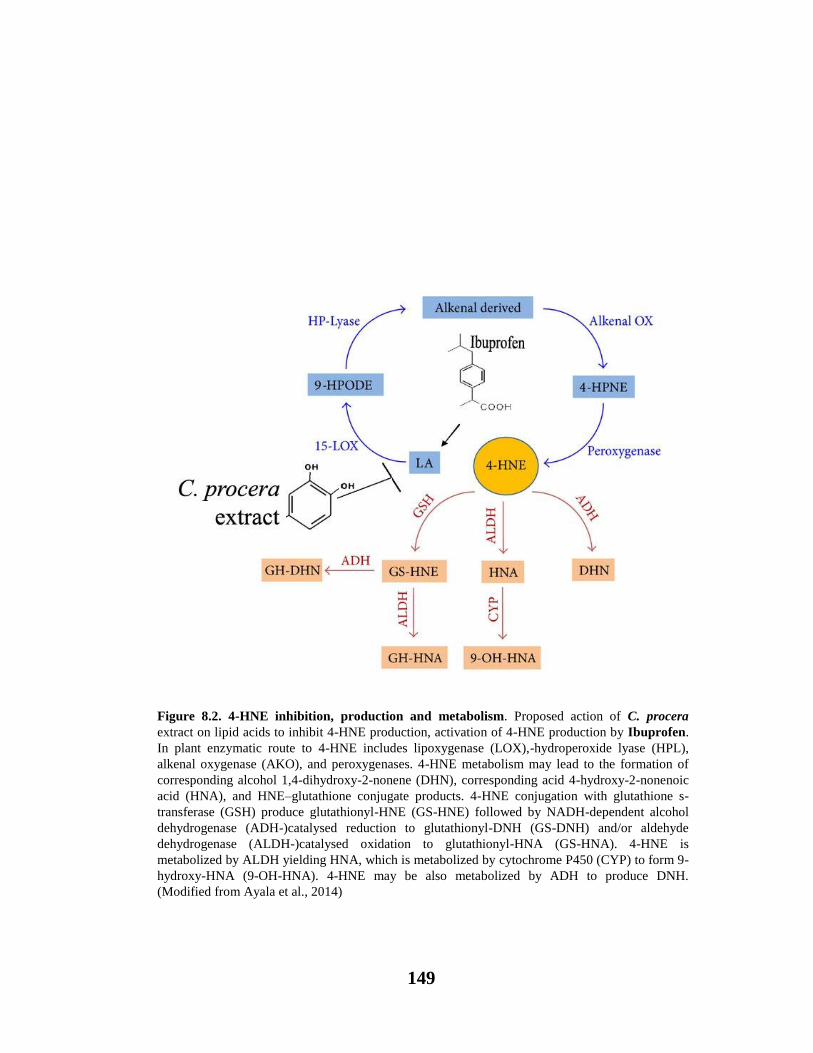
149
Figure 8.2. 4-HNE inhibition, production and metabolism. Proposed action of C. procera
extract on lipid acids to inhibit 4-HNE production, activation of 4-HNE production by Ibuprofen.
In plant enzymatic route to 4-HNE includes lipoxygenase (LOX),-hydroperoxide lyase (HPL),
alkenal oxygenase (AKO), and peroxygenases. 4-HNE metabolism may lead to the formation of
corresponding alcohol 1,4-dihydroxy-2-nonene (DHN), corresponding acid 4-hydroxy-2-nonenoic
acid (HNA), and HNE–glutathione conjugate products. 4-HNE conjugation with glutathione s-
transferase (GSH) produce glutathionyl-HNE (GS-HNE) followed by NADH-dependent alcohol
dehydrogenase (ADH-)catalysed reduction to glutathionyl-DNH (GS-DNH) and/or aldehyde
dehydrogenase (ALDH-)catalysed oxidation to glutathionyl-HNA (GS-HNA). 4-HNE is
metabolized by ALDH yielding HNA, which is metabolized by cytochrome P450 (CYP) to form 9-
hydroxy-HNA (9-OH-HNA). 4-HNE may be also metabolized by ADH to produce DNH.
(Modified from Ayala et al., 2014)
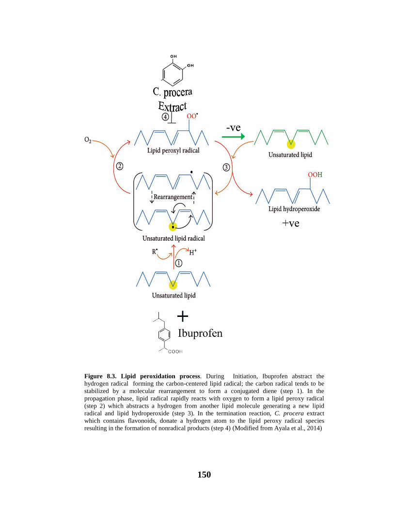
150
Figure 8.3. Lipid peroxidation process. During Initiation, Ibuprofen abstract the
hydrogen radical forming the carbon-centered lipid radical; the carbon radical tends to be
stabilized by a molecular rearrangement to form a conjugated diene (step 1). In the
propagation phase, lipid radical rapidly reacts with oxygen to form a lipid peroxy radical
(step 2) which abstracts a hydrogen from another lipid molecule generating a new lipid
radical and lipid hydroperoxide (step 3). In the termination reaction, C. procera extract
which contains flavonoids, donate a hydrogen atom to the lipid peroxy radical species
resulting in the formation of nonradical products (step 4) (Modified from Ayala et al., 2014)
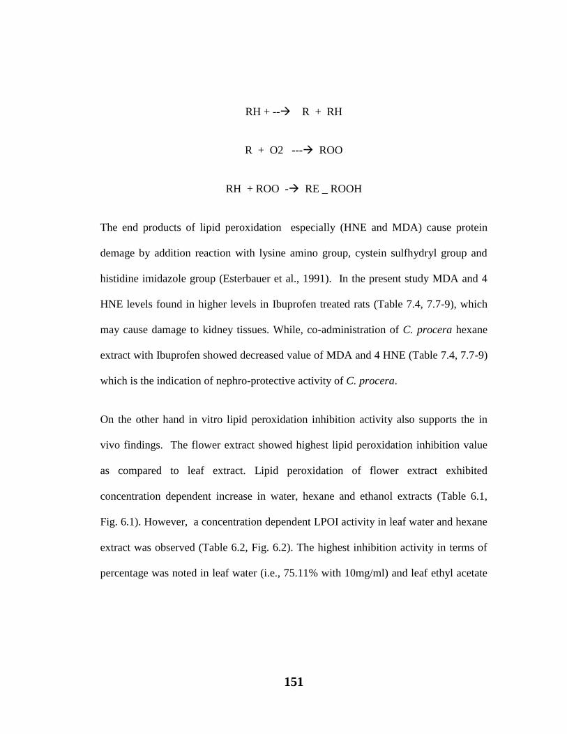
151
RH + -- R + RH
R + O2 --- ROO
RH + ROO - RE _ ROOH
The end products of lipid peroxidation especially (HNE and MDA) cause protein
demage by addition reaction with lysine amino group, cystein sulfhydryl group and
histidine imidazole group (Esterbauer et al., 1991). In the present study MDA and 4
HNE levels found in higher levels in Ibuprofen treated rats (Table 7.4, 7.7-9), which
may cause damage to kidney tissues. While, co-administration of C. procera hexane
extract with Ibuprofen showed decreased value of MDA and 4 HNE (Table 7.4, 7.7-9)
which is the indication of nephro-protective activity of C. procera.
On the other hand in vitro lipid peroxidation inhibition activity also supports the in
vivo findings. The flower extract showed highest lipid peroxidation inhibition value
as compared to leaf extract. Lipid peroxidation of flower extract exhibited
concentration dependent increase in water, hexane and ethanol extracts (Table 6.1,
Fig. 6.1). However, a concentration dependent LPOI activity in leaf water and hexane
extract was observed (Table 6.2, Fig. 6.2). The highest inhibition activity in terms of
percentage was noted in leaf water (i.e., 75.11% with 10mg/ml) and leaf ethyl acetate

152
extract showed (75.11%, with 8mg/ml) as compared to ethanol and hexane. Similarly
BHA exhibited 85% inhibition activity as compared to ascorbic acid (75%) lipid
peroxidation inhibition activity. The results revealed that the test extracts have strong
potential to inhibit lipid peroxidation in vitro as well.
1,1-Diphenyl-2-picryl-hydrazyl (DPPH) radical scavenging is a more sophisticated
method for in vitro determination of antioxidant ability. DPPH is a nitrogen
containing free radical which is largely being used to demonstrate antioxidant property
of any substance. It is also known that DPPH reacts quickly with substances which
have phenol group in their structure. It is a dark purple coloured solution which
contain an odd / unpaired electron which is responsible for transfer of electron from
antioxidant to DPPH radical, the colour of DPPH solution converts from purple to
yellow as the radical is scavenged by the antioxidant, due to this property DPPH is
being used for spectrophotometric analysis (Usmani, 2013; Kumar et al., 2013).
In the present study, flower extract showed highest scavenging activity as compared
to leaf extract (Table 6.3-4, Fig. 6.3-4). The highest activity is found in ethanol extract
(88.19%). While, leaf extract with water exhibited highest activity (75.95% with
2mg/ml). Whereas, BHA (85%) and ascorbic acid (75.5%) were used as standards
Table 6.3-4, Fig. 6.3-4).
Thus, C. procera found more effective as compared to standard DPPH values which
indicates that C. procera is more potent antioxidant for DPPH radical scavenging.
Besides this, in vitro antioxidant activity was also determined by the method of
reducing power capacity. The main principle of reducing power assay is that

153
compounds which have reduction potential may react with potassium ferricyanide
(Fe+3
) to form potassium ferrocyanide (Fe+2
) which then react with ferric chloride to
form ferrous complex. The colour intensity of the complex may be determined in
terms of absorbance at 532 nm. Higher absorbance indicates higher reduction
potential. The reducing ability (absorbance) of a given sample determines its
antioxidant potential with the ability to donate hydrogen atom to free radical chain
(Kumar et al., 2013; Patel et al., 2014).
In the present study different solvent extracts of C. procera flower and leaf were
analyzed for reducing power capacity. In general, flower shows highest reducing
power as compared to leaf extracts. However, C. procera flower and leaf water
extracts showed concentration dependent reducing power capacity (Table 6.5-6, Fig.
6.5-6).
Due to the global appearance of increasing drug-resistant cases there is a need to
search sources for production of natural antimicrobial compounds which may be
effective to destroy these drug resistant pathogens. During last few decades a lot of
natural antimicrobial compounds were discovered for the control of severe microbial
infections. Presently, the antimicrobial potential of the various fractions of C. procera
flower and leaf extracts against four human pathogenic strains viz., Salmonella typhi,
Escherecia coli, mithicillin resistant Staphylococcus aureus and Micrococcus luteus is
investigated. These pathogens are the major cause of infectious diseases such as M.
luteus is an opportunistic pathogen and cause infection in immune-compromised
individuals (Seifert et al., 1995). Similarly, E. coli is a toxin producing human
154
pathogen and cause severe diarrhoea leads to kidney failure through food. Whereas,
MRSA involved in various hospital acquired infections and found to be resistant to all
β-lactum antibiotics (Iqbal, 1998; Iqbal et al., 2005; Que and Moreillon, 2010).
However, S. typhi. mainly cause inflammatory bowel syndrome and typhoid fever.
Different soluble flower and leaf extracts of C. procera exhibited differential spectrum
of inhibition against all tested pathogenic strains. Similar to the previous findings of
Joshi and Kaur (2013), the hexane fractions of flower and leaf have been proved very
significant as an antimicrobial agent against all of the studied pathogens, as maximum
zone of inhibition was observed against M. luteus flower (22mm) and leaf ( 23mm).
While on the other hand, these findings are in contrast to the previous findings of
Parabia et al. (2008) and Doshi et al. (2011) where (7-9mm) maximum zone of
inhibition was observed.
Similarly, fractions of flower and leaf ethyl acetate showed significant inhibitory
activity against E. coli and MRSA (Table 4.1-2, Fig. 4.1-4). While, no inhibitory
activity was recorded against S. typhi. and only flower ethyl acetate fraction was found
to be significant inhibitor against M. luteus. Present inhibitory findings could be well
supported by the studies of Patil and Saini (2012) where they also found significant
role of ethyl acetate extract of C. procera against various pathogens. However,
aqueous leaf and flower extracts showed significant inhibitory activity only against M.
luteus. Whereas, butanol leaf extract showed insignificant inhibitory activities against
all of the studied pathogenic strains and flower butanol extract was found significant
only against M. luteus. Thus, amongst all of the tested extracts of C. procera flower
and leaf hexane, ethyl acetate, aqueous and butanol fractions were found significant
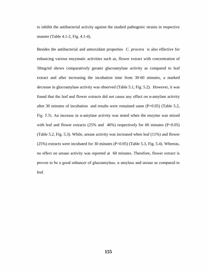
155
to inhibit the antibacterial activity against the studied pathogenic strains in respective
manner (Table 4.1-2, Fig. 4.1-4).
Besides the antibacterial and antioxidant properties C. procera is also effective for
enhancing various enzymatic activities such as, flower extract with concentration of
50mg/ml shows comparatively greater glucoamylase activity as compared to leaf
extract and after increasing the incubation time from 30-60 minutes, a marked
decrease in glucoamylase activity was observed (Table 5.1, Fig. 5.2). However, it was
found that the leaf and flower extracts did not cause any effect on α-amylase activity
after 30 minutes of incubation and results were remained same (P>0.05) (Table 5.2,
Fig. 5.3). An increase in α-amylase activity was noted when the enzyme was mixed
with leaf and flower extracts (25% and 46%) respectively for 60 minutes (P<0.05)
(Table 5.2, Fig. 5.3). While, urease activity was increased when leaf (11%) and flower
(25%) extracts were incubated for 30 minutes (P<0.05) (Table 5.3, Fig. 5.4). Whereas,
no effect on urease activity was reported at 60 minutes. Therefore, flower extract is
proven to be a good enhancer of glucoamylase, α amylase and urease as compared to
leaf.
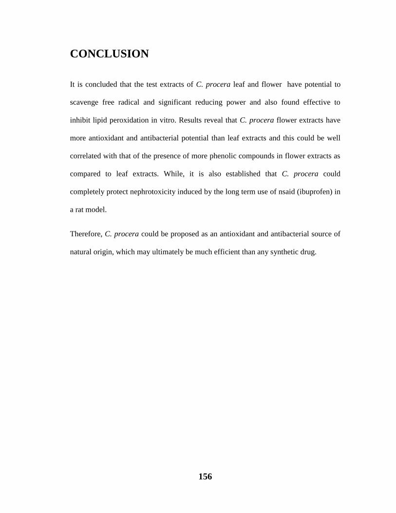
156
CONCLUSION
It is concluded that the test extracts of C. procera leaf and flower have potential to
scavenge free radical and significant reducing power and also found effective to
inhibit lipid peroxidation in vitro. Results reveal that C. procera flower extracts have
more antioxidant and antibacterial potential than leaf extracts and this could be well
correlated with that of the presence of more phenolic compounds in flower extracts as
compared to leaf extracts. While, it is also established that C. procera could
completely protect nephrotoxicity induced by the long term use of nsaid (ibuprofen) in
a rat model.
Therefore, C. procera could be proposed as an antioxidant and antibacterial source of
natural origin, which may ultimately be much efficient than any synthetic drug.

157
REFERENCES
1. Agharkar, S.P. (1991). Medicinal plants of Bombay Presidency. Scientific
Publications, India. pp.48-49.
2. Ahmad, N., Anwar, F., Hameed, S. and Boyce, M.C. (2011). Antioxidant
and antimicrobial attributes of different solvent extracts from leaves and
flowers of aak Calotropis procera. J. Med. Plant. Res., 5(19):4879-4887.
3. Ahmed, M., Khan, R. A., Shahzaib, S., Khan, A., Zaif, A. W., and Ahmed,
W. (2014). Antifungal, antioxidant and antibacterial activities of Calotropis
procera. Int. J. Biosci., 5(6):75-80.
4. Ajith, T.A. (2010). Ameliorating reactive oxygen species-induced invitro lipid
peroxidation in brain, liver, mitochondria and DNA damaged by Zingiber
officinalis Ind. J. Clin. Biochem. 25(1): 67-73.
5. Akhtar, N., Malik, A., Alia, S.N. and Kazmi, S.U. (1992). Proceragenin, an
antibacterial cardenolide from Calotropis procera, Phytochem , 31(8):2821–
2824.
6. Alam, P. and Ali, M. (2009). Phytochemical investigation of Calotropis
procera Ait roots. Ind. J. Chem., 48(3):443-446.
7. Alencar, N.M., Figueiredo, I.S., Vale, M.R., Bitencourt, F.S., Oliveira, J.S.,
Ribeiro, R.A. (2004). Anti-inflammatory effect of the latex from Calotropis
procera in three different experimental models peritonitis, paw, edema and
hemorrhagic cystitis. Pl. Med., 70:1144.
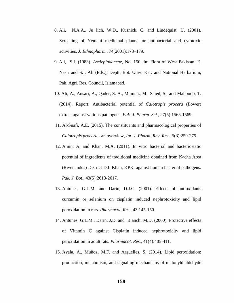
158
8. Ali, N.A.A., Ju lich, W.D., Kusnick, C. and Lindequist, U. (2001).
Screening of Yemeni medicinal plants for antibacterial and cytotoxic
activities, J. Ethnopharm., 74(2001):173–179.
9. Ali, S.I. (1983). Asclepiadaceae, No. 150. In: Flora of West Pakistan. E.
Nasir and S.I. Ali (Eds.), Deptt. Bot. Univ. Kar. and National Herbarium,
Pak. Agri. Res. Council, Islamabad.
10. Ali, A., Ansari, A., Qader, S. A., Mumtaz, M., Saied, S., and Mahboob, T.
(2014). Report: Antibacterial potential of Calotropis procera (flower)
extract against various pathogens. Pak. J. Pharm. Sci., 27(5):1565-1569.
11. Al-Snafi, A.E. (2015). The constituents and pharmacological properties of
Calotropis procera - an overview, Int. J. Pharm. Rev. Res., 5(3):259-275.
12. Amin, A. and Khan, M.A. (2011). In vitro bacterial and bacteriostatic
potential of ingredients of traditional medicine obtained from Kacha Area
(River Indus) District D.I. Khan, KPK, against human bacterial pathogens.
Pak. J. Bot., 43(5):2613-2617.
13. Antunes, G.L.M. and Darin, D.J.C. (2001). Effects of antioxidants
curcumin or selenium on cisplatin induced nephrotoxicity and lipid
peroxidation in rats. Pharmacol. Res., 43:145-150.
14. Antunes, G.L.M., Darin, J.D. and Bianchi M.D. (2000). Protective effects
of Vitamin C against Cisplatin induced nephrotoxicity and lipid
peroxidation in adult rats. Pharmacol. Res., 41(4):405-411.
15. Ayala, A., Muñoz, M.F. and Argüelles, S. (2014). Lipid peroxidation:
production, metabolism, and signaling mechanisms of malonyldialdehyde
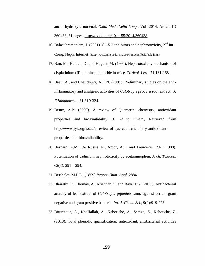
159
and 4-hydroxy-2-nonenal. Oxid. Med. Cellu Long., Vol. 2014, Article ID
360438, 31 pages. http://dx.doi.org/10.1155/2014/360438
16. Balasubramaniam, J. (2001). COX 2 inhibitors and nephrotoxicity, 2nd
Int.
Cong. Neph. Internet. http://www.uninet.edu/cin2001/html/conf/bala/bala.html)
17. Ban, M., Hettich, D. and Huguet, M. (1994). Nephrotoxicity mechanism of
cisplatinium (II) diamine dichloride in mice. Toxicol. Lett., 71:161-168.
18. Basu, A., and Chaudhury, A.K.N. (1991). Preliminary studies on the anti-
inflammatory and analgesic activities of Calotropis procera root extract. J.
Ethnopharma., 31:319-324.
19. Bentz, A.B. (2009). A review of Quercetin: chemistry, antioxidant
properties and bioavailability. J. Young Invest., Retrieved from
http://www.jyi.org/issue/a-review-of-quercetin-chemistry-antioxidant-
properties-and-bioavailability/.
20. Bernard, A.M., De Russis, R., Amor, A.O. and Lauwerys, R.R. (1988).
Potentiation of cadmium nephrotoxicity by acetaminophen. Arch. Toxicol.,
62(4): 291 – 294.
21. Berthelot, M.P.E., (1859) Report Chim. Appl. 2884.
22. Bharathi, P., Thomas, A., Krishnan, S. and Ravi, T.K. (2011). Antibacterial
activity of leaf extract of Calotropis gigantea Linn. against certain gram
negative and gram positive bacteria. Int. J. Chem. Sci., 9(2):919-923.
23. Bouratoua, A., Khalfallah, A., Kabouche, A., Semza, Z., Kabouche, Z.
(2013). Total phenolic quantification, antioxidant, antibacterial activities

160
and flavonoids of Algerian Calotropis procera (Asclepiadaceae). Der
Pharm. Lett., 5 (4):204-207.
24. Boya, P., Pena, A., Beloqui, O., Larrea, E., Conchillo, M. and Castleruiz,
Y. (1999). Antioxidant status and glutathione metabolism in peripheral
blood mononuclear cells from patients with chronic hepatitis, C. J.
Hepatol., 31:808-814.
25. Bradford, M. M. (1976). A rapid and sensitive method for the quantization
of microgram quantities of protein utilizing the principle of protein-dye
binding. Analy. Biochem., 72(1):248-254.
26. Brayer, G.D., Yaoguang, L. and Wither, S.G. (1995). The structure of
human pancreatic α-amylase at 1.8 A resolution and comparisons with
related enzymes. Prot. Sci., 4:1730-1742.
27. Butler, A.R., Hussain, I. and Leitch, E. (1981). The chemistry of the
diacetyl monoxime assay of urea in biological fluids. Clin. Chim. Acta.,
112:357-360.
28. Calderon-Montaño, J.M., Burgos-Moron, E., Perez-Guerrero, C., Lopez-
Lazaro, M. (2011). A review on the dietary flavonoid kaempferol. Mini.
Rev. Med. Chem., 11(4): 298–344. doi:10.2174/138955711795305335. PMID 21428901
29. Carlberg, I. and Mannervik, B. (1985). Glutathione reductase. Methods
Enzymol., 113:484-490.
30. Chavda, R., Vadalia, K.R., and Gokani, R. (2010). Hepatoprotective and
antioxidant activity of root bark of Calotropis procera R. Br.
(Asclepediaceae). Int. J. Pharmacol., 6(6):937-943.

161
31. Chen, C.Y., Pang, V.F., and Chen, C.S. (1994). Assessment of Ibuprofen-
associated nephrotoxicity in renal dysfunction. J. Pharmacol. Exp. Ther.,
270:1307-1312.
32. Chiranjeevi, T., Rao, K., Srinija, K., Rao, P.R., Sajjad, S.K., Lavanya, P.,
Kumar, A.R., Sathyanathan, V., (2013). Phytochemical Evaluation of
Calotropis procera, Gymnema sylvestre, Hemidesmus indicus. Int. Res. J.
Pharm. App Sci., 3(6):31-34.
33. Choudhary, N.K., Jha, A.K., Sharma, S., Goyal, S. and Dwivedi, J. (2011).
Antidiabetic potential of chloroform extract of flowers of Calotropis
gigantea: An in vitro and in vivo study. Int. J. Green Pharm., 5:296-301.
34. Clark, R. A. F. (2008). Oxidative stress and “senescent” fibroblasts in non-
healing wounds as potential therapeutic targets, J. Invest. Dermat., 128:
2361–2364. doi:10.1038/jid.2008.257
http://www.nature.com/jid/journal/v128/n10/fig_tab/jid2008257f1.html#figure-title.
35. Custodio, J.B., Cardoso, C.M., Santos, M.S., Almeida, L.M., Vicente, J.A.,
Fernances M.A. (2009). Cisplatin impairs rat liver mitochondrial functions
by inducing changes on membrane ion permeability: prevention by thiol
group protecting agent. Toxicol., 259(1-2):18-24.
36. Dalzeil, J.M. (1937). The useful plants of west tropical Africa: Crown
agent for the colonies, London. p.101-110.
37. David, M., Bharat, K.R. and Bhavani, M. (2011). Study of Calotropis
gigantea R. Br. extracts on growth and survival dynamics of selected
pathogenic microorganisms. Int. J. Biol. Engg., 1(1):1-5.
162
38. Davies, K.J.A. (1991). Oxidative Damage and Repair: Chemical,
Biological and Medical Aspects, Oxford: Pergamon Press.
39. De Crespigny, P.C., Hewitson, T., Birchall, I., Smith, P.K. (1990).
Caffeine potentiates the nephrotoxicity of mefenamic acid on the Rat Renal
Papilla, A.J. Nephrol., 10 (4):311 – 315.
40. Derle, D.V., Gugar, K.N., and Sagar, B.S.H. (2006). Adverse effects
associated with use of non-steroidal anti-inflammatory drugs: An overview.
J. Pharm. Ind. Sci., 68(4): 409 – 414.
41. Devi, S.K.M., Annaporani, S. and Murugesan, S. (2008). Antifungal
activity analysis of Calotropis procera. Madras Agric. J., 95(7-12):386-
389.
42. Dewan, S., Kumar, S., Kumar, V.L. (2000). Antipyretic effect of latex of
Calotropis procera. Ind. J. Pharmacol., 32:252.
43. Dianzani, M. and Barera, G. (2008). Pathology and physiology of lipid
peroxidation and its carbonyl products. In: Alverez, S., Evelson, P. (ed.),
Free radical Pathophysiology, pp. 19-38, Transworld Research Network:
Kerala, India, ISBN: 978-81-7895-311-3.
44. Doshi, H., Satodiya, H., Thakur, M.C. Parabia, F. and Khan, A. (2011).
Phytochemical screening and biological activity of Calotropis
Procera (Ait). R.Br. (Asclepiadaceae) against selected bacteria and
Anopheles stephansi Larvae, Int. J. Pl. Res., 1(1):29–33.
45. El-Fallal, A., Dobara, M.A., El-Sayed, A. and Omar, N. (2012). Starch and
Microbial α-Amylases: From Concepts to Biotechnological Applications,
163
Carbohydrates - Comprehensive Studies on Glycobiology and
Glycotechnology, Prof. Chuan-Fa Chang (Ed.), ISBN: 978-953-51-0864-1,
InTech, DOI: 10.5772/51571. Available from:
http://www.intechopen.com/books/carbohydrates-comprehensive-studies-
on-glycobiology-and-glycotechnology/starch-and-microbial-amylases-
from-concepts-to-biotechnological-applications.
46. Esterbauer, H., Schaur, J. and Zollner, H. (1991). Chemistry and
biochemistry of 4-hydroxynonenal, malondialdehyde and related
aldehydes. Free Rad. Bio. Med., 11:81–128.
47. Fackovcova, D., Kristova, V. and Kriska, M. (2000). Renal damage
induced by the treatment with non-opioid analgesics – theoretical
assumption or clinical significance. Bratisl. Lek Listy., 101(8):417 – 422.
48. Farag, M.M., Mikhail, M., Shehata, R., Abdel–Meguid, E. and Abdel–
Tawab S. (1996). Assessment of gentamicin induced nephrotoxicity in rats
treated with low doses of ibuprofen and diclofenac sodium. Clini. Sci.
(Lord), 91(2):187 – 91.
49. Fawcett, J.K. and Scott, J.E. (1960). A rapid and precise method for the
determination of urea. J. Chim. Pathol., 13: 156.
50. Fowler, B. A. (1992). Mechanisms of kidney cell injury from Metals,
Environ. Health Perspec., 100:57 – 63.
51. Frayn, K.N. (1998). Regulation of fatty acid delivery in vivo, Advan.
Experi. Med. Bio., 441:171–179.

164
52. Fruhbeck, G., Gomez-Ambrosi, J., Muruz´abal, F.J. and M. A. Burrell.
(2001).The adipocyte: a model for integration of endocrine and metabolic
signaling in energy metabolism regulation, The Am. J. Physio. Endocr.
Metabol., 280(6):E827–E847.
53. Gajare, S. M., Patil, M. V., and Mahajan, R. T. (2012). Phytochemical
screening and antimicrobial activity of ethanol extract of Calotropis
procera root. Int. J. Res. Phytochem. Pharmacol., 2(3):143-146.
54. Gao, X., Bjo, K.L., Trajkovski, V., and Uggla, M. (2000). Evaluation of
antioxidant activities of rosehip ethanol extract in different test systems, J.
Agri. Food Chem., 80:2021-2027.
http://dx.doi.org/10.1002/10970010(200011)80:14<2021::aid-
jsfa745>3.0.co;2-2
55. Ghani, M., Aman, A., Rehman, H., Siddiqui, N.N., and Qader, S.A.
(2013). Strain improvement by mutation for enhanced production of starch-
saccharifying glucoamylase from Bacillus licheniformis. Starch, 65: 875-
884.
56. Gholamshahi, S., Vakili, M. A., Shahdadi, F., and Salehi, A. (2014).
Comparison of total phenols and antiradical activity of flower, leaf, fruit
and latex extracts of milkweed (Calotropis procera) from Jiroft and Bam
cities. Int. J. Biosci., 4(7):159-164.
57. Goyal, M., Mathur, R. (2011). Antimicrobial potential and pytochemical
analysis of plant extract of Calotropis procera. Int. J. Drug Discov.
Herbal Res., 1(3):138-143.

165
58. Gupta, R., Gigras, P., Mohapatra, H., Goswami, V.K. and Chayban, B.
(2003). Microbial α-amylase: a biotechnological perspective. Pro.
Biochem., 38:1599-1616.
59. Halliwell, B. and Aruoma, O.I. (1993). DNA and Free Radicals, pp.315-
327, Ellis Horwood, Chichester.
60. Halliwell, B. and Gutteridge, J.N.A. (1999). Mechanism of damage of
cellular targets by oxidative stress: Lipid peroxidation, in Free Radicals in
Biology and Medicine, B. Halliwell and J.M.C Gutteridge, Ed., pp.284-
313, Oxford University Press, Oxford, UK.
61. Hanachi, P., Othman, F. and Motelleb, G. Effect of Berberis
vulgaris aqueous extract on the apoptosis, sodium and potsium in
hepatocarcinogenic rats. Iran. J. Bas. Med. Sci., 11(2): 62-69.
62. Harborne, J.B. (1984). Phytochemical Methods, 2nd
Edition. Chapman and
Hall, London, pp.288.
63. Hassan, S.W., Bilbis, F.L., Ladan, M.J., Umar, R.A., Dangoggo, S.M.,
Saidu, Y., Abubakar K., and Faruk, U.Z. (2006). Evaluation of antifungal
activity and phytochemical analysis of leaves, roots and stem barks extracts
of Calotropis procera (Asclepiadaceae). Pak. J. Bio. Sci., 9(14):2624-
2629.
64. Hemalatha, M., Arirudran, B., Thenmozhi, A. and Mahadeva Rao, U.S.
(2011). Antimicrobial effect of separate extract of acetone, ethylacetate,
methanol and aqueous from leaf of milkweed (Calotropis gigantea L.).
Asian J. Pharm. Res., 1(4):102-107.
166
65. Hesham, R., Seedi, E.L. and Nishiyama, S. (2002). Chemistry of
bioflavonoids. Ind. J. Pharm. Educ., 36:191-194.
66. Hickey, E.J., Raje, R.R., Reid, V.E., Gross, S.M. and Ray, S.D. (2001).
Diclofenac induced in vivo nephrotoxicity may involve oxidative stress
mediated massive genomic DNA fragmentation and apoptotic cell death.
Free Rad. Bio. Med., 31(2):139 – 152.
67. Hoitsma, A.J., Wetzels, J.F.M. and Koene, R.A.P. (1991). Drug Induced
Nephrotoxicity. Dr. Saf., 6(2):131-147.
68. Ikken, Y., Morales, P., Martinez, A., Martin, M.L., Haza, A.L. and
Cambero, M.I. (1999). Antimutagenic effect of fruit and vegetable
ethanolic extract against N-nitrosamine evaluated by the Ames test. J.
Agri. Food Chem., 47:3257-3264.
69. Iqbal, A. (1998). Production, purification and characterization of
bacteriocins from indigenous clinical staphylococci. Ph.D. Thesis,
University of Karachi, Pakistan. Research Repository, Higher Education
Commission, ID code 1112.
70. Iqbal, Z., Lateef, M.A., Muhammad G. and Khan, M.N. (2005). Anti-
helmintic activity of Calotropis procera Ait. flowers in sheep. J.
Ethnopharma., 102(2):256-261.
71. Ishnava, K.B., Chauhan, J.B., Garg, A.A. and Thakkar, A.M. (2012).
Antibacterial and phytochemical studies on Calotropis gigantia (L.) R. Br.
Latex against selected cariogenic bacteria, Saudi J. Bio. Sci., 19:87–91.

167
72. Jaiswal, J., Bhardwaj, H., Srivastava, S., Gautam, H., Sharma, S., Rao, Ch.
(2014). Anti-diabetic activity of methanolic extract of Calotropis gigantea
seeds on STZ induced diabetic rats. Int. J. Pharm. Pharm. Sci. 6(1):254-
257.
73. Javadian, F., Sahraei, S. and Azizi, A. (2014). Evaluation of the effect of
antimicrobial activity of ethanol extract of Calotropis procera in Extended
Spectrum Beta- Lactamase Producing E. coli, Int. J. Adv. Bio. Biomed.
Res., 2(3):764-768.
74. Javed, S., Khan, J.A., Khaliq, T., Javed, I. and Abbas, R.Z. (2015).
Experimental evaluation of nephroprotective potential of Calotropis
procera (Ait) flowers against gentamicin-induced toxicity in albino rabbits.
Pak. Vet. J., 35(2):222-226.
75. Jayaprakasha, G.K., Girennavar, B. and Patil B.S. (2008). Radical
scavenging activities of Rio Red grapefruits and sour orange fruit extracts
in different in vitro model systems, Biores. Techno., 99(10):4484–4494.
76. Jhonson, D.B., Shringi, B.N., Patida, B.K., Chalichem, N.S.S. and
Javvadi, A.K. (2011). Screening of antimicrobial activity of alcoholic and
aqueous extract of some indigenous plants. Indo. Global J. Pharm. Sci.,
1(2):186-193.
77. Joshi, M. and Kaur, S. (2013). In vitro evaluation of antimicrobial activity
and phytochemical analysis of Calotropis procera, Eichhornia crassipes
and Datura innoxia leaves. Asian J. Phar. Cli. Res., 6(5):25-28.

168
78. Jucá, T. L., Ramos, M. V., Moreno, F. B. M. B., Viana de Matos, M. P.,
Marinho-Filho, J. D. B., Moreira, R. A. and Monteiro-Moreira, A. C. D. O.
(2013). Insights on the Phytochemical Profile (Cyclopeptides) and
Biological Activities of Calotropis procera Latex Organic Fractions. The
Sci. World J., Article ID 61545:1-9. http://dx.doi.org/10.1155/2013/615454
79. Kamboj, V.P. (2000). Herbal medicine, Curr. Sci., 78:35-39.
80. Kaneko, J.J., Harvey, J.M. and Bruss, M.L. (2008). Clinical Biochemistry
of Domestic Animals. 6th (Ed.). pp,916, Academic Press, San Diego, USA.
81. Kareem, S.O., Akpan, I. and Ojo, O.P. (2008). Antimicrobial activities of
Calotropis procera on selected pathogenic microorganisms. Afr. J. Biomed.
Res., 11:105-110.
82. Kawo, A.H., Mustapha, A., Abdullahi, B.A., Rogo, L.D., Gaiya, Z.A. and
Kumurya, A.S. (2009). Phytochemical properties and antibacterial
activities of the leaf and latex extracts of Calotropis procera. Bayero J.
Pur. App. Sci., 2(1):34-40.
83. Kazemipour, N., Nikbin, M., Valizadeh, J., Ghadera, F. and
Sepehrimanesh, M. (2014). Antimicrobial and chemical properties of
Calotropis procera extracts. Onl. J. Vet. Res., 18(11):869-874.
84. Kew, F. (1985). The useful plants of west tropical Africa Vol.1. Families
A-D Edition 2 (ed Burkill, H.M). Royal Botanical Gardens. 219-222.
85. Kim, H., Xu, M. and Lin, Y. (1999). Renal dysfunction associated with the
perioperative use of diclofenac. Anesth. Analg., 89:999 – 1005.
169
86. Kinter, M. and Roberts, R.J. (1996). Glutathione comsumption and
glutathione peroxidase inactivation in fibroblast cell lines by 4 hydroxyl-2-
nonenal. Free Rad. Bio. Med., 21:457-462.
87. Kocaoglu, S., Karan, A., Berkan, T., Basdemir, G. and Akpinar, R. (1997).
Urinary gamma-glutamyl transferase activity in rats with non-steroidal
anti-inflammatory drug induced nephrotixicity. Arch. Immunol. Ther. Exp.
(Warsz), 45(1):73 – 7.
88. Kono, Y. (1978). Generation of superoxide radical during autooxidation of
hydroxylamine and an assay for super oxide dismutase. Arch. Biochem.
Biophys., 189-195.
89. Krishnaveni, M., Durairaj, S., Madhiyan, P., Amsavalli, L. and
Chandrasekar R. (2013). In vitro free radical scavenging activity of
aqueous leaf extract of Plants near thermal power plant, mettur, salem, Int.
J. Pharm. Sci. Res., 4(9):3659-3662.
90. Kristova, P. and Tissot, M. (1995). Sod-Anthroquinone pulping of
Hibiscus sabdariffa (Karkadeh) and Calotropis procera from Sudan.
Biores. Technol., 53:677-82.
91. Kumar, G., Karthik, L., Venkata, B.R.K., Kirthi, A.V., Jayaseelam, G.,
Rahuman, A., (2012). Phytochemical composition, mosquito larvicidal,
ovicidal and repellent activity of Calotropis procera against Culex
tritaeniorhynchus and Culex gelidus. Bangla. J. Pharma., 7: 63–69.

170
92. Kumar, S., Gupta, A., and Pandey, A. K. (2013). Calotropis procera root
extract has the capability to combat free radical mediated damage. ISRN
Pharma., Article ID 691372, 8 pages.
93. Langseth, L. (1995). Oxidants, antioxidants and disease prevention,
International Life Science Institute, Belgium.
94. Larhsini, M., Oumoulid, L., Lazrek, H.B., Wataleb, S., Bousaid, M.,
Bekkouche, K., Markouk, M. and Jana, M. (1999). Screening of
antibacterial and antiparasitic activities of six Moroccan medicinal plants,
Therapie, 54:763-765.
95. Leena, P. and Alaraman, B.R. (2005). Effect of gree tea extract on cisplatin
induced oxidative damage on kidney and testes of rats. Ars. Pharm., 46:5-
18.
96. Lim, J.H., Park, H.S., Choi, J.K., Lee I.S., Choi, H.J. (2007). Isoorientin
induces Nrf2 pathway-driven antioxidant response through
phosphatidylinositol 3-kinase signaling. Arch. Pharm. Res. 30(12):1590-8.
97. Lutskii, V.I., Gromova, A.S. and Tyukavkina, N.A. (1971).
Aromadendrin, apigenin, and kaempferol from the wood of Pinus
sibirica. Chem. Nat. Comp., 7(2):197. doi:10.1007/BF00568701
98. Mabberley, D.J. (2008). Mabberley’s Plant-Book: a portable dictionary of
plants, their classification and uses. Third edition. Cambridge University
Press. pp. xviii + 1021.
99. Mahalakshmi, R., Rajesh, P., Ramesh, N., Balasubramanian, V. and Rajesh
Kannan V. (2010). Hepatoprotective activity on Vitex negundo Linn.
171
(Verbenaceae) by using wistar albino rats in Ibuprofen induced model. Int.
J. Pharmacol., 6(5):658-663.
100. Mainasara, M. M., Aliero, B. L., Aliero, A. A., and Dahiru, S. S. (2011).
Phytochemical and antibacterial properties of Calotropis procera (Ait) R.
Br.(Sodom Apple) fruit and bark extracts. Int. J. Mod. Bot., 1(1):8-11.
101. Majewska, M., Skrzycki, M., Podsiad, M. and Czeczot H. (2011).
Evaluation of antioxidant potential of flavonoids: an in vitro study. Act.
Polo. Pharma. Dr. Res., 68(4):611-615.
102. Mako, G.A., Memon, A.H. and Mughal, U.R. (2012). Antibacterial
effects of leaves and root extract of Calotropis procera Linn. Pak. J. Agri.
Agri. Engg. Vet. Sci., 28 (2): 141-149.
103. Mascolo, N., Sharma, R., Jain, S.C. and Capasso, F. (1988).
Ethnopharmacology of Calotropis procera flowers. J. Ethnopharmacol.,
22(2):211-21.
104. Meena, A.K., Yadav, A. and Rao, M.M. (2011). Ayurvedic uses and
pharmacological activities of Calotropis procera Linn., Asi. J. Tradi.
Med., 6(2): 45-50.
105. Miller, G.L. (1959). Use of dinitrosalisylic acid reagent for determination
of reducing sugars. Analy. Chem., 31(3): 426-428.
106. Mishra, A., Sharma, A.K., Kumar, S., Saxena, A. K. and Pandey, A.K.
(2013). Bauhinia variegata leaf extracts exhibit considerable antibacterial,
antioxidant and anticancer activities, Bio. Med. Res. Int., Article ID
915436, 10 pages.
172
107. Mohanraj, R., Rakshit, J. and Nobre, M. (2010). Anti HIV – I and
antimicrobial activity of the leaf extract of Calotropis procera. Int. J.
Green Pharm., 4:242-6.
108. Morelle, J., Labriola, L., Lambert, M., Cosyns, J.P., Jouret, F. and Jadoul, M.
(2009). Tenofovir related acute kidney injury and proximal tubule dysfunction
precipitated by diclofenac a case of drug–drug interaction. Clin. Nephrol.,
71(5):567 – 70.
109. Motalleb, G., Hanachi, P., Kua, S.H., Othman, F. and Asmah R. (2005).
Evaluation of phenolic content and total antioxidant activity in Berberis vulgaris
fruit extract. J. Biol. Sci., 5(5):648-653.
110. Moteriya, P., Rinkal, S., and Chanda, S. (2015). Screening of
phytochemical constituents in some ornamental flowers of Saurashtra
region. J. Pharmacog. Phytochem., 3(5):112-120.
111. Mounnisamy, V.M., Kavimani, S. and Gunasegaran, R. (2002).
Antibacterial activity of gossypetin isolated from Hibiscus sabdariffa The
Antisep., 99(3):81-2. http://medind.nic.in/imvw/imvw289.html
112. Moustafa, A. M. Y., Ahmed, S. H., Nabil, Z. I., Hussein, A. A. and
Omran, M. A. (2010). Extraction and phytochemical investigation of
Calotropis procera: effect of plant extracts on the activity of diverse
muscles. Pharma. Bio., 48(10), 1080-1190.
113. Murray, M.D. and Brater, D.C. (1993). Renal toxicity of the non-steroidal
anti-inflammatory drugs: Ann. Rev. Pharmacol. Toxicol., 32:435 – 65.

173
114. Murti, Y., Singh, A. and Pathak, D. (2013). In-vitro anthelmintic &
cytotoxic potential of different extracts of Calotropis procera leaves. Asi.
J. Pharma. Cli. Res., 6(1):14-16.
115. Muzammal, M. (2014). Study on antibacterial activity of Calotropis
procera. PeerJ
PrePrints, 2:e430v1https://dx.doi.org/10.7287/peerj.preprints.430v1
116. Neenah, E.G. and Ahmed, M.E. (2011). Antimicrobial activity of extracts
and latex of Calotropis procera and synergistic effect with reference to
antimicrobials. Res. J. Med. Plants, 5(6):706-716.
117. Nenaah, G. (2013). Antimicrobial activity of Calotropis procera
Ait.(Asclepiadaceae) and isolation of four flavonoid glycosides as the
active constituents. World J. Micro. Biotech., 29(7):1255-1262.
118. Nicholson, J.K., Timbrell, J.A. and Sadler, P.J. (1985). Proton NMR
spectra of urine as indicators of renal damage. Mercury induced
nephrotoxicity in rats. Mole. Pharma., 27(6):644 – 651.
119. Ohkawa, H., Ohishi, N. and Yagi, K. (1979). Assay for lipid peroxide in
animal tissues by thyobarbituric acid reaction. Anal. Biochem., 95:351-
358.
120. Oloumi, H. (2014). Phytochemistry and ethno-pharmaceutics of
Calotropis procera, Ethno-Pharma. Prod., 1(2):1-8.
121. Ong, W.Y., Lu, X.R., Hu, C.Y. and Halliwell, B. (2000). Distribution of
hydroxynonenal-modified proteins in the kainate-lesioned rat hippocampus:
evidence that hydroxynonenal formation precedes neuronal cell death. Free Rad.
Bio. Med., 28:1214–1221.

174
122. Oyaizu, M. (1986). Studies on products of browning reactions:
antioxidative activities of products of browning reaction prepared from
glucosamine. Jap. J. Nut., 44:307–315.
123. Ozen, S., Akyol, O., Iraz M., Sogut, S., Ozugurlu, F., Ozyurt, H., Odaci,
E., Yildrim, Z. (2004). Role of caffeic acid phenethyl ester, an active
component of propolis, against cisplatin induced nephrotoxicity in rats. J.
App. Toxicol., 24:27-35.
124. Pandey, A., Agrawal, S., Bhatia, A.K. and Saxena, A. (2015). In vitro
assesment of antibacterial activity of Calotropis procera and Coriandrum
sativum against various pathogens. Int. J. Pharm. Res. All. Sci., 4(1):33-
44.
125. Parabia, F.M, Kothari, I.L. and Parabia, M.H. (2008). Antibacterial
activity of solvent fractions of crude water decoction of apical twigs and
latex of Calotropis procera (Ait.) R. Br. Nat. Prod. Radia., 7:30–4.
126. Parihar, G., Sharma, A., Ghule, S., Sharma, P., Deshmukh, P. and
Srivasta, D.N. (2011). Anti-inflammatory effect of Calotropis procera
root bark extract. Asi. J. Pharm. Life Sci., 1(1):29-44.
127. Parlakpinar, H., Tasdemir, S., Polat, A., Bay-Karabulut, A., Vardi, N.,
Ucar, M. and Acet. A. ( 2005). Protective role of caffeic acid phenethyl
ester (cape) on gentamicin induced acute renal toxicity in rats. Toxicol.,
207: 169-177.
128. Patel, H. V., Patel, J. D., and Patel, B. (2014). Comparative efficacy of
phytochemical analysis and antioxidant activity of methanolic extract of

175
Calotropis gigantea and Calotropis procera. Int. J. Life Sci. Biotechnol.
Pharm. Res., 5(2):107-13.
129. Patel, V.R., Prakash, R.P. and Sushil, S.K. (2010). Antioxidant Activity
of Some Selected Medicinal Plants in Western Region of India. Adv. Biol.
Res., 4:23–26.
130. Patil, S.M. and Saini, R. (2012). Antimicrobial activity of flower extracts
of Calotropis gigentea. Int. J. Pharm. Phytopharmacol. Res., 1(4):142-
145.
131. Pohanka, M., Bandouchova, H., Sobotka, J., Sedlackova, J., Soukupova,
I. and Pikula, J. (2009). Ferric reducing antioxidant power and square
wave voltametry for assay of low molecular weight antioxidants in blood
plasma: performance and comparison of methods, Sens., 9:9094-9103.
doi:10.3390/s91109094
132. Pooja, M., Rinka, S. and Sumitra, C. (2014). Evaluation of antioxidant
potential and phenol and flavonoid content of some flower extracts of
Saurashtra region ., World J. Pharm. Sci., 2(7):622-634.
133. Prabha, M.R., and Vasantha, K. (2012). Phytochemical and antibacterial
activity of Calotropis procera (Ait.) R. Br. Flowers. Int. J. Pharma. Bio.
Sci., 3(1):1-6.
134. Proctor, P. H. and Reynolds, E. S. (1984). Free radicals and disease in
man, Physiol. Chem. Phys., 16:175.
176
135. Quazi, S., Mathur, K. and Arora S. (2013). Calotropis procera: An
overview of its phytochemistry and pharmacology, Ind. J. Drugs, 1(2):63-
69.
136. Que, Y.A. and Moreillon, P. (2010). Staphylococcus aureus (including
staphylococcal toxic shock). In: Mandell, G.L., Bennett, J.E., Dolin, R.,
eds. Principles and Practice of Infectious Diseases. 7th ed. Philadelphia, Pa:
Chap. 195, Elsevier Churchill Livingstone.
137. Qureshi, A. A., Prakash, T., Patil, T., Swamy, A. H. M. V., Gouda, A. V.,
Prabhu, K., and Setty, S. R. (2007). Hepatoprotective and antioxidant
activities of flowers of Calotropis procera (Ait) R. Br. in CCl4 induced
hepatic damage. Ind. J. Exp. Bio., 45(3):304-310.
138. Rahimi, M. (2015). Pharmacognostical Aspects and Pharmacological
activities of Calotropis procera. Bull. Env. Pharmacol. Life Sci., 4:156-
162.
139. Rajesh, K., Priyadharshni, S. P., Kumar, K. E., and Satyanarayana, T.
(2014). Phytochemical Investigation on Stem of Calotropis Procera (Ait.)
R. Br. (Asclepiadaceae). J. Pharm. Bio. Sci., 9(3):25-29.
140. Ranjit, P.M., Santhipriya, T., Nagasri, S., Chowdary, Y.A., Gopal,
P.N.V. (2012). Preliminary phytochemical screening and antibacterial
activities of Ethanolic extract of Calotropis procera flowers against
human pathogenic Strains. Asi. J. Pharm. Cli. Res., 5(3):127-131.
141. Reddy, V.C., Amulya, V., Lakshmi, C.H., Reddy, K., Praveen, D.B.,
Pratima, D., Thirupathi, A.T., Kumar, K.P. and Sengottuvelu, S. (2012).
177
Effect of Simvastatin in gentamicin induced nephrotoxicity in albino rats.
Asi. J. Pharm. Clin. Res., 5:36-40.
142. Repetto, M., Boveris, A. and Semprine, J. (2012). Lipid peroxidation:
chemical mechanism, biological implications and analytical
determination. INTECH Open Acc. Pub., Argentina
http://dx.doi.org/10.5772/45943
143. Ricardo, G., Cheyla, R., Aluet, B., Frank, H., Nelson, M., Zullyt, Z. and Enis, R.
(2005). Lipid peroxides and antioxidant enzymes in cisplatin-induced chronic
nephrotoxicity in rats. Mediators Inflamm, 3:139-143.
144. Robertson, C.E., Someren, V.V., Ford, M.J., Dlugolecka, M. and Prescott, L.F.
(1980). Mefenamic acid nephropathy, The Lancet, 316(8188):232 – 233.
145. Robertson, S. (2014). Ibuprofen. News Medical http://www.news-
medical.net/health/Ibuprofen-Mechanism.aspx retrieved on 29/8/2015.
146. Roy, S., Sehgal, S., Padhy, B.M., Kumar, V.L. (2005). Antioxidant and
protective effect of latex of Calotropis procera against alloxan induced
diabetes in rats. J. Ethnopharma., 102(3):470-473.
147. Saleemi, M.K., Zargham, M.Z., Javed, I. and Khan, A. (2009).
Pathological effects of gentamicin administered intramuscularly to day-
old broiler chicks. Exp. Toxicol Pathol., 61:425-432.
148. Salem, W.M., Sayed, W.F., Haridi, M., and Hassan, N.H. (2014).
Antibacterial activity of Calotropis procera and Ficus sycomorus extracts
on some pathogenic microorganisms. Afr. J. Biotechnol., 13(32):3271-
3280.
178
149. Satoshi, T. (2007). A Journey of Twenty-Five Years through the
Ecological Biochemistry of Flavonoids. Biosci. Biotech.
Biochem., 71(6):1387–1404. doi:10.1271/bbb.70028.ISSN 0916-
8451.PMID 17587669.
150. Schafer, F.Q. and Buetner, G.R. (2001) Redox state of the cell as viewed
through the glutathione disulfide/glutathione couple. Free Rad. Biol.
Med., 30:1191-1212.
151. Seifert, H., Kaltheuner, M. and Perdreau-Remington, F. (1995).
Micrococcus luteus endocarditis: case report and review of the literature.
Zentra. Bakteriol., 282:431-5.
152. Sen, S., Chakraborty, R., Sridhar, C., Reddy, Y.S.R. and Biplab De,
(2010). Free radicals, antioxidants, diseases and phytomedicines: Current
status and future prospect. Int. J. Pharm. Sci. Rev. Res., 3(1):91-100.
153. Sengul M., Yildiz H., Gungor N., Cetin B., Eser Z. and Ercisli S.
(2009). Total phenolic content, antioxidant and antimicrobial activities of
some medicinal plants. Pak. J. Pharm. Sci., 22(1):102-6.
154. Setty, S.R., Qureshi, A.A., Swamy, A.H.M.V., Patil, T., Prakash, T.,
Prabhu, K. and Gouda, A.V. (2007). Hepatoprotective activity of
Calotropis procera flowers against paracetamol-induced hepatic injury in
rats. Fitoter., 78:451-454.
155. Shankar, K. R., Srividya, B. Y. and Kiranmayi, G. V. N. (2014).
Pharmacological Investigation of Antidiabetic and Antihyper-lipidemic

179
activity of Ethanolic fruit extract of Calotropis procera. Adv. Biores.,
5(2):30-37.
156. Sharma, A. K., Kharb, R. and Kaur, R. (2011). Pharmacognostical aspects
of Calotropis procera. Int. J. Pharma Biosci., 2(3):480-488.
157. Sharma, M.K., Sharma, A., Kumar A. and Kumar, M. (2007). Evaluation
of protective efficacy of Spirulina fusiform is against mercury induced
nephrotoxicity in swiss albino mice. Food Chem. Toxico., 45(6):879 –
887.
158. Shetty, V.G., Patil, M.G. and Dound, A.S. (2015). Evaluation of
phytochemical and antibacterial properties of Calotropis procera (Ait) r.
Br. Leaves. Int. J. Pharm. Pharma. Sci., 7(4):316-319.
159. Shihui, C., Xiaolan, Z., Jing, W., Li, R., Yu, Q., Xiaofang, W.,
Yanxiang, G., Furong, S., Yong, Z., Peng, L., Qianyong, Z., Jundong,
Z. and Mantian, M. (2015). Dihydromyricetin improves glucose and
lipid metabolism and exerts anti-inflammatory effects in nonalcoholic
fatty liver disease: A randomized controlled trial. Pharma. Res., 99:74–
81. doi:10.1016/j.phrs.2015.05.009.ISSN 1043-6618.
160. Shrivastava, A., Singh, S. and Singh, S. (2013). Phytochemical
Investigation of Different Plant Parts of Calotropis procera. Int. J. Sci.
Res. Pub., 3(8):1-4.
161. Siegers, C.P. and Moller, H. W. (1989). Cholestyramine as an antidote
against paracetamol induced hepato and nephrotoxicity in the rats.
Toxicol. Lett., 47(2): 179 – 184.

180
162. Sies, H. (1997). Physiological society symposium: impaired endothelial
and smooth muscle cell function in oxidative stress oxidative stress:
oxidants and antioxidants, Exp. Physio., 82:291-295.
163. Sinha, K.A. (1972). Colourimetric assay of catalase. Anal. Biochem.
47:389-394.
164. Somanawat, K., Thong-Ngam, D. and Klaikeaw, N. (2013), Curcumin
attenuated paracetamol overdose induced hepatitis. World J.
Gastroenterol., 19(12):1962-1967.
165. Somchit, N., Sanat, F., Gan, E.H., Shahrin, I.A. W. and Zuraini, A.
(2004). Liver injury induced by the non-steroidal anti-inflammatory drug
mefenamic acid, Singa. Med. J., 45(11):530 – 532.
166. Soubhia, R.M., Mendes, G.E., Mendonca, F.Z., Baptista, M.A., Cipulla,
J.P. and Burdmann, E.A. (2005). Tacrolimus and non-steroidal anti-
inflammatory drugs and association to be avoided. Am. J. Nephrol.,
25(4):327 – 34.
167. Spices, J.R. (1957). Colourimetric procedures for amino acids. In:
Methods of Enzymology (S.P. Calowick and N.O. Kaplon. Eds.).
Academic press, New York. 468 pp.
168. Spierto, F.W., Macneil, M.L. and Burtis, C.A. (1979).The effect of
temperature and wavelength on the measurement of creatinine with Jeff’s
procedure. Cli. Biochem., 12:18-21.
169. SPSS Inc., (2005). SPSS version 14.0 for Windows, SPSS Inc. Chicago.

181
170. Srivastava, N., Chauhan, A. S. and Sharma, B. (2012). Isolation and
Characterization of Some Phytochemicals from Indian Traditional Plants.
Biotech. Res. Int., Article ID 549850, 8 pages, doi:10.1155/2012/549850.
171. Srividya, B.Y., Ravishankar, K. and Bhandhavi, P.P. (2013). Evaluation
of in vitro antioxidant activity of Calotropis procera fruit extract, Int. J.
Res. Pharm. Chem., 3(3):573-578.
172. Stoilova, I., Gargova, S., Stoyanova, A. and Ho, L. (2005). Antimicrobial
and Antioxidant Activity of the polyphenol Mangiferin. Her.
Poloni., 51 (1/2):37–44.
173. Stojiljkovic, N., Veljkovic, S., Mihailovic, D., Stoiljkovic, M.,
Radovanovic, D. and Randelovic, D. (2008). The effect of calcium
channel blocker verapamil on gentamicin nephrotoxicity in rats. Bosn. J.
Bas. Med. Sci., 8:170-176.
174. Sugiyama, S. (1989). Adverse effect of antitumor drugs, cisplatin, on rat
kidney mitochondria; disturbances in glutathione peroxidase activity.
Biochem. Biophy. Res. Commun., 159:1121-1127.
175. Swain, T. and Hillis, W. E. (1959). Phenolic constituents of Prunus
domesticai quantitative analysis of phenolic constituents. J. Sci. Food
Agri., 10: 63–68.
176. Tagg, J.R. and McGiven, A.R. (1971). Assay system of bacteriocins. J.
Appl. Microbiol., 21: 943-948.

182
177. Tarloff, J. B., Goldstein, R.S., Silver, A.C., Hewitt, W.R. and Hook, J.B.
(1990). Intrinsic susceptibility of the kidney to acetaminophen toxicity in
middle-aged rats, Toxicol. Lett., 52(1):101 – 110.
178. Tiwari, A., Singh, S. and Singh, S. (2014). Chemical Analysis of Leaf
Extracts of Calotropis procera. Int. J. Sci. Res. Pub., 145.
179. Trumper, L., Girardi, G., and Elias, M.M. (1992). Acetaminophen
nephrotoxicity in male wistar rats. Arch. Toxicol., 66(2):107-11.
180. Usmani, S. (2013). Screening for antioxidant and free radical scavenging
potential of extracts of leaves and flowers of Calotropis gigantea. Asi. J.
Pharm. Cli. Res., 6(2):97-100.
181. Vadlapudi, V. and Naidu, C.K. (2009). Invitro bioactivity of Indian
medicinal plant Calotropis procera (Ait). J. Global Pharm. Tech.,
2(2):43-45.
182. Vadlapudi, V., Behara, M., Kaladhar, D. S. V. G. K., Kumar, S. S.,
Seshagiri, B. and Paul, M. J. (2012). Antimicrobial profile of crude
extracts Calotropis procera and Centella asiatica against some important
pathogens. Ind. J. Sci. Tech., 5(8):3132-3136.
183. Vaya, J., Belinky, P.A. and Aviram, M. (1997). Antioxidant constituents
from licorice roots:Isolation, structure elucidation and antioxidative
capacity toward LDL oxidation. Free Rad. Biol. Med., 23(2):302-313.
184. Velmurugan, S., Viji, V. T., Babu, M. M., Punitha, M. J. and Citarasu, T.
(2012). Antimicrobial effect of Calotropis procera active principles

183
against aquatic microbial pathogens isolated from shrimp and fishes. Asi.
Paci. J. Tropic. Biomed., 2(2):S812-S817.
185. Verma, R., Satsangi, G.P. and Shrivastava, J.N. (2013). Analysis of
phytochemical constituents of the ethanolic and chloroform extracts of
Calotropis procera using gas chromatography-mass spectroscopy (GC-
MS) technique. J. Med. Plt. Res., 7(40):2986 - 2991.
186. Windish, W.W. and Mhatre, N.S. (1965). Microbial amylase. In Wayne
WU, editor. Advan. Appl. Micbiol., 7:273-304.
187. Yadav, P., Kumar, A., Mahour, K. and Vihan, V.S. (2010) Phytochemical
Analysis of Some Indiegenous Plants Potent Against Endoparasite. J.
Adv. Lab. Res. Biol., 1(1):56-59.
188. Yasmeen, T., Qureshi, G.S. and Perveen, S. (2007). Adverse effects of
diclofenac sodium on renal parenchyma of adult albino rats. J. Pak. Med.
Assoc., 57(7):349 – 51.
189. Yemm, E.W. and Willis, A.J. (1954). The estimation of carbohydrates in
plant extracts by anthrone, Biochem. J., 57:508.
190. Yesmin, M. N., Uddin, S. N., Mubassara, S. and Akond, M. A. (2008).
Antioxidant and antibacterial activities of Calotropis procera Linn. Am.
Eura. J. Agric. Environ. Sci., 4(5):550-553.
191. Yildrim, Z., Sogut, S. and Odaci, E. (2003). Oral erdosteine
administration attenuates cisplatin induced renal tubular damage in rats.
Pharmacol. Res., 47:149-56.

184
192. Younes, M., Sauce, C., Siegers, C.P. and Lemoine, R. (1988). Effect of
deferrioxamine and diethyldithiocarbamate on paracetamol induced hepato – and
nephrotoxicity. The role of lipid peroxidation, J. Appl. Toxicol., 8(4):261 – 265.
193. Zhang, J.G. and Lindup, W.E. (1993). Role of mitochondria in cisplatin
induced oxidative damage exhibited by rat renal cortical slices. Biochem
Pharmacol, 45(11):2215-2222.
194. Zimmer, M. (2000). Molecular mechanics evaluation of the proposed
mechanisms for the degradation of urea by urease. J. Biomol. Struct.
Dyn., 17(5):787–97. doi:10.1080/07391102.2000.10506568.PMID 10798524.

185

Seediscussions,stats,andauthorprofilesforthispublicationat:http://www.researchgate.net/publication/264678996
AntibacterialpotentialofCalotropisprocera(flower)extractagainstvariouspathogens
ARTICLEinPAKISTANJOURNALOFPHARMACEUTICALSCIENCES·SEPTEMBER2014
ImpactFactor:0.68
READS
46
6AUTHORS,INCLUDING:
AbidAli
UniversityofKarachi
19PUBLICATIONS12CITATIONS
SEEPROFILE
shahAliUlQader
52PUBLICATIONS267CITATIONS
SEEPROFILE
SumayyaSaied
UniversityofKarachi
29PUBLICATIONS85CITATIONS
SEEPROFILE
TabassumMahboob
UniversityofKarachi
49PUBLICATIONS112CITATIONS
SEEPROFILE
Availablefrom:AbidAli
Retrievedon:10October2015

Pak. J. Pharm. Sci., Vol.27, No.5(Special), September 2014, pp.1565-1569 1565
REPORT
Antibacterial potential of Calotropis procera (flower) extract against various pathogens
Abid Ali1, Asma Ansari2, Shah Ali Ul Qader*2, Majid Mumtaz3, Sumayya Saied3 and Tabassum Mahboob1 1Department of Biochemistry, University of Karachi, Karachi, Pakistan 2The Karachi Institute of Biotechnology & Genetic Engineering (KIBGE), University of Karachi, Karachi, Pakistan 3Department of Chemistry, University of Karachi, Karachi, Pakistan
Abstract: Increased bacterial resistance towards commonly used antibiotics has become a debated issue all over the world in a last few decades. Due to this, consumer demand towards natural anti-microbial agents is increasing day by day. Natural anti-microbial agents have gained enormous attention as an alternative therapeutic agent in pharmaceutical industry. Current study is an effort to explore and identify a bactericidal potential of various solvent extracts of Calotropis procera flower. Flowers of C. procera were extracted with hexane, butanol, ethyl acetate and aqua to evaluate the antibacterial activity by agar well diffusion method against the various human pathogens. The microorganisms used in this study includes Salmonella typhi, Escherichia coli (O157:H7), Micrococcus luteus KIBGE-IB20 (Gen Bank accession: JQ250612) and methicillin resistant Staphylococcus aureus (MRSA) KIBGE-IB23 (Gen Bank accession: KC465400). Zones of inhibition were observed against all four pathogenic strains. Fraction soluble in hexane showed broad spectrum of inhibition against all the studied pathogens. However, fractions soluble in ethyl acetate inhibited the growth of E. coli, MRSA, and M. luteus. In case of butanol and aqueous extracts only growth of M. luteus was inhibited. Results revealed that the flower extracts of C. procera have a potential to be used as an antibacterial agent against these pathogenic organisms. Keywords: Calotropis procera, antibacterial potential, human pathogens, agar well diffusion method, hexane extract. INTRODUCTION In the last few decades, several new natural anti-microbial compounds were discovered for the control of severe infections. A discovery of new antibacterial agents against multidrug resistant organisms is still in full swing due to the development of continuous resistance developed by microbes. The multidrug resistant organisms have received great clinical attention because of increasingly reported cases around the globe. Along with this, there is an increase consumer demand for those drugs, which are isolated or derived from natural sources. Threat posed to general public health by various multidrug resistant organisms and pathogens can be resolved by the discovery of natural antibacterial compounds having effective broad spectrum inhibition against pathogens prevalent in the local community. The anti-microbial potential of Calotropis procera against human pathogens was previously investigated by several researchers. Calotropis procera belong to the family Asclepiadaceae and commonly known as “AAK”. The flower C. procera is widely distributed in Asia, Africa and Arab countries (Mohanraj et al., 2010). C. procera flowers (fig. 1) are arranged in terminal or axillary umbelloid cyme, consists
of five deeply lobed and dirty white sepals with purple tips and white base, corona of five fleshy laterally compressed lobes surrounding the pentagonal stigma (Ali 1983). C. procera is medicinally very important due to its anaesthetic properties (Kawo et al., 2009) and its crude extracts are commonly used in traditional medicines and also in veterinary practises (Dewan et al., 2000; Alencar et al., 2004; Kareen et al., 2008; Johnson et al., 2011). The milky sap of C. procera is also found to be very useful in alternative medicines (Goyal and Mathur, 2011). C. procera flowers are used as therapeutic agents to treat inflammation (Mascolo et al., 1988; Basu and Chaudhuri 1991; Neenah and Ahmed, 2011), cholera, wound, piles and asthma (Mohanraj et al., 2010). Sharma et al. (2001) and Mohanraj et al. (2010) also reported the use of C. procera as appetizer and tonic. Beside this the extracts of C. procera also used as an antibacterial agent against Gram’s positive and Gram’s negative bacteria (Mascolo et al., 1988; Sharma et al., 2001; Parabia et al., 2008; Devi et al., 2008; Varahalarao and Naido, 2010; Johnson et al., 2011; Ahmed et al., 2011; David et al., 2011; Doshi et al., 2011; Patil and Saini, 2012). The present study is an effort to evaluate the antibacterial potential of C. procera using different solvent fractions of flowers with butanol, hexane, ethyl acetate and aqueous against various human
*Corresponding author: e-mail: [email protected]

Antibacterial potential of Calotropis procera (flower) extract against various pathogens
Pak. J. Pharm. Sci., Vol.27, No.5(Special), September 2014, pp.1565-1569 1566
pathogens to substantiate the earlier findings for its significant use. MATERIALS AND METHODS
Plant materials The fresh flowers of Calotropis procera were collected from natural population growing around the vicinity of Karachi during 2010-2011. Voucher specimens were deposited in the Karachi University Herbarium (G.H. No. 86455).
Extract preparation About eight kilo-gram flowers of C. procera were collected and washed properly with tap water. The flowers were air dried at room temperature for one month. The dried flowers were then crushed into fine powder with the help of grinder. About 700gms of the dried flower was soaked in 80% ethanol for ten days. To obtain crude extract, the sample was filtered through a filter paper. The extract was concentrated by using Buchi Rotavapor R-200 (Buchi Labortechnik AG, Switzerland) rotary evaporator. The resulting residues were stored at 4°C until used for fractionation. Fraction preparation The ethanol concentrated extract was used for fractionation using separating funnel. A series of solvents were used to separate different fractions soluble in hexane, ethyl acetate and butanol. Aqueous fraction was collected during separating funnel fractionation. Fraction of hexane and ethyl acetate was concentrated on Buchi Rotavapor R-200 while butanol fraction was concentrated with the help of Eyela Rotary Vacuum Evaporator (Model No. N-10, Tokyo Rikakikai Co. Ltd. Japan). The resulting residues were then dried until it turns into solid form. The solid residue was stored at 4°C.
Indicator organisms The anti-bacterial activity of flower extracts was determined against four human pathogenic bacterial strains. Salmonella typhi and Escherichia coli (O157:H7) were Gram’s negative organisms isolated from contaminated water samples. Whereas, Micrococcus luteus KIBGE-IB20 (GenBank accession: JQ250612) and methicillin resistant Staphylococcus aureus (MRSA) KIBGE-IB23 (Gen Bank accession: KC465400) were Gram’s positive organisms isolated from soil sample and clinical specimen respectively. Culture conditions For the revival of the culture, all the strains were grown in nutrient broth at 37°C for 24 hours with the agitation of 135 rpm. For further studies strains were maintained on nutrient agar slants at 4°C. Anti-microbial activity assay To determine the anti-microbial potential of flower extracts fractionated in different solvents, agar well
diffusion method (Tagg and Mcgiven, 1971) was performed. Nutrient agar was poured in sterilized plates and was incubated at 37°C for 24 hours. Next day wells were punctured on nutrient agar plates previously spreaded with 100µl culture of each indicator strain containing 108cfu/ml compared with the 0.5 McFarland turbidity index. Concentrated fractions (100µl) were added in wells and plates were incubated at 37°C for 24 hours. Solvents without flower extracts were used as a negative control. Zones of inhibition were measured in millimeters to determine the anti-microbial activity. The values presented in table are means of three replicate experiments with the standard deviation of ±3. RESULTS The Current study was designed to explore the anti-bacterial potential of medicinally important flower C. procera against various pathogenic as well as drug resistant organisms of our community. Different soluble flower extracts of C. procera showed differential spectrum of inhibition against S. typhi, E. coli, methicillin resistant S. aureus (MRSA) and M. luteus (table 1). Amongst all the extracts, hexane fraction has been proved very significant as an antibacterial agent against all the studied pathogens. Maximum zone of inhibition (22mm) was observed against M. luteus (fig. 1). Butanol and Aqua fractions also exhibited inhibitory activity against M. luteus whereas; other indicator strains were resistant to both fractions. Fraction of ethyl acetate showed inhibitory activity not only against M. luteus (25mm) but also against MRSA (18mm) and E. coli (15mm). DISCUSSION Resistance to different broad-spectrum antibiotic has now become a global concern due to emerging cases of drug resistance (Mohanraj et al., 2010). Due to these emerging cases and also due to the increase consumer demand towards natural antibacterial agents there is a need of screening of natural anti-microbial compounds effective against different drug resistant pathogens. In the last few decades; several new natural anti-microbial compounds were discovered for the control of severe infections. Keeping this in view, the present study was designed to explore the anti-bacterial potential of medicinally important flower C. procera. Different soluble flower extracts of C. procera showed differential spectrum of inhibition against tested pathogenic organisms. Amongst all the extracts, hexane fraction has been proved very significant as an antibacterial agent against all the studied pathogens. Maximum zone of inhibition was observed against M. luteus which is an opportunistic pathogen and can cause infections in immune-compromised individuals (Seifert et

Abid Ali et al
Pak. J. Pharm. Sci., Vol.27, No.5(Special), September 2014, pp.1565-1569 1567
al., 1995). It is also noteworthy that present findings are in contrast to the earlier findings (Parabia et al., 2008) where hexane fraction of apical twig showed least antibacterial activity (7mm) against M. luteus. Fraction of ethyl acetate showed inhibitory activity not only against M. luteus but also against MRSA and E. coli, which are complementary with the previous study (Patil and Saini, 2012). E. coli is a toxin producing human pathogen. E. coli (O157:H7) is an enteric hemorrhagic strain and cause severe diarrhea leads to kidney failure through food. However, MRSA is also a potent human
pathogen, involved in various hospital acquired infections and found to be resistant to all β-lactam antibiotics (Que and Moreillon, 2010; Iqbal et al., 2005) but in the current study ethyl acetate and hexane extracts of C. procera significantly inhibited the growth of this multidrug resistant organisms. Varahalarao and Naido (2010) demonstrated the antibacterial potential of extracts of C. procera extracted in hexane, chloroform and methanol against Alternaria alternate, Aspergillus flavus, Aspergillus niger, Bipolaris bicolor, Curvularia lunata, Penicillin expansum, Pseudomonas marginalis and Rhizoctonia solani. In another study ethanolic flower
Table 1: Antibacterial activity of flower extracts against different pathogenic strains.
Extracts Zones of inhibition (mm) Salmonella typhi control Escherichia coli control MRSA control Micrococcus control
Butanol -ve -ve -ve -ve -ve -ve 30 -ve Ethyl acetate
-ve -ve 15 -ve 18 -ve 25 -ve
Aqua -ve -ve -ve -ve -ve -ve 30 -ve Hexane 13 -ve 12 -ve 15 -ve 22 -ve
Key: MRSA: Methicillin resistant Staphylococcus aureus, Significant zone: > 11 mm, -ve: No activity detected.
A B
C D
Fig. 1: Zone of inhibitions of flower extracts of Calotropis procera against various pathogens using agar well diffusion assay. Micrococcus luteus (A), Salmonella typhi (B), E.coli (C), MRSA (D).

Antibacterial potential of Calotropis procera (flower) extract against various pathogens
Pak. J. Pharm. Sci., Vol.27, No.5(Special), September 2014, pp.1565-1569 1568
extract was used against the larvae of A. stephansi (Doshi et al., 2008). Parabia et al. (2008) used acetone, methanol, ethanol, hexane, chloroform and ethyl acetate fractions against Staphylococcus aureus, Staphylococcus epidermidis, Bacillus cereus, Pseudomonas aeroginosa, Kleibsiella pneumonia, Serratia marcenes, Bacillus subtilis and Micrococcus luteus. Davis (2008) reported the anti-fungal potential of water, methanol and ethyl acetate flower extracts against Fusarium and T. vesiculatum. However, acetone and methanolic flower extracts were used against Bacillus pumilis, E.coli, A. niger, Fusarium oxysporum, (David et al., 2011) Salmonella para typhi A, Salmonella para typhi B, Bacillus subtilis, Bacillus thuringiensis, Proteus mirabilis, Proteus vulgaris, Pseudomonas aeroginosa, S. aureus and E. coli (Prabha et al., 2012). After reviewing the antibacterial potential C. procera it is concluded that flower extracts of C. procera found to be highly effective not only against the common human pathogenic organisms of our community but also against multidrug resistant organism. In a nutshell, extracts of C. procera can be used to treat infections caused by aforementioned organisms after performing its characterization and clinical trials. ACKNOWLEDGEMENT Authors are indebted to the Dr. Iqbal Chaudhry, Director, HEJ Research Institute of Chemistry, University of Karachi, for providing facility of Eyela Rotary Vacuum Evaporator Model No. N-10, Tokyo Rikakikai Co. Ltd. Japan. This paper is a part of PhD thesis of first author. REFERENCES Ahmad N, Anwar F, Hameed S and Boyce MC (2011).
Antioxidant and anti-microbial attributes of different solvent extracts from leaves and flowers of aak Calotropis procera. J. Med. Plant. Res., 5(19): 4879-4887.
Alencar NM, Figueiredo IS, Vale MR, Bitencourt FS, Oliveira JS and Ribeiro RA (2004) Anti-inflammatory effect of the latex from Calotropis procera in three different experimental models peritonitis, paw edema and hemorrhagic cystitis. Planta Medica, 70: 1144.
Ali SI (1983). Asclepiadaceae No. 150. In: (Ed. Nasir E and Ali SI) Flora of West Pakistan, Stewart Herbarium, Islamabad. pp. 1-65.
Basu A and Chaudhury AKN (1991). Preliminary studies on the anti-inflammatory and analgesic activities of Calotropis procera root extract. J. Ethnopharmacol., 31: 319-324.
David M, Bharat KR and Bhavani M (2011). Study of Calotropis gigantea R. Br. extracts on growth and survival dynamics of selected pathogenic micro-organisms. Intl. J. Biol. Enginee., 1(1): 1-5.
Devi SKM, Annaporani S and Murugesan S (2008). Anti-fungal activity analysis of Calotropis procera. Madras Agric. J., 95(7-12): 386-389.
Dewan S, Kumar S and Kumar VL (2000). Anti-pyretic effect of latex of Calotropis procera. Indian J. Pharmacol., 32: 252.
Doshi H, Satodiya H, Thakur MC and Parabia F (2011). Phytochemical screening and biological activity of Calotropis procera (Ait.) R. Br. (Asclepiadacea) against selected bacteria and Anopheles stephansi Larvae. Intl. J. Plant Res., 1(1): 29-33.
Goyal M and Mathur R (2011). Anti-microbial potential and pytochemical analysis of plant extract of Calotropis procera. Intl. J. Drug Discov. & Herbal Res., 1(3): 138-143.
Iqbal Z, Lateef MA, Muhammad G and Khan MN (2005). Anti-helmintic activity of Calotropis procera ait. Flowers in sheep. Journal of Ethnopharmacology, 102(2): 256-261.
Johnson DB, Shringi BN, Patida BK, Chalichem NSS and Javvadi AK (2011). Screening of anti-microbial activity of alcoholic and aqueous extract of some indigenous plants. Indo. Global J. Pharm. Sci., 1(2): 186-193.
Kareem SO, Akpan I and Ojo OP (2008). Anti-microbial activities of Calotropis procera on selected pathogenic microorganisms. African J. Biomed. Res., 11: 105-110.
Kawo AH, Mustapha A, Abdullahi BA, Rogo LD and Gaiya ZA (2009). Phytochemical properties and antibacterial activities of the leaf and latex extracts of Calotropis procera. Bayero J. Pure & Applied Sci., 2(1): 34-40.
Mascolo N, Sharma R, Jain SC and Capasso F (1988). Ethnopharmacology of Calotropis procera flowers. J. Ethnopharmacol., 22(2): 211-21.
Mohanraj R, Rakshit J and Nobre M (2010). Anti HIV-I and anti-microbial activity of the leaf extract of Calotropis procera. Intl. J. Green Pharm., 4: 242-246.
Neenah EG and Ahmed ME (2011). Anti-microbial activity of extracts and latex of Calotropis procera and synergistic effect with reference to anti-microbials. Res. J. Med. Plants., 5(6): 706-716.
Parabia FM, Kothari LL and Parabia MH (2008). Anti-bacterial activity of solvent fractions of crude water decoction of apical twigs and latex of Calotropis procera. Natural Product Radiance, 7(1): 30-34.
Patil SM and Saini R (2012). Anti-microbial activity of flower extracts of Calotropis gigentea. Int. J. Pharm. & Phytopharmacol. Res., 1(4): 142-145.
Prabha MR and Vasantha K (2012). Phytochemical and anti-bacterial activity of Calotropis procera flowers. Intl. J. Pharma & Biosciences, 3(1): 1-6.
Que YA and Moreillon P (2010). Staphylococcus aureus (including staphylococcal toxic shock). In: (Ed. Mandell GL, Bennett JE and Dolin R) Principles and Practice of Infectious Diseases; 7th ed. Elsevier Churchill Livingstone, Philadelphia. Pp.2543-2578.
Abid Ali et al
Pak. J. Pharm. Sci., Vol.27, No.5(Special), September 2014, pp.1565-1569 1569
Seifert H, Kaltheuner M and Perdreau-Remington F (1995). Micrococcus luteus endocarditis: Case report and review of the literature. Zentralbl Bakteriol, 282: 431-435.
Sharma AK, Kharb R and Kaur R (2001). Pharmacognostical aspects of Calotropis procera. Intl. J. Pharma and Bio Sci., 2(3): 480-488.
Tagg JR and McGiven AR (1971). Assay system of bacteriocins. J. Appl. Microbiol., 21: 943-948.
Varahalarao V and Naido CK (2010). Invitro bioactivity of Indian medicinal plant Calotropis procera (Ait). J. Global Pharm. Tech., 2(2): 43-45.
Related Documents