ANTIMICROBIAL AND WOUND HEALING ACTIVITY OF KIGELIA AFRICANA AND GUIERA SENEGALENESIS By HAJER ELNAZEIR IBRAHIM SAEED (B.Sc. (Honors) FMLS, University of Khartoum, 1999) A thesis submitted to the University of Khartoum for the degree of M.Sc. in microbiology (bacteriology) Supervisor: Professor AISHA ZOHEIR IBRAHIM ALMAGBOUL Medicinal and Aromatic Plants Research Institute, National Center for Research , Khartoum Co-Supervisor: Professor NASER ELDIN BILAL, PhD Faculty of Medical Laboratory Sciences, University of Khartoum May 2009

Welcome message from author
This document is posted to help you gain knowledge. Please leave a comment to let me know what you think about it! Share it to your friends and learn new things together.
Transcript
ANTIMICROBIAL AND WOUND HEALING ACTIVITY OF
KIGELIA AFRICANA AND GUIERA SENEGALENESIS
By
HAJER ELNAZEIR IBRAHIM SAEED
(B.Sc. (Honors) FMLS, University of Khartoum, 1999)
A thesis submitted to the University of Khartoum for the degree of M.Sc. in microbiology (bacteriology)
Supervisor:
Professor AISHA ZOHEIR IBRAHIM ALMAGBOUL
Medicinal and Aromatic Plants Research Institute, National
Center for Research , Khartoum
Co-Supervisor:
Professor NASER ELDIN BILAL, PhD
Faculty of Medical Laboratory Sciences,
University of Khartoum
May 2009
TABLE OF CONTENTS Dedication
Acknowledgments …………………………………………………… I
Arabic abstract………………………………………………………. II
English abstract………………………………………………………III
Table of contents…………………………………………………… V
List of tables……………………………………………………… XII
List of figures…………………………………………………… XIII
CHAPTER ONE
1.1 Introduction……………………………………………............... 1
1.1.2 Rationale and objectives…………………………………… 7
1.2 Literature review……………………………………………… . 8
1.2.1 Antimicrobial activity of medicinal plants…………………… 8
1.2.1.1 Mechanisms of actions of medicinal plants……………… .. 16
1.2.1.1.1Onmicroorganisms ……………………………………… .. 16
1.2.1.1.2 In wound healing………………………………………… 17
1.2.2 Wound healing activity of medicinal plants……………… 19
1.2.3 Antimicrobialagents……………………………………………25
1.2.3.1Modes of action of antimicrobial agents…………………… 29
1.2.3.1.1 Inhibition of cell wall synthesis…………………… ... 29
1.2.3.2Inhibition of protein synthesis………………………………..30
1.2.3.3 Inhibition of nucleic acid synthesis………………… ………31
V
1.2.4Antimicrobial susceptibility testing………………………… ………..32
1.2.4.1 Susceptibility testing techniques………………………………... 34
1.2.4.1.1 Dilution sensitivity tests …………………… …………………...34
1.2.4.1.2 Disc diffusion susceptibility tests……………… ………………...35
2.4.2Factors affecting diffusion test……………………………………… 35
1.2.4.2.1 Choice of medium ………………………………………………..36
1.2.4.2.2 Depth of medium………………………………………………… 36
1.2.4.2.3 Inoculum density……………………………………………….... 36
1.2.4.2.4 Pre- incubation and pre-diffusion…………………………. ……36
1.2.4.2.5 Antimicrobial discs……………………………………………… 36
1.2.4.2.6-Incubation ………………………………………………………. 36
1.2.4.2.7 Reading of zones………………………………………………… 37
1.2.5 Wound infection……………………………………………………... 42
1.2.6 Normal wound healing………………………………………………. 42
1.2.6.1 Phases of wound healing…………………………………. ………..45
1.2.7 Biochemical tests for identification of bacteria……………………… 47
1.2.7.1 Gram reaction……………………………………………… ………47
1.2.7.2 Catalase test………………………………………………………... 48
1.2.7.3 Citrate utilization test……………………………………….. ……..48
1.2.7.4 -Coagulase test……………………………………………………...48
1.2.7.5 DNase test………………………………………………….. ……...49
1.2.7.6 Indole test………………………………………………… ……….49
1.2.7.7 Oxidase test……………………………………............................... 50
VI
1.2.7.8 Urease……………………………………………………………… 51
1.2.7.9-Carbohydrate utilization test…………………………. ……………51
1.2.7.10 The MR (Methyl red) test and V-P(Voges–Proskauer)test………..51
1.2.7.11 Pigment production……………………………………………… 52
1.2.7.12 -Kligler iron agar…………………………………………………. 52
1.2.7.13 Novobiocin disc…………………………………………………... 53
1.2.7.14 Growth at 42°C…………………………………………………… 53
1.2.8 Selection of the appropriate laboratory animals……………………....53
1.2.9 Ointments……………………………………………………………. 54
1.2.9.1Ointment bases………………………………………………………54
1.2.9.1.1 Hydrocarbon bases: ……………………………..………………. 54
1.2.9.1.1.1. Petrolatum, USP…………………………………………. ……55
1.2.9.1.1.2Liquid paraffin…………………………………………………...55
1.2.9 1.2 Absorption bases………………………………………………... 55
1.2.9.1.2.1 Lanolin, USP…………………………………………………... 56
1. 2.9.1.3 Water removable bases (water miscible ) ……………………… 56
1.2.9.1.4.1 Polyethlene glycol ointment (natural formula) (macrogol or
carbowaxes): ……………………………………………… ………..57
1.2.9.1.4.1.1 Advantages of Polyethlene Glycol……………………………58
1.2.9.1.4.1.2 Disadvantages of Polyethylene Glycol:………………………58
1.2.9.2 Properties of the Ideal Base………………………………………... 59
1.2.9.3 Selection of the appropriate base……………………………… …..60
VII
1.2.9.4 Compounding of Ointments and pastes ……………………….. ….60
1.2.9.4.1Fusion mixing method……………………………………………..60
1.2.9.4.1.1Preparation of the Ointment base by fusion……………… …….61
1.2.9.4.1.2Preparation of Medicated Ointments and Pastes by Fusion……. 61
1.2.9.5 Application frequency……………………………………………... 61
1.2.9.6 Microbial contents……………………………………………. ……61
1.2.10. Plants used in this study…………………………………………… 62
2. MATERIALS AND METHODS……………………………………… ..64
2.2 Methods ……………………………………………………………….. 64
2.2.1. Identification of the clinical isolates………………………………... 64
22.1.1 Microscopical examination of aerobic bacterial isolates…………... 65
2.2.1.2 Simplified routine biochemical tests for identification of bacterial
isolates………………………………………………………………. 66
2.2.1.3.1 Fermentation tests……………………………………….. ………66
2.2.1.3.2. Methyl red tests………………………………………………… 66
2.2.1.3.3 Voges- Proskauer test……………………………………………. 66
2.2.1.3.4 Citrate utilization test……………………………………. ………67
2.2.1.3.5 Indole production test…………………………………….. ……...67
2.2.1.3.6 Hydrogen sulphide production test ………………………. ……68
2.2.1.3.7Catalase test………………………………………………………. 68
2.2.1.3.8. Coagulase test…………………………………………………… 68
2.2.1.3.9. Oxidase test…………………………………………………… 69
2.2.1.3.10 Urease test……………………………………………………….69
2.2.1.3.11. Deoxyribonucleic (DNase) test………………………………... 70
VIII
2.2.2. Plant materials ……………………………………………. ………...70
2.2.2.1. Preparation of Crude Extracts………………………… ………......71
2.2.3. Preparation of the test organisms…………………………………… 72
2.2.3.1 Preparation of bacterial suspensions………………………………. 72
2.2.4 In vitro testing of extracts for antimicrobial activity………………… 73
2.2.4.1 Testing for antibacterial Activity………………………………….. 73
2.2.4.2. Testing the susceptibility of clinical isolate to extracts …………....74
2.2.5. Determination of minimum inhibitory concentration (MIC) by agar
plate dilution method……………………………………………….. 74
2.2.6.Wound healing activity of Kigelia africana…………………………. 75
2.2.7.1.Ointment preparation ……………………………………………… 75
2.2.7.2. Experimental animals…………………………………………… ...75
2.2.7.3. In vivo wound healing activity of Kigelia africana extracts (non-
infected rats ………………………………………………………… 76
2.2.7.4. Evaluation method of wound healing percentage………………… 77
3. RESULTS……………………………………………………………….. 79
3.1 Isolations and Identification of Clinical Isolates………………………..79
3.1.1 Identification of Escherichia.coli…………………………………… 79
3.1.1.2 Microscopical examination………………………………………... 79
3.1.1.3 Biochemical reactions…………………………………………. …..79
3.1.2 Identification of Proteus vulgaris………………………………… …80
3.1.3.Identification of Pseudomonas eruginosa…………………………….81
3.1.4. Identification of Staphylococcus aureus ……………………. 82
IX
3.2 Screening of antibacterial activity of the two Sudanese medicinal
plants…………………………………………………………………84
3.3 Screning of antifungal activity of Guirea senegalensis and Kigelia
Africana…………………………………………………………… 85
3.4 Determination of the minimum inhibitory concentrations (MICs) …….86
3.5 Susceptibility of the clinical isolates to selected plant extracts exhibiting
high antibacterial activity…………………………………………… 86
3.6 Wound healing activity of kigelia Africana…………………………… 87
4. Discussion …………………………………………………………..116
4.1 The antimicrobiall activity of the two medicinal plants……………….116
4.1.1 Guiera senegalensis…………………………………………………116
4.1.2Kigelia Africana……………………………………………………...117
4.2 Discussion of wound healing activity 0f Kigelia Africana…………119
Conclusions ……………………………………………………….120
Recommandations………………………………………………..… 121
References… …………………………………………………… 122
Appendices
X

List of tables
1-Mechanism of bacterial Resistance to Antimicrobial Agents……. ……...38
2-Mechanisms of action of important antibacterial & antifungal drug……. 39
3-Mode of action of protein synthesis. Inhibitor antibiotic………………... 40
4-Factors affecting antimicrobial activity on culture media………………..41
5-Biochemical tests used for the identification of clinical isolates………... 83
6-Preliminary screening for antimicrobial and antifungal activity of Kigelia
Africana and Guiera senegalensis plant extract..................................89
7-Susceptibility of standard Organisms to Kigelia africana and Guiera
senegalensis plant extracts.................................................................. 90
8- Antibacterial Activity of reference drugs against standard organisms..... 91
9-Susceptibility of Staphylococcus aureus to Kigelia africana and Guiera
senegalensis plant extracts.................................................................. 92
10-Susceptibilty of Bacillus subtilis to Kigelia africana and Guiera
senegalensis plant extracts...................................................................93
11-Susceptibilty of Escherichia coli to Kigelia africana and Guiera
senegalensis plant extracts.................................................................. 94
12-Susceptibilty of Proteus vulagris to Kigelia africana and Guiera
senegalensis plant extracts...................................................................95
13-Susceptibilty of Pseudomonas aeruginosa to Kigelia africana and Guiera
senegalensis plant extrac)................................................................... 96
XI
14-Susceptibility of standard Fungi to Kigelia africana and Guiera
senegalensis plant extracts……………………………………… 97
15- Anti fungal activity of reference drugs used against the standard
organisms………………………………………………… 98
16-Susceptibility of Candida albicans to Kigelia africana and Guiera
senegalensis plant extracts……………………………… ….. 99
17-Susceptibility of Aspergilus niger to Kigelia africana and Guiera
senegalensis plant extracts……………………………… ... 100
18-Determination of the minimum inhibitory concentration (mg/ml) of crude
extract against standard organisms………………………………... 101
19-the activity of Guiera senegalensis leaves against 100clinicalisolates...102
20-The activity of kigelia africana fruit against clinical isolates………….103
21-Susceptibility of staphylococcus aureus clinical isolates against selected
plant extracts exhibiting high antibacterial activity ………………..104
22-Susceptibility of Escherichia coli clinical isolates against selected plant
extracts exhibiting high antibacterial activity……………………. 105
23-Susceptibility of Pseudomonas aeruginosa clinical isolates against
selected plant extracts exhibiting high antibacterial activity………106
XII
24-Susceptibility of Proteus vulgaris clinical isolates against selected plant
extracts exhibiting high antibacterial active……………………… ..107
25-Percentage of wound healing activity of Kigelia africana plant extract on
five Albino rats…………………………………………………... 108
List of figures
1-Antimicrobial activity of Kigelia Africana and Guiera senegalensis on
staphylococcus aureus……………………………………………...109
2- Antimicrobial activity of Guirea senegalensis leaves extracts against
clinical isolates………………………………………………... 110
3-Antimicrobial activity of Kigelia africana fruits extracts against clinical
isolates……………………………………………………. 111
4- Percentage wound healing activity of Kigelia African…… ……… 112
5- Wound healing activity of Kigelia africana ointment (day 3)…... 113
6- Wound healing activity of Kigelia africana ointment (day 6)…... 114
7- Wound healing activity of Kigelia africana ointment (day 10) 115
XIII

المستخلص
Guiera( لنبات الغبيش الكلوروفورمية ، الميثانولية والمائيةتم اختيار الخالصاتSengalensis (ر ونبات أم مشطو )Kigelia africana ( لمعرفة فعاليتها ضد خمسة أنواع
العصوية الرقيقة و العنقودية ( من البكتريا المعيارية وهي نوعان من البكتريا موجبة الجرام االشريكية القولونية والزائفة الزنجارية والمتقلبة ( وثالثة أنواع من البكتريا سالبة الجرام ) ذهبيةال
باستخدام ) المبيضة البيضاء و الرشاشية السوداء( ونوعين من الفطريات المعيارية ). االعتياديه طريقة االنتشار االجاري
ريات من الخالصة آثر فاعلية تجاه البكتريا و الفطاثبت ان الخالصة المائية و الميثانولية للنباتين ا الكللورفورمية
اشتملت الدراسة على تحديد اقل ترآيز مثبط لنمو البكتريا و الفطريات المعيارية الآثر الخالصات .فاعلية ، بطريقة تخفيف االجار
ات المعيارية و ة انواع من البكتريا و الفطريلسبعا حيوية مرجعية ضد مضاداتتم تحديد فاعلية ستة قورنت فاعليتها مع فاعلية خالصات النباتات المختبرة ضد البكتريا المعيارية و الفطريات المختبرة
100اختيرت اآثر الخالصات فاعلية ضد البكتريا المعيارية ومن ثم أختبرت هذه الخالصات ضد حي ،مستشفى الخرطوم تم عزلها عشوائيًا بالمعمل القومي الص. عينة بكتيرية معزولة من مرضى
. التعليمي نالتعليمي و مستشفى أمد رما
يعتبر نبات أم مشطور أحد النباتات الطبية واسعة االنتشار في السودان و يستخدم في العديد من وقد تم في هذه الدراسة التحقق من تأثير الخالصة الميثانولية لنبات أم .التقليديةالمستحضرات
استخدمت ثالسويسرية، حي من الفئران البيضاء، 15الجلد المفتوحة في مشطور على التئام جروح حلق شعر مؤخرة الظهر والجانب جرام )100 -80الفئران من آال الجنسين وبأوزان تتراوح بين
وتم تحضير خالصة ميثانولي .المحلوقة سم في المنطقة 1األيمن وإحداث جرح غاير دائري قطره Poly المن الخالصة في) وزن/وزن% (2يضا تحضير مرهم آم تم أ.النباتمن ثمرة
ethylene glycol . مع استخدام مرهمFusidinآدواء قياسي ، آل من المرهمين تم مسحه مرتين يوميًا
.الفئرانتم عمل تجربة مكونة من ثالث مجموعات من
اللتئام بالنقص في قورنت المجموعات المعالجة مع المجموعات غير المعالجة حيث تم تقدير ا glycol Poly ethylene في ال% 2 أآدت النتائج أن مرهم ثمرة أم مشطور .الجرحمنطقة
.المختبر Fusidinهو عامل التئام فعال ، بل وجد انه افضل من مرهم ال
II
ABSTRACT
The chloroform, methanol and aqueous extracts of the Guiera senegalensis
and Kigelia africana, were tested for their antimicrobial activity against five
standard bacteria ,two standard Gram-positive bacteria (Bacillus subtilis
NCTC 8236 and Staphylococcus aureus ATCC 25923), three standard
Gram-negative bacteria (Escherichia coli ATCC 25922, Proteus vulgaris
ATCC 6380 and Pseudomonas aeruginosa ATCC 27853) and the two fungi
(Aspergillus niger ATCC 27853 and Candida albicans ATCC 7596), using
the cup plate agar diffusion method.
The aqueous and methanolic extracts of the two plants proved to be more
effective against the bacteria and fungi tested than the chloroform extracts.
The minimum inhibitory concentrations (MICs) of the most active extracts
against the standard bacteria and fungi were determined using the agar plate
dilution method. The antimicrobial activity of six reference drugs were
determinated against the seven tested Gram positive ,Gram negative bacteria
and fungi and compared their activity with the activity of the tested plant
extracts .
The most active extracts against the standard bacteria were selected, and
III
then they were tested against 100 clinical isolates collected randomaly from
the National Health Laboratory, Khartoum Educational Hospital and
Omdurman Educational Hospital.
Kigelia africana is one of the most widely used medicinal plants in Sudan,
and is employed in numerous traditional preprations. In this study the wound
healing effect of Kigelia africana fruit extract was investigated on open skin
wound model on rats. 15 Swiss Wistar Albino rats of either sex weighing
(80-100 gm) were used during the study Hair of the lower back and right
flank of the animal was completely shaved. Full thickness circular excision
wound 1 cm in diameter was made on the shaved area. Methanolic extract of
Kigelia africana fruit was prepared. Ointment of 2% (w/w) extract in
polyethylene glycol was prepared. Fucidin ointment was used as standard
healing agent; both ointments were applied twice daily.
One trail was performed using three groups of rats. Treated groups were
compared with non-treated groups.
Healing was determined by reduction in the size of wound area. The results
of this study confirmed that the 2% Kigelia africana ointment is a potent
healing agent even better than the tested Fucidin ointment.
IV
1
1. Introduction and literature review
1.1 Introduction
The relationship between man and plants has always been a very
close one throughout the human culture, and no doubt, the herbalist is
probably one of the first professionals in the evolution of human cultures
(Elghazali et al., 1987). A sizeable number of plants are used in different
parts of the world for treatment of various ailments. The medicinal value
of these plants was recognized since the ancient times (Almagboul,
1992). The complexity of peculiarity of the secondary metabolism of
plants makes every plant species a chemical bank of potential interest for
the discovery of drugs. Since 1800, chiefly in the footsteps of traditional
medicine, some 30,000 plants have been investigated according to
scientific criteria for some biological action or for the presence of
secondary metabolites.
The number of plant species is, however, much greater (300,000 –
500,000) and the vegetable kingdom is still and almost unexplored source
of drugs, since the majority of plants have not yet been considered from
the pharmacological or chemical point of view. Every living species is the
outcome of a slow and irreversible process biological evolution and the
ecosystems represent a precious reverse of biodiversity that has been
steadily reduced by the advance of civilization (Michel, 1993). The
extinction of species through the agency of man has been documented
right from preclassical times (The first known example of legal protection
of a plant is probably an edict of the Assyrian kind Artaxerxes I, who in
450 BC tried to limit the felling of cedars of Lebanon, used in ship-
building) and, in regard to plants, even their medicinal use has been a
cause of extinction. The most well-known case is probably that of
2
Silphium, an umbelliferous species of the genus Ferula that grew in some
areas of North Africa and whose latex was used as a spice and
contraceptive (Silphium is the first documented case of oral
contraception). Its commercial importance was such that the plant figured
on the coins of cyrence and in the first century AD; its value was already
greater than that of sivler. Several fruitless attempts were made to
cultivate Silphium but in the end it becames extinct (Michel, 1993). The
information contained in ancient botanical and herbal writing is usually
the major source of medicinal folklore (Elghazali et al., 1994). The IX
congress of the Italian society of pharmacognosy was held in 1998 to
focus attention on modern "pharmacognosy" which is defined as the
isolation and elucidation of biologically active metabolites (Viller et al.,
1998). Medicinal plants continue to be of use for the treatment of disease
in a world-wide basis. Plants are a logical source of new drug discovery
and currently thousands are being screened for biological activities in
order to develop new drug entities (Phillipson, 1997). In recent years
novel anticancer and antimalarial drugs have been developed from plant
sources. Although there are many potent and specific drugs available to
day for the treatment of disease, there is a public swing to alternative,
complementary medicine, including the use of herbal medicine, in
developed countries (Phillipson, 1997).
Aroma therapy is one of the most actively growing forms of
alternative medicine combining massage together with counseling
combining massage together with counseling and a nice odour. Most
clients suffer from some kind of stress-related disease and aroma therapy
encourages the healing process largely through relaxation and the relief of
stress (Balchin, 1997).
3
Traditional Chinese medicine (TCM) plays an important role in
health-care systems in many parts of the world (Ueng et al., 1997). The
sale of herbal products in Europe during 1992 was 1.4 billion $ The
majority of herbal products in the United Kingdom (UK) are not Licensed
as medicines and are not assessed for their quality, safety and efficacy as
licensed medicines. This is a matter of concern to both consumers and
health-care professionals (Phillipson, 1997). Second world congress on
medicinal and aromatic plants for human welfare was held in Buenos
Aires Argentina in 1999. A total of 52 papers presented at this conference
were included covering aspects including toxicity, genotoxicity and
adverse effects of medicinal plants, and medicinal properties,
pharmacokinetics, ethnobotany and chemical composition of medicinal
plants (Martino et al., 1999). Many of the world's population can not
afford medicine and rely on traditional systems of medicine which are
mainly plant based. Medicinal plants require investigation in
collaborative research programmes between scientists in developing
countries (Phillipson, 1997). World Health Organization prepared a list of
20,000 medicinal plants used world wide, and indicated that 4000 drugs
from plant origin are used in a wide range world wide (Omer, 2000). The
demand for medicinal plants is contributing to the lost of plant species
and future demand should be met from cultivated sustainable species
(Phillipson, 1997). An ethnobotanical survey was conducted in 1995-
1996 in the Bouhmed district of the northern part of Morocco. The use of
plants by Bouhmed population for the preparation of herbal remedies has
been studied. Results revealed that 96 species from 49 plant families
serve for the treatment of 59 diseases (Merzouki et al., 1997).
Research on the ethnobotany of Mestizos in Suni Mirano in 1994
documented 60 plant species used for medicinal purposes. The scientific,

4
family and vernacular names, ailments treated, parts used and preparation
types are tabulated for 49 species. Some cultural data on traditional
healing and etiology were also collected (Jovel et al., 1996).
A new book about the healing with plants in the American and
Mexican west was published in USA in 1996. The first part of this book
covers the ethnohistory of this region, plant nomenclature and actions,
illnesses treated with plants, and healing illnesses in women and children.
In the second part there is an alphabetical list of 100 medicinal plants
giving information, on nomenclature, distribution, plant description,
historic, modern uses and phytochemistry (Kay, 1996). Skin care practice
in Africa is undertaken under several different practices. Among the
common practices is the skin care for beauty in addition to care against
wounds (Rukangira, 2001). Although several aspects of the use of herbal
remedies against psychiatric ailments in different parts of the world,
including tropical West Africa, have been reviewed. There is scanty
information on the application of herbal medicines in the successful
treatment of mental ailments variously known in Nigeria as Ala. These
include schizopherenia and other psychosomatic disorders (congenital or
acquired), "normal or moon-madness and spiritual madness, believed to
be caused by sorcery. The success rate of patients returning to normal
family life after being treated by herbalists promoted them focus our
attention on this old Nigerian medical practice on consideration of its
pharmacological and economic potentials (Nwosu,1999)
Field interviews brought the total species used for disease treatment by
herbalists of the majority of Baganda Tribe of southern Uganda to 168.
Literature searches provided support for the ethanomedical claims for a
number of these species, and provided criteria for the species
5
classification (Hamill et al., 2003). The traditional medicine programme
of the WHO defined traditional medicine as " The sum total of all the
knowledge and practices, whether explicable or not, used in diagnosis,
prevention and elimination of physical, mental or social imbalance and
relying exclusively on practical experience and observation handed down
from generation, whether verbally or in writing" (Rukangira, 2001).
Nearly half of all prescription drugs produced in West Germany
were initially derived from raw plant materials, and in USA, over 1/4 of
the 500 million prescriptions dispensed annually, were derived from
plants (Ayensu, 1978). In North Africa, plants were traditionally
prescribed and used for generations and probably for centuries with slight
or almost no change, and with strong belief leading in most cases to
satisfactory results (Boulos, 1983).
In Africa the application of herbs for internal and external uses has
always been a major factor in the practice of medicine. The treatment of
wounds with decoction prepared from leaves, bark and root is a daily
occurrence in an African community. Because of the astringent or
disinfectant properties of certain plant parts, such applications have been
highly successful for generations. The right shade, the poppy and the pea
have been well known for healing qualities to the herbalists over the
centuries. Modern man recognizes the familiar plant derivatives from
these families as alleviants in strychnine, Quinine, Nicotine, Cocaine and
morphine (Ayensu, 1978).
The relative ratios of traditional practitioners and university-trained
doctors in relation to the whole population in African countries are
revealing. In Ghana, for example, in Kwahu district, for every tradistional
practioner, there are 224 people, compared to I trained doctor for nearly
6
21,000 people (Rukangria, 2001). In Sudan, medicinal folklore passed
from one generation to another but was never documented. There exists
however some reports (Welcome Research laboratory Reports, Sudan
Notes and Records, and Brown and Massey 20). More organized
institutional research and documentation on medicinal plants wer initiated
by the department of Pharmacognosy-Faculty of Pharmacy. University of
Khartoum.These were further developed by the establishment of the
Medicinal and Aromatic Plants Research Institute in 1970, National
Centre further developed these for Research, in collaboration with the
department of Botany, Faculty of Science, University of Khartoum
(Elghazali etal., 1994). Sudan folklore-medicine represents a unique
blend of indigenous cultures with Egyptian, Indi.an, Arabian, East and
West African cultures (Elghazali etal., 1994) The Medicinal and
Aromatic plants Research Institute has drawn an urgent short-term
objective to issue an atlas of medicinal plants used in Sudanese folk
medicine. This in view of a number of factors such as drought,
desertification, expansion of agricultural schemes and the introduction of
health services to primitive areas which has initiated astonishingly rapid
changes leading to the least use of native medicines which would
eventually disappear (Elghazali etal., 1994).
In Sudan, people have been tapping their herbal remedies for
medication for time immemorial. Because of this purpose they used a
variety of plants ranging from the rain forest vegetation in the south to the
desert vegetation of the north and from the semi-Mediterranean climatic
zone of the red sea to the rich savanna of the west., The Ingassana area
represents one of the richest areas in Sudan, both in medicinal plants and
in the type of medication, with an interesting blend of herbal practitioners
(Elghazali etal., 1994).

7
1.1.1Rationale and objectives
1.1.2 Rationale:-
Kigelia africana and Guiera senegalensis are widely used by traditional healers in Sudan. Therefore this study will be carried out to provide scientific evidence for their use.
1.1.3General objectives:-
To study the antimicrobial activity of the chloroform, methanol and aqueous extracts of Kigelia africana and Guiera senegalensis against microorganisms.
1.1.4 Specific objectives:-
1-Testing the antimicrobial activity of plants extracts against standard microorganisms in vitro .
2- Susceptibility testing of the clinical isolates.
3- Determination of minimum inhibitory concentration (MIC) for Kigelia africana and Guiera senegalensis.
4- Wound healing activity of the most active plant extract.
8
1.2. Literature review
1.2.1 Antimicrobial activity of medicinal plants: -
There was a great progress achieved in the field of antimicrobial
agents of plant origin during the last years (Anil et al., 2000).
Rode et al.( 1989) showed that a garlic extract has bactericidal effects
against 3 of 6 Gram positive organisms tested (Streptococcus pyogenes,
Staphylococcus aureus, Streptococcus pneumoniae, with minimum
bactericidal concentrations (MBCs) equivalent to 3.2, 0.8 and 3.2 mg
allicin / ml, respectively); and 7 of 8 Gram-negative organisms tested
(Pseudomonas aeruginosa "2 strains”, Proteus vulgaris , Escherichia
coli, Serratia marcescens, Salmonella typhimurium and Klebsiella
pneumoniae, with MBCs equivalent to 1.6 , 0.4 , 1.6 , 0.8 , 1.6 and 0.4
mg allicin/ml ,respectively).
Bandara et al., (1990) investigated steam distillates prepared from the
leaves of 10 plant species (nearly all with reported medicinal uses in the
central province of Sirilanka for antimicrobial and insecticidal activity).
Murrya paniculata, Toddalia asiatica, Limonia acidissima, Acronychia
pedunculata and Glycosmis penntaphylla showed the highest antifungal
activity against Cladosporium cladssporioides. High antibacterial activity
was displayed by L. acidissima and M. paniculata against Staphylococcus
aureus, but none of the distillates tested was active against Escherichia
coli.
Chhabra & Uiso (1991) collected plant material of 31 species used
locally for treating infections from 4 regions of Tanzania. The methanolic
extracts were assayed against isolates of Staphylococcus aureus,
Pseudomonas aeruginosa, Escherichia coli, Neisseria gonorrhoeae,

9
Salmonella oranienburg and Shigella boydii. The results were tabulated
for each species; listed with its vernacular name, part used and medicinal
uses. The highest antimicrobial activity was shown by Dracaena
deremensis, Acacia xanthophloea and Maytenus senegalensis . Activity
was more common against Gram-positive (S.aureus) than against Gram-
negative bacteria. Of the latter pathogens, N.gonrrhoeae was most
affected.
Almagboul, (1992) screened 111 Sudanese medicinal plants for their
antimicrobial activity against four standard organisms (Bacillus subtilis,
Staphylococcus aureus, Escherichia coli, and Psendomonas aeruginosa).
Out Of the 573 extracts screened, 433 (76%) exhibited inhibitory activity
against one or more of the tested bacteria.
Khan et al. (1993) investigated the antibacterial activity of Withania
coagulans in Pakistan. The ethanol extract of the leaves and stems
displayed antibacterial activity which may be due to polar components
like salts, alkaloids, glycosides, saponins, polyols, resins and amino acids.
The antibacterial activity exhibited by the hexane extract of stems may be
due to the waxy nonpolar components of the fruit mainly fixed oil and
some other minor constituents were active against some of the bacteria
under investigation whereas the components of ethanol extract were
active against all the micro-organisms except S.cerevisiae. The aqueous
extract of the fruit was active and the activity might be attributed to
water-soluble components.
Garg et al. (1994) found that various parts of neem tree have been used
since ancient times in the Indian sub-continents, though the
ethnobotanical knowledge is poorly documented. Based on ethnomedical
reports, scientific investigations on the immunomodulatory, contraceptive
10
and antimicrobial activities of neem oil were undertaken. Purified extracts
of neem oil (praneem) and two other active components of herbal origin
were formulated as praneem herbal cream and pessaries using
pharmaceutically approved ingredients. Following the completion of
safety and efficacy studies, the preparations are undergoing clinical trails
for contraception and in the treatment of cervicitis / vaginitis caused by
various genital pathogens. Early results in these trials were very
promising.
Yuh et al. (1995) extracted Angelica pubescens (AP) with various
solvents in order to find the bioactive constituents that demonstrated
analgesic and anti-inflammatory effects. The results were obtained as
follows :( 1) Methanol, chloroform-, and ethylacetate extracts affectively
reduced the pain that was induced by 1% acetic and a hot plate. (2)
Methanol, chloroform and ethylacetate extracts reduced the odema that
was induced by 3% formalin or 1.5 carrageena. (3) Sixteen compounds
have been isolated and identified from the roots of Ap. Among these
compounds, columbianadin, columbiantein acetate, pergaptein, and
caffeic acid significantly demonstrated antimicrobial and analgesic
activities at 10mg/kg. However, only osthole and xanthotoxin revealed
antimicrobial activity. Impeaorin only demonstrated an analygesic effect.
These results revealed that the antimicrobial and analgesic constituents
from roots of Ap were related to the inhibition of microbial growth and to
the influence on the central nervous system.
Mahasneh et al. (1996) showed that petroleum ether, methanol, hexane,
butane and aqueous crude extracts of aerial parts of Suadeda vermiculata,
Prosopis farcta, Capparis spinosa and Salsola villosa exhibited
antimicrobial activity against 4 bacteria (2 Gram positive and 2 Gram
11
negative bacteria) and 2 fungal species. The petroleum ether extract of
Suaeda vermiculata and the butanol extract of Salsola villosa exhibited
antifungal activity against Candida albicans and Fusarium oxysporum
comparable with that exhibited by miconazole nitrate.
Valsaraj et al. (1997) selected from Indian traditional medicines, 78
plants on the basis of their use in the treatment of infectious diseases.
Different concentrations of 80% ethanol extracts were tested, using the
agar dilution method, against 4 bacteria (Bacillus subtilis, Staphylococcus
aureus, Echerichia coli and Pseudomonas aeruginosa) and using the
agar-well diffusion methods, against 2 fungi: Candida albicans and
Aspergillus niger. At the lowest tested concentration of 1.6 mg/ml, 10%
of the plant extracts were active.
Ali et al. (1998) investigated ethanolic and aqueous extracts of 20
Palestinian plant species used in folk medicine for the treatment of
dermatomucosal infections for their antimicrobial activities against 5
bacterial species (Staphylococcus aureus, Escherichia coli, Klebsiella
pneumoniae, Proteus vulgaris and Pseudomonas aeruginosa) and one
(Candida albicans). Of the plants tested, 90% showed antimicrobial
activity, with significant differences in activity between the plants. Only
10 of the tested plant extracts were active against Candida albicans. The
ethanolic extracts (70%) showed activity against both Gram- positive and
Gram-negative bacteria and 40% showed anticandidal activity, whereas
50% of the aqueous extracts showed antibactericidal and 20% showed
anticandicidal activity.
Bagchi et al. (1999) found that in a survey at Lucknow, India, the
seedlings of plant species which were prescribed in the Indian traditional
system of medicine for a variety of infectious diseases were predominate
12
on fresh or decomposing cattle dung, a harsh medium for plant growth
due to the high microbial load and other biotic factors, plants of most of
the common species did not occur on the cattle dung heaps. It was
hypothesized that plant species, which were able to grow on cattle dung,
may have antimicrobial compounds in their seeds to protect them from
microbial attack. In confirmation, the whole seeds of 15 of the
coprophilus plant species identified as occurring most frequently on fresh
decomposing cattle dung were directly tested against 8 bacterial and 3
fungal strains.
Interestingly, seeds of all the examined species exhibited
antimicrobial activity. The seeds of the species found more frequently on
the cattle dung heaps possessed higher levels of antimicrobial activities.
Ibrahim et al. (2000) investigated 70% ethanolic aerial part extracts of
Echium lonifolium, and Heliotropium digynum for their biological
activities at the National Research Centre, Cairo, Egypt. Different
concentrations of each plant extract were used and the cork-borer method
was applied to determine the antimicrobial activity. The degree of
sensitivity was determined by measuring the visible and clear zone of
growth Inhibition The ether extract of Echium longifolium was the most
effective extract against the eight microorganisms used in this study.
Bacillus anthracoid was sensitive to most of the extracts of the two
plants. These plant extracts exhibited significant antimicrobial and
analgesic activities.
Ramesh et al. (2001) found that women of the Paliyan tribes in
Tirunelveli district of Tamil Nadu in India consume a bark extract of
some plant species to cure menorrhagia. Aqueous and methanolic extracts
and their fractions were tested against 10 human pathogenic bacteria and
13
4 fungal strains. Inhibitory activities were maximum in the chloroform –
methanol (1: 1) fractions of the methanolic extract against E.coli,
K.pneumoniae and Pseudomonas aeruginosa, which were responsible for
the pathogenesis of urinary tract infection. The study provided a scientific
evidence for the efficacy of their use.
Atindehou et al. (2002) tested 148 crude ethanol extracts from 115 plant
species in vitro against Gram-negative strains (Escherichia coli,
Pseudomonas aeruginosa) and the Gram-positive Staphylococcus aureus
and Enterococcus faecalis. Moreover, they were submitted to antifungal
assays against Candida albicans and Cladosporium cucumerinum, a
human and plant pathogenic microorganisms respectively, known to be
good indicators of antifungal activity. No activity was detected against
the Gram-negative bacteria while 14.8 and 10.8% of the extracts showed
Gram-positive bactericidal or bacteriostatic effects on Staphylococcus
aureus and Enterococcus.faecalis, respectively. An antifungal activity
was observed with 15 extracts (10.1%). Two species were particularly
active against the fungi.
Faleiro et al. (2003) investigated Thymus species, which is a wild species
mostly, found in the arid lands of Portugal. Possible antimicrobial
properties of Thymus essential oils have been investigated. The chemical
composition of the essential oils was analysed. The antimicrobial activity
was tested by the disc agar diffusion technique against Candida albicans,
Escherichia coli, Listeria monocytogenes, Proteus mirabilis, Salmonella
species and Staphylococcus aureus. This study concluded that the
antimicrobial activity of essential oils might be related to more than one
component.
14
Manhal et al. (2004) tested the volatile oil, gum and resin ethanolic
extracts of Pistacia lentiscus L. (Misteca), for antibacterial activity
against one Gram-positive and three Gram-negative microorganisms. All
extracts exhibited high antibacterial activity against the tested
microorganisms. Therefore they were further tested against 14 clinical
isolates. The standard bacteria were tested against two antibiotics and the
results were compared with the activity of the plant extract.
Elhadi et al. (2005) showed that the oily extract of Nigella sativa L.seeds
showed a marked antibacterial activity against both Gram-negative
Escherichia coli and Gram-positive Staphylococcus aureus, and a
promising antifungal activity against Candida albicans. These findings
were quite similar to those reported in the literature concerning the
different extracts and oil of Nigella sativa seeds, which were reported to
possess considerable antimicrobial activities when tested against various
organisms.
Mohammed et al. (2006) studied the antimicrobial activity of the
chloroformic, methanolic and aqueous extracts of Borreria seniensis in
vitro against five standard bacterial species(Bacillus subtilis,
Staphylococcus aureus, Escherichia coli, Proteus vulgaris and
Pseudomonas aeruginosa) and two fungal species (Aspergillus niger and
Candida albicans) by the agar diffusion method.The results indicated that
the stem chloroformic extract was active against both Gram-positive and
Gram-negative organisms.The stem methanolic extract showed high
activity against Bacillus subtilis, low activity against Escherichia coli and
no activity against Staphylococcus aureus,Proteus vulgaris and
Pseudomonas aeruginosa.The stem aqueous extract showed high activity
against both Gram-positive organisms, two Gram-negative organisms,

15
namely Escherichia coli and Proteus vulgaris, and was inactive against
Pseudomonas aeruginosa.All the extracts were inactive against the two
standard fungi, Aspergillus niger and Candida albicans.The active
extracts were further tested against sixty clinical isolates, fifteen of each
of Staphylococcus aureus, Escherichia coli, Proteus vulgaris and
Pseudomonas aeruginosa, collected randomly from specimens from
Sudanese patients.
The stem chloroformic extract of Borreria seniensis at 200mg/ml was
more effective than Ampicillin at 40µg/ml against Bacillus subtilis and
Proteus vulgaris. Compared to Gentamycin 40µg/ml concentration, the
extract was more effective against Staphylococcus aureus, Escherichia
coli and Pseudomonas aeruginosa. The stem methanolic extract of
Borreria seniensis at 200mg/ml was almost similar to Gentamicin
15µg/ml against Bacillus subtilis and Gentamycin 5µg/ml against
Escherichia coli.
The stem aqueous extract at 200mg/ml concentration was found to be
more effective than Ampicillin at 40µg/ml against Bacillus subtilis,
Staphylococcus aureus, Escherichia coli and Pseudomonas aeruginos.
This aqueous extract was found to be similar in action to Gentamicin
15µg/ml against Bacillus subtilis and Escherichia coli and to Gentamicin
at more than 40µg/ml against Staphylococcus aureus and Proteus
vulgaris. The clinical isolates exhibited low susceptibility compared to
the standard organisms.
Ahmed et al. (2007) tested the methanol and aqueous extracts of four
Sudanese medicinal plants used in traditional medicine (Acacia nilotica,
Artemisia absinthium, Cyperus longus, and Monchma ciliatum) for their
antimicrobial activity against five standard bacteria: Bacillus subtilis,
16
Staphylococcus aureus, Escherichia coli, Proteus vulgaris and
Pseudomonas aeruginosa, and three fungi: Aspergillus flavus, Aspergillus
niger and Candida albicas using the agar diffusion method.
Antimicrobial activity of these plants extracts used had antimicrobial
activity against at least one of the tested standard microorganisms.
Methanolic extrats were found to be more active at the five different
concentrations, compared to aqueous extracts at the two different
concentrations. Acacia nilotica showed the highest antimicrobial activity
against the standard microorganisms compared to the other plant extracts,
followed by Artemisia absinthium, Cyperus longus and Monechma
ciliatum did not show a potent antimicrobial activity.
1.2.1.1 Mechanisms of actions of medicinal plants plants:
1.2.1.1.1 On microorganisms:
A review is given to various antimicrobial compounds from higher plants,
covering their occurrence, chemical structures and antimicrobial
properties. Like microbial antibiotics, they have specific mechanisms of
antimicrobial action. Some sesquiterpenoids (e.g. in Polygonum
hydropiper, Warburgia stuhlmannii and W. ugandensis, Warburiga
stuhlmannii and W.ugandensis) damage fungal cell membranes, as do
labadane-type diterpenes from Alpinia galangal, which also interfere with
fungal lipid metabolism, other sesquiterpenoids from Warburgia inhibit
sulfhydryl enzymes or from (Commiphora guidottii) cause bacterial lysis
respiratory metabolism is inhibited in bacteria by diterpenes in
Podocarpus nagi root bark, a benzoquinone derivative from Maesa
pancealate fruits assists the antifungal action of membrane-active
antibiotics. Some flavonoids (e.g. robinetin and myricetin) interfere with
17
DNA and RNA synthesis in bacterial cells, the unsaturated lactone
porotoanemonin from Pulsatilla alpine acts similarly in yeasts.
Anacarolic acids from Anacardium occidentale assist the action of beta-
Lactam antibiotics by inhibiting bacterial beta-Lactamase (Haraguchi
1998).
Tegos et al. (2002) showed that Gram-negative bacteria have an effective
permeability barrier, comprised of the outer membrane, which restricts
the penetration of amphipathic compounds, and multidrug resistance
pumps (MDRs), which extrude toxins across this barrier. It is possible
that the apparent in effectiveness of plant antimicrobials is largely due to
the permeability barrier. This hypothesis was tested in study done in
USA.
1.2.1.1.2 In wound healing:
Roa et al. (1991) found that two Indian herbs had a favourable influence
on wound healing by enhancing the wound breaking strength and reduced
the period of epithelization. Pieters et al. (1995) found that Dragon's
blood improve wound healing in vivo by stimulating the formation of
fibroblast and collagen.
Bodeker et al. (1998) showed in expermental evidence, that some plant
extracts stimulates cell proliferation and inhibits collagen contraction,
while others has antioxidant properties.
Sidhu et al. (1999) found that Arnebin-1 promoted cell proliferation,
migration and vessel formation to form a thick granulation tissue and re-
epithelization of wounds. An increase in the synthesis of collagen,
fibronectin and transforming growth factor-betal was seen in arnebin-1-
treated wounds compared with controls.
18
As transforming-betal known to enhance wound healing, and associated
with wound healing defect in hydrocortisone-treated wounds, the
enhanced expression of transforming growth factor-betal at both
translational and transcription level by arnebin-1 may be responsible for
the enhancement of wound healing during normal and impaired wound
repair.
Rasik et al. (1999) showed that Calotropis procera enhance wound
healing by markedly increasing collagen, DNA and protein synthesis and
epithelization leading to reduction in wound area.
Austin et al.(2001) in Canada found that some plant species were
effective in wound healing mechanism by significantly decreasing beta I
integrin expression in human gingival fibroblasts that may affect cell and
cell-substratum adehesion during wound healing.
Thang et al. (2001) showed that in cutaneous tissue repair, oxidants and
antioxidants play very important roles.In local acute and chronic wounds,
oxidants are known to have the ability to cause cell damage and may
function as inhibitor factors to wound healing. The administration of
antioxidants or free radical scavengers is reportedly helpful, notably in
order to limit delayed sequelae of thermal trauma and to enhance the
healing process. Extracts from the leaves of Chromolaena odorata have
been shown to be beneficial for treatment of wounds. Studies in vitro of
these extracts demonstrated enhanced proliferation of fibroblasts,
endothelial cells and keratinocytes, stimulation of keratinocytes migration
in an in vitro wound assay, vp-regulation of production by keratinocytes
of extracellular matrix proteins and basement membrane components, and
inhibition of collagen lattice contraction by fibroblasts. In this study, the
anti-oxidant effects of both total ethanol and polyphenolic extracts from
19
the plant leaves on hydrogen peroxide and hypoxanthine-xanthine
oxidase induced damage to human fibroblasts and keratinocytes were
investigated.
Cell viability was monitored by a colorimetric assay. The results
showed that for fibroblasts, toxicity of hydrogen peroxide or
hypoxanthine- xanthine oxidase on cells was dose-dependant.
Total ethanol extract (TEE) at 400 and 800 microg/ml showed maximum
and consistent protective cellular effect on oxidant toxicity at law or high
doses of oxidants. Protection of cells against destruction by inflammatory
mediators may be one of the ways in which the extracts from the plants,
contribute to wound healing.
1.2.2 Wound healing activity of medicinal plants: -
The treatment of wound is a major reason for people seeking healthcare.
Many traditional systems of medicine employ materials for this purpose
but the study and use of these have been largely neglected by the medical
profession in the west and by many international organizations. The
relevance of research to healthcare delivary is often tenuous and must be
strengthened.
There is a need to provide adequate funding for research the production
of safe medicines, the training of personnel and also for the sustainable
harvesting of medicinal plants. Examples to illustrate these points are
given particularly from the experience of the Wound Healing Institute at
Oxford, UK (Bodeker et al., 1998).
Pieters et al. (1995) evaluated the wound healing activity of Dragon’s
blood(latex from croton spp.), a traditional South American drug, and
some of its constituents, including the alkaloid tapsine(applied in
20
polyethylene glycol(PEG )ointment or as tapsine hydrochloride in
polyethylene glycol( PEG ) ointment or in distilled water, the
dichlorobenzufuran lignan 3,4-O-dimethylcedrusin(applied in PEG
ointment or in solution in plyethylene glycol( PEG) 400) and
proanthocyanidins, in vivo in on epidermal excision wounds in rats, and
compared with the wound healing activity of synthetic drug
proanthocyanidins. The beneficial effect of dragon’s blood on wound
healing was confirmed. Dragon’s blood was found to be very effective in
wound healing.
Adupa et al.(1991 )showed that filtered leaf sap of medicinal plant
Tridax procumbens increase the tensile strength of wound granulation
tissue in rats with concomitant reduction in granuloma weight .This
may indicate its potential in the management of keloid and hypertropic
scars.
Palanichamy, (1992) assessed oinments containing a leaf extract of
Cassia alata (a species with a wide range of medicinal uses in India and
the west Indies) for wound healing effects in rabbits .The best results
were obtained when the extract was formulated in polyethylene glycol
base compared with bases of emulsifying wax and h-bentonit).
Nayer et al. (1994) used the herbal drug - Himax - to treat maggot –
infected- septic and lacerated wounds- foot and mouth disease and hoot
lesions- and abscesses in 33 bovines. Complete cures were achieved
within 7-28 days depending on the nature and severity of wound. The
fly repellent nature and antimicrobial actions of himax helped in the
healing process.

21
Ahmed et al. (1995) made experimental wound in 6groups of calves
5goats and 5 sheep. Two wounds were made in each animal, one wound
was used as control and the other was treated topically with either
Matricaria chamomilla {Chamomilla recutita} lotion or ointment; Salix
fragilis lotion; M.chamomilla lotion and Polygonum bistorta ointment;
S. fragilis lotion and P.bistorta ointment Nigella sativa lotion or left as
control.
Clinical, histopathological, histochemical and microbial studies showed
that healing was best with M.chamomilla lotion‚ followed by
M.chamomilla lotion and P.bistorta oinment‚ N.sativa lotion ‚S.fragilis
lotion and P.bistorta oinment ‚with S.fragilis lotion the least effective.
Dilika et al. (1996) found that among the Xhosa- speaking tribes in South
Africa‚ circumcision is not just surgery‚ it is a cultural cermony by which
men are seperated from boys.Traditionally‚ the wound caused by
circumcision is bandaged with mashed leaves of some herbs.
As traditional circumcision has a high risk of infection‚ the
antimicrobial properties of the plants used to bandage circumcision
wounds were examined. Sterilized plant extracts were tested against the
common bacteria infecting circumcision wounds (Staphylococcus
aureus‚ Streptococcus pyogenes‚ Streptococcus viridans and
Escherichia coli)‚ using the agar plant diffusion method. These plants
exihibited inhibitory effects against all tested bacteria and possessed high
wound healing activities.
Kakali, (1997) investigated L.lavandulaetolia‚ commonly known as
Hallcusha‚ a well-known plant in Indian traditional medicine for its
wound healing activity. A methnol extract of this plant (collected in West
22
Bengal) was examined for its wound healing activty both in the form of
an ointment as well as an injection in 2 types of wound model in rats: (I)
the excision wound model and (II) the incision wound model.
The injection and the ointment produced significant responses in both of
the wound types tested. The results were also comparable to those of the
standard drug, nitrofurazone {nitrofural} ‚ in terms of wound contraction
ability ‚wound clusure time‚ tensile strength and regeneration of tissues at
the wound site.
Bhacta, (1998) collected the leaves of C.fistula‚ used in traditional
medicine in India to treat ringworm‚ as a purgative and for many other
diseases‚ from Agartala‚Tripura in India. The methanol extract of
C.fistula leaves was examined for its wound healing property in the form
of an ointment in two types of wound models in rats‚excision wound
model and incision wound model. The ointment of the leaf extract of
two different concentrations (5 and 10/ w/w ointment of base) responded
significantly in both models of wounds tested. The results were also
comparable to that of the standard drug‚ nitrofurazone‚ in terms of wound
contraction ability‚epithelization period‚ tensile strength and regeneration
of tissue at wound area.
Shukla et al. (1999) studied the activity of asiaticoside, isolated from
C.asiatica in normal as well as delayed-type wound healing.
In guinea pig punch wounds (full thickness‚ 8mm in diameter, made
using a biopsy punch), topical applications of 0.2% solution of asiacoside
produced a 56% increase in hydroxyproline‚ 57% increase in tensile
strength‚ increased collagen content and better epitheligation.
23
In streptozotocin-diabetic rat‚ where healing is delayed‚ topical
application of a 0.4% solution of asiaticoside over punch wounds
increased hydroxyproline content‚ tensile strength‚ collagen content. It
promoted angiogensis in the chick chorioallantoic membrane model at
40µg/disk. These results indicate that asiaticoside exhibits a significant
wound healing activity in normal as well as delayed healing models and
is the main active constituent of C.asiatica.
Nagappa et al. (2000) found that villagers have traditionally used the
poultice prepared from the fruits of Thespesia populnea to treat a variety
of skin ailments including wounds. The aqueous extract of T. populnea
fruit showed significant wound healing activity in the excision
wound and incision wound models in rats following topical and oral
administration ‚respectively .
Kostava, (2001) has reviewed the literature on the chemical constituents
and the biological activity of Fraxinus ornus bark‚ leaves and flowers.
Chemical studies showed that the presence of many compounds
belonging mainly to the groups of hydroxycoumarines‚ secoiridoid
glucosides‚ phenylethanoids and flavonoids.
Biological studies reveal significant antimicrobial‚ antioxidative‚
photodynemic damage prevention‚ wound healing‚ anti-inflamatory‚
immunomodulatory and antiviral activities‚ and support the use of the
folk medicine.
Martins et al.(2002 )reported the antimicrobial and wound healing
activity of the bark oil‚ a plant widely used by traditional healers
specially for wound healing‚ for the first time.The essential oil
was active against both bacterial and fungal strains.
24
Arzi et al. (2003) investigated Glycyrrhiza glabra (Licorise), one of the
widely used medicinal plant employed in numerous traditional and
modern preparation. The healing effect of Licorise extract was
investigated on open skin wounds in rabbits. The results of this study
confirmed that Licorise cream of 10 % is a potent healing agent even
better than phenytoin cream.
Abdrabo et al. (2005) investigated the wound healing activity of
Solenostemma argel which is one of the most widely used medicinal
plants in Sudan, and is employed in numerous traditional preperations. In
this study the wound healing effect of Solenostemma argel leaves extract
was investigated on open skin wound model in rats. Thirty Swiss Wister
Albino rats of either sex weighing 80-100ġ were used during the
study.Hair of the lower back and right flank of animal was completely
shaved. Full- thickness circular excision wound one cm in diameter was
made on the shaved area.
Methanolic extract of Solenostemma argel leaves was prepared. Ointment
of 2 %( w/w) extract in polyethylene glycol was prepared. Tetracycline
ointment 3% was used as standard control; both oinments were applied
twice daily.
Two trials were performed; the first using three groups of non-infected
rats and the second using three groups of rats artificially infected with
standardized Staphylococcus aureus. Treated groups were compared with
non-treated groups. Healing was determined by reduction in wound area.
The results of this study confirmed that the 2% Solenostemma argel
ointment is a potent healing agent even better than the tested 3%
Tetracycline ointment.
25
1.2.3 Antimicrobial agents: -
The year 1935 was an important one for the chemotherapy of systemic
bacterial infections, although antiseptics had been applied topically to
prevent the growth of microorganisms. Systemic bacterial infections had
not as yet responded to any existing agents. In 1935, the red azo dye
protosil was shown to protect mice against systemic streptococcal
infection and to be curative in patients suffering from such infections. It
was soon found that protosil was cleaved in the body to release P-
aminobenzene sulfonamide, or sulfanilamide, which was subsequently
shown to have antibacterial activity. These observations regarding the
first sulfa drug ashered in a new era in medicine. Compounds (antibiotics)
produced by microorganisms were evently discovered to inhibit the
growth of microorganisms (Patric et al., 2005). For example, in 1928,
Alexander Fleming observed that a contaminante mold was growing in a
culture dish that had been carelessly left open to the air. In addition
staphlococcal colonies growing adjacent to the mold were undergoing
lysis. Fleming correctly concluded that the mold, later identified as strain
of pencillium notatum, was producing a diffusible bacteriolytic substance
capable of killing staphylococci. Fleming’s unkown antibiotic, which was
later named penicillin, heralded the advent of the modern antibiotic era.
More than a decade passed before Fleming’s discovery had practical
application to the treatment of infectious disease, although injection of
antimicrobial chemicals into humans was not a new concept. In 1912,
Paul Ehrlich discovered his magic bullet salvarson was the first injectable
substance effective in vivo against the spirochate of syphilis. In
1939,Florey and Chain developed a practical technique by which the
antimicrobial extract of penicillium species molds could be obtained in
sufficient purity and quantity for use in humans (Elmer et al., 1990)

26
Streptomycin and the tetracyclines were developed in 1940s and 1950s,
follwed rapidly by development of additional aminoglycosides,
semisynthetic pencillins, cephalosporins, quinolones, and other
antimicrbials. All these antibacterial agents greatly increased the range of
infectious diseases that could be prevented or cured (Patrick et al., 2005).
Antimicrobial agents include naturally occurring antibiotics, synthetic
derivativees of naturally occurring antibiotics (semi-synthetic antibiotics)
and chemical antimirobial compouds (chemotherapeutic agents).
Generally, however, the term antibiotic is used to describe antimicrobial
agents (usually antibacterial) that can be used to treat infection.
Compared with antibacterial agents, only a few antiviral and antifungal
agents have been developed. Many antiviral agents have serious side-
effects (Cheesbrough, 2004). Despite the rapidity with which new
chemotherapeutic agents are introduced, bacteria have shown a
remarkable ability to develop resistance to these agents. Thus antibiotics
therapy will not be the magical cure for all infections, as predicted; rather,
it is only one weapon, albeit an important one, against infectious disease.
It is also important to recognize that because resistance to antibiotics is
often not predictable. Physicians should rely on their clincal experience
for the initial selection of empirical therapy (Patrick et al., 2005), the
need for antimicrobial susceptibility testing became evident soon after
antibiotics became commercially available. Before world war, penicillin
production was limited and extremely expensive. Thus, a means for
predicting when the use of penciillin might cure a patient of an infectious
disease was needed. During world war, additional antibiotics were
discovered, and patterns of susceptibility against various organisms were
established through this long- time interest in soil microbes, Waksman
discovered sterptomycin in 1943, and Dubos discvered gramicidin and
27
tyrocidin soon thereafter. Duggar’s research at Pearl River resulted in the
discovery of chlortetracycline Aureomycin, by lederle laboratories (Pearl
River NY) in 1944. Although these new antibiotics were truly “wounder
drugs” at the time of their introduction, it was not long before resistant
bacterial strains emerged. Susceptibility testing became a practical
necessity. Intial optimism that antibiotics woud put an end to bacterial
infection has given way to reluctant acceptance that chemotherpeutics
resources must be managed wisely in order to control disease. A few
bacteria such as Streptococcus pyogenes (Group A β –hemolytic
streptococci), have maintained their predictable susceptibility to
penicillin. This persistent susceptibility is, unfortunately, the exception
rather than the rule. The mechanisms of bacterial resistant are complex,
varied, and not completely understood. Lorian and colleagues have
provided a detailed discussion on this complex subject. Some
mechanisms are encoded by chromosomal DNA, produced by genetic
mutation, and can be transferred to other bacteria by transformation or
transduction. Others are mediated by extrachromosomal DNA fragments
(plasmids) that can be passed from one bacterium to another and perhaps
from species to anthoer, by conjugation if transfer factors are present.
Even worse, some of the DNA is on transposons-genetic segments that
can move between chromosomes or between chromosomes and plasmids.
The major types of defects and mechanisms of resistance are summarized
in Table 1. Note the multiple mechanisms of resistance may be present in
a single bacterial species (Elmer et al., 1990). The most important
concept underlying antimiccrobial therapy is selective toxicity, ie,
selective inhibition of the growth of the microorganism without damage
to the host. Selective toxicity achieved by exploiting the differences
between the metabolism and structure of the microorganism and the
28
corresponding features of human cells For example, penicillins and
cephalosporins are effective antibacterial agents because they prevent the
synthesis of peptidoglycan, thereby inhibiting the growth of bacteria but
not human cells. There are four major sites in the bacterial cell that are
sufficiently different from the human cell that they serve as the basis for
the action of clinically effective drugs. Cell wall, ribosomes, nucleic
acids, and cell membrane, are far more antibacterial drugs than antiviral
drugs. This is a consequence of the difficulty of designing a drug that will
selectively inhibit viral replication. Because viruses use many of the
normal cellular functions of the host in their growth .It is not easy to
develop a drug that specifically inhibits viral functions and does not
damagethe host cell. Broad-spectrum antibiotics are active against several
types of microorganisms, eg, tetracyclines are active against many Gram-
negative rods, Chlamydiae, Mycoplasmas, and Rickettsiae, Narrow –
spectrum antibiotics are active against one or very few types.Vancomycin
is primarily used against certain Gram- postive cocci , namely
Staphylococci and enterococci (Warren & Ernest, 2002). In some clinical
situations, it is essential to use a bactericidal drug rather than a
bacteriostatic one. A bacterial drug kills bacteria whereas a bacteriostatic
drug inhibits their growth but does not kill them. The salient features of
the behavior of bacteriostati drugs are that: -
(1) The bacteria can grow again when the drug is withdrawn.
(2) Host defense mechanisms, such as phagocytosis, are required to kill
the bacteria.
Batericidal drugs are particularly useful in certain infections, eg, those
that are immediately life- threatening; those in patients below 500/ml; and
endocarditis, in which phagocytosis is limted by the fibrinous network of
29
the regetations and bacteriostatic drugs do not effect a cure (Warren et al.,
2002). Not all antimicrobials, at the concentration required to be effective
are completely non-toxic to human cells. Most, however, show sufficient
selective toxicity to be of value in the treatment of microbial disease.
Antimicrobial agents can be grouped by their mode of action .i e, their
ability to inhibit the synthesis of the cell membrane, cell wall, proteins,
and the nucleic acids of bacteria (Cheesbrough, 2004).
1.2.3.1 Modes of action of antimicrobial agents: -
Modes of action of antibacterial & antifungal drugs are summerized in
Table (2) and they include: -
1.2.3.1.1 Inhibition of cell wall synthesis:
The most important and common mechanism of antibiotic activity is
interference with baterial cell wall synthesis. Most of the cell wall –active
antibiotics are classified as β –lactam antibiotics (e.g, penicillins,
cephalosporins, cephamycins, carbapenems, monobactams, β=lactamase
inhibitors), so named because they share a common β-lactam ring
structure, other antibiotics that interfere with construction of the bacterial
cell wall include vancomycin, bacitracin, and the following
antimycobaterial agents : isoniazid, ethambutol , cycloserine, and
ethionamide.
β-lactam antibiotics: - The major structural component of bacterial cell
wall is the peptidoglycan layer. The basic structure is a chain of 10 to 65-
disaccharide residue consisting of alternating molecules of N-
acetylglocosmine and N- acetylmuramic acid. These chains are cross-
linked with peptide bridges that create a rigid mesh coating for the
bacteria.
30
(1) Penicillin antibiotics are highly effective antibiotics with an
extremely low toxicity. The basic compound is an organic acid with a β-
lactam ring obtained from culture of mold penicillium chrysogenum.
(2) Cephalosporins and cephamycins: - The cephalosporins are β- lactam
antibiotics derived from 7aminocephalosporanic acid (the B-lactam ring
is fused with a dihydrothiazine ring) that was originaly isolated from the
mold cephalosporium.
(3) Other B-lactam antibiotics.
(4) Glycopeptides: Vancomycin.
(5) Polypeptides: Bacitracin and polymyxins.
(6)Isoniazid, ethionamide, Ethambutol and cycloserine (Patrick et al.,
2005).
1.2.3.2 Inhibition of protein synthesis: - Several drugs inhibit protein
synthesis in bacteria without significantly interfering with protein syntesis
in human cells. This selectivity is due to the differences between bacterial
and human ribosomal proteins, RNAs, and, associated enzymes. Bacteria
have 70s ribosomes with 50s and 30s subunits, whereas human cells have
80s ribosomes with 60s and 40s subunits. Chloramphenicol,
erythromycin, clindamycin, and linezolid act on the 50s subunit, whereas
tetracyclines and aminoglycosides act on the30s subunit. A summary of
the modes of action of these drugs is presented in Table (3).
1- Drugs that act on the 30s subunit.
Aminoglycosides: Aminoglycosides are bacterial drugs especially useful
against many Gram-negative rods. Certain aminoglycosides are used

31
against other organisms; eg, streptomycins used in multidrug thearpy of
tuberculosis.
Tetracyclines: tetracyclines are a family of antibiotics with bacteriostatic
activity against a variety of Gram- postive and Gram –negative bacteria.
2- Drugs that act on the 50s subunit: -
Chloramphenicol: chloramphenicol is active against a broad range of
organisms’ inculding Gram negative and Gram postive bacteria.
Erythromycin: erythromycin is a bacterostatic drug with a wide spectrum
of activity.
Clindamycin: the most useful clinical activity of this bacterostatic drug is
against anaerobes, both Gram postive and Gram negative bacteria
(Warren and Ernest 2002).
1.2.3.3 Inhibition of nucleic acid synthesis: -
Quinolones: The quinolones are one of the most widely used classes of
antibiotics.
These are synthetic chemotherapeutic agents that inhibit bacterial DNA
topoisomerase type ii (gyrase) or topoisomerase type IV, which are
required for DNA replication, recombination, and repair.
Rifampin and Rifabutin: Rifampin, a semisynthetic derivative of
rifampcin B produced by Streptomyces mediterranei –binds to DNA
dependent RNA polymerase and inhibits the intiation of RNA synthesis.
Metronidazole: metronidazole was originally introduced as an oral agent
for the treatment of Trichomonas vaginitis. However, it was also found to
be effective in the treatment of amoebiasis (Patrick et al., 2005),
32
Antimetabolites: The sulfanomides are antimetabolites that compete
with P- aminobenzoic acid therepy by preventing the synthesis of the
folic acid required by certain microorganism.Because mammalian
organisms don’t synthesize folic acid (required as a vitamin).
Trimethoprim is other antimetabolites that interfere with folic acid
metabolism by inhibiting dihydrofolate to tetrahydrofolate (Patrick et al.,
2005).
1.2.3.4 Additional drugs mechanisms: -
-. Isonizid: inhibits mycolic acid synthesis.
Metronidazole (flagyl): This drug has two possible mechanisms of
action; the first one is its ability to act as electron sink. The second mode
of action of metronidazole is related to its ability to inhibit DNA
synthesis by unknown mechanism (Warren and Ernest 2002).
1.2.4 Antimicrobial susceptibility testing: -
The use of in vitro susceptibility testing in clinical laboratories is an
attempt to predict the likely in vivo response of the infecting organism to
selected range of antimicrobial agents. Such tests are carried out very
widely, but their limitations need to be appreciated in that organisms are
tested under conditions favouring rapid growth on highly nutritional
media and no account is taken of factors outside the organism – antibiotic
interaction. Susceptibility tests are designed to give a result interpreted as
susceptible, intermediate or resistant (S, I or R). A patient infected with a
susceptible organism should respond to the manufacturer’s recommended
dosage regimen, whereas one infected with a resistant organism would be
unlikely to respond. For an organism categorized as intermediate (or
moderately susceptible), there is uncertainity whether or not the patient
33
will respond to standard doses, but he or she will be more likely to
respond to higher doses or if concentrations in excess of those in the
plasma are obtained at the site of infection. However, it is term which
clinicans generally find unhelpful (Hawkey and Lewis, 1989). The
primary role of the clinical microbiology laboratory is to provide
information with which physicians can diagnose and treat infectious
diseases. If a communicable disease is present, the identification of a
specific pathogen is of ulmost important to a hospital epidemiologist or
puplic health worker. Identification of a microbe has been recovered from
a clinical specimen often benefits the patient by definitively identifying
apuzzling disease and assisting in the provisional selection of
chemotherapy, but the two most important pieces of information for
clinicians are: -
(1) Whether an infectious agent is present and
(2) Which antimicrobial agent should provide adequate therapy? These
priorities were derived from one of the great medical advances of this
century (Elmer et al., 1990). In the treatment and control of infectious
diseases, especially when caused by pathogens that are often drug
resistant, susceptibility testing is used to select effective antimicrobial
drugs susceptiblity testing is not usually indicated when the sensitivity
reactions of a pathogen can be predicted, for example:
-Proteus species are generally resistant to nitrofurantoin and tetracyclines
-S.pyogenes is usually sensitive to penicillin, K.pneumoniae is generally
ampicillin resistant.
- Anaerobes are sensitive to metronidazole, sensitivity tests must never
be performed on commensal organisms or contaminants because this
34
would mislead the clinician and could result in the patient receiving
ineffective and unnecesary antimicrobial thearpy, causing possible side
effects and resistance to other potentially pathogenic organisms
(Cheesbrough, 2004). Such information forms the basis for best guess
thearpy when patients have to be treated before laboratory results are
available. The ssusceptibility pattern may also help in identification of
infecting organism (Hawkey and Lewis, 1989)
1.2.4.1 Susceptibility testing techniques: -
Antimicrobial susceptibility testing can be performed using: -
-A dilution technique.
- A disc diffusion technique.
1.2.4.1.1 Dilution sensitivity tests: Manual or semi-automated dilution
sensitivity tests are performed in microbiological reference laboratory for
epidemiological purposes or when a patient does not respnod to treatment
thought to adequate, relapses while being treated or when there is
immunosupression. Dilution techniques measure the minimum inhibitory
cocentration (MIC). They can also be used to measure the minimum
bacterial cocentration (MBC), which is the lowest concentration of
antimicrobial required to kill bacteria.
Adding dilutions of an antimicrobial to a broth or agar medium carries
out a dilution test. A standardized inoculum of the test organism is then
added. After over night incubation, the MIC is reported as the lowest
concentration of antimicrobial required to prevent visible growth. By
comparing the MIC value with known concentrations of the drug
obtained in serum or other body fluids, the likely clinical response can be
assessed. When required the MBC can be determined by subculturing the
35
last tube to show visible growth and all the tubes in which there is no
growth (Cheesbrough, 2004).
1.2.4.1.2 Disc diffusion susceptibility tests: -
The disc diffusion method is the technique most commonly used for
routine antimicrobial susceptibility testing .The method is convenient,
technically simple, cheap, and, if correctly performed, reasonably
reliable. The surface of an agar plate is evenly inoculted with organism
and a filter paper disc containg a defined amount of antimicrobial agent is
applied to the inoculated plate .After incubation (usually over night at
(35-37 °C) there is a circular zone of inhibition around the disc as a result
of diffusion of the agent into the agar and inhibition of growth of the
organism .The size of the zone of inhibition is an indication of the
susceptibility of the organism. More resistant organisms giving small
zone sizes, the size of the zone of inhibition is, however, influensed by
technical variables that must be controlled to produce meaningful results.
The theoretical aspects of zone formation developed by Cooper and
Linton have been interpreted in relation to more recent diffusion
procedures described by Barry (1986).
1.2.4.2Factors affecting diffusion test: -
These have been extensively reviewed and summarized below:
1.2.4.2.1 Choice of medium: The culture medium should support the
growth of organism normally tested without being antagonistic to the
activity or diffusion of agents. Some of the factors influencing the activity
of various antibiotics are shown in Table (4).
36
1.2.4.2.2 Depth of medium: Zones of inhibition increase as the depth of
agar decreases, and the effect is more marked with very thin plates. Plates
should therefore have a constant level depth of 3-4 mm.
1.2.4.2.3 Inoculum density: Increasing the inoculum size reduces zone
size with all antimicrobial agents to some extent. Variation in inoculum
size is one of the main sources of error in susceptibilty testing. Most disc
diffusion methods recommended an inoculum resulting in semi- confluent
growth of colonies. This has the advantage that an incorrect inoculum can
be seen and the test repeated .The inoculum is generally acceptable if the
density is between almost confluent and colonies separated to the extent
that zones cannot be measured (Hawkey and Lewis 1989).
1.2.4.2.4 Pre- incubation and pre-diffusion: -
Pre-incubation of inoculated plates before discs are applied reduces zone
size. And pre-diffusion of antimicrobial agents prior to incubation has the
opposite effect. Athough a set prediffusion time of 30-60 min may
improve reproducibility of tests; attempts to standardize pre-incubation
and pre-diffusion times present practical difficulties.
1.2.4.2.5 Antimicrobial discs: -Commercially produced filter paper discs
are almost universally used. Although problems with the discs are
occasionally due to manufacturning failures, most faults are related to
inadequate handling of discs in the laboratory. . High temperture, and
particularly high humidity, lead to more rapid deterioration of labile
agents, especially β-lactams. Discs should therefore be stored and
handled in optimumm conditions.
1.2.4.2.6-Incubation: Plates are incubated at 35-36°C in air unless
another atomosphere is essential for growth. An atmosphere containing
37
additional carbon dioxide should be avoided because this reduces the pH
and thus may give false result with some agents. Stacks of plates should
be as small as possible, preferably no more than5 plates high, as plates in
the center of large stacks take considerably longer to warm to incubator
temperature than at top and bottom.
1.2.4.2.7 Reading of zones: - Reproducibility of reading zones is related
to the clarity of zone edges. Hence the reading of tests on sulphanomides
and streptococci tends to be most variable. Generally there is an obvious
zone edge. Small colonies or a film of growth at zone edges, swarming of
Proteus, spp, into zones, or haemolytic effects on media should be
ignored. If it is necessary to measure zones, calipers (preferably) or a
ruler should be used (Hawkey and Lewis, 1989). For clinical and
surveillnace purposes and to promote reproducibility and comparability
of results between laboratories, WHO recommends the National
Committee for Clinical Laboratory standard (NCCLs) modified Kirby-
Baur disc diffution technique. The validity of this carefully standardized
tehcnique depends on using disc of correct antimicrobial content, an
inoculum that gives confluent growth, and a reliable Mueller-Hinton agar.
The test method must be followed exactly in every detail. After
incubation at 35°C for 16-18 hours, zone sizes are measured and
interpreted using NCCLs standards. These are derived from the
correlation, which exists between zone size and MICs (Cheesbrough,
2004).
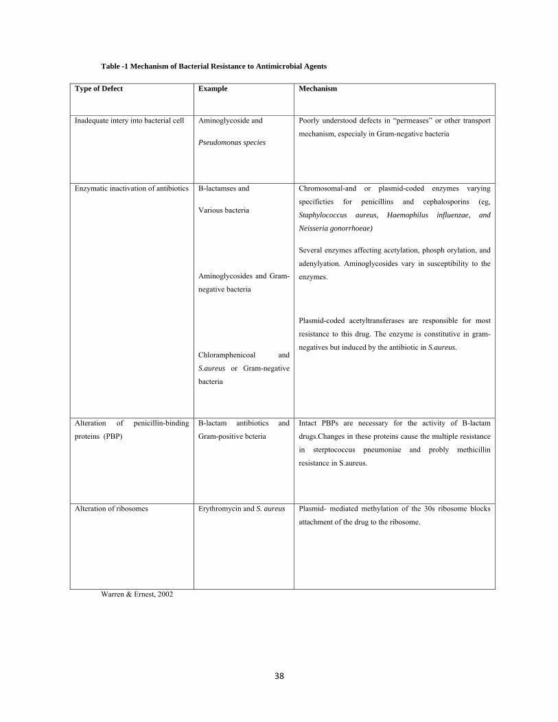
38
Table -1 Mechanism of Bacterial Resistance to Antimicrobial Agents
Mechanism Example Type of Defect
Poorly understood defects in “permeases” or other transport
mechanism, especialy in Gram-negative bacteria
Aminoglycoside and
Pseudomonas species
Inadequate intery into bacterial cell
Chromosomal-and or plasmid-coded enzymes varying
specificties for penicillins and cephalosporins (eg,
Staphylococcus aureus, Haemophilus influenzae, and
Neisseria gonorrhoeae)
Several enzymes affecting acetylation, phosph orylation, and
adenylyation. Aminoglycosides vary in susceptibility to the
enzymes.
Plasmid-coded acetyltransferases are responsible for most
resistance to this drug. The enzyme is constitutive in gram-
negatives but induced by the antibiotic in S.aureus.
B-lactamses and
Various bacteria
Aminoglycosides and Gram-
negative bacteria
Chloramphenicoal and
S.aureus or Gram-negative
bacteria
Enzymatic inactivation of antibiotics
Intact PBPs are necessary for the activity of B-lactam
drugs.Changes in these proteins cause the multiple resistance
in sterptococcus pneumoniae and probly methicillin
resistance in S.aureus.
B-lactam antibiotics and
Gram-positive bcteria
Alteration of penicillin-binding
proteins (PBP)
Plasmid- mediated methylation of the 30s ribosome blocks
attachment of the drug to the ribosome.
Erythromycin and S. aureus Alteration of ribosomes
Warren & Ernest, 2002
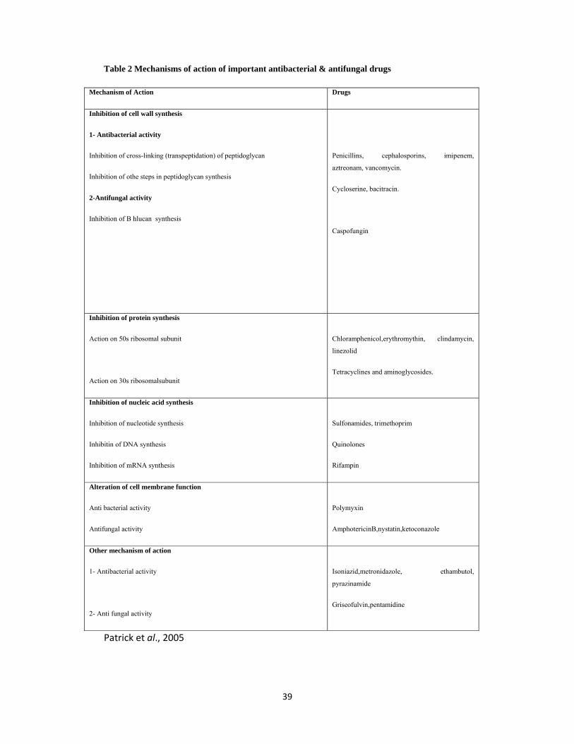
39
Table 2 Mechanisms of action of important antibacterial & antifungal drugs
Drugs Mechanism of Action
Penicillins, cephalosporins, imipenem,
aztreonam, vancomycin.
Cycloserine, bacitracin.
Caspofungin
Inhibition of cell wall synthesis
1- Antibacterial activity
Inhibition of cross-linking (transpeptidation) of peptidoglycan
Inhibition of othe steps in peptidoglycan synthesis
2-Antifungal activity
Inhibition of B hlucan synthesis
Chloramphenicol,erythromythin, clindamycin,
linezolid
Tetracyclines and aminoglycosides.
Inhibition of protein synthesis
Action on 50s ribosomal subunit
Action on 30s ribosomalsubunit
Sulfonamides, trimethoprim
Quinolones
Rifampin
Inhibition of nucleic acid synthesis
Inhibition of nucleotide synthesis
Inhibitin of DNA synthesis
Inhibition of mRNA synthesis
Polymyxin
AmphotericinB,nystatin,ketoconazole
Alteration of cell membrane function
Anti bacterial activity
Antifungal activity
Isoniazid,metronidazole, ethambutol,
pyrazinamide
Griseofulvin,pentamidine
Other mechanism of action
1- Antibacterial activity
2- Anti fungal activity
Patrick et al., 2005
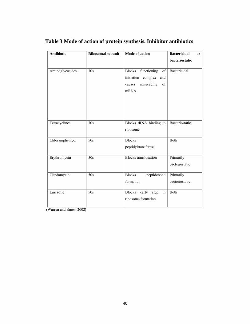
40
Table 3 Mode of action of protein synthesis. Inhibitor antibiotics
Bactericidal or
bacteriostatic
Mode of action Ribosomal subunit Antibiotic
Bactericidal Blocks functioning of
initiation complex and
causes misreading of
mRNA
30s Aminoglycosides
Bacteriostatic Blocks tRNA binding to
ribosome
30s Tetracyclines
Both Blocks
peptidyltransferase
50s Chloramphenicol
Primarily
bacteriostatic
Blocks translocation 50s Erythromycin
Primarily
bacteriostatic
Blocks peptidebond
formation
50s Clindamycin
Both Blocks early step in
ribosome formation
50s Linezolid
(Warren and Ernest 2002)

41
Table 4. Factors affecting antimicrobial activity on culture media
Effect on activity Agent affected Factor
Reduced Sulphonamides
Trimethoprin
Thymidine
Increased Aminoglycosides
Macrolides
Lincosamides
Nitrofurantoin
Rasied pH
Increased Tetracycline
Methicillin
Fusidic acid
Novobiocin
Lower pH
Increased against
staphylococci
Increased against Proteus
spp
Reduced
Bacitracin
Fusidic acid
Novobiocin
Penicillin
Tetracycline
Monovalent
cations (eg. Na+)
Reduced against
Pseudomonas spp.
Polymyxins
Aminoglycosides
Divalent cations
(eg. Mg2+ and
Na2+)
Elmer et al. ,1
42
1.2.5 Wound infection: -
Wound infections follow surgery or trauma that disrupts the skin or
mucosal surface (e. g. road accidents and bites). Post –operative wound
infections commonly follows gastro-intestinal surgery and some
gynaecological surgery (Bushell, 1989). It is so difficult to list all
pathogens that may be found in pus (Cheesbrough, 2004) .The type of
infecting organsim depends on the site and nature of the surgery or
trauma (Bushell, 1989). Woud infections following colo- rectal surgery
often contain bacteria from the large bowel (E.coli, Bacteroides spp. etc.).
Wound infections follow bites will contain mouth organisms from the
biting animal. Wound infections are often caused by organisms resident
on the skin surface which has been breashed. The main culprit here is
Staphylococcus aureus, and this is the only constituent of the normal skin
flora that is worth looking for routinely.
i) Common: S. aureus, β-haemolytic Streptococcus, E.coli, Bacteroides
fragilis, Proteus spp, other Enterobacteriaceae, CL-perfringens,
anaerobic cocci.
ii) Less common: mirco-aerophilic Streptococcus, Pasteurella multocida
(animal bites) other Clostridia, other Bacteroides, Fusobacterium spp,
Pseudomonas spp, Salmonella spp, Capnocytophaga canimrsus (formerly
D FZ).
iii) Rare: Vibrios (infected marine wounds).Cl.tetani,fungi (Bushell,1989)
Note on pathogens: S.aureus is the commonest pathogen isolated from
subcutaneous abscesss and skin wound. It also causes imetigo (small
pustules that form yellow crusty sores, usually around the mouth).
43
Penicilln and methecillin resistant strains of S.aureus are common causes
of hospital-acquired wound infections.
Ps.aeruginosa is associated with infected burns and hospital acquired
infections.
E.coli, Proteus spp, Pseudomonas aeruginosa, and Bacteroides spp are
the pathogens most frequently isolated from abdominal abscesses and
wounds .Pus containing Bacteroides spp has a very unpleasant smell (as
also pus containing other anaerobes).
Cl.perfringensis found mainly in deep wounds where anaerobic
conditions exist. The toxins produced cause putrefactive decay of the
infected tissue with gas production .The death and decay of tissue by
Cl.perfringens is called gas gangrene.
Chronic leg ulceration is common in those with sickle cell disease. The
commonest pathogens isolated are S, aureus. Ps.aeruginosa, S. pyogenes,
and Bacteroides species.
Mycobacterium tuberculosis is associated with cold abscesses.
Actinomycetes filamentous bacteria and several species of fungi cause
mycetoma. Specimens of pus from the draining sinuses contain granules.
Examination of which helps to differentiate wheather the Mycetoma is
bacterial (treatable) or fungal (less treated).
Actinomycetes israeli and other species of actinomyces cause
actinomycosis. Small yellow granules can be found in pus from draining
sinuses (often in the neck).
Vincent’s organsims (Borrelia vincenti with Gram- negative anaerobic
Fusiform bacilli) are asociated with tropical ulcer. The ulcer is commonly
44
found on the leg. Often of malnourished persons, especially children.
Staphylococcus and Streptococcus are frequently secondary invadors
(Cheesbrough, 2004).
1.2.6 Normal wound healing: -
The entire wound healing process is a complex series of events that
begins at the moment of injury and can continue for months to years. This
overview will help in identifying the various phases of wound healing
(Mathieu et al., 1999). Healing of wounds, whether from accidental
injury or surgical intervention involves the activity of intricate network of
blood cells, tissue types, cytokines, and growth factors .This results in
increased cellular activity, which causes an intensified metabolic demand
for nutrients (Thom et al., 1997) .Nutritional deficiencies can impede
wound healing, and several nutritional factors required for wound repair
may improve healing time and wound outcome.Vitamin A is required for
epithelial and bone formation, cellular differentiation, and immune
function .Vitamin C is necessary for collagen formation, proper immune
function, and as a tissue antioxidant. Vitamin E is the major lipid- soluble
antioxidant in the skin; however, the effect of vitamin E on surgical
wounds is inconclusive. Bromelain reduces edema, bruising, pain, and
healing time following trauma and surgical procedures. Glucosamine
appears to be the rate- limiting substrate for hyaluronic acid production in
the wound. Adequate dietary protein is absolutely essential for proper
wound healing, and tissue levels of the aminoacids arginine and
glutamine may influence wound repair and immune function (Douglas et
al., 2003).
The botanical medicines Centella asitica and Aloe vera have been used
for decades, both topically and internally, to enhance wound repair, and
45
scientific studies are now beginning to validate efficacy and explore
mechanisms of action for these botanicals. To promote wound healing in
the shortest time possible, with minmal pain, discomfort, and scarring to
the patient. It is important to explore nutritional and botanical influences
on wound out come (Douglas et al., 2003).
Wound repair or restoration of tissue integrity is natural reaction to
injury. Many drugs of natural or chemical origin (e.g. quince seed
mucilage and Ginko bilba, phenytoin, Zink oxide, Ketanserin,
dexpanthenol) have been used topically (Arzi et al., 2003).
1.2.6.1 Phases of wound healing: -
1-Inflammatory phase
A) Immediate to 2-5days.
B) Hemostasis: -
Vasoconstriction.
Platelet aggregation.
Tromboplastin makes clot.
C) Inflammation:
Vasodilation.
Phagocytosis.
Proliferative phase
A) 2 days to 3 weeks.
B) Granulation
46
Fibroblasts lay bed of collagen.
Fills defect and produce new capillaries.
C) Construction
Wound edges pull together to reduce defect
D) Epithelization
Crosses moist surface
Cell travel about 3 cm from point of origin in all directions.
Remodeling phase
A.) 3weeks to 2 years
B) New collagen forms which increases tensile strength to wounds.
C) Scar tissue is only 80 percent as strong as original tissue (Thom et al.,
1997).
Tissue injury initiates a response that first clears the wound of devitalized
tissue and foreign material, setting the stage for subsequent tissue healing
and regeneration. The intial vascular response involves a brief and
trasient period of intese vasoconstriction and hemosttasis 5-10 minute;
period of intense vasoconstriction is followed by active vasodilation
accompanied by an increase in capillary permeability. Platelets
aggregated whithin afibrin clot secrete a variety of growth factors and
cytokines that set the stage for an orderly series of events leading to tissue
repair (Douglas et al., 2003).
The second phase of wound healing the inflammatory phase, presents
itself as erythema, swelling, and warms, and is often associated with pain
47
.The inflamatory response increase vascular permeability,resultingin
migration of neutrophils and monocytes into the surrouding tissue .The
neutrophils engulf debris and microorgnisms, providing the first line of
defence against infection .Neutrophil migration ceases after the first few
days post- injury if the wound is not contaminated If this acute
inflammatory phase persist, due to wound hypoxia, infection ,nutritional
deficiencies, medication use, or other factors related to the patient
immune response, it can interfere with the late inflammatory phase
(Douglas et al., 2003).
1.2.7 Biochemical tests for identification of bacteria:- While several commercial systems for identifying bacteria are available,
these are often difficult to obtain or too expensive to use in developing
countries (Cheesbrough, 2004). However, the following of conventional
biochemical tests are used for identification of bacteria.
1.2.7.1 Gram reaction: -
Gram did not describe a stain but a method in which he used stains and
solutions devised by others, to this day we do not fully understand its
mechanism, but we do know that the reaction to Gram stain method is
stable charateristic of bacterium. Gram positivity (the ability to resist
decolorization with ethanol or acetone) is a feature of relatively young
bacterial cells of some species, as they age, the cells loose this
characteristic and apparetly become Gram –negative. It is important,
therefore, to examine young cultures, preferably before the end of the
logarithmic growth phase. Arecent modification by Preston and Morrell is
claimed to be fool proof (Cowan and Stell 1970).
48
Morphology: May be affected by the medium on which the organism is
grown and the temperture of incubation (Cowan and Stell 1970)
1.2.7.2 Catalase test: -
This is used to differentiate bacteria that produce the enzyme catalase,
such as Staphylococcus, from non –catalase producing bacteria such as
Streptococcus.Catalase acts as a catalyst in the breakdown of hydrogen
peroxide to oxgen and water .An organism is tested for catalase
production by bringing it into contact with hydrogen peroxide. Bubbles of
oxgen are relased if the organism is a catalase produser .The culture
shoud not be more than 24 hours old (Cheesbrough, 2004)
1.2.7.3 Citrate utilization test: -
This test is one of several technique used occasionally to assist in the
identification of Enterobacteria .The test is based on the abilityof an
organism to use citrate as its only sourse of carbon. There are two ways
for performing this test. Using a rasco citrate identification tablet. This is
the most economical method when only a few tests are performed. The
tablets have along shelf life and good stability in tropical climates.
Using Simmon citrate agar but the dehydrated medium is only availble in
500g-pack size from manufactures. After being opened the medium does
have good stability in tropical climates.
1.2.7.4 -Coagulase test: -
This test is used to identify S.aureus which produces the enzyme
coagulase causes plasma to clot by converting fibrinogen to fibrin.
Two types of coagulase are produced by most strain of S.aureus.
49
Free coagulase which convert fibrinogen to fibrin by activating a
coagulase- reacting fator present in plasma. Free coagulase is detected by
clotting in the tube test.
Bound coagulase (clumbing factor) which converts fibrinogen directly to
fibrin without requiring a coagulase –reacting factor .IT can be detected
by the clumping of bacterial cells in the rapid slide test. A test tube most
always be performed when the result of a slide test is not clear, or when
the slide test is negative and Staphylococcus has been isolated from a
serious infection. A tube test is reguired to detect some MRSA
(methicillin resist Staphylococcus aureus strains. Before performing a
coagulase test, examine a Gram stained smear to confirm that the
organism is a Gram-positive cocci (Cheesbrough, 2004).
1.2.7.5 DNase test: -
This test is used to identify Staphylococcus aureus which produses
dexoyribonucease enzyme. The Dnase test is particularly used when
plasma is not available to perform coagulase test or when the result of the
coagulase test is difficult to interpret
Deoxyribonuclease hydrolysis deoxyribonucleic acid (DNA). The test
organism is cultured on a medium, which contains DNA. After over night
incubation, the colonies are tested for Dnase production by flooding the
plate with a weak hydrochloric acid solution. The acid precipitates
hydrolyzed DNA. Dnase –producing colonies are therefore surrounded by
clear areas due toDNA hydrolysis (Hawkey and Lewis 1989).
1.2.7.6 Indole test: -
Testing for indole production is important in the identification of
Enterobacteria. Most strains of E.coli, P.vulgaris. P.rettgeri, M.morganii
50
and providencia species break down the aminoacid tryptophan with the
release of indole .The test organism is cultured in a medium, which
contains tryptophan. Indole production is detected by Kovac or Elrlich’s
reagent, which contains 4(p)-dimethylaminobenzaldehyde. This reacts
with the indole to produce a red coloured compound. Kovac’s reagent is
recommended in preference to Ehrlichs reagent for the detection of indole
from Enterobacteria (Cheesbrough, 2004).
1.2.7.7 Oxidase test: -
The oxidase test is used to assist in the identification of Pseudomonas,
Neisseria, Vibrio, Brucella, and Pasteurella species, all of which produce
the enzyme cytochrome oxidase .A piece of filter paper is soaked with a
few drops of oxidase reagent. A colony of the test organism is then
smeared on the filter paper. Alternatively an oxidase reagent strip can be
used .If the organism is oxidase –producing, the phenyleneiamine in the
reagent will be oxidized to deep puple colour. Occasionally the test is
performed by flooding the cultue plate with oxidase reagent but this
technique is not recommended for routine use because the reagent rapidly
kills bacteria. It can however be useful when attempting to isolate
N.gonorrhoeae coloies from mixed culture in the absence of a selective
medium .The oxidase positve colony must be removed and subcultured
within 30 seconds of flooding the plate. Acidity inhibits oxidase enzyme
activity .The oxidase test must not be performed on colonies that produce
fermentation on carbohydrte-cotaining media such as TCBS or
MacConkey agar. Subculture on nutrient agar is required before the
oxidase test can be performed. Colonies tested from a medium that
contain nitrate may give unreleable oxidase test results (Hawkey and
Lewis 1989).

51
1.2.7.8 Urease test: -Testing for urease enzyme activity is important in
differentiating Enterobacteria. Proteus strains are strong urease
producers.Y.enterocolitica also shows urease activity (weakly at 35-
37°C). Salmonellae and Shigellae do not produce urease. The test
organism is cultured in a medium contains urea and the indicator phenol
red. When the strain is urease producing, the enzyme will brake down
urea (by hydrolysis) to give ammonia and carbon dioxide. With the
release of ammonia, the medium becomes alkaline as shown by a change
in colour of the indicator to pink-red (Cheesbrough, 2004).
1.2.7.9-Carbohydrate utilization test: -The so-called“fermentation
tests”were used by the early bacterilogists to distinguish one organism
from another and elaborate diagnostic tables were based on them. The
introduction of the simple gas tube (Durham, 1898) and indicators
enabled the production of gas and acid to be detected by inspection
(Cowan, 1970). Peptone water, sugars and Rasco sugar fermentation
tablets are used to identify bacteria by their fermentation reactions
(Cheesbrough, 2004).
1.2.7.10 The MR (Methyl red) test and V-P (Voges–Proskauer) test
for acetylemethylcarbinol or acetoin may be carried out on the same tube
of culture and are discussed together. The tests mainly used to distinguish
various Coliform organisms from each othor; all ferment glucose
vigrously and the pH value of the glucose medium falls quickly. When
methyl red is added after overnight incubation the cultures of all these
organisms will be found to be acid to the dye, i.e.MR positive. After
further incubation Escherichia coli cultures produce even more acid and
inspite of phosphate buffer in the medium may be self-sterilizing; The
MR test remains positive. Klebsiella pneumoniae cultures, on the other
52
hand, decarboxylate and condense the pyruvic acid to form
acetylmethylcarbinl, the pH value rises and, when methyl red is added,
the colour is yellow. i.e. MR –negative. Nowadays there is a tendency to
do biochemical tests earlier but the temptation to speed up the MR test
must be resisted, the MR shoud never read until the cultures have been
incubated for at least two days at 37°C or three days at 30°c. The VP test
can be obtained. It is now generally thought that the older methods are
too slow and insenstive, but there is less agreement about the method to
be recommended or the senstivity that gives the best differentiation
between species (Cowan and Stell 1970)
1.2.7.11 Pigment production: -
Often has considerable diagnostic value and it is an advantage to know
how to induce it. Although the pigments produced are seldom
photosynthetic, most bacteria dealt within laboratories form pigment
better in the light; this is most noticeable in Staphylococcus and Serratia,
and also occurs in the Pseudomonas and in Chromobacteria. The effect of
light on pigment production by bacteria has become a means of
distinguishing species. Temperatures and medium also influence the
intensity of pigmentation; most bacteria produce pigments better at
temperatures below the optimum for growth. Medium probably has the
biggest effect on the development of pigment. In some cases the addition
of glucose will enhance pigmentation; in other cases this will inhibit it.
(Hawkey and Lewis 1989).
1.2.7.12 -Kligler iron agar: -KIA reactions are based on the
fermentation of lactose and glucose (dextrose) and the production of
hydrogen sulphide. A yellow but (acid production) and red- pink slope
indicate the fermentation of glucose only. The slope is pink-red due to a
53
revertion of the acid reaction under aerobic conditions. This reaction is
seen with Salmonella and Shigella species and other enteric pathogens
cracks and bubles in the medium indicate gas prodution by S. paratyphi
and some faecal commensals. A yellow slope and a yellow butt indicate
the fermentation of lactose and possiblly glucose. This occurs with E.coli
and other Enterobacteria. Ared –pink slope and butt indicate no
fermention of glucose or lactose. This is seen with most strains of
Ps.aeruginosa. Blackening along the stab line or throughout the medium
indicates hydrogen sulphide (H2S) production e.g. S.typhi produces a
small amount of blackening whereas S.typhimurium causes extensive
blackening (Cheesbrough, 2004).
1.2.7.13 Novobiocin disc:
This senstivity test is used to differetiate between Staphylococcus species
(Cheesbrough, 2004).
1.2.7.14 Growth at 42°C: -
Differentiates Ps.aeruginosa from the less commonly isolated
Pseudomonads, P.putida and P.fluorescens (Cheesbrough, 2004).
1.2.8 Selction of the appropriate laboratory animals:
Animals are used to show standard response to experimental
manipulation. Animals must be of the same: species, subspecies, strain,
and sex, the same age range, reared under similar conditions and not
subject to previous experimental interference. One must try to achieve
maximum accuracy with the minimum number of animals. Rodent
animals can be used in the preliminary screening experiments.
54
Swiss Wistar Albino rats are popular for their size, ease of handling, low
cost of housing, short life span, the broad similarity to human physiology
and usefulness in wound healing evaluation experiments (Poole and
Robinson 1989).
1.2.9 Ointments: -
Ointments are semisolid preparations intended for external application to
the skin or mucous membrane. They may be medicated or non-medicated.
Non-medicated ointments are used for the physical effects that they
provide as protectants, emollients or lubricants. Ointment bases, as
described may be used for their physical effects or as a vehicle in the
preparation of medicated ointments (Ansel et al., 2002).
1.2.9.1 Ointment bases:- Ointment bases are classified by the United State Pharmacopea (USP)
into four general groups:
i) Hydrocarbon bases.
ii) Absorption bases.
iii) Water removable bases.
iv) Water soluble bases.
1.2.9.1.1 Hydrocarbon bases:
Also termed oleaginouss bases on application to the skin:
i) They have an emollient effect.
ii) Protection against the scape of moisure.
iii) Are effective as occlusive dressings.
55
iv) Can remain on the skin for prolonged periods of time without drying
out.
v) Due to immiscibility with water are difficult to wash-off.
vi) Water and aqueous preperations may be incorporated into them, but
only in small amounts and with some difficulty.
Petrolatum, white petrolatum, white ointment and yellow ointment are
examples of Hydrocarbon bases.
1.2.9.1.1.1. Petrolatum USP:
Petrolatum, USP is a purified mixture of semisolid hydrocarbons obtained
from petroleum. It is an unctuous mass, varying in colour from yellowish
to light amber.
It melts at temperature between 38°C and 60°C and may be used alone or
in combination with other agents as an ointment base. Petrolatum is also
known as "yellow petrolatum and petroleum jelly". A commercial
product is Vaseline'Chesebrough-Ponds’ (Ansel et al., 2000).
1.2.9.1.1.2 Liquid paraffin:
This is a mixture of liquid hydrocarbons obtained from petroleum. It is a
transparent, colourless, almost odorless oily liquid. On long storage it is
liable to oxidation with production of peroxides and therefore it may
require an antioxidant, e.g. Tocopherol or butylated hydroxytoluene
(BHT). It is used to soften ointment bases and to reduce the viscosity of
creams (Collet and Aulton, 1991).
1.2.9 1.2 Absorption bases They are two types:
56
i) Those that permit the incorporation of aqueous solutions resulting in
the formation of water in oil emulsions (e.g. hydrophilic petrolatum).
ii) Those that are water in oil emulsions (syn.: emulsion bases) and permit
the incorporation of additional quantities of aqueous solutions (e.g.
Lanolin).
These bases may be used as emollients although they do not provide the
degree of occlusion afforded by the hydrocarbon bases. Absorption bases
are not easily removed from the skin with water washing since the
external phase of the emulsion is oleaginous (Ansel et al., 2002).
1.2.9.1.2.1 Lanolin, USP:
It is obtained from the wool of sheep (Ovis Aries) is a purified, wax-like
substance that has been cleaned, deodorized and decolorized. It contains
no more than 0.25% water. Additional water may be incorporated into
lanolin by mixing.
1.2.9.1.3 Water removable bases (water miscible):
i) Oil-in-water emulsions resembling creams in appearance.
ii) Because the external phase of the emulsion is aqueous, they are easily
washable from skin and are often called “water washable bases”
iii) They may be diluted with water or aqueous solutions.
iv) They have the ability to absorb serous discharges.
Hydrophilic ointment, USP is an example of this type of bases (Ansel et
al., 2002)
The three emulsifying ointments from water-miscible bases, i.e.
57
Emulsifyingg ointment BP (anionic), cetrimide emulsifying ointment BP
(cationic) and cetomacrogol emulsifying ointment BP (non-ionic)
These contain paraffins and an o/w emulgent and have the general
formula:
Anionic, cationic or non-ionic emulsifying Wax 30 %
White soft paraffin 50 %
Liquid paraffin 20%
They are used for preparing o/w creams (Collett and Aulton, 1991).
1.2.9.1.4 Water-soluble bases:
They do not contain oleaginous components. They are completely water
washable and often referred to, as ‘Greaseless’ because they soften
greatly with addition of water, large amounts of aqueous solutions are not
effectively incorporated into these bases. They are mostly used for the
incorporation of solid substances. Polyethlene glycol ointment, natural
formula (NF) is the prototype example of water-soluble base.
1.2.9.1.4.1 Polyethlene glycol ointment (natural formula) (macrogol
or carbowaxes):
Polyethlene Glycol (PEG) is a polymer of ethylene oxide and water
presented by the formulation H (OCH2CH2) n OH in which n represents
the average number of oxyethlene groups. The numerical designations
associated with PEGs refer to the average molecular weights of the
polymer. PEGs having average molecular weights below 600 areclear,
colorless liquids, those with molecular weights above 1000 are wax like
white materials, and those with molecular weights in between are
semisolids. The greater the molecular weight the greater is the viscosity
58
.The natural formula (NF) lists the viscosities of PEGs ranging from
average molecular weights of 200 to 8000. The general formula for the
preparation of 1000g of polyethene glycol ointment is:
Polyethlene glycol 3350 400grams
Polyethlene glycol 400 600grams
Shayoub (1985) listed the advantages and disadvantages of Polyethylene
Glycol.
1.2.9.1.4.1.1 Advantages of Polyethlene Glycol:
1/ PEGs vehicles form non-occlusive films on skin.
2/ PEGs are anhydrous and can easily wash from the skin.
3/ Good absorption by the skin.
4/ Good solvent properties. Some water- immissible dermatological
drugs such as hydrocortisone, salicylic acid, sulphonamides, sulphur and
redsonoic acid are soluble in macrogols.
5/ Freedom from greasiness.
6/ Satifactory agening properties. They do not hydrolyzse, rancidify or
support microbial growth.
7/ Compatibility with many deratological medicaments e.g. ammoniated
mercury; yellow mercuric oxide, icthammol and sulphur (Robinson et al.,
1964).
1.2.9.1.4.1.2 Disadvantages of Polyethylene Glycol:- 1/ Limited uptake of water, macrogols dissolve when the proportion of
water reaches about5% bases may be thinned with liquid macrogol or
59
with propylene glycol. Inclusion of a higher fatty alcohol, such
ascetostearly, which allows incorporation of larger quantites of water, and
improves the texture of the bases (Nexon, 1951 cited in Shayoub 1985).
2/ They are less bland than paraffins, possibly due to their hygroscopic
nature.
3/ They cause reduction in activity of certain antibacterial agents,e,g.
phenols. Hydroxyl benzoates and quaterary ammonium componunds
(Pattel and Foss, 1964). Some antibiotics are rapidly inactivated such as
penicillin and bacitracin (Couster et al., 1961).
4/ They have a solvent action on polyethlene and backelite; these plastics
should not be used as containers or closures for macrogel ointments.
1.2.9.2 Properties of the Ideal Base:
1- It does not retard wound healing.
2-It has a low sensitization index.
3/ It must be neutral.
4/ It has a non-dehydrating effect.
5/ It has an acceptable pharmaceutical elegance.
6/It must be compatible with common medications.
7/ It has a good keeping quality.
8/ It has alow index of irritation.
9/ It has non-greasy property.
10/ It has a minimum number of ingredients.
60
11/ It has an efficient release of medicament at the site of application.
12/ It is washable.
13/ It has an easy compounding property (Shayoub, 1985).
1.2.9.3 Selection of the appropriate base:
The selection of the base to be used in formulation of an ointment
depends upon the careful assessment of a number of factors inculding:
1/ The desired release rate of the drug substance from the ointment base.
2/Desirability for topical or percutaneous drug absorption.
3/Desirability of occlusion of moisture from the skin.
4/ Stability of the drug in the ointment base.
5/ Effect.if any, of the drug on the consistency or other features of the
ointment base.
6/ The desire for base that it is easily removed by washing with water.
The base that provides the majority of the most desired attributes should
be selected (Ansel et al., 2000).
1.2.9.4 Compounding of Ointments and pastes:
The basic techniques for the preparation of ointments and pastes are:
Weighing, measuring of liquids, size reduction and size separation, and
mixing.
61
1.2.9.4.1 Fusion mixing method: -
In this method, the ingeredients are melted together and stirred to ensure
homgeneity, on a small scale; fusion is usually carried out in an
evaporating basin over a water- bath. Stainless basins are to be preferred.
1.2.9.4.1.1Preparation of the Ointment base by fusion:
The constituents of the base should be placed together in the basin and
allowed to melt together. Melting time is shortened if high melting point
ingerdients, such as hard paraffin and the emulsifying waxes are grated
into the basin and heated while other ingerdients are being prepared.
After melting, the ingerdients should be stirred until cool.
1.2.9.4.1.2 Preparation of Medicated Ointments and Pastes by
Fusion:
Solid that is completely or partialy soluble in the base should be added in
fine powder to the molten base at a low temperature as possible and the
mixture stirred until cooled (Collett and Aulton, 1991).
1.2.9.5 Application frequency:
Topical agents are often applied twice daily (Hardman et al., 1996).
1.2.9.6 Microbial contents:
With the exception of ophthalmic preparations, topical applications are
not required to be sterile. They must, however, meet acceptable standards
for microial contents. Preparations which are prone to microial growth
must be presented with antimicrobial preservatives (Ansel et al., 2000).
62
1.2.10 The plants used in this study are: -
1.2 10 .1 Guiera senegalensis: -
Vernacular name: Gubeish (Arabic).
Family: Combretaceae (Elghazali, 1997).
Botanical description: Grey tomentose shrubs up to 3m high. Leaves
opposite, or subopposite elliptic- oblong, 6-12 x 0.5 - 2.8 cm, apex
mucronate, base slightly cordate to attenuate, margin entire.
Inflorescences dense terminal heads up to 2 m across. Fruit capsules,
spindle- shaped, angled, 2.5-3.0 cm, grey-brown (Elghazali, et al., 1987).
Habitat: Sandy lowland plains, degarded savanna.
Distribution: Tendelti, Rashad, Wad ashana and central Sudan.
Chemical constituents: Flavonoids, saponins, alkaloids, mucilages and
tanins100 were isolated (Elghazali, 1997). A new methoxylated naphthyl
butenone, guieranone A were isolated from the leaves of Guiera
senegalensi and it is the first derivative that has been extracted from the
family Combretaceae.
Uses: The macerations of the leaves used as antidiabetic, antipyretic, anti-
vomiting and antileprosy (Elghazali, 1997). In African traditional
medicine the leaves are also used for gastrointestinal disorders, coughs
and topically for wound healing (Bosiso et al., 1997). Plant is used for
malaria in Mali and Sao-tome traditional medicine (Ancolio et al., 2002).
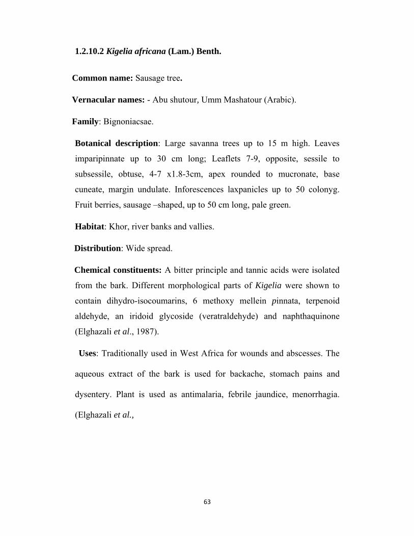
63
1.2.10.2 Kigelia africana (Lam.) Benth.
Common name: Sausage tree.
Vernacular names: - Abu shutour, Umm Mashatour (Arabic).
Family: Bignoniacsae.
Botanical description: Large savanna trees up to 15 m high. Leaves
imparipinnate up to 30 cm long; Leaflets 7-9, opposite, sessile to
subsessile, obtuse, 4-7 x1.8-3cm, apex rounded to mucronate, base
cuneate, margin undulate. Inforescences laxpanicles up to 50 colonyg.
Fruit berries, sausage –shaped, up to 50 cm long, pale green.
Habitat: Khor, river banks and vallies.
Distribution: Wide spread.
Chemical constituents: A bitter principle and tannic acids were isolated
from the bark. Different morphological parts of Kigelia were shown to
contain dihydro-isocoumarins, 6 methoxy mellein pinnata, terpenoid
aldehyde, an iridoid glycoside (veratraldehyde) and naphthaquinone
(Elghazali et al., 1987).
Uses: Traditionally used in West Africa for wounds and abscesses. The
aqueous extract of the bark is used for backache, stomach pains and
dysentery. Plant is used as antimalaria, febrile jaundice, menorrhagia.
(Elghazali et al.,
64
2. MATERIALS AND METHODS
2.2 Methods
2.2.1. Identification of the clinical isolates:
One hundred clinical isolates of Escherichia coli, Proteus vulgaris,
Pseudomonas aeruginosa and Staphylococcus aureus, were collected
randomly during the period from June - December 2005 from National
Health Laboratory, Khartoum and Omdourman Educational
Hospitals.These clinical isolates were obtained from, urine, wounds, ear
swab, abscesses and eye swab. Streaking on mannitol salt agar,
MacConkey agar crystal violet blood agar, cetrimide agar and cooked
meat medium purified them. They were identified on the basis of the
results of microscopical examination (Gram stain), cultural characteristics
and biochemical tests (Cruickshank et al., 1975).
Media used for identification of clinical isolates:
All specimens were inoculated on two blood agar plates, one incubated
aerobically and the other anaerobically using gas-generating kits. The
obtained isolates were then purified by streaking on mannitol salt agar
plates, MacConkey's agar crystal violet agar, cetrimide agar and cooked
meat medium.
65
The purified isolates were then subcultured on mannitol, MacConkey
agar, crystal violet blood agar, cetrimide agar and cooked meat medium
slopes and then stored in a refrigerator until they were used.
2.2.1.1 Microscopical examination of aerobic bacterial isolates.
All isolates were subjected to microscopical examination to study
their staining properties (using the Gram’s staining technique), the shapes
and cellular arrangements.
The bacterial film is fixed then flooded with crystal violet for one minute,
then the stain washed off with clean water and coverd with iodine for one
minute and decolorized rapidly with acetone- alcohol, washed clearly
with clean water, decolorized smears coverd with safranin for two
minutes and then examined microscopically.
2.2.1.2 Simplified routine biochemical tests for identification of
bacterial isolates:-
The biochemical activities of the purified isolates were then
studied for identification and confirmation of these organisms. The
biochemical tests carried out include:-
66
2.2.1.3.1 Fermentation tests:
Different bacteria are variously able to ferment carbohydrates (e.g.
glucose, lactose, and sucrose). Sterile peptone water was used, with 1 %
of the sugar, and suitable pH indicator, is inoculated with the test
organism and incubated at 37ºC for 1-3 days. Acid production is detected
by a colour change of the indicator, and gas production by an inverted
Durham׳s tube, which has been completely filled with the medium (with
phenol red the colour changed from pink to yellow indicating
fermentation and production of acid). The production of gas was detected
by the presence of air-bubbles in the tube (Harris, 1964).
2.2.1.3.2. Methyl red tests:
It is used to detect the ability of some bacteria to produce sufficient
amounts of acidic substances due to fermentation of glucose using methyl
red- Voges- Proskauer medium. Colour changes from yellow (pH 6.2) to
red (pH4.2) with acid production on addition of methyl red indicator.
(Cruickshank et al., 1975).
2.2.1.3.3 Voges- Proskauer test:
It is based upon the production of acetyl methyl carbinol as a product of
dextrose metabolism by certain bacteria. This substance is readily
oxidized by atmospheric oxygen, in alkaline medium, giving diacetyl
67
which, in turn, reacts with the amino acid arginine in the medium to give
a pink colour when alpha naphthol is added (Cruickshank et al., 1975)
2.2.1.3.4 Citrate utilization test:
It is based on the ability of some organisms to utilize citrate as the
sole carbon and energy source for growth, and an ammonium salt as the
sole source of nitrogen. Inoculation of Simmon's citrate medium
(modification of Koser's citrate medium with agar and an indicator) with
a 24 hours culture of the tested organism and incubation at 37oC for 2-3
days. Growth and a change of colour of the indicator from light green to
blue, due to alkaline reaction following citrate utilization (Collee et al.,
1996)
2.2.1.3.5 Indole production test:
It is based on the ability of certain bacteria to decompose the amino
acid tryptophane to indole. By inoculation of peptone water with the test
organism and incubation for 2 days at 37 °C and addition of Kovac’s
reagent (ρ-dimethyl-aminobenzaldehyde), a red colour indicates the
presence of indole (Collee et al., 1996)
68
2.2.1.3.6 Hydrogen sulphide production test:
It is based upon the ability of some bacteria to produce H2S from sulphur
containing amino acids by reduction. H2S may be tested by suspending
strip of filter paper impregnated with lead acetate above the culture, H2S
is demonstrated by its ability to form black insoluble ferrous sulfide after
incubation of the inoculated peptone water at 37ºC for 2-3 days (Collee et
al.,1996)
2.2.1.3.7Catalase test:
This demonstrates the presence of catalase , an enzyme that catalyses
the release of oxygen from hydrogen peroxide.One dropl of hydrogen
peroxide solution was poured over a 24 h nutrient agar slope culture of
the test organism and the tube is held in slanting position.The production
of gas bubbles from the surface of the solid culture material indicates a
positive reaction. (Collee et al., 1996)
2.2.1.3.8. Coagulase test:
It is based on the presence of the enzyme coagulase in the cell of
some bacteria. In a test tube, 1 ml of a l in 10 dilution of sterile citrated
human plasma in saline is added to few drops of a 24 hrs. Inoculated
broth culture of the test organism, and the mixture incubated at 37 °C and

69
examined for coagulation after 1, 3, and 6 hrs. The formation of a clearly
visible clot indicates a positive coagulase test. (Harris, 1964).
2.2.1.3.9. Oxidase test:
It is based upon the presence of the enzyme oxidase in the cells of
certain bacteria. The oxidase enzyme catalyzes the transport of electrons
between electron donors in the bacteria and a redox dye- tetra methyl,
para-phenylene diamine, a freshly prepared oxidase reagent when added
in a solid growth media, rapidly develops a purple colour at the colonies
of oxidase- positive organism (Cruickshank et al., 1975; Salle, 1961).
2.2.1.3.10 Urease test:
It is based upon the presence of enzyme urease in the cells of
certain bacteria. The test organism cultured in a medium which contains
urea and the indicator phenol red. When the organism is urease
producing, the enzyme will break down the urea (by hydrolysis) to give
ammonia and carbon dioxide. With the realese of ammonia, the medium
becomes alkaline and the indicator colour change from yellow to pink
(Cheesbrough, 1996)
70
2.2.1.3.11. Deoxyribonucleic (DNase) test:
Staphylococcus aureus produces the enzyme deoxyribonuclease
(DNase). The test organism was cultured on a medium containing (DNA).
After over night incubation the colonies were tested for DNase
production by flooding the plate with a (1N) HCl acid solution, the acid
precipitated unhydrolysed DNA. DNase producing colonies were
therefore surrounded by clear areas indicating DNA hydrolysis
(Cheesbrough, 1996).
2.2.2. Plant materials
The two medicinal plants used in this study were collected from different
parts of Sudan, by herbalists in collaboration with the Institute of
Traditional Medicine, during 2005. They were authenticated by the
researcher Haider Abdelgadir and Wail Elsadig Abdalla, Medicinal and
Aromatic Plants Research Institute (MAPRI). Voucher specimens were
deposited by the herbarium of the Institute.
Data concerning the description of the habitat, the local names, traditional
methods of preparing the herbal preparations, the way of application and
the diseases they treat were obtained from the local herbalists and
recorded.50 grams of each plant sample were powdered by grinder and
71
extracted, as described below in the experimental section and then
subjected to antimicrobial activity screening.
Plants screened are listed in Table (6) with their botanical names,
synonyms, families, morphological part used, place of collection,
vernacular names and folkloric uses.
2.2.2.1. Preparation of crude extracts:
Each of the coarsely powdered plant material (50 g) was exhaustively
extracted for 20 hours with chloroform in Soxhlet apparatus. The
chloroform extract was filtered and evaporated under reduced pressure
using Rota-vap. The extracted plant material was then air-dried, repacked
in the Soxhlet and exhaustively extracted with methanol. The methanolic
extract was filtered and evaporated under reduced pressure again using
Rota-vap.Each residue was weighed and the yield percentage was
determined. The chloroform residue (2 g) was dissolved or suspended in a
mixture containing methanol: petroleum ether (2:1) to a final volume
20ml (con. 100 mg/ml). The methanol residue (2g) was dissolved in
methanol 20 ml (con. 100mg/ml), and kept in refrigerator until used.
For aqueous extract 100 g of each plant sample was soacked with 500 ml
hot water for 4 hours then filtered with Whattman filter paper. Extracts
kept in deep freezer for 48 hours, then induced in freeze dryer till
72
completely dried. The residue was weighed and the yield percentage was
determined. The aqueous residue (2g) was dissolved in sterile distilled
water 20 ml (con. 100mg/ml), and kept in refrigerator until used.
2.2.3. Preparation of the test organisms:
2.2.3.1 Preparation of bacterial suspensions:
One ml aliquots of a 24 hours broth culture of the test organisms were
aseptically distributed onto nutrient agar slopes and incubated at 37º C for
24 hours. The bacterial growth was harvested and washed off with 100 ml
sterile normal saline, to produce a suspension containing about 108- 109
C.F.U/ ml. The suspension was stored in the refrigerator at 4° C till used.
The average number of viable organisms per ml of the stock suspension
was determined by means of the surface viable counting technique (Miles
and Misra, 1938). Serial dilutions of the stock suspension were made in
sterile normal saline solution and 0.02 ml volumes of the appropriate
dilution were transferred by micro pipette onto the surface of dried
nutrient agar plates. The plates were allowed to stand for two hours at
room temperature for the drops to dry and then incubated at 37 °C for 24
hours. After incubation, the number of developed colonies in each drop
was counted. The average number of colonies per drop (0.02 ml) was
multiplied by 50 and by the dilution factor to give the viable count of the
73
stock suspension, expressed as the number of colony forming units per ml
suspension.
Each time a fresh stock suspension was prepared. All the above
experimental conditions were maintained constant so that suspensions
with very close viable counts would be obtained.
2.2.4 In vitro testing of extracts for antimicrobial activity:-
2.2.4.1 Testing for antibacterial Activity:
The cup-plate agar diffusion method (Kavanagh, 1972) was
adopted with some minor modifications to assess the antibacterial activity
of the prepared extracts.
One ml of the standardized bacterial stock suspension 108 –109 C.F.U/ ml
were thoroughly mixed with 100ml of molten sterile nutrient agar which
was maintained at 45 ºC. 20ml aliquots of the inoculated nutrient agar
were distributed into sterile Petri-dishes.
The agar was left to set and in each of these plates 4 cups (10 mm in
diameter) was cut using a sterile cork borer (No. 4) and agar discs were
removed.
Alternate cups were filled with 0.1 ml sample of each of the 4
extracts in methanol using automatic microlitre pipette, and allowed to
74
diffuse at room temperature for two hours. The plates were then
incubated in the upright position at 37 ºC for 18 hours. Two replicates
were carried out for each extract against each of the test organisms. After
incubation the diameters of the resultant growth inhibition zones were
measured, averaged and the mean values were tabulated.
2.2.4.2. Testing the susceptibility of clinical isolate to extracts:
Using the standard cup plate agar diffusion technique, the clinical strains
were examined for susceptibility to the extracts, which showed activity
against standard bacterial organisms.
2.2.5. Determination of minimum inhibitory concentration (MIC) by
agar plate dilution method:
The principle of the agar plate dilution is the inhibition of growth
on the surface of the agar by the plant extracts incorporated into the
medium.
Plates were prepared in the series of increasing concentrations of
the plant extract. The bottom of each plate was marked off into 6
segments. The organisms tested were grown in broth over night to contain
108 organisms per ml.
A loop-full of diluted culture is spotted with a calibrated loop that delivers 0.001 ml on the surface of each segment. The end point of
75
MIC is the least concentration of antimicrobial agent that completely inhibits the growth. Results are reported as the MIC in mg/ml of crude extract.
2.2.6. Wound healing activity of Kigelia africana:-
The wound evaluation model (Arzi et al., 2003) was adopted with some
minor modification to assess the wound healing activity of a selected
plant extract.
2.2.7.1. Ointment preparation
Polyethylene glycol was used as a water soluble base to prepare
ointments of Kigelia africana extracts in 1, 2, 5% concentrations
Polyethylene glycol used (1:1) mixture of 200: 2000 PEGs, The mixtures
were stirred gently by infusion in waterbath till they are homogenously
distributed and then cooled with continuous stirring.
2.2.7.2. Experimental animals:
Swiss Wistar Albino rats of either sex, weighing 80-100g were
used. Animals were supplied by the National Experimental Animal House
(NEAH), Medicinal and Aromatic Research Institute (MAPRI), National
Center for Research (NCR), Ministry of Science and Technology
(MOST), Sudan.
76
The rats were housed individually in a ventilated Animal house before
and after surgery. They had access to standard diet which has been
prepared in National Experimental Animal House (NEAH) supplemented
with water adlibitum (as much as one likes).The holding room was
illuminated with 12 hours.Light/dark cycles. Room temperature was
between 30-35 Co with 45% to 55% humidity.
2.2.7.3. In vivo wound healing activity of Kigelia africana extracts
(non-infected rats):
Full thickness wounds were made in the skin of the tested animals
according to the model of (Arzi et al., 2003).
Hair of the lower back and right flank of animals was fully shaved.
Rats were lightly anaesthetized by inhalation using Halothane.
The animals were held in standard crouching position, and the
mobile skin of flank was gently stretched and held by fingers. A metal
circular object measuring 1 cm in diameter was placed on stretched skin
and an outline of the object was traced on the skin using a fine tipped pen.
The wound was made by excising the skin within the border of the
object to level of loose subcutaneous tissue, using sterile forceps and
scalpel blade
77
The artificial wounds were circular with a diameter of 1 cm.
The first day of the experiment was regarded as day Zero.
Animals were divided into five groups, each containing five
animals:
Group 1 (wound only):
Untreated control group, wounds were left without treatment.
Group 2 (wound + Fucidin ointment):
Wounds of these animals were treated topically with fucidin ointment
every 24 hours as standard healing agent starting from first day.
Group 3 (wound+ Kigelia africana ointment in polyethylineglycol):
Wounds of these animals were treated topically with Kigelia
africana ointment every 24 hours starting from first day.
2.2.7.4. Evaluation method of wound healing percentage:
In order to determine the rate of wound healing, every 24 hours,
each animal was held in the standard crouching position and two
diameters of the wound circle (horizontal and vertical) were measured
using a transparent ruler. Measurement errors were minimized by
78
repeating each measurement three times at the same moment and using an
average of the calculations.
The area of the wound in day zero was considered as 100% and the
wound areas on subsequent days were compared with the wound on day
zero.
Healing percentage in a certain day was the difference between the initial
wound (in zero days) and healing wound on that certain day.
79
3. RESULTS
3.1 Isolations and Identification of Clinical Isolates:
3.1.1 Identification of Eschericha coli 3.1.1.1 On MacConkey agar medium, red colonies were observed as a
result of lactose fermentation.
3.1.1.2 Microscopical examination:
With Gram staining technique, Gram-negative rods with no special
arrangement were seen.
3.1.1.3 Biochemical reactions:
The biochemical properties of the isolates are summarized in Table 5.
All isolates fermented lactose, mannitol and glucose with production of
acid and gas. All of them fermented sucrose with acid production and gas
formation.
All of the isolates were methyl red positive and Voges Proskauer
negative. All isolates gave indole positive result. None of the isolates
produced urease, utilize citrate or gave positive oxidase.
All isolates did not change the yellow colour of K.I.A. both in slant and butt, with the absence of H2S production, but all isolates produced gas.
All these led to identifying these clinical isolates as Escherichia coli. Table (5).
80
3.1.2 Identification of Proteus vulgaris:
Cultural characteristics:
On nutrient agar, fishy smell and swarming appearance was clear. On
MacConkey agar medium, pale coloured colonies were observed as a sign
of non-lactose fermentation.
Microscopical examination:
With Gram staining technique, Gram-negative rods with no special
arrangement were seen
Biochemical reactions:
All isolates were non-lactose and non-mannitol fermenters, but were
sucrose fermenters.
Most of them did not ferment sucrose with acid production and some
formed gas. Most of them fermented glucose; methyl red positive and all
was Voges Proskaur negative.
All of them were oxidase negative, most utilized citrate and produced
urease, and most were indole negative.
All isolates changed the colour of slope of KIA from yellow to red and
maintained the yellow colour of butt. Most were H2S producers and gas
non-producers.
All these led to identifying these clinical isolates as Proteus vulgaris.
(Table 5).
81
3.1.3. Identification of Pseudomonas aeruginosa
Cultural characteristics:
On nutrient agar, most of the isolates produced blue-green pigments,
which diffused in the surrounding medium.
Microscopical examination:
With Gram staining technique, Gram-negative rods with no arrangement
were seen.
Biochemical reactions:
All isolates were non-lactose and non-sucrose fermenters, but glucose
fermenters.
Most of them were methyl red negative and all were Voges Proskaur
negative. Most were oxidase positive, indole negative, did not utilize
citrate and did not produce urease.
Regarding KIA, all of them changed the slope colour to red, and most
changed the butt to red, without H2S or gas production.
All these led to identifying these clinical isolates as Pseudomonas
aeruginosa.Table (5).
82
3.1.4. Identification of Staphylococcus aureus (S.aureus)
Cultural characteristics:
On nutrient agar, golden yellow colonies were observed. On mannitol salt
agar, it changed the colour of medium from red to yellow.
Microscopical examination:
With Gram staining technique, Gram-positive cocci arranged in grape
like clusters were seen.
Biochemical reactions:
All the 25 S. aureus strains were isolated from wound and abscess
fermented lactose with production of acid and did not form gas. All of
them fermented sucrose and mannitol with acid production. All of them
fermented glucose with either acid or acid and gas production.
All of them were catalase positive, coagulase positive, most were DNase positive.
All these led to identifying these clinical isolates as Staphylococcus
aureus.

83
Table (5). Biochemical tests used for the identification of clinical isolates
K. I. A
slant
Ur. Oxid Ind. Cit. V.P M.R. Gluc. Mann. Lact. Suc Type &no of
isolates
Y ‐ ‐
+ ‐ ‐ + + + + + E.col i(25)
R + ‐ ‐ + ‐ + + ‐ ‐ + Pr.vulgaris (10)
R + ‐ ‐ + ‐ + + ‐ ‐ + Pr.vulgaris 15)
R + ‐ ‐ + ‐ + + ‐ ‐ + P.aeruginosa (13)
R + ‐ ‐ + ‐ + + ‐ ‐ + P.aeruginosa
(7) R + ‐ ‐ + ‐ + + ‐ ‐ +
P.aeruginosa (5)
Suc=sucrose. Lact= lactose. Mann=mannitol. Gluc= glucose.M.R=methyl red. V.P=voges
proskauer.Cit=citrat Ind=indol. Oxid=oxidase. Ur=urease
84
3.2 Screening of antibacterial activity of Guiera senegalensis &
Kigelia africana:
In the preliminary screening for antibacterial activity of two Sudanese medicinal plants, belonging to two families, the total number of extracts examined against the five tested standard organisms was 6. Of these extracts, 5 exhibited inhibitory activity against the five tested standard bacteria. The other extract was devoid of any activity.
All methanolic and aqueous extracts of the two medicinal plants exhibited
innhibitory effects against all the tested standard organisms.
All chloroformic extracts of the two medicinal plants exhibited low
inhibitory effects against the tested standard organisms Table (6).
Interpretation of results:
The means of the diameters of the growth inhibition zones obtained in the
experiments were shown in Table (6) and the results were interpreted
susceptible, intermediate and resistant Table (7).
On the basis of the results obtained with standard chemotherapeutic
agents against the same standard tested microorganisms Table (8) plant
extracts resulting in more than 18 mm growth inhibition zones are
considered to possess relatively high antibacterial activity, and those
resulting in 14-18 mm inhibition zones are of intermediate activity, and
those resulting in zones below 14 mm were considered of low activity.
(Cruickshank et al., 1975).
Five plant extracts exhibited inhibitory activity against Staphylococcus
aureus Table (9) and Figure (1).
Four plant extracts exhibited inhibitory activity against Bacillus subtilis
Table (10).
85
Escherichia coli were inhibited by four-plant extracts. Table (11).
Four plant extracts were found to be effective against Proteus vulgaris
Table (12).
Pseudomonas aeruginosa was inhibited by five plant extracts. Table (13).
3.3 Screening of antifungal activity of Guiera senegalensis & Kigelia
africana:-
In this study, the two medicinal plant extracts were also screened for their
antifungal activity against two fungi Aspergillus niger and Candida
albicans. Table (14).
The antifungal activities of these two plant extracts were compared with
those of Nystatin and Clotrimazole as reference antifungal agents. Table
(15) (Cruickshank et al., 1975).
Out of the 6 extracts screened, 4 (66, 7%) exhibited inhibitory activity
against the tested fungi. The other 2 (33, 3%) extracts were devoid of any
activity against the two-tested fungi.
Methanolic and aqueous extracts of the two plants exhibited antifungal
activity against the two tested fungi.
Chloroformic extracts of the two medicinal plant extracts did not exhibit
antifungal activity against any of the tested fungi.
Interpretation of results: -
According to the interpretation of the results in terms of susceptible, moderate and inactive it had been found that four extracts (66, 7%) were moderate and two extracts (33, 3) were inactive.
86
Four plant extracts exhibited inhibitory effect against Candida albicans,
Table (16). Aspergilus niger inhibited by four plant extracts. Table (17).
3.4 Determination of the minimum inhibitory concentrations (MICs):
The minimum inhibitory concentrations of the most active extracts (i.e.
the methanolic and aqueous extract of each plant) were determined
against the standard organisms (Bacillus subtilis, Staphylococcus aureus,
Escherichia coli, Proteus vulgaris, Pseudomonas aeruginosa, Aspergillus
niger and Candida albicans.
The results were summarized inTable (18) as mg/ml of crude extract.
Aspergillus niger and Candida albicans were the most resistant (less susceptible) organisms.
3.5 Susceptibility of the clinical isolates to selected plant extracts
exhibiting high antibacterial activity: ‐
Depending on the results of testing 6 plants extracts against the standard
bacteria,the most active plant extracts were tested against 100 clinical
isolates (any inactive plant extract against standard bacteria, was
excluded from being tested against the clinical isolates).
Aqueous leaves extract of Guiera sengalensis exhibited high inhibitory
activity against all clinical isolates, while methanolic leaves extract of the
plant exhibited inhibitory activity against 97% of the clinical isolates.
Table (19) and Figure (2).
Methanolic fruit extract of Kigelia africana exhibited high inhibitory
activity against all clinical isolates, while aqueous extract of the plant
87
exhibited activity against 93% of the clinical isolates.Ttable (20) and
Figure (3).
Two Staphylococcus aureus clinical isolates were resistant in their
activity to the medicinal plant extracts.Table (21).
Four Escherichia coli clinical isolates showed resistance to the two plant
extracts. Table (22).
Two isolates of Pseudomonas aeruginosa were found to be resistant to
the two medicinal plant extracts.Table (23).
Two Proteus vulgaris strains were not susceptible to the two plant
extracts.Table (24).
3.6 Wound healing activity of Kigelia africana:-
Interpretation of the results:-
In the first (untreated wounded) group, healing was completed in 11 days.
In the group (wounded /treated with fucidin ointment) more than 11 days
were required for the completion of healing. In the third group (wouded
/treated with PEG containing Kigelia africana 2% ointment) the healing
period was reduced to 10 days in three rats. Figure (4) .The healing rate in
the other two rats was higher than the healing rate of the rats of the other
two groups on the same day.Table (25) and Figure (5).
88
Sign of inflammation (pus, exudates, black debris redness and pictured)
were not observed on the rats treated with Kigelia africana ointment.
These signs observed until the last of the experiment on the untreated and
fucidin treated groups.
Significant differences between PEG containing Kigelia africana 2%
ointment and fucidin ointment groups were observed at the first day of
the treatment until the end of the treatment course.Significant differences
between groups was calculated.
89
Table (6) Preliminary screening for antimicrobial activity of Kigelia africana and Guiera
senegalensis plant extracts:‐
Test organism usd */MDIZ mm**
C.al A.nig P.a Pr.v E.c S.a B.s
Yield % Solvent
system
Part used
(extracted
Folkloric use
(local)
of
on
‐
15
16
‐
15
17
11
18
22
‐
22
18
13
19
21
14
18
24
12
25
16
3.2
18.4
6.2
CHCL3
MeOH
H2O
Fruits Wound and
Abscesses
um
12
18
17
‐
18
16
14
22
24
12
20
22
‐
20
27
11
23
28
12
29
20
.65
20.2
3.5
CHCL3
MeOH
H2O
Leaves Antipyretic,
Antidiabetic ains
B.s= Bacillus subtilis, S.a =Staph aureus, E.c=Escherichia coli, Pr.v=Proteus vulgaris,
P.a=Pseudomonas aeruginosa,A.nig= Aspergillus niger, C.al= Candida albicans .
M.D.I.Z.=Mean diameter of growth inhibi on zone in (mm),Average of 2 replicates
concentra on used= 100mg/ml of 0.1 ml/cup.
‐ = No activity.
Table 7: Susceptibility of standard Organisms to Kigelia africana and Guiera senegalensis plant extracts:-

90
NO. of extracts *
Low**** Moderate*** Active **
Organisms
2 1 3 Bacillus subtilis
1 2 3 S. aureus
2 - 4 Escherichia coli
2 1 3 Proteus vulgaris
1 2 3 Pseudomonas aeruginosa
* No. of extracts = 6
** Active= M.D.I.Z. >18 mm
*** Moderately active = M.D.I.Z.= 14-18 mm
**** Low = M.D.I.Z. <14
-=No inhibition zone
M.D.I.Z. = Mean dimeter of growth inhibition zone in
Table 8: Antibacterial Activity of reference drugs against standard organisms
*Standard organisms used /**MDIZ ( mm) Drug Concentration used
µg /ml B.s S.a E.c Pr.v Ps.a*
91
Ampicillin 40
20
10
5
16
14
13
12
-
-
-
-
-
-
-
-
20
18
16
-
-
-
-
-
Benzyl penicillin 40
20
10
5
-
-
-
-
38
33
28
24
-
-
-
-
-
-
-
-
-
-
-
-
Cloxacillin 40
20
10
5
-
-
-
-
29
27
22
18
-
-
-
-
-
-
-
-
-
-
-
-
Gentamcin 40
20
10
5
30
20
16
16
20
16
14
12
22
18
15
11
20
16
14
-
18
16
12
-
Key:*Standard organisms tested: B.s. = Bacillus subtilis,
S.a. = Staphylococcus aureus, E.c. = Escherichia coli, Pr.v. = Proteus vulgaris, Ps.a. = Pseudomonas aeruginosa.
**MDIZ : Mean diameter of growth inhibition zone in (mm)
Interpretation of results
MIZD (mm): >18 mm : Active
: 14 – 18 mm: Moderate
: < 14 mm
-: No inhibition zone
Table 9: Susceptibility of Staphylococcus aureus to Kigelia africana and Guiera senegalensis plant extracts:-
92
No. of extracts *
Low**** Moderate***Active**
Solvent (extracts)
1 1 - CHCL3
- 1 1 MeOH
- - 2 H2O
* No. of extracts = 6
** Active= M.D.I.Z. >18 mm
*** Moderately active = M.D.I.Z. = 14-18 mm
**** Low = M.D.I.Z. <14
-=No inhibition zone
M.D.I.Z. = Mean diameter of growth inhibition zone in (mm)
Table 10: Susceptibilty of Bacillus subtilis to Kigelia africana and Guiera senegalensis
plant extracts:-
No. of extracts* Solvent (extracts)

93
Low***** Moderate***Active**
2 - - CHCL3
- - 2 MeOH
- 1 1 H2O
* No. of extracts = 6
** Active= M.D.I.Z. >18 mm
*** Moderately active = M.D.I.Z.= 14-18 mm
**** Low = M.D.I.Z. <14
-=No inhibition zone
M.D.I.Z. = Mean diameter of growth inhibition zone in (mm)
Table 11: Susceptibilty of Escherichia coli to Kigelia africana and Guiera senegalensis plant extracts:
94
No. of extracts *
Low**** Moderate**Active**
Solvent (extracts)
2 - - CHCL3
- - 2 MeOH
- - 2 H2O
* No. of extracts = 6
** Active= M.D.I.Z. >18 mm *** Moderately active = M.D.I.Z.= 14-18 mm
**** Low = M.D.I.Z. <14
-=No inhibition zone
M.D.I.Z. = Mean diameter of growth inhibition zone in (mm)
Table 12: Susceptibilty of Proteus vulagris to Kigelia africana and Guiera senegalensis
plant extracts:
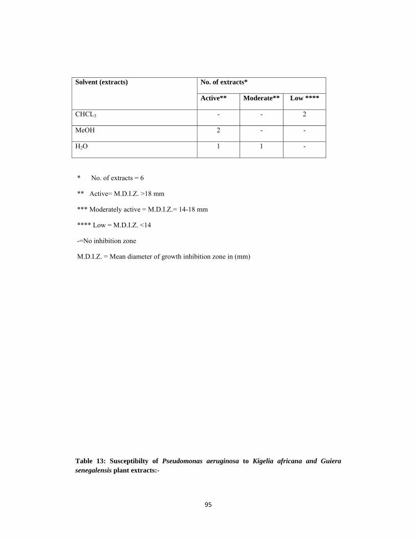
95
No. of extracts*
Low **** Moderate**Active**
Solvent (extracts)
2 - - CHCL3
- - 2 MeOH
- 1 1 H2O
* No. of extracts = 6
** Active= M.D.I.Z. >18 mm
*** Moderately active = M.D.I.Z.= 14-18 mm
**** Low = M.D.I.Z. <14
-=No inhibition zone
M.D.I.Z. = Mean diameter of growth inhibition zone in (mm)
Table 13: Susceptibilty of Pseudomonas aeruginosa to Kigelia africana and Guiera senegalensis plant extracts:-

96
No. of extracts*
Low***** Moderate*** Active**
Solvent (extracts)
1 1 - CHCL3
- 1 1 MeOH
- - 2 H2O
* No. of extracts = 6 ** Active= M.D.I.Z. >18 mm
*** Moderately active = M.D.I.Z.= 14-18 mm
**** Low = M.D.I.Z. <14
-=No inhibition zone
M.D.I.Z. = Mean dimeter of growth inhibition zone in (mm)
Table 14: Susceptibility of standard fungi to Kigelia africana and Guiera senegalensis
plant extracts:-
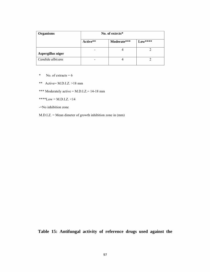
97
No. of extrcts*
Low**** Moderate*** Active**
Organisms
2 4 - Aspergillus niger
2 4 - Candida albicans
* No. of extracts = 6
** Active= M.D.I.Z. >18 mm
*** Moderately active = M.D.I.Z.= 14-18 mm
****Low = M.D.I.Z. <14
-=No inhibition zone
M.D.I.Z. = Mean dimeter of growth inhibition zone in (mm)
Table 15: Antifungal activity of reference drugs used against the

98
standard organisms.
MDIZ *( mm) Drug Concentration
used(µg /ml )
Aspergillus niger Candida albicans
Clotrimazole 20
10
5
24
19
16
43
33
30
Nystatin 50
25
12.5
17
14
-
28
28
23
MDIZ * : Mean diameter of growth inhibition zone (mm)
Interpretation of results
MDIZ (mm): > 18 mm : Active
: 14 – 18 mm: Moderate
:< 14 mm : Low
- : No inhibition zone
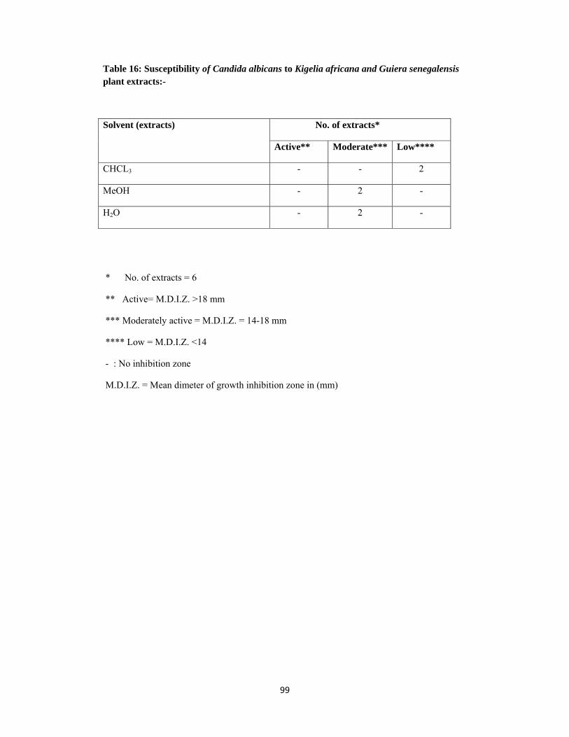
99
Table 16: Susceptibility of Candida albicans to Kigelia africana and Guiera senegalensis plant extracts:-
No. of extracts*
Low**** Moderate***Active**
Solvent (extracts)
2 - - CHCL3
- 2 - MeOH
- 2 - H2O
* No. of extracts = 6
** Active= M.D.I.Z. >18 mm
*** Moderately active = M.D.I.Z. = 14-18 mm
**** Low = M.D.I.Z. <14
- : No inhibition zone
M.D.I.Z. = Mean dimeter of growth inhibition zone in (mm)
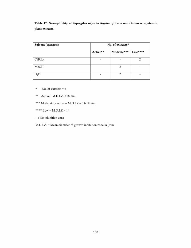
100
Table 17: Susceptibility of Aspergilus niger to Kigelia africana and Guiera senegalensis
plant extracts: -
No. of extracts*
Low**** Modrate***Active**
Solvent (extracts)
2 - - CHCL3
- 2 - MeOH
- 2 - H2O
* No. of extracts = 6
** Active= M.D.I.Z. >18 mm
*** Moderately active = M.D.I.Z.= 14-18 mm
**** Low = M.D.I.Z. <14
- : No inhibition zone
M.D.I.Z. = Mean diameter of growth inhibition zone in (mm
101
Table18: Determination of the minimum inhibitory concentration. (MIC)(mg/ml) of crude extract against standard organisms Plant Part Solvent B.s S.a E.c P.v Ps.a A.nig C.a
G. se Leaves MeOH >75 9.38 >75 >75 37.5 >100 >100
Ki. a Fruits MeOH 37.5 37.5 75 75 75 >100 >100
B.s=Bacillus subtilis. S.a= Staphylococcus aureus. E.c= Escherichia coli. P.v= Protus vulgaris. Ps.a= Pseudomonas aeruginosa. A.nig= Aspergillus niger. C.a= Candida albicans.
G. se= Guiera senegalensis; Ki.a = Kigelia Africana

102
Table 19: The activity of Guiera senegalensis leaves against 100 clinical isolates
No. of clinical isolates
Total Resistant Intermediate
Susceptible
Solvent
Organism tested
25
1
-
6
3
18
22
MeOH
H2O E. coli
25
-
-
8
1
17
24
MeOH
H2O
Pr. Vulgaris
25
2
-
1
1
22
24
MeOH
H2O
Ps.aeruginosa
25
-
-
1 3
24
22
MeOH
H2O S. aureus
100 Total
103
Table 20: The activity of Kigelia africana fruits against clinical isolates
No. of clinical isolates
Total Resistant Intermediate
Sensitive
Solvent
Organism tested
25
-
3
3
3
22
19
MeOH
H2O E. coli
25
-
2
1
9
24
14
MeOH
H2O
Pr. Vulgaris
25
-
-
-
14
25
11
MeOH
H2O
Ps.aeruginosa
25
-
2
4
5
21
18
MeOH
H2O S. aureus
100 Total
104
Table 21: Susceptibility of Staphylococcus aureus clinical isolates against selected plant
extracts exhibiting high antibacterial activity
iameter of inhibition zones,in( mm )
25 24 23 22 21 20 19 18 17 16 15 14 13 12 11 10 9 8 7 6 5
18
25
21
20
20
18
20
18
24
20
19
23
22
20
19
18
22
25
24
22
25
25
20
20
25
24
25
26
22
29
22
21
26
24
25
25
24
23
25
22
24
20
1
2
32
20
22
19
24
15
30
15
22
12
15
13
26
18
23
15
21
19
32
20
23
20
22
19
19
21
29
19
24
20
16
14
31
25
20
22
25
24
25
21
16
21
9
2
G.se =Guiera senegalensis
Ki.a= Kigelia africana
105
Table 22: Susceptibility of Escherichia coli clinical isolates against selected plant extracts exhibiting high antibacterial activity
meter of inhibition zones,in (mm)
25 24 23 22 21 20 19 18 17 16 15 14 13 12 11 10 9 8 7 6 5
25
15
25
23
20
20
20
21
19
19
20
20
19
20
25
20
15
20
20
22
23
22
15
22
20
20
15
15
20
25
20
20
20
15
15
22
24
30
15
22
24
26
0
5
25
20
25
20
25
21
25
20
15
20
30
21
15
20
24
21
24
24
25
23
30
0
20
15
25
20
19
20
15
22
20
15
25
11
21
11
32
16
32
23
25
22
5
2
G.se =Guiera senegalensis
Ki.a= Kigelia africana
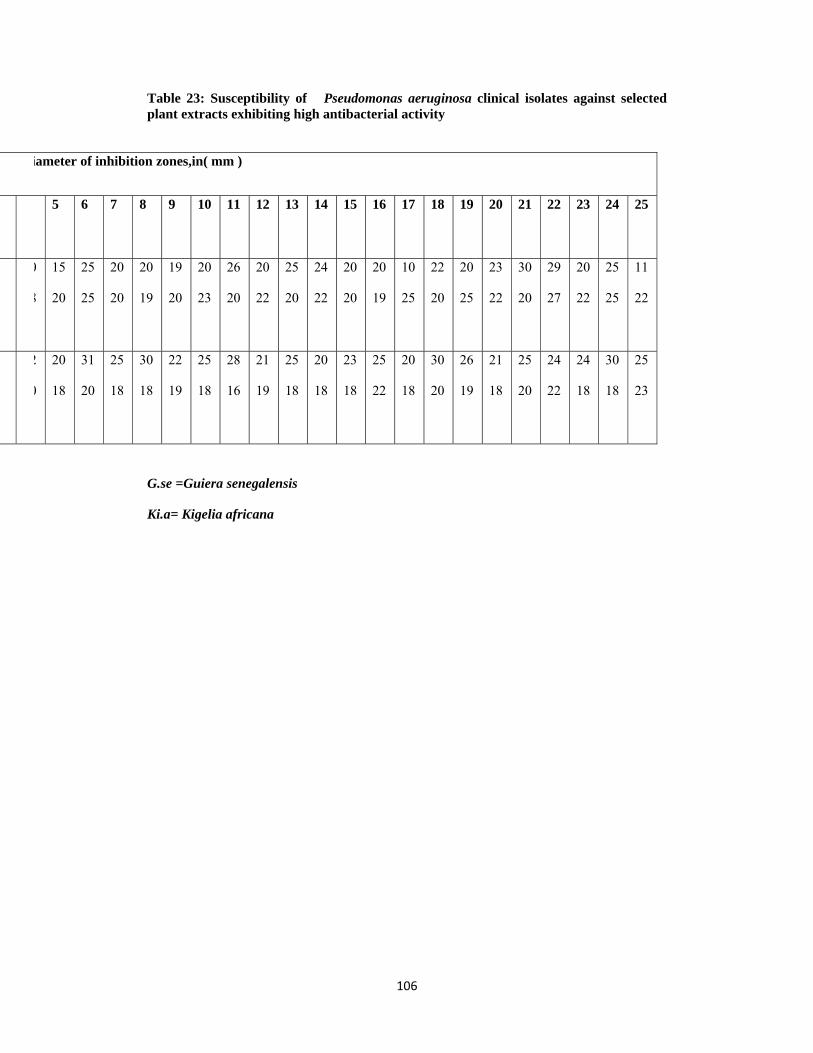
106
Table 23: Susceptibility of Pseudomonas aeruginosa clinical isolates against selected plant extracts exhibiting high antibacterial activity
iameter of inhibition zones,in( mm )
25 24 23 22 21 20 19 18 17 16 15 14 13 12 11 10 9 8 7 6 5
11
22
25
25
20
22
29
27
30
20
23
22
20
25
22
20
10
25
20
19
20
20
24
22
25
20
20
22
26
20
20
23
19
20
20
19
20
20
25
25
15
20
0
8
25
23
30
18
24
18
24
22
25
20
21
18
26
19
30
20
20
18
25
22
23
18
20
18
25
18
21
19
28
16
25
18
22
19
30
18
25
18
31
20
20
18
2
0
G.se =Guiera senegalensis
Ki.a= Kigelia africana
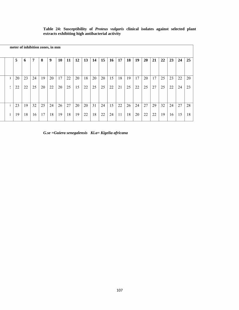
107
Table 24: Susceptibility of Proteus vulgaris clinical isolates against selected plant extracts exhibiting high antibacterial activity
meter of inhibition zones, in mm
25 24 23 22 21 20 19 18 17 16 15 14 13 12 11 10 9 8 7 6 5
20
23
22
24
23
22
25
25
17
27
20
25
17
22
19
25
18
21
15
22
20
25
20
25
18
22
20
15
22
25
17
20
20
22
19
20
24
25
23
22
20
22
0
2
28
18
27
15
24
16
32
19
29
22
27
22
24
20
26
18
22
11
15
24
24
22
31
18
20
22
20
19
27
18
26
19
24
18
25
17
32
16
19
18
23
19
5
4
G.se =Guiera senegalensis Ki.a= Kigelia africana
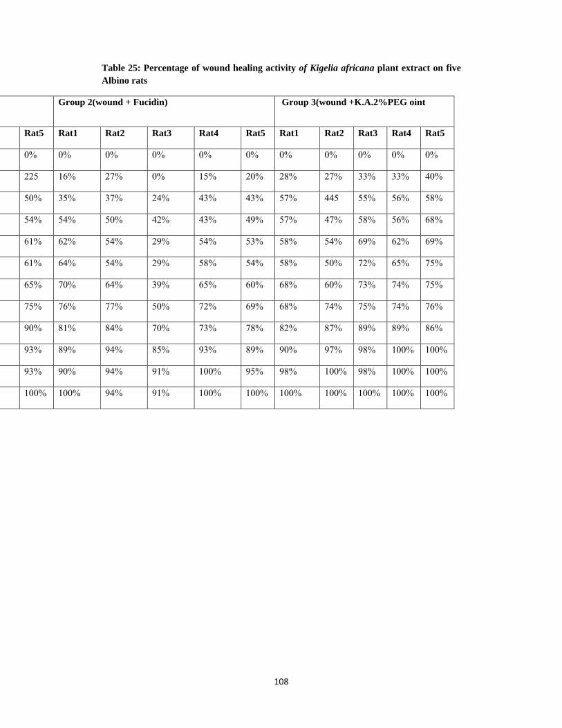
108
Table 25: Percentage of wound healing activity of Kigelia africana plant extract on five Albino rats
Group 3(wound +K.A.2%PEG oint Group 2(wound + Fucidin)
Rat5 Rat4 Rat3 Rat2 Rat1 Rat5 Rat4 Rat3 Rat2 Rat1 Rat5
0% 0% 0% 0% 0% 0% 0% 0% 0% 0% 0%
40% 33% 33% 27% 28% 20% 15% 0% 27% 16% 225
58% 56% 55% 445 57% 43% 43% 24% 37% 35% 50%
68% 56% 58% 47% 57% 49% 43% 42% 50% 54% 54%
69% 62% 69% 54% 58% 53% 54% 29% 54% 62% 61%
75% 65% 72% 50% 58% 54% 58% 29% 54% 64% 61%
75% 74% 73% 60% 68% 60% 65% 39% 64% 70% 65%
76% 74% 75% 74% 68% 69% 72% 50% 77% 76% 75%
86% 89% 89% 87% 82% 78% 73% 70% 84% 81% 90%
100% 100% 98% 97% 90% 89% 93% 85% 94% 89% 93%
100% 100% 98% 100% 98% 95% 100% 91% 94% 90% 93%
100% 100% 100% 100% 100% 100% 100% 91% 94% 100% 100%

109
Figure (1)
Antimicrobial activity of Kigelia africana (upper ) and Guiera
senegalensis (lower ) on Staphylococcus aureus
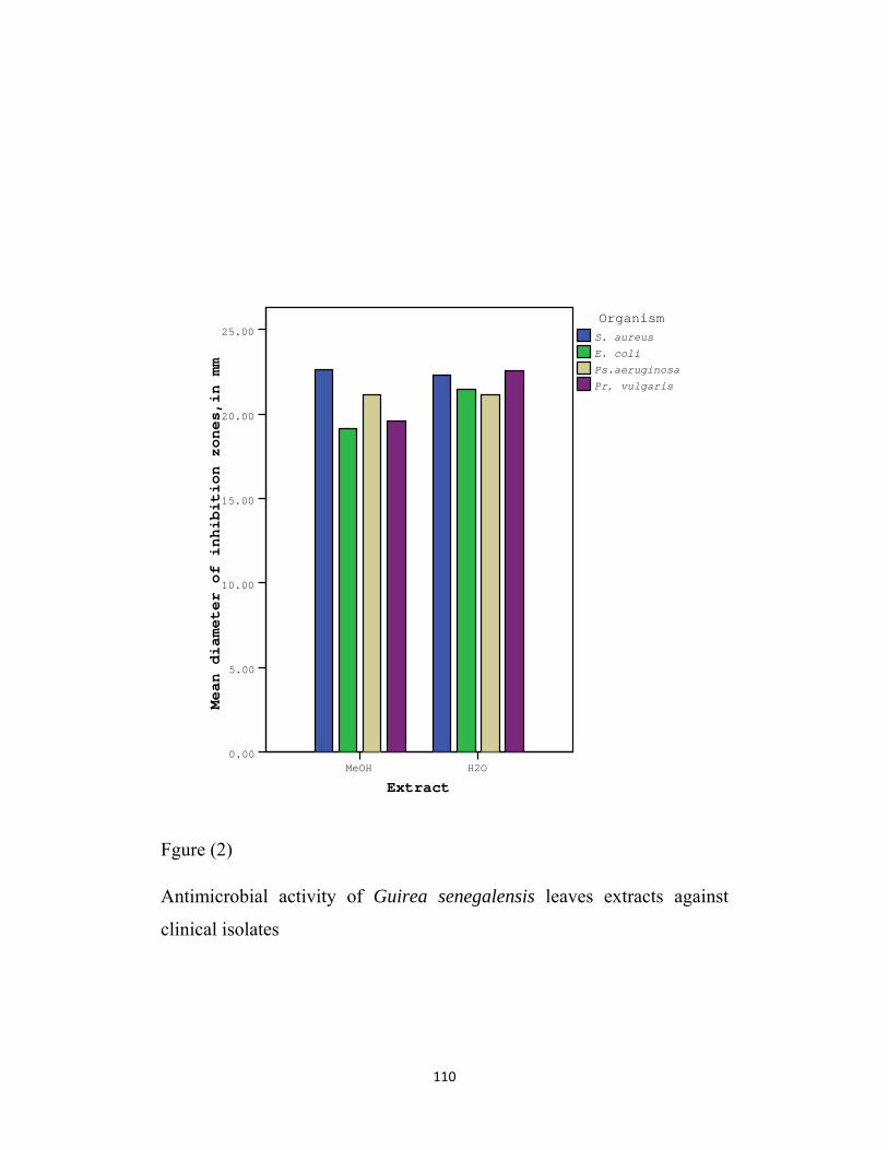
110
ExtractH2OMeOH
Mean diameter of inhibition zones,in mm
25.00
20.00
15.00
10.00
5.00
0.00
Pr, vulgaris
Ps.aeruginosa
E. coli
S. aureus
Organism
Fgure (2)
Antimicrobial activity of Guirea senegalensis leaves extracts against
clinical isolates

111
ExtractH2OMeOH
Mean diameter of inhibition zones,in mm
30.00
20.00
10.00
0.00
Pr, vulgaris
Ps.aeruginosa
E. coli
S. aureus
Organism
Figure (3)
Antimicrobial activity of Kigelia africana fruits extracts against
clinical isolates
112
DaysDay11
Day10Day9
Day8Day7
Day6Day5
Day4Day3
Day2Day1
Day0
Mean Reading
100
80
60
40
20
0
Group 3(woud +K.A.2%PEG oint
Group 2(wound + Fucidin)
Group 1(wound untreated )
groups
Figure (4)
Percentage wound healing activity of Kigelia Africana
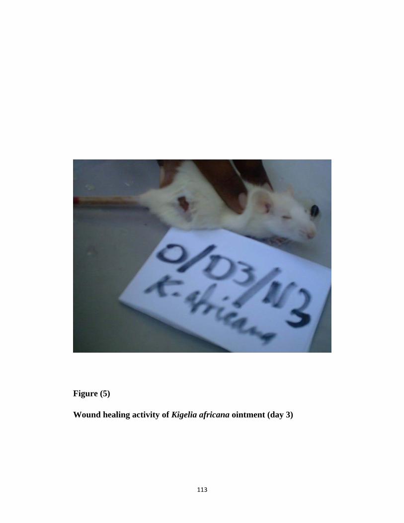
113
Figure (5)
Wound healing activity of Kigelia africana ointment (day 3)
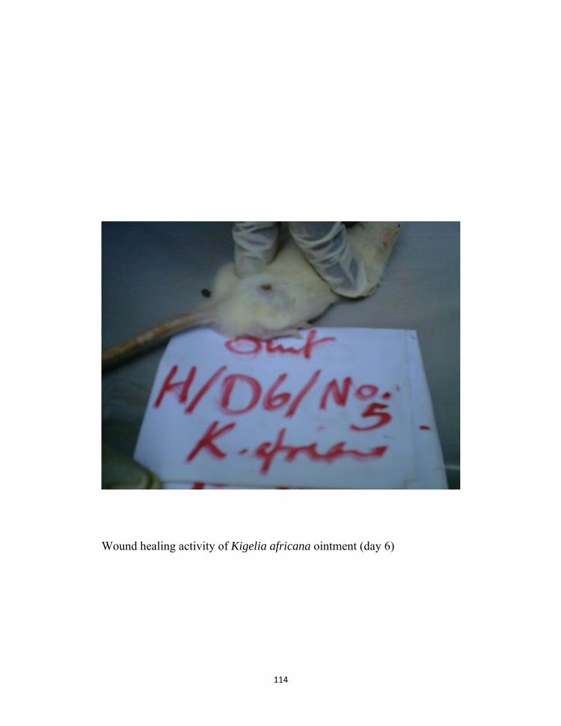
114
Wound healing activity of Kigelia africana ointment (day 6)
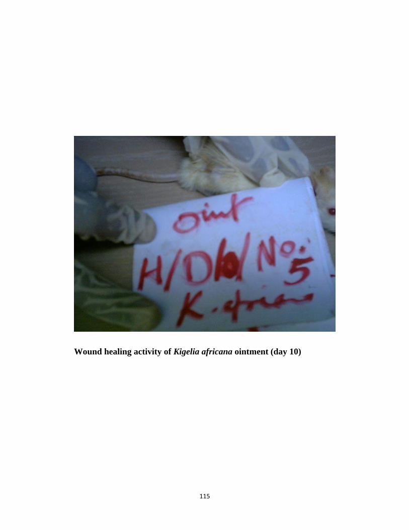
115
Wound healing activity of Kigelia africana ointment (day 10)
116
4. Discussion
4.1The antimicrobial activity of the two medicinal plants:
4.1.1 Kigelia africana
In this study the chloroform fruit extract of Kigelia africana showed low activity (11-13mm) against Bacillus subtilis, Escherichia coli, Pseudomonas aeruginosa, intermediate activity (14mm) against Staphylococcus aureus, and no activity against Proteus vulgaris, Aspergillus niger and Candida albicans.
The methanol fruit extract of Kigelia africana exhibited pronounced activity (25mm) against B.subtilis, (22mm) against Proteus vulgaris, high activity (19mm) against Escherichia coli and intermediate activity (15-18mm) against Staphylococcus aureua, Pseudomonas aeruginosa and against both fungi Aspergillus niger and Candida albicans.
Its aqueous fruit extract showed pronounced activity(24mm) against Staphylococcus aureus, high activity(21-22)against Escherichia coli and Pseudomonas aeruginosa and intermediate activity(16-18mm) against Bacillus subtilis Proteus vulgaris,Aspergillus niger and Candida albicans.
This result is different from that reported by Almagboul (1992) who found that the chloroform and the methanol extracts of Kigelia africana gave good results while the aqueous extract was inactive. This could be due to the unknown concentrations used.
Grace etal. (2002) tested the stem and fruit of Kigelia africana aqueous, ethanol and ethyl acetate extracts, which is the most popular source of traditional medicine throughout Africa, for their antibacterial activity using the microtitre assay. All extracts showed significant activity against the tested Gram positive and Gram negative bacteria, thus supporting the traditional use of the plant in the therapy of bacterial infections. These results are similar to our results.
Ijah and Oyebanji,(2003)determined the activity of the crude extracts of the medicinal plants Kigelia afrcana,Bridelia ferruginea,Trema nitems and Drypetes gossweileri against Escherichia coli,Staphylococcus aureus
117
Pseudomonas aeruginosa,Klebsiella sp. and Proteus sp., causing urinary tract infection. The extracts of the medicinal plants inhibited the growth of the bacteria tested. These results are in agreement with our result.
Gram positive and Gram negative bacteria were equal in their susceptibility to the different plant extracts used in this study and this result contrary to that obtained by Abdalla (2004) who proved that the Gram negative organisms were less susceptible to the extracts than the Gram positive bacteria. It is evident that the patterns of inhibition varied with the plant part, the solvent used for extraction and the organisms tested.
The methanol extract of Kigelia afrcana exhibited intermediate activity
against Aspergillus niger which is almost similar 5µg/ml Clotrimazole
and 25µg/ml Nystatin i.Its aqueous extract exerted intermediate activity
against Aspergillus niger which is similar to50µg/ml Nystatin 4.1.2Gueria senegalensis In the present study, the chloroform leaves extracts of Gueria senegalensis showed low activity (11-12) against Bacillus subtilis, Staphylococcus aureus, Proteus vulgar and Candida albicans and an intermediate activity against Pseudomonas aeruginosa while no activity against Escherichia coli and the fungus Aspergillus niger.
Both its methanol and aqueous extracts exhibited pronounced activity (20-29mm) against both Gram positive and Gram negative organisms and intermediate activity (16-18mm) against both fungi. These results are similar to that reported by Grand (1989) who found that the leaves of Gueria senegalensis showed antimicrobial activity against the bacteria and fungi tested. Similarly Sanago etal. (1998) found that a decoction and methanol extracts of Gueria senegalensis exhibited significant activity against all strains of bacteria tested and that is suggested to the use of the same solvent.Our results are different from Bosis etal. (1997) who found that the crude extract of the same plant possessed a mild antimicrobial effect only on Gram positive bacteria.
118
The methanol and aqueous extracts of Gueria senegalensis leaves showed high activity against both Gram positive and Gram negative organisms which was more than that produced by40µg/ml Ampicillin.The methanol extract inhibited Bacillus subtilis, Proteus vulgaris similar to 40µg/ml of Gentamicin, Pseudomonas aeruginosa Staphylococcus aureus higher than 40µg/ml of Gentamicin and inhibited Escherichia coli similar to 40µg/ml Gentamicin.
The aqueous extract of the same plant inhibited Bacillus subtilis similar to 20µg/ml Gentamicin and inhibited Staphylococcus aureus, Escherichia coli, Proteus vulgaris and Pseudomonas aeruginosa higher than 40µg/ml of Gentamicin.
The methnol extract of Gueria senegalensis showed intermediate activity against Aspergillus niger which is almost similar to 10µg/ml Clotrimazole and 50µg/ml Nystatin .Its aqueous extract exhibited intermediate activity against Aspergillus niger which is similar to 5µg/ml Clotrimazole and almost similar to 50µg/ml Nystatin.
119
4.2 Discussion of wound healing activity of Kigelia africana
In the present study the methanolic extract of Kigelia africana was able to reduce the time required for wound healing. The results were obtained from the faster contraction of the wound treated with Kigelia africana extract in comparison with fucidin ointment or untreated groups.
Kigelia africana ointment in 2% Poly ethylene Glycol was more potent than Fucidin ointment as standard healing agent.The percentage of healing with Polyethylene Glycol contaning Kigelia africana 2% ointment was significantly higher than Fucidin group from the first day of treatment until the closure of wounds. This is similar with Arzi et al. (2003) who found that the healing effect of Licorice cream of 10 % is a potent healing agent even better than phenytoin cream
Owolabi&Omogbai. (2007) evaluated the ethanolic extract of Kigelia africana for analgesic property using acetic acid induced mouse writhing and hotplate reaction time and anti-inflammatory activity using the carrageenan induced paw odema and its probable mechanism evaluated in mice and guinea pigs.Kigelia africana extract showed a dose dependant significant reduction of the number of writhes (p0.001) with 500mg /kg body weight dose giving the highest reduction. The extract showed significant analgesic and anti-inflammatory activity.
Inhibition of the synthesis of prostaglandins and other inflammatory mediators probable accounts for the analagesic and anti inflammatory.
Asekum etal.(2007) analysed the volatile constituents of the oil form the leaves and flowers of Kigelia africana from Lagos( Nigeria), isolated by hydrodistllation using GC and GC/MS.The leaf oil was found to contain 25 components, while the flower oil contained nine.These components responsible for the bioactivity of this plant extracts.
Not to my knowledge there is a literature concerning wound healing activity of methanolic extracts of the fruits of Kigelia africana.However more studies are reguired to elucidate the extract mechanism of Kigelia africana in wound healing modles.
120
5.1 Conclusions
Sudan has huge resources of plants. Exploitation of these plants
represents an important means of obtaining cheap and effective drugs,
which can participate in the solution of the health problems in the
country.
The results of the present work indicate that there are plants with
promising high and broad antimicrobial activity, when compared with
standard antimicrobial drugs in current use. These findings verified the
claimed bioactivity of these plants and their employment in traditional
medicine in Sudan.
The methanolic and aqueous extracts of Guiera senegalensis and Kigelia
africana proved to have significant antimicrobial activity and this
justifies their traditional uses.
Methanolic extract of Kigelia africana was proved to have wound healing
activity, and this justifies its traditional use as a wound healing agent.
121
Recommendations
Pharmacological, toxicological and clinical studies should be carried out
on the selected medicinal plants to assess their safety, therapeutic efficacy
and potential for commercial utilization.
Formulation of the active extracts and/ or principles in suitable dosage
form, with special reference to Kigelia africana fruits which proved to be
a potent wound healing agent.
Bio-assay-guided fractionation and purification may lead to isolation of
the active compounds. The chemical structures of these compounds can
then be elucidated. This can then help in:-
i- The standardization of the active.
ii- The study of the structure activity relationship for the
production of compounds with improved characteristics.
iii- The study of pharmacokinetics of the pure active compounds
and also it helps in the formulation procedures.
122
References
Abdrabo, AN; Omer, MEA; Elnima, EI; Shayoub,MAA.; Almagboul
AZ; (2005) .Wound healing activity of methanolic extract of Solenostema
argel in poletyyleneglycol ointment.Omdurman Journal of Pharmceutical
Sciences 1(1): 43-54.
Adupa, SL; Udopa, AL; Kulk Arni, DR. (1991). Influence of Tridax
procumbens on dead space wound healing. Fitoterapia. 62(2): 146-1
Ahmed ,AM ; Gumaa, SA.; Abdelmageed, MAM.; Almagboul,
AZ.(2007).IN vitro antimicrobial activity of some Sudanese plants used
in traditional medicine. Omdurman Journal of Pharmaceutical Sciences
1(3): 367-378.
Ahmed, IH; Awad, MA; Elmahdi, M.; Gohar, HM.; Ghanem,
AM.(1995). The effect of some medicinal plants extracts on wound
healing in farm animals .Assiut Veterinary Medicinal Journal. 32(64):
236-244.
Ali, MS.; Yaghmour, RMR; Faidi, YR; Salem, K; Al-Nuri, MA. (1998).
Antimicrobial activity of 20 plants used in folkloric medicine in
Palestinian area. Journal of Ethnopharmacology, 60(3): 265-271.
123
Almagboul, AZ. (1992). Antimicrobial and Phytochemical Investigations
of Vernonia amygdalina and other Sudanese Medicinal Plants. Doctorate
thesis, Khartoum, University of Khartoum, Sudan.
Ancolio,C; Azaz,N; Mahiou,V; Ollivier, E; Giorgio,CD; Keita,A;
Timon,D.; Palansard,G; Giorgio,DC.(2002). Antimicrobial activity of
extracts and alkaloid isolated from six plants used in traditional medicine.
Phytotherapy Research.16 (7):646-649.
Anil, S; Shukla, NY; Sushil, K; Srivastava, A; Kumars, S. (2000). Recent
development in plant derived antimicrobial constituents-a review. Journal
of Medicinal and Aromatic Plant Sciences, 2(3): 349-405.
Ansel, Hc; Allen, LV.; Popovich, NG (2000). Pharmaceutical Dosage, 7th
edition, Lippinctt Willams and Wilkins (pub), New York
Arzi,A .; Hemmati, AA; Amin, M. (2003). Stimulation of wound healing
by licorice in rabbits. Saudi Pharmaceutical Journal. 1(2): 57-60.
Asekun, OT. ; Olusegum, E.; Adebola, O. (2007). The volatile
constituents of the leaves and flowers of Kigelia africana. Flavour and
Fragrance Journal. 22(1) 21-23.
124
Atindehou, KK. ; Kone, M.; Terreaux, C.; Traore, D.; Hostettmann, K.;
Dosso, M. (2002). Evaluation of the antimicrobial potential of medicinal
plants from the Ivory Coast. Phytotherapy Research, 16(5): 497-50
Austin, GW. ; Guenim, MF. ; Hokett, SD. (2001). Effect of nicotine on
fibroblast beta-1. J. Perodontol. 72(4) 438-44.
Ayensu, ES. (1978).Medicinal plants of West Africa. Reference
publications, Inc. (pub.), New York.
Bagchi, GD.; Amrita, S.; Khanuja, SPS. ; Bansal, Rp. ; Singh, SC.;
Sushil,K.(1999). Wide spectrum antibacterial and antifungal activities in
the seeds of some coprophilous plants of North Indian Plains. Journal of
Ethnopharmacology, 64(1): 69-77.
Balchin , M. (1997). Essential oils and 'aromotherapy': their modern role
in healing. Journal of the Royal Society of Health, 117(5): 324-329.
Bandara, BMR. ; Hewage, C M .; Jayamanne, DHLW.; Krunaratne, V.;
Pinti, MRM. (1990). Biological activity of some steam distillates from
leaves of ten species of rutaceous plants.Journal of the National Science,
18(1): 17-77.
125
Bhacta, T.; Mukherjee, Pk.; Pal, M.; Saha, BP. (1998): Studies on in vivo
wound healing activity of Cassia fistula leaves in rats. Natural Product
Sciences. 4(2): 84-87.
Bodeker,G.; Hughes, AM.; Prendergast, HDV.; Etkin, NL.; Harris, DR.;
Houghton, PJ.(1998). Wound healing, traditional treatments and research
policy. Plants for food and medicine, 1(6): 345-359.
Bosisio, E.; Mascetti, D.; Verotta, L.; Zani, F.; Mazza, P.; Talbot, M.
(1997). Guiera senegalensis J.F. Gmelin(Combretaceae): Biological
activities and chemical investigation. Phytomedicine, 3(4): 339-348.
Boulos, L. (1983). Medicinal plants of North Africa. Reference
Publications, Inc. (Pub). USA.
Bushell, AC. (1989). Medical Bacteriology, first Edition, Oxford
University Press, New York, 167-193.
Cheesbrough, M. (1996). Medical Laboratory Manual for Tropical
Counteries (Microbiology) 14 Bevills Close, Paddington Cambridge
Shire (Pub). England.Vol II.
Cheesbrough, M. (2000). District laboratory Practice for Tropical
countries. Part2. University Press. Cambridge.
126
Cheesbrough, M. (2004). District Laboratory Practice in Tropical
Countries, Part 2, Low price edition, Cambridge.
Chhabra, SC.; Uiso, FC. (1991).Antibacterial activity of some
Tanzanian plants used in traditional medicine. Fitoterapia, 62(6): 499-
503.
Collee,JG.;Fraser,AG.;Marmion,BP.;Simmons,A.;Mackie and
McCartiny.(1996).Practical Medical Microbiology.14th edition.Churchill
Livingstone . New York.
Collett, DM., Aulton, Me. (1991): Pharmaceutical Practice, First edition,
Churchill, Livingstone, London.
Couster, LV.; Pashley, MM .; Tattersall, KJ. (1961). Ointments
Pharm.Pharma.3 (1): 620-624.
Cowan, ST.; Stell, KJ. (1970). Manual for the Identification of Medical
Bacteria.First edition.Cambridge. University Press.
Cruickshank, R.; Duguid, JP.; marmion, BP.; Swain, RH. (1975).
Medical microbiology 12th edition. Churchill Livingstone (pub) Edinburg,
London and New York. Vol II.
127
Dilika, NF. ; NI Kolova, RV.; Jacobs, TV. (1996).Plants used in the
circumcision rise of the Xhosa tribe in South Africa. International
Sympsium on Medicinal and Aromatic Plants.27 (30) 165-169. S
Douglas, J.; Mackay,A; Miller. L. (2003). Nutritional support for wound
healing. Alternative Medicine Review. 28(10):1-3
Elghazali, GEB. ; EA.; Bashir, AK; Salih, AKM. (1987). Medicinal
Plants of the Sudan: Medicinal plants of the Eastern Nuba Mountains.
Part II. Khartoum, Khartoum University Press. Sudan
Elghazali, GEB. ; Eltohami, MS.; Elegami, AAB. (1994): Medicinal
plants of the Sudan: Medicinal plants of the White Nile Provinces. Part
III. Khartoum, Khartoum University Press.Sudan.
Elghazali, GEB. (1997).Sudanese Promissing Medicinal
Plants.Omdurman Islamic University Press (Pub.)Sudan
El-Hadi, MMM.; Hassan, SK.; Yousif, GM. (2005): Antimicrobial
activity of Nigella sativa L.seeds oil. Fitoterapia, 1(1): 103-116.
Elmer, WK.; Steven, DA.; Dwell, VR.; Willam,MJ.; Herbert, MS.;
Washington, CW (1990).Colour Atlas and Text book of Dignostic
Microbiology. Third edition, Lippincott Company, Philadelphia, 473-475
128
Faleiro, ML. ; Miguel, MG.; Ladeiro, F.; Venancio, F.; Tavares, R.;
Brito, JC.; Figueiredo, AC.(2003). Antimicrobial activity of essential oils
isolated from Portuguese endemic species of Thymus. Lett Appl
Micobiol, 36(1): 35-4
Garg, S.; Chhabra, R.; Talwar, GB.(1994). Neem from ethnomedicine to
modern system of medicine. Ethnobiology in Human welfare: abstracts of
the fourth international congress of ethnobiology, Lucknow, India, 17-21.
Grace, OM.; Light, ME.; Lindsey,KL.;Standen,(2002). Antimicrobial
activity and isolation of active compounds from fruit of Kigelia
africana.South Africa Journal of Botany.68 (2):220-222.
Grand, AL. (1989).Anti-infectious phytotherapies of the tree-savanna,
Senegal (West Africa).Journal of Ethnopharmacology. 25(3):315-338.
Hamill, FA; Apio, NK. ; Bukenya, Z R.; Monsango, M.; Maganyi, OW;
Soejarto, DD. (2003). Traditional herbal drugs of Southern Uganda,
literature analysis and antimicrobial assays. Journal of
Ethnopharmacology, 84(1): 57-78.
Haraguchi, H. (1998). Mode of action of antimicrobial substances in
plants. Recent Research Developments in Agricultural and Food
Chemistry, 2(1): 259-268.
129
Hardman, JG.; Limird, LE.; Molinoff, PB.; Puddon, RW.; Gilman,
AG.(1996).The Pharmacological Basis of Therapeutics. 9th Edition.
McGraw Hill. New York
Harries, M. (1964). Pharmaceutical Microbiology. Mailiene, Tindal and
Cox. London.
Hawkey, PM.; Lewwis, DA. (1989). Medical Bacteriology, a practical
approach, first edition, Oxford University Press, Oxford.
Ibrahim, LF. ; Kawashty, SA.; El-Eraky, WI.; Shabana, MM.; El-
Negoumy, SI.(2000). Acomprative study of the flavonoids,
pharmacological and antimicrobial effects. Egyptian Journal of
Physiological-Sciences, 24(1): 63-82.
Ijah, UJJ and Oyebanji, FO. (2003). Effect of Tannins and Polyphenols
of some Medicinal plants on bacterial agents of urinary tract infection.
Global journal of Pure and Applied Sciences, 9(2): 193-198.
Jovel, EM .; Cabanillus, j.; Towers, GHN. (1996) .An ethnobotanical
study of the traditional medicine of the Mestizo people of Suni-Marino.
Journal of Ethnopharmacology, 53(3): 149-156.
130
Kakali., S.; Mukherjee, PK.; Das,J.; Pal,M.(1997).Wound healing
activity of Leucas eavadulaefolia Rees. Journal of Ethnopharmacology.
56(2): 139-144.
Kavanagh, F. (1972). Analytical Microbiology. Vol II, Academic Press
(Pub), New York and London.p.11
Kay, MA. (1996). Healing with plants in the American and Mexican
West, First edition, Arizona, University of Arizona press.
Khan, MT.; Ashraf, M.; Bukhtair, MK.; Ashraf, S. (1993) Antimicrobial
activity of Withania coaulans. Fitoterapia 4 (1) 431-436.
Kostova, I. (2001). Fvaxinus ornusl. Fitoterapia. 72(5): 4
Kwo,Vt.;Craker,LE.;Nolan,L.;Shelly,K.(1996). Screening Camerron
MedicinalPlant extracts for antimicrobial activity.Acta Horiculturare.42
(6):147-155.
Mahasneh, AM.; Abaas, J A .; El-oqlah, AA.(1996). Antimicrobial
activity of extracts of herbal plants used in the traditional medicine of
Bahrain. Phytotherapy Research, 10(3):251-253.
Manhal, H; Almagboul, AZ.; Omer, MEA. ; Elshiekh, OM. (2004). In
vitro antibacterial activity of Pistacia lentiscus against some human
bacterial infections. Journal of Science and Technology, 5(1):65-72.
131
Martino, V.; Caffini, N.; lappa, A.; Ferraro, G.; Schilcher, H.; Phillipson,
JD. (1999). Second world congress on medicinal and aromatic plants for
human welfare. Acta-Horticulturae, 501:336.
Martins, AP.; Salgueiro, LR. ; Vila, R.; Gunha, A.(2002) .” Essential oil
composition and antimicrobial actitivity. Planta Medica. 69(1) 77-79.
Mathieu, D.; Watell. F.; Billard, V.; Defoin, IF. (1999). Post traumatic
limb ischemia. Journal of Trauma.30 (3): 307-314.
Merzouki, A.; Derfoufi, F.; El-Aallali, A.; Molero, MJ. (1997). Wild
medicinal plants used by local Bouhmed population (Morocco).
Fitoterapia, 68(5): 444-460.
Michel,DA.and Demontaig,NE.( 1993).Medicinal plants. Fitoterapia.
Volume 54(1): 5-8.
Miles, M. and Misra, SS. (1938). The estimation of the bactericidal
power of the blood. Journal Hyg. 8(3): 732.
Mohammed, TO. ; Almagboul, AZ.; Omer, MAA. ; Abd Rabo, AN.;
Ahmed, SOM. (2006). In vitro antimicrobial activity of Borreria sinesis.
Omdurman Journal of Pharmceutical Sciences 1(2): 188-199.
Nagappa, AN.; Cheriyan, B. (2002). Wound healing activity of the
aqueous extract of Thespesia populnea fruit. Fitoterapia. 72(5): 503-506.
132
Nayar, Sr.; Balagopian, TP; Raveevdran, NS. (1994).Clinical traial with
Himax lotion on wounds in large animals.Indian Journal of Indigenous
Medicines. 11(1): 61-62.
Nexon, W. (1951). Polyethylene Glycol. Parma. J. 16(7): 213.
Nwosu, MO. (1999).The use of herbal remedies against Psychiatric
ailments in Tropical West Africa. Fitoterapia, 70(1):58-63.
Palanichamy, S.; Bhaskar, Ea.; Bakthava thsal, R. (1992). Wound
healing activity of Cassia alate.Fitoterapia. 62(2): 153-156.
Patric, RM.; Ken, SR.; Michael, AP. (2005). Medical Microbiology,
Fifth edition, Library of Congress, Philadelphia, 203-212
Pattel, NK and Foss, NE. (1964): Polyethelene Glycol.Journal Pharm.
Sci. (3): 367-369.
Pieters, L.; Bruyne, T.; Poel, BV. ; Vingerhoets, R.; Totte, J.; Berghe, D.
(1995). In vivo wound healing activity of Dragon’s blood(croton spp.), A
traditional South American drug, and some of its constituents.
Phytomedicine, 2(1): 17-22.
Phillipson, JD. (1997). Biology in action: Medicinal plants. Journal of
Biological Education, 31(2): 109-115.
133
Poole, TB and Robinson, R. (1989): The Hand bookon the care and
mangement of laboratory animals. 6th Edition. Longman Scientific and
Technical (Pub). London.
Omer, MEA. ( 2000). Medicinal and Aromatic Plants as a Source of
Herbal Medicines, National Symposium on the Research Application in
the iIndustrial Sector(ISESCO).16-18 October, Khartoum, Sudan.
Owolabi, OJ andOmogabi, EK.(2007). Ethanolic stem- bark extract of
Kigelia africana. African Journal of Biotechnology. 6(5) 582-585.
Ramesh, N.; Viswanathan, MB.; Saraswathy, A.; Balakrishna, K.;
Brindha, P.; Lakshmanaperumalsamy, P.(2001). Phytochemical and
antimicrobial studies of Bridelia crenulata. Pharmaceutical Biology,
39(6): 460-464.
Rao, SG. ; Udupa, AL.; Udupa, SL.; Kulkarni, DR. (1991): Calendula
and Hypericum: Two homeopathic drugs promoting wound healing in
rats.Fitoterapia, 62(6): 508-510.
Rasik, Am.; Ram, R.; Gupta, A.; Shukla, A.; Dubey, MP.; Srivastava, S.;
Jain, HK.; Kulshrestha,DK.; Raghubir, R.(1999) Healing potential of
Calotropis procera. Journal of Ethnopharmacology, 1(3): 261-266.
134
Robinson, J.; Gibaldi, M.; Weiner, ND. ; Kanig , J I. ;( 1964).
Polyethylene Glycol, Journal of Pharm. Sci .5 (3): 1245-1247.
Rode, H.; Wet, PM.; Cywes, S. (1989).The antimicrobial effect of Allium
sativum L. (garlic). South African Journal of Science, 85(7): 462-464.
Rukangira and Ernest. (2001). The African herbal industry constrains
and challenges.The natural products and cosmeceuticals conference.
Erboristeria Domani (Pub.).
Salle, AT. (1961). Fundamental Principal of Bacteriology. 5th Edition
McGraw hill Book CO (Pub.). London.
Sanogo,R.;GrisafiG.;Germano,Mp.;Depasquale,R.;Bisignano,G.;Cappaso
,F;.Basso,F;Evanos,FJ.;Mascolo,N.(1998).Evalulation of Malian
traditional medicines.Phytotherapy research.12(1):5154-5156.
Shayoub, MAA. (1985). Polyethylene Glycol ointment bases evaluations
and application in Sudan, Master Thesis, and University of Khartoum,
Sudan.
Shukla, A.; Rasik, AM.; Shankkar, R.; Dhawan, BN. (1999). Invitro and
in vivo wound healing activity of asiaticoside isolated from Centella
asiatica. Journal of Ethnopharmacology. 65(1): 1-11.
135
Sidhu,GS.; Singh, AK.; Banaudha, KK.; Gaddipata, JP.; Patnaik, GK.;
Maheshwari,PK. (1999). Arnebin-1 accelerates normal wound healing.
Journal of Investigative Dermatology, 113(5): 773-781.
Silva, O and Gomes, ET. (2003). Guieranone A naphthyl butanone from
the leaves of Guiera senegalensis with antifungal activity.Journal of
Natural Products.66 (3):447-449.
Thang, PT.; Patrrick, S.; Teic, LS. ; Yung, CS.(2001). Anti-oxidant
effect of the extracts from the leaves of Chromolaena odorata on human
dermal fibroblasts. Burns, 27(4): 319-327.
Tego, G.; Stermitz, FR.; Lomovskaya, O.; Lewis, K. (2002). Multidrug
pump inhibitors uncover remarkable activity of plant antimicrobials.
Antimicrobial Agents and Chemotherapy, 46(10): 3133-3141.
Thom, SR.; Bolotin, T.; Hardy, K.; Nebolon, M.; Kilpatric, L.
(1997).Inhibition of human neutrophil. Amercan Journal of physiology.
3(1):770-777.
Ueng, TH; Kang, JJ; Wang, HW; Lin, PC. (1997): An overview of the
toxicology of commonly used traditional Chinese medicine. Journal of
Food and Drug Analysis 5(4): 241-264.
136
Valsaraj, R.; Pushpangadan, P; Smitt, UW; Adsersen, A. (1997).
Antimicrobial screening of selected medicinal plants from India. Journal
of Ethnopharmacology, 58(2):75-83
Viller, AM; Schorn, PJ; Abatangelo, G; Nencini, P; Baradli, M. (1998).
IX Congress of the Italian Society of Pharmacognosy. Fitoterapia, 5: 82.
Warren, MD and Ernest, MD. (2002). Medical Microbiology and
Immunology, Seventh edition, Donnelley and Sons Company, New York
59-80.
Yuh, FC; Huei, YT; Tian, SW. (1995). Antimicrobial and analgesic
activities from roots of Angelica pubescens. Planta Medica: 2(8), 2-8.
Related Documents