International Journal of Pharmaceutical Chemistry ISSN: 2249-734X (Online) Journal DOI: 10.7439/ijpc CODEN: IJPCH3 (American Chemical Society) * Correspondence Info G. Panduranga Murthy Department of Engineering Chemistry, Sampoorna Institute of Technology and Research, (Visvesvaraya Technological University, Belgaum) Belakere, Channapatna taluk, Ramanagara district (Karnataka) -560162. E mail: [email protected] Research Article Antimicrobial and Antioxidant Activities of Tribal Medicine formulation (TMF) accomplished for Wound related remedies in Biligirirangana Hill area of Chamarajanagara district, Karnataka (India) G. Panduranga Murthy* 1 , K. B Chandrasekhar 2 , B. C. Leelaja 3 and S. Lokesh 4 1 Department of Engineering Chemistry, Sampoorna Institute of Technology and Research, (Visvesvaraya Technological University, Belgaum) Belakere, Channapatna taluk, Ramanagara district (Karnataka) 560162. 2 Department of Chemical Engineering and Biotechnology, Jawaharlal Nehru Technological University (JNTU), Ananthapuramu-515 002 (Andhrapradesh), India. 3 Bhoomigeetha Institute of Research and Development (BIRD), B.H. Road, Tumkur-572 102, Karnataka, India. 4 Department of Studies in Biotechnology, University of Mysore, Manasagangothri, Mysore-570006, India. 1. Introduction Plant-derived substances have recently become of great interest owing to their versatile applications in the ‘Health care system’. Ethno-medicinal plants are the main source of large amount of herbal drugs comprising to different groups such as antimicrobials, anti-inflammatory, anti-cancer, anti-viral, anti-venom activities etc. Medicinal plants are the richest bio-resource of drugs of traditional systems of medicine, modern medicines, nutraceuticals, food supplements, folk medicines, pharmaceutical intermediates and chemical entities for synthetic drugs [1]. Globally, a large number of the ethno-medicinal plants are claimed to possess the antibiotic properties in the traditional system and are used extensively by the tribal/ folk medicinal community. It is now believed that Abstract The present investigation looking at some less known medicinal uses of ethno-medicinal plants drug formulation which is practiced by Tribal Medicine Men (TMM) at Biligirirangana Hill area of Chamarajanagara district, Karnataka, India. This was mainly based in the light of traditional practice of herbal drugs comprising of diverse ethno-medicinal plants for other related ailments. The contents and amount of EMP present in the formulations were validated scientifically. The previous studies of these ethno-medicinal plants confirm that, the physico-chemical analysis and the active status of pharmacognostic parameters which evidently indicates the efficiency of these plant drugs. To ascertain these findings, a study on biological activities in the vein of Antimicrobial and Antioxidant activities were attempted. The antimicrobial activity was tested against standard strains such as Escherichia coli, Bacillus subtilis, Staphylococcus aureus, Kliebsiella pneumonia and Pseudomonas aerugenosa using disc diffusion method. The significant zone of inhibition was observed in all the ethno-medicinal plant drugs followed by TMF and the result was found to be superior over standard antibiotics. The antioxidant activity has been evaluated by DPPH radical scavenging and ABTS methods, respectively. All the EMP drugs registered significantly higher in phenolic content, individually (107.67mg GAE/g) than that of TMF (102.45 mg GAE/g). Similarly, antioxidant potential was found to be noteworthy as determined by DPPH (12.24 EC 50 μg/ml) radical scavenging activity which was registered in the EMF individually. A superior ABTS radical scavenging (ranging between12.06-15.40 EC 50 μg/ml), Frap assay (ranging between 8.25-21.26μmol Fe 2+ /mg) and significantly higher total reduction capacity (1.28%) were realized in ethanolic extract of TMF. As a result, the EMF and TMF drugs can be used as functional foods to increase shelf-life of food items for human consumption and neutraceuticals to dissuade deleterious free radical-induced life threatening diseases. Keywords: Antioxidant activity, Antimicrobial activities, Tribal Medicine Formulation (TMF), Tribal / Traditional Medicine Men (TMM), B.R. Hills, Karnataka.

Welcome message from author
This document is posted to help you gain knowledge. Please leave a comment to let me know what you think about it! Share it to your friends and learn new things together.
Transcript

International Journal of Pharmaceutical Chemistry ISSN: 2249-734X (Online) Journal DOI: 10.7439/ijpc
CODEN: IJPCH3 (American Chemical Society)
* Correspondence Info G. Panduranga Murthy
Department of Engineering Chemistry,
Sampoorna Institute of Technology and Research,
(Visvesvaraya Technological University, Belgaum) Belakere,
Channapatna taluk, Ramanagara district (Karnataka) -560162.
E mail: [email protected]
Research Article
Antimicrobial and Antioxidant Activities of Tribal Medicine
formulation (TMF) accomplished for Wound related remedies in Biligirirangana Hill area of Chamarajanagara district, Karnataka
(India)
G. Panduranga Murthy*1, K. B Chandrasekhar
2, B. C. Leelaja
3 and S. Lokesh
4
1Department of Engineering Chemistry, Sampoorna Institute of Technology and Research, (Visvesvaraya
Technological University, Belgaum) Belakere, Channapatna taluk, Ramanagara district (Karnataka) 560162. 2Department of Chemical Engineering and Biotechnology, Jawaharlal Nehru Technological University (JNTU),
Ananthapuramu-515 002 (Andhrapradesh), India. 3Bhoomigeetha Institute of Research and Development (BIRD), B.H. Road, Tumkur-572 102, Karnataka, India.
4Department of Studies in Biotechnology, University of Mysore, Manasagangothri, Mysore-570006, India.
1. Introduction Plant-derived substances have recently become of great interest owing to their versatile applications in the
‘Health care system’. Ethno-medicinal plants are the main source of large amount of herbal drugs comprising to
different groups such as antimicrobials, anti-inflammatory, anti-cancer, anti-viral, anti-venom activities etc.
Medicinal plants are the richest bio-resource of drugs of traditional systems of medicine, modern medicines,
nutraceuticals, food supplements, folk medicines, pharmaceutical intermediates and chemical entities for synthetic
drugs [1]. Globally, a large number of the ethno-medicinal plants are claimed to possess the antibiotic properties in
the traditional system and are used extensively by the tribal/ folk medicinal community. It is now believed that
Abstract The present investigation looking at some less known medicinal uses of ethno-medicinal plants drug
formulation which is practiced by Tribal Medicine Men (TMM) at Biligirirangana Hill area of Chamarajanagara
district, Karnataka, India. This was mainly based in the light of traditional practice of herbal drugs comprising of
diverse ethno-medicinal plants for other related ailments. The contents and amount of EMP present in the
formulations were validated scientifically. The previous studies of these ethno-medicinal plants confirm that, the
physico-chemical analysis and the active status of pharmacognostic parameters which evidently indicates the
efficiency of these plant drugs. To ascertain these findings, a study on biological activities in the vein of
Antimicrobial and Antioxidant activities were attempted. The antimicrobial activity was tested against standard
strains such as Escherichia coli, Bacillus subtilis, Staphylococcus aureus, Kliebsiella pneumonia and
Pseudomonas aerugenosa using disc diffusion method. The significant zone of inhibition was observed in all the
ethno-medicinal plant drugs followed by TMF and the result was found to be superior over standard antibiotics.
The antioxidant activity has been evaluated by DPPH radical scavenging and ABTS methods, respectively. All
the EMP drugs registered significantly higher in phenolic content, individually (107.67mg GAE/g) than that of
TMF (102.45 mg GAE/g). Similarly, antioxidant potential was found to be noteworthy as determined by DPPH
(12.24 EC50µg/ml) radical scavenging activity which was registered in the EMF individually. A superior ABTS
radical scavenging (ranging between12.06-15.40 EC50µg/ml), Frap assay (ranging between 8.25-21.26µmol
Fe2+
/mg) and significantly higher total reduction capacity (1.28%) were realized in ethanolic extract of TMF. As
a result, the EMF and TMF drugs can be used as functional foods to increase shelf-life of food items for human
consumption and neutraceuticals to dissuade deleterious free radical-induced life threatening diseases.
Keywords: Antioxidant activity, Antimicrobial activities, Tribal Medicine Formulation (TMF), Tribal /
Traditional Medicine Men (TMM), B.R. Hills, Karnataka.
G. Panduranga Murthy et al 261
IJPC (2015) 05 (08) www.ssjournals.com
nature has given the cure of every disease in one way or another. Plants have been known to relieve various diseases
in Ayurveda [2-5].
In India, almost 95% of the prescriptions were plant based in the traditional health care systems like,
Ayurveda, Unani, Homeopathy and Siddha [6]. Therefore, the researchers today are emphasizing on evaluation,
characterization of various plants and plant constituents against a number of diseases based on their traditional
claims of the plants given in Ayurveda. The exploration of the bioactive constituents in the plant drugs has always
been a challenging task for the researchers. The role of natural products, herbal medicine, tribal and traditional
medicines is being increasingly appreciated in recent years for the prevention and cure of several human serious
ailments. Therefore, standardization of plant drug formulation is the order of the day [7-12]. The increasing demand
for herbal drugs both in the developing and developed countries inevitably led to maintaining the quality, purity of
the herbal raw materials and finished products [5]. The study of ethno-medicinal plants continues principally for the
discovery of novel secondary metabolites.
The present research study is undertaken to carry-out antimicrobial and antioxidant activities in the selected
ethno-medicinal plants viz., Andrographis serphyllifolia Vahl., Dioscorea hispida Dennst., Glycosmis mauritiana
Tanaka., Nothapodytes nimmoniana Blume. And Rauvolfia densiflora (Wall.) Benth & Hook and Tribal Medicine
formulation. These plant drugs and their formulations used by the tribal practitioners that could be an evidence for
getting into the further investigation on these ethno-medicinal plant drugs for the biological activity and isolation of
active constituents using bio-assay guided fractionation.
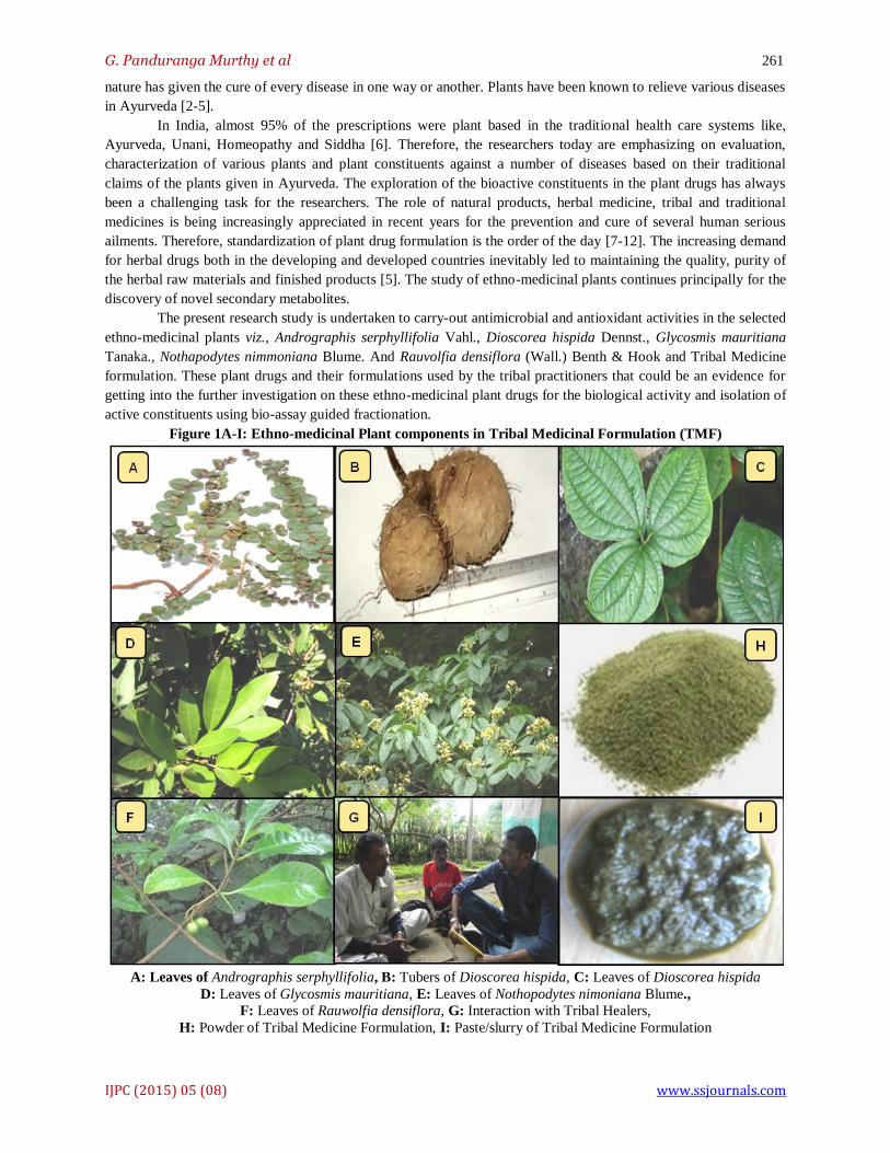
Figure 1A-I: Ethno-medicinal Plant components in Tribal Medicinal Formulation (TMF)
A: Leaves of Andrographis serphyllifolia, B: Tubers of Dioscorea hispida, C: Leaves of Dioscorea hispida
D: Leaves of Glycosmis mauritiana, E: Leaves of Nothopodytes nimoniana Blume.,
F: Leaves of Rauwolfia densiflora, G: Interaction with Tribal Healers,
H: Powder of Tribal Medicine Formulation, I: Paste/slurry of Tribal Medicine Formulation

G. Panduranga Murthy et al 262
IJPC (2015) 05 (08) www.ssjournals.com
2. Materials and Methods
2.1 Base-line survey and collection of Ethno-medicinal plants
The data on ethno-medicinal plants, Andrographis serphyllifolia, Vahl (leaves), Dioscorea hispida, Dennst,
(tubers); Glycosmis mauritiana Tanaka, (leaves); Nothapodytes nimmoniana Blume (leaves) and Rauvolfia
densiflora (Wall.) Benth & Hook (whole plant) were collected during the survey conducted between 2012 and 2014,
at Biligirirangana Hill area, Karnataka. The species identified appropriately and authenticated by consulting a flora
of Mysore and the same have been deposited in the Centre for Shridevi Research Foundation, SIET in collaboration
with Bhoomigeetha Institute of Research & Development (Tumkur), Karnataka. The baseline informations of
selected ethno-medicinal plants are represented in the Table 1.
Table 1: Validated Tribal Medicine formulation (TMF)* and its components practiced for wound healing and
related ailments at Biligirirangana Hill Tracts, Karnataka.
Sl.
No.
Ethno-medicinal plants with
Vernacular Name.
Family Plant
parts
used
Quantity
(powder)
(g/kg)
Validated
Quantity of
TMF (g)*
1 Andrographis serphyllifolia (A)
Vr. Name: Kasinasara
Acanthaceae Whole
plant
20
(A) 20+
(D)15+
(G) 25+
(N) 25+
(R) 15+
TMF
ADGNR
= 100
2 Dioscorea hispida (D) Vr. Name: Noolana hambu
Dioscoreaceae Tubers 15
3 Glycosmis mauritiana (G)
Vr. Name: Orrange berry
Rutaceae leaves 25
4 Nothapodytes nimoniana (N)
Vr. Name: Durvasane mara
Icacinaceae Leaves 25
5 Rauwolfia densiflora (R) Vr. Name: Snake root
Apocynaceae Leaves 15
DOSAGE, DURATION AND MODE OF TREATMENTS OF TRIBAL MEDICINE FORMULATION
Paste of TMF
It is applied on affected part of wound followed by snake bite and Scorpion bite region with few drops of Honey and Lime
juice for wounds and infected area.
Duration: Apply paste at wound area & cover with a
thin cloth 3times/week
Decoction of
TMF
Ground & juice boiled with warm water & swallowed for
internal problems Decoction with warm water/ goat milk for. inflammation and related ailments
Duration: One tsp two times a day for 8 days.
*TMF obtained from TMM was validated by Authorized Ayurvedic Practitioner
2.2 Validation of Tribal Medicine Formulation (TMF)
Interactions were conducted with Tribal Medicine Men with a semi-structured questionnaire and the data
on Medicine formulation responsible plant components were obtained. Further, the Tribal Medicine Formulations
(TMF) and Ethno-medicinal plant materials were obtained from the Tribal Medicine Men (Fig. 3A-J) and then the
samples were scientifically validated based on its physical characteristics in association with an authorized
Ayurvedic practitioner, Nisarga Ayurveda Research Foundation, Sakaleshpur, Hassan district, India. The standard
protocols were identified and the methodology was employed in the present study, based on the description of
Authorized Herbal Practitioner [13].
2.3 Preparation and Processing of TMF
The collected Ethno-medicinal plant materials were subjected for separating different desirable parts like
leaves, stem, root/ tubers from the main plants or whole plant parts were shade dried for 20 days to ensure the active
constituents free from decomposition and also to avoid any photo-chemical degradation. The different parts of plant
materials were powdered using suitable electric blender. The powdered samples of both EMP and TMF were stored
in airtight containers and kept in a cool, dark, dry place until the further analysis is commenced following the
standard procedures [6].
2.4 Successive solvent extraction
2.4.1 Tribal Medicine Formulation: Medicine Formulation is a mixture of the tribal medicinal components of
various parts of plants that are used to treat various abnormalities. The parts used for the mixture can be leaves,
roots, stem, tubers, twigs, fruits, seeds, flowers and whole plants. The formulation is usually prepared by mixing the
components in variable amounts and pasting it using cold or warm water. It can be directly applied on to the exterior
parts of the body with honey drops or given for the intake depending on the abnormality being treated through warm

G. Panduranga Murthy et al 263
IJPC (2015) 05 (08) www.ssjournals.com
water or goat milk. These formulations are based on the indigenous knowledge of Tribal people in India (Fig. 3A-J
and Table 2).
The bio-chemical activity of Tribal Medicine Formulation (TMF) will not be known to the tribal healers
but their action will be known, because the TMF is practiced by them, since many years. The components react with
each other and show the suitable activity on the patient. The TMF constituent was subjected for devastating into
small pieces using pestle and mortar and then powdered in an electric grinder for further analysis.
50g powder of the Tribal Medicine Formulation (TMF) was subjected to successive solvent extraction with
different solvents in increasing order of polarity from petroleum ether to ethanol and methanol finally to the level of
crude extraction with water. The organic solvent was made particular based on the dissolving efficiency and total
recovery of the ethno-medicinal plant drugs amongst the organic solvents used in the study, Meanwhile, the extracts
were kept for evaporation to dryness and set aside for further bio-physical analysis.
2.4.2 Ethno-medicinal plants: The powdered sample (170g) was extracted by maceration method using soxhlet
apparatus with different solvents from polar to less polar solvents such as Water, Ethanol, Methanol and Petroleum
ether. The macerated extract was centrifuged at 5000rpm for 15 min and the supernatant was taken for the excess
solvent evaporation. After evaporation of the excess solvent, the crude extract was taken for further analysis. The
macerated extracts were then placed in shaker incubator for 24 hours and later subjected to filtration using Whatman
No. 1 filter paper.
The test solvents were confirmed based on the yield and feasibility of the solvents during extraction
processes and then, organic solvents were removed, firstly by means of a water bath, then in an oven, yielding the
extracted compound. The concentrate was designated as crude ethanolic extract of EMP. These extracts were used to
conduct the phyto-chemical and pharmacological evaluation of ethno-medicinal plant drugs (EMP).
2.5 Water extraction of Crude EMP Samples
Crude plant extract was prepared by Soxhlet extraction method. The powdered plant material was
uniformly packed into a thimble for extraction. The mixture was heated on a hot plate with continuous stirring at
30-40ºC for 20 minutes. Then, the water extract was filtered through Whatmann No. 1 filter paper. The filtrate was
subjected to dryness and the dried extract was kept in refrigerator at 4ºC for their future use for biological activities
and bio-chemical analysis.
2.6 Antimicrobial activity
2.6.1 Test Microorganisms: Test organisms were used in the study, gram-positive bacterium: Staphylococcus
aureus, Bacillus subtilis and gram-negative bacteria: Escherichia coli, Kliebsiella pneumoniae and Pseudomonas sp.
were collected from the infected individuals at Siddhartha Medical College, Tumkur. The Cultures were maintained
as nutrient agar slants in screw-capped bottles and stored at 4°C. All the cultures were checked for viability, purity
by regular plating and biochemical tests. Test cultures were prepared by transferring a loop full of bacteria from
stock culture to nutrient broth in the flasks and incubated at 37°C for 24 h.
2.6.2 Preparation of Inocula: Stock cultures were maintained at 4°C on slopes of nutrient agar. The active cultures
for experiments were prepared by transferring a loop-full of cells from the stock cultures to test tubes of Mueller-
Hinton broth (MHB) and were incubated as still cultures for 24h at 37°C and were used for the determination of
Antimicrobial activity.
2.6.3 Disc diffusion method: In vitro antimicrobial activity was screened through disc diffusion method as proposed
by Kirby-Bauer using Mueller Hinton Agar (MHA). The MHA plates were prepared by pouring 15 ml of molten
media into sterile petriplates. The plates were allowed to solidify for 5 min and inoculum suspension was swabbed
uniformly and the inoculum was allowed to dry for 5 min. the discs were formed at appropriate distance in the
petriplates and loaded with the Control (solvent), Standard Antibiotic ( Chloramphenicol - 0.025%) and Plant
extracts (concentration- 100 mg/ml) in polar, non-polar and not-so-polar solvents [14]. The plates were incubated at
370C for 24 h. at the end of incubation; the inhibition zones formed around the disc were measured with transparent
ruler and recorded.
2.7 Minimum Inhibitory Concentration (MIC)
The minimum inhibitory concentrations (MIC), MBC and MFCs were performed by a serial dilution
technique using 96-well microtiter plates as described by [15]. The different plant extracts viz., Methanol, Ethanol,
Petroleum Ether, Aqueous were taken (1 mg/ml) and serial dilution of the extract with Luria broth for bacterial
culture and potato dextrose broth medium for fungi with respective inoculums were used. The extracts were serially

G. Panduranga Murthy et al 264
IJPC (2015) 05 (08) www.ssjournals.com
diluted (two-fold) to a working concentration ranging from 400mg/ml to 12.5mg/ml using nutrient broth and later
inoculated with 0.2ml suspension of the test organisms namely Escherichia coli, Bacillis subtilis, Staphylococcus
aureus, Kliebsiella pneumoniae and Pseudomonas sp and the microplates were incubated for 72 hours at 280C,
respectively. After incubation period, the test tubes were observed for turbidity. The least concentration where no
turbidity was observed was determined and noted as the minimum inhibitory concentration (MIC) value. The lowest
concentrations without visible growth (under binocular microscope) were defined as MICs and the calculation was
done as per the following formula.
MIC=Minimum Inhibitory Concentration, C=Concentration of Antibiotic in mg/ml in total volume,
X=Length of Microbial growth in cm. and Y=Total length of possible growth in cm.
2.8 Determination of Minimum Bacterial Concentration (MBC)
The MBCs were determined by serial sub-culturing of 2 μl into microtitre plates containing 100μl of broth
per well and further incubation for 72 hours. The lowest concentration with no visible growth was defined as the
MBC, indicating assassination (99.5%) of the original inoculum. The optical density of each well was measured at a
wavelength of 655nm by Microplate reader (Perlong, ENM8602) and compared with the standard Ampicillin for
Bacteria (Hi-media lab, India) as the positive control. All the experiments were performed in duplicate and repeated
three times.
2.9 Determination of MFC
The fungicidal concentrations (MFCs) were determined by serial sub-culturing of a 2 μl into microtiter
plates containing 100μl of broth per well and further incubation for 72 hours at 28oC. The lowest concentration with
no visible growth was defined as MFC indicated 99.5% killing of the original inoculums. The commercial standard,
Flucanazole (Sigma), was used as positive controls (1–3000 μg/ml) for fungi. All experiments were performed in
duplicate and repeated three times.
2.10 Antioxidant Activity
2.10.1 DPPH (1, 1-diphenyl-2-picrylhydrazyl) free radical scavenging activity: The molecule of 1,1-diphenyl-2-
picrylhydrazyl (α,α-diphenyl-β- picryl hydrazyl; DPPH) is characterized as a stable free radical by virtue of the
delocalization of the spare electron over the molecule as a whole, so that the molecules do not dimerise, as would be
the case with most other free radicals. The delocalization also gives rise to the deep violet colour, characterized by
an absorption band in ethanol solution centered at about 517 nm. When a solution of DPPH is mixed with that of a
substance that can donate a hydrogen atom, then this gives rise to the reduced form with the loss of this violet
colour. The free radical scavenging capacity of the methanol extracts was determined using DPPH [16,17].
The DPPH solution (0.006% w/v) was prepared in 95% methanol. The different concentrations of the test
sample which is to be examined for antioxidant activity is prepared (50-300μg/ml). 3 ml of different concentration
of test samples of EMP and TMF extracts were mixed with 1 ml of DPPH solution in dark. Ascorbic acid which is
strong anti-oxidizing agent is taken as standard. The different concentrations (3 ml) of standard solution of ascorbic
acid were mixed with 1 ml of DPPH solution in dark. The prepared solution of ascorbic acid and plant extracts
samples was incubated for half an hour and then the absorbance was taken with the help of U.V. Spectrophotometer
at 517nm. The concerned extract or solvent served as a blank and the experiment was expressed as the inhibition
percentage of free radical by the sample and was calculated using the following formula.
2.10.2 ABTS (2, 2-azinobis 3-ethylbenzothiazoline-6-sulfonic acid) radical scavenging activity: ABTS assay was
performed according to the standard protocol; the stock solution was prepared by mixing equal volumes of 7mM
ABTS solution and 2.45mM potassium per-sulfate solution followed by incubation for 12h at room temperature in
the dark to yield a dark-colored solution containing ABTS radicals. The working solution was prepared freshly
before each assay by diluting the stock solution by mixing of stock solution to 50% methanol for an initial
absorbance of about 0.700 (± 0.02) at 745 nm, with temperature control set at 30°C. The varying concentrations (50-
3000μg/ml) of the plant extracts of EMP and TMF were allowed to react with 3 ml of the ABTS solution and the
absorbance readings were recorded at 734 nm. Ascorbic acid was used as positive controls. The scavenging activity
was estimated based on the percentage of ABTS radicals scavenged by the following formula as described by Lie-
fan et al., (2011).
DPPH radical scavenging activity (%) = Control OD – Sample OD ×100
Control OD

G. Panduranga Murthy et al 265
IJPC (2015) 05 (08) www.ssjournals.com
2.11 FRAP Assay and Determination of Total Phenolic Content
The FRAP assay was done for the extracts of EMP drugs as per the standard protocol [17] and total
phenolic content was determined based on the standard formula [18].
1.12. Assay for Total reducing ability
The total reduction capabilities of the aqueous extract of TMF were estimated by using the standard method
[19]. The absorbance of the reaction mixture after incubation was measured at 700nm using a spectrophotometer
(UV-Cintra Spectrophotometer). Higher absorbance of the reaction mixture indicated greater reducing power.
2.13 Statistical Analysis
Statistical analysis for animal experiment was carried out using one-way ANOVA followed by Dunnet’s
multiple comparisons. The results obtained from all the test samples of EMP and TMF were compared with each
other. The p values <0.05 were considered to be statistically significant. The concentration producing 50% of the
maximum response (LC50 or IC50) was obtained by the best visual fit from the plot of the individual experiments.
3. Results
The data on ethno-medicinal plant drugs comprising of family, vernacular name, parts used, medicinal
value, formulation, treatment of ailment, dosage and duration of the treatment are well represented (Table 1).
Similarly, the details on medicinal formulation were obtained from the tribal medicine men. Further, the collected
tribal medicine formulation (TMF) was validated with an authorized Ayurvedic Medical Practitioner (Table 2).
3.1 Antimicrobial activity
In the study, Gram-positive: S. aureus; B. subtilis and Gram-negative; E. coli, P. aeruginosa,
K. pneumonia were used for evaluation. In all, two Gram-positive bacteria were found to be sensitive to the extracts
of EMP and TMF, whereas in case of Gram-negative organisms, they were considerably susceptible to solvent
extracts such as Ethanol, Methanol, Petroleum ether compared to aqueous extracts of both EMP and TMF (Table 2).
The antimicrobial potential of both the EMP and TMF plant drugs was evaluated according to their zone of
inhibition against various pathogens and the results (zone of inhibition) were compared with the activity of the
standards viz., Ampicillin (1.0 mg/disc), Flucanazole (1.0 mg/disc). The results revealed that, all the extracts are
potent antimicrobials against all the microorganisms studied. Among the different solvent extracts studied, methanol
and ethanol showed high degree of inhibition followed by aqueous and Petroleum ether extracts. For all the tested
microorganisms, Ethanol and Methanol showed maximum antibacterial activity in EMP. In model organism, E.coli,
the Ethanol extract showed maximum inhibition zone diameter with A. serphyllifolia (14±0.2), D. hispida (14±0.6),
G. mauritiana (16±3.5) N. nimmoniana (14±1.5) and R. densiflora (16±1.0). Similarly in TMF, the zone of
inhibition was (19±0.2) significantly superior than other solvent extracts. For S. aureus, the ethanolic extract
exhibited radical increase in zone of inhibition at A. serphyllifolia (13±0.3), D. hispida (16±2.5), G. mauritiana
(19±1.5) N. nimmoniana (15±1.0) and R. densiflora (21±3.0) and the maximum inhibition (23±2.0) was recorded.
Correspondingly, the Methanol extract showed maximum inhibition zone with diameter of 24±2.5 mm in S. aureus
and similar values (20±1.6) were observed in B. subtilis and Pseudomonas sp. Further, the Petroleum Ether (10-20
mm) and aqueous extract (12-19 mm) showed poised status and the activity was notable. More specifically, aqueous
extract was found to be equally effective and represented trivial susceptibility to all bacterial strains as compared to
other tested organisms (Table 2).
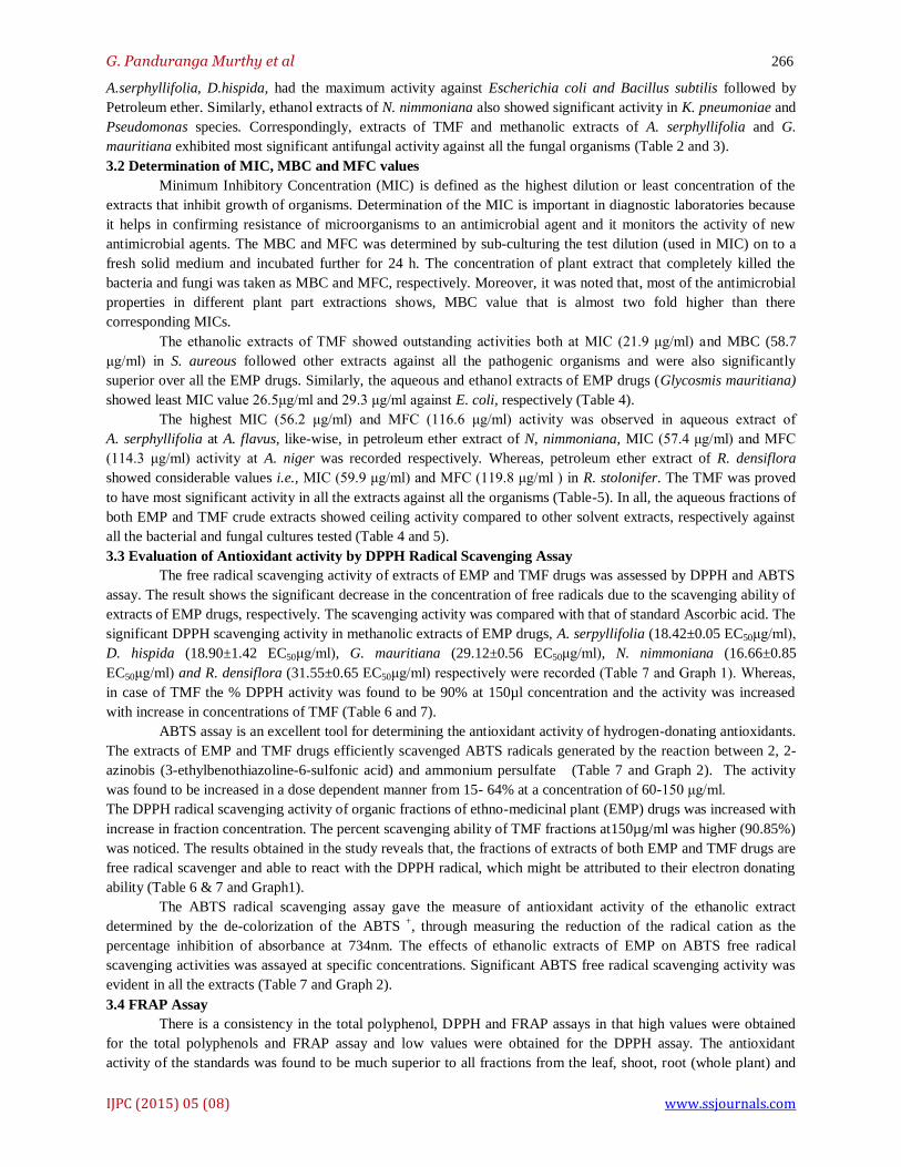
In the antifungal activity, ethanol extracts of EMP drugs showed efficient antifungal activity against A.
flavus viz., A. serphyllifolia (19.6±0.6), D. hispida (16.9±0.5), G. mauritiana (17±0.4) N. nimmoniana (16±0.3) and
R. densiflora (15.5±0.4). Consequently, the TMF Methanol drug extract alone showed proficient activity in A. niger
(21±0.3), A. flavus (22.1±0.6), F. oxysporum (24.8±0.11) and R. stolonifer (26.1±0.5) respectively. In contrast,
Aqueous and Petroleum Ether extracts showed lowest inhibition zone with diameter ranging between 11-18 mm and
14-21 mm against all the pathogenic fungal strains, respectively (Table 3).
Among all the extracts employed in the antimicrobial activity, the TMF drug and the EMP namely
G. mauritiana and R. densiflora demonstrated relatively strong antibacterial activity with hops and presenting a
highest activity in Staphylococcus aureus compared to model organism. Of these, ethanol extracts of
ABTS radical scavenging activity (%) = Control OD – Sample OD × 100
Control OD
G. Panduranga Murthy et al 266
IJPC (2015) 05 (08) www.ssjournals.com
A.serphyllifolia, D.hispida, had the maximum activity against Escherichia coli and Bacillus subtilis followed by
Petroleum ether. Similarly, ethanol extracts of N. nimmoniana also showed significant activity in K. pneumoniae and
Pseudomonas species. Correspondingly, extracts of TMF and methanolic extracts of A. serphyllifolia and G.
mauritiana exhibited most significant antifungal activity against all the fungal organisms (Table 2 and 3).
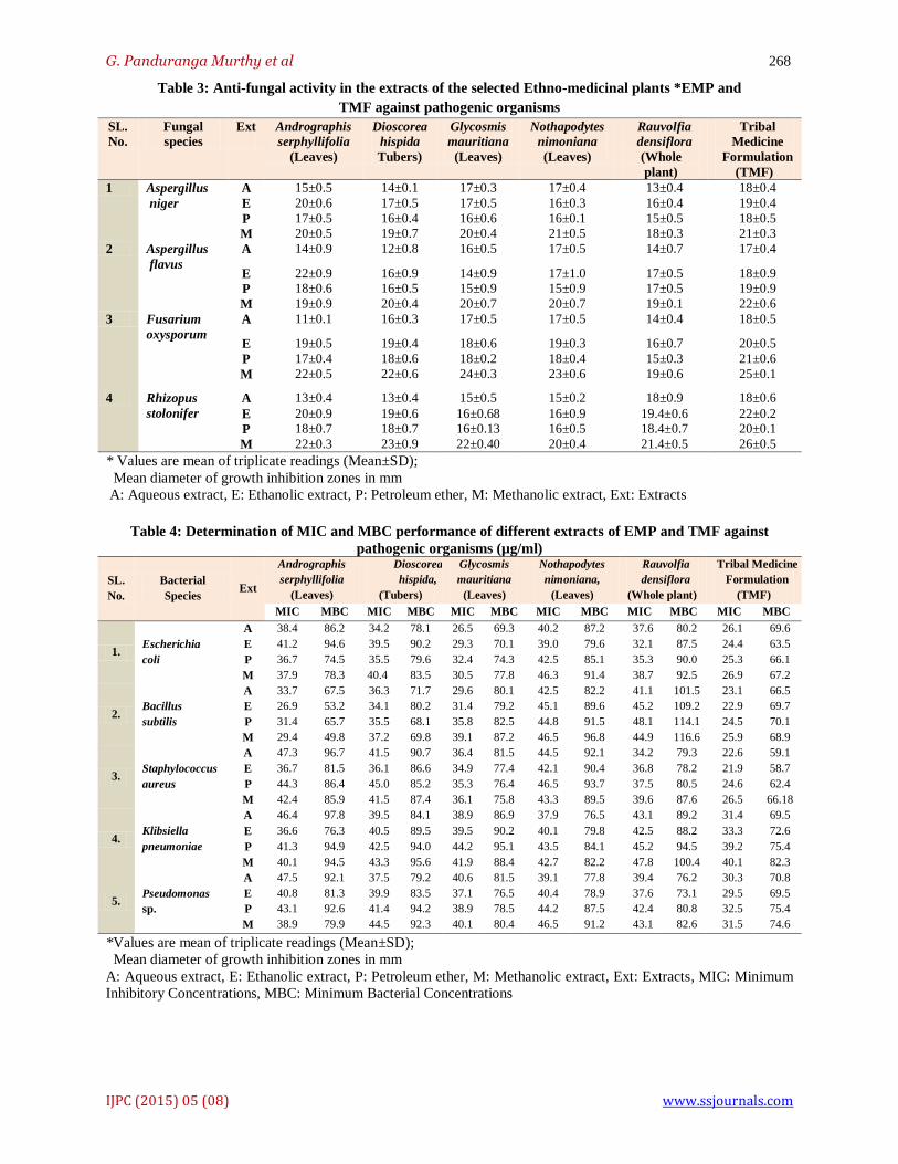
3.2 Determination of MIC, MBC and MFC values
Minimum Inhibitory Concentration (MIC) is defined as the highest dilution or least concentration of the
extracts that inhibit growth of organisms. Determination of the MIC is important in diagnostic laboratories because
it helps in confirming resistance of microorganisms to an antimicrobial agent and it monitors the activity of new
antimicrobial agents. The MBC and MFC was determined by sub-culturing the test dilution (used in MIC) on to a
fresh solid medium and incubated further for 24 h. The concentration of plant extract that completely killed the
bacteria and fungi was taken as MBC and MFC, respectively. Moreover, it was noted that, most of the antimicrobial
properties in different plant part extractions shows, MBC value that is almost two fold higher than there
corresponding MICs.
The ethanolic extracts of TMF showed outstanding activities both at MIC (21.9 μg/ml) and MBC (58.7
μg/ml) in S. aureous followed other extracts against all the pathogenic organisms and were also significantly
superior over all the EMP drugs. Similarly, the aqueous and ethanol extracts of EMP drugs (Glycosmis mauritiana)
showed least MIC value 26.5μg/ml and 29.3 μg/ml against E. coli, respectively (Table 4).
The highest MIC (56.2 μg/ml) and MFC (116.6 μg/ml) activity was observed in aqueous extract of
A. serphyllifolia at A. flavus, like-wise, in petroleum ether extract of N, nimmoniana, MIC (57.4 μg/ml) and MFC
(114.3 μg/ml) activity at A. niger was recorded respectively. Whereas, petroleum ether extract of R. densiflora
showed considerable values i.e., MIC (59.9 μg/ml) and MFC (119.8 μg/ml ) in R. stolonifer. The TMF was proved
to have most significant activity in all the extracts against all the organisms (Table-5). In all, the aqueous fractions of
both EMP and TMF crude extracts showed ceiling activity compared to other solvent extracts, respectively against
all the bacterial and fungal cultures tested (Table 4 and 5).
3.3 Evaluation of Antioxidant activity by DPPH Radical Scavenging Assay
The free radical scavenging activity of extracts of EMP and TMF drugs was assessed by DPPH and ABTS
assay. The result shows the significant decrease in the concentration of free radicals due to the scavenging ability of
extracts of EMP drugs, respectively. The scavenging activity was compared with that of standard Ascorbic acid. The
significant DPPH scavenging activity in methanolic extracts of EMP drugs, A. serpyllifolia (18.42±0.05 EC50μg/ml),
D. hispida (18.90±1.42 EC50μg/ml), G. mauritiana (29.12±0.56 EC50μg/ml), N. nimmoniana (16.66±0.85
EC50μg/ml) and R. densiflora (31.55±0.65 EC50μg/ml) respectively were recorded (Table 7 and Graph 1). Whereas,
in case of TMF the % DPPH activity was found to be 90% at 150µl concentration and the activity was increased
with increase in concentrations of TMF (Table 6 and 7).
ABTS assay is an excellent tool for determining the antioxidant activity of hydrogen-donating antioxidants.
The extracts of EMP and TMF drugs efficiently scavenged ABTS radicals generated by the reaction between 2, 2-
azinobis (3-ethylbenothiazoline-6-sulfonic acid) and ammonium persulfate (Table 7 and Graph 2). The activity
was found to be increased in a dose dependent manner from 15- 64% at a concentration of 60-150 μg/ml.
The DPPH radical scavenging activity of organic fractions of ethno-medicinal plant (EMP) drugs was increased with
increase in fraction concentration. The percent scavenging ability of TMF fractions at150µg/ml was higher (90.85%)
was noticed. The results obtained in the study reveals that, the fractions of extracts of both EMP and TMF drugs are
free radical scavenger and able to react with the DPPH radical, which might be attributed to their electron donating
ability (Table 6 & 7 and Graph1).
The ABTS radical scavenging assay gave the measure of antioxidant activity of the ethanolic extract
determined by the de-colorization of the ABTS +
, through measuring the reduction of the radical cation as the
percentage inhibition of absorbance at 734nm. The effects of ethanolic extracts of EMP on ABTS free radical
scavenging activities was assayed at specific concentrations. Significant ABTS free radical scavenging activity was
evident in all the extracts (Table 7 and Graph 2).
3.4 FRAP Assay
There is a consistency in the total polyphenol, DPPH and FRAP assays in that high values were obtained
for the total polyphenols and FRAP assay and low values were obtained for the DPPH assay. The antioxidant
activity of the standards was found to be much superior to all fractions from the leaf, shoot, root (whole plant) and

G. Panduranga Murthy et al 267
IJPC (2015) 05 (08) www.ssjournals.com
tuber samples. This is probably due to the low concentration of antioxidant compounds present in the fractions. In
all the samples, a correlation between the total phenol assay and antioxidant activity (FRAP assay) was observed for
the aqueous fractions of EMP drugs with the value of 18.57, 21.26, 8.25, 11.68 and 10.67 μmol/mg, respectively
(Table 7). In all, the sample of D. hispida the ethanol fraction (31.22) and methanol fraction gave the highest value
(29.70) when compared to all other extracts of EMP drugs, respectively and therefore, the ethanol and methanol
fractions of the EMP drugs was analyzed for comparison in the study (Table 7).
3.5 Antioxidant activity in TMF drug
The ethanolic extract of tribal medicine formulation was used to evaluate Antioxidant activity using DPPH
Radical scavenging assay. The absorbance value for TMF is represented in the Table 6. The value of % inhibition
for TMF drug was noticed (Graph-3). The percentage of inhibition was found to be increasing with the increasing
concentration of TMF extracts. The O.D values were found to be decreasing.
Another comparison was made with the Standard O.D values of Ascorbic acid, which is an antioxidant by itself. The
O.D values of Ascorbic acid were found to be decreasing and so as the TMF drug values. This also demonstrated the
presence of the antioxidant activity for the samples (Graph-3 and Table 6).
3.6 Evaluation of Total Phenolic content
The amount of total phenolics varied in different extracts of EMP drugs and the values of aqueous extracts
of all the EMP drugs ranged from 56.24, 52.17, 72.46, 89.5 to 107.67 mg GAE/g of EMP. The highest total phenolic
levels were detected in the extract of R. densiflra and the lowest in the extract of D. hispida (Table 7). The amount
of total phenolic compounds in all tested plant extracts was higher than the TMF drug. The ranking order of eight
plant species from point of view of antioxidant (phenolic compounds) amounts was as follows: R. densiflora > N.
nimoniana > G. mauritiana > A. serphyllifolia > D. hispida.
3.7 Evaluation of Total Reduction capability
In the extracts of both EMP drugs, the IC50 values of reducing activities of aqueous fractions revealed the
order of activity as: 0.36% in Andrographis serphyllifolia, 0.45% in Dioscorea hispida, 1.17% in Glycosmis
mauritiana, 1.28% in Nothapodytes nimoniana and 1.12% in Rauwolfia densiflora were recorded. Similarly, the
solvent fractions were also found to be significant as compared to aqueous crude extract. The reducing potentiality
of EMP drugs is directly proportional to different extracts at variable concentrations (Table 7).
Table 2: Antibacterial activity in the extracts of the selected Ethno-medicinal plants EMP and
TMF against pathogenic organisms
SL.
No
Bacterial
species
Ex
Extracts
Andrographis
serphyllifolia
(Leaves)
Dioscorea
hispida
(Tubers)
Glycosmis
mauritiana
(Leaves)
Nothapodytes
nimoniana
(Leaves)
Rauvolfia
densiflora
(Whole
plant)
Tribal
Medicine
Formulation
(TMF)
1 Escherichia
coli
A 12± 0.3 13±0.6 17±1.2 12±0.2 16±0.3 18±0.5
E 14±0.2 14±0.6 16±3.5 14±1.5 16±1.0 19±0.2
P 10±1.2 16±0.2 19±2.2 13±1.2 20±2.5 17±1.2
M 13±0.6 15±0.5 17±1.6 15±2.2 18±1.5 18±1.5
2 Bacillus
subtilis
A 11±0.5 13±1.2 11±0.3 14±0.4 15±0.0 16±1.0
E 9±0.4 12±0.6 13±1.2 10±0.6 17±1.5 19±2.0
P 10±2.2 14±1.0 14±0.5 11±0.0 15±2.0 16±2.2
M 10±1.6 11±0.2 12±2.0 13±0.2 17±0.1 20±1.6
3 Staphylococcus
aureus
A 15±1.5 14±1.6 17±2.6 13±2.6 18±0.5 19±1.2
E 13±0.3 16±2.5 19±1.5 15±1.0 21±3.0 23±2.0
P 18±1.6 18±3.0 16±2.5 14±0.5 19±1.5 20±1.6
M 19±0.5 19±1.5 21±0.6 15±3.0 20±3.0 24±2.5
4 Klibsiella
pneumoniae
A 10±0.6 10±2.0 13±0.0 13±1.5 14±2.0 17±0.6
E 12±0.1 13±00 12±1.0 12±2.0 13±0.5 15±1.0
P 10±0.2 16±1.5 13±0.6 12±1.5 11±0.2 18±2.5
M 13±0.5 15±1.0 15±0.5 14±1.0 14±0.5 17±0.5
5 Pseudomonas
sp.
A 07±0.3 13±3.0 11±0.2 10±1.0 14±1.2 15±1.5
E 10±0.5 12±0.3 13±0.5 13±0.4 15±1.0 16±1.6
P 13±0.3 10±0.5 10±0.0 14±0.0 17±3.0 18±1.2
M 12±1.5 14±1.5 13±2.0 14±2.2 16±0.5 20±2.5
*Data are the Mean values of triplicate and expressed as M±SD (P<0.05).
Mean diameter of growth inhibition zones in mm
A: Aqueous extract, E: Ethanolic extract, P: Petroleum ether, M: Methanolic extract
G. Panduranga Murthy et al 268
IJPC (2015) 05 (08) www.ssjournals.com
Table 3: Anti-fungal activity in the extracts of the selected Ethno-medicinal plants *EMP and
TMF against pathogenic organisms
SL.
No.
Fungal
species
Ext
Andrographis
serphyllifolia
(Leaves)
Dioscorea
hispida
Tubers)
Glycosmis
mauritiana
(Leaves)
Nothapodytes
nimoniana
(Leaves)
Rauvolfia
densiflora
(Whole
plant)
Tribal
Medicine
Formulation
(TMF)
1 Aspergillus
niger
A 15±0.5 14±0.1 17±0.3 17±0.4 13±0.4 18±0.4
E 20±0.6 17±0.5 17±0.5 16±0.3 16±0.4 19±0.4
P 17±0.5 16±0.4 16±0.6 16±0.1 15±0.5 18±0.5
M 20±0.5 19±0.7 20±0.4 21±0.5 18±0.3 21±0.3
2 Aspergillus
flavus
A 14±0.9 12±0.8 16±0.5 17±0.5 14±0.7 17±0.4
E 22±0.9 16±0.9 14±0.9 17±1.0 17±0.5 18±0.9
P 18±0.6 16±0.5 15±0.9 15±0.9 17±0.5 19±0.9
M 19±0.9 20±0.4 20±0.7 20±0.7 19±0.1 22±0.6
3 Fusarium
oxysporum
A 11±0.1 16±0.3 17±0.5 17±0.5 14±0.4 18±0.5
E 19±0.5 19±0.4 18±0.6 19±0.3 16±0.7 20±0.5
P 17±0.4 18±0.6 18±0.2 18±0.4 15±0.3 21±0.6
M 22±0.5 22±0.6 24±0.3 23±0.6 19±0.6 25±0.1
4 Rhizopus
stolonifer
A 13±0.4 13±0.4 15±0.5 15±0.2 18±0.9 18±0.6
E 20±0.9 19±0.6 16±0.68 16±0.9 19.4±0.6 22±0.2
P 18±0.7 18±0.7 16±0.13 16±0.5 18.4±0.7 20±0.1
M 22±0.3 23±0.9 22±0.40 20±0.4 21.4±0.5 26±0.5
* Values are mean of triplicate readings (Mean±SD);
Mean diameter of growth inhibition zones in mm
A: Aqueous extract, E: Ethanolic extract, P: Petroleum ether, M: Methanolic extract, Ext: Extracts
Table 4: Determination of MIC and MBC performance of different extracts of EMP and TMF against
pathogenic organisms (µg/ml)
SL.
No.
Bacterial
Species Ext
Andrographis
serphyllifolia
(Leaves)
Dioscorea
hispida,
(Tubers)
Glycosmis
mauritiana
(Leaves)
Nothapodytes
nimoniana,
(Leaves)
Rauvolfia
densiflora
(Whole plant)
Tribal Medicine
Formulation
(TMF)
MIC MBC MIC MBC MIC MBC MIC MBC MIC MBC MIC MBC
1. Escherichia
coli
A 38.4 86.2 34.2 78.1 26.5 69.3 40.2 87.2 37.6 80.2 26.1 69.6
E 41.2 94.6 39.5 90.2 29.3 70.1 39.0 79.6 32.1 87.5 24.4 63.5
P 36.7 74.5 35.5 79.6 32.4 74.3 42.5 85.1 35.3 90.0 25.3 66.1
M 37.9 78.3 40.4 83.5 30.5 77.8 46.3 91.4 38.7 92.5 26.9 67.2
2. Bacillus
subtilis
A 33.7 67.5 36.3 71.7 29.6 80.1 42.5 82.2 41.1 101.5 23.1 66.5
E 26.9 53.2 34.1 80.2 31.4 79.2 45.1 89.6 45.2 109.2 22.9 69.7
P 31.4 65.7 35.5 68.1 35.8 82.5 44.8 91.5 48.1 114.1 24.5 70.1
M 29.4 49.8 37.2 69.8 39.1 87.2 46.5 96.8 44.9 116.6 25.9 68.9
3. Staphylococcus
aureus
A 47.3 96.7 41.5 90.7 36.4 81.5 44.5 92.1 34.2 79.3 22.6 59.1
E 36.7 81.5 36.1 86.6 34.9 77.4 42.1 90.4 36.8 78.2 21.9 58.7
P 44.3 86.4 45.0 85.2 35.3 76.4 46.5 93.7 37.5 80.5 24.6 62.4
M 42.4 85.9 41.5 87.4 36.1 75.8 43.3 89.5 39.6 87.6 26.5 66.18
4. Klibsiella
pneumoniae
A 46.4 97.8 39.5 84.1 38.9 86.9 37.9 76.5 43.1 89.2 31.4 69.5
E 36.6 76.3 40.5 89.5 39.5 90.2 40.1 79.8 42.5 88.2 33.3 72.6
P 41.3 94.9 42.5 94.0 44.2 95.1 43.5 84.1 45.2 94.5 39.2 75.4
M 40.1 94.5 43.3 95.6 41.9 88.4 42.7 82.2 47.8 100.4 40.1 82.3
5. Pseudomonas
sp.
A 47.5 92.1 37.5 79.2 40.6 81.5 39.1 77.8 39.4 76.2 30.3 70.8
E 40.8 81.3 39.9 83.5 37.1 76.5 40.4 78.9 37.6 73.1 29.5 69.5
P 43.1 92.6 41.4 94.2 38.9 78.5 44.2 87.5 42.4 80.8 32.5 75.4
M 38.9 79.9 44.5 92.3 40.1 80.4 46.5 91.2 43.1 82.6 31.5 74.6
*Values are mean of triplicate readings (Mean±SD);
Mean diameter of growth inhibition zones in mm
A: Aqueous extract, E: Ethanolic extract, P: Petroleum ether, M: Methanolic extract, Ext: Extracts, MIC: Minimum
Inhibitory Concentrations, MBC: Minimum Bacterial Concentrations

G. Panduranga Murthy et al 269
IJPC (2015) 05 (08) www.ssjournals.com
Table 5: Determination of MIC and MFC performance of different extracts of EMP and TMF against
pathogenic organisms (µg/ml)
SL.
No.
Fungal
species
Ext Andrographis
serphyllifolia
(Leaves)
Dioscorea
hispida,
(Tubers)
Glycosmis
mauritiana
(Leaves)
Nothapodytes
nimoniana,
(Leaves)
Rauvolfia
densiflora
(Whole
plant)
Tribal
Medicine
Formulation
(TMF)
MIC MFC MIC MFC MIC MFC MIC MFC MIC MFC MIC MFC
1. Aspergillus
niger
A 53.1 110.2 47.5 101.5 41.5 97.5 55.1 110.2 47.1 99.2 34.1 72.4
E 49.4 101.6 46.2 104.3 39.5 95.1 52.4 106.1 50.2 105.2 36.3 76.5
P 54.2 111.5 49.9 108.2 42.6 99.1 57.4 114.3 54.1 109.1 40.5 79.6
M 50.5 99.6 51.2 105.1 49.5 101.2 54.3 109.3 56.5 116.4 43.2 82.5
2. Aspergillus
flavus
A 56.2 116.6 53.1 112.2 42.6 101.9 51.5 102.4 53.3 111.5 37.2 77.6
E 48.4 95.7 46.5 97.9 43.5 99.2 52.1 106.3 56.1 115.4 40.4 79.7
P 56.8 113.9 52.3 115.6 45.1 95.2 55.2 112.4 54.3 113.3 42.3 83.5
M 51.2 102.8 54.3 105.4 47.5 93.6 54.8 111.2 58.3 118.5 41.7 80.2
3. Fusarium
oxysporum
A 57.2 118.6 55.3 106.4 44.1 97.9 53.5 103.5 49.1 100.3 40.2 79.4
E 53.6 108.7 56.5 115.5 46.4 98.2 48.3 100.2 52.5 103.5 41.4 82.8
P 56.9 109.8 59.2 116.5 45.4 101.5 53.5 104.1 54.3 107.6 39.2 78.5
M 48.3 97.9 52.7 100.4 48.5 102.6 56.2 110.5 53.3 105.7 43.2 85.2
4. Rhizopus
stolonifer
A 48.9 98.8 49.1 101.6 49.2 98.4 45.5 92.1 50.3 101.6 38.1 77.7
E 38.5 81.6 42.5 90.7 52.4 104.2 49.5 101.6 53.4 106.2 41.2 83.3
P 46.4 97.8 49.2 99.1 57.1 107.2 44.3 95.6 59.9 119.8 46.4 90.2
M 41.6 96.5 47.1 102.5 52.3 102.4 42.5 92.7 55.5 114.4 40.6 89.9
* Values are mean of triplicate readings (Mean±SD); Mean diameter of growth inhibition zones in mm
A: Aqueous extract, E: Ethanolic extract, P: Petroleum ether, M: Methanolic extract, Ext: Extracts, MIC: Minimum
Inhibitory Concentrations, MFC:Minimum Fungal Concentrations
Table 6: DPPH Radical scavenging assay for TMF practiced by Tribal Medicine Men at B.R. Hills, Karnataka.
SL.
No.
Concentration of Sample
(TMF extract) Absorbance of
Sample
Control DPPH radical scavenging activity
(%)
1. 30µl 1.449
2.788
48.01
2. 60µl 1.334 52.13
3. 90µl 1.158 58.44
4. 120µl 1.058 62.03
5. 150µl 0.255 90.85
Table 7: Antioxidant activities and Total Phenolic content of Tribal medicine formulation practiced by Tribal
Medicine Men at B.R. Hills, Karnataka
SL.
No
Parameters* Extracts
Andrographis
serphyllifolia
(Leaves)
Dioscorea
hispida
(Tubers)
Glycosmis
mauritiana
(Leaves)
Nothapodytes
nimoniana,
(Leaves)
Rauvolfia
Densiflora
(Whole plant)
1. DPPH Assay
(EC50µg/ml) A 12.24±0.2 06.40±0.2 07.22±0.2 04.14±1.2 06.15±1.7
E 16.14±1.0 17.10±0.2 26.45±1.2 07.14±0.6 11.14±1.3
P 07.16±0.7 09.25±1.3 14.00±1.4 09.18±1.5 23.20±0.9
M 18.42±0.1 18.90±1.4 29.12±0.6 16.66±0.9 31.55±0.7
2. FRAP Assay
(µmol Fe2+ /mg) A 18.57±2.7 21.26±1.6 08.25±1.2 11.68±0.5 10.67±0.3
E 11.12±1.5 31.22±0.0 19.80±1.5 16.40±0.5 10.12±1.5
P 13.65±0.1 24.32±1.5 17.85±0.1 13.33±0.1 15.42±1.5
M 24.51±0.7 28.90±0.5 29.70±0.5 11.90±1.1 16.30±0.8
3. ABTS Radical
Scavenging Activity
(%)
A 12.64±0.7 15.40±0.5 12.20±0.5 13.71±0.6 12.06±1.4
E 15.45±1.5 19.00±1.6 13.32±0.0 16.32±1.5 19.10±0.0
P 13.55±1.5 14.33±0.5 10.20±0.1 11.00±0.6 14.44±0.1
M 13.89±0.5 18.74±0.5 15.80±0.1 17.30±0.9 22.53±0.1
4. Total Phenolic content
(mg of GAE/g) A 56.24±0.1 52.17±1.0 72.46±0.7 89.65±0.8 107.67±1.7
E 61.20±1.2 48.64±0.1 67.21±1.6 78.52±0.7 102.32±0.4
P 57.60±0.3 59.80±1.0 53.63±1.0 23.12±0.0 83.55±0.9
M 36.42±0.6 55.00±1.5 76.50±0.7 19.00±0.1 31.32±0.6
5. Total reduction
capacity (%)
(A700nm)
A 00.36±0.0 0.45±0.02 01.17±0.0 01.28±0.0 01.12±0.5
E 00.26±0.7 0.36±0.00 00.90±0.0 00.96±0.0 01.08±0.5
P 00.94±0.1 00.43±0.0 00.74±0.0 00.88±0.0 00.96±1.5
M 00.41±0.0 00.52±0.0 00.94±0.1 01.12±0.0 01.21±0.0
Data are the Mean values of triplicate and expressed as M±SD (P<0.05). A: Aqueous extract, E: Ethanolic extract &
P: Petroleum ether & M: Methanolic extract.

G. Panduranga Murthy et al 270
IJPC (2015) 05 (08) www.ssjournals.com
Graph 1: Radicals scavenging activity by DPPH method in aqueous extracts of EMP
Graph 2: Radicals scavenging activity by ABTS method in aqueous extracts of EMP
Graph 3: Radical scavenging activity by DPPH method in aqueous extracts of Tribal Medicine Formulation
(TMF) and Ascorbic acid

G. Panduranga Murthy et al 271
IJPC (2015) 05 (08) www.ssjournals.com
Figure 2A-C: Bioactive constituents present in the A. serphyllifolia-a component of Tribal Medicinal
Formulation (TMF) (Rao et al., 2014)
Figure 3A-B: Bioactive constituents present in the D.hispida- and G.mauritiana-components of Tribal
Medicinal Formulation (TMF) (Javed et al., 2011; Pinder, 1951)

G. Panduranga Murthy et al 272
IJPC (2015) 05 (08) www.ssjournals.com
Figure 4A-C: Bioactive constituents present in the N. nimmoniana- and R. densiflora-components of
Tribal Medicinal Formulation (TMF) ( Khan, et al., 2013; Amjad et al., 2013)
4. Discussion
The evaluation of pharmacognostic parameters will help for setting standards for crude herbal drugs. The
curative properties of medicinal plants are perhaps due to the presence of various secondary metabolites.
Several studies have described the antioxidant properties of medicinal plants which are rich in phenolic
compounds [20-22]. Many factors could contribute to this variation, such as the plant variety, growing condition,
maturity, season, geographic location, soil type, storage conditions and amount of sunlight received. Other
contributing factor for this difference may be also due to sample preparation and analytical procedures [23].
The results obtained in this study thus suggest the identified phyto-chemical compounds may be the
bioactive constituents and these plants are proving to be an increasingly valuable reservoir of bioactive compounds
of substantial medicinal merit [24,25]. The most active extracts of both EMP and TMF were compared with the
standard antibiotics, pencillin, Streptomycin and Ampicillin 100mg/disc). The results obtained in the present study
suggest that, all the EMP and TMF drugs could be used in treating diseases caused by these pathogenic organisms
[2, 8, 15, 26- 32].
The observed antimicrobial activity against the tested organisms could be due to the presence of tannins
and cyano-genetic glycosides in the extracts of EMP as these have previously been reported to possess antimicrobial
activities. These could explain the rationale for the use of the plant in the treatment of the various conditions in
traditional medical practice. The results seem to justify their continued use in the treatment of Wound related
ailments and microbial infections [33,34,].
Minimum inhibitory concentrations are important in diagnostic laboratories to confirm resistance of
microorganisms to an antimicrobial agent and also to monitor the activity of new antimicrobial agents. MIC is
generally regarded as the most basic laboratory measurement of the activity of an antimicrobial agent against an
organism [35]. Clinically, the minimum inhibitory concentrations are used not only to determine the amount of

G. Panduranga Murthy et al 273
IJPC (2015) 05 (08) www.ssjournals.com
antibiotic that the patient will receive but also the type of antibiotic used, which in turn lowers the opportunity for
microbial resistance to specific antimicrobial agents.
The ethno-medicinal plants contain generally different class of phenolic compounds with antioxidant
activity at significant level and the result reveals, the fractions of extracts of ethno-medicinal plants and formulations
are free radical scavenger and able to react with the DPPH radical, which might be attributed to their electron
donating ability. The free radical scavenging (antioxidant) activities of these ethno-medicines probably contribute to
the effectiveness in various therapeutic applications possessing majority of phyto-chemical classes of compounds
and these uphold the phenomenon that is to say, ‘natural products are the source of synthetic and traditional herbal
medicine’ [36]. More than 4000 phenol com-pounds (flavonoids monophenols and polyphenols) are found in
vascular plants. Phenolic compounds, such as querecetin, rutin, narigin, catechine, caffeic acid, gallic acid and
chlorogenic acid are very important plant constituents. This is in accordance with the reports of [23,30,37- 40].
DPPH is a kind of stable free radical and accepts an electron (or) hydrogen radical to become a stable
diamagnetic molecule which is widely used to investigate radical scavenging activity. In the DPPH radical
scavenging assay, antioxidants react with DPPH and exist naturally in deep violet colour to turn into a yellow
coloured diphenyl- picryl hydrazine. The degree of discoloration indicates the radical-scavenging potential of the
antioxidant [41- 43].
Natural antioxidants mainly come from plants in the form of phenolic compounds such as flavonoid,
phenolic acids, tocopherols etc. [4,44,45]. Tannins bind to proline rich protein and interfere with protein synthesis.
Flavonoids are hydroxylated phenolic substances known to be synthesized by plants in response to microbial
infection and they have been found to be antimicrobial substances against wide array of microorganisms in vitro.
Their activity is probably due to their ability to complex with extracellular and soluble proteins and to complex with
bacterial cell wall [46-48].
They also are effective antioxidant and show strong anticancer activities [38,37,23, 28, 44, 49]. The plant
extracts were also revealed to contain saponins which are known to produce inhibitory effect on inflammation.
Saponins have the property of precipitating and coagulating red blood cells. Some of the characteristics of saponins
include formation of foams in aqueous solutions, hemolytic activity, cholesterol binding properties and bitterness
[50,40]. Steroids have been reported to have antibacterial properties [28] and they are very important compounds
especially due to their relationship with compounds such as sex hormones [23,51,52].
From the literature survey, it is evident that, all these EMP drugs were of ethnic use, because, there are
some specific active chemical constituents present in the EMP drugs namely, A. serpyllifolia possess, flavones like,
Apigenin, Serpyllin and 5-hydroxy-7, 8, 2′, 3′, 4′-pentamethoxy flavone [53] which validates the biological
activities attempted in the study (Fig. 2A-C).
In D. hispida, an alkaloid called Dioscorine, C13H19O2N, is present along with some steroidal sapogenin
and suggested that, the alkaloid belonging to the tropane group [40] and projected with these possible biological
activities (Fig. 3A).
In the study, G. mauritiana showed positive test for sugar and flavonoid moiety suggested that, the
compound might be a flavanoid glycoside. Besides, among the other bioactive compounds, Luteolin-4'-O-[α-L-
rhamnopyranosyl-(1→2)-{α-L-rhamnopyranosyl-(1→6)}-β-D-glycopyranoside is new for this plant (Fig. 3B). The
presence of these active constituents may lead a key role in all the biological activities analyzed individually [54].
Similarly, a potent alkaloid, namely Camptothecin present in N. nimmoniana of a wide spectrum of pharmacological
activities like anti-cancer, anti-HIV, anti-malarial, antibacterial, anti-oxidant, anti-inflammatory, anti-fungal and also
applied in the treatment of anemia (Fig. 4A). The Camptothecin is still not synthesized; therefore, its production
entirely depends on natural sources [55].
Consequently, R. densiflora showed considerable pharmacological status which may be due to presence of
medicinally important phyto-chemicals such as reserpine, densiflorine which are falling under the group of alkaloids
(Fig. 4B and C). Apart from being used in the treatment of maternity complications, beri beri, syphilis, dysentery,
diabetes, asthma, snake bite in the traditional system of medicine, the treatment of skin diseases and wound related
infections etc. is of great interest which is critically evaluated and documented [56].
So far, little work has been done to bridge up the vast ethno-medicinal utilization of these EMP drugs and
their active principles related to treatment of wound related ailments. It has been confirmed that, the tribal use of
these EMP drugs in the form of TMF against Wound related ailments apart from other diseases. Further, the

G. Panduranga Murthy et al 274
IJPC (2015) 05 (08) www.ssjournals.com
mechanism of action needs to be stabilized through complete purification by employing specific biophysical
techniques, which can be recommended for development of an appropriate drug specifically for Wound related
ailments.
In conclusion, the evaluation of extracts of both TMF and EMP reveals some interesting activities like
Phyto-chemicals, Antibacterial activity, Antioxidant activities of all the plant drugs respectively. From these we can
assume that different active secondary metabolites are present in its extracts and perhaps some of these compounds
may function in a synergistic manner. Screening of selected ethno-medicinal plant drugs and Tribal medicinal
formulation clearly indicate the presence of maximum classes of active phyto-constituents is present in the extracts
of both EMP and TMF drugs respectively. Efforts should be geared up to exploit the biomedical applications of
these screened plants due to the presence of certain class of phyto-compounds for their full utilization. Now a day
the standardization of crude drugs has become very important for identification and authentication of drug. For this
reason, the above plant extract could be explored for its highest therapeutic efficacy by pharmaceutical companies in
order to develop safe drugs towards wound related ailments.
The ethno-medicinal plants studied are of great importance due to the presence of both antimicrobials and
antioxidant constituents. Since these plants have also been used by tribal practitioners for the treatment of
supplementary ailments in association with additional plant drugs, the medicinal roles of these plants could be
related to such identified bioactive compounds. The identification of these active phyto-compounds would be an
interesting area for further study.
The observed antimicrobial activity against the tested organisms could be due to the presence of tannins
and glycosides in the extracts of EMP as these have previously been reported to possess antimicrobial activities.
These could explain the rationale for the use of the plant in the treatment of the various conditions in traditional
medical practice. The results seem to justify their continued use in the treatment of ‘Wound related’ ailments and
microbial infections.
However, this report may serve as a stepping stone for future research on the biological and
pharmacological activities in the extracts of EMP and TMF drugs. In addition, many evidences gathered in earlier
studies which confirmed the identified phyto-chemicals to bioactive. Several earlier studies confirmed that, the
presence of some active phyto-chemicals contribute medicinal as well as physiological properties to the plants
studied in the treatment of different ailments. Therefore, extracts from EMP and TMF plant drugs could be seen as a
good source for useful drugs. The traditional medicine practice is strongly recommended for these plants as well as
it is suggested that further work should be carried out to purify and characterize the chief active constituents
responsible for the activity of the extracts of EMP and TMF. Besides, extension investigation is encouraged to
elucidate the possible mechanism of action lying with effects of these extracts against the ailments to develop a
novel drug with the status of functional food and nutraceuticals.
Acknowledgement
The first Author is awfully grateful to the authorities of Jawaharlal Nehru Technological University,
Anantapur (AP) for providing this opportunity to execute Research study. In addition, the Researcher also thankful
to the Research Supervisors from University of Mysore, Mysuru and JNTU, Anantapur (AP), Ananthapuramu, India
for their valuable guidance, counsel and cooperation for completing the study.
References
[1] Criagg, G.M., David, J.N. Natural product drug discovery in the next millennium. J. Pharm. Biol., 2001; 39: 8-
17.
[2] Ahmad I, Mehamood Z and Mohammed F. Screening of some Indian medicinal plants for their antimicrobial
properties, Journal of Etnopharmacology, 1998; 62 (2), 183-193.
[3] Alali FQ. Antioxidant activity and total phenolic content of aqueous and methanolic extracts of Jordinian plants:
an ICBG project; 2002.
[4] Ali, S.S., Kasoju, N., Luthra, A., Singh, A., Sharanabasava, H.,Sahuand, A., Bora, U. Indian medicinal herbs as
source of antioxidants. Food Res. Int., 2008; 41: 1-15.
[5] Acharya, D and Shrivastava, A. Indigenous Herbal Medicines: Tribal Formulations and Traditional Herbal
Practices, Aavishkar Publishers Distributor, Jaipur- India, 2008; ISBN, P.440.
[6] Anonymous. Wealth of India. First supplementary Series, Vol-3, (D-I), Raw materials, 2002; Niscom. 130.
G. Panduranga Murthy et al 275
IJPC (2015) 05 (08) www.ssjournals.com
[7] Cordell GA. Changing strategies in natural products chemistry. Phytochemistry 40: 1585-1612.
[8] Cowan, M.M. 1999. Plant products as Antimicrobial agents. Clin. Microbiol., 1995; Rev. 564-582.
[9] Fabricant DS and Farnsworth NR. "The value of plants used in traditional medicine for drug discovery". PMID,
2001, p.69–75.
[10] Auddy B , Ferreria and Lafon L . Screening of antioxidant activity of three Indian medicinal plants traditionally
used for the management of neurodegenerative diseases. J. Ethanopharmacol., 2003; p.131-138.
[11] Arunkumar, G.S., Seema kumara, B., Chandrasekhar raju, B and Ramaro, M. Biological activity of Mathanolic
and Aqueous extract of Glycosmis mauritiana and Streblus asper; International Research Journal of Pharmacy,
2011; 2(12):267-269.
[12] Chithrashree, Narasimha Murthy. K and Srinivas. C. Phytochemical screening and In vitro assessment of
antimicrobial and antioxidant potential of Andrographis serpyllifolia - An endemic medicinal plant from South
India. International Journal of Advanced Research, 2014; 2 (2): 917-928.
[13] Chaithra, D (Registered Ayurvedic Practitioner and consultanat of traditional herbal drugs), Nisaraga Ayurvedic
Research Foundation, Sakaleshpur, Hassan district (India): Validated Tribal Medicine formulation (TMF): Ref.
No.176/2013.
[14] Raman N, Phytochemical Methods, New Indian Publishing Agencies, New Delhi, 2006, p. 19.
[15] Usman, H., F.I. Abdulrahman and A.H. Ladan. Phytochemical and Antimicrobial Evaluation of Tribulus
terrestris L. (Zygophylaceae). Growing in Nigeria. Res. J. Bio. Sci. Medwell Journals, 2007 2(3): 244-247.
[16] Chen Y, Wang M, Rosen R, Ho C. 1.1-Diphenilyl-2-picrylhydrazyl radical-scavenging active components from
polygonum multiform Thumb. J Agric Food Chem, 1999; 47 (2): 26–28.
[17] Wang L, Yen JH, Ling HL, Wu MJ. Antioxidant effect of methanol extracts from Lotus plumule and Blossom
(Nelumbo nucifera Gertn). J Food Drug. 2003; 11:60-66.
[18] Sudawadee Theerasin and A.T. Baker. Analysis and identification of Phenolic compounds in Dioscorea hispida
Dennst; Asian Journal of Food and Agro-Industry, 2009; 2(04):547- 560.
[19] Vijayabaskar P, Shiyamala. Antioxidant properties of seaweed polyphenol from Turbinaria ornata (Turner) J.
Agardh, 1848. Asian Pac J Trop Biomed, 2012; 1(1): S90-S98.
[20] Vedavathy, S., Mrudula, V. and Sudhakar, A. Tribal medicine in Chitoor district, Andhra Pradesh, India,
Vedams e Books (P) Ltd. 1997.
[21] Oyedemi, S.O., G. Bradley and A.J. Afolayan. In vivo and in vitro antioxidant activities of aqueous stem bark
extract of Strychnos henningsii (Gilg). Afr. J. Pharm. Pharmacol., 2010; 4: 70-78.
[22] Coulidiati, T.H., H. Millogo-Kone, A. Lamien-Meda, M. Yougbare-Ziebrou, J. Millogo-Rasolodimby and
Nacoulma, O.G. Antioxidant and antibacterial activities of two Combretum species from burkina faso. Res. J.
Med. Plant., 2011; 5: 42-53.
[23] Okwu, D.E. Phytochemicals and vitamin content of indigenous species of southeastern Nigeria. J.Sustain.
Agric. Environ., 2004;6(1): 30-37.
[24] Nyarko, A.A and Addy, M.E. Effects of Aqueous extracts of Adenia cissampeloides on Blood pressure and
serum anlyte of Hypertensive patients, Phytotherapy Res., 1990; 4(1):25-28.
[25] Murthy, P.G., Mamatharani, D.R., Tejas, T.S and Niranjan M. Suralikerimath.. Phyto-chemical analysis, invitro
antibacterial and antioxidant activities of wild onion sps. International Journal of Pharma and Biosciences,
2011a; 2(3): Pp-230-237.
[26] Baur AW, Kirby WM, Sherris JC, Turck M. Antibiotic susceptibility testing by a standard single disk method.
Am J Clin Path, 1996; 45: 493-496.
[27] Elzaa, W.A . Antioxidant and Antibacterial Activities of Rumex japonicus HOUTT. Aerial Parts, Biol Pharm
Bull, 2005; 28; No.12; 2225-2230.
[28] Raquel, F.E. Bacterial lipid composition and antimicrobial efficacy of cationic steroid coppounds. Biochemica
et Biophysica Acta., 2007; 2500-2509.
[29] Priscila Ikeda Ushimaru, Mariama Tomaz Nogueira da Silva, Luiz Claudio Di Stasi, Luciano Barbosa, Ary
Fernandes Junior . Antibacterial Activity of Medicinal Plant Extracts. Brazilian Journal of Microbiology. 2007;
38:717-719.
[30] Reuben, K.D., F. I. Abdulrahman., J.C.Akan., H. Usman., O.A.Sodipo., G.O. Egwu. Phytochemical Screening
and In Vitro Antimicrobial Investigation of the Methanolic Extract of Croton Zambesicus Muell ARG. Stem
Bark. European Journal of Scientific Research., 2008; 23(1):134-140.
[31] Saurabh, A., Patra, A and Prasanta K. Bag. Evaluation of the Antimicrobial activity of Some Medicinal Plants
against Enteric Bacteria with Particular Reference to Multi-Drug Resistant Vibrio cholerae. Tropical Journal of
Pharmaceutical Research. 2009; 8(3): 231-237.
[32] Mohamed, S., Shihabudeen. H., Hansi Priscilla. D and Kavitha Thirumurugan. Antimicrobial activity and
phytochemical analysis of selected Indian folk medicinal plants., International Journal of Pharma Sciences and
Research, 2010; 1(10):430-434.

G. Panduranga Murthy et al 276
IJPC (2015) 05 (08) www.ssjournals.com
[33] Antara, S
and Batra, A. Evaluation of Antimicrobial Activity of Different Solvent Extracts of Medicinal Plant:
Melia Azedarach L. International Journal of Current Pharmaceutical Research, 2012; 4(2): 67-73.
[34] Vedhanarayanan, P., Unnikannan,P and Sundaramoorthy, P. Antimicrobial activity and phyto-chemical
screening of Wrightia tinctoria (Roxb.) R.Br. Journal of Pharmacognosy and Phytochemistry, 2013; 2 (4): 123-
125.
[35] Murthy, P. G., Mokshith, M.C., Ravishankar, H.G. Isolation, partial purification of protein and detection of
Antibacterial acivity in leaf extracts of Tephrosia cinerea (L.) Pers.- An Ethno-medicinal plant practiced by
Tribal community at Biligirirangana Hills of Karnataka, India, International Journal of Pharma & Biosciences,
2011b; 2(3):513-519.
[36] Effat Souri, Gholamreza Amin, Hassan Farsam, Hassan Jalalizadeh and Saba Barezi. Screening of Thirteen
Medicinal Plant Extracts for Antioxidant Activity., Iranian Journal of Pharmaceutical Research., 2008; 7(2):
149-154.
[37] Del-Rio, A., Obdululio, B.G., Casfillo, J., Main, F.G., Ortuno, A. Uses and properties of citrus flavonoids, J.
Agric. Food Chem., 1997; 45: 4505-4515.
[38] Salah, N., Miller, N.J., Pagange, G., Tijburg, L., Bolwell, G.P, Rice, E., Evans, C. Polyphenolic flavonoids as
scavenger of aqueous phase radicals as chai breaking antioxidant. Arc. Biochem. Broph., 1995; 2: 339-346.
[39] Parekh, J., Chanda, S. Antibacterial and phytochemical studies on twelve species of Indian medicinal plants.
Afr. J. Biomed. Res., 2007; 10: 175-181.
[40] Javed Intekhab, Mohammad Aslam, Hira Khalid. Phytochemical Study of Glycosmis Mauritiana, American
Journal of Plant Sciences, 2011; 2, 657-659.
[41] Suter M, Richter C. Anti and Pro-oxidative properties of PADMA 28, a Tibetan herbal formulation. Redox Rep.,
2000; 5:17-22.
[42] Jimoh, F.O. Antioxidant Properties of the Methanol Extracts from the Leaves of Paullina pinnata. Journal of
Medicinal Food, 2007; 10(4):707-711.
[43] Uma, G., Jagathes Kumar, S and Balasubramaniam.V. In vitro Antioxidant properties of Nothapodytes
nimmoniana (Grah.) Mabb. (Icacinaceaae); Asian Journal of Pharmaceutical and Clinical Research, 2013;
6(1):53-55.
[44] Singh, R., Singh, S.K., Arora, S. Evaluation of antioxidant potential of ethyl acetate extract/fractions of Acacia
auriculiformis A. Cunn. Fod Chem. Toxicol., 2007; 45: 1216-1223.
[45] Lie-Fen Shyur, Jieh-Hen Tsung, Je-Hsin Chen, Chih-Yang Chiu and Chiu-Ping Lo. Antioxidant Properties of
Extracts from Medicinal Plants Popularly Used in Taiwan. International Journal of Applied Science and
Engineering, 2005; (3)3: 195-202.
[46] Marjorie, C. 1996. Plant products as antimicrobial agents. Clincal Microbiol. Rev., 12: 564-582.
[47] Han, X., Shen, T., Lou, H. Dietry polyphenols and their biological significance. Int. J. Mol. Sci: 2007; 950-988.
[48] Francois Muanda, Donatien Kone, Amadou Dicko, Rachid Soulimani and Chafique Younos. Phytochemical
Composition and Antioxidant Capacity of Threev Malian Medicinal Plant Parts., eCAM ., 2009; 10:1-8.
[49] Aiyegroro, O.A., Okoh, A.I. Preliminary phytochemical screening and in vitro antioxidant activities of aqueous
extract of Helichrysum longifolium DC. BMC compl. and Alt. Med., 2010; 10:21.
[50] Sodipo, O.A, Mohammad, S.L., Nigeria. Journal of Basic and Applied Science, 1990.4(182) 41-52.
[51] Makari, H.K., Haraprasad, N.; Ravikumar, Patil H. S. In Vitro Antioxidant Activity of the hexane and
Methanolic Extracts of Cordia Wallichii and Celastrus paniculata. Internet Journal of Aesthetic and antiaging
Medicine, 2008; Vol-1; p-4.
[52] Chakraborty, K., Praveen, N.K., Vijayan, K.K and Rao, G.S. Evaluation of phenolic contents and antioxidant
activities of brown seaweeds belonging to Turbinaria spp. (Phaeophyta, Sargassaceae) collected from Gulf of
Mannar, Asian Pacific Journal of Tropical Biomedicine, 2013; 3(1): 8-16.
[53] Rao, V., Lubna Azmi, Shyam Sundar Gupta. Phyto-chemical and Pharmacological Review on Andrographis
serpyllifolia: Potential herbal cure-all, Mintage journal of Pharmaceutical & Medical Sciences, 2014; 3(3):1-3.
[54] Pinder, A.R. An Alkaloid of Dioscorea hispida, Dennst, Nature 168, 1951; 1090; doi: 10.1038/1681090a0.
[55] Khan, N., Tamboli, E.T., Sharma, V.K and Kumar, S. Phyto-chemical and pharmacological aspects of
Nothapodytes nimmoniana. An overview; Herba Rolonica, 2013; 59(1):54-66.
[56] Amjad Ali M. Iqbal, Firoz A. Kalam Khan, Imtiyaz Ansari, Altamash Quraishi, Mohib Khan. Ethno-Phyto-
Pharmacological Overview on Rauwolfia densiflora (Wall) Benth.ex Hook.f; International Journal of
Pharmaceutical and Phytopharmacological Research, 2013; 2(5): 372-376.
Related Documents