ANTIMICROBIAL ACTIVITY OF SOIL IN THE EASTERN CAPE AGAINST INFECTIVE DIARRHOEA (RESEARCH TREATISE) By SITUMBEKO LIWELEYA (s213459531) Submitted in fulfilment of the requirements for the degree of BACHALOROUS TECHNOLOGIEA: BIOMEDICAL TECHNOLOGY At the HEALTH SCIENCES FACULTY At Nelson Mandela Metropolitan University Port Elizabeth, 2013. SUPERVISOR- PROFESSOR SMITH. N.

Welcome message from author
This document is posted to help you gain knowledge. Please leave a comment to let me know what you think about it! Share it to your friends and learn new things together.
Transcript
ANTIMICROBIAL ACTIVITY OF SOIL INTHE EASTERN CAPE AGAINST INFECTIVE
DIARRHOEA(RESEARCH TREATISE)
BySITUMBEKO LIWELEYA(s213459531)
Submitted in fulfilment of therequirements for the degree ofBACHALOROUS TECHNOLOGIEA: BIOMEDICAL
TECHNOLOGYAt the
HEALTH SCIENCES FACULTY
AtNelson Mandela Metropolitan University
Port Elizabeth, 2013.
SUPERVISOR- PROFESSOR SMITH. N.
DECLARATION
I, the undersigned, hereby declare that the research work
contained in this study is my own original work, and all the
sources I have used or quoted have been indicated and
acknowledged by means of complete references.
Situmbeko Liweleya

ABSTRACTAnecdotes, both historical and recent, recount the curing of
skin infections, including diaper rash, by using red soils
from the Hashemite Kingdom of Jordan. Following inoculation of
red soils isolated from geographically separate areas of
Jordan, Micrococcus luteus and Staphylococcus aureus were rapidly
killed. Over the 3-week incubation period, the number of
specific types of antibiotic-producing bacteria increased, and
high antimicrobial activity (MIC, ∼10 μg/ml) was observed in
methanol extracts of the inoculated red soils. Antibiotic-
producing microorganisms whose numbers increased during
incubation included actinomycetes, Lysobacter spp.,
and Bacillus spp. The actinomycetes produced actinomycin C2 and
actinomycin C3. No myxobacteria or lytic bacteriophages with
activity against either M. luteus or S. aureus were detected in
either soil before or after inoculation and incubation. These
results suggest that the antibiotic activity of Jordan's red
soils is due to the proliferation of antibiotic-producing
bacteria.
(Falkinham, 2009)
A total of 51 Actinomycetes were isolated from different soil
samples of Palestine. Preliminary screening by cross-streak
method was carried out for all the 51 isolates. After
preliminary screening, 17 isolates which showed antimicrobial
(antibacterial, antifungal) activity were selected for further
study. Among these 17 isolates tested, 5 isolates which were
found to be promising were subjected to detailed taxonomic
iii
studies. A novel strain of S. albovinaceus (isolate 10/2) which
was found to be maximum antibiotic producer and which has
shown both broad spectrum antibacterial and antifungal
activities was isolated and is been selected for further
detailed optimization studies.
(Abdelghani, 2009)
A total of 29 Bacillus species isolated from the soil was
analysed using the agar diffusion method in terms of their
general inhibition effects to some test bacteria. It has been
found that isolates are effective against Gram-positive and
Gram-negative bacteria whereas their extensive inhibition
effect is particularly against Gram-positive bacteria. On the
other hand, B. cereus M15 strain has an inhibitory effect against
both Gram-positive and Gram-negative bacteria. Furthermore
some isolates are more effective against test bacteria when
compared to some antibiotics.
(Yilmaz, 2006)
Soil samples were mixed with human saliva, incubated in media
suitable for bacterial and fungal growth and filtered.
Eighteen bacterial and five fungal species were isolated and
identified. The bacterial and fungal filtrates as well as the
isolated species were evaluated for their antimicrobial
activities against some pathogenic microbes causing
dermatological diseases (Staphylococcus aureus, methicillin
resistant S. aureus (MRSA) and Aspergillus niger). The bacterial
filtrate showed significant antagonistic effect against S.
iv
aureus and methicillin resistant S. aureus (MRSA), whereas showed
non inhibitory action on the pathogenic fungus.
(Sheikh, 2010)
During screening for antibiotic producing microorganisms from
environmental soil samples, the supernatant of a bacterial
isolate was found to have antibacterial and antifungal
activity on the standard indicator species. The standard
cylinder-plate method was used to determine the inhibitory
effect of the crude supernatant of each isolate on 6 bacterial
and 3 fungal standard strains by measuring the diameter of
inhibition zone. The highest inhibition zone on Aspergillus niger
belonged to culture broth of isolate FAS1 by 25 mm, and this
isolate was the most efficient microorganism to inhibit
standard bacterial and fungal species. Based on morphological
and biochemical properties as well as 16S rDNA gene analysis,
the selected isolate (isolate FAS(1)) belonged to Bacillus
genus.
(Moshafi, 2011)
The main aim of this study included determining whether or not
soil bound microbes’ exhibit antimicrobial properties against
known bacteria of medical importance. The objectives of this
research included investigating the development of a new
antimicrobial agent from soil. The second objective of this
research was to reduce the expanding problem of microbial
resistance in treatment of bacterial infections such as
infective diarrhoea.
v
Three soil samples were collected from 3 different soil sites
(Summerstrand, NMMU North Campus main building round-about and
NMMU South Campus water pond) and cultured for microbial
growth on suitable culture media. Pure colonies isolated from
the mixed growth were categorised by gram stain, where all of
them were gram positive bacilli. The pure colonies were tested
for antimicrobial activity against Escherichia coli, Staphylococcus
aureus, Shigella flexneri, Bacillus cereus and Salmonella spp. All the
three pure colonies antimicrobial activity was resisted by the
bacteria tested against. Positive controls of antibiotics were
tested against the known microorganisms and demonstrated
antimicrobial activity by zones of inhibition on growth media.
Key words: Antimicrobial; Bacillus cereus ; Escherichia coli ;
Staphylococcus aureus ; Shigella flexneri; Salmonella spp.; pure colony.
vi
ACKNOWLEDGMENTS
I, the author, would like to express my sincerest gratitude
and appreciation to the following people who have all
contributed in their own special ways towards the completion
of this study:
Professor N. Smith her invaluable guidance, encouragement,
support, assistance and patience with me during the duration
of this project.
Mrs. L Beyleveldt for the ordering of supplies.
The entire Department of Biochemistry and Microbiology for
their invaluable assistance and encouragement.
My Beautiful Wife Taonga, My Mother and family for their
unconditional love, perseverance and endurance during the time
when I was away from them.
My Supervisor, Mr. K. Mwiinga, Collegues, Mazabuka District
Hospital Human Resource Office and the entire Hospital
Administration for granting me the permission to undertake
this study.
vii
My fellow students of the Department of Biochemistry and
Microbiology for their invaluable assistance and
encouragement.
My heavenly Father, for being my Rock and Shelter and without
whom nothing is possible and for leaving me with the gift of a
peace of mind and heart throughout the study period.
TABLE OF CONTENT
SABSTRACT.........................................................iiiACKNOWLEDGMENTS...................................................vi
TABLE OF FIGURES................................................viiiLIST OF TABLES....................................................ix
LIST OF ABBREVIATIONS..............................................xCHAPTER ONE........................................................1
1.1 INTRODUCTION.................................................11.2 AIM..........................................................3
1.3 OBJECTIVES...................................................3CHAPTER 2..........................................................4
2.1 LITERATURE REVIEW............................................42.2 MATERIALS AND METHODS........................................6
2.2.1 SOIL SAMPLES.............................................6
viii
2.2.2 ISOLATION OF PURE CULTURE OF MICROBES....................72.2.3 SCREENING OF ANTIMICROBIAL ACTIVITIES OF PURE ISOLATES. . .9
2.2.4 TEST ORGANISMS...........................................92.2.5 RESULTS AND DISCUSSION...................................9
CHAPTER 3.........................................................143.1 CONCLUSION..................................................14
3.2 RECOMMENDATIONS.............................................16REFERENCES........................................................17
TABLE OF FIGURES
FIGURE 2.1 1..................................................7
FIGURE 2.1 2..................................................8
FIGURE 2.1 3..................................................8
FIGURE 2.1 4.................................................11
FIGURE 2.1 5.................................................12
ix
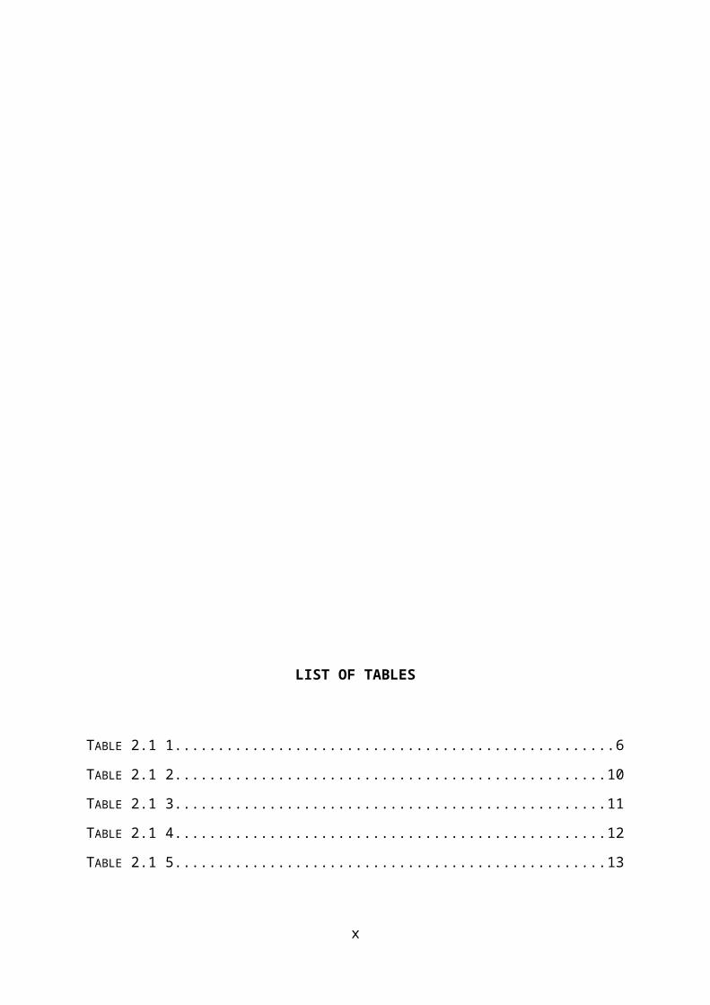
LIST OF TABLES
TABLE 2.1 1...................................................6
TABLE 2.1 2..................................................10
TABLE 2.1 3..................................................11
TABLE 2.1 4..................................................12
TABLE 2.1 5..................................................13
x

LIST OF ABBREVIATIONS
WHO……………………………………………………………..World Health Organisation
MRSA…………………………………………………………….Methicillin-Resistant Staphylococcus
aureus
xi
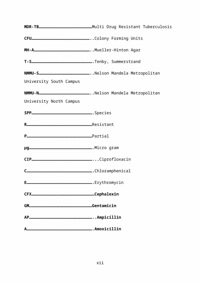
MDR-TB…………………………………………………………Multi Drug Resistant Tuberculosis
CFU………………………………………………………………..Colony Forming Units
MH-A……………………………………………………………..Mueller-Hinton Agar
T-S………………………………………………………………….Tenby, Summerstrand
NMMU-S………………………………………………………..Nelson Mandela Metropolitan
University South Campus
NMMU-N………………………………………………………..Nelson Mandela Metropolitan
University North Campus
SPP………………………………………………………………….Species
R………………………………………………………………………Resistant
P………………………………………………………………………Partial
µg…………………………………………………………………….Micro gram
CIP…………………………………………………………………...Ciprofloxacin
C……………………………………………………………………….Chloramphenical
E……………………………………………………………………….Erythromycin
CFX……………………………………………………………………Cephalexin
GM……………………………………………………………………Gentamicin
AP……………………………………………………………………..Ampicillin
A……………………………………………………………………….Amoxicillin
xii
CHAPTER ONE
1.1 INTRODUCTION
The increasing incidence of antibiotic-resistant bacteria,
especially the methicillin-resistant Staphylococcus aureus in
communities and hospitals, has placed great emphasis on the
need for new antimicrobial agents to treat infectious
diseases. In an attempt to uncover such resources researchers
are exploring some historically recognized natural remedies
which are still being used in some communities as an
alternative to expensive pharmaceutical drugs. (Historical
Anecdote Of Jordan's Red Soils May Offer New Antibiotic, 2009)
Antimicrobial chemotherapy has conferred huge bene ts on humanfi
health. A variety of microorganisms were elucidated to cause
infectious diseases in the latter half of the 19th century.
Thereafter, antimicrobial chemotherapy made remarkable
advances during the 20th century, resulting in the overly
optimistic view that infectious diseases would be conquered in
the near future. However, in response to the development of
antimicrobial agents, microorganisms that have acquired
resistance to drugs through a variety of mechanisms have
emerged and continue to plague human beings. In Japan, as in
other countries, infectious diseases caused by drug resistant
bacteria are one of the most important problems in daily
clinical practice. In the current situation, where multidrug-
resistant bacteria have spread widely, options for treatment
with antimicrobial agents are limited, and the number of brand
new drugs placed on the market is decreasing. Since drug-1
resistant bacteria have been selected by the use of
antimicrobial drugs, the proper use of currently available
antimicrobial drugs, as well as efforts to minimize the
transmission and spread of resistant bacteria through
appropriate infection control would be the rst step in fi
resolving the issue of resistant organisms. (Saga, 2009)
In 1928, Fleming discovered penicillin. He found that the
growth of Staphylococcus aureus was inhibited in a zone
surrounding a contaminated blue mold (a fungus from the
Penicillium genus) in culture dishes, leading to the nding fi
that a microorganism would produce substances that could
inhibit the growth of other microorganisms. The antibiotic was
named penicillin, and it came into clinical use in the 1940s.
In 1944, streptomycin, an aminoglycoside antibiotic, was
obtained from the soil bacterium Streptomyces griseus. Thereafter,
chloramphenicol, tetracycline, macrolide, and glycopeptide
(e.g., vancomycin) were discovered from soil bacteria. The
synthesized antimicrobial agent nalidixic acid, a quinolone
antimicrobial drug, was obtained in 1962. (Saga, 2009)
Historical anecdotes of the red soils from the Hashemite
Kingdom of Jordan tell of people using the soils to treat skin
infections and diaper rash. A multinational group of
researchers suggest the healing power may be due to
antibiotic-producing bacteria they have found living in the
soil. This discovery may ultimately lead to new antibiotic
treatments against harmful pathogens such as Staphylococcus
aureus. (Historical Anecdote Of Jordan's Red Soils May Offer
New Antibiotic, 2009)2
1.2 AIM
The main purpose of this study is to investigate the
biological activity of soil. Challenges in
biological screening remain a key focus in drug discovery from
soil.
1.3 OBJECTIVES
To determine antimicrobial activity of soil against bacteria
of medical importance,
3
To combat the problem of antimicrobial resistance,
To reduce the cost in development of drugs by using soil as a
source,
4
CHAPTER 2
2.1 LITERATURE REVIEW
Soils typically contain 109 to 1010 microorganisms per gram
(dry weight), which may represent more than a million
bacterial species. However, characterization of the small
fraction of microbes that has been cultivated provides only a
glimpse of their potential physiological capacity and
influence on soil ecosystems. The absence of pure cultures or
genome sequences makes it difficult to ascertain the roles of
specific microbes in soil environments: this is particularly
true for bacteria in the phylum Acidobacteria, which are broadly
distributed in soils but poorly represented in culture.
(Eichorst, 2007)
Diarrhoea caused by Shigella species is estimated by World health
organisation (WHO) to cause 50% of dysentery cases. S. dysenteriae
serotype 1 is particularly virulent, causing endemic and
epidemic dysentery with high death rate. Salmonella organisms
are endemic in many tropical and developing countries, while
other salmonellas cause food poisoning and bacteraemia. It is
highly infectious and resistance to common available
antimicrobials is an increasing problem.
(Cheesbrough, 2000)
Infections caused by resistant microorganisms often fail to
respond to conventional treatment, resulting in prolonged
illness, greater risk of death and higher costs.
5
Tuberculosis strains resistant to isoniazid and rifampicin
(multidrug-resistance - MDR-TB) require treatment courses that
are much longer and less effective. World Health Organisation
estimates that there are about 630 000 MDR-TB cases in the
world.
A high percentage of hospital-acquired infections are caused
by highly resistant bacteria such as methicillin-resistant
Staphylococcus aureus (MRSA) or multidrug-resistant Gram-negative
bacteria.
New resistance mechanisms have emerged, making the latest
generation of antibiotics virtually ineffective.
The evolution of resistant strains is a natural phenomenon
that happens when microorganisms are exposed to antimicrobial
drugs, and resistant traits can be exchanged between certain
types of bacteria. The misuse of antimicrobial medicines
accelerates this natural phenomenon. Poor infection control
practices encourage the spread of AMR. Infections caused by
resistant microorganisms often fail to respond to the standard
treatment, resulting in prolonged illness and greater risk of
death. The death rate for patients with serious infections
treated in hospitals is about twice that in patients with
infections caused by non-resistant bacteria.
(Antimicrobial resistance, 2013)
Synthesis of medicinally important compounds is very difficult
and thus the cost of medicine is also high because of the non-
availability of source materials especially aromatic
compounds.
6
(Gopalakrishnan, 2011)
2.2 MATERIALS AND METHODS
2.2.1 SOIL SAMPLES
Soil samples were collected from the different places Port
Elizabeth, Eastern Cape. Samples were collected from various
depth of the earth surface, ranging from layers just beneath
the upper surface to 6 inches depth. They were collected in
the sterile small plastic tubes and properly labelled with the
date of collection. Nine soil samples were collected within a
period of 3 weeks with regard to different weather conditions
7

(After rain, on a sunny day and in on a cold day). The
collected soil samples were dried in a hot air oven at 60–65°C
for 3 hours and stored in 4°C until examined.
Sample
Number
Date of
collection
Collection
site
Depth Weather
1 3rd August Tenby,
Summerstrand
5 inches Sunny
2 5th August NMMU South
Campus pond
6 inches Cold
3 14th August NMMU North
Campus
Round- about
6 Inches After rains
Table 2.1 1
Showing collection sites, dates, weather at collection and
depth of soil from where the isolates were collected using
Nutrient agar media
8
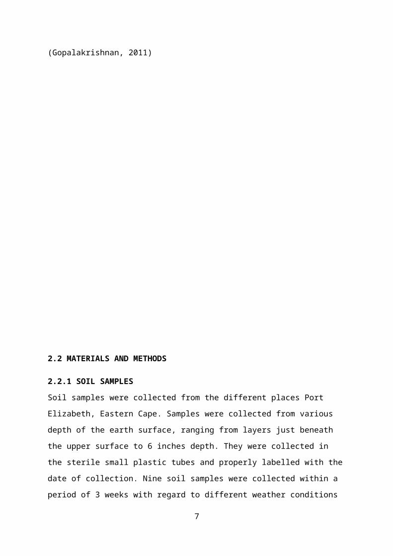
2.2.2 ISOLATION OF PURE CULTURE OF MICROBES
Three microorganisms were isolated and obtained as pure
culture by using standard microbiological method. From each
soil sample, 1 gm of dried soil was suspended in 9 mL sterile
water, and successive serial dilutions were made by
transferring 1mL of aliquots to 2nd test tube containing 9 mL
of sterile water, and in this way dilutions up to 10−4 were
prepared. Each time the contents were vortexed to form uniform
suspension. An aliquot of 0.1 mL of each dilution was taken
and spread evenly over the surface of Nutrient-agar medium on
16 cm petri dishes. Plates were incubated at 37°C and
monitored for 24 hours. The colonies were carefully counted by
visual observation and c.f.u per gram of soil was determined.
Plates those gave 100–150 colonies were chosen for further
isolation in pure culture. Suitable colonies those that showed
an anonymous appearance under light microscope were re-
cultivated several times for purity on blood agar.
Figure 2.1 19
Shows the serial dilution technique used to dilute the 1 gram
soil specimens for each of the three samples
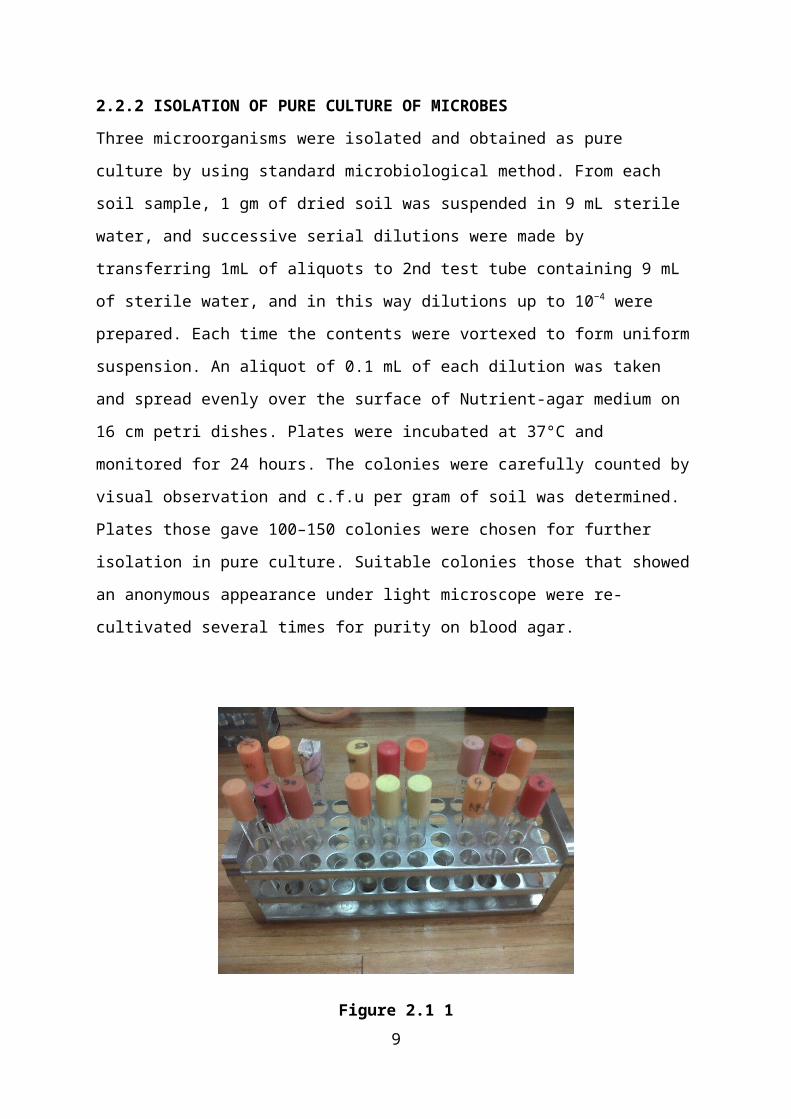
Figure 2.1 2
Showing colonies observation and c.f.u per gram of soil on
Nutrient agar
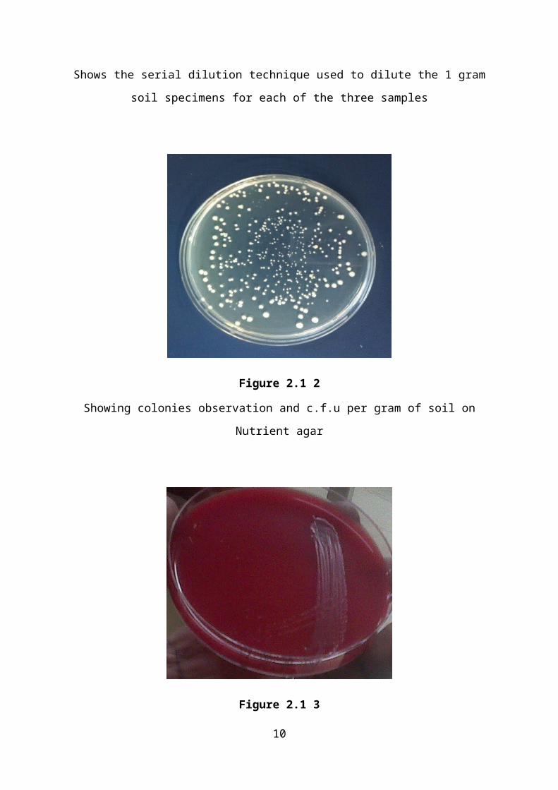
Figure 2.1 3
10
Showing isolation in pure culture on blood agar
2.2.3 SCREENING OF ANTIMICROBIAL ACTIVITIES OF PURE ISOLATES
Preliminary screening for antibiotic activity of the isolates
was done by using streak-plating technique Mueller Hinton-agar
medium. Each pure isolates were streaked individually on
different agar plates in a single line. The plates were then
incubated at 32°C for 5 days to allow the isolates to secrete
antibiotics into the medium. After the incubation period, the
properly diluted test organisms were cross-streaked along the
line of fully grown isolates. Each streaking was started near
the edge of the plates and streaked toward
the Streptomyces growth line. The plates were then incubated
for 12 hours at 37°C, for the zone of inhibition to be
observed.
2.2.4 TEST ORGANISMS
Five test organisms were used to test the antibiotic activity
of the isolates. Four of them were gram-positive and four were
gram-negative bacteria. Gram-positive species were Staphylococcus
aureus and Bacillus cereus. Gram-negative strains were Escherichia
coli, Shigella flexneri, and Salmonella spp. They were maintained in
nutrient agar medium and mackonkey agar medium respectively at
4°C.
11
2.2.5 RESULTS AND DISCUSSION
This study was performed with an aim of isolating soil
microbial strains with antimicrobial activities using the
selective isolation media. Three different gram positive cocci
strains were isolated from 3 soil samples collected from
different locations in the Eastern Cape in the year of 2013.
All of these strains were collected by using Nutrient-agar
media. Nutrient agar is a microbiological growth
medium commonly used for the routine cultivation of non-
fastidious bacteria. It is useful because it remains solid
even at relatively high temperatures. Also, bacteria grown in
nutrient agar grows on the surface, and is clearly visible as
small colonies.
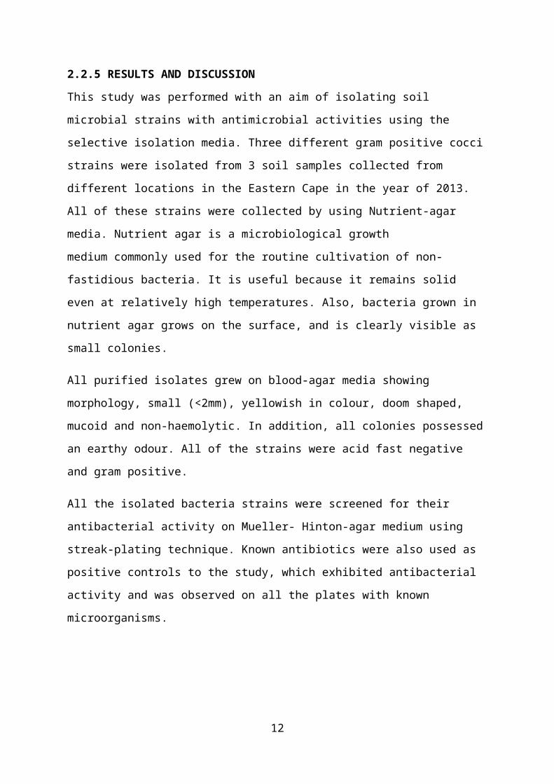
All purified isolates grew on blood-agar media showing
morphology, small (<2mm), yellowish in colour, doom shaped,
mucoid and non-haemolytic. In addition, all colonies possessed
an earthy odour. All of the strains were acid fast negative
and gram positive.
All the isolated bacteria strains were screened for their
antibacterial activity on Mueller- Hinton-agar medium using
streak-plating technique. Known antibiotics were also used as
positive controls to the study, which exhibited antibacterial
activity and was observed on all the plates with known
microorganisms.
12
Source Colony
Appearance
Colour
and Odour
Colony Size
(Diameter)
1(T-S) Doom
Shaped,
Mucoid And
Non-
Haemolytic
Cream
White-
Yellow
And
Earthy
Odour
<2mm
2(NMMU-S) Doom
Shaped,
Mucoid And
Non-
Haemolytic
Cream
White-
Yellow
And
Earthy
Odour
<2mm
3(NMMU-N) Doom
Shaped,
Mucoid And
Non-
Haemolytic
Cream
White-
Yellow
And
Earthy
Odour
<2mm
Table 2.1 2
Describes the colonies of purified isolates grown on blood-
agar media with morphology; small (<2mm), yellowish in colour,
doom shaped, mucoid and non-haemolytic
13
Source Gram Stain Zielh Neelsen
Stain1(T-S) Gram Positive
Cocci
Negative
2(NMMU-S) Gram Positive
Cocci
Negative
3(NMMU-N) Gram Positive
Cocci
Negative
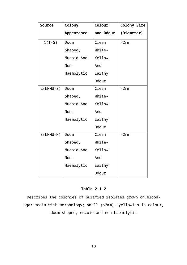
Table 2.1 3
Shows the microscopic results of the pure colonies from soil
14
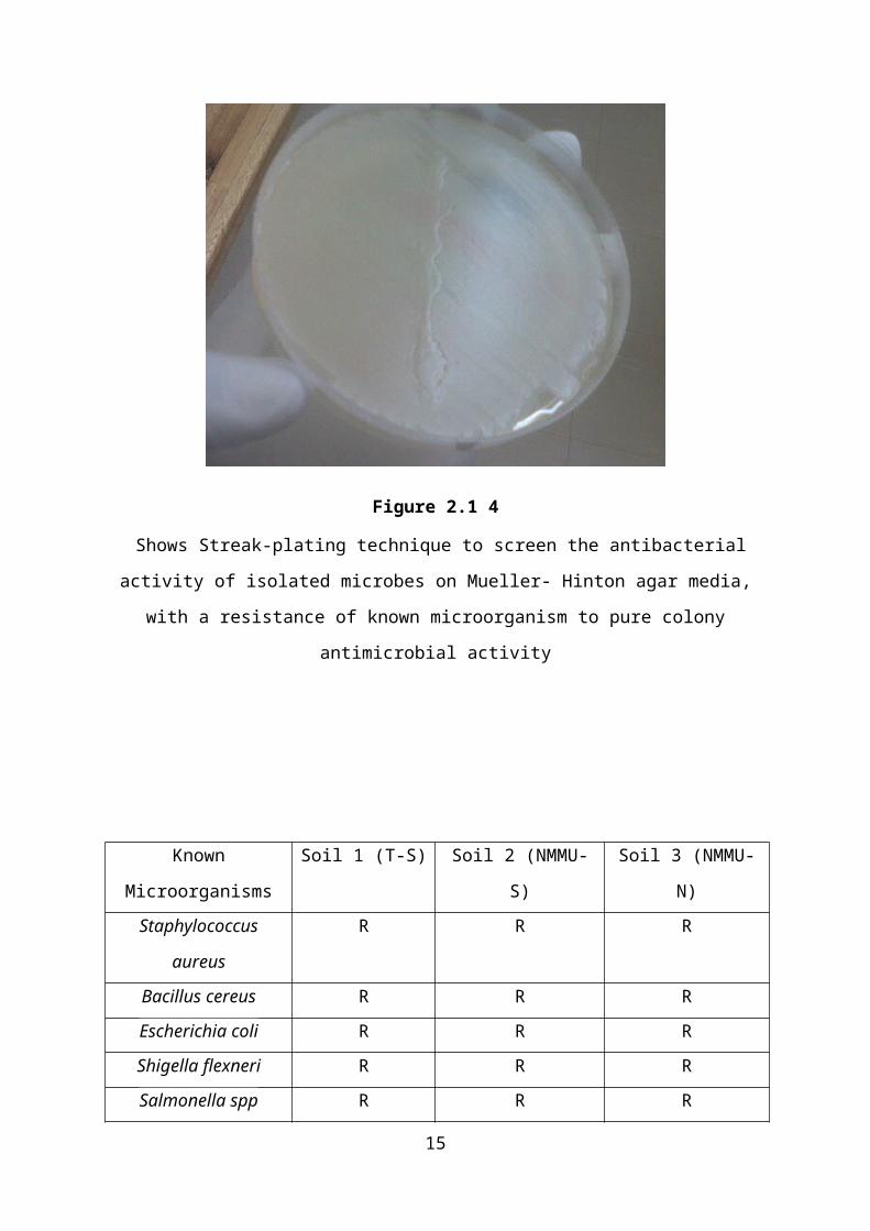
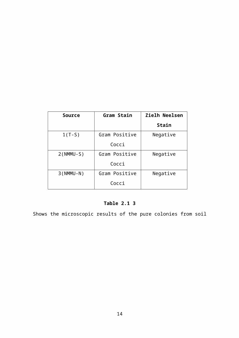
Figure 2.1 4
Shows Streak-plating technique to screen the antibacterial
activity of isolated microbes on Mueller- Hinton agar media,
with a resistance of known microorganism to pure colony
antimicrobial activity
Known
Microorganisms
Soil 1 (T-S) Soil 2 (NMMU-
S)
Soil 3 (NMMU-
N)Staphylococcus
aureus
R R R
Bacillus cereus R R REscherichia coli R R RShigella flexneri R R RSalmonella spp R R R
15
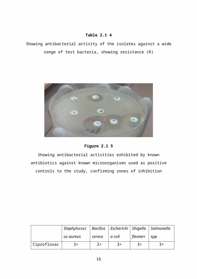
Table 2.1 4
Showing antibacterial activity of the isolates against a wide
range of test bacteria, showing resistance (R)
Figure 2.1 5
Showing antibacterial activities exhibited by known
antibiotics against known microorganisms used as positive
controls to the study, confirming zones of inhibition
Staphylococc
us aureus
Bacillus
cereus
Escherichi
a coli
Shigella
flexneri
Salmonella
spp
Ciprofloxac 3+ 2+ 3+ 3+ 3+
16
in (CIP)-
Dose- 5µgChloramphen
ical
(C)- 30µg
2+ 1+ 2+ 3+ 1+
Erythromyci
n (E)- 5µg
R - 2+ - 2+
Cephalexin
(CFX)- 30µg
1+ R P R P
Gentamicin
(GM)- 10µg
R 1+ P 1+ P
Ampicillin
(AP)
Dose- 10µg
- - P P P
Amoxycillin
(A)
Dose- 10µg
P R R R R
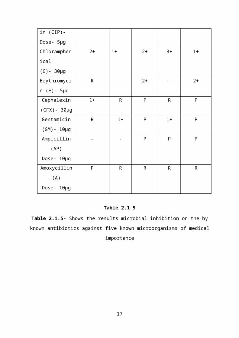
Table 2.1 5
Table 2.1.5- Shows the results microbial inhibition on the by
known antibiotics against five known microorganisms of medical
importance
17
CHAPTER 3
3.1 CONCLUSION
Historically the most commonly isolated actinomycete genera
have been Streptomyces and Micromonospora. As a result, the
majority of metabolites identi ed in screening programmes fi
searching for new antibiotics were derived from a relatively
limited pool of organisms. The genus Streptomyces is in fact
known as one of the major sources of bioactive natural
products. In the last decades, the intensive screening for new
secondary metabolites has also focused on minor groups of
actinomycetes, including species that are dif cult to isolate fi
and culture, and those that grow under extreme conditions.
In an effort to improve a screening programme in search of new
secondary metabolites with antimicrobial activity, alternative
selective conditions of pH and salinity for the isolation of
minor groups of actinomycetes not usually recovered in neutral
and low osmolality conditions was tested. The effect of this
expanded range of isolation conditions on the patterns of
detection of antibiotic activity was then evaluated.
(Basilio, 2003)
Actinomycetes comprise an extensive and diverse group of Gram-
positive, aerobic, mycelial bacteria that play an important
ecological role in soil cycles. Many are well known for their
economic importance as producers of biologically active
substances, such as antibiotics, vitamins and enzymes. In
addition, they are one of the major communities of the
microbial population present in soil, and their occurrence is
18
greatly in uenced by the environmental conditions of humidity,fl
temperature, pH and vegetation.
Soil actinomycetes for the most part show their optimum growth
in neutral and slightly alkaline conditions, and isolation
procedures have been traditionally based on this neutrophilic
character. Previous works showed the existence of a large
diversity of acidophilic actinomycetes that differed
morphologically and physiologically from neutrophilic species.
(Basilio, 2003)
In a study, from the soil samples of Karanjal regions of
Sundarbans of Bangladesh, about 55 actinomycetes of different
genera were isolated and screened for antibacterial activity.
In their screening work, they found that 20 isolates were
active against the test organisms. In another study,
356 Streptomyces isolates were obtained from soils in the
Aegean and East Black Sea regions of Turkey, and 36% of the
isolates were found to be active against tested
microorganisms. In a recent study performed in 2010 by Dehand,
the antibacterial activity of streptomyces isolates from soil
samples of West of Iran was investigated. Out of 150
actinomycetes, only 20 isolates (13.30%) showed activity
against the test bacteria.
(Sheikh, 2010)
In this study, in-vitro antimicrobial susceptibility tests
were performed using a panel which included both clinical
pathogens and laboratory control strains. All bacteria used
19
for the tests were resistant to at least one known
antimicrobial agent. The active soil isolates exhibited no
inhibitory pattern against the test organisms Staphylococcus
aureus, Bacillus cereus, Shigella flexneri, Escherichia coli and Salmonella spp.
Comparing the above mentioned results of other soil
antimicrobial studies, with this study, we can conclude that
the soil samples of three sites of choice are not rich source
of actinomycetes or any other bacteria which produce
metabolites inhibitory to bacterial pathogens. We found that
none of the isolated colonies were active against the test
bacteria. The known antibiotic positive controls were very
active and showed very large zone of inhibition.
3.2 RECOMMENDATIONS
Future investigations with different soil samples of different
properties still have to be carried out since these results
indicate that the selected soil samples stimulate growth of
all the pathogens. For future reference, there is need of
using further microbial identification techniques on the soil
isolates, other than gram stain and Ziehl Neelsen stain. The
precise identification of these soil pure isolates may be
20
studied on other bacteria of medical importance. Also, more
research on their biological properties may be carried out.
It is also clear that the wrong use of antimicrobial agents
resulted in the selection of resistant bacteria. Since the
advent of new mighty drugs is highly dif cult, the proper use fi
of currently available antimicrobial agents as well as efforts
to minimize the spread of resistant bacteria through
appropriate infection control would be quite important, and
may represent a rst step in solving the issue of resistant fi
microorganisms.
(Saga, 2009)
21
REFERENCES
(2009, May 21). Retrieved September 2013, from Science daily: http://www.sciencedaily.com/releases/2009/05/090518222202.htm
(2013, May). Retrieved Sepetember 2013, from World Health
Organisation:
http://www.who.int/mediacentre/factsheets/fs194/en/
Abdelghani, K. P. (2009, April). Antibacterial activity of
bacterial isolates of soil bacteria collected from Palestine.
Current Trends in Biotechnology and Pharmacy, 3(2). Retrieved September
2013, from http://www.pharmainfo.net/articles/antibacterial-
activity-bacterial-isolates-soil-bacteria-collected-palestine
Basilio, G. V. (2003, August). Patterns of antimicrobial
activities from soil actinomycetes isolated under different
conditions of pH and salinity. Journal of Applied Microbiology, 95(4),
814-823. Retrieved October 2013, from
http://onlinelibrary.wiley.com/doi/10.1046/j.1365-
2672.2003.02049.x/pdf
Cheesbrough. (2000). District laboratory practice in tropical countries (Vol.
2). Cambridge, United Kingdom: Cambridge University Press.
Retrieved September 2013
Eichorst, B. S. (2007, April). Isolation and Characterization
of Soil Bacteria That Define Terriglobus gen. nov., in the
Phylum Acidobacteria. Applied and Enviromental Microbiology, 73(8),
2708-2713. Retrieved September 2013, from
http://www.ncbi.nlm.nih.gov/pmc/articles/PMC1855589/#fn1
22
Falkinham, W. T. (2009, March). Proliferation of Antibiotic-
Producing Bacteria and Concomitant Antibiotic Production as
the Basis for the Antibiotic Activity of Jordan's Red Soils.
American Society For Microbiology, 75(9), 2735-2741. Retrieved
September 2013, from http://aem.asm.org/content/75/9/2735
Gopalakrishnan, N. M. (2011). Antibacterial activity of azo
compounds synthesized from the natural. Journal of Chemical and
Pharmaceutical Research, 3(4), 490-497. Retrieved September 2013,
from http://jocpr.com/vol3-iss4-2011/JCPR-2011-3-4-490-497.pdf
Moshafi, F. A.-N. (2011, July). Antimicrobial activity of
Bacillus sp. strain FAS1 isolated from soil. Pakistan Journal of
Pharmaceutical science, 24(3), 269-275. Retrieved September 2013,
from http://www.ncbi.nlm.nih.gov/pubmed/21715259
Saga, Y. (2009, April). History of Antimicrobial Agents and
Resistant. Japan Medical Association, 52(2), 103-108. Retrieved
September 2013, from
http://www.med.or.jp/english/journal/pdf/2009_02/103_108.pdf
Sheikh, H. M. (2010, October 4). Antimicrobial activity of
certain bacteria and fungi isolated from soil mixed with human
saliva against pathogenic microbes causing dermatological
diseases. Saudi Journal of Biological Sciences, 17(4), 331-339. Retrieved
September 2013, from
http://www.sciencedirect.com/science/article/pii/S1319562X1000
0732
23
Yilmaz, M. (2006, February 13). Antimicrobial activities of
some Bacillus spp. strains isolated from the soil. Microbiological
Research, 161(2), 127-131. Retrieved September 2013, from
http://www.sciencedirect.com/science/article/pii/S094450130500
0704
24
Related Documents


![Available Online through (or) www ... · diarrhoea and anti-inflammatory, anti cancer and anti oxidative. It also known to possess antiviral and anti fungal [19, 20] and antimicrobial](https://static.cupdf.com/doc/110x72/5f8fe9b432d03476e9579f60/available-online-through-or-www-diarrhoea-and-anti-inflammatory-anti-cancer.jpg)