CANCER STEM CELLS Antigen-Specific T-Cell Response from Dendritic Cell Vaccination Using Cancer Stem-Like Cell-Associated Antigens QIJIN XU,GENTAO LIU,XIANGPENG YUAN,MINLIN XU,HONGQIANG WANG,JIANFEI JI,BINDU KONDA, KEITH L. BLACK,JOHN S. YU Department of Neurosurgery, Cedars-Sinai Medical Center, Los Angeles, California, USA Key Words. Dendritic cell • Vaccination • Glioblastoma • Cancer • Stem-like cells • Cytotoxic T lymphocyte ABSTRACT Glioblastoma multiforme (GBM) is the most aggressive pri- mary brain tumor, with current treatment remaining palli- ative. Immunotherapies harness the body’s own immune system to target cancers and could overcome the limitations of conventional treatments. One active immunotherapy strategy uses dendritic cell (DC)-based vaccination to initi- ate T-cell-mediated antitumor immunity. It has been pro- posed that cancer stem-like cells (CSCs) may play a key role in cancer initiation, progression, and resistance to cur- rent treatments. However, whether using human CSC anti- gens may improve the antitumor effect of DC vaccination against human cancer is unclear. In this study, we explored the suitability of CSCs as sources of antigens for DC vacci- nation again human GBM, with the aim of achieving CSC- targeting and enhanced antitumor immunity. We found that CSCs express high levels of tumor-associated antigens as well as major histocompatibility complex molecules. Fur- thermore, DC vaccination using CSC antigens elicited anti- gen-specific T-cell responses against CSCs. DC vaccination- induced interferon-c production is positively correlated with the number of antigen-specific T cells generated. Finally, using a 9L CSC brain tumor model, we demon- strate that vaccination with DCs loaded with 9L CSCs, but not daughter cells or conventionally cultured 9L cells, induced cytotoxic T lymphocytes (CTLs) against CSCs, and prolonged survival in animals bearing 9L CSC tumors. Understanding how immunization with CSCs generates superior antitumor immunity may accelerate development of CSC-specific immunotherapies and cancer vaccines. STEM CELLS 2009;27:1734–1740 Disclosure of potential conflicts of interest is found at the end of this article. INTRODUCTION Glioblastoma multiforme (GBM) is the most common and most aggressive type of primary brain tumor, accounting for 52% of all primary brain tumor cases. Current treatment of GBM remains palliative and includes surgery, radiotherapy, and chemotherapy [1, 2]. Immunotherapies harness the body’s own immune system to target brain tumor and could over- come the limitations in conventional treatments [3–5]. One of the most promising strategies may be active immunotherapy using dendritic cell (DC)-based vaccination to initiate T-cell- mediated antitumor immunity [6–10]. In practice, vaccination strategies have often used DCs pulsed with tumor-derived whole lysates/peptides as modalities to present a broad range of tumor antigens to T cells ex vivo to stimulate effective antitumor T-cell immunity [8]. This process includes two stages: in vitro DC maturation and antigen loading and in vivo DC migration and antigen presentation in the draining lymph node (DLN). Human DCs are commonly generated from peripheral blood-derived monocytes, followed by a dif- ferentiation step to produce immature DCs (iDCs). The iDCs undergo maturation and antigen loading steps to produce mature DCs [11]. Mature DCs loaded with tumor antigens are administrated subcutaneously into patients. The goal is to gen- erate ex vivo a population of antigen-loaded DCs that stimu- lates robust and long-lasting CD4 þ and CD8 þ T-cell responses in the patient with cancer. Recent studies identifying cancer stem-like cells (CSCs) as brain tumor-initiating cells may have implications for mod- ifying GBM treatments, including DC vaccination-based immunotherapy [8, 12–14]. Therapies targeting CSCs may prevent tumor recurrences seen after conventional radiation and chemotherapies. Furthermore, it is likely that certain stem cell markers expressed by CSCs may have distinct antigenic- ity and thus provide opportunities for enhanced immunother- apy. Some proteins expressed by CSCs are normally seen only in early development stages. Antibodies against the stem Author contributions: Q.X.: concept and design; culture and characterization of human CSCs; human dendritic cell characterization; rat DC vaccination model and animal experiments; writing the manuscript; G.L.: human CSC expression of TAA, IFN-c assays, tetramer assays, data analysis; X.Y.: human dendritic cell endocytosis, and supporting roles in tissue culture and rat DC vaccination model and animal experiments; M.X.: PCR and FACS experiments with G.L.; H.W.: human DC culture and antigen loading; J.J.: supporting DC culture and genotyping; B.K.: assisting with animal experiments; K.L.B.: financial support and material; J.S.Y.: concept and design, advisor and supervising role, cowriting. Q.X. and G.L. contributed equally to this work. Correspondence: John S. Yu, M.D., Maxine Dunitz Neurosurgical Institute, Suite 800E East, 8631 West 3rd Street, Los Angeles, CA 90048, USA. Telephone: 310-423-0845; Fax: 310-423-1038; e-mail: [email protected] Received January 20, 2009; accepted for publi- cation April 14, 2009; first published online in STEM CELLS EXPRESS April 23, 2009. V C AlphaMed Press 1066-5099/2009/$30.00/0 doi: 10.1002/stem.102 STEM CELLS 2009;27:1734–1740 www.StemCells.com

Welcome message from author
This document is posted to help you gain knowledge. Please leave a comment to let me know what you think about it! Share it to your friends and learn new things together.
Transcript
CANCER STEM CELLS
Antigen-Specific T-Cell Response from Dendritic Cell Vaccination
Using Cancer Stem-Like Cell-Associated Antigens
QIJIN XU, GENTAO LIU, XIANGPENG YUAN, MINLIN XU, HONGQIANG WANG, JIANFEI JI, BINDU KONDA,
KEITH L. BLACK, JOHN S. YU
Department of Neurosurgery, Cedars-Sinai Medical Center, Los Angeles, California, USA
Key Words. Dendritic cell • Vaccination • Glioblastoma • Cancer • Stem-like cells • Cytotoxic T lymphocyte
ABSTRACT
Glioblastoma multiforme (GBM) is the most aggressive pri-mary brain tumor, with current treatment remaining palli-ative. Immunotherapies harness the body’s own immunesystem to target cancers and could overcome the limitationsof conventional treatments. One active immunotherapystrategy uses dendritic cell (DC)-based vaccination to initi-ate T-cell-mediated antitumor immunity. It has been pro-
posed that cancer stem-like cells (CSCs) may play a keyrole in cancer initiation, progression, and resistance to cur-
rent treatments. However, whether using human CSC anti-gens may improve the antitumor effect of DC vaccinationagainst human cancer is unclear. In this study, we explored
the suitability of CSCs as sources of antigens for DC vacci-nation again human GBM, with the aim of achieving CSC-
targeting and enhanced antitumor immunity. We found
that CSCs express high levels of tumor-associated antigensas well as major histocompatibility complex molecules. Fur-thermore, DC vaccination using CSC antigens elicited anti-gen-specific T-cell responses against CSCs. DC vaccination-induced interferon-c production is positively correlatedwith the number of antigen-specific T cells generated.Finally, using a 9L CSC brain tumor model, we demon-
strate that vaccination with DCs loaded with 9L CSCs, butnot daughter cells or conventionally cultured 9L cells,
induced cytotoxic T lymphocytes (CTLs) against CSCs, andprolonged survival in animals bearing 9L CSC tumors.Understanding how immunization with CSCs generates
superior antitumor immunity may accelerate developmentof CSC-specific immunotherapies and cancer vaccines.
STEM CELLS 2009;27:1734–1740
Disclosure of potential conflicts of interest is found at the end of this article.
INTRODUCTION
Glioblastoma multiforme (GBM) is the most common andmost aggressive type of primary brain tumor, accounting for52% of all primary brain tumor cases. Current treatment ofGBM remains palliative and includes surgery, radiotherapy,and chemotherapy [1, 2]. Immunotherapies harness the body’sown immune system to target brain tumor and could over-come the limitations in conventional treatments [3–5]. One ofthe most promising strategies may be active immunotherapyusing dendritic cell (DC)-based vaccination to initiate T-cell-mediated antitumor immunity [6–10]. In practice, vaccinationstrategies have often used DCs pulsed with tumor-derivedwhole lysates/peptides as modalities to present a broad rangeof tumor antigens to T cells ex vivo to stimulate effectiveantitumor T-cell immunity [8]. This process includes twostages: in vitro DC maturation and antigen loading and invivo DC migration and antigen presentation in the draining
lymph node (DLN). Human DCs are commonly generatedfrom peripheral blood-derived monocytes, followed by a dif-ferentiation step to produce immature DCs (iDCs). The iDCsundergo maturation and antigen loading steps to producemature DCs [11]. Mature DCs loaded with tumor antigens areadministrated subcutaneously into patients. The goal is to gen-erate ex vivo a population of antigen-loaded DCs that stimu-lates robust and long-lasting CD4þ and CD8þ T-cellresponses in the patient with cancer.
Recent studies identifying cancer stem-like cells (CSCs)as brain tumor-initiating cells may have implications for mod-ifying GBM treatments, including DC vaccination-basedimmunotherapy [8, 12–14]. Therapies targeting CSCs mayprevent tumor recurrences seen after conventional radiationand chemotherapies. Furthermore, it is likely that certain stemcell markers expressed by CSCs may have distinct antigenic-ity and thus provide opportunities for enhanced immunother-apy. Some proteins expressed by CSCs are normally seenonly in early development stages. Antibodies against the stem
Author contributions: Q.X.: concept and design; culture and characterization of human CSCs; human dendritic cell characterization; ratDC vaccination model and animal experiments; writing the manuscript; G.L.: human CSC expression of TAA, IFN-c assays, tetramerassays, data analysis; X.Y.: human dendritic cell endocytosis, and supporting roles in tissue culture and rat DC vaccination model andanimal experiments; M.X.: PCR and FACS experiments with G.L.; H.W.: human DC culture and antigen loading; J.J.: supporting DCculture and genotyping; B.K.: assisting with animal experiments; K.L.B.: financial support and material; J.S.Y.: concept and design,advisor and supervising role, cowriting. Q.X. and G.L. contributed equally to this work.
Correspondence: John S. Yu, M.D., Maxine Dunitz Neurosurgical Institute, Suite 800E East, 8631 West 3rd Street, Los Angeles, CA90048, USA. Telephone: 310-423-0845; Fax: 310-423-1038; e-mail: [email protected] Received January 20, 2009; accepted for publi-cation April 14, 2009; first published online in STEM CELLS EXPRESS April 23, 2009. VC AlphaMed Press 1066-5099/2009/$30.00/0doi: 10.1002/stem.102
STEM CELLS 2009;27:1734–1740 www.StemCells.com
cell-associated antigen SOX2 was identified in a humanpatient [15]. CSC-associated proteins may be used for cancervaccination. It was reported recently that vaccination usingprostate stem cell antigen induced long-term protectiveimmune response against prostate cancer without autoimmun-ity [16]. Even without identification of specific antigens,CSCs can be a useful source of tumor antigens in DC vacci-nation-based immunotherapy. Using a mouse GL261 gliomamodel, Pellegatta et al. demonstrated that vaccination withDCs loaded with glioma CSC antigens elicited robust antitu-mor T-cell immunity [17]. In this study, DC vaccination usingCSC antigens cured up to 80% GL261 tumors, whereas DCvaccination using regular GL261 antigens cured none of theCSC-initiated tumors. However, whether using human CSCantigens may improve the antitumor effect of DC vaccinationagainst human cancer is unclear.
In the current study, we investigate expression of tumor-associated antigens (TAAs) and major histocompatibility com-plex (MHC) molecules by GBM-derived CSCs. We reportthat CSCs express MHC I and increased levels of a range ofTAAs. CSCs can be recognized by T cells generated afterDCs were pulsed with CSC tumor antigens. DC vaccination-induced interferon (IFN)-c production is positively correlatedwith the number of antigen-specific T cells generated. Finally,using a 9L CSC brain tumor model, we demonstrate that DCvaccination with 9L CSCs induced higher IFN-c productionthan vaccination using parent cells and daughter cells and thatDC vaccination with only 9L CSCs prolongs survival of tu-mor-bearing animals.
MATERIALS AND METHODS
Primary Culture of Glioblastoma-Derived CancerStem-Like Cells
Primary brain tumor spheres were cultured as previouslydescribed [14]. Briefly, brain tumor stem-like cells were grown inDulbecco’s modified Eagle’s medium (DMEM)/F12 medium sup-plemented with B-27 (Invitrogen, Carlsbad, CA, http://www.invi-trogen.com), 20 ng/ml of basic fibroblast growth factor, and 20ng/ml of endothelial-derived growth factor (Peprotech, RockyHill, NJ, http://www.peprotech.com). Alternatively, dispersedbrain tumor stem-like cells were grown on a laminin-coated sur-face in the same medium as described above. Primary human fe-tal neural stem cells were derived from primary cells obtainedfrom Cambrex (East Rutherford, NJ, http://www.cambrex.com).GBM cell line and adherent primary glioma cells were culturedin DMEM/F-12 containing 10% fetal bovine serum. Some frozenprimary GBM tissues were used to compare gene expression pro-files of CSCs and their parental tumors.
Human Glioblastoma Cell Antigen Preparation
Human brain tumor cells were prepared following InstitutionalReview Board (IRB)-approved Standard Operating Procedure.Fresh tumor specimens were divided and processed under sterileconditions. Tumors were cleaned and minced in a dissection me-dium (Hanks’ balanced salt solution, 0.36% glucose, 0.8 mMMgCl2, 0.03 mg/ml catalase, 6.6 mg/l deferoxamine, 25 mg/l N-acetyl cysteine, 94 mg/L cystine-Cl, 1.25 mg/L superoxide dismu-tase, 100 U/ml Fungi-Bact, 0.11 mg/ml sodium pyruvate, 10 mMHEPES). After digestion with trypsin-EDTA for 10 minutes at37�C, cells were washed and passed through 100-lm meshes.CSCs were isolated from primary tumor cells as described previ-ously [8, 14]. At lease 1 to 3 million cells in suspension wereirradiated to prepare apoptotic CSCs as antigens. Tumor antigenprotein concentrations were determined using Bio-Rad ProteinAssay reagents (Hercules, CA, http://www.bio-rad.com).
Priming of Human DCs with Apoptotic CSCs
Human immature DCs were prepared from peripheral mononu-clear blood cells (PBMCs). PBMCs obtained from a HLA-A2þ
healthy donor were prepared by Ficoll/Paque (Invitrogen) densitygradient centrifugation. Cells were seeded (1 � 107 cells/3 ml/well) into 6-well plates (Corning Costar Corp., Cambridge, MA,http://www.corning.com/index.aspx) in RPMI 1640 supplementedwith 10% human AB serum, 2 mM L-glutamine, 10 mM HEPES,and antibiotics. After 2 hours of incubation at 37�C, adherentcells were used for DC generation as described. The nonadherentlymphocytes were stimulated with autologous DCs loaded withirradiated CSC line no. 66 (HLA-A2-positive, CD133-positive) atthe ratio of 10:1 (the CSCs and DCs were cocultured overnightbefore they were seeded with lymphocytes). Interleukin-2 (IL-2;300 IU/ml) was added to the cultures the next day and every 3days thereafter. The lymphocytes were restimulated with DCs ev-ery week for up to 3 stimulations. The stimulated cells weretested for their ability to recognize antigen epitopes by incubatingthe stimulated cells with T2 cells pulsed with the epitopes as wellas peptides derived from other tumor antigens and CD133-posi-tive CSC lines. The stimulated cells (1 � 105) were incubatedwith 1 � 105 target cells for 24 hours, and the release of IFN-c(pg/ml) was measured by commercial enzyme-linked immunosor-bent assay (ELISA) kit (Endogen, Cambridge, MA, http://www.piercenet.com). The percentage of CD133-specific cytotoxicT lymphocytes (CTLs) in the stimulated cells was analyzed bytetramer technology.
Rat Glioma Cell Antigen Preparation and DCCulture
Rat 9L glioma cell antigens are prepared using a procedure simi-lar to the one described above. All animal procedures were per-formed in strict accordance with the Institutional Animal Careand Use Committee guidelines at Cedars-Sinai Medical Center.For rat immature DCs isolation, F344 Fisher rats were euthanizedand bone marrow cells were collected by flushing femurs andtibias with RPMI 1640 media. Bone marrow cells were incubatedwith IL-4 and granulocyte macrophage-colony stimulation factor(150 U/ml; Peprotech), with medium renewed every 2 days. After7 days, cells were analyzed using fluorescence activated cell sort-ing (FACS) with antibodies against CD11c, CD14, CD80, CD86,and MHCII (obtained from BD Pharmingen, San Diego, http://www.bdbiosciences.com). Immature DCs of 90%--100% puritywere used for further studies. For rat DC stimulation, cells werestimulated by addition of lipopolysaccharide (50 lg/ml, from Ecoli 055:B5; Sigma-Aldrich, St. Louis, http://www.sigmaaldrich.com) or tumor lysate (100 lg/ml) for 1 day. Supernatant weretaken after 1 day for further analysis.
Synthetic Peptides and HLA Typing
All of the peptides used in this study were synthesized by Macro-molecular Resource (Fort Collins, CO, http://www.macromolecular.colostate.edu). The identity and purity of each of the peptides wereconfirmed by mass spectrometer and high-performance liquid chro-matography analysis. Peptides were dissolved in dimethyl sulfoxideat 1 mM concentration for future use. GBM cells were stained withbiotin-conjugated HLA-A2- or HLA-A1-specific monoclonal anti-body (US Biological, Swampscott, MA, http://www.usbio.net) orbiotin-conjugated isotype control antibody. After streptavidin-PerCP(BD Pharmingen) staining for 30 minutes, the mean fluorescenceintensity of HLA-A2 staining was analyzed by flow cytometry.
Tetramer Staining
Various antigen-specific peptide tetramers (phycoerythrin-peptide-loaded HLA tetramer complexes) were synthesized [18, 19] andprovided by Beckman Coulter (San Diego, http://www.beckman-coulter.com). Specific CTL clone CD8þ cells were resuspendedat 105 cells/50 ll fluorescence-activated cell-sorting buffer (phos-phate buffer plus 1% inactivated fluorescence-activated cell-
Xu, Liu, Yuan et al. 1735
www.StemCells.com
sorting buffer). Cells were incubated with 1 ll of tHLA for 30minutes at room temperature, and incubation was then continuedfor 30 minutes at 4�C with anti-CD8 monoclonal antibody (Bec-ton, Dickinson and Company, Franklin Lakes, NJ, http://www.bd.com). Cells were washed twice in 2 ml of cold fluores-cence-activated cell-sorting buffer before analysis by fluores-cence-activated cell sorting (Becton, Dickinson and Company).
T-Cell Stimulation and Cytotoxic T-Cell Assays
For human cells, CTL precursor frequency was determined toassess effective immunization to autologous tumor cells. PBMCs(1 � 106 cells/ml) were stimulated in 10% human AB serumwith 1 � 106/ml irradiated CSC-pulsed DCs, with recombinanthuman IL-2 (300 units/ml) added on day 2. Expanded CTLs fromPBMCs were restimulated on day 11 with 150 lg/ml tumor anti-gens for 2 hours. RNA was extracted and IFN-c production wasanalyzed using ELISAs. Controls include DC alone, PBMCalone, and tumor antigens with PBMCs. Rat T-cell stimulationand CTL assays were performed in a similar fashion. FACS anal-ysis of CD69 and MHC class II expression were performed foralloreactive T-cell activation index.
Real-time reverse-transcription PCR, ELISA, andFACS analysis
For quantitative polymerase chain reaction (PCR), total RNA wasisolated from stimulated cells using Trizol (GIBCO Invitrogen,San Diego, CA, http://www.invitrogen.com) and transcribed usingrandom hexamers. Cytokines and reference cDNA and quantifiedplasmid DNA standards were amplified using quantitative PCRprimers or probes (Qiagen, Alameda, CA, http://www1.qiagen.-com), with greater than a 1.5-fold increase in CD8-normalizedIFN-c production following vaccination indicating a positiveresponse. ELISA analysis was performed to determine IFN-c pro-duction using ELISA kits from R&D Systems Inc. (Minneapolis,http://www.rndsystems.com) following the manufacturer’s instruc-tions. DCs and PBMCs were rinsed in FACS buffer (phosphate-buffered saline with 1% FCS, 0.1% wt/vol sodium azide) andincubated with Fc Block (BD Biosciences, San Diego, http://www.bdbiosciences.com) for 20 minutes on ice, and stained withfluorescein isothiocyanate- or phycoerythrin-conjugated antibodies(CD86, CD69, MHC class II; from BD Biosciences) for 30minutes on ice. After rinsing 3 times, cells were analyzed using aFACScan system (BD Biosciences).
DC Vaccination in a Rat Glioma Model
Adult F344 Fisher rats were anesthetized and placed in the ste-reotactic frame. The skin was cut with a scalpel and the skullpenetrated using a dental drill. A needle was placed 5-mm deepat the following coordinates from bregma (anterior-posterior: þ1mm; medial-lateral: 3 mm; dorsal-ventral: 5 mm). Duramater waspunctured at the specific site of injection. Initial tumor inoculumswere prepared using 25,000 luciferase-labeled 9L-gliosarcomacells. For DC vaccination, at days 7, 14, and 21, animals receivedsubcutaneous DC vaccinations into the flank, or via intracranialinjection. Before vaccination, 50,000 freshly cultured immaturedendritic cells were incubated with tumor antigens acid-elutedfrom tumor cells or saline control for 1 day.
Survival and Statistical Analysis
Data were analyzed with a SAS statistical software package (SASInstitute, Cary, NC, http://www.sas.com). Means of at least 3 in-dependent experiments were reported with standard deviations.The estimated probability of survival was demonstrated using theKaplan-Meier method. The Mantel Cox log-rank tests were usedto compare curves between study and control groups. Any P val-ues less than .05 were considered statistically significant.
RESULTS
Expression of Tumor-Associated Antigens inGlioblastoma-Derived Cancer Stem-Like Cells
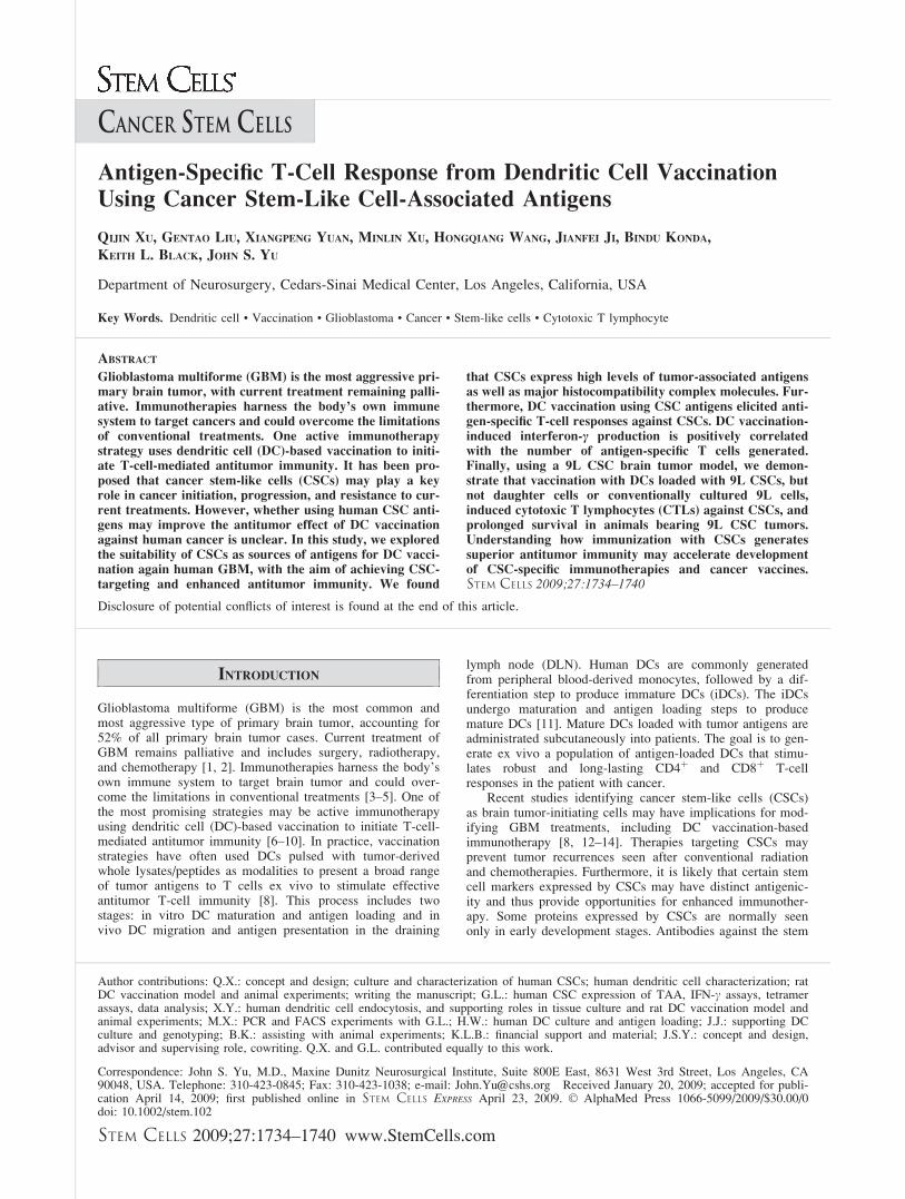
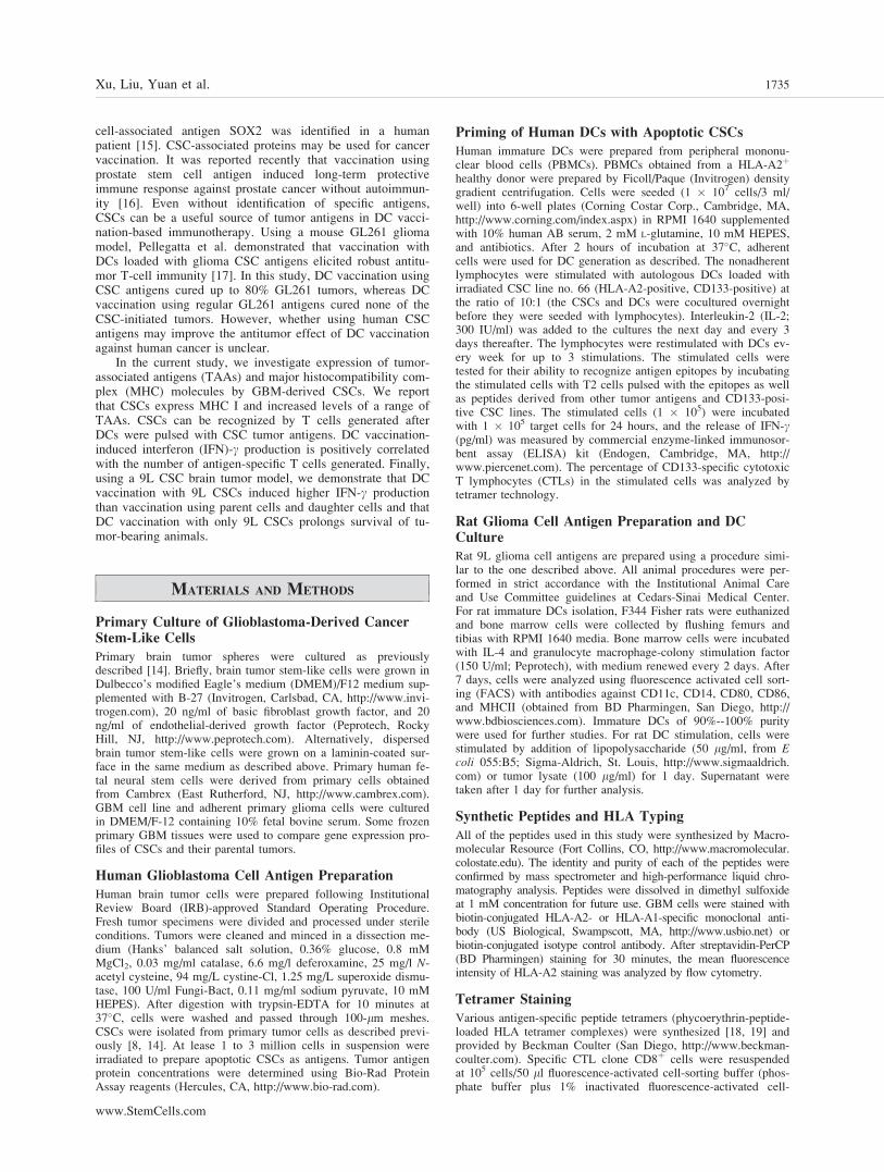
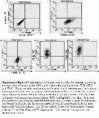
We had previously isolated CSCs from human GBM anddemonstrated their self-renewal capability and multipotent dif-ferentiation in vitro and tumor-initiating ability in vivo [8,14]. Although these CSCs manifest different gene expressionprofiles and signal pathway activities [14], they share theexpression of neural stem cell marker Nestin and the cell sur-face marker CD133 (Fig. 1A). Another cell surface markerLeX/CD15 was recently identified in murine neural stem cells[5]. Flow cytometry analysis indicated that most of theCD133-positive CSCs also express CD15 (Fig. 1B). Theexpression of CD133 and CD15 in the differentiated daughtercells, however, is greatly reduced (supporting information Fig.1). To study whether CSCs express certain TAAs, we meas-ured the expression levels of several GBM-associated tumorantigens in both conventional cultured adherent cells and cor-responding CSCs. We found that both adherent cells that con-tain differentiated daughter cells and CSCs express EGFR,HER2, TRP2, MRP3, AIM2, SOX2, and IL13Ra2, but theexpression levels were 2-fold to more than 200-fold higher inCSCs than those in adherent cells, with the exception ofIL13Ra2, whose expression levels are comparable in both ad-herent cells and CSCs (Fig. 2). Some of these tumor antigenswere shown in our previous studies to be expressed in humanGBM and recognized by CTLs [7, 18, 19]. The higher expressionof TAAs in CSCs suggested that CSCs may be a better source oftumor antigens in DC vaccination-based immunotherapy.
Expression of Class I MHC Molecules on CellSurface of Cancer Stem-Like Cells
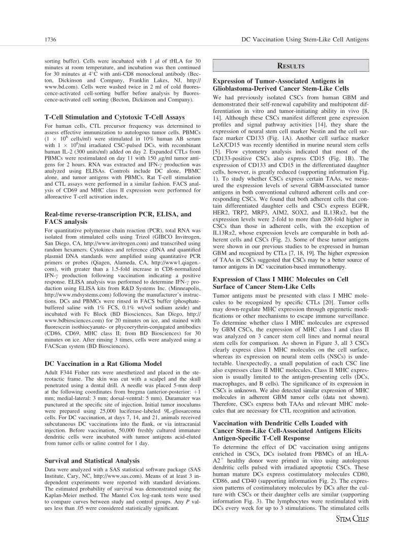
Tumor antigens must be presented with class I MHC mole-cules to be recognized by specific CTLs [20]. Tumor cellsmay down-regulate MHC expression through epigenetic modi-fications or other mechanisms to escape immune surveillance.To determine whether class I MHC molecules are expressedby GBM CSCs, the expression of MHC class I and class IIwas analyzed on 3 cancer stem cell lines and normal neuralstem cells for comparison. As shown in Figure 3, all 3 CSCsclearly express class I MHC molecules on the cell surface,whereas its expression on neural stem cells (NSCs) is unde-tectable. Unexpectedly, a small population of each CSC linealso expresses class II MHC molecules. Class II MHC expres-sion is usually limited to the antigen-presenting cells (DCs,macrophages, and B cells). The significance of its expression inCSCs is unknown. We also detected similar expression of MHCmolecules in adherent GBM tumor cells (data not shown).Therefore, CSCs express both TAAs and relevant MHC mole-cules that are necessary for CTL recognition and activation.
Vaccination with Dendritic Cells Loaded withCancer Stem-Like Cell-Associated Antigens ElicitsAntigen-Specific T-Cell Response
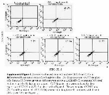
To determine the effect of DC vaccination using antigensenriched in CSCs, DCs isolated from PBMCs of an HLA-A2þ healthy donor were primed in vitro using autologousdendritic cells pulsed with irradiated apoptotic CSCs. Thesehuman mature DCs express costimulatory molecules CD80,CD86, and CD40 (supporting information Fig. 2). The expres-sion patterns of costimulatory molecules by DCs after the cul-ture with CSCs or their daughter cells are similar (supportinginformation Fig. 3). The lymphocytes were restimulated withDCs every week for up to 3 stimulations. The stimulated cells
1736 DC Vaccination Using Stem-Like Cell Antigens
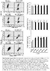
were then tested for their ability to recognize specific epitopesderived from TRP2, HER2, IL13Ra2, and SOX2 by incubat-ing the stimulated cells with T2 cells pulsed with the epi-topes. As shown in Figure 4A, tumor antigen-loaded DCsstimulated Th1 response and induced significant IFN-c pro-duction. Next, we tested the effect of DC vaccination using 3different CSCs and NSCs as antigens. Stimulation of PBMCsusing each of the 3 CSC antigens induced significant T-cellresponses as indicated by robust IFN-c production in responseto analysis stimulation (Fig. 4B). Although some antigens(such as EGFR) were shared between CSCs and NSCs, cocul-turing with NSCs did not induce immune response and IFN-cproduction, consistent with the fact that NSCs do not express
MHC molecules. These data suggested that DC vaccinationusing apoptotic CSCs can elicit a strong specific T-cell response.
To determine whether the stimulated T cells contain anti-gen-specific CTLs, we performed tetramer analyses usingantigen tetramers. Figure 4C shows that tumor antigen-spe-cific CTLs were present in the in vitro-stimulated T cells. Thepercentages of antigen-specific CTLs vary from 0.3% to 1.2%(Fig. 4C). Clearly, optimization of peptide and tetramerdesigns and conditions may improve the detection of antigen-
Figure 2. Elevated expression of tumor-associated antigens inhuman glioblastoma-derived stem-like cells. Representative tumor-associated antigen expression in glioblastoma multiforme cancerstem-like cells. Cancer stem-like cells single-cell suspension wasstained with specific monoclonal antibodies to each tumor-associatedantigen and an isotype-matched control antibody, followed by fluores-cence activated cell sorting analysis. Results are given as the ratio ofantigen expression levels in CSCs to those in daughter cells. Abbrevi-ations: CSCs, cancer stem-like cells; dpi, dots per inch; GBM1-5,glioblastoma multiforme cancer stem-like cells 1-5.
Figure 3. Glioblastoma-derived stem-like cells express class I MHCmolecules on the cell surface. A subset of CSCs expresses both classI and class II MHC molecules. Single-cell suspension of neural stemcells and cancer stem-like cells was stained with specific antibody toclass I MHC (HLA-A, B, C), class II MHC (HLA-DR) and an iso-type-matched control antibody, followed by fluorescence activatedcell sorting analysis. Results were representative of 3 independentexperiments. Abbreviations: CSC3-5, cancer stem-like cells 3-5; dpi,dots per inch; FITC, fluorescein isothiocyanate; MHC, major histo-compatibility complex; NSCs, neural stem cells; PE, phycoerythrin.
Figure 1. Glioblastoma-derived spheresare enriched in cells expressing cancerstem-like cell markers. (A): Glioblastoma-derived cancer stem-like cells expressCD133 and Nestin. Primary cultured glio-blastoma-derived spheres were processedfor immunofluorescence staining usingantibodies against human CD133 and Nes-tin. DAPI staining was used to reveal cellnuclei. (B): Glioblastoma-derived spheresare enriched in cells expressing bothCD133 and CD15. Glioblastoma-derivedspheres were processed into single-cellsuspension before fluorescence activatedcell sorting analysis using antibodiesagainst human CD133 and CD15. Abbrevi-ations: CSC, cancer stem-like cell; DAPI,40,6-diamidino-2-phenylindole; dpi, dotsper inch; FITC, fluorescein isothiocyanate;PE, phycoerythrin.
Xu, Liu, Yuan et al. 1737
www.StemCells.com
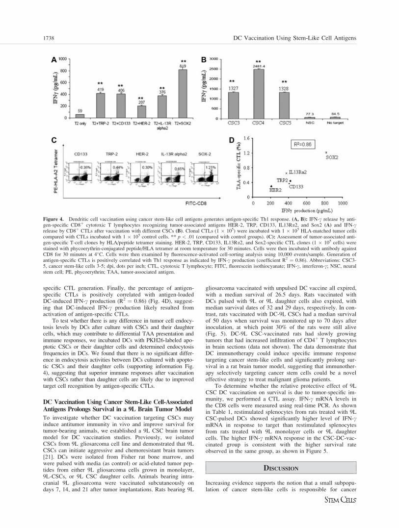
specific CTL generation. Finally, the percentage of antigen-specific CTLs is positively correlated with antigen-loadedDC-induced IFN-c production (R2 ¼ 0.86) (Fig. 4D), suggest-ing that DC-induced IFN-c production likely resulted fromactivation of antigen-specific CTLs.
To test whether there is any difference in tumor cell endocy-tosis levels by DCs after culture with CSCs and their daughtercells, which may contribute to differential TAA presentation andimmune responses, we incubated DCs with PKH26-labeled apo-ptotic CSCs or their daughter cells and determined endocytosisfrequencies in DCs. We found that there is no significant differ-ence in endocytosis activities between DCs cultured with apopto-tic CSCs and their daughter cells (supporting information Fig.4), suggesting that superior immune responses after vaccinationwith CSCs rather than daughter cells are likely due to improvedtarget cell recognition by antigen-specific CTLs.
DC Vaccination Using Cancer Stem-Like Cell-AssociatedAntigens Prolongs Survival in a 9L Brain Tumor Model
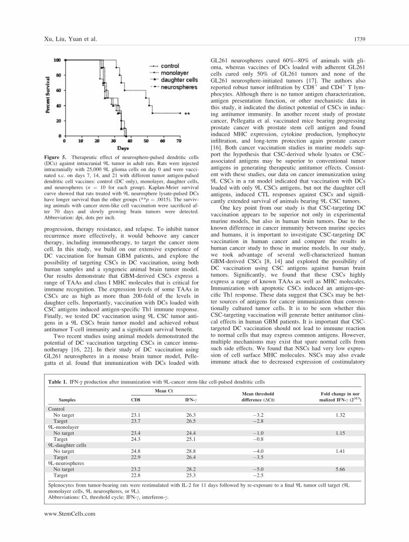
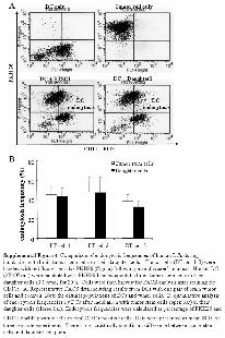
To investigate whether DC vaccination targeting CSCs mayinduce antitumor immunity in vivo and improve survival fortumor-bearing animals, we established a 9L CSC brain tumormodel for DC vaccination studies. Previously, we isolatedCSCs from 9L gliosarcoma cell line and demonstrated that 9LCSCs can initiate aggressive and chemoresistant brain tumors[21]. DCs were isolated from Fisher rat bone marrow, andwere pulsed with media (as control) or acid-eluted tumor pep-tides from either 9L gliosarcoma cells grown in monolayer,9L-CSCs, or 9L CSC daughter cells. Animals bearing intra-cranial 9L gliosarcoma were vaccinated subcutaneously ondays 7, 14, and 21 after tumor implantations. Rats bearing 9L
gliosarcoma vaccinated with unpulsed DC vaccine all expired,with a median survival of 26.5 days. Rats vaccinated withDCs pulsed with 9L or 9L daughter cells also expired, withmedian survival dates of 32 and 29 days, respectively. In con-trast, rats vaccinated with DC-9L CSCs had a median survivalof 50 days when survival was monitored up to 70 days afterinoculation, at which point 30% of the rats were still alive(Fig. 5). DC-9L CSC-vaccinated rats had slowly growingtumors that had increased infiltration of CD4þ T lymphocytesin brain sections (data not shown). The data demonstrate thatDC immunotherapy could induce specific immune responsetargeting cancer stem-like cells and significantly prolong sur-vival in a rat brain tumor model, suggesting that immunother-apy selectively targeting cancer stem cells could be a noveleffective strategy to treat malignant glioma patients.
To determine whether the relative protective effect of 9LCSC DC vaccination on survival is due to tumor-specific im-munity, we performed a CTL assay. IFN-c mRNA levels inthe CD8 cells were measured using real-time PCR. As shownin Table 1, restimulated splenocytes from rats treated with 9LCSC-pulsed DCs showed significantly higher level of IFN-cmRNA in response to target than restimulated splenocytesfrom rats treated with 9L monolayer cells or 9L daughtercells. The higher IFN-c mRNA response in the CSC-DC-vac-cinated group is consistent with the higher survival rateobserved in the same group, as shown in Figure 5.
DISCUSSION
Increasing evidence supports the notion that a small subpopu-lation of cancer stem-like cells is responsible for cancer
Figure 4. Dendritic cell vaccination using cancer stem-like cell antigens generates antigen-specific Th1 response. (A, B): IFN-c release by anti-gen-specific CD8þ cytotoxic T lymphocytes recognizing tumor-associated antigens HER-2, TRP, CD133, IL13Ra2, and Sox2 (A) and IFN-crelease by CD8þ CTLs after vaccination with different CSCs (B). Clonal CTLs (1 � 105) were incubated with 1 � 105 HLA-matched tumor cellscompared with CTLs incubated with 1 � 105 control cells. ** p < .01 (compared with control groups). (C): Assessment of tumor-associated anti-gen-specific T-cell clones by HLA/peptide tetramer staining. HER-2, TRP, CD133, IL13Ra2, and Sox2-specific CTL clones (1 � 105 cells) werestained with phycoerythrin-conjugated peptide/HLA tetramer at room temperature for 30 minutes. Cells were then incubated with antibody againstCD8 for 30 minutes at 4�C. Cells were then examined by fluorescence-activated cell-sorting analysis using 10,000 events/sample. Generation ofantigen-specific CTLs is positively correlated with Th1 response as indicated by IFN-c production (coefficient R2 ¼ 0.86). Abbreviations: CSC3-5, cancer stem-like cells 3-5; dpi, dots per inch; CTL, cytotoxic T lymphocyte; FITC, fluorescein isothiocyanate; IFN-c, interferon-c; NSC, neuralstem cell; PE, phycoerythrin; TAA, tumor-associated antigen.
1738 DC Vaccination Using Stem-Like Cell Antigens
progression, therapy resistance, and relapse. To inhibit tumorrecurrence more effectively, it would behoove any cancertherapy, including immunotherapy, to target the cancer stemcell. In this study, we build on our extensive experience ofDC vaccination for human GBM patients, and explore thepossibility of targeting CSCs in DC vaccination, using bothhuman samples and a syngeneic animal brain tumor model.Our results demonstrate that GBM-derived CSCs express arange of TAAs and class I MHC molecules that is critical forimmune recognition. The expression levels of some TAAs inCSCs are as high as more than 200-fold of the levels indaughter cells. Importantly, vaccination with DCs loaded withCSC antigens induced antigen-specific Th1 immune response.Finally, we tested DC vaccination using 9L CSC tumor anti-gens in a 9L CSCs brain tumor model and achieved robustantitumor T-cell immunity and a significant survival benefit.
Two recent studies using animal models demonstrated thepotential of DC vaccination targeting CSCs in cancer immu-notherapy [16, 22]. In their study of DC vaccination usingGL261 neurospheres in a mouse brain tumor model, Pelle-gatta et al. found that immunization with DCs loaded with
GL261 neurospheres cured 60%--80% of animals with gli-oma, whereas vaccines of DCs loaded with adherent GL261cells cured only 50% of GL261 tumors and none of theGL261 neurosphere-initiated tumors [17]. The authors alsoreported robust tumor infiltration by CD8þ and CD4þ T lym-phocytes. Although there is no tumor antigen characterization,antigen presentation function, or other mechanistic data inthis study, it indicated the distinct potential of CSCs in induc-ing antitumor immunity. In another recent study of prostatecancer, Pellegatta et al. vaccinated mice bearing progressingprostate cancer with prostate stem cell antigen and foundinduced MHC expression, cytokine production, lymphocyteinfiltration, and long-term protection again prostate cancer[16]. Both cancer vaccination studies in murine models sup-port the hypothesis that CSC-derived whole lysates or CSC-associated antigens may be superior to conventional tumorantigens in generating therapeutic antitumor effects. Consist-ent with these studies, our data on cancer immunization using9L CSCs in a rat model indicated that vaccination with DCsloaded with only 9L CSCs antigens, but not the daughter cellantigens, induced CTL responses against CSCs and signifi-cantly extended survival of animals bearing 9L CSC tumors.
One key point from our study is that CSC-targeting DCvaccination appears to be superior not only in experimentalmurine models, but also in human brain tumors. Due to theknown difference in cancer immunity between murine speciesand humans, it is important to investigate CSC-targeting DCvaccination in human cancer and compare the results inhuman cancer study to those in murine models. In our study,we took advantage of several well-characterized humanGBM-derived CSCs [8, 14] and explored the possibility ofDC vaccination using CSC antigens against human braintumors. Significantly, we found that these CSCs highlyexpress a range of known TAAs as well as MHC molecules.Immunization with apoptotic CSCs induced an antigen-spe-cific Th1 response. These data suggest that CSCs may be bet-ter sources of antigens for cancer immunization than conven-tionally cultured tumor cells. It is to be seen whether thisCSC-targeting vaccination will generate better antitumor clini-cal effects in human GBM patients. It is important that CSC-targeted DC vaccination should not lead to immune reactionto normal cells that may express common antigens. However,multiple mechanisms may exist that spare normal cells fromsuch side effects. We found that NSCs had very low expres-sion of cell surface MHC molecules. NSCs may also evadeimmune attack due to decreased expression of costimulatory
Figure 5. Therapeutic effect of neurosphere-pulsed dendritic cells(DCs) against intracranial 9L tumor in adult rats. Rats were injectedintracranially with 25,000 9L glioma cells on day 0 and were vacci-nated s.c. on days 7, 14, and 21 with different tumor antigen-pulseddendritic cell vaccines: control (DC only), monolayer, daughter cells,and neurospheres (n ¼ 10 for each group). Kaplan-Meier survivalcurve showed that rats treated with 9L neurosphere lysate-pulsed DCshave longer survival than the other groups (**p ¼ .0015). The surviv-ing animals with cancer stem-like cell vaccination were sacrificed af-ter 70 days and slowly growing brain tumors were detected.Abbreviation: dpi, dots per inch.
Table 1. IFN-c production after immunization with 9L-cancer stem-like cell-pulsed dendritic cells
Samples
Mean CtMean threshold
difference (DCt)Fold change in nor
malized IFN-c (2DCt)CD8 IFN-c
ControlNo target 23.1 26.3 �3.2 1.32Target 23.7 26.5 �2.8
9L-monolayerNo target 23.4 24.4 �1.0 1.15Target 24.3 25.1 �0.8
9L-daughter cellsNo target 24.8 28.8 �4.0 1.41Target 22.9 26.4 �3.5
9L-neurospheresNo target 23.2 28.2 �5.0 5.66Target 22.8 25.3 �2.5
Splenocytes from tumor-bearing rats were restimulated with IL-2 for 11 days followed by re-exposure to a final 9L tumor cell target (9Lmonolayer cells, 9L neurospheres, or 9L).Abbreviations: Ct, threshold cycle; IFN-c, interferon-c.
Xu, Liu, Yuan et al. 1739
www.StemCells.com
proteins [23]. We are in the process of initiating a clinicaltrial to study the safety and efficacy of DC vaccination usingCSC antigens.
To date there is a very limited number of studies of CSC-targeting DC vaccination in animal models or in patients.And detailed immunological analysis data on the developmentof antitumor immunity after DC vaccination are not available.Questions remain regarding mechanisms underlying the appa-rent superior outcomes from CSC-targeting DC vaccination.For example, is there any difference between CSC antigensand conventional tumor lysates in promoting DC maturationand polarization, or in effector cell differentiation and mem-ory T-cell generation in vivo? Although higher expression ofTAAs in CSCs, as shown in our study, may be one factorcontributing to the outcomes, it is likely other factors in addi-tion to TAA expression levels also play a role. Finally, out-comes of DC vaccination may be improved when it is admin-istered in combination with chemotherapy, radiotherapy, orother therapies [9, 24, 25].
SUMMARY
In summary, we explored the suitability of CSCs as sourcesof antigens for DC vaccination against human GBM, with theaims of achieving CSC-targeting and better antitumor immu-
nity. We found that CSCs express increased levels of TAAsas well MHC molecules. Furthermore, DC vaccination usingCSC antigens elicited a potent antigen-specific Th1 response.Finally, we show that vaccination with DCs loaded with 9LCSCs, but not the daughter cells or conventionally cultured9L cells, induced CTLs that recognized CSCs and prolongedsurvival of animals bearing 9L CSC tumors. Understandinghow immunization with CSCs generates superior antitumorimmunity may help develop novel and more effective cancerimmunotherapies.
ACKNOWLEDGMENTS
We thank Iman Abdulkadir for technical assistance in bone mar-row cell culture and animal experiments. This work was sup-ported in part by R01 NS048959 and grant NS048879 to J.S.Y.
DISCLOSURE OF POTENTIAL CONFLICTS
OF INTEREST
John Yu owned stock in, performed contract work for, served asan officer or board member for, and was employed byImmunoCellular Theraputics, Ltd.
REFERENCES
1 Reardon DA, Rich JN, Friedman HS et al. Recent advances in thetreatment of malignant astrocytoma. J Clin Oncol 2006;24:1253–1265.
2 Liang XJ, Choi Y, Sackett DL et al. Nitrosoureas inhibit the stathmin-mediated migration and invasion of malignant glioma cells. CancerRes 2008;68:5267–5272.
3 Ehtesham M, Black KL, Yu JS. Recent progress in immunotherapyfor malignant glioma: treatment strategies and results from clinical tri-als. Cancer Control 2004;11:192–207.
4 Okada H, Low KL, Kohanbash G et al. Expression of glioma-associ-ated antigens in pediatric brain stem and non-brain stem gliomas. JNeurooncol 2008;88:245–250.
5 Eguchi J, Hatano M, Nishimura F et al. Identification of interleukin-13 receptor alpha2 peptide analogues capable of inducing improvedantiglioma CTL responses. Cancer Res 2006;66:5883–5891.
6 Liau LM, Black KL, Martin NA et al. Treatment of a patient by vac-cination with autologous dendritic cells pulsed with allogeneic majorhistocompatibility complex class I-matched tumor peptides. CaseReport. Neurosurg Focus 2000;9:e8.
7 Liu G, Khong HT, Wheeler CJ et al. Molecular and functional analysisof tyrosinase-related protein (TRP)-2 as a cytotoxic T lymphocyte targetin patients with malignant glioma. J Immunother 2003;26:301–312.
8 Yuan X, Curtin J, Xiong Y et al. Isolation of cancer stem cells fromadult glioblastoma multiforme. Oncogene 2004;23:9392–9400.
9 Liu G, Akasaki Y, Khong HT et al. Cytotoxic T cell targeting ofTRP-2 sensitizes human malignant glioma to chemotherapy. Oncogene2005;24:5226–5234.
10 Liau LM, Prins RM, Kiertscher SM et al. Dendritic cell vaccination inglioblastoma patients induces systemic and intracranial T-cellresponses modulated by the local central nervous system tumor micro-environment. Clin Cancer Res 2005;11:5515–5525.
11 Gilboa E. DC-based cancer vaccines. J Clin Invest 2007;117:1195–1203.12 Singh SK, Hawkins C, Clarke ID et al. Identification of human brain
tumour initiating cells. Nature 2004;432:396–401.13 Lee J, Kotliarova S, Kotliarov Y et al. Tumor stem cells derived from
glioblastomas cultured in bFGF and EGF more closely mirror the phe-
notype and genotype of primary tumors than do serum-cultured celllines. Cancer Cell 2006;9:391–403.
14 Xu Q, Yuan X, Liu G et al. Hedgehog signaling regulates brain tumorinitiating cell proliferation and portends shorter survival for patientswith PTEN-coexpressing glioblastomas. Stem Cells 2008;26:3018–3026.
15 Spisek R, Kukreja A, Chen LC et al. Frequent and specific im-munity to the embryonal stem cell-associated antigen SOX2 inpatients with monoclonal gammopathy. J Exp Med 2007;204:831–840.
16 Garcia-Hernandez Mde L, Gray A, Hubby B et al. Prostate stem cellantigen vaccination induces a long-term protective immune responseagainst prostate cancer in the absence of autoimmunity. Cancer Res2008;68:861–869.
17 Pellegatta S, Poliani PL, Corno D et al. Neurospheres enriched in can-cer stem-like cells are highly effective in eliciting a dendritic cell-mediated immune response against malignant gliomas. Cancer Res2006;66:10247–10252.
18 Liu G, Yu JS, Zeng G et al. AIM-2: a novel tumor antigen isexpressed and presented by human glioma cells. J Immunother 2004;27:220–226.
19 Liu G, Ying H, Zeng G et al. HER-2, gp100, and MAGE-1 areexpressed in human glioblastoma and recognized by cytotoxic T cells.Cancer Res 2004;64:4980–4986.
20 Wu A, Wiesner S, Xiao J et al. Expression of MHC I and NK ligandson human CD133þ glioma cells: possible targets of immunotherapy. JNeurooncol 2007;83:121–131.
21 Ghods AJ, Irvin D, Liu G et al. Spheres isolated from 9L gliosarcomarat cell line possess chemoresistant and aggressive cancer stem-likecells. Stem Cells 2007;25:1645–1653.
22 Manzo G. Cancer genesis: stem tumour cells as an MHC-null/HSP70- very high ‘primordial self’ escaping both MHC-restricted and MHC-non-restricted immunesurveillance. Med Hypotheses 2001;56:724–730.
23 Odeberg J, Piao JH, Samuelsson EB et al. Low immunogenicity of invitro-expanded human neural cells despite high MHC expression. JNeuroimmunol 2005;161:1–11.
24 Heisel SM, Ketter R, Keller A et al. Increased seroreactivity to gli-oma-expressed antigen 2 in brain tumor patients under radiation.PLoS ONE 2008;3:e2164.
25 Okada H, Lieberman FS, Walter KA et al. Autologous glioma cellvaccine admixed with interleukin-4 gene transfected fibroblasts in thetreatment of patients with malignant gliomas. J Transl Med 2007;5:67.
See www.StemCells.com for supporting information available online.
1740 DC Vaccination Using Stem-Like Cell Antigens
Related Documents


