1 Antigen recognition by T and B cells - T and B cells exhibit fundamental differences in antigen recognition - B cells recognize antigen free in solution (native antigen). - T cells recognize antigen after it has been phagocytosed, degraded and small pieces of the antigen have been bound by MHC molecules. Role of Antigen-Presenting Cells (APC) - Helper T cells: recognize antigen after processing and presentation by MHC-II on APC (dendritic cells, macrophages, B cells). - Cytotoxic T cells: recognize antigen when it is presented on MHC-I. - Since most nucleated cells in the body express class I MHC, most cells in the body can present antigen to cytotoxic T cells. Although they are presenting antigen, these cells are usually not referred to as “antigen-presenting cells”. If they are presenting antigen that will cause them to be killed by cytotoxic T cells, they are referred to as “target cells”. Antigen presenting cells • Remember: 1) MHC-II, 2) deliver co-stimulatory signals • Professional APC: DC> MΦ > B cells, why? • DC: Always express high levels of MHC-II molecules and co-stimulatory activity (B7 molecule) • MΦ: requires activation to up-regulate MHC-II molecules and co-stimulatory molecules (B7 molecules) • B cells: always express MHC-II molecules but needs to be activated to express co-stimulatory activity (B7 molecule) Professional vs Non-Professional APCs Self MHC Restriction • Both MHC-I and MHC-II molecules can only recognize antigens when presented by SELF-MHC molecules. • No value for individual to have T cells that recognize foreign antigen associated with foreign MHC • Self MHC restriction occurs in thymus Classic Experiment to show self -MHC restriction H-2 K H-2 K H-2 b H-2 K CD8 T cells (-) (+) (-)

Welcome message from author
This document is posted to help you gain knowledge. Please leave a comment to let me know what you think about it! Share it to your friends and learn new things together.
Transcript
1
Antigen recognition by T and B cells
- T and B cells exhibit fundamental differences in
antigen recognition
- B cells recognize antigen free in solution (native
antigen).
- T cells recognize antigen after it has been
phagocytosed, degraded and small pieces of the
antigen have been bound by MHC molecules.
Role of Antigen-Presenting Cells (APC)
- Helper T cells: recognize antigen after processing and
presentation by MHC-II on APC (dendritic cells,
macrophages, B cells).
- Cytotoxic T cells: recognize antigen when it is presented on
MHC-I.
- Since most nucleated cells in the body express class I MHC, most cells in the body can present antigen to cytotoxic T
cells. Although they are presenting antigen, these cells are
usually not referred to as “antigen-presenting cells”. If they
are presenting antigen that will cause them to be killed by
cytotoxic T cells, they are referred to as “target cells”.
Antigen presenting cells
• Remember: 1) MHC-II, 2) deliver co-stimulatory signals
• Professional APC: DC> MΦΦΦΦ > B cells, why?
• DC: Always express high levels of MHC-II molecules and co-stimulatory activity (B7 molecule)
• MΦΦΦΦ: requires activation to up-regulate MHC-II molecules and co-stimulatory molecules (B7 molecules)
• B cells: always express MHC-II molecules but needs to be activated to express co-stimulatory activity (B7 molecule)
Professional vs Non-Professional APCs
Self MHC Restriction
• Both MHC-I and MHC-II molecules can
only recognize antigens when presented by
SELF-MHC molecules.
• No value for individual to have T cells
that recognize foreign antigen
associated with foreign MHC
• Self MHC restriction occurs in thymus
Classic Experiment to
show self -MHC restriction
H-2K
H-2K H-2bH-2K
CD8 T cells
(-) (+) (-)
2
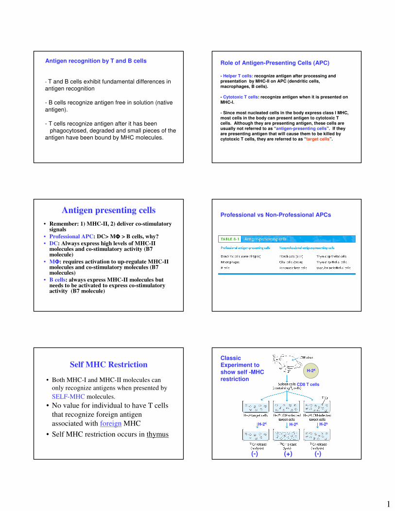
Ag processing is required
• Classical experiment showing that B and T
cells have different requirement for antigen
recognition.
• Processing is required for Th activation
• Processing is a metabolic active process
Role for APC?
Points Concerning Antigen
Processing and Presentation
1. Location of pathogen
• viruses in cytosol, MHC class I pathway, Tcresponse (Cytosolic pathway)
• extracellular bacteria, MHC class II pathway, Th2 response � Ab formation (Endocytic pathway)
• intracellular bacteria, MHC class II pathway, Th1 response � cellular response(Endocytic)
Points Concerning Antigen
Processing and Presentation
2. Peptides derived from both self and
non-self proteins can associate with
MHC class I and class II molecules.
3. Chemical nature of MHC groove
determines which peptides it will
bind.
MHC-I and MHC-II associated with peptides processed in different intracellular compartments
A) Class I MHC binds peptides derived from endogenous antigens
B) Class I MHC binds peptides from antigens that have been processed via the cytosolic pathway (derived from the cytoplasm of the cell)
C) Class II MHC molecules bind peptides derived from exogenous antigens. These antigens were internalized by phagocytosis or endocytosis.
D) These peptides are said to have been processed within the endocytic pathway.
3
Endogenous Pathway
• Peptides are generated by proteasome
degradation
• Peptides are transported from cytosol to the
RER
• Peptides loading onto MHC-I is aided by
chaperones
Kuby Figure 8-5
- Size
- Hydrophobicity
- The cytosolic antigen processing pathway - 2. The role of the TAP
(Transporter associated with Antigenic Processing).
- Peptides from proteasome degradation of cytoplasmic proteins are transported
across the membrane of the rough endoplasmic reticulum by a heterodimeric
protein designated as TAP.
- TAP is composed of two subunits - TAP1 and TAP-2
- TAP-mediated transport is ATP-dependent
The genes for TAP1 and TAP2 are encoded within the MHC.
Kuby Figure 8-6a
The cytosolic antigen processing pathway - 3. Assembly of the class I-peptide
complex
Kuby Figure 8-6b
1. The class I alpha chain is stabilized by calnexin.
2. When the alpha chain binds beta-2-microglobulin:
- calnexin is lost- calreticulin and tapasin
bind
3. Tapasin & calreticulinbrings the class I molecules into the vicinity of the TAP.
4. A cytoplasmic peptide transported through the TAP is loaded onto the class I molecule.
5. Class I MHC dissociates from calreticulin and tapasin.
Class I MHC-peptide complex is transported to Golgi and to the cell surface.
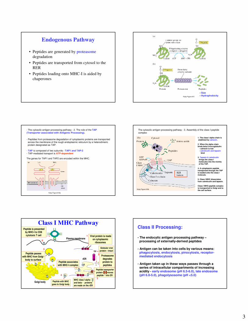
Class I MHC Pathway
Viral protein is made
on cytoplasmicribosomes
Plasma membrane
Proteasome
degrades protein to
peptides
Peptide transporter
protein moves
peptide into ER
MHC class I alpha
and beta proteins
are made on the rER
Peptide associates
with MHC-I complex
Peptide with MHC
goes to Golgi body
Peptide passeswith MHC from Golgi
body to surface
Peptide is presentedby MHC-I to CD8
cytotoxic T cell
Golgi body
rER
Globular viral
protein - intact
1
2
3
4
5
6
Class II Processing:
- The endocytic antigen processing pathway –processing of externally-derived peptides
- Antigen can be taken into cells by various means: phagocytosis, endocytosis, pinocytosis, receptor-mediated endocytosis
- Antigen taken up in these ways passes through a series of intracellular compartments of increasing acidity - early endosome (pH 6.5-6.0), late endosome(pH 6.0-5.0), phagolysosome (pH <5.0)
4
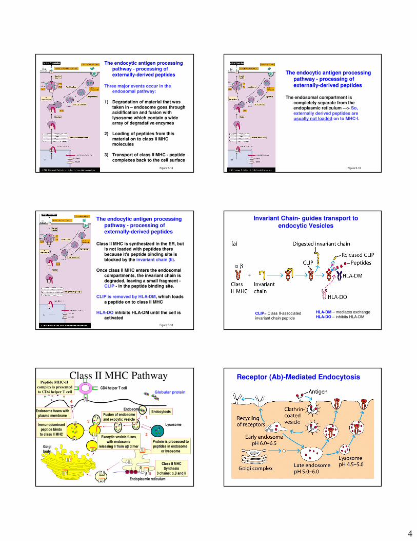
The endocytic antigen processing pathway - processing of externally-derived peptides
Three major events occur in the endosomal pathway:
1) Degradation of material that was taken in – endosome goes through acidification and fusion with lysosome which contain a wide array of degradative enzymes
2) Loading of peptides from this material on to class II MHC molecules
3) Transport of class II MHC - peptide complexes back to the cell surface
Figure 5-18 Figure 5-18
The endocytic antigen processing pathway - processing of externally-derived peptides
The endosomal compartment is completely separate from the endoplasmic reticulum ---> So, externally derived peptides are usually not loaded on to MHC-I.
Figure 5-18
The endocytic antigen processing pathway - processing of externally-derived peptides
Class II MHC is synthesized in the ER, but is not loaded with peptides there because it's peptide binding site is blocked by the invariant chain (Ii).
Once class II MHC enters the endosomal compartments, the invariant chain is degraded, leaving a small fragment -CLIP - in the peptide binding site.
CLIP is removed by HLA-DM, which loads a peptide on to class II MHC
HLA-DO inhibits HLA-DM until the cell is activated
CLIP= Class II-associated
invariant chain peptide
HLA-DM – mediates exchange
HLA-DO – inhibits HLA-DM
Invariant Chain- guides transport to
endocytic Vesicles
Class II MHC PathwayGlobular protein
Endocytosis
Protein is processed to
peptides in endosome
or lysosome
Endosome
Lysosome
Fusion of endosome
and exocytic vesicle
Endoplasmic reticulum
Class II MHC
Synthesis
3 chains: α,β and Ii
Golgi
body
Exocytic vesicle fuses
with endosome
releasing Ii from αβ dimer
α
β Ii
Immunodominant
peptide binds
to class II MHC
Endosome fuses with
plasma membrane
Peptide MHC-II
complex is presented
to CD4 helper T cellCD4 helper T cell
Globular protein
1
2
3
4
Receptor (Ab)-Mediated Endocytosis
5
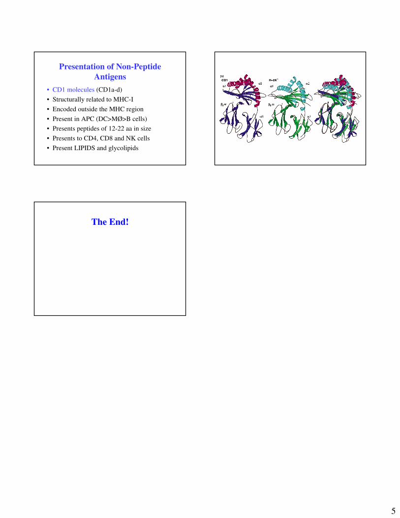
Presentation of Non-Peptide
Antigens
• CD1 molecules (CD1a-d)
• Structurally related to MHC-I
• Encoded outside the MHC region
• Present in APC (DC>MØ>B cells)
• Presents peptides of 12-22 aa in size
• Presents to CD4, CD8 and NK cells
• Present LIPIDS and glycolipids
The End!
Related Documents