Antigen Presenting Cell-Mediated Expansion of Human Umbilical Cord Blood Yields Log-Scale Expansion of Natural Killer Cells with Anti-Myeloma Activity Nina Shah 1 *, Beatriz Martin-Antonio 1 , Hong Yang 1 , Stephanie Ku 2 , Dean A. Lee 3 , Laurence J. N. Cooper 3 , William K. Decker 2,4 , Sufang Li 1 , Simon N. Robinson 1 , Takuya Sekine 1 , Simrit Parmar 1 , John Gribben 5 , Michael Wang 6 , Katy Rezvani 1 , Eric Yvon 1 , Amer Najjar 7 , Jared Burks 8 , Indreshpal Kaur 1 , Richard E. Champlin 1 , Catherine M. Bollard 2 , Elizabeth J. Shpall 1 1 Department of Stem Cell Transplantation and Cellular Therapy, The University of Texas M.D. Anderson Cancer Center, Houston, Texas, United States of America, 2 Center for Cell and Gene Therapy, Baylor College of Medicine, Houston, Texas, United States of America, 3 Department of Pediatrics, The University of Texas M.D. Anderson Cancer Center, Houston, Texas, United States of America, 4 Department of Pathology and Immunology, Baylor College of Medicine, Houston, Texas, United States of America, 5 Institute of Cancer, Queen Mary University of London, Centre for Medical Oncology, Barts and The London School of Medicine, London, United Kingdom, 6 Department of Lymphoma, The University of Texas M.D. Anderson Cancer Center, Houston, Texas, United States of America, 7 Department of Experimental Diagnostic Imaging, The University of Texas M.D. Anderson Cancer Center, Houston, Texas, United States of America, 8 Department of Leukemia Research, The University of Texas M.D. Anderson Cancer Center, Houston, Texas, United States of America Abstract Natural killer (NK) cells are important mediators of anti-tumor immunity and are active against several hematologic malignancies, including multiple myeloma (MM). Umbilical cord blood (CB) is a promising source of allogeneic NK cells but large scale ex vivo expansion is required for generation of clinically relevant CB-derived NK (CB-NK) cell doses. Here we describe a novel strategy for expanding NK cells from cryopreserved CB units using artificial antigen presenting feeder cells (aAPC) in a gas permeable culture system. After 14 days, mean fold expansion of CB-NK cells was 1848-fold from fresh and 2389-fold from cryopreserved CB with .95% purity for NK cells (CD56 + /CD3 2 ) and less than 1% CD3 + cells. Though surface expression of some cytotoxicity receptors was decreased, aAPC-expanded CB-NK cells exhibited a phenotype similar to CB- NK cells expanded with IL-2 alone with respect to various inhibitory receptors, NKG2C and CD94 and maintained strong expression of transcription factors Eomesodermin and T-bet. Furthermore, CB-NK cells formed functional immune synapses with and demonstrated cytotoxicity against various MM targets. Finally, aAPC-expanded CB-NK cells showed significant in vivo activity against MM in a xenogenic mouse model. Our findings introduce a clinically applicable strategy for the generation of highly functional CB-NK cells which can be used to eradicate MM. Citation: Shah N, Martin-Antonio B, Yang H, Ku S, Lee DA, et al. (2013) Antigen Presenting Cell-Mediated Expansion of Human Umbilical Cord Blood Yields Log- Scale Expansion of Natural Killer Cells with Anti-Myeloma Activity. PLoS ONE 8(10): e76781. doi:10.1371/journal.pone.0076781 Editor: Evren Alici, Karolinska Institutet, Sweden Received April 16, 2013; Accepted August 29, 2013; Published October 18, 2013 Copyright: ß 2013 Shah et al. This is an open-access article distributed under the terms of the Creative Commons Attribution License, which permits unrestricted use, distribution, and reproduction in any medium, provided the original author and source are credited. Funding: This work was supported by the National Institutes of Health K12 CA088084 (Shah) and Cancer Prevention and Research Institute of Texas RP#100430 (Shpall). The funders had no role in study design, data collection and analysis, decision to publish, or preparation of the manuscript. Competing Interests: The authors have declared that no competing interests exist. * E-mail: [email protected] Introduction Multiple myeloma (MM) is the second most common hemato- logic malignancy in adults [1]. It is currently considered incurable, even after high dose chemotherapy and autologous hematopoietic stem cell transplantation (HSCT) [2]. Natural killer (NK) cells are CD56 + /CD3 2 cytotoxic lymphocytes that are increasingly recog- nized as a potent cellular therapy. NK cells have been shown to be active against MM in several preclinical studies [3,4]. In addition, a relative decrease in NK cell frequency or function in MM patients has been shown to correlate with more advanced disease or poorer outcome [5,6]. NK cell cytotoxic activity can be triggered by cytokines, antibodies or a shift in the balance between their activating and inhibitory receptors. Specifically, NK cells are cytotoxic to cells lacking appropriate self-major histocompatibility complex (MHC) class I molecules via disinhibition of the killer immunoglobulin-like receptor (KIR). This forms the basis for the ‘‘missing self’’ hypothesis [7] and is thought to mediate donor NK cell alloreactivity in the setting of allogeneic HSCT. However the precise role of KIR-ligand mismatch in HSCT is not known. In some patients treated with allogeneic-HSCT, PB-NK cell allor- eactivity as determined by missing KIR ligands appears to predict reduced rates of relapse and graft versus host disease (GVHD) [8,9]. Additionally, in MM patients undergoing matched alloge- neic-HSCT, an activated donor KIR haplotype (Bx) has been associated with a significantly lower risk of relapse and better PFS [10]. In contrast, other studies have suggested that the effect of KIR-ligand incompatibility is not consistent, particularly as it relates to conditioning regimen, donor source and GVHD outcomes [11,12,13,14]. Although allogeneic NK cells appear promising in MM, autologous PB-NK cells from MM patients appear to be hypofunctional [15]. This may be due to inhibitory cytokines PLOS ONE | www.plosone.org 1 October 2013 | Volume 8 | Issue 10 | e76781

Welcome message from author
This document is posted to help you gain knowledge. Please leave a comment to let me know what you think about it! Share it to your friends and learn new things together.
Transcript

Antigen Presenting Cell-Mediated Expansion of HumanUmbilical Cord Blood Yields Log-Scale Expansion ofNatural Killer Cells with Anti-Myeloma ActivityNina Shah1*, Beatriz Martin-Antonio1, Hong Yang1, Stephanie Ku2, Dean A. Lee3, Laurence J. N. Cooper3,
William K. Decker2,4, Sufang Li1, Simon N. Robinson1, Takuya Sekine1, Simrit Parmar1, John Gribben5,
Michael Wang6, Katy Rezvani1, Eric Yvon1, Amer Najjar7, Jared Burks8, Indreshpal Kaur1,
Richard E. Champlin1, Catherine M. Bollard2, Elizabeth J. Shpall1
1 Department of Stem Cell Transplantation and Cellular Therapy, The University of Texas M.D. Anderson Cancer Center, Houston, Texas, United States of America, 2 Center
for Cell and Gene Therapy, Baylor College of Medicine, Houston, Texas, United States of America, 3 Department of Pediatrics, The University of Texas M.D. Anderson
Cancer Center, Houston, Texas, United States of America, 4 Department of Pathology and Immunology, Baylor College of Medicine, Houston, Texas, United States of
America, 5 Institute of Cancer, Queen Mary University of London, Centre for Medical Oncology, Barts and The London School of Medicine, London, United Kingdom,
6 Department of Lymphoma, The University of Texas M.D. Anderson Cancer Center, Houston, Texas, United States of America, 7 Department of Experimental Diagnostic
Imaging, The University of Texas M.D. Anderson Cancer Center, Houston, Texas, United States of America, 8 Department of Leukemia Research, The University of Texas
M.D. Anderson Cancer Center, Houston, Texas, United States of America
Abstract
Natural killer (NK) cells are important mediators of anti-tumor immunity and are active against several hematologicmalignancies, including multiple myeloma (MM). Umbilical cord blood (CB) is a promising source of allogeneic NK cells butlarge scale ex vivo expansion is required for generation of clinically relevant CB-derived NK (CB-NK) cell doses. Here wedescribe a novel strategy for expanding NK cells from cryopreserved CB units using artificial antigen presenting feeder cells(aAPC) in a gas permeable culture system. After 14 days, mean fold expansion of CB-NK cells was 1848-fold from fresh and2389-fold from cryopreserved CB with .95% purity for NK cells (CD56+/CD32) and less than 1% CD3+ cells. Though surfaceexpression of some cytotoxicity receptors was decreased, aAPC-expanded CB-NK cells exhibited a phenotype similar to CB-NK cells expanded with IL-2 alone with respect to various inhibitory receptors, NKG2C and CD94 and maintained strongexpression of transcription factors Eomesodermin and T-bet. Furthermore, CB-NK cells formed functional immune synapseswith and demonstrated cytotoxicity against various MM targets. Finally, aAPC-expanded CB-NK cells showed significantin vivo activity against MM in a xenogenic mouse model. Our findings introduce a clinically applicable strategy for thegeneration of highly functional CB-NK cells which can be used to eradicate MM.
Citation: Shah N, Martin-Antonio B, Yang H, Ku S, Lee DA, et al. (2013) Antigen Presenting Cell-Mediated Expansion of Human Umbilical Cord Blood Yields Log-Scale Expansion of Natural Killer Cells with Anti-Myeloma Activity. PLoS ONE 8(10): e76781. doi:10.1371/journal.pone.0076781
Editor: Evren Alici, Karolinska Institutet, Sweden
Received April 16, 2013; Accepted August 29, 2013; Published October 18, 2013
Copyright: � 2013 Shah et al. This is an open-access article distributed under the terms of the Creative Commons Attribution License, which permitsunrestricted use, distribution, and reproduction in any medium, provided the original author and source are credited.
Funding: This work was supported by the National Institutes of Health K12 CA088084 (Shah) and Cancer Prevention and Research Institute of Texas RP#100430(Shpall). The funders had no role in study design, data collection and analysis, decision to publish, or preparation of the manuscript.
Competing Interests: The authors have declared that no competing interests exist.
* E-mail: [email protected]
Introduction
Multiple myeloma (MM) is the second most common hemato-
logic malignancy in adults [1]. It is currently considered incurable,
even after high dose chemotherapy and autologous hematopoietic
stem cell transplantation (HSCT) [2]. Natural killer (NK) cells are
CD56+/CD32 cytotoxic lymphocytes that are increasingly recog-
nized as a potent cellular therapy. NK cells have been shown to be
active against MM in several preclinical studies [3,4]. In addition,
a relative decrease in NK cell frequency or function in MM
patients has been shown to correlate with more advanced disease
or poorer outcome [5,6].
NK cell cytotoxic activity can be triggered by cytokines,
antibodies or a shift in the balance between their activating and
inhibitory receptors. Specifically, NK cells are cytotoxic to cells
lacking appropriate self-major histocompatibility complex (MHC)
class I molecules via disinhibition of the killer immunoglobulin-like
receptor (KIR). This forms the basis for the ‘‘missing self’’
hypothesis [7] and is thought to mediate donor NK cell
alloreactivity in the setting of allogeneic HSCT. However the
precise role of KIR-ligand mismatch in HSCT is not known. In
some patients treated with allogeneic-HSCT, PB-NK cell allor-
eactivity as determined by missing KIR ligands appears to predict
reduced rates of relapse and graft versus host disease (GVHD)
[8,9]. Additionally, in MM patients undergoing matched alloge-
neic-HSCT, an activated donor KIR haplotype (Bx) has been
associated with a significantly lower risk of relapse and better PFS
[10]. In contrast, other studies have suggested that the effect of
KIR-ligand incompatibility is not consistent, particularly as it
relates to conditioning regimen, donor source and GVHD
outcomes [11,12,13,14].
Although allogeneic NK cells appear promising in MM,
autologous PB-NK cells from MM patients appear to be
hypofunctional [15]. This may be due to inhibitory cytokines
PLOS ONE | www.plosone.org 1 October 2013 | Volume 8 | Issue 10 | e76781

such as TGF-b, IL-6 and IL-10 present in the MM microenvi-
ronment [16,17,18] or dysregulation of IL-15 signaling in favor of
MM cells over activation of NK cells [19,20]. While some pre-
clinical studies suggest that this NK cell dysfunction can be
reversed via ex vivo expansion/activation [4,21,22], the potentially
unpredictable nature of autologous NK cells from heavily pre-
treated patients warrants further optimization of techniques for
allogeneic adoptive NK cell therapy. Furthermore, in advanced
disease states, MM cells may upregulate Class I expression [23].
This suggests that KIR-MHC class I mismatched, allogeneic NK
cell therapy would be advantageous over autologous NK cell
therapy, as allogeneic NK cells would be less inhibited by cognate
MHC class I in contrast to autologous NK cells.
To date, the majority of clinical trials of NK cell therapy for
various malignancies have used allogeneic PB as a source of NK
cells. We are interested in NK cells derived from human umbilical
cord blood (CB) as an alternative and more readily available
source of NK cells. Our group has previously demonstrated that ex
vivo expansion with IL-2 activates otherwise quiescent CB-NK
cells. These CB-NK cells exhibit a mature phenotype, similar to
PB-NK cells, and are as active as PB-NK cells against leukemia
targets [24].
The limited number of NK cells in an unmanipulated CB unit
requires an efficient and robust NK cell ex vivo expansion strategy.
Several groups have recently reported expansion of PB-NK cells
using genetically engineered artificial antigen presenting cells
(aAPCs) derived from the K562 cell line [25,26]. In this study, we
build upon recently developed technology with aAPCs [26] and
describe a novel technique for expanding CB-NK cells for use in
MM. This good manufacturing practice (GMP)-compliant method
yields clinical scale expansion of phenotypically mature CB-NK
cells which are cytotoxic to MM cells in vitro and demonstrate
in vivo anti-MM activity in a xenogenic model. Taken together, our
results provide the basis for further exploration of CB-NK cell
therapy for patients with MM.
Materials and Methods
Ethics StatementAll research involving human materials was approved by the
MD Anderson (MDACC) Institutional Review Board (IRB). Cord
blood units were obtained from healthy donors who gave written
informed consent. All animal work was performed under an
MDACC Institutional Animal Care and Use Committee (IA-
CUC)-approved protocol specific to this study.
Cells and Cell LinesK562-based aAPCs expressing membrane bound IL-21 ‘‘Clone
9.mbIL21’’ were generously provided by Dr. Laurence Cooper
(MDACC, Houston TX). Clone 9.mbIL21 cells express mem-
brane-bound IL-21, 41BB ligand, CD64 (FccRI) and CD86. This
cell line has recently been shown to promote PB NK cell
expansion [26].and is GMP-grade for clinical use. Targets for NK
cell functional assays consisted of K562 cells (American Type
Culture Collection (ATCC), Rockville, MD) and MM cell lines
RPMI 8226 (ATCC), ARP-1 (Multiple Myeloma Research
Center, Little Rock AK), and U266 (ATCC). Autologous,
unselected CB cells (from the same CB unit as the NK cells) were
used as a negative control for 51chromium (Cr) experiments.
Generation of eGFP-FFLuc-expressing ARP-1 Cell Line forin vivo Experiments
The generation of retrovirus vectors encoding green fluorescent
protein (eGFP)-Firefly Luciferase (eGFP-FFLuc) and production of
transient retroviral supernatant have been previously described
[27,28]. Briefly, the fusion protein eGFP-FFLuc was cloned into
an SFG retroviral vector and retroviral supernatant was produced
using 293-T cells co-transfected with the following retroviral
vectors: eGFP-FFLuc SFG plasmid, the Peg-Pam-e plasmid
containing the sequence for the MoMLV gag-pol and the RDF
plasmid encoding for the RD114 envelope. Retroviral supernatant
was collected at 48 and 72 hours after transfection and stored at
-80uC for further use. For the generation of eGFP-FFLuc-
expressing ARP-1 tumor cells, 50,000 cells were plated in presence
of retroviral supernatant encoding eGFP-FFLuc in one well of a
24-well plate pre-coated with recombinant fibronectin fragment
(CH-296; Takara Shuzo, Otsu, Japan). Transduced ARP-1 cells
were expanded and eGFP expression evaluated by fluorescence-
activated cell sorter (FACSCalibur; Becton-Dickinson (BD), San
Jose, CA) analysis, whereas expression of FFLuc was detected
using D-luciferin (Promega, Madison, WI) and bioluminescence
measured with a luminometer (Modulus; Turner BioSystems,
Sunnyvale, CA). Because of the absence of selection gene in the
eGFP-FFLuc retroviral construct, single cell cloning of the ARP-1-
transduced cells was performed to isolate and expand an ARP-1
clone (clone # 24) with high level of eGFP and FFLuc expression.
As ARP-1 expresses both CD138 and kappa light chain [29,30],
Clone 24 was further validated by FACS analysis for CD138 and
Kappa light chain expression and ELISA for Kappa light chain
secretion.
Isolation and Expansion of Umbilical Cord Blood-derivedNK Cells
CB units were obtained from healthy donors who gave informed
consent under MDACC IRB-approved protocols. Culture media
was comprised of 45% RPMI-1640 (Cellgro, Manassas, VA) and
45% Click’s media (Irvine Scientific, Santa Ana, CA) supplement-
ed with 10% AB human serum (Atlanta Biologicals, Lawrenceville,
GA) and 100 IU/mL IL-2 (Proleukin; Chiron, Emeryville, CA).
CB mononuclear cells (MNCs) were isolated from fresh or
frozen CB units by ficoll density gradient centrifugation. Twenty
million MNCs were plated in 400 mL media in a GP500 gas
permeable bioreactor (Wilson Wolf Corporation, New Brighton,
MN) with irradiated (100 Gy) aAPC feeder cells (2:1 feeder
cell:MNC ratio) at 37uC. IL-2 was replenished every 2–3 days. On
day 7, cultured cells were CD3-depleted via immunomagnetic
depletion according to manufacturer’s instructions (Miltenyi
Biotech, Auburn, CA). Remaining cells were then re-plated in
the same conditions, re-stimulated with aAPC feeder cells and
cultured for an additional 7 days (Figure 1). Flow cytometric
analysis was performed on Days 0, 7 and 14 during the expansion.
NK cell number was determined by multiplying the live total
nucleated cell count by the percentage of CD56+/CD32 cells.
Differences in cell growth were calculated using a 2-tailed student’s
t-test (Microsoft Excel 2010, Redmond, WA).
Original Expansion TechniquesFor comparison, CB-NK cells were also expanded by a method
already known to be successful in our laboratory [24]. Fresh CB
MNCs were isolated as above and then subjected to CD56+
immunomagnetic selection. These cells were then suspended at
16106 cells/mL culture media with IL-2 at 500 IU/mL. The cells
were cultured for 14 days at 37uC; IL-2 was replenished every 2–3
days.
Expanded Cord Blood NK Cells Kill Myeloma
PLOS ONE | www.plosone.org 2 October 2013 | Volume 8 | Issue 10 | e76781

NK Cell Phenotyping via Flow CytometryThe following antibodies were used: FITC-conjugated CD45,
CD158a, CD158b, CD94; PE-conjugated CD16, CD56, NKp30,
NKp46, NKp44, NKG2C; PerCP-conjugated CD3; APC-conju-
gated CD56, NKG2A; Alexa Fluor 647- conjugated Eomesodermin,
T-bet (BD Biosciences); FITC-conjugated CD158e1 (BioLegend,
San Diego, CA); aAPC-conjugated NKG2A (Beckman Coulter,
Brea, CA). Intracellular staining for Eomes and T-bet was
performed per manufacturer’s guidelines (BD Cytofix/Cytoperm,
BD Biosciences). Data were acquired by the BD FACSCalibur
device using BD CellQuest-Pro software. Flow cytometry analysis
was performed using CellQuest and FlowJo (Tree Star, Ashland,
OR) software. Differences in MFI were calculated using a two-sided
paired t-test (Microsoft Excel 2010).
Immunofluorescence and Confocal Microscopy ImageAcquisition
Immunofluorescent labeling was performed as previously
described [31]. Target cells were labeled with CellTracker Blue
CMAC (7-amino-4-chloromethylcoumarin, Molecular Probes,
Eugene, OR). NK cell-target cell conjugates were formed by
suspending equal volumes and cell numbers of NK effector cells
and target cells (56106/mL) in culture media for 15 min at 37uC.
Cells were then transferred onto microscope slides using a cell
concentrator (Cytofuge 2, IRIS International, and Chatsworth,
CA), fixed with 3% methanol-free formaldehyde and then
permeabilized. NK effector cell F-actin was stained with rhoda-
mine-phalloidin (Molecular Probes, Invitrogen, Carlsbad, CA).
Images were acquired using an Olympus IX81 microscope (Center
Valley, PA).
NK Cell 51Cr Cytotoxicity AssaySerial dilutions of NK cells were co-incubated in triplicate for 4
hours with 5000 51Cr-labeled target cells (Amersham Pharmacia
Biotech, Piscataway, NJ), in a total volume of 100 ml in a V-
bottom 96-well plate (Corning, Corning, NY). Thereafter,
supernatants (50 ml) were harvested and transferred to a Luma-
Plate-96 (Perkin-Elmer, Waltham, MA). After drying overnight,51Cr release was measured on a TOPCount NXT microplate
scintillation and luminescence counter (Perkin-Elmer). Cytotoxic-
ity was determined by the formula: cytotoxicity = (sample value-
spontaneous lysis)/(max-lysis-spontaneous lysis) 6 100%.
ARP-1 Myeloma Murine ModelNOD/SCID IL-2Rcnull (NSG) mice (Jackson Laboratories, Bar
Harbor, ME) were irradiated with 300 cGy and inoculated with
1606 eGFP-FFLuc -transduced ARP-1 cells (Clone 24) intrave-
nously on day 21. Where indicated, 106106 ex vivo, fresh, aAPC-
expanded CB NK cells were given retro-orbitally on days 0, 12
and 19 with IL-2 (2000 IU intrapertioneally (IP) three times per
week). Mice were subjected to twice weekly bioluminescence
imaging (BLI) and weekly serum kappa light chain measurements.
Prior to image acquisition mice were anesthetized with 2%
isoflurane in 98% oxygen. BLI was performed using a Xenogen
IVIS 200 system (Caliper, Waltham, MA) 10 minutes following a
100 mL IP injection of D-luciferin (20 mg/mL PBS). BLI images
were acquired at 5-minute exposures and superimposed on bright
field photographs of the animals. Signal quantitation in photons/
second (p/s) was performed by determining the photon flux rate
within standardized regions of interest (ROI) using Living Image
software (Caliper). Serum kappa levels were measured by a
commercially available enzyme-linked immunosorbent assay
(ELISA) kit (Bethyl Laboratories, Montgomery, TX) according
to manufacturer’s instructions. Results reported are a representa-
tive experiment with 5 mice in each group. Differences in BLI and
serum kappa levels were calculated using a 2-tailed student’s t-test
(Microsoft Excel 2010). Survival was calculated using the Kaplan-
Meier method (SAS statistical software, version 9.2, Cary, NC).
Results
aAPC-mediated CB-NK Expansion from Fresh orCryopreserved CB Units yields Significantly Greater FoldExpansion of NK Cells than Expansion of CD56+ Cells withIL-2 Alone
In comparison with our original expansion approach of CD56-
selected cells cultured with IL2 alone, culture of either fresh or
frozen CB MNCs with aAPC feeder cells resulted in greater
Figure 1. Culture of CB-NK cells. Unselected CB MNCs were cultured for 7 days in a GP500 bioreactor with IL-2 (100 IU/mL) and aAPCs at 2:1aAPC:MNC ratio. Cells were immunomagnetically CD3-depleted on Day 7 and re-cultured in same conditions for an additional 7 days. On day 7 cellswere again CD3-depleted and subject to phenotypic and functional studies.doi:10.1371/journal.pone.0076781.g001
Expanded Cord Blood NK Cells Kill Myeloma
PLOS ONE | www.plosone.org 3 October 2013 | Volume 8 | Issue 10 | e76781

expansion of NK cells after culture for 14 days (p,0.05 for both
fresh or frozen conditions, Figures 2A and 2B). Culturing of fresh
CB MNCs (n = 8) with aAPC feeder cells yielded a mean fold
expansion of 1848 fold (609 fold –4778 fold) while culturing of
frozen CB MNCs (n = 6) with feeder cells yielded a mean fold
expansion of 2389 fold (103 fold –4931 fold). This was in
comparison to 20 fold (11 fold -27 fold) expansion from culture of
fresh CD56+-selected cells with IL-2 alone (n = 3). The difference
in NK cell yield was apparent by day 7 for the fresh CB culture
with aAPC feeders (p,0.05) but did not reach statistical
significance for the frozen CB condition until day 14 (p = 0.06 at
day 7). As seen in Figure 2C, the final culture contained very few
(#1%) CD3+ cells and this was not significantly different between
the 3 culture conditions: mean value of 0.44% CD3+ cells from the
culture with IL-2 alone, 0.74% CD3+ cells from fresh CB MNCs
with aAPC feeders and 0.66% CD3+ cells from frozen CB MNCs
with aAPC feeders (p.0.5 for all comparisons).
aAPC-mediated Expansion Yields a Pure Population of NKCells with a Mature Phenotype
As seen in Figure 3A, co-culture of CB MNCs with IL-2 and
aAPC feeder cells yielded a population that was pure for NK cells
at the end of the 2 week expansion period. After CD3-depletion,
96% of cells were CD56+/CD32 and less than 1% were CD3+.
CB-NK cells expanded with aAPCs demonstrated a CD56hi
phenotype similar to CB-NK cells expanded with IL-2 alone. Of
note, culture of unselected CB MNCs with IL-2 and soluble IL-21
yielded a relatively pure CD56+/CD32 NK cell population but
with limited expansion of cells (mean expansion of 14 fold, data
not shown). In addition, after log-fold expansion, aAPC-expanded
CB-NK cells did not appear exhausted; rather, CB-NK cells
continued to strongly express Eomesodermin and T-bet, tran-
scription factors recently recognized as necessary for NK cell
maturation and activation [32,33] (Figure 3B). Interestingly, the
surface expression of NK cytotoxicity receptors (NCRs) NKp30,
NKp46 and NKp44 was significantly lower for aAPC-expanded
CB-NK cells versus IL-2-expanded CB-NK cells (p#0.05 for all
three NCRs). However, the expression of KIR antigens, NKG2A,
co-receptor CD94 and the activating receptor NKG2C was similar
between the two expansion methods (Figure 3C).
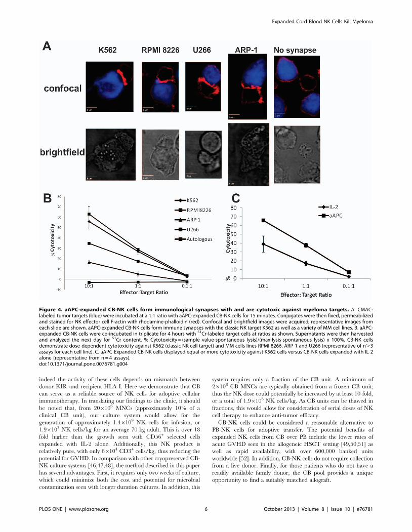
CB-NK Cells Cultured with aAPCs Demonstrate in vitroAnti-myeloma Activity
In order to kill targets, NK cells must directly contact the cell of
interest and form the ‘‘NK immune synapse’’ (NKIS) [34,35]. Our
lab has previously demonstrated that expansion of CB-NK cells is
necessary to repair the defective NKIS exhibited by naı̈ve CB-NK
cells [24]. To demonstrate that this synapse ability is maintained in
CB-NK cells expanded with aAPC feeder cells, we performed a
series of synapse assays with various MM targets. As shown in
Figure 4A, NK cells cultured with aAPC feeder cells formed a
functional NKIS (demonstrated by F-actin polarization) with the
classic NK cell target K562, MM cell lines RPMI 8226, aARP-1
and U266.
To demonstrate the functionality of CB-NK cells expanded with
aAPC feeder stimulation, we performed a standard 51Cr
cytotoxicity assay. aAPC-expanded CB-NK cells were cytotoxic
to all of the MM cell line targets (Figure 4B). Furthermore, despite
the differences in phenotype with regard to the NCRs, in
comparison with CB-NK cells expanded with IL-2 alone, the
aAPC-mediated expanded CB-NK cells demonstrated equal or
greater cytotoxicity against K562 (Figure 4C). This finding was
consistent across the MM cell lines as well (Figure S1). Neither of
the CB-NK preparations demonstrated autologous cytotoxicity.
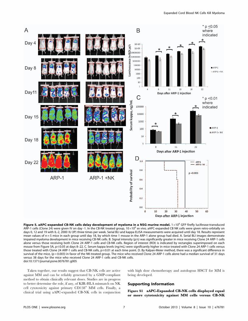
Treatment with Expanded CB-NK Cells DelaysDevelopment of Myeloma in a Murine Model
To investigate whether ex vivo expanded CB-NK cells can inhibit
the growth of MM cells in vivo, we studied NSG mice treated with
GFP firefly luciferase-transduced ARP-1 cells (Clone 24). Using
the bioluminescent signal intensity as a surrogate for tumor cell
density, serial images demonstrated that mice treated with CB-NK
cells had a delay in the onset of MM (Figure 5A). After 1 week, the
signal intensity (p/s) was significantly greater in those mice who
Figure 2. Co-culture of CB MNCs with IL-2 and aAPCs yieldssignificantly greater expansion of NK cells than culture with IL-2 alone. A. Mean fold growth of CD56+/CD32 NK cells from 8 fresh and6 frozen cord blood expansions with aAPCs and IL-2 versus 3expansions with IL-2 alone (14 day culture). B. Time course of NK cellgrowth over 14 day culture between all 3 conditions. By day 7, the freshCB aAPC-containing culture demonstrated greater NK cell growth thanculture with IL-2 alone (p,0.05). The frozen CB showed a similar trendat day 7, which did not reach statistical significance (p = 0.06). C. Allthree culture conditions yielded comparable, low percentages of CD3+
cells:. 0.44%, 0.74% and 0.66% CD3+ cells from the culture with IL-2alone, fresh CB MNCs with aAPC feeders or frozen CB MNCs with aAPCfeeders respectively (p.0.5 for all comparisons). Mean +/2 SD is shownfor each figure. P,0.05 where indicated (*).doi:10.1371/journal.pone.0076781.g002
Expanded Cord Blood NK Cells Kill Myeloma
PLOS ONE | www.plosone.org 4 October 2013 | Volume 8 | Issue 10 | e76781

received Clone 24 ARP-1 cells alone versus those who received
Clone 24 ARP-1 cells and CB-NK cells (Figure 5B, p,0.05 from
Day 8–22) This was consistent with the ELISA analysis of serum
kappa light chains; mice receiving Clone 24 ARP-1 cells alone had
significantly more measurable serum kappa than mice who
received Clone 24 ARP-1 cells and CB-NK cells, (Figure 5C,
p,0.01 at each time point). Finally, there was also a difference in
survival between the 2 groups with a median survival of 31 days in
the mice who received Clone 24 ARP-1 cells alone versus 38 days
for the mice who received Clone 24 ARP-1 cells and CB-NK cells,
(Figure 5D, p = 0.003).
Discussion
To our knowledge, this is the first study exploring ex vivo
expanded CB-NK cells for the treatment of MM. Clinical trials
with allogeneic HSCT for MM consistently show an enhanced
complete remission rate in comparison with autologous HSCT
regimens [36,37,38], suggesting a true graft versus MM effect.
However, this benefit is off-set by increased treatment-related
mortality associated with GVHD [39]. MM is thus an ideal disease
candidate for NK cell therapy: in comparison with a T cell replete
allograft, NK cells exert an allogeneic graft versus tumor effect but
do not appear to increase the risk of GVHD [40,41]. Indeed a
clinical trial with allogeneic PB-derived NK cells for MM has
demonstrated safety and no increase in GVHD [42], though the
role of KIR-HLA I incompatibility on NK cell alloreactivity
remains to be defined.
The in vitro and in vivo data presented here support the use of
CB-NK cells against MM. Expanded CB-NK cells exhibited
impressive cytotoxicity and immune synapse formation against
MM targets. In addition, CB-NK cells were able to significantly
delay establishment of disease in a murine MM model. The
eventual tumor burden in our in vivo model suggests that cellular
therapy would likely have greatest success if administered in
combination with other conventional therapies, which could
include alkylating or immunomodulatory agents. In addition, the
timing of serial NK cell doses may be further optimized to exert
greater anti-tumor activity, as has been done in a similar in vivo
assay [4].
In comparison to expansion with IL-2 alone, CB-NK cells
expanded with aAPCs demonstrated a decreased surface expres-
sion of the activating NCRs NKp30, NKp46 and NKp44.
However the expression of KIR antigens, inhibitory receptor
NKG2a, co-receptor CD94 and activating receptor NKG2C was
similar between the 2 conditions. The reason for the decrease in
NCR expression is not completely clear. It is possible that the
interaction between the CB-NK cells and the K562-based aAPCs
during co-culture mediated a transfer of the receptors to the target
cells, as has been seen with other NK cell receptors and target cell
lines [43]. Interestingly, the differences in NCR surface expression
did not appear to impair the functional cytotoxicity of the aAPC-
expanded CB NK cells, suggesting that the gain in cell number is
not accompanied by a compromise in function. In addition,
aAPC-expanded CB-NK cells showed preservation of Eomeso-
dermin and T-bet expression, two transcription factors which have
recently been recognized as integral to NK cell function
[32,44,45]. Recent murine studies have reported that down-
regulation of these two transcription factors in NK cells following
adoptive NK cell transfer and homeostatic proliferation is
accompanied by an exhausted phenotype and limited NK cell
anti-tumor activity [32]. While one might expect a similar
reduction of Eomesodermin and T-bet expression after the log-
fold expansion of our CB-NK, this was not the case. Additional
in vivo studies are in progress to investigate if expanded CB NK
cells are intrinsically less susceptible to exhaustion and more likely
to maintain the expression of these transcription factors following
adoptive transfer.
The challenge of expanding allogeneic NK cells to a clinically
relevant dose remains, as does finding the appropriate donor, if
Figure 3. Phenotype of CB-NK cells cultured with aAPCs. A. Over the 14-day expansion, CB-NK cells cultured with aAPC feeder cellsdemonstrated a progressively pure, CD56+/CD32 population, (representative dot plots of 17 expansions). B. aAPC-expanded CB-NK cells maintainedEomesoderminhi and T-bethi phenotype after expansion. Representative histograms from 3 different CB-NK expansions; cells are gated on the liveCD56+ population. C. CB MNCs from the same CB unit were expanded with aAPCs +IL-2 or IL-2 alone (n = 3 separate CB units). Representative dotplots of NK cell surface receptor expression on day 14 are shown. D. By median fluorescence intensity (MFI), aAPC-expanded CB-NK demonstrated adecreased surface expression of the NCRs NKp30, NKp46 and NKp44. However there was a similar expression between the conditions of the KIRantigens, inhibitory receptor NKG2A, co-receptor CD94 and activating receptor NKG2C) (n = 3 paired expansions, mean +/2 SD is shown, p#0.05where indicated).doi:10.1371/journal.pone.0076781.g003
Expanded Cord Blood NK Cells Kill Myeloma
PLOS ONE | www.plosone.org 5 October 2013 | Volume 8 | Issue 10 | e76781
indeed the activity of these cells depends on mismatch between
donor KIR and recipient HLA I. Here we demonstrate that CB
can serve as a reliable source of NK cells for adoptive cellular
immunotherapy. In translating our findings to the clinic, it should
be noted that, from 206106 MNCs (approximately 10% of a
clinical CB unit), our culture system would allow for the
generation of approximately 1.46109 NK cells for infusion, or
1.96107 NK cells/kg for an average 70 kg adult. This is over 18
fold higher than the growth seen with CD56+ selected cells
expanded with IL-2 alone. Additionally, this NK product is
relatively pure, with only 66104 CD3+ cells/kg, thus reducing the
potential for GVHD. In comparison with other cryopreserved CB-
NK culture systems [46,47,48], the method described in this paper
has several advantages. First, it requires only two weeks of culture,
which could minimize both the cost and potential for microbial
contamination seen with longer duration cultures. In addition, this
system requires only a fraction of the CB unit. A minimum of
26108 CB MNCs are typically obtained from a frozen CB unit;
thus the NK dose could potentially be increased by at least 10-fold,
or a total of 1.96108 NK cells/kg. As CB units can be thawed in
fractions, this would allow for consideration of serial doses of NK
cell therapy to enhance anti-tumor efficacy.
CB-NK cells could be considered a reasonable alternative to
PB-NK cells for adoptive transfer. The potential benefits of
expanded NK cells from CB over PB include the lower rates of
acute GVHD seen in the allogeneic HSCT setting [49,50,51] as
well as rapid availability, with over 600,000 banked units
worldwide [52]. In addition, CB-NK cells do not require collection
from a live donor. Finally, for those patients who do not have a
readily available family donor, the CB pool provides a unique
opportunity to find a suitably matched allograft.
Figure 4. aAPC-expanded CB-NK cells form immunological synapses with and are cytotoxic against myeloma targets. A. CMAC-labeled tumor targets (blue) were incubated at a 1:1 ratio with aAPC-expanded CB-NK cells for 15 minutes. Conjugates were then fixed, permeabilizedand stained for NK effector cell F-actin with rhodamine-phalloidin (red). Confocal and brightfield images were acquired; representative images fromeach slide are shown. aAPC-expanded CB-NK cells form immune synapses with the classic NK target K562 as well as a variety of MM cell lines. B. aAPC-expanded CB-NK cells were co-incubated in triplicate for 4 hours with 51Cr-labeled target cells at ratios as shown. Supernatants were then harvestedand analyzed the next day for 51Cr content. % Cytotoxicity = (sample value-spontaneous lysis)/(max-lysis-spontaneous lysis) x 100%. CB-NK cellsdemonstrate dose-dependent cytotoxicity against K562 (classic NK cell target) and MM cells lines RPMI 8266, ARP-1 and U266 (representative of n.3assays for each cell line). C. aAPC-Expanded CB-NK cells displayed equal or more cytotoxicity against K562 cells versus CB-NK cells expanded with IL-2alone (representative from n = 4 assays).doi:10.1371/journal.pone.0076781.g004
Expanded Cord Blood NK Cells Kill Myeloma
PLOS ONE | www.plosone.org 6 October 2013 | Volume 8 | Issue 10 | e76781
Taken together, our results suggest that CB-NK cells are active
against MM and can be reliably generated by a GMP-compliant
method to obtain clinically relevant doses. Studies are in progress
to better determine the role, if any, of KIR-HLA mismatch on NK
cell cytotoxicity against primary CD138+ MM cells. Finally, a
clinical trial using aAPC-expanded CB-NK cells in conjunction
with high dose chemotherapy and autologous HSCT for MM is
being developed.
Supporting Information
Figure S1 aAPC-Expanded CB-NK cells displayed equalor more cytotoxicity against MM cells versus CB-NK
Figure 5. aAPC-expanded CB-NK cells delay development of myeloma in a NSG murine model. 16106 GFP firefly luciferase-transducedARP-1 cells (Clone 24) were given IV on day -1. In the CB-NK treated group, 106106 ex vivo, aAPC-expanded CB NK cells were given retro-orbitally ondays 0, 12 and 19 with IL-2, 2000 IU (IP) three times per week. Serial BLI and kappa ELISA measurements were acquired until day 18. Results representmean values of n = 5 mice in each group until day 18, by which time 1 mouse in the ARP-1 alone group had died. A. Serial BLI images demonstrateimpaired myeloma development in mice receiving CB-NK cells. B. Signal intensity (p/s) was significantly greater in mice receiving Clone 24 ARP-1 cellsalone versus those receiving both Clone 24 ARP-1 cells and CB-NK cells. Region of interest (ROI) is indicated by rectangles superimposed on eachmouse from Figure 5A, p#0.05 at days 8–22. C. Serum kappa levels (ng/mL) were significantly higher in mice treated with Clone 24 ARP-1 cells versusthose treated with Clone 24 ARP-1 cells and CB-NK cells, p#0.01 at each time point. D. By Kalpan-Meier method, there was a significant difference insurvival of the mice, (p = 0.003) in favor of the NK-treated group. The mice who received Clone 24 ARP-1 cells alone had a median survival of 31 daysversus 38 days for the mice who received Clone 24 ARP-1 cells and CB-NK cells.doi:10.1371/journal.pone.0076781.g005
Expanded Cord Blood NK Cells Kill Myeloma
PLOS ONE | www.plosone.org 7 October 2013 | Volume 8 | Issue 10 | e76781

cells expanded with IL-2 alone. IL-2 expanded or aAPC-
expanded CB-NK cells were co-incubated in triplicate for 4 hours
with 51Cr-labeled target cells as detailed for Figure 4. Cytotoxicity
of aAPC-expanded CB-NK cells was equal to or greater than that
of CB-NK cells expanded without aAPCs against various MM cell
lines (A: RPMI 8226, B: U266, C: ARP-1; representative data
from n = 3 experiments).
(TIF)
Acknowledgments
The authors wish to thank Dr. Qing Yi and Jin He for assistance with the
multiple myeloma murine model and Wilson Wolf Corporation for
providing GP500 bioreactors. We would also like to thank the MD
Anderson Cord Blood Bank and Myeloma Tissue Bank for the normal and
malignant cells used in these studies.
Author Contributions
Conceived and designed the experiments: NS BMA HY SK DL LC WD
SL TS SP JG KR EY AN JB IK CB EJS. Performed the experiments: NS
BMA HY SK WD SL TS EY AN JB IK. Analyzed the data: NS BMA HY
SK WD SL SR TS SP JG MW KR EY AN JB IK CB EJS. Contributed
reagents/materials/analysis tools: DL LC JG MW AN JB RC CB EJS.
Wrote the paper: NS BMA HY LC WD KR EY AN IK RC CB EJS.
References
1. Raab MS, Podar K, Breitkreutz I, Richardson PG, Anderson KC (2009)
Multiple myeloma. Lancet 374: 324–339.
2. Harousseau JL, Moreau P (2009) Autologous hematopoietic stem-cell trans-
plantation for multiple myeloma. N Engl J Med 360: 2645–2654.
3. Alici E, Konstantinidis KV, Sutlu T, Aints A, Gahrton G, et al. (2007) Anti-
myeloma activity of endogenous and adoptively transferred activated natural
killer cells in experimental multiple myeloma model. Exp Hematol 35: 1839–
1846.
4. Garg TK, Szmania SM, Khan JA, Hoering A, Malbrough PA, et al. (2012)
Highly activated and expanded natural killer cells for multiple myeloma
immunotherapy. Haematologica 97: 1348–1356.
5. Sawanobori M, Suzuki K, Nakagawa Y, Inoue Y, Utsuyama M, et al. (1997)
Natural killer cell frequency and serum cytokine levels in monoclonal
gammopathies: correlation of bone marrow granular lymphocytes to prognosis.
Acta Haematol 98: 150–154.
6. Jurisic V, Srdic T, Konjevic G, Markovic O, Colovic M (2007) Clinical stage-
depending decrease of NK cell activity in multiple myeloma patients. Med
Oncol 24: 312–317.
7. Passweg JR, Stern M, Koehl U, Uharek L, Tichelli A (2005) Use of natural killer
cells in hematopoetic stem cell transplantation. Bone Marrow Transplant 35:
637–643.
8. Ruggeri L, Capanni M, Mancusi A, Urbani E, Perruccio K, et al. (2004)
Alloreactive natural killer cells in mismatched hematopoietic stem cell
transplantation. Blood Cells Mol Dis 33: 216–221.
9. Hsu KC, Gooley T, Malkki M, Pinto-Agnello C, Dupont B, et al. (2006) KIR
ligands and prediction of relapse after unrelated donor hematopoietic cell
transplantation for hematologic malignancy. Biol Blood Marrow Transplant 12:
828–836.
10. Kroger N, Zabelina T, Berger J, Duske H, Klyuchnikov E, et al. (2011) Donor
KIR haplotype B improves progression-free and overall survival after allogeneic
hematopoietic stem cell transplantation for multiple myeloma. Leukemia 25:
1657–1661.
11. Willemze R, Ruggeri A, Purtill D, Rodrigues CA, Gluckman E, et al. (2010) Is
there an impact of killer cell immunoglobulin-like receptors and KIR-ligand
incompatibilities on outcomes after unrelated cord blood stem cell transplan-
tation? Best Pract Res Clin Haematol 23: 283–290.
12. Brunstein CG, Wagner JE, Weisdorf DJ, Cooley S, Noreen H, et al. (2009)
Negative effect of KIR alloreactivity in recipients of umbilical cord blood
transplant depends on transplantation conditioning intensity. Blood 113: 5628–
5634.
13. Miller JS, Cooley S, Parham P, Farag SS, Verneris MR, et al. (2007) Missing
KIR ligands are associated with less relapse and increased graft-versus-host
disease (GVHD) following unrelated donor allogeneic HCT. Blood 109: 5058–
5061.
14. Kanga U, Mourya M, Seth T, George J, Sood P, et al. (2012) Role of killer
immunoglobulin-like receptor-ligand interactions in human leukocyte antigen-
matched sibling hematopoietic stem cell transplantation. Transplant Proc 44:
919–921.
15. Fauriat C, Mallet F, Olive D, Costello RT (2006) Impaired activating receptor
expression pattern in natural killer cells from patients with multiple myeloma.
Leukemia 20: 732–733.
16. Ghiringhelli F, Menard C, Terme M, Flament C, Taieb J, et al. (2005)
CD4+CD25+ regulatory T cells inhibit natural killer cell functions in a
transforming growth factor-beta-dependent manner. J Exp Med 202: 1075–
1085.
17. Tanner J, Tosato G (1991) Impairment of natural killer functions by interleukin
6 increases lymphoblastoid cell tumorigenicity in athymic mice. J Clin Invest 88:
239–247.
18. D’Andrea A, Aste-Amezaga M, Valiante NM, Ma X, Kubin M, et al. (1993)
Interleukin 10 (IL-10) inhibits human lymphocyte interferon gamma-production
by suppressing natural killer cell stimulatory factor/IL-12 synthesis in accessory
cells. J Exp Med 178: 1041–1048.
19. Tinhofer I, Marschitz I, Henn T, Egle A, Greil R (2000) Expression of functional
interleukin-15 receptor and autocrine production of interleukin-15 as mecha-
nisms of tumor propagation in multiple myeloma. Blood 95: 610–618.
20. Godfrey J, Benson DM Jr. (2012) The role of natural killer cells in immunity
against multiple myeloma. Leuk Lymphoma 53: 1666–1676.
21. Alici E, Sutlu T, Bjorkstrand B, Gilljam M, Stellan B, et al. (2008) Autologous
antitumor activity by NK cells expanded from myeloma patients using GMP-
compliant components. Blood 111: 3155–3162.
22. Katodritou E, Terpos E, North J, Kottaridis P, Verrou E, et al. (2011) Tumor-
primed natural killer cells from patients with multiple myeloma lyse autologous,
NK-resistant, bone marrow-derived malignant plasma cells. Am J Hematol 86:
967–973.
23. Carbone E, Neri P, Mesuraca M, Fulciniti MT, Otsuki T, et al. (2005) HLA
class I, NKG2D, and natural cytotoxicity receptors regulate multiple myeloma
cell recognition by natural killer cells. Blood 105: 251–258.
24. Xing D RA, Gribben JG, Decker WK, Burks JK, Li S, et al. (2010) Cord Blood
Natural Killer Cells Exhibit Defective Lytic Immunological Synapse Formation
that is Reversed with IL-2 Ex Vivo Expansion. J Immunother 33(7): 684–96.
25. Lapteva N, Durett AG, Sun J, Rollins LA, Huye LL, et al. (2012) Large-scale ex
vivo expansion and characterization of natural killer cells for clinical
applications. Cytotherapy 14: 1131–1143.
26. Denman CJ, Senyukov VV, Somanchi SS, Phatarpekar PV, Kopp LM, et al.
(2012) Membrane-bound IL-21 promotes sustained ex vivo proliferation of
human natural killer cells. PLoS One 7: e30264.
27. Hoyos V, Savoldo B, Quintarelli C, Mahendravada A, Zhang M, et al. (2010)
Engineering CD19-specific T lymphocytes with interleukin-15 and a suicide
gene to enhance their anti-lymphoma/leukemia effects and safety. Leukemia 24:
1160–1170.
28. Savoldo B, Rooney CM, Di Stasi A, Abken H, Hombach A, et al. (2007) Epstein
Barr virus specific cytotoxic T lymphocytes expressing the anti-CD30zeta
artificial chimeric T-cell receptor for immunotherapy of Hodgkin disease. Blood
110: 2620–2630.
29. Yang J, Cao Y, Hong S, Li H, Qian J, et al. (2009) Human-like mouse models
for testing the efficacy and safety of anti-beta2-microglobulin monoclonal
antibodies to treat myeloma. Clin Cancer Res 15: 951–959.
30. Feinman R, Koury J, Thames M, Barlogie B, Epstein J, et al. (1999) Role of NF-
kappaB in the rescue of multiple myeloma cells from glucocorticoid-induced
apoptosis by bcl-2. Blood 93: 3044–3052.
31. Ramsay AG, Johnson AJ, Lee AM, Gorgun G, Le Dieu R, et al. (2008) Chronic
lymphocytic leukemia T cells show impaired immunological synapse formation
that can be reversed with an immunomodulating drug. J Clin Invest 118: 2427–
2437.
32. Gill S, Vasey AE, De Souza A, Baker J, Smith AT, et al. (2012) Rapid
development of exhaustion and down-regulation of eomesodermin limit the
antitumor activity of adoptively transferred murine natural killer cells. Blood
119: 5758–5768.
33. Intlekofer AM, Takemoto N, Wherry EJ, Longworth SA, Northrup JT, et al.
(2005) Effector and memory CD8+ T cell fate coupled by T-bet and
eomesodermin. Nat Immunol 6: 1236–1244.
34. Stinchcombe JC, Bossi G, Booth S, Griffiths GM (2001) The immunological
synapse of CTL contains a secretory domain and membrane bridges. Immunity
15: 751–761.
35. Orange JS (2008) Formation and function of the lytic NK-cell immunological
synapse. Nat Rev Immunol 8: 713–725.
36. Bjorkstrand B, Iacobelli S, Hegenbart U, Gruber A, Greinix H, et al. (2011)
Tandem autologous/reduced-intensity conditioning allogeneic stem-cell trans-
plantation versus autologous transplantation in myeloma: long-term follow-up.
J Clin Oncol 29: 3016–3022.
37. Krishnan A, Pasquini MC, Logan B, Stadtmauer EA, Vesole DH, et al. (2011)
Autologous haemopoietic stem-cell transplantation followed by allogeneic or
autologous haemopoietic stem-cell transplantation in patients with multiple
myeloma (BMT CTN 0102): a phase 3 biological assignment trial. Lancet Oncol
12: 1195–1203.
Expanded Cord Blood NK Cells Kill Myeloma
PLOS ONE | www.plosone.org 8 October 2013 | Volume 8 | Issue 10 | e76781

38. Rosinol L, Perez-Simon JA, Sureda A, de la Rubia J, de Arriba F, et al. (2008) A
prospective PETHEMA study of tandem autologous transplantation versus
autograft followed by reduced-intensity conditioning allogeneic transplantation
in newly diagnosed multiple myeloma. Blood 112: 3591–3593.
39. Giaccone L, Storer B, Patriarca F, Rotta M, Sorasio R, et al. (2011) Long-term
follow-up of a comparison of nonmyeloablative allografting with autografting for
newly diagnosed myeloma. Blood 117: 6721–6727.
40. Ruggeri L, Capanni M, Urbani E, Perruccio K, Shlomchik WD, et al. (2002)
Effectiveness of donor natural killer cell alloreactivity in mismatched
hematopoietic transplants. Science 295: 2097–2100.
41. Lundqvist A, McCoy JP, Samsel L, Childs R (2007) Reduction of GVHD and
enhanced antitumor effects after adoptive infusion of alloreactive Ly49-
mismatched NK cells from MHC-matched donors. Blood 109: 3603–3606.
42. Shi J, Tricot G, Szmania S, Rosen N, Garg TK, et al. (2008) Infusion of haplo-
identical killer immunoglobulin-like receptor ligand mismatched NK cells for
relapsed myeloma in the setting of autologous stem cell transplantation.
Br J Haematol 143: 641–653.
43. Roda-Navarro P, Vales-Gomez M, Chisholm SE, Reyburn HT (2006) Transfer
of NKG2D and MICB at the cytotoxic NK cell immune synapse correlates with
a reduction in NK cell cytotoxic function. Proc Natl Acad Sci U S A 103:
11258–11263.
44. Paley MA, Gordon SM, Bikoff EK, Robertson EJ, Wherry EJ, et al. (2012)
Technical Advance: Fluorescent reporter reveals insights into eomesodermin
biology in cytotoxic lymphocytes. J Leukoc Biol.
45. Townsend MJ, Weinmann AS, Matsuda JL, Salomon R, Farnham PJ, et al.
(2004) T-bet regulates the terminal maturation and homeostasis of NK andValpha14i NKT cells. Immunity 20: 477–494.
46. Spanholtz J, Tordoir M, Eissens D, Preijers F, van der Meer A, et al. (2010)
High log-scale expansion of functional human natural killer cells from umbilicalcord blood CD34-positive cells for adoptive cancer immunotherapy. PLoS One
5: e9221.47. Spanholtz J, Preijers F, Tordoir M, Trilsbeek C, Paardekooper J, et al. (2011)
Clinical-grade generation of active NK cells from cord blood hematopoietic
progenitor cells for immunotherapy using a closed-system culture process. PLoSOne 6: e20740.
48. Beck RC, Padival M, Yeh D, Ralston J, Cooke KR, et al. (2009) The Notchligands Jagged2, Delta1, and Delta4 induce differentiation and expansion of
functional human NK cells from CD34+ cord blood hematopoietic progenitorcells. Biol Blood Marrow Transplant 15: 1026–1037.
49. Rocha V, Labopin M, Sanz G, Arcese W, Schwerdtfeger R, et al. (2004)
Transplants of umbilical-cord blood or bone marrow from unrelated donors inadults with acute leukemia. N Engl J Med 351: 2276–2285.
50. Laughlin MJ, Eapen M, Rubinstein P, Wagner JE, Zhang MJ, et al. (2004)Outcomes after transplantation of cord blood or bone marrow from unrelated
donors in adults with leukemia. N Engl J Med 351: 2265–2275.
51. Anasetti C, Aversa F, Brunstein CG (2012) Back to the future: mismatchedunrelated donor, haploidentical related donor, or unrelated umbilical cord blood
transplantation? Biol Blood Marrow Transplant 18: S161–165.52. Gluckman E, Ruggeri A, Volt F, Cunha R, Boudjedir K, et al. (2011) Milestones
in umbilical cord blood transplantation. Br J Haematol 154: 441–447.
Expanded Cord Blood NK Cells Kill Myeloma
PLOS ONE | www.plosone.org 9 October 2013 | Volume 8 | Issue 10 | e76781
Related Documents