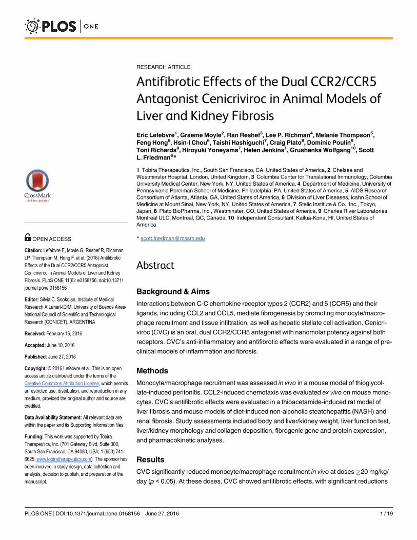
RESEARCH ARTICLE Antifibrotic Effects of the Dual CCR2/CCR5 Antagonist Cenicriviroc in Animal Models of Liver and Kidney Fibrosis Eric Lefebvre 1 , Graeme Moyle 2 , Ran Reshef 3 , Lee P. Richman 4 , Melanie Thompson 5 , Feng Hong 6 , Hsin-l Chou 6 , Taishi Hashiguchi 7 , Craig Plato 8 , Dominic Poulin 9 , Toni Richards 8 , Hiroyuki Yoneyama 7 , Helen Jenkins 1 , Grushenka Wolfgang 10 , Scott L. Friedman 6 * 1 Tobira Therapeutics, Inc., South San Francisco, CA, United States of America, 2 Chelsea and Westminster Hospital, London, United Kingdom, 3 Columbia Center for Translational Immunology, Columbia University Medical Center, New York, NY, United States of America, 4 Department of Medicine, University of Pennsylvania Perelman School of Medicine, Philadelphia, PA, United States of America, 5 AIDS Research Consortium of Atlanta, Atlanta, GA, United States of America, 6 Division of Liver Diseases, Icahn School of Medicine at Mount Sinai, New York, NY, United States of America, 7 Stelic Institute & Co., Inc., Tokyo, Japan, 8 Plato BioPharma, Inc., Westminster, CO, United States of America, 9 Charles River Laboratories Montreal ULC, Montreal, QC, Canada, 10 Independent Consultant, Kailua-Kona, HI, United States of America * [email protected] Abstract Background & Aims Interactions between C-C chemokine receptor types 2 (CCR2) and 5 (CCR5) and their ligands, including CCL2 and CCL5, mediate fibrogenesis by promoting monocyte/macro- phage recruitment and tissue infiltration, as well as hepatic stellate cell activation. Cenicri- viroc (CVC) is an oral, dual CCR2/CCR5 antagonist with nanomolar potency against both receptors. CVC’s anti-inflammatory and antifibrotic effects were evaluated in a range of pre- clinical models of inflammation and fibrosis. Methods Monocyte/macrophage recruitment was assessed in vivo in a mouse model of thioglycol- late-induced peritonitis. CCL2-induced chemotaxis was evaluated ex vivo on mouse mono- cytes. CVC’s antifibrotic effects were evaluated in a thioacetamide-induced rat model of liver fibrosis and mouse models of diet-induced non-alcoholic steatohepatitis (NASH) and renal fibrosis. Study assessments included body and liver/kidney weight, liver function test, liver/kidney morphology and collagen deposition, fibrogenic gene and protein expression, and pharmacokinetic analyses. Results CVC significantly reduced monocyte/macrophage recruitment in vivo at doses 20 mg/kg/ day (p < 0.05). At these doses, CVC showed antifibrotic effects, with significant reductions PLOS ONE | DOI:10.1371/journal.pone.0158156 June 27, 2016 1 / 19 a11111 OPEN ACCESS Citation: Lefebvre E, Moyle G, Reshef R, Richman LP, Thompson M, Hong F, et al. (2016) Antifibrotic Effects of the Dual CCR2/CCR5 Antagonist Cenicriviroc in Animal Models of Liver and Kidney Fibrosis. PLoS ONE 11(6): e0158156. doi:10.1371/ journal.pone.0158156 Editor: Silvia C. Sookoian, Institute of Medical Research A Lanari-IDIM, University of Buenos Aires- National Council of Scientific and Technological Research (CONICET), ARGENTINA Received: February 16, 2016 Accepted: June 10, 2016 Published: June 27, 2016 Copyright: © 2016 Lefebvre et al. This is an open access article distributed under the terms of the Creative Commons Attribution License, which permits unrestricted use, distribution, and reproduction in any medium, provided the original author and source are credited. Data Availability Statement: All relevant data are within the paper and its Supporting Information files. Funding: This work was supported by Tobira Therapeutics, Inc. (701 Gateway Blvd, Suite 300, South San Francisco, CA 94080, USA; 1 (650) 741- 6625; www.tobiratherapeutics.com). The sponsor has been involved in study design, data collection and analysis, decision to publish, and preparation of the manuscript.

Welcome message from author
This document is posted to help you gain knowledge. Please leave a comment to let me know what you think about it! Share it to your friends and learn new things together.
Transcript
RESEARCH ARTICLE
Antifibrotic Effects of the Dual CCR2/CCR5Antagonist Cenicriviroc in Animal Models ofLiver and Kidney FibrosisEric Lefebvre1, Graeme Moyle2, Ran Reshef3, Lee P. Richman4, Melanie Thompson5,Feng Hong6, Hsin-l Chou6, Taishi Hashiguchi7, Craig Plato8, Dominic Poulin9,Toni Richards8, Hiroyuki Yoneyama7, Helen Jenkins1, GrushenkaWolfgang10, ScottL. Friedman6*
1 Tobira Therapeutics, Inc., South San Francisco, CA, United States of America, 2 Chelsea andWestminster Hospital, London, United Kingdom, 3 Columbia Center for Translational Immunology, ColumbiaUniversity Medical Center, New York, NY, United States of America, 4 Department of Medicine, University ofPennsylvania Perelman School of Medicine, Philadelphia, PA, United States of America, 5 AIDS ResearchConsortium of Atlanta, Atlanta, GA, United States of America, 6 Division of Liver Diseases, Icahn School ofMedicine at Mount Sinai, New York, NY, United States of America, 7 Stelic Institute & Co., Inc., Tokyo,Japan, 8 Plato BioPharma, Inc., Westminster, CO, United States of America, 9 Charles River LaboratoriesMontreal ULC, Montreal, QC, Canada, 10 Independent Consultant, Kailua-Kona, HI, United States ofAmerica
Abstract
Background & Aims
Interactions between C-C chemokine receptor types 2 (CCR2) and 5 (CCR5) and their
ligands, including CCL2 and CCL5, mediate fibrogenesis by promoting monocyte/macro-
phage recruitment and tissue infiltration, as well as hepatic stellate cell activation. Cenicri-
viroc (CVC) is an oral, dual CCR2/CCR5 antagonist with nanomolar potency against both
receptors. CVC’s anti-inflammatory and antifibrotic effects were evaluated in a range of pre-
clinical models of inflammation and fibrosis.
Methods
Monocyte/macrophage recruitment was assessed in vivo in a mouse model of thioglycol-
late-induced peritonitis. CCL2-induced chemotaxis was evaluated ex vivo on mouse mono-
cytes. CVC’s antifibrotic effects were evaluated in a thioacetamide-induced rat model of
liver fibrosis and mouse models of diet-induced non-alcoholic steatohepatitis (NASH) and
renal fibrosis. Study assessments included body and liver/kidney weight, liver function test,
liver/kidney morphology and collagen deposition, fibrogenic gene and protein expression,
and pharmacokinetic analyses.
Results
CVC significantly reduced monocyte/macrophage recruitment in vivo at doses�20 mg/kg/
day (p < 0.05). At these doses, CVC showed antifibrotic effects, with significant reductions
PLOSONE | DOI:10.1371/journal.pone.0158156 June 27, 2016 1 / 19
a11111
OPEN ACCESS
Citation: Lefebvre E, Moyle G, Reshef R, RichmanLP, Thompson M, Hong F, et al. (2016) AntifibroticEffects of the Dual CCR2/CCR5 AntagonistCenicriviroc in Animal Models of Liver and KidneyFibrosis. PLoS ONE 11(6): e0158156. doi:10.1371/journal.pone.0158156
Editor: Silvia C. Sookoian, Institute of MedicalResearch A Lanari-IDIM, University of Buenos Aires-National Council of Scientific and TechnologicalResearch (CONICET), ARGENTINA
Received: February 16, 2016
Accepted: June 10, 2016
Published: June 27, 2016
Copyright: © 2016 Lefebvre et al. This is an openaccess article distributed under the terms of theCreative Commons Attribution License, which permitsunrestricted use, distribution, and reproduction in anymedium, provided the original author and source arecredited.
Data Availability Statement: All relevant data arewithin the paper and its Supporting Information files.
Funding: This work was supported by TobiraTherapeutics, Inc. (701 Gateway Blvd, Suite 300,South San Francisco, CA 94080, USA; 1 (650) 741-6625; www.tobiratherapeutics.com). The sponsor hasbeen involved in study design, data collection andanalysis, decision to publish, and preparation of themanuscript.
in collagen deposition (p < 0.05), and collagen type 1 protein and mRNA expression across
the three animal models of fibrosis. In the NASH model, CVC significantly reduced the non-
alcoholic fatty liver disease activity score (p < 0.05 vs. controls). CVC treatment had no
notable effect on body or liver/kidney weight.
Conclusions
CVC displayed potent anti-inflammatory and antifibrotic activity in a range of animal fibrosis
models, supporting human testing for fibrotic diseases. Further experimental studies are
needed to clarify the underlying mechanisms of CVC’s antifibrotic effects. A Phase 2b
study in adults with NASH and liver fibrosis is fully enrolled (CENTAUR Study 652-2-203;
NCT02217475).
IntroductionFibrosis results from a sustained inflammatory response to chronic organ injury and is charac-terized by the deposition of extracellular matrix proteins, including collagen types 1 and 3 [1].Hepatic fibrosis is associated with chronic liver disease, a significant global burden that con-tributes to cirrhosis and hepatocellular carcinoma [2]. Likewise, renal fibrosis is a commonmanifestation of chronic kidney disease [3], associated with elevated risks of morbidity andmortality [4]. Effective, well-tolerated antifibrotic and anti-inflammatory pharmacotherapiesthat can be integrated into current disease-management approaches are urgently needed.
The inflammatory response to hepatocyte injury plays a key role in hepatic fibrogenesis andinvolves recruitment of bone marrow-derived monocytes and macrophages to the site ofinjury, which is triggered by the activation of resident macrophages (i.e. Kupffer cells [KCs])[1]. In turn, infiltrating monocytes/macrophages amplify this immune response by producinginflammatory cytokines and chemokines, which further promote recruitment of inflammatorycells and upregulate the activation of hepatic stellate cells (HSCs) [1,5]. Fibrogenic cytokines(e.g. transforming growth factor-beta [TGF-beta]), produced by activated macrophages, pro-mote transdifferentiation of HSCs into myofibroblasts, which are the primary source of scar-forming matrix proteins, including fibrillary collagen types 1 and 3, and the contractile proteinalpha-smooth muscle actin (alpha-SMA) [1,6–8].
Recruitment of extra-hepatic inflammatory cells to the site of hepatic injury is largely medi-ated by interactions between chemokines and their receptors. Monocytes, KCs and HSCscan express C-C chemokine receptor types 2 (CCR2) and 5 (CCR5) on their surface [9–12].Increasing evidence implicates CCR2/CCR5 and their ligands (including C-C chemokineligand type 2 [CCL2, aka monocyte chemotactic protein-1 (MCP-1)] and type 5 [CCL5, akaRegulated on Activation, Normal T-cell Expressed and Secreted (RANTES)]), in the pathogenesisof liver fibrosis through promotion of monocyte/macrophage recruitment and tissue infiltra-tion, and HSC activation following liver injury [9–15]. Hepatocytes, KCs and infiltratingmonocytes/macrophages are the main sources of TGF-beta, a major fibrogenic cytokine pro-moting collagen production by activated HSCs [12]. Additional evidence substantiates theroles of CCR2/CCR5 and their ligands in renal fibrosis [16–20]. Thus, CCR2 and CCR5 havebecome attractive targets for antifibrotic therapy.
Cenicriviroc (CVC) is a novel, oral, once-daily (QD) dual CCR2/CCR5 antagonist withnanomolar potency, and a long plasma half-life (30–40 hours in humans) [21–24]. It has afavorable safety profile and was well tolerated in approximately 600 subjects, including those
Cenicriviroc as an Antifibrotic Agent
PLOS ONE | DOI:10.1371/journal.pone.0158156 June 27, 2016 2 / 19
Competing Interests: EL and HJ are employees ofTobira Therapeutics, Inc. GM is on the ScientificAdvisory Board of Tobira Therapeutics, Inc. and hasserved on the Board of Directors of TobiraTherapeutics, Inc. and holds stock options in TobiraTherapeutics, Inc. RR, FH, HC, TH and HY havereceived research funding from Tobira Therapeutics,Inc. LPR, CP, DP and TR have no conflicts of interestto declare. MT is a member of an institution thatreceived clinical trial funding or research grants fromTobira Therapeutics, Inc. GW is a consultant forTobira Therapeutics, Inc. and holds stock options inTobira Therapeutics, Inc. SLF is on the ScientificAdvisory Board of Tobira Therapeutics, Inc., hasperformed research studies supported by TobiraTherapeutics, Inc. and holds stock options in TobiraTherapeutics, Inc. This does not alter the authors'adherence to PLOS ONE policies on sharing dataand materials.
with mild or moderate hepatic impairment (Child–Pugh A and B) [25,26]. CVC is currentlyunder evaluation in a Phase 2b study in 289 adults with non-alcoholic steatohepatitis (NASH)and liver fibrosis, for which it received Fast Track Designation by the Food and Drug Adminis-tration (CENTAUR Study 652-2-203; NCT02217475) [27]. An ex vivo study of human periph-eral blood mononuclear cells found that CVC leads to receptor occupancies of ~98% for CCR2on monocytes (at 6 nmol/L) and�90% for CCR5 on CD4+ and CD8+ T-cells (at 3.1 and 2.3nmol/L, respectively) [28]. As a shorter half-life (~2 hours in mice) and a lower potency havebeen observed for CVC in rodents versus humans, this was considered in dose selection for dis-ease models. An ex vivo study conducted on mouse monocytes and macrophages showed thatCVC concentrations of 250 nmol/L or higher achieve>87% CCR2/CCR5 occupancy in thesecells [29,30]. Collectively, these findings suggest that rodent models are well suited to evaluatethe anti-inflammatory and antifibrotic properties of CVC, resulting from effective CCR2/CCR5 blockade.
A number of in vitro and in vivomodels of fibrosis are commonly used to assess recruitmentof inflammatory cells and antifibrotic activity of therapeutic agents [31–33]. Multiple modelsof fibrosis allow assessment of the broad effect of an antifibrotic agent across species andorgans, and reduce the likelihood that efficacy is restricted to one model. Here we provide evi-dence for the antifibrotic effects of CVC, as demonstrated in models that have evaluated: (1)the ex vivo and in vivo effects of CVC on recruitment/migration of monocytes/macrophages;and (2) the in vivo antifibrotic effects of CVC in liver and kidney fibrosis.
Materials and MethodsAll animal procedures were approved by each institution’s animal care and use committee(IACUC), and were conducted in accordance with national guidelines. CVC is cenicrivirocmesylate, provided by Tobira Therapeutics, Inc., USA. The vehicle control used in all in vivostudies was 0.5% [w/v] methylcellulose + 1% Tween1-80 (pH ~1.3).
Effect of CVC on recruitment/migration of monocytes/macrophagesIn vivomouse model of peritonitis. Amurine thioglycollate (TG)-induced model of peri-
tonitis, where acute inflammation induced by intraperitoneal (IP) injection of TG resultsin a rapid increase in monocyte/macrophage migration into the peritoneal cavity [34], wasemployed to assess the effects of CVC on cell recruitment in vivo. The protocol was approvedby the IACUC of Charles River Laboratories Preclinical Services, Montreal (PCS-MTL). Thecare and use of animals was conducted in accordance with the guidelines of the US NationalResearch Council and the Canadian Council on Animal Care.
Male C57BL/6 mice (n = 44; 8–10 weeks of age; Charles River Laboratories, Canada) wereallocated to receive treatments via oral gavage (PO) on Days 1–5 in the following groups: non-disease control, vehicle control twice daily (BID), CVC 5 mg/kg/day (CVC5) BID, CVC 20 mg/kg/day (CVC20) BID, CVC 100 mg/kg/day (CVC100) BID, CVC20 QD, and positive controldexamethasone (corticosteroid known to reduce inflammation in a variety of animal models) 1mg/kg QD (S1 Table). On Day 4, peritonitis was induced via IP injection of TG 3.85% (1 mL/animal) 2 hours post-dose in all groups except non-disease controls. Study endpoints included:peritoneal lavage cell counts and pharmacokinetic (PK) evaluation. Animals were sacrificed 48hours post-TG injection by isoflurane inhalation, and peritoneal lavage and blood samples (0.7mL) were collected. Differential cell counts were assessed in peritoneal lavage samples using anAdvia1 Hematology System (Siemens Healthcare Diagnostics, USA) with multispecies soft-ware and an analysis software designed for mouse peritoneal fluid on Advia1 120 (LabThru-Put, USA). A 0.3 mL aliquot of the blood sample was processed to plasma for PK analysis.
Cenicriviroc as an Antifibrotic Agent
PLOS ONE | DOI:10.1371/journal.pone.0158156 June 27, 2016 3 / 19
Ex vivomigration of mouse monocytes. The protocol was approved by the IACUC of theUniversity of Pennsylvania (protocol number 804755) and animals were maintained accordingto the National Institutes of Health (NIH) guidelines. Animals were euthanized by CO2 inhala-tion followed by cervical dislocation.
Mouse monocyte migration in response to CVC treatment was assessed ex vivo in triplicate.TG was injected intraperitoneally into male C57BL/6 mice (n = 3; 8–10 weeks of age; JacksonLaboratory, USA) and activated macrophages were collected 48 hours later by peritoneal lavage.Chemotaxis was assayed using a Transwell1 Chamber (Costar, USA) with a 5 μm-pore sizepolycarbonate filter, as previously described [35]. Briefly, cells were incubated for 2 hours in thepresence of 1 nM CCL2 and/or 1 μMCVC (dissolved in dimethyl sulfoxide with 0.5% aceticacid and diluted 1:1000 with serum-free Roswell Park Memorial Institute-1640 medium and0.5% bovine serum albumin). Cells were harvested from the lower compartment and analyzedby flow cytometry to enumerate F4/80+CD11b+ macrophages using a 3-laser BD FACSCanto™(BD Biosciences, Canada). Results were analyzed using FlowJo software (Tree Star Inc., USA).
Antifibrotic effects of CVC in animal models of fibrosisRat model of thioacetamide (TAA)-induced liver fibrosis (TAA model). The TAA
model is commonly used for the evaluation of treatment at various stages of disease, frominflammation to cirrhosis [36]. The protocol was approved by the Mount Sinai IACUC(approval number: LA12-00318) and animals were maintained according to the NIH guide-lines. Anesthesia was performed with 1–5% isoflurane through inhalation; surgery wasterminal.
Using male Sprague-Dawley rats (n = 72, 10–12 weeks of age; Harlan Laboratories, USA),fibrosis was induced by IP administration of TAA at a dose of 150 mg/kg three times per weekfor 8 weeks. Rats (n = 4–8/group) received vehicle control, CVC 30 mg/kg/day (CVC30) orCVC100 QD PO during Weeks 0–8 (early intervention), Weeks 4–8 (established fibrosis) orWeeks 8–12 (cirrhosis reversal) and were sacrificed at Weeks 8, 8 or 12, respectively (S1 Table).Study endpoints included: body and liver weights, liver biochemistry (e.g. serum alanine andaspartate aminotransferase [ALT/AST]), extracellular matrix protein expression in liver tissue(collagen type 1 and alpha-SMA), mRNA expression (collagen type 1, alpha-SMA, beta-plate-let-derived growth factor-beta receptor, TGF beta-receptor, matrix metalloproteinase 2, tissueinhibitor of metalloproteinases 1 [TIMP1] and 2 [TIMP2]) and liver morphology. For liver-function testing, plasma samples were obtained from blood collected from the vena cava. Liverswere sectioned and fixed in 3.7% formalin, embedded in paraffin, cut at 4 mm thickness andstained with hematoxylin and eosin (H&E) for histological examination.
Mouse model of diet-induced NASH (NASHmodel). The protocol was approved by Ste-lic IACUC (approval number: RP-131). All animals were housed and cared for in accordancewith the Japanese Pharmacological Society Guidelines for Animal Use.
NASH was induced in male C57BL/6 mice (Charles River Laboratories, Japan) via subcuta-neous injection of 200 μg of streptozotocin 2 days post-birth (causing mild islet inflammationand islet destruction) plus a high-fat diet (57 kcal% fat) from 4 weeks of age (sequentially caus-ing fatty changes to the liver, NASH and fibrosis) [37]. FromWeeks 6 to 9, three groups (n = 9/group) received vehicle control, CVC20 or CVC100 BID PO (S1 Table). At Week 9, six animalsper group were sacrificed for assessment of liver fibrosis and Non-Alcoholic Fatty Liver Disease(NAFLD) Activity Score (NAS). The animals were sacrificed by exsanguination through directcardiac puncture under ether anesthesia. Study endpoints included: body and liver weights,plasma biochemistry (e.g. ALT and CCL2), extracellular matrix protein in liver tissue(hydroxyproline content), mRNA expression (collagen type 1, tumor necrosis factor-alpha,
Cenicriviroc as an Antifibrotic Agent
PLOS ONE | DOI:10.1371/journal.pone.0158156 June 27, 2016 4 / 19
MCP-1, TIMP1) and histopathological analyses. Blood samples were collected and plasmasamples derived for biochemistry assessments. Sections were cut from paraffin blocks of livertissue, prefixed in Bouin’s solution and stained with H&E for histological examination.
Mouse model of unilateral ureter obstruction (UUO)-induced renal fibrosis (UUOmodel). Unilateral ureter obstruction is a commonly used experimental model of kidneyinjury, in which interstitial inflammation, tubular cell injury/death and fibrosis, ensue [32].The protocol was approved by Plato BioPharma, Inc. IACUC (PBI; approval number 2013–04). The care and use of animals was conducted in accordance with the NIH Guide for theCare and Use of Laboratory Animals (2010), Action against Medical Accidents Guidelines onEuthanasia (2007), Office of Laboratory Animal Welfare Institutional Animal Care and UseCommittee Guidebook (2002), Public Health Service Policy on Humane Care and Use ofLaboratory Animals (2002), PBI standard operating procedures on a) moribund sacrifice, b)vivarium maintenance and animal care, and c) animal handling and dosing. Anesthesia wasperformed by isoflurane injection; animals were euthanized by diaphragm laceration followedby heart laceration while under isoflurane anesthesia.
Male CD-1 mice (n = 51; 7–8 weeks of age; Charles River Laboratories, USA) were allocatedto weight-matched treatment groups on Day -1 (1 day prior to either sham [one group] or per-manent right UUO surgery [five groups] via aseptic laparotomy). From Days -1 to 4, micereceived phosphate-buffered saline (PBS) IP QD, apart from a positive-control group, whichreceived 1D11 (anti-TGF-beta1 antibody) 3 mg/kg IP QD. From Days 0 to 5, mice receivedvehicle control (sham surgery, UUO-control and UUO+positive-control groups) or CVC 7mg/kg/day (CVC7), CVC20 or CVC100 PO BID (S1 Table). The CVC100 group was termi-nated due to poor clinical condition (6/9 animals died and 3/9 euthanized prior to end ofstudy). Of note, a similar dose of CVC100 administered for 3 weeks was well tolerated in themouse NASH model; therefore, it is plausible that the laparotomy and UUO surgery in combi-nation with CVC may have contributed to the loss of animals. Study endpoints included: bodyand kidney weights, mRNA expression (TGF-beta, connective tissue growth factor, MCP-1,alpha-SMA, collagen 1a1, fibronectin-1 and collagen 3a1), extracellular matrix protein in renalcortical tissue (hydroxyproline content) and histological analyses. Blood and tissue sampleswere collected from anesthetized mice 4 hours post-dose on Day 5, prior to sacrifice. A mid-transverse section of the right obstructed kidney was collected for histological analysis.
Outcome measures in animal models of fibrosisBody and liver/kidney weights. Body weight (measured before and during treatment) and
sacrificed animal body and liver/kidney/spleen weights were recorded.Plasma biochemistry. In the TAA model, serum ALT/AST levels were measured using
VITROS1 5,1 FS (Ortho Clinical Diagnostics, USA), with plasma diluted with VITROS1 7%bovine serum albumin if needed. In the NASH model, plasma ALT levels were measured byFUJI DRI-CHEM 7000 (Fujifilm, Japan) and plasma CCL2 concentration quantified by themouse CCL2/JE/MCP-1 immunoassay kit (R&D, USA).
Liver or obstructed kidney morphology. Collagen deposition (the extent of fibrosis) wasvisualized in liver/kidney sections with picrosirius red staining. In the TAA model, collagenquantification was performed using computerized Life Science morphometry system (BIO-QUANT, USA) on a total of 36 images per animal at 100x magnification (four picrosirius red-stained slides per animal, with nine images taken randomly per slide).
In the NASHmodel, bright field images of picrosirius red-stained sections were capturedaround the central vein using a digital camera (DFC280; Leica, Germany) at 200x magnification;the ‘positive’ areas in five fields/sections were measured using ImageJ software (National
Cenicriviroc as an Antifibrotic Agent
PLOS ONE | DOI:10.1371/journal.pone.0158156 June 27, 2016 5 / 19
Institutes of Health, USA). Perivascular areas were subtracted from the total positive areas foreach field (modified fibrosis area). The NAS, a histological tool developed to assess diseaseseverity in humans, was assessed in a blinded fashion and calculated according to Kleiner’s crite-ria on H&E-stained sections [38]. It is based on the semi-quantification of steatosis, lobularinflammation and hepatocellular ballooning.
In the UUOmodel, ten images/depth/kidney were assessed in a blinded fashion using AxioImager.A2 (Zeiss, USA) light microscopy (at 200x magnification to enable 60–70% sampling ofrenal cortical area) and quantified by a composite Collagen Volume Fraction (CVF [% totalarea imaged]) score expressed as the average positive stain across three anatomically distinct(200–250 μM apart) tissue sections, or depths, from the obstructed kidney.
Immunohistochemistry. In the NASH model, liver sections fixed in acetone were incu-bated with anti-F4/80 antibody (BMA Biomedicals, Switzerland) to assess inflammation. Dou-ble immunohistochemical analyses were performed with anti-CD206 (RayBiotec, USA) oranti-CD16/32 (BD Biosciences, USA) antibodies. Cells were counted and the M1/M2 polariza-tion ratio was calculated as mean percentage of F4/80+CD16/32+ cells/mean percentage of F4/80+CD206+ cells.
Extracellular matrix proteins. Extracellular matrix protein content in tissue samples wasmeasured by evaluating collagen type 1 and alpha-SMA protein expression in the TAA modeland hydroxyproline content in the NASH and UUOmodels.
In the TAAmodel, total protein was extracted from liver cells and expression levels of collagentype 1 and alpha-SMA were assessed by western blotting. Protein expression levels were normal-ized to the reference protein (glyceraldehyde-3-phosphate dehydrogenase [GAPDH] or calnexin).
In the NASH model, frozen liver samples were subjected to an alkaline-acid hydrolysis, thencentrifuged, and the supernatant collected. Hydroxyproline content was quantified against ahydroxyproline standard curve, with a BCA protein assay kit (Thermo Fisher Scientific, USA)used to normalize the calculated hydroxyproline values.
In the UUOmodel, frozen renal cortical tissue biopsies were hydrolyzed and centrifuged,and the supernatant was analyzed by OD absorbance at 560 nM on a SpectraMax1 190(Molecular Devices, USA). Standard curves for conversion of ODs to concentrations were gen-erated using linear regression and sample concentrations were determined using SoftMax1
Pro5 software (Molecular Devices, USA).Gene expression of fibrotic or inflammatory biomarkers. In the hepatic-fibrosis models,
RNA was extracted from liver tissues and purified, and 1 μg of total mRNA was reverse-tran-scribed into complementary DNA. Expression levels were determined by quantitative polymerasechain reaction (PCR) (iQ™ SYBR1Green Supermix [Bio-Rad Laboratories, USA] on the LightCy-cler1 480 Real-Time PCR System [Roche, Switzerland]) in the TAAmodel at Week 8 (early inter-vention and established fibrosis) andWeek 12 (cirrhosis reversal), and by real-time PCR (PCRDice1 and SYBR1 Premix Ex Taq™ [Takara Bio Inc., Japan]) at Week 9 in the NASHmodel.
In the UUOmodel, mRNA expression levels in renal cortical tissue were evaluated usingthe QuantiGene1 Plex 2.0 profiling platform (Affymetrix, USA).
Relative mRNA expression levels were normalized to the following reference genes: TAAmodel, GAPDH; NASH model, 36B4 (gene symbol: Rplp0); UUOmodel, hypoxanthinephosphoribosyltransferase.
PK analysis in the TG-induced model of peritonitis and the UUOmodelCVC plasma levels (minimum and maximum) were determined by KCAS Bioanalytical Ser-vices, USA, using a validated liquid chromatography-tandem mass spectrometry (LC/MS/MS)plasma method (50 μL assay, range 10.0−1920.0 ng/mL).
Cenicriviroc as an Antifibrotic Agent
PLOS ONE | DOI:10.1371/journal.pone.0158156 June 27, 2016 6 / 19
Statistical analysisIn all studies, statistical significance level was set as p< 0.05.
Recruitment/migration of monocytes/macrophages. In the peritonitis mouse model,one-way analysis of variance with post-hoc Dunnett’s test was performed, using GraphPadPrism1 (GraphPad Software, Inc., USA), on differential cell counts obtained from lavage intreatment groups versus the vehicle-control group. For the ex vivomigration of mouse mono-cytes, statistical significance was determined by a Student’s t test.
Animal models of fibrosis. Data are presented as ±standard error of the mean (SEM) inthe TAA model. Statistical significance was determined by the two-tailed Student’s t test.
In the NASHmodel, data are expressed as mean ±standard deviation (SD). Statistical analyseswere performed using the Bonferroni multiple comparison test (GraphPad Prism1 4 software).
In the UUOmodel, data are expressed as mean ±SEM. All statistical analyses were per-formed using GraphPad Prism1 6 software. Unpaired ‘t’-tests were used to analyze treatmentdifferences between control groups (sham-surgery and UUO+positive-control 1D11 each vs.UUO control). One-way analysis of variance with post-hoc Dunnett’s test was used to comparetreatment differences between CVC groups and UUO controls.
Results
Effect of CVC on recruitment/migration of monocytes/macrophagesIn vivomouse model of peritonitis. In the TG-induced model of peritonitis, CVC treat-
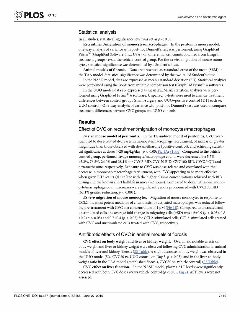
ment led to dose-related decreases in monocyte/macrophage recruitment, of similar or greatermagnitude than those observed with dexamethasone (positive control), and achieving statisti-cal significance at doses�20 mg/kg/day (p< 0.05; Fig 1A; S1 Fig). Compared to the vehicle-control group, peritoneal lavage monocyte/macrophage counts were decreased by: 5.7%,45.2%, 76.5%, 26.0% and 38.1% for CVC5 BID, CVC20 BID, CVC100 BID, CVC20 QD anddexamethasone, respectively. Exposure to CVC was dose-related and correlated with thedecrease in monocyte/macrophage recruitment, with CVC appearing to be more effectivewhen given BID versus QD, in line with the higher plasma concentrations achieved with BIDdosing and the known short half-life in mice (~2 hours). Compared to dexamethasone, mono-cyte/macrophage-count decreases were significantly more pronounced with CVC100 BID(62.1% greater reduction, p< 0.001).
Ex vivomigration of mouse monocytes. Migration of mouse monocytes in response toCCL2, the most potent mediator of chemotaxis for activated macrophages, was reduced follow-ing pre-treatment with CVC at a concentration of 1 μM (Fig 1B). Compared to untreated andunstimulated cells, the average fold change in migrating cells (±SD) was 4.6±0.9 (p< 0.05), 0.8±0.2 (p> 0.05) and 0.7±0.4 (p> 0.05) for CCL2-stimulated cells, CCL2-stimulated cells treatedwith CVC and unstimulated cells treated with CVC, respectively.
Antifibrotic effects of CVC in animal models of fibrosisCVC effect on body weight and liver or kidney weight. Overall, no notable effects on
body weight and liver or kidney weight were observed following CVC administration in animalmodels of liver and kidney fibrosis (S2 Table). A slight decrease in body weight was observed inthe UUOmodel (5%, CVC20 vs. UUO control on Day 5, p< 0.05), and in the liver-to-bodyweight ratio in the TAA model (established fibrosis, CVC30 vs. vehicle control) (S2 Table).
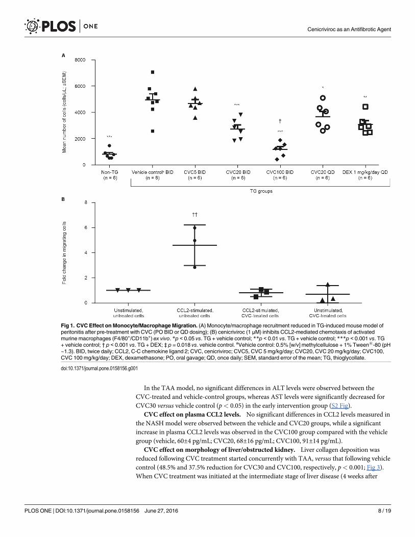
CVC effect on liver function. In the NASH model, plasma ALT levels were significantlydecreased with both CVC doses versus vehicle control (p< 0.05; Fig 2). AST levels were notassessed.
Cenicriviroc as an Antifibrotic Agent
PLOS ONE | DOI:10.1371/journal.pone.0158156 June 27, 2016 7 / 19
In the TAA model, no significant differences in ALT levels were observed between theCVC-treated and vehicle-control groups, whereas AST levels were significantly decreased forCVC30 versus vehicle control (p< 0.05) in the early intervention group (S2 Fig).
CVC effect on plasma CCL2 levels. No significant differences in CCL2 levels measured inthe NASH model were observed between the vehicle and CVC20 groups, while a significantincrease in plasma CCL2 levels was observed in the CVC100 group compared with the vehiclegroup (vehicle, 60±4 pg/mL; CVC20, 68±16 pg/mL; CVC100, 91±14 pg/mL).
CVC effect on morphology of liver/obstructed kidney. Liver collagen deposition wasreduced following CVC treatment started concurrently with TAA, versus that following vehiclecontrol (48.5% and 37.5% reduction for CVC30 and CVC100, respectively, p< 0.001; Fig 3).When CVC treatment was initiated at the intermediate stage of liver disease (4 weeks after
Fig 1. CVC Effect on Monocyte/MacrophageMigration. (A) Monocyte/macrophage recruitment reduced in TG-induced mouse model ofperitonitis after pre-treatment with CVC (PO BID or QD dosing); (B) cenicriviroc (1 μM) inhibits CCL2-mediated chemotaxis of activatedmurine macrophages (F4/80+/CD11b+) ex vivo. *p < 0.05 vs. TG + vehicle control; **p < 0.01 vs. TG + vehicle control; ***p < 0.001 vs. TG+ vehicle control; † p < 0.001 vs. TG + DEX; ‡ p = 0.018 vs. vehicle control. aVehicle control: 0.5% [w/v] methylcellulose + 1% Tween1-80 (pH~1.3). BID, twice daily; CCL2, C-C chemokine ligand 2; CVC, cenicriviroc; CVC5, CVC 5 mg/kg/day; CVC20, CVC 20 mg/kg/day; CVC100,CVC 100 mg/kg/day; DEX, dexamethasone; PO, oral gavage; QD, once daily; SEM, standard error of the mean; TG, thioglycollate.
doi:10.1371/journal.pone.0158156.g001
Cenicriviroc as an Antifibrotic Agent
PLOS ONE | DOI:10.1371/journal.pone.0158156 June 27, 2016 8 / 19
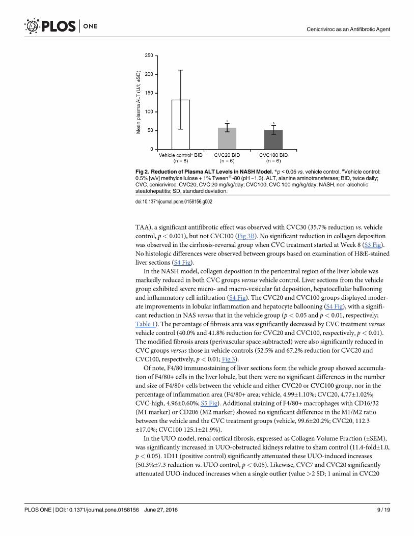
TAA), a significant antifibrotic effect was observed with CVC30 (35.7% reduction vs. vehiclecontrol, p< 0.001), but not CVC100 (Fig 3B). No significant reduction in collagen depositionwas observed in the cirrhosis-reversal group when CVC treatment started at Week 8 (S3 Fig).No histologic differences were observed between groups based on examination of H&E-stainedliver sections (S4 Fig).
In the NASH model, collagen deposition in the pericentral region of the liver lobule wasmarkedly reduced in both CVC groups versus vehicle control. Liver sections from the vehiclegroup exhibited severe micro- and macro-vesicular fat deposition, hepatocellular ballooningand inflammatory cell infiltration (S4 Fig). The CVC20 and CVC100 groups displayed moder-ate improvements in lobular inflammation and hepatocyte ballooning (S4 Fig), with a signifi-cant reduction in NAS versus that in the vehicle group (p< 0.05 and p< 0.01, respectively;Table 1). The percentage of fibrosis area was significantly decreased by CVC treatment versusvehicle control (40.0% and 41.8% reduction for CVC20 and CVC100, respectively, p< 0.01).The modified fibrosis areas (perivascular space subtracted) were also significantly reduced inCVC groups versus those in vehicle controls (52.5% and 67.2% reduction for CVC20 andCVC100, respectively, p< 0.01; Fig 3).
Of note, F4/80 immunostaining of liver sections form the vehicle group showed accumula-tion of F4/80+ cells in the liver lobule, but there were no significant differences in the numberand size of F4/80+ cells between the vehicle and either CVC20 or CVC100 group, nor in thepercentage of inflammation area (F4/80+ area; vehicle, 4.99±1.10%; CVC20, 4.77±1.02%;CVC-high, 4.96±0.60%; S5 Fig). Additional staining of F4/80+ macrophages with CD16/32(M1 marker) or CD206 (M2 marker) showed no significant difference in the M1/M2 ratiobetween the vehicle and the CVC treatment groups (vehicle, 99.6±20.2%; CVC20, 112.3±17.0%; CVC100 125.1±21.9%).
In the UUOmodel, renal cortical fibrosis, expressed as Collagen Volume Fraction (±SEM),was significantly increased in UUO-obstructed kidneys relative to sham control (11.4-fold±1.0,p< 0.05). 1D11 (positive control) significantly attenuated these UUO-induced increases(50.3%±7.3 reduction vs. UUO control, p< 0.05). Likewise, CVC7 and CVC20 significantlyattenuated UUO-induced increases when a single outlier (value>2 SD; 1 animal in CVC20
Fig 2. Reduction of Plasma ALT Levels in NASHModel. *p < 0.05 vs. vehicle control. aVehicle control:0.5% [w/v] methylcellulose + 1% Tween1-80 (pH ~1.3). ALT, alanine aminotransferase; BID, twice daily;CVC, cenicriviroc; CVC20, CVC 20 mg/kg/day; CVC100, CVC 100 mg/kg/day; NASH, non-alcoholicsteatohepatitis; SD, standard deviation.
doi:10.1371/journal.pone.0158156.g002
Cenicriviroc as an Antifibrotic Agent
PLOS ONE | DOI:10.1371/journal.pone.0158156 June 27, 2016 9 / 19
Fig 3. Reduction in Fibrosis. (A) Representative micrographs of Sirius red-stained liver sections in the rat TAA model (early interventionand established fibrosis; 100x), mouse NASHmodel (200x) and mouse UUOmodel (200x). (B) Reduction in collagen deposition in the ratTAAmodel (early intervention and established fibrosis), mouse NASHmodela and mouse UUOmodelb. *p < 0.05 vs. sham control;**p < 0.01 vs. vehicle control; ***p < 0.001 vs. vehicle control; †p < 0.05 vs. UUO + vehicle control. aPerivascular area subtracted; bDatapresented exclude a single outlier from an animal in the CVC20 group, which had a CVF value >2 SDs higher than any other animal in the
Cenicriviroc as an Antifibrotic Agent
PLOS ONE | DOI:10.1371/journal.pone.0158156 June 27, 2016 10 / 19
group) was removed (28.6%±8.8 and 31.8%±6.8 reduction vs. UUO control, respectively,p< 0.05; Fig 3).
CVC effect on extracellular matrix protein content. Compared to vehicle control, pro-tein expression of collagen type 1 in the TAA model was reduced by 29% and 12% for CVC30and CVC100, respectively, in early intervention, and 30% for CVC30 in established fibrosis.Protein expression of alpha-SMA was reduced following CVC treatment by 17% and 22% forCVC30 and CVC100, respectively, versus vehicle control in early intervention, and 14% forCVC30 in established fibrosis. No reductions were observed in protein expression in theadvanced disease cirrhosis group (Fig 4). The differences in collagen type 1 and alpha-SMAprotein levels between treatment arms were not statistically significant; however, these resultslikely reflect the significant decrease of fibrosis observed in histological analyses.
In the NASH model, the liver hydroxyproline content tended to decrease in the CVC20 andCVC100 groups compared with the vehicle group (vehicle, 0.75±0.18 μg/mg; CVC20, 0.63±0.05 μg/mg; CVC100, 0.62±0.09 μg/mg; Fig 4).
Compared with sham control, UUO increased hydroxyproline content in obstructed kid-neys (1.6±0.2 fold; vehicle control). 1D11 attenuated these increases (37.1±4.9%); however,CVC did not affect these increases in obstructed kidney hydroxyproline content (Fig 4).
CVC effect on gene expression of fibrotic/inflammatory biomarkers. In the TAAmodel, there were no significant changes between CVC and vehicle-control groups in mRNAexpression of the fibrosis markers assessed (S6 Fig).
group. CVF, Collagen Volume Fraction; CVC, cenicriviroc; NASH, non-alcoholic steatohepatitis; SD, standard deviation; SEM, standarderror of the mean; TAA, thioacetamide; UUO, unilateral ureteral obstruction.
doi:10.1371/journal.pone.0158156.g003
Table 1. Reduction of NAS in NASHModela.
Score Vehicle controlb (n = 6) CVC20 (n = 6) CVC100 (n = 6)
Steatosis
0 0 0 1
1 4 6 5
2 2 0 0
3 0 0 0
Lobular inflammation
0 0 0 0
1 0 3 3
2 6 3 3
3 0 0 0
Hepatocyte ballooning
0 0 0 1
1 0 3 2
2 6 3 3
NAS, mean (±SD) 5.3 (±0.5) 4.0 (±0.6)* 3.7 (±0.8)**
*p = 0.0013 vs. vehicle control;
**p = 0.0009 vs. vehicle control.aInterpretation performed by pathologist blinded by treatment group;bVehicle control: 0.5% [w/v] methylcellulose + 1% Tween1-80 (pH ~1.3).
CVC, cenicriviroc; CVC20, CVC 20 mg/kg/day; CVC100, CVC 100 mg/kg/day; n, number of animals; NAS, non-alcoholic fatty liver disease activity score;
NASH, non-alcoholic steatohepatitis; SD, standard deviation.
doi:10.1371/journal.pone.0158156.t001
Cenicriviroc as an Antifibrotic Agent
PLOS ONE | DOI:10.1371/journal.pone.0158156 June 27, 2016 11 / 19
Collagen type 1 mRNA expression levels in the NASH model decreased with CVC treat-ment (37% and 27% reduction vs. vehicle control for CVC20 and CVC100, respectively). Thisreduction was statistically significant only for the CVC20 group (p< 0.05). There were no sta-tistically significant differences in mRNA expression levels between the vehicle-control andCVC groups for the other fibrosis markers tested in this model (S7 Fig).
In the UUOmodel, UUO significantly increased collagen 1a1, collagen 3a1, fibronectin-1and TGF-beta1 mRNA expression in vehicle-treated animals versus sham controls, and 1D11(positive control) significantly attenuated these UUO-induced increases (p< 0.05) (S8 Fig).There was a trend for CVC7 and CVC20 to inhibit UUO-induced increases in the obstructedrenal cortical mRNA expression of some fibrogenic genes; however, it did not reach statisticalsignificance relative to UUO control.
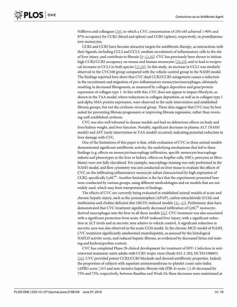
CVC PK analysisIn the TG-induced model of peritonitis, following BID dosing, plasma levels of CVC ~14 hourspost-dose (trough levels) increased in a dose-dependent manner (Table 2). At CVC20, lower
Fig 4. CVC Effects on Extracellular Matrix Protein Content.Mean expression of (A) collagen type I and (B) alpha-SMA in the TAAmodelcompared to vehicle control; mean expression of hydroxyproline content in (C) NASHmodel and (D) UUOmodel. alpha-SMA, alpha-smoothmuscle actin; BID, twice daily; CVC, cenicriviroc; NASH, non-alcoholic steatohepatitis; QD, once daily; SD, standard deviation; TAA,thioacetamide; UUO, unilateral ureter obstruction.
doi:10.1371/journal.pone.0158156.g004
Cenicriviroc as an Antifibrotic Agent
PLOS ONE | DOI:10.1371/journal.pone.0158156 June 27, 2016 12 / 19
trough plasma levels were seen with QD (n = 5) versus BID dosing (n = 5) (25.7±8.1 ng/mL [26hours post-dose] vs. 81.2±22.8 ng/mL [14 hours post-dose], respectively).
In the UUOmodel, all dose groups had exposure to CVC and plasma levels of CVC at theapproximate maximum concentration (Cmax) 4 hours post-dose on Day 5, and these increasedin a dose-dependent manner (Table 2).
DiscussionCVC displayed significant anti-inflammatory and antifibrotic effects across a range of in vivomodels, including liver fibrosis, NASH and kidney fibrosis. CVC reduced monocyte/macro-phage recruitment in an in vivomodel of peritonitis, and monocyte migration in vitro. A signif-icant decrease in monocyte/macrophage recruitment versus vehicle control (p< 0.05) and thepositive control dexamethasone (p< 0.001) was obtained following treatment with CVC100BID in vivo. These findings support CVC’s anti-inflammatory mode of action, and are furthersubstantiated by results from the NASH model, where a significantly lower NAS score wasobserved in the CVC treatment groups relative to the vehicle-control group and a greater num-ber of CVC-treated animals had decreased lobular inflammation and prominent hepatocellularballooning.
In all models, reductions in collagen deposition and production were observed (a significantreduction in collagen deposition, as well as decreased protein and mRNA expression of colla-gen type 1). While the significant decreases in liver collagen deposition were dose-dependentin the NASH model, where CVC was administered BID given its short half-life in mice (~2hours), CVC30 in the TAA model lead to the greater reduction in collagen deposition versusCVC100 (48.5% and 37.5% reduction vs. vehicle control, respectively, p< 0.001). The QDadministration of CVC in the TAA model and short half-life of CVC in rats (4–5 hours) mayhave led to increased clearance of CVC, explaining the lack of apparent dose response. In theUUOmodel, where gavage was performed twice daily in mice, a significant antifibrotic effectwas observed at the low dose (CVC7; p< 0.05) suggesting that this could represent the mini-mum effective dose. The lack of apparent dose response in this model may also be explained bythe short treatment duration (5 days) and the fact that adequate and sustained receptor occu-pancy (necessary to disrupt CCL2- and CCL5-induced migration) may have already beenachieved at lower CVC doses. In fact, CCR2 and CCR5 receptor occupancy by CVC wasassessed ex vivo on murine cryopreserved peripheral blood mononuclear cells by Lishomwa
Table 2. CVC Plasma Trough Levels (Mean±SD).
Dose Time post-dose CVC concentration (mean±SD,ng/mL)
TG-induced mouse model of peritonitis (Day 6)
CVC5 BID, n = 2 ~14 hours 13.5a
CVC20 BID, n = 5 ~14 hours 81.2±22.8
CVC100 BID, n = 5 ~14 hours 551±253
Mouse model of UUO-induced renal fibrosis (Day 5)
CVC7 BID, n = 8 ~4 hours 251±364
CVC20 BID, n = 8 ~4 hours 971±507
a3 of 5 samples below the lower limit of quantification (10 ng/mL).
BID, twice-daily; CVC, cenicriviroc; CVC5, CVC 5 mg/kg/day; CVC7, CVC 7 mg/kg/day; CVC20, CVC 20
mg/kg/day; CVC100, CVC 100 mg/kg/day; n, number of animals; SD, standard deviation; TG,
thioglycollate; UUO, unilateral ureter obstruction
doi:10.1371/journal.pone.0158156.t002
Cenicriviroc as an Antifibrotic Agent
PLOS ONE | DOI:10.1371/journal.pone.0158156 June 27, 2016 13 / 19
Ndlhovu and colleagues [39], in which a CVC concentration of 250 nM achieved>90% and87% occupancy for CCR2 (blood and spleen) and CCR5 (spleen), respectively, in proinflamma-tory monocytes.
CCR2 and CCR5 have become attractive targets for antifibrotic therapy, as interactions withtheir ligands, including CCL2 and CCL5, mediate recruitment of inflammatory cells to the siteof liver injury, and contribute to fibrosis [9–15,40]. CVC has previously been shown to initiatehigh CCR2/CCR5 occupancy on mouse and human monocytes [28,29], and to lead to recipro-cal increases in CCL2 in both species [25,26]. In this study, an increase in CCL2 was similarlyobserved in the CVC100 group compared with the vehicle-control group in the NASH model.The findings reported here show that CVC dual CCR2/CCR5 antagonism causes a reductionin the recruitment and migration of pro-inflammatory monocytes/macrophages, ultimatelyresulting in decreased fibrogenesis, as measured by collagen deposition and gene/proteinexpression of collagen type 1. In line with this, CVC does not appear to impact fibrolysis, asshown in the TAA model, where reductions in collagen deposition, as well as in collagen type 1and alpha-SMA protein expression, were observed in the early intervention and establishedfibrosis groups, but not the cirrhosis-reversal group. These data suggest that CVC may be bestsuited for preventing fibrosis progression or improving fibrosis regression, rather than revers-ing well-established cirrhosis.
CVC was also well tolerated in disease models and had no deleterious effects on body andliver/kidney weight, and liver function. Notably, significant decreases in plasma ALT (NASHmodel) and AST (early intervention in TAA model) occurred, indicating potential reduction inliver damage with CVC.
One of the limitations of this paper is that, while evaluation of CVC in these animal modelsdemonstrated significant antifibrotic activity, the underlying mechanisms that led to thesefindings (e.g. effects on monocyte/macrophage infiltration, specific monocyte/macrophagesubsets and phenotypes in the liver or kidney, effects on Kupffer cells, HSCs, pericytes or fibro-blasts) were not fully elucidated. For example, macrophage staining was only performed in theNASH model, and flow cytometry was not conducted on liver tissue to evaluate the effects ofCVC on the infiltrating inflammatory monocyte subset characterized by high expression ofCCR2, specifically Ly6Chi. Another limitation is the fact that the experiments presented herewere conducted by various groups, using different methodologies and on models that are notwidely used, which may limit interpretation of findings.
The effects of CVC are currently being evaluated in established animal models of acute andchronic hepatic injury, such as the acetaminophen (APAP), carbon tetrachloride (CCl4) andmethionine and choline deficient diet (MCD)-induced models [41–43]. Preliminary data havedemonstrated that CVC treatment significantly decreased infiltration of Ly6Chi monocyte-derived macrophages into the liver in all three models [42]. CVC treatment was also associatedwith a significant protection from acute APAP-induced liver injury, with a significant reduc-tion in ALT levels and in necrotic area relative to vehicle control. A significant reduction innecrotic area was also observed in the acute CCl4 model. In the chronic MCDmodel of NASH,CVC treatment significantly ameliorated steatohepatitis, as assessed by the histologicalNAFLD activity score, and reduced hepatic fibrosis, as evidenced by decreased Sirius red stain-ing and hydroxyproline content.
CVC has completed Phase 2b clinical development for treatment of HIV-1 infection in anti-retroviral treatment-naïve adults with CCR5-tropic virus (Study 652-2-202; NCT01338883)[26]. CVC provided potent CCR2/CCR5 blockade and showed antifibrotic properties. Indeed,the proportion of subjects with aspartate aminotransferase-to-platelet count ratio index(APRI) score�0.5 and non-invasive hepatic fibrosis risk (FIB-4) score�1.45 decreased by75% and 73%, respectively, between Baseline and Week 24; these decreases were maintained at
Cenicriviroc as an Antifibrotic Agent
PLOS ONE | DOI:10.1371/journal.pone.0158156 June 27, 2016 14 / 19
Week 48 [44]. More recently, an independent analysis of study samples showed that CVCtreatment led to significant reduction in the enhanced liver fibrosis (ELF) scores at Week 48(p< 0.0001) [45]. In addition, CVC exhibited a good tolerability profile in humans, includinghepatically impaired subjects [21,25,26]. Overall, these results establish proof of principle forhuman testing of CVC for fibrosis-related conditions, including NASH.
NASH, a hepatic manifestation of the metabolic syndrome typically associated with insulinresistance, is the most serious form of highly prevalent NAFLD [46–48]. Mild to moderatefibrosis is present in 76–100% of patients with NASH, and severe fibrosis in 15–50%; cirrhosisaffects 7–16% of adults with the condition [49]. CVC 150 mg QD is in Phase 2b clinical devel-opment for the treatment of adults with NASH and liver fibrosis (CENTAUR Study 652-2-203;NCT02217475). No drugs are currently approved to treat NASH but, in addition to CVC, sev-eral other agents are under investigation. These have different mechanisms of action to CVC,and include high-dose vitamin E, pioglitazone (peroxisome proliferator-activated receptor[PPAR]-gamma agonist), obeticholic acid (a farnesoid X receptor agonist) and elafibranor (for-merly GFT505, a peroxisome proliferator-activated receptor-alpha/delta agonist) [50–52]. Pio-glitazone, obeticholic acid and elafibranor have shown amelioration of insulin resistance and/or antifibrotic activity in animal models [53–55] and have undergone Phase 2 clinical evalua-tion [50,51,56] (NCT01694849). Taken together, these findings provide a strong rationale fortargeting both inflammatory and metabolic pathways in NASH, and warrant the need to evalu-ate combination therapies as a means to further improve treatment outcomes.
In conclusion, the comprehensive and consistent preclinical data demonstrating anti-inflammatory and antifibrotic effects of CVC, existing human safety data, oral availability, andtherapeutic targets strongly implicated in experimental and human liver diseases [9–15] pro-vide a strong rationale to further evaluate CVC as a treatment for NASH with liver fibrosis.
Supporting InformationS1 Table. Experimental Designs. aThe number of animals at the start of the study is indicated,with the number of animals at the end of the study on which analyses were conducted indicatedin brackets; bVehicle control: 0.5% [w/v] methylcellulose + 1% Tween1-80. BID, twice daily;CVC, cenicriviroc; DEX, dexamethasone; IP, intraperitoneal; NASH, non-alcoholic steatohepa-titis; PBS, phosphate buffer saline; PO, oral gavage; QD, once daily; SC, subcutaneous; STAM,stelic animal model; TAA, thioacetamide; TG, thioglycollate; UUO, unilateral ureter obstruc-tion.(DOCX)
S2 Table. Effects of CVC on Body, Liver or Kidney Weight. �p< 0.05 vs. vehicle control;CVC, cenicriviroc; DEX, dexamethasone; NASH, non-alcoholic steatohepatitis; SD, standarddeviation; SEM; standard error of mean; TAA, thioacetamide; UUO, unilateral ureter obstruc-tion.(DOCX)
S1 Fig. Representative Plots of Peritonitis Lavage Cell Counts. Total and differential cellcounts were assessed in peritoneal lavage samples using an Advia1 Hematology System (Sie-mens Healthcare Diagnostics, USA) with multispecies software and an analysis softwaredesigned for mouse peritoneal fluid on Advia1 120 (LabThruPut, New York, USA). The soft-ware applies cluster analysis on the two channels (peroxidase and basophil channels) pictured.In the peroxidase channel, eosinophils are shown in yellow, neutrophils in magenta and mono-nuclear cells (lymphocytes, monocytes and macrophages) in cyan. In the basophil channel,neutrophils and eosinophils are shown in magenta and cellular debris in white. Information
Cenicriviroc as an Antifibrotic Agent
PLOS ONE | DOI:10.1371/journal.pone.0158156 June 27, 2016 15 / 19
from both channels are combined to obtain mononuclear cells and neutrophil counts. Theperitoneal fluid white-blood-cell count, and the absolute and differential mononuclear cell,neutrophil and eosinophil counts are then calculated. BID, twice daily; CVC, cenicriviroc;CVC5, CVC 5 mg/kg/day; CVC20, CVC 20 mg/kg/day; CVC100, CVC 100 mg/kg/day; QD,once daily; TG, thioglycollate.(TIF)
S2 Fig. CVC effects on Liver Function in the TAAModel. (A) Average ALT levels and (B)Average AST levels in the early intervention, established fibrosis and cirrhosis reversal groups.�p< 0.05 vs. vehicle control; ALT, alanine aminotransferase; AST, aspartate aminotransferase;CVC, cenicriviroc; QD, once daily; SD, standard deviation; TAA, thioacetamide.(TIF)
S3 Fig. Liver Collagen Deposition in the Cirrhosis Reversal Intervention (TAAModel).CVC, cenicriviroc; QD, once daily; SD, standard deviation; TAA, thioacetamide.(TIF)
S4 Fig. Histological Examination of H&E Stained Liver Sections. Representative micro-graphs of H&E-stained liver sections in (A) the rat TAA model (40x) and (B) the mouse NASHmodel (50x). CVC, cenicriviroc; H&E, hematoxylin and eosin; NASH, non-alcoholic steatohe-patitis; TAA, thioacetamide.(TIF)
S5 Fig. Representative F4/80 micrographs in the NASHmodel. CVC, cenicriviroc; NASH,non-alcoholic steatohepatitis.(TIF)
S6 Fig. mRNA Expression of Fibrosis Markers in the TAAModel. (A) Collagen type I; (B)alpha-SMA; (C) beta-PDGFR; (D) TGF-beta; (E) MMP2; (F) TIMP1; (G) TIMP2. CVC, ceni-criviroc; MMP2, matrix metalloproteinase 2; beta-PDGFR, beta-platelet-derived growth fac-tor-beta receptor; QD, once daily; SE, standard error; alpha-SMA, alpha-smooth muscle actin;TAA, thioacetamide; TGF-beta, transforming growth factor-beta; TIMP, tissue inhibitor ofmetalloproteinase.(TIF)
S7 Fig. mRNA Expression of Fibrosis Markers in the NASHModel. �p< 0.05 vs. vehiclecontrol; BID, twice daily; CVC, cenicriviroc; MCP-1, monocyte chemotactic protein-1; NASH,non-alcoholic steatohepatitis; SD, standard deviation; TIMP, tissue inhibitor of metalloprotei-nase; TNF, tumor necrosis factor.(TIF)
S8 Fig. mRNA Expression of Fibrosis Markers in the UUOModel. (A) Collagen 1a1; (B)Collagen 3a1; (C) alpha-SMA; (D) TGF-beta; (E) MCP-1; (F) Fibronectin; (G) CTFG.�p< 0.05 vs. sham control; †p< 0.05 vs. UUO control; BID, twice daily; CTFG, connective tis-sue growth factor; CVC, cenicriviroc; MCP-1, monocyte chemotactic protein-1; QD, oncedaily; SEM, standard error of the mean; alpha-SMA, alpha-smooth muscle actin; TGF-beta,transforming growth factor-beta; UUO, unilateral ureter obstruction.(TIF)
AcknowledgmentsEditorial support was provided by Camille Bonomelli of Alpharmaxim Healthcare Communi-cations. Analyses of the pharmacokinetic samples were performed by KCAS, Shawnee, KS.
Cenicriviroc as an Antifibrotic Agent
PLOS ONE | DOI:10.1371/journal.pone.0158156 June 27, 2016 16 / 19
Charles River Preclinical Services Montreal, InVivo Pharmacology technical team conductedthe TG model of peritonitis study.
Author ContributionsConceived and designed the experiments: EL LPR FH HC TH CP TR HY HJ GW SLF. Per-formed the experiments: RR LPR FH HC TH DP TR HY. Analyzed the data: EL GM RR LPRMT HC TH CP DP TR HY HJ GW SLF. Contributed reagents/materials/analysis tools: N/A.Wrote the paper: EL GM RR LPR MT FH HC TH CP DP TR HY HJ GW SLF. Obtained fund-ing and provided administrative, technical or material support: RR SLF. Provided study super-vision: LPR TH DP HY SLF.
References1. Lee YA, Wallace MC, Friedman SL. Pathobiology of liver fibrosis: a translational success story. Gut.
2015; 64: 830–841. doi: 10.1136/gutjnl-2014-306842 PMID: 25681399
2. Lim YS, KimWR. The global impact of hepatic fibrosis and end-stage liver disease. Clin Liver Dis.2008; 12: 733–746. doi: 10.1016/j.cld.2008.07.007 PMID: 18984463
3. Eddy AA. Overview of the cellular and molecular basis of kidney fibrosis. Kidney Int Suppl. 2014; 4: 2–8.
4. Jha V, Garcia-Garcia G, Iseki K, Li Z, Naicker S, Plattner B, et al. Chronic kidney disease: global dimensionand perspectives. Lancet. 2013; 382: 260–272. doi: 10.1016/S0140-6736(13)60687-X PMID: 23727169
5. Baeck C, Wehr A, Karlmark KR, Heymann F, Vucur M, Gassler N, et al. Pharmacological inhibition ofthe chemokine CCL2 (MCP-1) diminishes liver macrophage infiltration and steatohepatitis in chronichepatic injury. Gut. 2012; 61: 416–426. doi: 10.1136/gutjnl-2011-300304 PMID: 21813474
6. Tacke F, Zimmermann HW. Macrophage heterogeneity in liver injury and fibrosis. J Hepatol. 2014; 60:1090–1096. doi: 10.1016/j.jhep.2013.12.025 PMID: 24412603
7. Pradere JP, Kluwe J, De Minicis S, Jiao JJ, Gwak GY, Dapito DH, et al. Hepatic macrophages but notdendritic cells contribute to liver fibrosis by promoting the survival of activated hepatic stellate cells inmice. Hepatology. 2013; 58: 1461–1473. doi: 10.1002/hep.26429 PMID: 23553591
8. DeMinicis S, Seki E, Uchinami H, Kluwe J, Zhang Y, Brenner DA, et al. Gene expression profiles duringhepatic stellate cell activation in culture and in vivo. Gastroenterology. 2007; 132: 1937–1946. PMID:17484886
9. Saiman Y, Friedman SL. The role of chemokines in acute liver injury. Front Physiol. 2012; 3: 213. doi:10.3389/fphys.2012.00213 PMID: 22723782
10. Seki E, De Minicis S, Gwak GY, Kluwe J, Inokuchi S, Bursill CA, et al. CCR1 and CCR5 promotehepatic fibrosis in mice. J Clin Invest. 2009; 119: 1858–1870. PMID: 19603542
11. Seki E, DeMinicis S, Inokuchi S, Taura K, Miyai K, van Rooijen N, et al. CCR2 promotes hepatic fibrosisin mice. Hepatology. 2009; 50: 185–197. doi: 10.1002/hep.22952 PMID: 19441102
12. Zimmermann HW, Tacke F. Modification of chemokine pathways and immune cell infiltration as a noveltherapeutic approach in liver inflammation and fibrosis. Inflamm Allergy Drug Targets. 2011; 10: 509–536. PMID: 22150762
13. Mitchell C, Couton D, Couty JP, Anson M, Crain AM, Bizet V, et al. Dual role of CCR2 in the constitutionand the resolution of liver fibrosis in mice. Am J Pathol. 2009; 174: 1766–1775. doi: 10.2353/ajpath.2009.080632 PMID: 19359521
14. Miura K, Yang L, van Rooijen N, Ohnishi H, Seki E. Hepatic recruitment of macrophages promotes non-alcoholic steatohepatitis through CCR2. Am J Physiol Gastrointest Liver Physiol. 2012; 302: G1310–G1321. doi: 10.1152/ajpgi.00365.2011 PMID: 22442158
15. Berres ML, Koenen RR, Rueland A, Zaldivar MM, Heinrichs D, Sahin H, et al. Antagonism of the che-mokine Ccl5 ameliorates experimental liver fibrosis in mice. J Clin Invest. 2010; 120: 4129–4140. doi:10.1172/JCI41732 PMID: 20978355
16. Kang YS, Lee MH, Song HK, Ko GJ, Kwon OS, Lim TK, et al. CCR2 antagonism improves insulin resis-tance, lipid metabolism, and diabetic nephropathy in type 2 diabetic mice. Kidney Int. 2010; 78: 883–894. doi: 10.1038/ki.2010.263 PMID: 20686445
17. Panzer U, Steinmetz OM, Stahl RA, Wolf G. Kidney diseases and chemokines. Curr Drug Targets.2006; 7: 65–80. PMID: 16454700
18. Segerer S, Mac KM, Regele H, Kerjaschki D, Schlöndorff D. Expression of the C-C chemokine receptor5 in human kidney diseases. Kidney Int. 1999; 56: 52–64. PMID: 10411679
Cenicriviroc as an Antifibrotic Agent
PLOS ONE | DOI:10.1371/journal.pone.0158156 June 27, 2016 17 / 19
19. Vielhauer V, Anders HJ, MacK M, Cihak J, Strutz F, Stangassinger M, et al. Obstructive nephropathy inthe mouse: progressive fibrosis correlates with tubulointerstitial chemokine expression and accumula-tion of CC chemokine receptor 2- and 5-positive leukocytes. J Am Soc Nephrol. 2001; 12: 1173–1187.PMID: 11373340
20. Xia Y, Entman ML, Wang Y. CCR2 regulates the uptake of bone marrow-derived fibroblasts in renalfibrosis. PLoS One. 2013; 8: e77493. doi: 10.1371/journal.pone.0077493 PMID: 24130892
21. Lalezari J, Gathe J, Brinson C, Thompson M, Cohen C, Dejesus E, et al. Safety, efficacy, and pharma-cokinetics of TBR-652, a CCR5/CCR2 antagonist, in HIV-1-infected, treatment-experienced, CCR5antagonist-naive subjects. J Acquir Immune Defic Syndr. 2011; 57: 118–125. doi: 10.1097/QAI.0b013e318213c2c0 PMID: 21317794
22. Marier JF, Trinh M, Pheng LH, Palleja SM, Martin DE. Pharmacokinetics and pharmacodynamics ofTBR-652, a novel CCR5 antagonist, in HIV-1-infected, antiretroviral treatment-experienced, CCR5antagonist-naïve patients. Antimicrob Agents Chemother. 2011; 55: 2768–2774. doi: 10.1128/AAC.00713-10 PMID: 21486960
23. Baba M, Takashima K, Miyake H, Kanzaki N, Teshima K, Wang X, et al. TAK-652 inhibits CCR5-medi-ated human immunodeficiency virus type 1 infection in vitro and has favorable pharmacokinetics inhumans. Antimicrob Agents Chemother. 2005; 49: 4584–4591. PMID: 16251299
24. Kuroshima,K, Inanami M, Kanzaki N, Takeda Chemical Industries, Ltd. Receptor binding properties ofTAK-652, a small molecular weight CCR5 antagonist (Report No. TAK-652/00045). 2003.
25. Lefebvre E, Gottwald M, Lasseter K, ChangW, Willett M, Smith PF, et al. Pharmacokinetics, Safety,and CCR2/CCR5 Antagonist Activity of Cenicriviroc in ParticipantsWith Mild or Moderate HepaticImpairment. Clin Transl Sci. 2016; 9: 139–148. doi: 10.1111/cts.12397 PMID: 27169903
26. Thompson M, Saag M, Dejesus E, Gathe J, Lalezari J, Landay AL, et al. A 48-week randomized Phase2b study evaluating cenicriviroc vs. efavirenz in treatment-naive HIV-infected adults with CCR5-tropicvirus. AIDS. 2016; 30: 869–878. doi: 10.1097/QAD.0000000000000988 PMID: 26636929
27. Friedman SL, Sanyal A, Goodman Z, Lefebvre E, Gottwald M, Fischer L, et al. Efficacy and safety studyof cenicriviroc for the treatment of non-alcoholic steatohepatitis in adult subjects with liver fibrosis: CEN-TAUR Phase 2b study design. Contemp Clin Trials. 2016; 47: 356–365. doi: 10.1016/j.cct.2016.02.012PMID: 26944023
28. Jalbert E, Byron MM, Shikuma C, Jenkins H, Lefebvre E, Barbour JD. Cenicriviroc achieves high CCR5receptor occupancy at low nanomolar concentrations. Presented at: 21st Conference on Retrovirusesand Opportunistic Infections (CROI 2014); 2014 Mar 3; Boston, MA, USA. 530.
29. Byron MM, D'Antoni ML, Premeaux T, Lefebvre E, Ndhlovu LC. Dual CCR2/CCR5 antagonism by ceni-criviroc efficiently inhibits both MCP-1 and RANTES induced chemokine receptor internalization inmurine pro-inflammatory monocytes. Presented at: Immunology 2015™—The American Association ofImmunologists (AAI) Annual Meeting; 2015 May 11; NewOrleans, LA, USA.
30. Tobira Therapeutics, Inc., Ndhlovu Laboratory. In Vitro CCR2 and CCR5 Receptor Affinity Studies ofCenicriviroc Mesylate CVC (TBR-652), Maraviroc (MVC) or BMS-22, on Murine Blood and SplenicMonocytes (Study Number 652-9-1024). 2015.
31. Hayashi H, Sakai T. Animal models for the study of liver fibrosis: new insights from knockout mousemodels. Am J Physiol Gastrointest Liver Physiol. 2011; 300: G729–G738. doi: 10.1152/ajpgi.00013.2011 PMID: 21350186
32. Ucero AC, Benito-Martin A, Izquierdo MC, Sanchez-Niño MD, Sanz AB, Ramos AM, et al. Unilateralureteral obstruction: beyond obstruction. Int Urol Nephrol. 2014; 46: 765–776. doi: 10.1007/s11255-013-0520-1 PMID: 24072452
33. Kanuri G, Bergheim I. In vitro and in vivomodels of non-alcoholic fatty liver disease (NAFLD). Int J MolSci. 2013; 14: 11963–11980. doi: 10.3390/ijms140611963 PMID: 23739675
34. Henderson RB, Hobbs JA, Mathies M, Hogg N. Rapid recruitment of inflammatory monocytes is inde-pendent of neutrophil migration. Blood. 2003; 102: 328–335. PMID: 12623845
35. Reshef R, Luger SM, Hexner EO, Loren AW, Frey NV, Nasta SD, et al. Blockade of lymphocyte chemo-taxis in visceral graft-versus-host disease. N Engl J Med. 2012; 367: 135–145. doi: 10.1056/NEJMoa1201248 PMID: 22784116
36. Traber PG, Chou H, Zomer E, Hong F, Klyosov A, Fiel MI, et al. Regression of fibrosis and reversal ofcirrhosis in rats by galectin inhibitors in thioacetamide-induced liver disease. PLoS One. 2013; 8:e75361. doi: 10.1371/journal.pone.0075361 PMID: 24130706
37. Fujii M, Shibazaki Y, Wakamatsu K, Honda Y, Kawauchi Y, Suzuki K, et al. A murine model fornon-alcoholic steatohepatitis showing evidence of association between diabetes and hepatocellularcarcinoma. Med Mol Morphol. 2013; 46: 141–152. doi: 10.1007/s00795-013-0016-1 PMID:23430399
Cenicriviroc as an Antifibrotic Agent
PLOS ONE | DOI:10.1371/journal.pone.0158156 June 27, 2016 18 / 19
38. Kleiner DE, Brunt EM, Van Natta M, Behling C, Contos MJ, Cummings OW, et al. Design and validationof a histological scoring system for nonalcoholic fatty liver disease. Hepatology. 2005; 41: 1313–1321.PMID: 15915461
39. Byron MM, D'Antoni ML, Premeaux T, Lefebvre E, Ndhlovu LC. Dual CCR2/CCR5 antagonism by ceni-criviroc efficiently inhibits both MCP-1 and RANTES induced chemokine receptor internalization inmurine pro-inflammatory monocytes. J Immunol. 2015; 194: 187.8.
40. Marra F, Tacke F. Roles for chemokines in liver disease. Gastroenterology. 2014; 147: 577–594. doi:10.1053/j.gastro.2014.06.043 PMID: 25066692
41. Krenkel O, Mossanen JC, Ergen C, Heymann F, Lefebvre E, Eulberg D, et al. CCR2+ infiltrating mono-cytes promote acetaminophen-induced acute liver injury—therapeutic implications of inhibiting CCR2and CCL2. J Hepatol. 2015; 62: S206.
42. Krenkel O, Püngel T, Mossanen J, Ergen C, Heymann F, Lefebvre E, et al. Dual CCR2/CCR5 anatago-nist cenicriviroc leads to potent and significant reduction in proinflammatory CCR2+monocyte infiltra-tion in experimental acute liver injury. Presented at: The 66th Annual Meeting of the AmericanAssociation for the Study of Liver Diseases: The Liver Meeting 2015; 2015 Nov 17; San Francisco, CA,USA. 1756.
43. Puengel T, Krenkel O, Mossanen J, Longerich E, Lefebvre E, Trautwein C, et al. The dual CCR2/CCR5antagonist cenicriviroc ameliorates steatohepatitis and fibrosis in vivo by inhibiting the infiltration ofinflammatory monocytes into injured liver. J Hepatol. 64: s159–s182.
44. Thompson M, ChangW, Jenkins H, Flynt A, Gottwald M, Lefebvre E. Improvements in APRI and FIB-4fibrosis scores correlate with decreases in sCD14 in HIV-1 infected adults receiving cenicriviroc over48 weeks. Hepatology 2014; 60: 424A.
45. Sherman KE, Abdel-Hameed E, Rouster SD. CCR2/CCR5 antagonism with cenicriviroc decreasesfibrosis scores in HIV-infected patients. Presented at: HEP DART 2015, Frontiers in Drug Developmentfor Viral Hepatitis; 2015 Dec 6; Hawaii, USA.
46. Sanyal AJ, Campbell-Sargent C, Mirshahi F, RizzoWB, Contos MJ, Sterling RK, et al. Nonalcoholicsteatohepatitis: association of insulin resistance and mitochondrial abnormalities. Gastroenterology.2001; 120: 1183–1192. PMID: 11266382
47. Hashimoto E, Taniai M, Tokushige K. Characteristics and diagnosis of NAFLD/NASH. J GastroenterolHepatol. 2013; 28: 64–70. doi: 10.1111/jgh.12271 PMID: 24251707
48. Wree A, Broderick L, Canbay A, Hoffman HM, Feldstein AE. From NAFLD to NASH to cirrhosis—newinsights into disease mechanisms. Nat Rev Gastroenterol Hepatol. 2013; 10: 627–636. doi: 10.1038/nrgastro.2013.149 PMID: 23958599
49. Sheth SG, Gordon FD, Chopra S. Nonalcoholic steatohepatitis. Ann Intern Med. 1997; 126: 137–145.PMID: 9005748
50. Neuschwander-Tetri BA, Loomba R, Sanyal AJ, Lavine JE, Van Natta ML, Abdelmalek MF, et al. Far-nesoid X nuclear receptor ligand obeticholic acid for non-cirrhotic, non-alcoholic steatohepatitis(FLINT): a multicentre, randomised, placebo-controlled trial. Lancet. 2015; 385: 956–965. doi: 10.1016/S0140-6736(14)61933-4 PMID: 25468160
51. Sanyal AJ, Chalasani N, Kowdley KV, McCullough A, Diehl AM, Bass NM, et al. Pioglitazone, vitaminE, or placebo for nonalcoholic steatohepatitis. N Engl J Med. 2010; 362: 1675–1685. doi: 10.1056/NEJMoa0907929 PMID: 20427778
52. Staels B, Rubenstrunk A, Noel B, Rigou G, Delataille P, Millatt LJ, et al. Hepatoprotective effects of thedual peroxisome proliferator-activated receptor alpha/delta agonist, GFT505, in rodent models of nonal-coholic fatty liver disease/nonalcoholic steatohepatitis. Hepatology. 2013; 58: 1941–1952. doi: 10.1002/hep.26461 PMID: 23703580
53. Ding SY, Shen ZF, Chen YT, Sun SJ, Liu Q, Xie MZ. Pioglitazone can ameliorate insulin resistance inlow-dose streptozotocin and high sucrose-fat diet induced obese rats. Acta Pharmacol Sin. 2005; 26:575–580. PMID: 15842776
54. Han JY, Kim YJ, Kim L, Choi SJ, Park IS, Kim JM, et al. PPARgamma agonist and angiotensin II recep-tor antagonist ameliorate renal tubulointerstitial fibrosis. J Korean Med Sci. 2010; 25: 35–41. doi: 10.3346/jkms.2010.25.1.35 PMID: 20052345
55. Higashi K, Oda T, Kushiyama T, Hyodo T, Yamada M, Suzuki S, et al. Additive antifibrotic effects of pio-glitazone and candesartan on experimental renal fibrosis in mice. Nephrology (Carlton). 2010; 15: 327–335.
56. Ratziu V, Harrison S, Francque S, Bedossa P, Lehert P, Serfaty L, et al. Elafibranor, an Agonist of thePeroxisome Proliferator-activated Receptor-ɑ and -δ, Induces Resolution of Nonalcoholic Steatohepa-titis Without Fibrosis Worsening. Gastroenterology. 2016; 150: 1147–1159. doi: 10.1053/j.gastro.2016.01.038 PMID: 26874076
Cenicriviroc as an Antifibrotic Agent
PLOS ONE | DOI:10.1371/journal.pone.0158156 June 27, 2016 19 / 19
Related Documents