ANTIDIABETIC SCREENING AND PHYTOCHEMICAL INVESTIGATION OF SELECTED MEDICINAL PLANTS THESIS SUBMITTED TO THE TAMILNADU DR. M. G. R. MEDICAL UNIVERSITY, CHENNAI, IN PARTIAL FULFILLMENT OF THE REQUIREMENTS FOR THE DEGREE OF DOCTOR OF PHILOSOPHY IN PHARMACEUTICAL SCIENCES BY LAKSHMINARASIMHAIAH UNDER THE GUIDANCE OF DR. M. J. N. CHANDRASEKAR DEPARTMENT OF PHARMACEUTICAL CHEMISTRY, J. S. S. COLLEGE OF PHARMACY, OOTACAMUND-643 001, THE NILGIRIS, TAMILNADU, INDIA. MARCH 2012

Welcome message from author
This document is posted to help you gain knowledge. Please leave a comment to let me know what you think about it! Share it to your friends and learn new things together.
Transcript
ANTIDIABETIC SCREENING AND PHYTOCHEMICAL
INVESTIGATION OF SELECTED MEDICINAL PLANTS
THESIS SUBMITTED TO
THE TAMILNADU DR. M. G. R. MEDICAL UNIVERSITY, CHENNAI,
IN PARTIAL FULFILLMENT OF THE REQUIREMENTS FOR THE DEGREE
OF
DOCTOR OF PHILOSOPHY
IN
PHARMACEUTICAL SCIENCES
BY
LAKSHMINARASIMHAIAH
UNDER THE GUIDANCE OF
DR. M. J. N. CHANDRASEKAR
DEPARTMENT OF
PHARMACEUTICAL
CHEMISTRY,
J. S. S. COLLEGE OF PHARMACY,
OOTACAMUND-643 001, THE NILGIRIS,
TAMILNADU, INDIA.
MARCH 2012
Date: 10.04.2012
Dr. M. J. N. ChandrasekarProfessor,Department of Pharmaceutical Chemistry,J. S. S College of Pharmacy,Ootacamund-643 001.
CERTIFICATE
This is to certify that the thesis entitled “Antidiabetic screening and phytochemical
investigation of selected medicinal plants” submitted by Mr. Lakshminarasimhaiah,
to The Tamilnadu DR. M. G. R. Medical University, Chennai, for the award of the degree
of Doctor of Philosophy in Pharmaceutical Sciences, is a record of the independent
research work carried out by him at J. S. S. College of Pharmacy, Ootacamund, under my
supervision, during 2007-2012. I also certify that this thesis or any part thereof has not
formed the basis for the award of any other research degree, of this or any other
University, previously.
Dr. M. J. N. Chandrasekar Research Supervisor
Date: 10.04.2012
CERTIFICATE
This is to certify that the thesis entitled “Antidiabetic screening and phytochemical
investigation of selected medicinal plants” submitted by Mr. Lakshminarasimhaiah,
to The Tamilnadu Dr. M. G. R. Medical, University, Chennai, for the award of the
Degree of Doctor of Philosophy in Pharmaceutical Sciences, is based on the research
work carried out by him under the supervision of Dr. M. J. N. Chandrasekar, Professor,
J. S. S. College of Pharmacy, Ootacamund. The thesis or any part thereof has not formed
the basis for the award of any other research degree, of this or any other University,
previously.
Principal

DECLARATION
I hereby declare that the thesis entitled “Antidiabetic screening and phytochemical
investigation of selected medicinal plants” submitted by me to The Tamilnadu
Dr. M. G. R. Medical University, Chennai, for the award of the Degree of Doctor of
Philosophy in Pharmaceutical Sciences, is the result of my original and independent
research work carried out at Department of Pharmaceutical Chemistry, J. S. S. College of
Pharmacy, Ootacamund, under the supervision of Dr. M. J. N. Chandrasekar, Professor,
J. S. S. College of Pharmacy, Ootacamund. The thesis or any part thereof has not formed
the basis for the award of any degree, diploma, associateship, fellowship or any other
similar title, of this or any other University, previously.
Date: 10.04.2012 Lakshminarasimhaiah
ACKNOWLEDGEMENT
I owe a deep dept of gratitude to my guide Dr. M. J. N. Chandrasekar, Professor, Department ofPharmaceutical Chemistry, JSS College of Pharmacy, Ootacamund, under whose guidance andconstant encouragement, this work was carried out.
I express my thanks and gratitude to Dr. K. Elango, Principal, JSS College of Pharmacy,Ootacamund for providing all necessary facilities and encouragement for the completion of mywork.
I sincerely thank Dr. P. Vijayan, Professor, Department of Biotechnology for his valuableguidance for my research work. Iam thankful to Dr. S. Rajan, Medicinal Plants Survey andCollection Unit, Government Arts College, Ootacamund for identification of plant and othersupports. Iam thankful to Dr. S. Ravi, Professor, Karpagam University, Coimbatore for help ininterpretation of isolated compounds.
Iam thankful to Dr. M. J. Nanjan, Dr. B. Duraiswamy, Dr. S. N. Meyyanathan, Dr. S. Shankar,Mr. T. K. Praveen, Mr. Prashath Kumar, Dr. M. N. Satishkumar and other staff of JSS College ofPharmacy for their support during the course of my research.
I sincerely thank my research colleagues, Mr. Pandian, Mrs. B. Geetha, Mr. Prashanth,Mr. S. Alexander, Mrs. Rohini Divedi, Mr. Ankur Gupta, Mr. Pankaj Masih, Mr. L. Raju and allothers for their co-operation and helpful discussions.
I wish to express my thanks to my father Mr. Doddanarasaiah, brother Mr. D. Hanumanthappa,son M. L. Hithesh and wife Bhanu G. L for their constant support and encouragement.
I would like to thank Mr. S. Puttarajappa, Administrative Officer, for his cooperation and supportand non-teaching faculty of the institution, Mr. Sukumar, Mr. Mahadevaswamy, Mr. Lingaraj, Mr.A. Venkatesh, Mr. Ramachandra, Mr. Nagendrappa and Mr. Shivkumar for their help andcooperation during the work.
I submit my sincere pranams to His Holiness Jagadguru Sri Sri Shivarathri DeshikendraMahaswamiji of Sri Suttur Mutt, Mysore.
Lakshminarasimhaiah
CONTENTS---------------------------------------------------------------------------------------------------- Sl. no Name of the Chapter Page no--------------------------------------------------------------------------------------------------------------------
1 Introduction 1
1.1 Drug discovery 1
1.2 Herbal medicine 8
1.3 Antidiabetic herbal drugs 14
1.4 Free radicals 16
1.5 Oxidative stress and human disease 18
1.6 Antioxidant defense system 19
1.7 Role of medicinal plants as antioxidants 19
1.8 Oxidative stress and diabetes 20
1.9 Diabetes mellitus 22
2 Scope, objectives and plan of work 25
2.1 Scope of the work 25
2.2 Objectives of the work 25
2.3 Plan of work 26
3 Plant profile and review of literature 27
3.1 Actiniopteris radiate 27
3.2 Phytochemical investigation and biological activity 28
4 Materials and methods 31
4.1 Plant material 31
4.2 Materials 31
4.3 Preparation of the plant extract 32
4.4 Preliminary phytochemical analysis of successive extracts
of Actiniopteris radiata 32
4.5 Physicochemical analysis 33
4.6 Isolation of compounds and characterization 34
4.7 Quantitative phytochemical screening 38
4.8 Quantitative and qualitative analysis of extract and fraction 40
4.9 In vitro antioxidant activity 42
4.10 In vitro alpha glucosidase inhibition activity 48
4.11 In vivo antidiabetic activity 49
5 Results and analysis 52
5.1 Plant material and extraction 52
5.2 Preparation of plant extracts 52
5.3 Preliminary phytochemical studies 52
5.4 Physicochemical analysis 53
5.5 Quantitative phytochemical analysis 53
5.6 Isolation of compounds and characterization 54
5.7 Qualitative and quantitative HPTLC estimation 67
5.8 In vitro antioxidant studies of Actiniopteris radiata 74
5.9 In vitro alpha glucosidase inhibition activity 76
5.10 In vivo antidiabetic activity 76
6 Discussion 80
7 Summary and conclusion 82
8 References 86
Appendix-1: Ethical committee certificate 100
------------------------------------------------------------------------------------------------------------------
1.INTRODUCTION
1.1 Drug Discovery
Drug discovery is the identification of novel active chemical compounds. The drug discovery
is made through the observations of biological effects of new or existing natural products
from micro organisms, plants etc. The drug discovery is also bound to therapeutic targets
such as enzymes, receptors etc. Pharmacophore approaches have become one of the major
tools in drug discovery. The ligand based and structure based methods have been developed
for improved pharmacophore modeling [1, 2]. The drug target is the naturally existing
cellular or molecular structure involved in the pathology of interest that the drug in
development is meant to act on. The drug target may be a established target or new target.
The process of finding a new drug against a chosen target for a particular disease usually
involves high-throughput screening. Two major approaches exist for the finding of new
bioactive chemical compound from natural sources. Screening the chemical compounds for
biological activity and structure elucidation of chemical compounds by NMR, Mass
spectroscopy [3].
In the post genomic era, pharmaceutical researchers are evaluating vast numbers of protein
sequences to formulate novel strategies for identifying valid targets and discovering leads
against them. Modern drug discovery often involves screening small molecules for their
ability to bind to a preselected protein target. Drug discovery can also involve screening
small molecules for their ability to modulate biological pathways in cells or organisms,
without regard to any particular protein target. Thus the establishment of various techniques
of genomic sciences such as rapid DNA sequencing, together with combinatorial chemistry,
cell based assays and automated high throughput screening (HTS) has led to a new concept
of drug discovery. In this concept, interaction between biologists and chemists, as well as
scientific reasoning has been replaced by a very high number of samples processed. With
rapid industrialization, an HTS system has been developed to screen hundreds of thousands
of chemical compounds in a short amount of time. HTS was created in the early 1990 for
rapid screening of large number of extracts or compounds [4, 5]. This requires the
identification of disease specific targets by basic research or by genomic approach, which is
used to develop a bioassay used in the HTS system. About 50 million screening tests have
been conducted so far using different molecules, different concentrations and different
bioassays. These technologies generated vast amounts of information on natural products
obtained from plants and microorganisms.
Plant cells produce two types of metabolites. Primary metabolites are involved directly in
growth and metabolism, viz. carbohydrates, lipids and proteins. Primary metabolites are
produced as a result of photosynthesis and are additionally involved in cell component
synthesis. Most natural products are compounds derived from primary metabolites such as
amino acids, carbohydrates and fatty acids and are generally categorized as secondary
metabolites. Secondary metabolites are considered products of primary metabolism and are
generally not involved in metabolic activity, viz. alkaloids, phenolics, essential oils, terpenes,
sterols, flavonoids, lignins, tannins, glycosides, etc. These secondary metabolites are the
major source of pharmaceuticals, food additives, fragrances and pesticides [6].
Primary metabolites obtained from higher plants for commercial use are high volume, low
value bulk chemicals. They are primarily used as industrial raw materials, foods or food
additives such as vegetable oils, carbohydrates and proteins. Medicinal plants are rich in
secondary plant products. These secondary metabolites exert a profound physiological effect
on mammalian systems. Thus they are known as the active principles of plants. Besides
secondary plant products, several primary metabolites exert strong physiological effects.
Primary metabolites exert a strong physiological effect include certain antibiotics, vaccines
and several polysaccharides acting as hormones [7, 8, 9]. Secondary metabolites of plants are
given below.
According to Pelletier, an alkaloid is a cyclic organic compound containing nitrogen in a
negative oxidation state which is limited distribution among living organisms. Sometimes it
is not possible to draw a clear line between true alkaloids and certain plant bases. Simple
bases such as methylamine, trimethylamine and other straight chain alkylamines are not
considered alkaloids. Other compounds such as betaines, choline and muscarine are also
excluded from alkaloids by some experts. Some authorities even exclude the
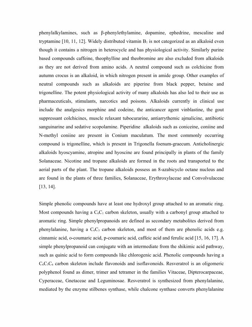
phenylalkylamines, such as β-phenylethylamine, dopamine, ephedrine, mescaline and
tryptamine [10, 11, 12]. Widely distributed vitamin B1 is not categorized as an alkaloid even
though it contains a nitrogen in heterocycle and has physiological activity. Similarly purine
based compounds caffeine, theophylline and theobromine are also excluded from alkaloids
as they are not derived from amino acids. A neutral compound such as colchicine from
autumn crocus is an alkaloid, in which nitrogen present in amide group. Other examples of
neutral compounds such as alkaloids are piperine from black pepper, betaine and
trigonelline. The potent physiological activity of many alkaloids has also led to their use as
pharmaceuticals, stimulants, narcotics and poisons. Alkaloids currently in clinical use
include the analgesics morphine and codeine, the anticancer agent vinblastine, the gout
suppressant colchicines, muscle relaxant tubocurarine, antiarrythemic ajmalicine, antibiotic
sanguinarine and sedative scopolamine. Piperidine alkaloids such as coniceine, coniine and
N-methyl coniine are present in Conium maculatum. The most commonly occurring
compound is trigonelline, which is present in Trigonella foenum-graecum. Anticholinergic
alkaloids hyoscyamine, atropine and hyoscine are found principally in plants of the family
Solanaceae. Nicotine and tropane alkaloids are formed in the roots and transported to the
aerial parts of the plant. The tropane alkaloids possess an 8-azabicyclo octane nucleus and
are found in the plants of three families, Solanaceae, Erythroxylaceae and Convolvulaceae
[13, 14].
Simple phenolic compounds have at least one hydroxyl group attached to an aromatic ring.
Most compounds having a C6C1 carbon skeleton, usually with a carbonyl group attached to
aromatic ring. Simple phenylpropanoids are defined as secondary metabolites derived from
phenylalanine, having a C6C3 carbon skeleton, and most of them are phenolic acids e.g.
cinnamic acid, o-coumaric acid, p-coumaric acid, caffeic acid and ferulic acid [15, 16, 17]. A
simple phenylpropanoid can conjugate with an intermediate from the shikimic acid pathway,
such as quinic acid to form compounds like chlorogenic acid. Phenolic compounds having a
C6C3C6 carbon skeleton include flavonoids and isoflavonoids. Resveratrol is an oligomeric
polyphenol found as dimer, trimer and tetramer in the families Vitaceae, Dipterocarpaceae,
Cyperaceae, Gnetaceae and Leguminosae. Resveratrol is synthesized from phenylalanine,
mediated by the enzyme stilbenes synthase, while chalcone synthase converts phenylalanine
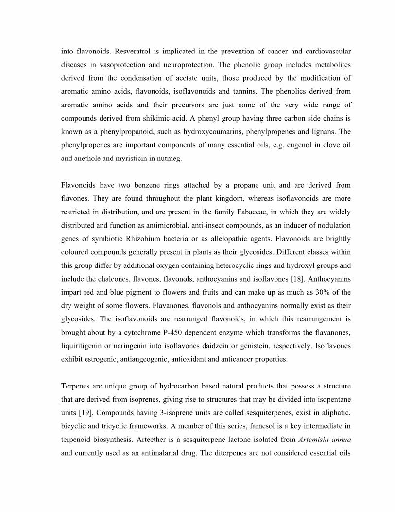
into flavonoids. Resveratrol is implicated in the prevention of cancer and cardiovascular
diseases in vasoprotection and neuroprotection. The phenolic group includes metabolites
derived from the condensation of acetate units, those produced by the modification of
aromatic amino acids, flavonoids, isoflavonoids and tannins. The phenolics derived from
aromatic amino acids and their precursors are just some of the very wide range of
compounds derived from shikimic acid. A phenyl group having three carbon side chains is
known as a phenylpropanoid, such as hydroxycoumarins, phenylpropenes and lignans. The
phenylpropenes are important components of many essential oils, e.g. eugenol in clove oil
and anethole and myristicin in nutmeg.
Flavonoids have two benzene rings attached by a propane unit and are derived from
flavones. They are found throughout the plant kingdom, whereas isoflavonoids are more
restricted in distribution, and are present in the family Fabaceae, in which they are widely
distributed and function as antimicrobial, anti-insect compounds, as an inducer of nodulation
genes of symbiotic Rhizobium bacteria or as allelopathic agents. Flavonoids are brightly
coloured compounds generally present in plants as their glycosides. Different classes within
this group differ by additional oxygen containing heterocyclic rings and hydroxyl groups and
include the chalcones, flavones, flavonols, anthocyanins and isoflavones [18]. Anthocyanins
impart red and blue pigment to flowers and fruits and can make up as much as 30% of the
dry weight of some flowers. Flavanones, flavonols and anthocyanins normally exist as their
glycosides. The isoflavonoids are rearranged flavonoids, in which this rearrangement is
brought about by a cytochrome P-450 dependent enzyme which transforms the flavanones,
liquiritigenin or naringenin into isoflavones daidzein or genistein, respectively. Isoflavones
exhibit estrogenic, antiangeogenic, antioxidant and anticancer properties.
Terpenes are unique group of hydrocarbon based natural products that possess a structure
that are derived from isoprenes, giving rise to structures that may be divided into isopentane
units [19]. Compounds having 3-isoprene units are called sesquiterpenes, exist in aliphatic,
bicyclic and tricyclic frameworks. A member of this series, farnesol is a key intermediate in
terpenoid biosynthesis. Arteether is a sesquiterpene lactone isolated from Artemisia annua
and currently used as an antimalarial drug. The diterpenes are not considered essential oils
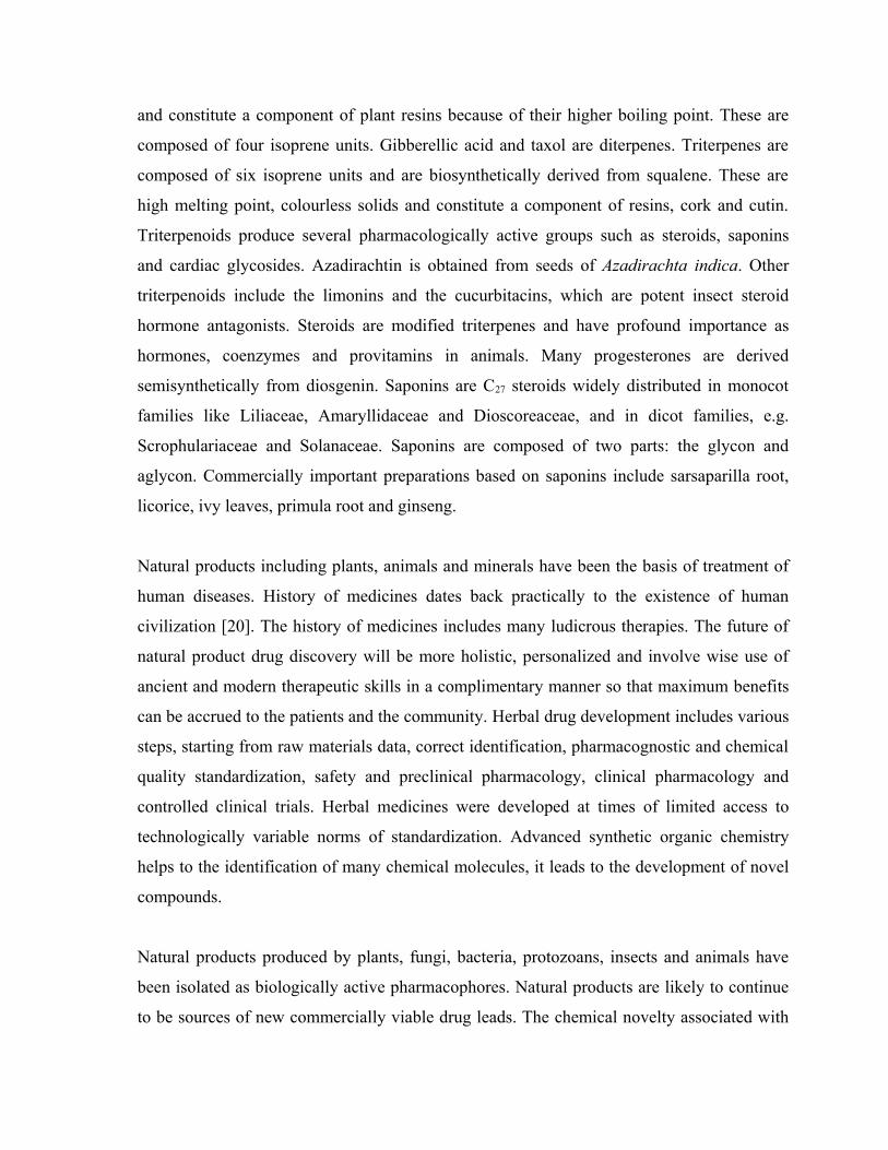
and constitute a component of plant resins because of their higher boiling point. These are
composed of four isoprene units. Gibberellic acid and taxol are diterpenes. Triterpenes are
composed of six isoprene units and are biosynthetically derived from squalene. These are
high melting point, colourless solids and constitute a component of resins, cork and cutin.
Triterpenoids produce several pharmacologically active groups such as steroids, saponins
and cardiac glycosides. Azadirachtin is obtained from seeds of Azadirachta indica. Other
triterpenoids include the limonins and the cucurbitacins, which are potent insect steroid
hormone antagonists. Steroids are modified triterpenes and have profound importance as
hormones, coenzymes and provitamins in animals. Many progesterones are derived
semisynthetically from diosgenin. Saponins are C27 steroids widely distributed in monocot
families like Liliaceae, Amaryllidaceae and Dioscoreaceae, and in dicot families, e.g.
Scrophulariaceae and Solanaceae. Saponins are composed of two parts: the glycon and
aglycon. Commercially important preparations based on saponins include sarsaparilla root,
licorice, ivy leaves, primula root and ginseng.
Natural products including plants, animals and minerals have been the basis of treatment of
human diseases. History of medicines dates back practically to the existence of human
civilization [20]. The history of medicines includes many ludicrous therapies. The future of
natural product drug discovery will be more holistic, personalized and involve wise use of
ancient and modern therapeutic skills in a complimentary manner so that maximum benefits
can be accrued to the patients and the community. Herbal drug development includes various
steps, starting from raw materials data, correct identification, pharmacognostic and chemical
quality standardization, safety and preclinical pharmacology, clinical pharmacology and
controlled clinical trials. Herbal medicines were developed at times of limited access to
technologically variable norms of standardization. Advanced synthetic organic chemistry
helps to the identification of many chemical molecules, it leads to the development of novel
compounds.
Natural products produced by plants, fungi, bacteria, protozoans, insects and animals have
been isolated as biologically active pharmacophores. Natural products are likely to continue
to be sources of new commercially viable drug leads. The chemical novelty associated with
natural products is higher than that of any other source. Natural products are traditional,
empirical and molecular [21]. The traditional approach makes the use of material that has
been found by trial and error over many years in different cultures and systems of medicines.
Examples include drugs such as morphine, quinine and ephedrine. The empirical approach
builds on an understanding of a relevant physiological process and develop therapeutic agent
from a naturally occurring lead molecule. Examples include tubocurarine and other muscle
relaxants, propranolol and other β-adrenergic antagonists, cimetidine and H2 receptor
antagonist. The molecular approach is based on the availability or understanding of a
molecular target for the medicinal agent. The development of molecular biological
techniques and advances in genomics, the majority of drug discovery is based on the
molecular approach [22].
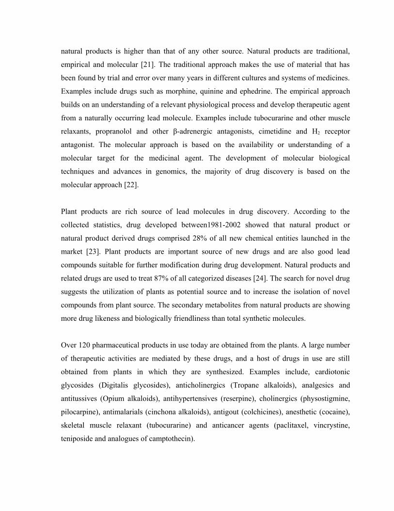
Plant products are rich source of lead molecules in drug discovery. According to the
collected statistics, drug developed between1981-2002 showed that natural product or
natural product derived drugs comprised 28% of all new chemical entities launched in the
market [23]. Plant products are important source of new drugs and are also good lead
compounds suitable for further modification during drug development. Natural products and
related drugs are used to treat 87% of all categorized diseases [24]. The search for novel drug
suggests the utilization of plants as potential source and to increase the isolation of novel
compounds from plant source. The secondary metabolites from natural products are showing
more drug likeness and biologically friendliness than total synthetic molecules.
Over 120 pharmaceutical products in use today are obtained from the plants. A large number
of therapeutic activities are mediated by these drugs, and a host of drugs in use are still
obtained from plants in which they are synthesized. Examples include, cardiotonic
glycosides (Digitalis glycosides), anticholinergics (Tropane alkaloids), analgesics and
antitussives (Opium alkaloids), antihypertensives (reserpine), cholinergics (physostigmine,
pilocarpine), antimalarials (cinchona alkaloids), antigout (colchicines), anesthetic (cocaine),
skeletal muscle relaxant (tubocurarine) and anticancer agents (paclitaxel, vincrystine,
teniposide and analogues of camptothecin).
Analysis of the number and sources of anticancer and anti-infective agents, reviewed mainly
in Annual Reports of Medicinal Chemistry from 1984 to 1995, indicates that over 60% of the
approved drugs and pre-NDA candidates (for the period 1989-1995), excluding biologics,
developed in these disease areas are of natural origin. According to Newmann et al., 2003,
during the period 1981-2002 a vast majority of New Chemical Entities is from natural
products source. Thus natural products have been playing an invaluable role in the drug
discovery process, particularly in the areas of diabetes, cancer and infectious diseases.
Plants have thus been a prime source of highly effective conventional drugs for the treatment
of diabetes. While the actual compounds isolated from plants frequently may not serve as
drugs, they provide leads for the development of potential novel agents. As new technologies
are developed, some of the agents which failed earlier in clinical studies are now stimulating
renewed interest. The appreciation of the significance of natural products as sources for
structurally novel and mechanistically unique drugs and the presence of an enormous
biodiversity of India, prompted the writer’s interest in evaluating the traditional medicinal
plants for their antioxidant and antidiabetic properties.
The chemical, pharmacological and clinical studies of the traditional medicines, which were
derived from plants are the most early medicines such as aspirin, digitoxin, morphine,
quinine and pilocarpine. High-throughput screening and mechanism based screening has
become mainstay in drug discovery. The mechanism based screening methods included
clavulanic acid, mevastatin and amoxicillin [25]. Natural products are source of new drugs
for many diseases and natural product derived drugs are well represented in the top 35
worldwide selling ethical drug sales of 2000, 2001 and 2002. The percentage of natural
product derived drugs was 40% in 2000 and remained approximately constant at 24% in
2001 and 26% in 2002. Natural products have historically provided many novel drug leads.
The natural product is extracted from the source, concentrated, fractionated and purified,
yielding essentially a single biologically active compound. Determination of the molecular
formula is done by high resolution mass spectrometry on microgram quantities of material.
Combining the tools of high resolution mass spectrometry with two-dimensional NMR
spectroscopy allows structure determination on milligram amount of compound in hours or
days [26].
1.2 Herbal Medicine
Man has been using herbs and plant products for combating diseases since times
immemorial. The Indian subcontinent is enriched by a variety of flora both aromatic and
medicinal plants. This is due to the wide diversity of climatic conditions of India. Numerous
types of herbs have been well recognized and catalogued by botanists from Himalaya to
Kanyakumari. This extensive flora has been utilized as a source of many drugs in the Indian
traditional system of medicine [27].
The WHO is actively encouraging the developing countries to use herbal medicine which
they have been traditionally used for centuries. There are 3000 plants have been identified in
the forests of India which can be used as medicine. The active ingredients from these plants
are worth Rs 2000 crores in the US market. The science of medicine developed around these
plants had curative properties. A continued search for medicinal plants during the last several
centuries has given rise to long list of plants which are of use in the treatment of diseases and
for promoting health. Drugs used in medicine today are either obtained from nature or are of
synthetic origin. Natural drugs are obtained from plants, animals, microbes or minerals. The
drugs obtained from plants and animals are called drugs of biological origin and produced in
the living cells of plants or animals [28].
There are 6000 plant constituents have been isolated and studied. The plants are
inexhaustible source of medicine, remains incompletely explored. This unexplored world
provides the most challenging aspects of pharmaceutical and medical science to scientists in
search for new and more potent drugs with negligible side effects. During the last few
decades, tremendous progress has been made in the study of phytochemicals.
Plants have been one of the important source of medicine since the dawn of human
civilization. The Chinese drug Mahung was in use for over 5000 years for the treatment of
different types of fever and respiratory disorders. Cinchona was in use in Peru in 1825 for
controlling malaria. The tremendous development in the field of synthetic drugs and
antibiotics during 21st century, plants still contribute one of the major sources of drugs in
modern and traditional medicine throughout the world. One-third of the world’s population
treat themselves with traditional medicines. Some of the compounds now commonly used in
medicine were isolated from plant sources and used in the 19th century. Examples are
morphine, quinine, atropine, papaverine, cocaine, digitoxine and pilocarpine. Examples of
some important compounds isolated in 20th century include ergotamine, labeline, digoxine,
reserpine, tubocurarine, diosgenin, vincrystine and vinblastine. Plants are the important
source of a number of well established and important source of drugs. They are also source
of chemical intermediates needed for the production of drugs [29].
Before independence of India, the production of plant based drugs in India was confined
mainly to cinchona, opium alkaloids, galanicals and tinctures. In the last three decades, bulk
production of plant drugs has become an important aspect of the Indian pharmaceutical
industry. Some of the drugs which are manufactured today include morphine, codeine,
papaverine, thebaine, emetine, quinine, quinidine, digoxine, caffeine, hyoscine,
hyoscyamine, atropine, xanthotoxin, sennosides, colchicines, berberine, vinblastine,
vincrystine, ergot alkaloids, papaine, nicotine, strychnine, brucine and pyrethroids.
In India, there are about 20 well recognized manufacturers of herbal drugs, 140 medium or
small scale manufacturers and about 1200 licensed small manufacturers on record, in
addition to many vaidyas having small manufacturing facilities. The estimated current annual
production of herbal drugs is around Rs 100 crores. The demand for herbal remedies is ever-
increasing. There are 1650 herbal formulations in the Indian market and 540 major plants
involved in their formulations. During the last two decades, over 3000 plants have been
screened in India for their biological activities. As a result, a number of new drugs have been
introduced in clinical practice and some are in advance stages of clinical development. There
are well documented scientific data on a good number of medicinal plants that have been
investigated.
Herbal medicines are the use of plants and plant extracts as medicines. In 2001 researchers
identified122 compounds used in medicine which were derived from ethnomedical plant
sources, 80% of these compounds were used in traditional ethnomedical use. Plants have
evolved the ability to synthesize chemical compounds that help them to defend against attack
from a wide variety of predators such as insects, fungi and herbivorous mammals. Some of
these compounds being toxic to plant predators have beneficial effect when used to treat
human diseases. People on all continents have used hundreds to thousands of indigenous
plants for treatment of ailments since prehistoric times. Medicinal herbs were found in the
personal effects of Otzi the iceman, whose body was frozen in the Otztal Alps for more than
5300 years [30].
In Indian Ayurveda medicine has used many herbs such as turmeric, pepper, garlic in 1900
B.C. Many other herbs and minerals used in ayurveda were described by Charaka and
Sushruta. Sushruta described 700 medicinal plants, 64 preparations from mineral sources and
57 preparations based on animal sources. Many of the pharmaceuticals currently available to
physicians have a long history of use as herbal remedies including opium, aspirin, digitalis
and quinine. The WHO estimates that 80 percent of the world’s population presently uses
herbal medicine for primary health care. Herbal medicines are available in the market from
health food stores without prescriptions and are widely used in India, China, USA and all
over the world. Herbal products are classified as dietary supplements and are marketed
pursuant to the dietary supplements Health and Education act of 1994. The herbal products
are regulated differently in other countries. In United Kingdom any product that is not
granted a licence as a medical product by Medicine Control Agency is treated as food and no
health claim or medical advice can be given on the label. Labeling of herbal products may
not actually reflect the content and adverse events or interactions attributed to specific herb
[31].
The commonly used many herbal medicines in their irregular, high doses or with other
medications in long term are toxic. The toxic effects of herbal medicines range from allergic
reactions to cardiovascular, hepatic, renal, neurological and dermatological toxic effects.
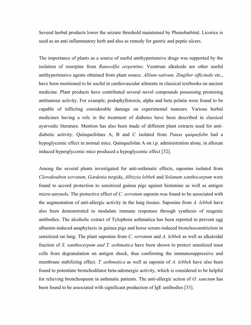
Several herbal products lower the seizure threshold maintained by Phenobarbital. Licorice is
used as an anti inflammatory herb and also as remedy for gastric and peptic ulcers.
The importance of plants as a source of useful antihypertensive drugs was supported by the
isolation of reserpine from Rauwolfia serpentine. Veratrum alkaloids are other useful
antihypertensive agents obtained from plant source. Allium sativum, Zingiber officinale etc.,
have been mentioned to be useful in cardiovascular ailments in classical textbooks on ancient
medicine. Plant products have contributed several novel compounds possessing promising
antitumour activity. For example, podophyllotoxin, alpha and beta pelatin were found to be
capable of inflicting considerable damage on experimental tumours. Various herbal
medicines having a role in the treatment of diabetes have been described in classical
ayurvedic literature. Mention has also been made of different plant extracts used for anti-
diabetic activity. Quinquefolans A, B and C isolated from Panax quiquefolin had a
hypoglycemic effect in normal mice. Quinquefolan A on i.p. administration alone, in alloxan
induced hyperglycemic mice produced a hypoglycemic effect [32].
Among the several plants investigated for anti-asthmatic effects, saponins isolated from
Clerodendron serratum, Gardenia turgida, Albizzia lebbek and Solanum xanthocarpum were
found to accord protection to sensitized guinea pigs against histamine as well as antigen
micro-aerosols. The protective effect of C. serratum saponin was found to be associated with
the augmentation of anti-allergic activity in the lung tissues. Saponins from A. lebbek have
also been demonstrated to modulate immune responses through synthesis of reagenic
antibodies. The alcoholic extract of Tylophora asthmatica has been reported to prevent egg
albumin-induced anaphylaxis in guinea pigs and horse serum-induced bronchoconstriction in
sensitized rat lung. The plant saponins from C. serratum and A. lebbek as well as alkaloidal
fraction of S. xanthocerpum and T. asthmatica have been shown to protect sensitized mast
cells from degranulation on antigen shock, thus confirming the immunosuppressive and
membrane stabilizing effect. T. asthmatica as well as saponin of A. lebbek have also been
found to potentiate bronchodilator beta-adrenergic activity, which is considered to be helpful
for relieving bronchospasm in asthmatic patients. The anti-allergic action of O. sanctum has
been found to be associated with significant production of IgE antibodies [33].
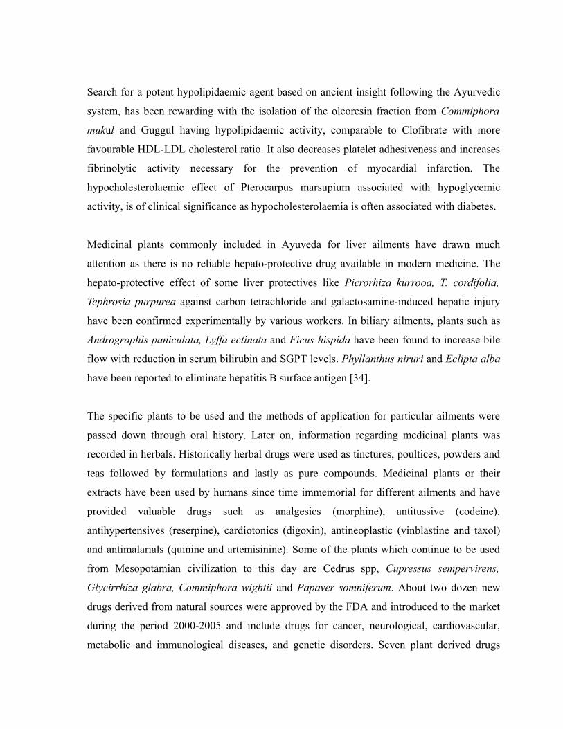
Search for a potent hypolipidaemic agent based on ancient insight following the Ayurvedic
system, has been rewarding with the isolation of the oleoresin fraction from Commiphora
mukul and Guggul having hypolipidaemic activity, comparable to Clofibrate with more
favourable HDL-LDL cholesterol ratio. It also decreases platelet adhesiveness and increases
fibrinolytic activity necessary for the prevention of myocardial infarction. The
hypocholesterolaemic effect of Pterocarpus marsupium associated with hypoglycemic
activity, is of clinical significance as hypocholesterolaemia is often associated with diabetes.
Medicinal plants commonly included in Ayuveda for liver ailments have drawn much
attention as there is no reliable hepato-protective drug available in modern medicine. The
hepato-protective effect of some liver protectives like Picrorhiza kurrooa, T. cordifolia,
Tephrosia purpurea against carbon tetrachloride and galactosamine-induced hepatic injury
have been confirmed experimentally by various workers. In biliary ailments, plants such as
Andrographis paniculata, Lyffa ectinata and Ficus hispida have been found to increase bile
flow with reduction in serum bilirubin and SGPT levels. Phyllanthus niruri and Eclipta alba
have been reported to eliminate hepatitis B surface antigen [34].
The specific plants to be used and the methods of application for particular ailments were
passed down through oral history. Later on, information regarding medicinal plants was
recorded in herbals. Historically herbal drugs were used as tinctures, poultices, powders and
teas followed by formulations and lastly as pure compounds. Medicinal plants or their
extracts have been used by humans since time immemorial for different ailments and have
provided valuable drugs such as analgesics (morphine), antitussive (codeine),
antihypertensives (reserpine), cardiotonics (digoxin), antineoplastic (vinblastine and taxol)
and antimalarials (quinine and artemisinine). Some of the plants which continue to be used
from Mesopotamian civilization to this day are Cedrus spp, Cupressus sempervirens,
Glycirrhiza glabra, Commiphora wightii and Papaver somniferum. About two dozen new
drugs derived from natural sources were approved by the FDA and introduced to the market
during the period 2000-2005 and include drugs for cancer, neurological, cardiovascular,
metabolic and immunological diseases, and genetic disorders. Seven plant derived drugs
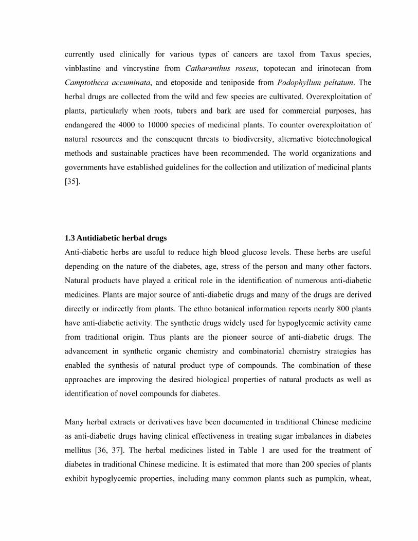
currently used clinically for various types of cancers are taxol from Taxus species,
vinblastine and vincrystine from Catharanthus roseus, topotecan and irinotecan from
Camptotheca accuminata, and etoposide and teniposide from Podophyllum peltatum. The
herbal drugs are collected from the wild and few species are cultivated. Overexploitation of
plants, particularly when roots, tubers and bark are used for commercial purposes, has
endangered the 4000 to 10000 species of medicinal plants. To counter overexploitation of
natural resources and the consequent threats to biodiversity, alternative biotechnological
methods and sustainable practices have been recommended. The world organizations and
governments have established guidelines for the collection and utilization of medicinal plants
[35].
1.3 Antidiabetic herbal drugs
Anti-diabetic herbs are useful to reduce high blood glucose levels. These herbs are useful
depending on the nature of the diabetes, age, stress of the person and many other factors.
Natural products have played a critical role in the identification of numerous anti-diabetic
medicines. Plants are major source of anti-diabetic drugs and many of the drugs are derived
directly or indirectly from plants. The ethno botanical information reports nearly 800 plants
have anti-diabetic activity. The synthetic drugs widely used for hypoglycemic activity came
from traditional origin. Thus plants are the pioneer source of anti-diabetic drugs. The
advancement in synthetic organic chemistry and combinatorial chemistry strategies has
enabled the synthesis of natural product type of compounds. The combination of these
approaches are improving the desired biological properties of natural products as well as
identification of novel compounds for diabetes.
Many herbal extracts or derivatives have been documented in traditional Chinese medicine
as anti-diabetic drugs having clinical effectiveness in treating sugar imbalances in diabetes
mellitus [36, 37]. The herbal medicines listed in Table 1 are used for the treatment of
diabetes in traditional Chinese medicine. It is estimated that more than 200 species of plants
exhibit hypoglycemic properties, including many common plants such as pumpkin, wheat,
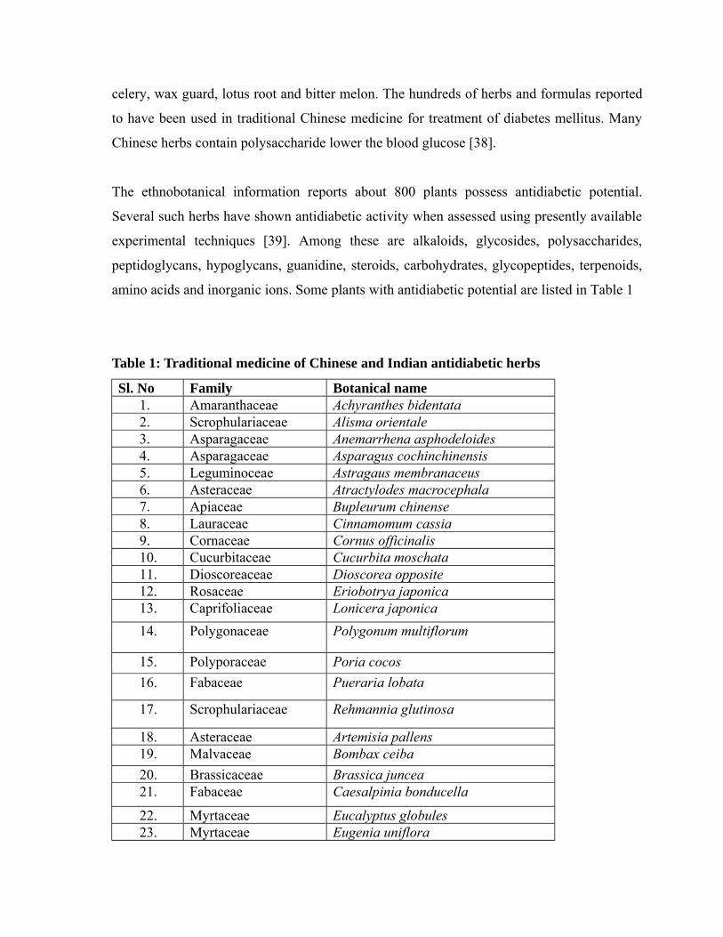
celery, wax guard, lotus root and bitter melon. The hundreds of herbs and formulas reported
to have been used in traditional Chinese medicine for treatment of diabetes mellitus. Many
Chinese herbs contain polysaccharide lower the blood glucose [38].
The ethnobotanical information reports about 800 plants possess antidiabetic potential.
Several such herbs have shown antidiabetic activity when assessed using presently available
experimental techniques [39]. Among these are alkaloids, glycosides, polysaccharides,
peptidoglycans, hypoglycans, guanidine, steroids, carbohydrates, glycopeptides, terpenoids,
amino acids and inorganic ions. Some plants with antidiabetic potential are listed in Table 1
Table 1: Traditional medicine of Chinese and Indian antidiabetic herbs
Sl. No Family Botanical name1. Amaranthaceae Achyranthes bidentata2. Scrophulariaceae Alisma orientale3. Asparagaceae Anemarrhena asphodeloides4. Asparagaceae Asparagus cochinchinensis5. Leguminoceae Astragaus membranaceus6. Asteraceae Atractylodes macrocephala7. Apiaceae Bupleurum chinense8. Lauraceae Cinnamomum cassia9. Cornaceae Cornus officinalis10. Cucurbitaceae Cucurbita moschata11. Dioscoreaceae Dioscorea opposite12. Rosaceae Eriobotrya japonica13. Caprifoliaceae Lonicera japonica14. Polygonaceae Polygonum multiflorum
15. Polyporaceae Poria cocos16. Fabaceae Pueraria lobata
17. Scrophulariaceae Rehmannia glutinosa
18. Asteraceae Artemisia pallens19. Malvaceae Bombax ceiba20. Brassicaceae Brassica juncea21. Fabaceae Caesalpinia bonducella
22. Myrtaceae Eucalyptus globules23. Myrtaceae Eugenia uniflora
24. Asclepiadaceae Gymnema sylvestre25. Anacardiaceae Mangifera indica
26. Melastomataceae Memecylon umbellatum27. Fabaceae Mucuna pruriens
The indigenous diet may not be useful in lowering the blood glucose to the same extent as
insulin and other hypoglycemic agent. But it has some other influences, which may be useful
for the management of the disease and its complications. The juices of bitter gourd,
decoction of chirata, neem leaves, betel leaves, fenugreek seeds and sada bahar flowers
achieve 10-20% lowering of blood glucose. It is useful as supplement to other therapies.
Vegetables have antidiabetic potency. Vegetables such as cabbage, capsicum, green leafy
vegetables, beans and tubers have shown the hypoglycemic effect in both experimental
animals and humans.
1.4 Free radicals
A free radical is an atom or a molecule that contains one or more unpaired electrons [40].
Unpaired electrons alter the chemical reactivity of an atom or molecule; usually make it
more reactive than the corresponding non-radical. The actual chemical reactivity of radicals,
however, varies enormously. The hydrogen radical, which contain one proton and one
electron, is the simplest free radical.
Free radicals in the body are generated by multiple mechanisms and are often initiated by
removal of an H atom from other molecules. Living organisms are exposed to
electromagnetic radiation from the environment, both natural and from man made sources.
Low wavelength electromagnetic radiation (i.e. gamma rays) can split water in the body to
generate hydroxyl radicals(OH). Hydroxyl radical has a very short in vivo half-life, reacting
at its site of formation, usually leaving behind a legacy of free radical chain reactions [41].
The body, through metabolic process, makes an oxygen radical called superoxide (O2),
where the unpaired electron is located on oxygen. Superoxide is made by adding one
electron to the oxygen molecule, and is generally poorly reactive [42]. Many molecules in
the body react directly with oxygen to make superoxide, including the catecholamines,
tetrahydrofolate and some constituents of mitochondrial and other electron transport chains.
Even when this mode of superoxide generation is not available, activated phagocytes
generate large amounts of superoxide as part of the mechanism by which foreign organisms
are killed. During chronic inflammation, this normal protective mechanism may become
damaging.
Another physiological free radical is nitric oxide (NO), which is made by vascular
endothelium as a relaxing factor [43]. Nitric oxide has many useful physiological functions,
but excess nitric oxide can be toxic. Neither superoxide nor nitric oxide is highly reactive
chemically, but under certain circumstances they can generate more reactive toxic products.
When oxygen is reduced in the electron transport chain, oxygen derived free radical
intermediates are formed. The O2 and H2O2 intermediates can escape from the system, and in
the presence of transition metal ions form the more reactive hydroxyl radicals. While O2 are
toxic to cells, the high reactivity of OH and O2 renders these activated forms most cytotoxic
due to deleterious peroxidation reactions with lipids, proteins and DNA. Lipid peroxidation
is an example of this oxidative damage [44]. Free radicals may attack polyunsaturated fatty
acids within membranes, forming peroxyl radicals. These newly formed free radicals can
then attack adjacent fatty acids within membranes causing a chain reaction of lipid
peroxidation. The lipid hydroperoxide end products are also harmful, and may be responsible
for some of the overall effect, which can lead to tissue and organ damage.
Antioxidants may be defined as radical scavengers which protect the human body against
free radicals that may cause pathological conditions such as ischemia, anaemia, asthma,
arthritis, inflammation, neurodegeneration, parkinson’s disease, mongolism, ageing and
dementias. Flavonoids and flavones are widely distributed secondary metabolites with
antioxidant and antiradical properties [45]. Reactive oxygen species (ROS) including
superoxide radicals, hydroxyl radicals, singlet oxygen and hydrogen peroxide are often
generated as by products of biological reaction or from exogenous factors. In vivo, some of
these ROS play an important role in cell metabolism including energy production,
phagocytosis and intercellular signaling. These ROS produced by sunlight, ultraviolet light,
ionizing radiation, chemical reactions and metabolic process have a wide variety of
pathological effects such as DNA damage, carcinogenesis and various degenerative disorders
such as cardiovascular diseases, ageing and neurodegenerative diseases [46]. A potent broad
spectrum scavenger of these species may serve as a possible preventive intervention for free
radical mediated cellular damage and diseases. Recent studies have shown that a number of
plant products including polyphenols, terpenes and various plant extracts exerted an
antioxidant action. Several medicinal plants have been extensively used in the Indian
traditional system of medicine for treatment of number of diseases. Some of these plants
have shown potent antioxidant activity.
Oxidative stress is exerted by all peroxides, which can damage cells and tissues, or directly
through their more reactive breakdown products such as malonaldehyde and
hydroxynonenals [47]. Moreover, metals such as iron and copper interact with free radicals
which contribute to the propagation of the lipid peroxidation chain reaction. It is evident
then that a single initiating event, caused by a prooxidant, may cascade into a widespread
chain reaction that produces many deleterious products in concentrations greater than that of
the initiator. This is exemplified by the fact that thousands of molecules may be destroyed by
a lipid peroxidation chain reaction initiated by a single radical. It is imperative that in order
to prevent this vicious chain reaction, the O2 radical cascade to O2 and H2O2 must be
attenuated, and the peroxides converted to innocuous metabolites. All aerobic organisms
therefore possess elaborate defense mechanisms to prevent the formation of toxic forms of
oxygen and to remove any peroxides formed.
1.5 Oxidative stress and human disease
Reactive oxygen species (ROS) such as superoxide anions, hydrogen peroxide, and
hydroxyl, nitric oxide and peroxy nitrite radicals, play an important role in the pathogenesis
of various diseases. The constant attack by oxyradicals and reactive oxygen species (ROS)
contributes to both the initiation and the progression of many major diseases. The oxidation
of lipid, DNA, proteins, carbohydrates and other biological molecules by toxic ROS may
cause mutation and damage to cells or tissues. The last decade has yielded considerable
evidence that implicates oxidative stress as a factor in the etiology and progression of a
spectrum of diseases, which include atherosclerosis, cancer, eye disorders, Parkinson
disease, diabetes, gastric ulcers, liver diseases etc. The mechanism may differ in specific
diseases, but generation of ROS is found in all cases [48].
1.6 Antioxidant defense system
All aerobic forms of life maintain elaborate defense systems known as antioxidant systems to
protect the body against free radical damage. The body needs to strike the right balance
between the number of free radicals generated and the defense and repair mechanism
available. The current view of cellular oxidant defenses can be categorized into primary and
secondary defense systems [49, 50]. The primary defenses consists of the broadly studied
antioxidant compounds, such as α-tocopherol, ascorbic acid, β-carotene and uric acid, along
with variety of antioxidant enzymes, where superoxide dimutase (SOD), catalase (CAT) and
glutathione peroxidase (GSH-Px) are notable examples.
Secondary defenses are predominantly a series of enzyme systems that act to repair or
eliminate molecules or cell components that were damaged by oxidants or free radical
reactions, which escape the primary antioxidant defense [51].
1.7 Role of medicinal plants as antioxidants
The widespread use of traditional herbs and medicinal plants has been traced to the
occurrence of natural products with medicinal properties. In recent years, the traditional
medicine, the world has revalued by an extensive activity of research on different plant
species and their therapeutic principles. Various medicinal properties have been ascribed to
natural herbs and medicinal plants constitute one of the main source of new pharmaceuticals
and healthcare products. Many studies have been performed to identify antioxidant
compounds with pharmacological activity with limited toxicity. A whole range of plant
derived dietary supplements, phytochemicals and pro-vitamins that assist in maintaining
good health and combating disease are now being described as functional foods,
nutriceuticals and nutraceuticals [52].
Potential sources of antioxidant compounds have been searched in many types of plant
materials such as fruits, seeds and leaves etc. As plants produce a lot of antioxidants to
control the oxidative stress caused by sunbeams and oxygen, they can represent a source of
new compounds with antioxidant activity. It has been observed that phytochemicals like
tannic acid, flavonoids, tocopherol, curcumin, ascorbate, carotenoids, polyphenols, etc. were
reported to have potent antioxidant properties [53].
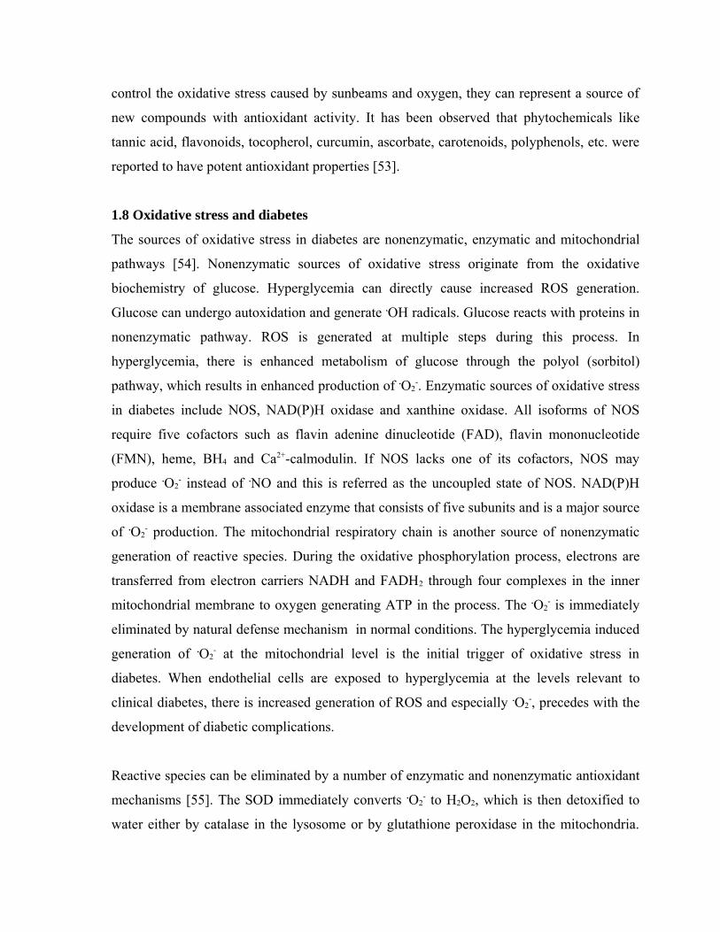
1.8 Oxidative stress and diabetes
The sources of oxidative stress in diabetes are nonenzymatic, enzymatic and mitochondrial
pathways [54]. Nonenzymatic sources of oxidative stress originate from the oxidative
biochemistry of glucose. Hyperglycemia can directly cause increased ROS generation.
Glucose can undergo autoxidation and generate .OH radicals. Glucose reacts with proteins in
nonenzymatic pathway. ROS is generated at multiple steps during this process. In
hyperglycemia, there is enhanced metabolism of glucose through the polyol (sorbitol)
pathway, which results in enhanced production of .O2-. Enzymatic sources of oxidative stress
in diabetes include NOS, NAD(P)H oxidase and xanthine oxidase. All isoforms of NOS
require five cofactors such as flavin adenine dinucleotide (FAD), flavin mononucleotide
(FMN), heme, BH4 and Ca2+-calmodulin. If NOS lacks one of its cofactors, NOS may
produce .O2- instead of .NO and this is referred as the uncoupled state of NOS. NAD(P)H
oxidase is a membrane associated enzyme that consists of five subunits and is a major source
of .O2- production. The mitochondrial respiratory chain is another source of nonenzymatic
generation of reactive species. During the oxidative phosphorylation process, electrons are
transferred from electron carriers NADH and FADH2 through four complexes in the inner
mitochondrial membrane to oxygen generating ATP in the process. The .O2- is immediately
eliminated by natural defense mechanism in normal conditions. The hyperglycemia induced
generation of .O2- at the mitochondrial level is the initial trigger of oxidative stress in
diabetes. When endothelial cells are exposed to hyperglycemia at the levels relevant to
clinical diabetes, there is increased generation of ROS and especially .O2-, precedes with the
development of diabetic complications.
Reactive species can be eliminated by a number of enzymatic and nonenzymatic antioxidant
mechanisms [55]. The SOD immediately converts .O2- to H2O2, which is then detoxified to
water either by catalase in the lysosome or by glutathione peroxidase in the mitochondria.
The gluatathione reductase acts as hydrogen donor during the elimination of H2O2.
Nonenzymatic antioxidants include vitamin A, C and E, glutathione, α-lipoic acid,
carotenoid, trace elements like copper, zinc and selenium, coenzyme Q10 (CoQ10) and
cofactors like folic acid, uric acid, albumin and vitamin B1, B2, B6 and B12. Glutathione
(GSH) acts as a direct scavenger and co-substrate for GSH peroxidase. Vitamin E is a fat
soluble vitamin that prevents lipid peroxidation. CoQ10 is a lipid soluble antioxidant, in
higher concentrations it scavenges .O2- and improves endothelial dysfunction in diabetes.
Vitamin C increases NO production in endothelial cells by stabilizing NOS cofactor BH4. α-
Lipoic acid is reduced to dihydrolipoate. Dihydrolipoate is able to regenerate antioxidants
such as vitamin C, vitamin E and reduced glutathione through redox cycling.
Free radicals and other reactive species play an important role in many human diseases.
Plants have long been regarded as having considerable health benefits, due to their main
antioxidant compounds [56]. In living system, free radicals are generated as part of the
body’s normal metabolic process. The free radical chain reactions are usually produced in the
mitochondrial respiratory chain, liver mixed function oxidases, through xanthine oxidase
activity, atmospheric pollutants and from transitional metal catalysts, drugs and xenobiotics.
Oxygen free radical can initiate peroxidation of lipids, which in turn stimulates glycation of
proteins, inactivation of enzymes and alteration in the structure and function of collagen
basement and other membranes and play a role in the long term complication of diabetes.
Diabetes is a risk factor for cardiovascular disease. The microvascular complications of
diabetes include nephropathy and retinopathy, macrovascular complications are coronary
artery disease, cerebrovascular disease and peripheral vascular disease are the leading cause
of death in the diabetes [57]. The control of blood glucose is effective in reducing the clinical
complications. The oxidative stress mediated mainly by hyperglycemia induced generation
of free radicals. The antioxidants treatments are effective in reducing diabetic complications.
Several clinical trials investigated the effect of antioxidant vitamin E on the prevention of
diabetic complications. These clinical trials are failed to demonstrate relevant clinical
benefits of this antioxidant on cardiovascular disease. The negative results of the clinical
trials with antioxidants lead to focus on mechanism of oxidative stress in diabetes to develop
antioxidant therapy.
1.9 Diabetes mellitus
Diabetes mellitus is a chronic disease of metabolic disorder caused by deficiency in
production of insulin by the β-cells of pancreas. This results in increased concentration of
blood glucose. This uncontrolled hyperglycemia after long duration leads to retinopathy,
neuropathy, nephropathy, cardiovascular problems and damage to blood vessels [58, 59].
The blood glucose level in the human body is balanced by insulin and glucagon. The normal
blood sugar of human body should be between 70 mg/dl to 110 mg/dl at fasting state and
below 140 mg/dl at two hours after eating. If blood glucose level is less than 70 mg/dl is
termed as hypoglycemia and more than 110 mg/dl is termed as hyperglycemia.
Insulin deficiency is the major cause in Type-1 diabetes, in which pancreas stop producing
insulin. In Type-2 diabetes, the cause may be inefficient utilization of glucose by human
body cells [60, 61]. The Type-3 diabetes is termed as Gestational diabetes and it is due to
development of insulin resistance. Gestational diabetes affects the mother and the baby.
According to W.H.O estimate, by 2025, a total of 300 million of the worldwide population
will be affected by diabetes and W.H.O recommended to include traditional medicines in
primary healthcare centers of third world countries, where 80% of the population depend on
traditional medicines. The traditional medicines constitute the plant products and plant
derived products. The plant products, plant derived active principles and synthetic drugs are
used in the treatment of Type-2 diabetes.
The pathophysiology of all types of diabetes is related to the hormone insulin, which is
secreted by the beta cells of the pancreas [62, 63]. In a healthy person, insulin is produced in
response to the increased level of glucose in the bloodstream, and its major role is to control
glucose concentration in the blood. What insulin does is, allowing the body cells and tissues
to use glucose as a main energy source. Also, this hormone is responsible for conversion of
glucose to glycogen for storage in the muscles and liver cells. This way, sugar level is
maintained at a near stable amount.
In a diabetic person, there is an abnormal metabolism of insulin hormone. The actual reason
for this malfunction differs according to the type of diabetes. Whatever the cause is, the body
cells and tissues do not make use of glucose from the blood, resulting in elevated blood
glucose (a typical symptom of diabetes called hyperglycemia). This condition is also
exacerbated by the conversion of stored glycogen to glucose, i.e., increased hepatic glucose
production. Over a period of time, high glucose level in the bloodstream can lead to severe
complications, such as eye disorders, cardiovascular diseases, kidney damage, and nerve
problems.
In Type 1 diabetes, the pancreas cannot synthesize enough amounts of insulin as required by
the body. The pathophysiology of Type 1 diabetes mellitus suggests that it is an autoimmune
disease, wherein the body's own immune system generates secretion of substances that attack
the beta cells of the pancreas. Consequently, the pancreas secretes little or no insulin. Type 1
diabetes is more common among children and young adults (around 20 years). Since it is
common among young individuals and insulin hormone is used for treatment, Type 1 diabetes
is also referred to as Juvenile Diabetes or Insulin Dependent Diabetes Mellitus (IDDM).
In case of Type 2 diabetes mellitus, the insulin hormone secreted by the beta cells is normal
or slightly lower than the ideal amount. However, the body cells are not responding to insulin
as they do in a healthy person. Since the body cells and tissues are resistant to insulin, they do
not absorb glucose, instead it remains in the bloodstream. Thus, the Type 2 diabetes is also
characterized by elevated blood sugar. It is commonly manifested by middle-aged adults
(above 40 years). As insulin is not necessary for treatment of Type 2 diabetes, it is known as
Non-insulin Dependent Diabetes Mellitus (NIIDM).
The third type of diabetes is called Gestational diabetes. As the term clearly suggests, it is
exhibited by pregnant women. Over here, high level of blood glucose is caused by hormonal
fluctuations during pregnancy. Usually, the sugar concentration returns to normal after the
baby is born. However, there are also instances, in which it remains high even after childbirth.
This is an indication for increased risks of developing diabetes in the near future.
As already mentioned, the symptoms and effects of all the three forms of diabetes are similar
[64, 65]. The noticeable symptoms include increased thirst (polydipsia), increased urination
(polyuria), and increased appetite (polyphagia). Other diabetes signs and symptoms include
excessive fatigue, presence of sugar in the urine (glycosuria), body irritation, unexplained
weight loss, and dehydration. Elevated blood sugar and glycosuria are interrelated; when
sugar amount in the blood is abnormally high, the reabsorption by proximal convoluted
tubule is reduced, thereby retaining some glucose in the urine.
2. SCOPE, OBJECTIVES AND PLAN OF WORK
2.1 SCOPE OF THE WORK
Actiniopteris radiata is a desert fern belong to family Adiantaceae (Pteridaceae). It is a tiny
terrestrial fern, found throughout India and also in Burma, Sri Lanka, Afghanistan, Persia,
Arabia, Yemen, South Eastern Egypt, Tropical Africa, Australia and Madagascar. It is of
limited distribution, and in areas where it occurs, is restricted to depleted walls and rocky
crevices of steep slops of exposed hilly areas, up to the altitude of 1200 m. The term
Actiniopteris has its origin from the Greek aktis (ray) and pteris (fern); refers to the radiating
leaf segments. Its vernacular names include Mayursikha : Sanskrit; Mapursika :Bombay and
Peacock’s tail :English.
Adiantaceae family has cosmopolitan ferns, about 17 species occur in India, most of which
possess medicinal properties. The ferns are primarily plants of lower elevation, growing upto
600 m above sea level, but a few survive at higher elevations also. The plants of this family
are reported to contain kaemferol, quercetol, luteol, adiantone, isoadiantone, fernene, β-
sitosterol and quercetin. The plants are used as hypoglycaemic, hair tonic, in skin diseases,
leprosy and fever. An ointment prepared from fern is used as hair tonic. The decoction of the
plant is used to cure cough and cold.
The ethnomedical uses of this plant are anthelmintic, haemostatic, antileprotic, used in
dysentery, diabetes, skin diseases and fever. The reported biological activities of this plant
are analgesic, antihistaminic, antimicrobial, antifungal and antifertility activity. The plant
contain rutin, hentricontane, hentricontanol, β-sitosterol, β-sitosterol palmitate, unidentified
glucoside, glucose and fructose. The reported phytochemical work is less.
2.2 OBJECTIVES OF THE WORK
1. To select the plants based on their ethnomedical uses and preparation of their extracts.
2. To screen the extracts for in vitro antioxidant activity.
3. To screen the extracts for in vitro antidiabetic activity.
4. To screen the plant extract for in vivo antidiabetic activity
5. To isolate the chemical constituents from the plant extract and structure elucidation.
2.3 PLAN OF WORK
• Selection of the plant, authentication, the whole plant to be dried at room temperature.
• The coarsely powdered plant to be extracted with different solvents of increasing
polarity.
• Qualitative phytochemical analysis and quantitative phytochemical estimation of
extracts.
• Column chromatography of ethyl acetate extract.
• Fractionating the ethyl acetate extract.
• Evaluation of in vitro anti-diabetic activity by alpha glucosidase inhibition activity.
• Quantitative and qualitative estimation of ethyl acetate extract and fractions by
HPTLC.
• Characterisation of isolated compounds by melting point, UV, IR, NMR, and mass
spectrums.
• Evaluation of the extracts for in vitro antioxidant activity.
• Evaluation of the extracts for in vivo anti-diabetic activity.
3. PLANT PROFILE AND REVIEW OF LITERATURE
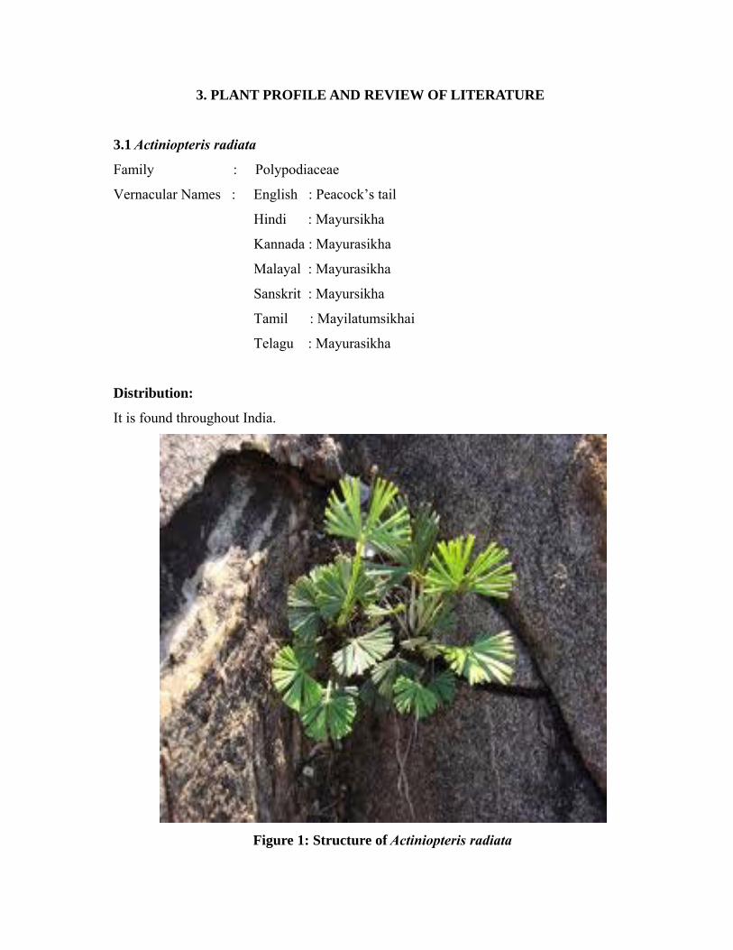
3.1 Actiniopteris radiata
Family : Polypodiaceae
Vernacular Names : English : Peacock’s tail
Hindi : Mayursikha
Kannada : Mayurasikha
Malayal : Mayurasikha
Sanskrit : Mayursikha
Tamil : Mayilatumsikhai
Telagu : Mayurasikha
Distribution:
It is found throughout India.
Figure 1: Structure of Actiniopteris radiata
Description:
A herbaceous miniature palm like fern upto 25 cm hight with densely tufted stipe. Fronds fan
like with numerous dichotomous segments which are rush like in texture, veins few,
subparallel with distinct midrib, segments of fertile frond longer than those of the barren one,
sori linear, elongate, submarginal.
Ethnomedical information:
The plant is bitter, astringent, anthelmintic, haemostatic, antileprotic and febrifuge. It is
useful in vitiated conditions of kapha and pitta, diarrhea, dysentery, helminthiasis,
haemoptysis, leprosy, skin diseases, diabetes and fever [66].
Chemical Constituents:
The plant contains rutin, hentriacontane, hentricontanol, β-sitosterol, β-sitosterol palmitate,
β-sitosterol-D-(+)-glycoside, an unidentified glycoside, glucose and fructose.
3.2 Phytochemical investigation and biological activity
The research papers have been collected for phytochemical investigation, in-vivo anti-
diabetic screening and in-vitro antioxidant activity for the selected plant Actiniopteris
radiata and related plants. Bambie, et al., have reported the gametophytic and sporophytic
generations of this plant [67]. Actiniopteris radiata is one of the apogamously developed
xerophytic ferns of Actiniopteridaceae. It has been worked out in detail regarding its anatomy
and morphology. The development of its gametophytes has also been studied up to the 8-
celled stage after which they did not grow on artificial medium. The spores are trilete with
slightly convex sides and rounded corners. The leasurae are long, crassimarginate with
undulate surface. Spores bear large, irregularly closely-set verucae like ridge with wavy
margins. They are yellowish to dark brown when mature. The average dimensions of the
spores are 49.39 × 54.83 µ. On germination the spore forms a densely chlorophyllous germ
filament composed of 3 to 8 short barrel shaped cells. The gametophytic and sporophytic
generations of actiniopteris radiata clearly indicate that this plant has adapted itself very well
to the xeric environment where it usually grows.
Bambie, et al., have reported the preliminary study of the chemical constitution of the plant
Actiniopteris radiata [68]. Dried stems and leaves of the plant (500 g) were extracted with
petroleum ether and ethanol respectively. The petroleum ether extract was concentrated
under reduced pressure to a green solid mass (10 g). It was put over an alumina column and
eluted successively with petroleum ether (40-60oC), petroleum ether: benzene (4:1), benzene
and benzene : chloroform (1:1). The first few fractions from petroleum ether on evaporation
gave a 20 mg of hentricontane. The fractions after elution with petroleum ether : benzene
(4:1) gave a 100 mg of β-sitosterol palmitate. The elutes from pure benzene on evaporation
gave 100 mg of hentricontol. The alcoholic extract of the plant was concentrated to one-tenth
of its original volume and was kept at 0oC for few days. A yellow crystalline substance was
accumulated at the bottom. This on repeated crystallization from methanol gave yellow
crystalline substance mp 190oC. It failed to give the test for steroids and flavanoids. It gave
positive response for Molisch’s test and blood red coloration with conc.H2SO4 indicates the
glycosidic nature of the compound.
Taneja, et al., have reported the isolation of compounds from petroleum ether and ethanol
extract and reported the presence of 3-hydroxy flavones in the ethanol extract [69].
Actiniopteris radiata was evaluated for analgesic activity using ethanolic and aqueous extract
by acetic acid induced writhing method and tail flick method [70]. Albino mice weigh 20-25
g were divided into four groups consisting of six animals. Group one served as negative
control, group second served as positive control (Pentazocine 5 mg/kg b.w ip), group third
received aqueous extract (300 mg/kg b.w ip) and group fourth received ethanolic extract (300
mg/kg b.w ip) of Actiniopteris radiata. The writhing movements were observed and counted
for a period of 15 minutes after acetic acid administration. The mean writhing scores in
control, extracts and pentazocine treated groups were calculated. All animals were
individually exposed to tail flick apparatus maintained at 55oC. The tail withdrawn from the
heat is taken as the end point. Cut off period of 10-12 sec is observed to prevent damage to
tail. The reaction time was noted from 0, 30, 60, 90, 120 and 180 minutes time interval. The
aqueous and ethanolic extracts shows significant analgesic activity in writhing method.
Whereas intraperitoneal administration of the aqueous and ethanolic extracts of Actiniopteris
radiata showed non significant change in the tail flick latency till 120 minutes.
Actiniopteris radiata was tested for in-vitro antihistaminic and anticholinergic activity [71].
Male wistar rats were sacrificed and a segment from ileum was dissected from the terminal
ileum and mounted in organ bath containing tyrode solution. A dose response curve for
histamine and acetylcholine was recorded in the following groups. Group 1- control
(Histamine and Ach), group 2- vehicle, group 3- test extract (2 mg/ml), group 4- test extract
(4 mg/ml), group 5 – test extract (10 mg/ml). The ethanolic extract of Actiniopteris radiata
shown significant antihistaminic and anticholinergic activity.
4. MATERIALS AND METHODS
4.1 Plant Material
The whole plant of Actiniopteris radiata was collected from Nilgiri district, Tamil Nadu,
India, in November 2007. The plant was identified by Dr. S. Rajan, Field Botanist, Survey of
Medicinal Plants and Collection Unit, Emerald, Nilgiri (Voucher No: 135). A voucher
specimen was deposited at Survey of Medicinal Plants and Collection Unit, Emerald, Nilgiri.
4.2 Materials
4.2.1 Instruments
Melting points were determined using a Lab India melting point apparatus. UV-Visible
spectrums were recorded using a Shimadzu UV-1700. IR spectrums were recorded on a
Shimadzu FTIR-8400s. 1H (500 MHz) and 13C (100 MHz) spectrums were recorded on a
BRUKER AV-400. EIMS was recorded by GC-MS on a P-POS/TOP MICRO, HITACHI.
ESIMS spectrums were recorded on a HCT-Ultra PTM discovery, BRUKER. ELISA reader
data recorded on a BIO - RAD 550.
4.2.2 Chemicals
2, 2 –diphenyl -1- picryl hydrazyl (DPPH) and 2, 21- azino-bis (3-ethylbenz-thiazoline-6-
sulfonic acid) diammonium salt (ABTS) were procured from Sigma-Aldrich, California,
USA. Rutin and p-nitroso dimethyl aniline (p-NDA) were procured from Acros Organics,
New Jersy, USA. Naphthyl ethylene diamine dihydrochloride (NEDD) was procured from
Roch – Light Ltd, Suffolk, UK. Nitro blue tetrazolium (NBT) was procured from S.D Fine
Chem Ltd, Biosar, India. Glibenclamide was procured from Inga labs Ltd, Mumbai, India.
Streptozotocin was procured from Hi media, Mumbai, India. All the other chemicals used
were of analytical grade.
4.3 Preparation of the plant extract
The plant was dried under shade for 7 days. The coarsely powdered plant material (500g)
was packed in soxhlet apparatus. The packed plant material was extracted successively with
petroleum ether, chloroform, ethyl acetate and ethanol for 18-20 hrs. These extracts were
filtered and dried under vacuum.
4.4 Preliminary phytochemical analysis of successive extracts of Actiniopteris
radiata
The qualitative chemical tests were carried out for successive extracts of Actiniopteris
radiata to identify the chemical constituents.
• Test for alkaloids
Mayer test
Dragendroff’s test
Wagner test
Hager test
• Test for saponins
Foam test
• Test for carbohydrates
Molisch test
Benedict test
• Test for glycosides
Borntrager test
Test for reducing sugar
• Test for steroids
Libermann Buchard test
• Test for fatty acids
Saponification test
• Test for flavanoids
Ferric chloride test
4.5 Physicochemical analysis
4.5.1 Ash value
Total ash
The powdered plant (3 g) was accurately weighed and spread in a silica crucible which was
previously ignited and weighed. The crucible was incinerated at a temperature not exceeding
450°C to make the powder free from carbon. The procedure was repeated to get a constant
weight. The percentage of a total ash was calculated with reference to the dry weight of the
powdered plant [72].
Acid insoluble ash
The acid insoluble ash was determined from the total ash. The total ash was boiled with 25
ml of 2 N HCl for 5 min. The insoluble ash was collected on an ash less filter paper and
washed with hot water. The insoluble ash was transferred to pre-weighed silica crucible,
ignited, cooled and weighed. The procedure was repeated to get a constant weight. The
percentage of an acid insoluble ash was calculated with reference the dry weight of the
powdered plant.
Water soluble ash
The water soluble ash was determined from the total ash. The total ash was boiled with 25
ml. of distilled water for 5 min. The insoluble ash was collected on an ash less filter paper
and washed with hot water. The insoluble ash was transferred to pre-weighed silica crucible,
ignited, cooled and weighed. The procedure was repeated to get a constant weight. The
percentage of water soluble ash was calculated with reference to the dry weight of the
powdered plant.
4.5.2 Extractive value
Extractive value determines the amount of active constituents in a given amount of medicinal
plant material when extracted with solvent [73].
Alcohol soluble extractive value
The powdered plant (3 g) was macerated with alcohol (50 ml) in stoppered flask for 24 h and
filtered. The filtrate was evaporated at 105°C to get a residue. The dry weight of the residue
was taken and percentage of alcohol soluble extractive value was calculated from the dry
weight of the powder.
Water soluble extractive value
The powdered plant (3 g) was macerated with water (50 ml) in stoppered flask for 24 h and
filtered. The filtrate was evaporated at 105°C to get a residue. The dry weight of the residue
was taken and percentage alcohol soluble extractive value was calculated from the dry
weight of the powder.
Moisture content
Moisture content was determined by subjecting the plant material at 105˚C to constant
weight and total loss of weight was calculated. The moisture content of the plant material
was determined by using Sartorius electronic moisture balance, a process of drying and its
simultaneous weight recording up to the point of constant weight.
4.6 Isolation of compounds and characterisation
4.6.1 Column Chromatography
Isolation of compounds from extracts was done by selection of silica gel (60-120 mesh size)
column chromatography. The column was prepared by wet packing method. The mobile
phase was allowed to flow down through the column. The plant extract was dried to free flow
powder, packed in a column chromatography. The solvents were allowed to flow down in the
order of increasing polarity [74, 75, 76].
4.6.2 Column chromatography of ethyl acetate extract
The ethyl acetate extract showed significant activity in the preliminary studies carried out and
hence it was selected for further fractionation and isolation. Fractionation was carried out
using silica gel column. The column was packed by wet packing method using petroleum
ether as solvent. The extract dried under vacuum was found to be 16.0 g. It was packed in a
column chromatography with a silica gel 60-120 mesh size as adsorbent (300.0 g). The
mobile phase was allowed to flow through the column in the increasing order of polarity [77,
78, 79]. The fractions were collected as follows.
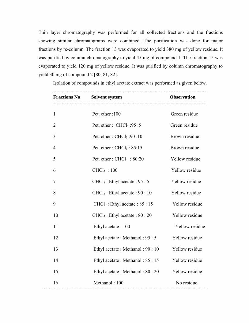
Thin layer chromatography was performed for all collected fractions and the fractions
showing similar chromatograms were combined. The purification was done for major
fractions by re-column. The fraction 13 was evaporated to yield 380 mg of yellow residue. It
was purified by column chromatography to yield 45 mg of compound 1. The fraction 15 was
evaporated to yield 120 mg of yellow residue. It was purified by column chromatography to
yield 30 mg of compound 2 [80, 81, 82].
Isolation of compounds in ethyl acetate extract was performed as given below.
---------------------------------------------------------------------------------------------- Fractions No Solvent system Observation
----------------------------------------------------------------------------------------------
1 Pet. ether :100 Green residue
2 Pet. ether : CHCl3 :95 :5 Green residue
3 Pet. ether : CHCl3 :90 :10 Brown residue
4 Pet. ether : CHCl3 : 85:15 Brown residue
5 Pet. ether : CHCl3 : 80:20 Yellow residue
6 CHCl3 : 100 Yellow residue
7 CHCl3 : Ethyl acetate : 95 : 5 Yellow residue
8 CHCl3 : Ethyl acetate : 90 : 10 Yellow residue
9 CHCl3 : Ethyl acetate : 85 : 15 Yellow residue
10 CHCl3 : Ethyl acetate : 80 : 20 Yellow residue
11 Ethyl acetate : 100 Yellow residue
12 Ethyl acetate : Methanol : 95 : 5 Yellow residue
13 Ethyl acetate : Methanol : 90 : 10 Yellow residue
14 Ethyl acetate : Methanol : 85 : 15 Yellow residue
15 Ethyl acetate : Methanol : 80 : 20 Yellow residue 16 Methanol : 100 No residue ----------------------------------------------------------------------------------------------------
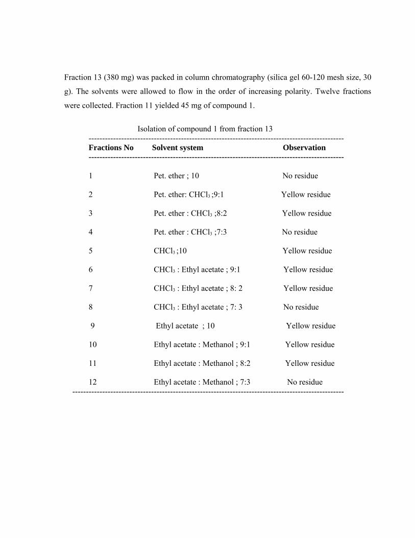
Fraction 13 (380 mg) was packed in column chromatography (silica gel 60-120 mesh size, 30
g). The solvents were allowed to flow in the order of increasing polarity. Twelve fractions
were collected. Fraction 11 yielded 45 mg of compound 1.
Isolation of compound 1 from fraction 13----------------------------------------------------------------------------------------------
Fractions No Solvent system Observation----------------------------------------------------------------------------------------------
1 Pet. ether ; 10 No residue
2 Pet. ether: CHCl3 ;9:1 Yellow residue
3 Pet. ether : CHCl3 ;8:2 Yellow residue
4 Pet. ether : CHCl3 ;7:3 No residue
5 CHCl3 ;10 Yellow residue
6 CHCl3 : Ethyl acetate ; 9:1 Yellow residue
7 CHCl3 : Ethyl acetate ; 8: 2 Yellow residue
8 CHCl3 : Ethyl acetate ; 7: 3 No residue
9 Ethyl acetate ; 10 Yellow residue
10 Ethyl acetate : Methanol ; 9:1 Yellow residue
11 Ethyl acetate : Methanol ; 8:2 Yellow residue
12 Ethyl acetate : Methanol ; 7:3 No residue ----------------------------------------------------------------------------------------------------
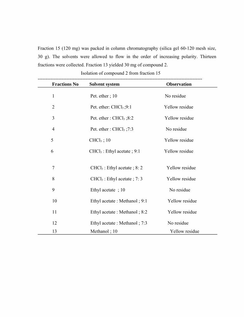
Fraction 15 (120 mg) was packed in column chromatography (silica gel 60-120 mesh size,
30 g). The solvents were allowed to flow in the order of increasing polarity. Thirteen
fractions were collected. Fraction 13 yielded 30 mg of compound 2.
Isolation of compound 2 from fraction 15------------------------------------------------------------------------------------------------------- Fractions No Solvent system Observation
1 Pet. ether ; 10 No residue
2 Pet. ether: CHCl3 ;9:1 Yellow residue
3 Pet. ether : CHCl3 ;8:2 Yellow residue
4 Pet. ether : CHCl3 ;7:3 No residue
5 CHCl3 ; 10 Yellow residue
6 CHCl3 : Ethyl acetate ; 9:1 Yellow residue
7 CHCl3 : Ethyl acetate ; 8: 2 Yellow residue
8 CHCl3 : Ethyl acetate ; 7: 3 Yellow residue
9 Ethyl acetate ; 10 No residue
10 Ethyl acetate : Methanol ; 9:1 Yellow residue
11 Ethyl acetate : Methanol ; 8:2 Yellow residue
12 Ethyl acetate : Methanol ; 7:3 No residue
13 Methanol ; 10 Yellow residue
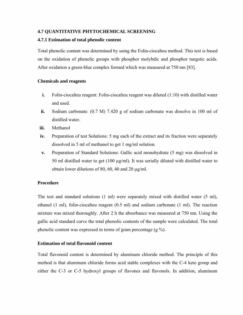
4.7 QUANTITATIVE PHYTOCHEMICAL SCREENING
4.7.1 Estimation of total phenolic content
Total phenolic content was determined by using the Folin-ciocalteu method. This test is based
on the oxidation of phenolic groups with phosphor molybdic and phosphor tungstic acids.
After oxidation a green-blue complex formed which was measured at 750 nm [83].
Chemicals and reagents
i. Folin-ciocalteu reagent: Folin-ciocalteu reagent was diluted (1:10) with distilled water
and used.
ii. Sodium carbonate: (0.7 M) 7.420 g of sodium carbonate was dissolve in 100 ml of
distilled water.
iii. Methanol
iv. Preparation of test Solutions: 5 mg each of the extract and its fraction were separately
dissolved in 5 ml of methanol to get 1 mg/ml solution.
v. Preparation of Standard Solutions: Gallic acid monohydrate (5 mg) was dissolved in
50 ml distilled water to get (100 µg/ml). It was serially diluted with distilled water to
obtain lower dilutions of 80, 60, 40 and 20 µg/ml.
Procedure
The test and standard solutions (1 ml) were separately mixed with distilled water (5 ml),
ethanol (1 ml), folin-ciocalteu reagent (0.5 ml) and sodium carbonate (1 ml). The reaction
mixture was mixed thoroughly. After 2 h the absorbance was measured at 750 nm. Using the
gallic acid standard curve the total phenolic contents of the sample were calculated. The total
phenolic content was expressed in terms of gram percentage (g %).
Estimation of total flavonoid content
Total flavonoid content is determined by aluminum chloride method. The principle of this
method is that aluminum chloride forms acid stable complexes with the C-4 keto group and
either the C-3 or C-5 hydroxyl groups of flavones and flavonols. In addition, aluminum
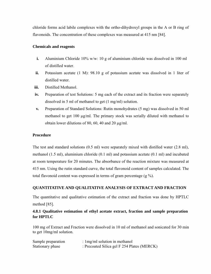
chloride forms acid labile complexes with the ortho-dihydroxyl groups in the A or B ring of
flavonoids. The concentration of these complexes was measured at 415 nm [84].
Chemicals and reagents
i. Aluminium Chloride 10% w/w: 10 g of aluminium chloride was dissolved in 100 ml
of distilled water.
ii. Potassium acetate (1 M): 98.10 g of potassium acetate was dissolved in 1 liter of
distilled water.
iii. Distilled Methanol.
iv. Preparation of test Solutions: 5 mg each of the extract and its fraction were separately
dissolved in 5 ml of methanol to get (1 mg/ml) solution.
v. Preparation of Standard Solutions: Rutin monohydrates (5 mg) was dissolved in 50 ml
methanol to get 100 µg/ml. The primary stock was serially diluted with methanol to
obtain lower dilutions of 80, 60, 40 and 20 µg/ml.
Procedure
The test and standard solutions (0.5 ml) were separately mixed with distilled water (2.8 ml),
methanol (1.5 ml), aluminium chloride (0.1 ml) and potassium acetate (0.1 ml) and incubated
at room temperature for 20 minutes. The absorbance of the reaction mixture was measured at
415 nm. Using the rutin standard curve, the total flavonoid content of samples calculated. The
total flavonoid content was expressed in terms of gram percentage (g %).
QUANTITATIVE AND QUALITATIVE ANALYSIS OF EXTRACT AND FRACTION
The quantitative and qualitative estimation of the extract and fraction was done by HPTLC
method [85].
4.8.1 Qualitative estimation of ethyl acetate extract, fraction and sample preparationfor HPTLC
100 mg of Extract and Fraction were dissolved in 10 ml of methanol and sonicated for 30 minto get 10mg/ml solution.
Sample preparation : 1mg/ml solution in methanolStationary phase : Precoated Silica gel F 254 Plates (MERCK)
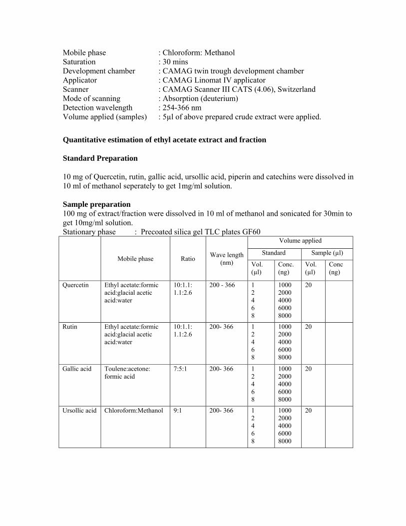
Mobile phase : Chloroform: Methanol Saturation : 30 mins Development chamber : CAMAG twin trough development chamber Applicator : CAMAG Linomat IV applicatorScanner : CAMAG Scanner III CATS (4.06), SwitzerlandMode of scanning : Absorption (deuterium) Detection wavelength : 254-366 nm Volume applied (samples) : 5µl of above prepared crude extract were applied.
Quantitative estimation of ethyl acetate extract and fraction
Standard Preparation
10 mg of Quercetin, rutin, gallic acid, ursollic acid, piperin and catechins were dissolved in 10 ml of methanol seperately to get 1mg/ml solution.
Sample preparation100 mg of extract/fraction were dissolved in 10 ml of methanol and sonicated for 30min to get 10mg/ml solution.Stationary phase : Precoated silica gel TLC plates GF60
Mobile phase Ratio Wave length(nm)
Volume applied
Standard Sample (µl)
Vol. (µl)
Conc. (ng)
Vol. (µl)
Conc (ng)
Quercetin Ethyl acetate:formic acid:glacial acetic acid:water
10:1.1: 1.1:2.6
200 - 366 12468
10002000400060008000
20
Rutin Ethyl acetate:formic acid:glacial acetic acid:water
10:1.1: 1.1:2.6
200- 366 12468
10002000400060008000
20
Gallic acid Toulene:acetone: formic acid
7:5:1 200- 366 12468
10002000400060008000
20
Ursollic acid Chloroform:Methanol 9:1 200- 366 12468
10002000400060008000
20
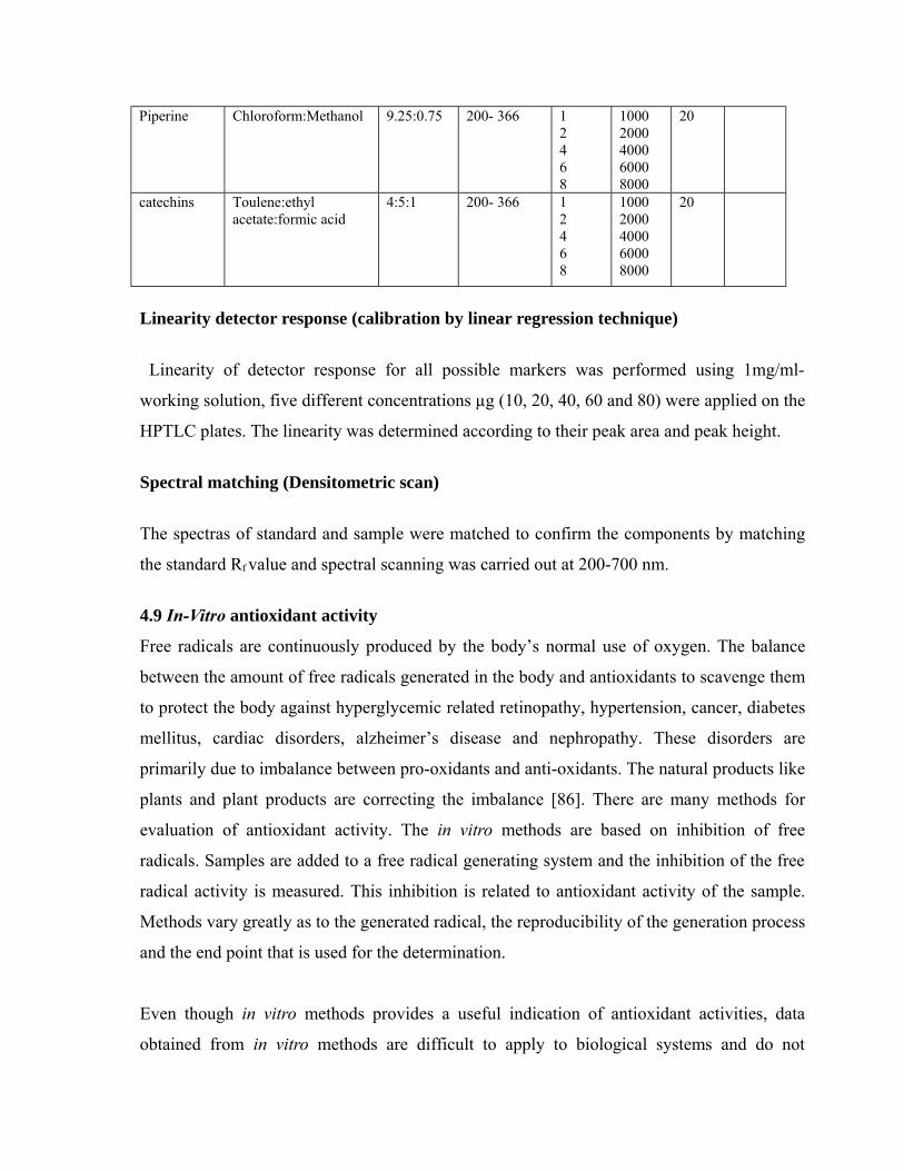
Piperine Chloroform:Methanol 9.25:0.75 200- 366 12468
10002000400060008000
20
catechins Toulene:ethyl acetate:formic acid
4:5:1 200- 366 12468
10002000400060008000
20
Linearity detector response (calibration by linear regression technique)
Linearity of detector response for all possible markers was performed using 1mg/ml-
working solution, five different concentrations µg (10, 20, 40, 60 and 80) were applied on the
HPTLC plates. The linearity was determined according to their peak area and peak height.
Spectral matching (Densitometric scan)
The spectras of standard and sample were matched to confirm the components by matching
the standard Rf value and spectral scanning was carried out at 200-700 nm.
4.9 In-Vitro antioxidant activity
Free radicals are continuously produced by the body’s normal use of oxygen. The balance
between the amount of free radicals generated in the body and antioxidants to scavenge them
to protect the body against hyperglycemic related retinopathy, hypertension, cancer, diabetes
mellitus, cardiac disorders, alzheimer’s disease and nephropathy. These disorders are
primarily due to imbalance between pro-oxidants and anti-oxidants. The natural products like
plants and plant products are correcting the imbalance [86]. There are many methods for
evaluation of antioxidant activity. The in vitro methods are based on inhibition of free
radicals. Samples are added to a free radical generating system and the inhibition of the free
radical activity is measured. This inhibition is related to antioxidant activity of the sample.
Methods vary greatly as to the generated radical, the reproducibility of the generation process
and the end point that is used for the determination.
Even though in vitro methods provides a useful indication of antioxidant activities, data
obtained from in vitro methods are difficult to apply to biological systems and do not
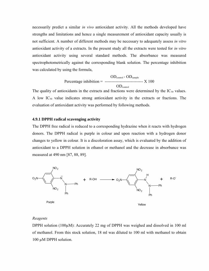
necessarily predict a similar in vivo antioxidant activity. All the methods developed have
strengths and limitations and hence a single measurement of antioxidant capacity usually is
not sufficient. A number of different methods may be necessary to adequately assess in vitro
antioxidant activity of a extracts. In the present study all the extracts were tested for in vitro
antioxidant activity using several standard methods. The absorbance was measured
spectrophotometrically against the corresponding blank solution. The percentage inhibition
was calculated by using the formula,
ODcontrol - ODsample
Percentage inhibition = ---------------------------- X 100 ODcontrol
The quality of antioxidants in the extracts and fractions were determined by the IC50 values.
A low IC50 value indicates strong antioxidant activity in the extracts or fractions. The
evaluation of antioxidant activity was performed by following methods.
4.9.1 DPPH radical scavenging activity
The DPPH free radical is reduced to a corresponding hydrazine when it reacts with hydrogen
donors. The DPPH radical is purple in colour and upon reaction with a hydrogen donor
changes to yellow in colour. It is a discoloration assay, which is evaluated by the addition of
antioxidant to a DPPH solution in ethanol or methanol and the decrease in absorbance was
measured at 490 nm [87, 88, 89].
NO2
O2N
NO2
N
N
Ph
Ph
..+ R-OH
NO2
O2N
NO2
N
N
Ph
Ph
HR-O.+
PurpleYellow
Reagents
DPPH solution (100µM): Accurately 22 mg of DPPH was weighed and dissolved in 100 ml
of methanol. From this stock solution, 18 ml was diluted to 100 ml with methanol to obtain
100 µM DPPH solution.
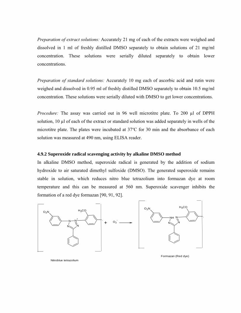
Preparation of extract solutions: Accurately 21 mg of each of the extracts were weighed and
dissolved in 1 ml of freshly distilled DMSO separately to obtain solutions of 21 mg/ml
concentration. These solutions were serially diluted separately to obtain lower
concentrations.
Preparation of standard solutions: Accurately 10 mg each of ascorbic acid and rutin were
weighed and dissolved in 0.95 ml of freshly distilled DMSO separately to obtain 10.5 mg/ml
concentration. These solutions were serially diluted with DMSO to get lower concentrations.
Procedure: The assay was carried out in 96 well microtitre plate. To 200 µl of DPPH
solution, 10 µl of each of the extract or standard solution was added separately in wells of the
microtitre plate. The plates were incubated at 37oC for 30 min and the absorbance of each
solution was measured at 490 nm, using ELISA reader.
4.9.2 Superoxide radical scavenging activity by alkaline DMSO method
In alkaline DMSO method, superoxide radical is generated by the addition of sodium
hydroxide to air saturated dimethyl sulfoxide (DMSO). The generated superoxide remains
stable in solution, which reduces nitro blue tetrazolium into formazan dye at room
temperature and this can be measured at 560 nm. Superoxide scavenger inhibits the
formation of a red dye formazan [90, 91, 92].
H3COO2N
N
N+
N
N .+ O2
-
H3COO2N
N
N
N
NH .
Nitroblue tetrazoliumFormazan (Red dye)
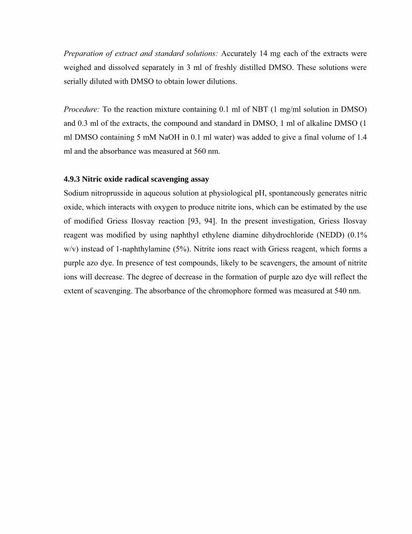
Preparation of extract and standard solutions: Accurately 14 mg each of the extracts were
weighed and dissolved separately in 3 ml of freshly distilled DMSO. These solutions were
serially diluted with DMSO to obtain lower dilutions.
Procedure: To the reaction mixture containing 0.1 ml of NBT (1 mg/ml solution in DMSO)
and 0.3 ml of the extracts, the compound and standard in DMSO, 1 ml of alkaline DMSO (1
ml DMSO containing 5 mM NaOH in 0.1 ml water) was added to give a final volume of 1.4
ml and the absorbance was measured at 560 nm.
4.9.3 Nitric oxide radical scavenging assay
Sodium nitroprusside in aqueous solution at physiological pH, spontaneously generates nitric
oxide, which interacts with oxygen to produce nitrite ions, which can be estimated by the use
of modified Griess Ilosvay reaction [93, 94]. In the present investigation, Griess Ilosvay
reagent was modified by using naphthyl ethylene diamine dihydrochloride (NEDD) (0.1%
w/v) instead of 1-naphthylamine (5%). Nitrite ions react with Griess reagent, which forms a
purple azo dye. In presence of test compounds, likely to be scavengers, the amount of nitrite
ions will decrease. The degree of decrease in the formation of purple azo dye will reflect the
extent of scavenging. The absorbance of the chromophore formed was measured at 540 nm.
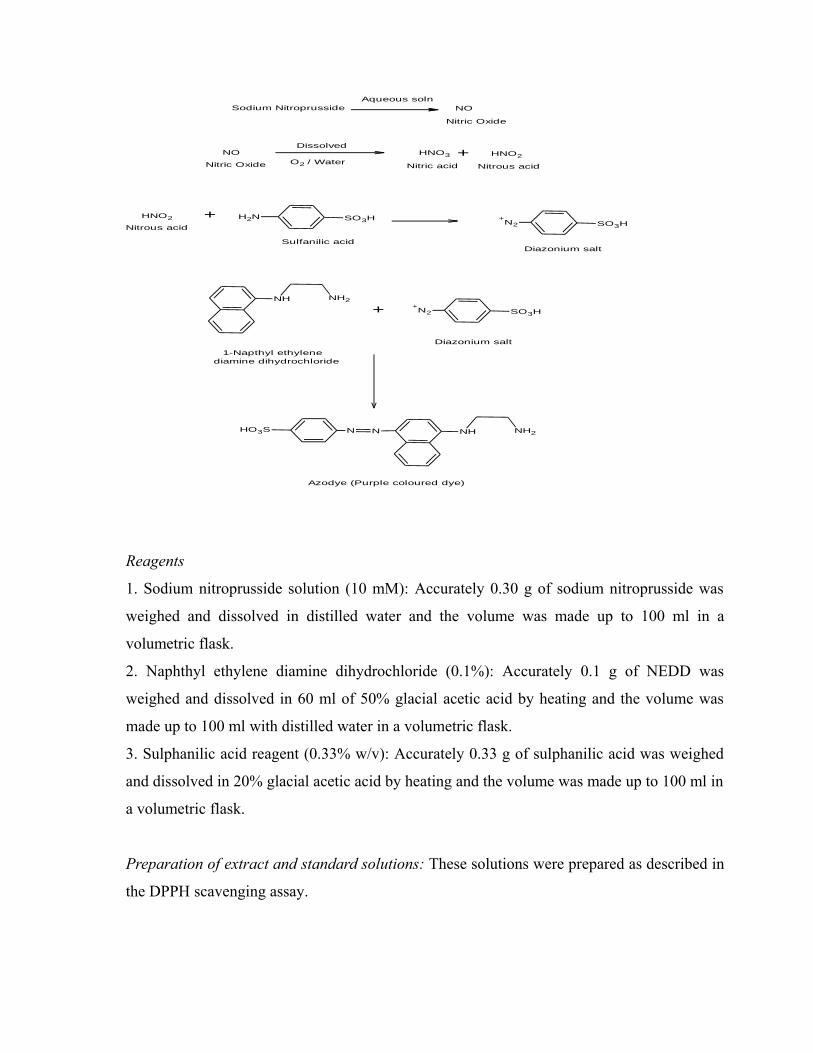
NO
Nitric Oxide
Dissolved
O2 / WaterHNO3 + HNO2
Nitrous acidNitric acid
HNO2
Nitrous acid
+ NH2 SO3H
Sulfanilic acid
+N2 SO3H
Diazonium salt
NH NH2
+ +N2 SO3H
Diazonium salt1-Napthyl ethylene
diamine dihydrochloride
HO3S N NH NH2N
Azodye (Purple coloured dye)
Sodium NitroprussideAqueous soln
NO
Nitric Oxide
Reagents
1. Sodium nitroprusside solution (10 mM): Accurately 0.30 g of sodium nitroprusside was
weighed and dissolved in distilled water and the volume was made up to 100 ml in a
volumetric flask.
2. Naphthyl ethylene diamine dihydrochloride (0.1%): Accurately 0.1 g of NEDD was
weighed and dissolved in 60 ml of 50% glacial acetic acid by heating and the volume was
made up to 100 ml with distilled water in a volumetric flask.
3. Sulphanilic acid reagent (0.33% w/v): Accurately 0.33 g of sulphanilic acid was weighed
and dissolved in 20% glacial acetic acid by heating and the volume was made up to 100 ml in
a volumetric flask.
Preparation of extract and standard solutions: These solutions were prepared as described in
the DPPH scavenging assay.
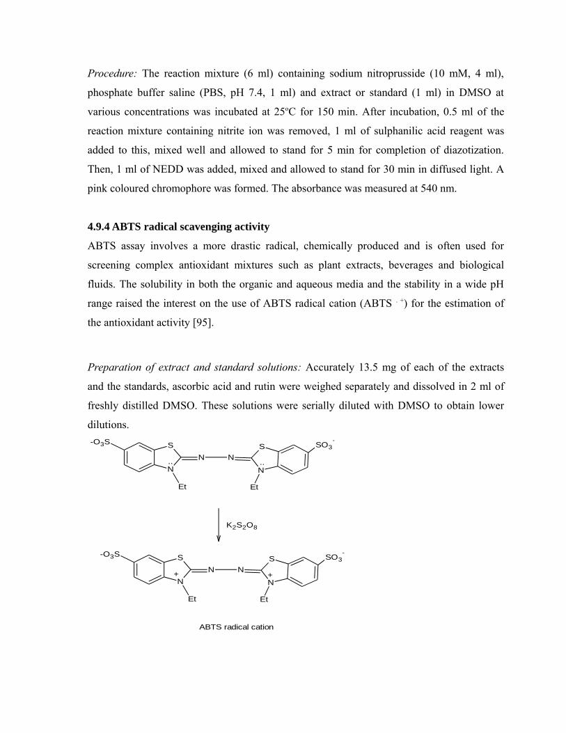
Procedure: The reaction mixture (6 ml) containing sodium nitroprusside (10 mM, 4 ml),
phosphate buffer saline (PBS, pH 7.4, 1 ml) and extract or standard (1 ml) in DMSO at
various concentrations was incubated at 25oC for 150 min. After incubation, 0.5 ml of the
reaction mixture containing nitrite ion was removed, 1 ml of sulphanilic acid reagent was
added to this, mixed well and allowed to stand for 5 min for completion of diazotization.
Then, 1 ml of NEDD was added, mixed and allowed to stand for 30 min in diffused light. A
pink coloured chromophore was formed. The absorbance was measured at 540 nm.
4.9.4 ABTS radical scavenging activity
ABTS assay involves a more drastic radical, chemically produced and is often used for
screening complex antioxidant mixtures such as plant extracts, beverages and biological
fluids. The solubility in both the organic and aqueous media and the stability in a wide pH
range raised the interest on the use of ABTS radical cation (ABTS . +) for the estimation of
the antioxidant activity [95].
Preparation of extract and standard solutions: Accurately 13.5 mg of each of the extracts
and the standards, ascorbic acid and rutin were weighed separately and dissolved in 2 ml of
freshly distilled DMSO. These solutions were serially diluted with DMSO to obtain lower
dilutions.
SO3--O3S S
NN
N
SN
Et Et
K2S2O8
SO3--O3S S
NN
N
SN
Et Et
.. ..
++
ABTS radical cation
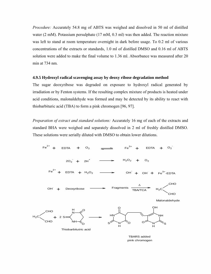
Procedure: Accurately 54.8 mg of ABTS was weighed and dissolved in 50 ml of distilled
water (2 mM). Potassium persulphate (17 mM, 0.3 ml) was then added. The reaction mixture
was left to stand at room temperature overnight in dark before usage. To 0.2 ml of various
concentrations of the extracts or standards, 1.0 ml of distilled DMSO and 0.16 ml of ABTS
solution were added to make the final volume to 1.36 ml. Absorbance was measured after 20
min at 734 nm.
4.9.5 Hydroxyl radical scavenging assay by deoxy ribose degradation method
The sugar deoxyribose was degraded on exposure to hydroxyl radical generated by
irradiation or by Fenton systems. If the resulting complex mixture of products is heated under
acid conditions, malonaldehyde was formed and may be detected by its ability to react with
thiobarbituric acid (TBA) to form a pink chromogen [96, 97].
Preparation of extract and standard solutions: Accurately 16 mg of each of the extracts and
standard BHA were weighed and separately dissolved in 2 ml of freshly distilled DMSO.
These solutions were serially diluted with DMSO to obtain lower dilutions.
Fe2+
EDTA O2 Fe3+ EDTA O2
-+ + + +
2O2- + 2H
+ H2O2 O2+
Fe2+
EDTA H2O2 OH-
OH.+ + + + Fe
3+-EDTA
OH.
Deoxyribose FragmentsΔ
TBA/TCA H2C
CHO
CHO
Malonaldehyde
+
H2C
CHO
CHO
NH
NH
O
O
S2+
Thiobarbitutric acid
NH
NH
S
O
O
NH
NH
SO
OH
TBARS added pink chromogen
Procedure: Various concentrations of the extracts, the compound and standard in DMSO (0.2
ml) were added to the reaction mixture containing deoxyribose (3 mM, 0.2 ml), ferric
chloride (0.1 mM, 0.2 ml), EDTA (0.1 mM, 0.2 ml), ascorbic acid (0.1 mM, 0.2 ml) and
hydrogen peroxide (2 mM, 0.2 ml) in phosphate buffer (PH 7.4, 20 mM) to give a total
volume of 1.2 ml. The solutions were then incubated for 30 min at 37°C. After incubation, ice
cold trichloroacetic acid (0.2 ml, 15% w/v) and thiobarbituric acid (0.2 ml, 1% w/v) in 0.25
N HCl were added. The reaction mixture was kept in a boiling water bath for 30 min, cooled
and the absorbance was measured at 532 nm.
4.10 In Vitro α-glucosidase inhibition activity
Isolation of α-glucosidase enzyme from rat small intestine
A male rat (200 g) was sacrificed by cervical dislocation. The small intestine was obtained
and flushed several times with ice-cold NaCl (0.9% w/w). The intestine was cleaned from
adipose tissue and cut longitudinally. The mucosa was scraped with a glass slide on an ice-
cold glass surface. The obtained material containing α glucosidase was homogenized with 20
ml of sodium phosphate buffer and stored at −250C until used. Total protein content was
determined by the Lowry method [98].
Determination of α-glucosidase inhibition
To all the test tubes 0.5 ml of Sodium phosphate buffer (80 Mm), pH 7.0 containing 37 mM
sucrose was taken and to the test tubes 1 ml of various concentrations of test sample and
standard was added. For the control and blank wells 1 ml of phosphate buffer pH 7.0 was
added. The reaction was initiated by adding 50µl of crude enzyme to all the tubes except
blank.
All the samples were incubated at 37°C for 20 min. The reaction was then stopped by heating
the test tubes at 95°C for 1.5 min. The liberated glucose was measured using commercial
glucose kit. The percentage inhibition was calculated by using the following formula.
% inhibition = 100 - [A sample / A control x 100]
A sample = absorbance of the sample,
A control = absorbance of the control
4.11 In vivo Antidiabetic activity
All the glucose lowering agents available today for treatment of diabetes resulted from in
vivo anti-diabetic drug discovery approach. The ethyl acetate extract has significant activity
in in vitro antidiabetic experiment [99, 100]. Hence ethyl acetate extract was selected for in
vivo antidiabetic experiment.
4.11.1 Animals and treatment
Wistar rats of either sex weighing 180-220g (6 to 8 weeks) with no prior drug treatment were
used for the present experiment. The animals were fed with standard laboratory chow (Amrut
laboratory Animal feeds, Pranav Agro industries Ltd, Sangli) and provided water ad libitum
[101]. Animal experiment was performed in the department of pharmacology, J.S.S college
of pharmacy, Ootacamund, after approval from the Institutional Animal Ethics Committee
(registration number 118/1999/CPCSEA) and animal care was taken as per the guidelines of
Committee for the Purpose of Control and Supervision of Experiments on Animals
(CPCSEA)(PH. D/PH. CHEM/03/2009-2010).
4.11.2 Acute toxicity studies
The acute toxicity study of the ethyl acetate extract was determined according to the OECD
guidelines No.425. Female wistar rats weighing 180 – 220g (6 to 8 weeks) were used for this
study. The general procedure was as follows: one rat was dosed at 400 mg/kg body weight
and if no mortality or over toxicity occurred within 48 h, another rat was dosed at 800 mg/kg
body weight. In the absence of toxicity, a third rat was dosed at 2000 mg/kg body weight and
if again no evidence of toxicity was observed, two additional rats were dosed at this level. In
all cases the dosing volume was fixed at 10 ml/kg body weight. The rats were observed for
clinical signs of toxicity at 0-0.5, 0.5-1, 1-2, 2-4 and 4-8 h post dosing. The body weights of
all the rats were recorded prior to the administration of test sample and at 7 and 14 days post
dosing The animals were observed for 24 hours and monitored for 14 days to record general
behaviour and mortality [102]. No mortality was observed till the end of the study.
4.11.3 Induction of diabetes
Streptozotocin was dissolved in sterilized citrate buffer pH 4.5. The wistar rats were fast
overnight and Streptozotocin 55mg/kg b.w was administered intraperitoneally. After a period
of 7 days blood glucose was estimated to confirm the diabetes. The rats were maintained for
a period of 14 days to stabilize the diabetic condition. The rats with blood glucose level
above 200 mg/dl were considered diabetic and used in the experiment [103, 104].
4.11.4 Experimental protocol
The animals were divided into following groups. Each group contain 5 animals [105].
Group 1 - Untreated Control
Group 2 - Diabetic Control
Group 3 - Positive Control (glibenclamide 10 mg/kg body weight)
Group 4 - Diabetic rats given (100 mg/kg b.w) ethyl acetate extract
Group 5 - Diabetic rats given (200 mg/kg b.w) ethyl acetate extract
Group 6 - Diabetic rats given (400 mg/kg b.w) ethyl acetate extract
The ethyl acetate extract was administered orally, twice daily for 7days and biochemical
parameters were estimated [106].
4.11.5 Determination of serum biochemical parameters
Blood was collected by retroorbital sinus. Blood samples were centrifuged at 4300 rpm for
20 min to obtain serum. Serum biochemical parameters were estimated using biochemical
kits according to instructions.
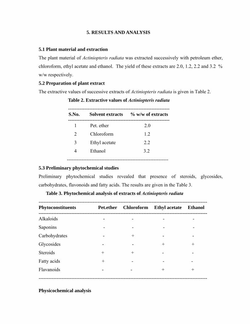
5. RESULTS AND ANALYSIS
5.1 Plant material and extraction
The plant material of Actiniopteris radiata was extracted successively with petroleum ether,
chloroform, ethyl acetate and ethanol. The yield of these extracts are 2.0, 1.2, 2.2 and 3.2 %
w/w respectively.
5.2 Preparation of plant extract
The extractive values of successive extracts of Actiniopteris radiata is given in Table 2.
Table 2. Extractive values of Actiniopteris radiata
--------------------------------------------------------------S.No. Solvent extracts % w/w of extracts
-------------------------------------------------------------- 1 Pet. ether 2.0
2 Chloroform 1.2
3 Ethyl acetate 2.2
4 Ethanol 3.2
--------------------------------------------------------------
5.3 Preliminary phytochemical studies
Preliminary phytochemical studies revealed that presence of steroids, glycosides,
carbohydrates, flavonoids and fatty acids. The results are given in the Table 3.
Table 3. Phytochemical analysis of extracts of Actiniopteris radiata
-------------------------------------------------------------------------------------------------------Phytoconstituents Pet.ether Chloroform Ethyl acetate Ethanol -------------------------------------------------------------------------------------------------------Alkaloids - - - -
Saponins - - - -
Carbohydrates - + - -
Glycosides - - + +
Steroids + + - -
Fatty acids + - - -
Flavanoids - - + +
-------------------------------------------------------------------------------------------------------
Physicochemical analysis
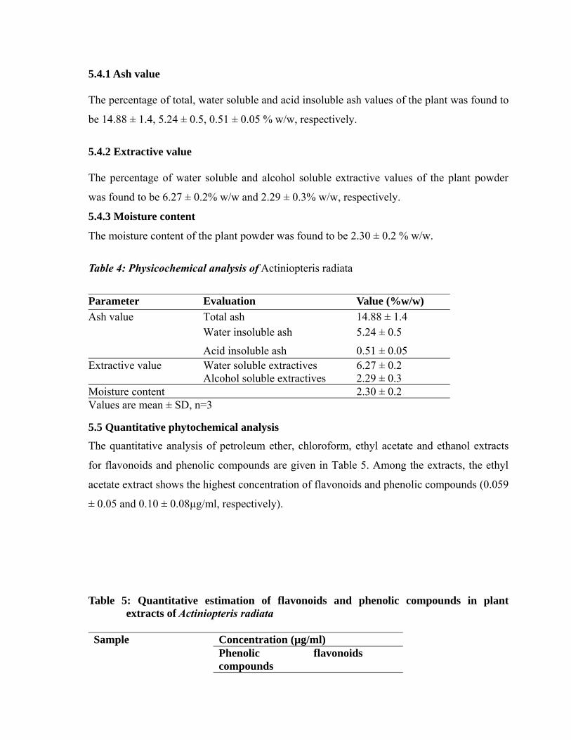
5.4.1 Ash value
The percentage of total, water soluble and acid insoluble ash values of the plant was found to
be 14.88 ± 1.4, 5.24 ± 0.5, 0.51 ± 0.05 % w/w, respectively.
5.4.2 Extractive value
The percentage of water soluble and alcohol soluble extractive values of the plant powder
was found to be 6.27 ± 0.2% w/w and 2.29 ± 0.3% w/w, respectively.
5.4.3 Moisture content
The moisture content of the plant powder was found to be 2.30 ± 0.2 % w/w.
Table 4: Physicochemical analysis of Actiniopteris radiata
Parameter Evaluation Value (%w/w)Ash value Total ash 14.88 ± 1.4
Water insoluble ash 5.24 ± 0.5
Acid insoluble ash 0.51 ± 0.05Extractive value Water soluble extractives 6.27 ± 0.2
Alcohol soluble extractives 2.29 ± 0.3Moisture content 2.30 ± 0.2Values are mean ± SD, n=3
5.5 Quantitative phytochemical analysis
The quantitative analysis of petroleum ether, chloroform, ethyl acetate and ethanol extracts
for flavonoids and phenolic compounds are given in Table 5. Among the extracts, the ethyl
acetate extract shows the highest concentration of flavonoids and phenolic compounds (0.059
± 0.05 and 0.10 ± 0.08µg/ml, respectively).
Table 5: Quantitative estimation of flavonoids and phenolic compounds in plantextracts of Actiniopteris radiata
Sample Concentration (µg/ml)Phenolic compounds
flavonoids
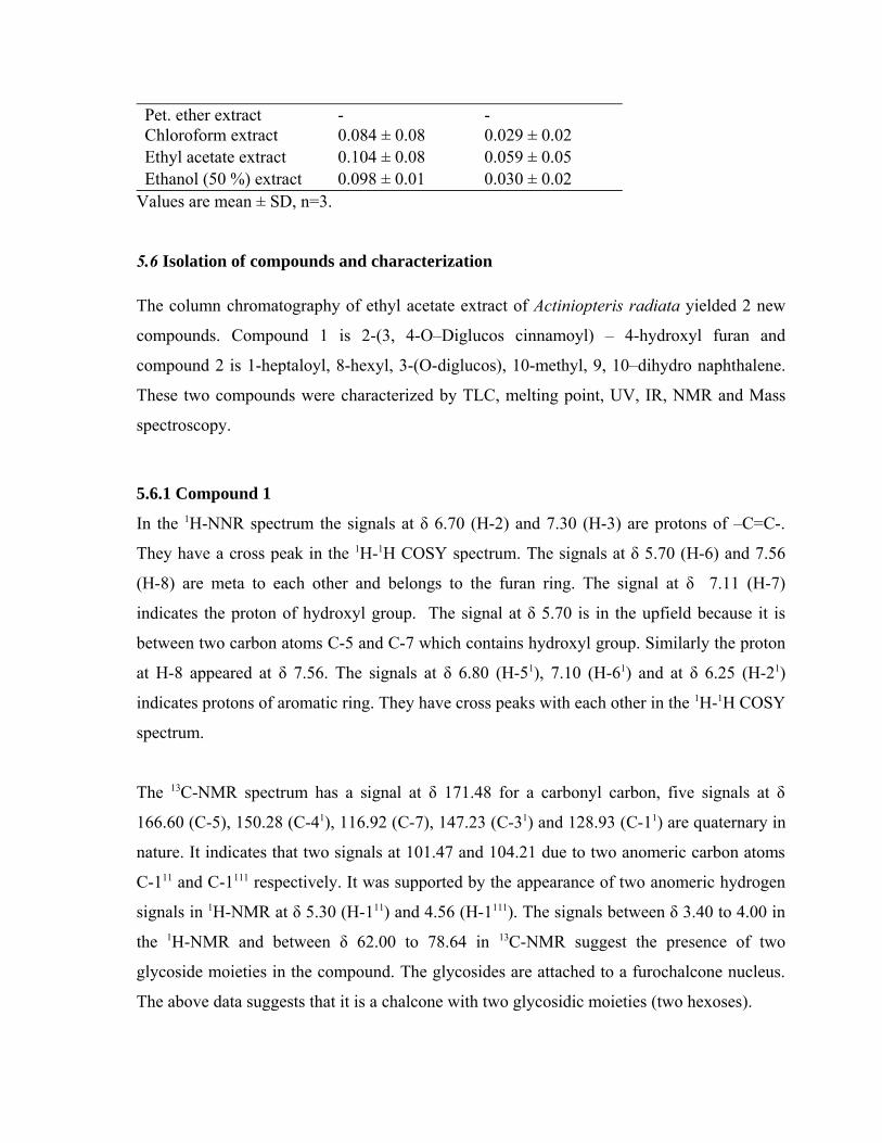
Pet. ether extract - -Chloroform extract 0.084 ± 0.08 0.029 ± 0.02Ethyl acetate extract 0.104 ± 0.08 0.059 ± 0.05Ethanol (50 %) extract 0.098 ± 0.01 0.030 ± 0.02
Values are mean ± SD, n=3.
5.6 Isolation of compounds and characterization
The column chromatography of ethyl acetate extract of Actiniopteris radiata yielded 2 new
compounds. Compound 1 is 2-(3, 4-O–Diglucos cinnamoyl) – 4-hydroxyl furan and
compound 2 is 1-heptaloyl, 8-hexyl, 3-(O-diglucos), 10-methyl, 9, 10–dihydro naphthalene.
These two compounds were characterized by TLC, melting point, UV, IR, NMR and Mass
spectroscopy.
5.6.1 Compound 1
In the 1H-NNR spectrum the signals at δ 6.70 (H-2) and 7.30 (H-3) are protons of –C=C-.
They have a cross peak in the 1H-1H COSY spectrum. The signals at δ 5.70 (H-6) and 7.56
(H-8) are meta to each other and belongs to the furan ring. The signal at δ 7.11 (H-7)
indicates the proton of hydroxyl group. The signal at δ 5.70 is in the upfield because it is
between two carbon atoms C-5 and C-7 which contains hydroxyl group. Similarly the proton
at H-8 appeared at δ 7.56. The signals at δ 6.80 (H-51), 7.10 (H-61) and at δ 6.25 (H-21)
indicates protons of aromatic ring. They have cross peaks with each other in the 1H-1H COSY
spectrum.
The 13C-NMR spectrum has a signal at δ 171.48 for a carbonyl carbon, five signals at δ
166.60 (C-5), 150.28 (C-41), 116.92 (C-7), 147.23 (C-31) and 128.93 (C-11) are quaternary in
nature. It indicates that two signals at 101.47 and 104.21 due to two anomeric carbon atoms
C-111 and C-1111 respectively. It was supported by the appearance of two anomeric hydrogen
signals in 1H-NMR at δ 5.30 (H-111) and 4.56 (H-1111). The signals between δ 3.40 to 4.00 in
the 1H-NMR and between δ 62.00 to 78.64 in 13C-NMR suggest the presence of two
glycoside moieties in the compound. The glycosides are attached to a furochalcone nucleus.
The above data suggests that it is a chalcone with two glycosidic moieties (two hexoses).
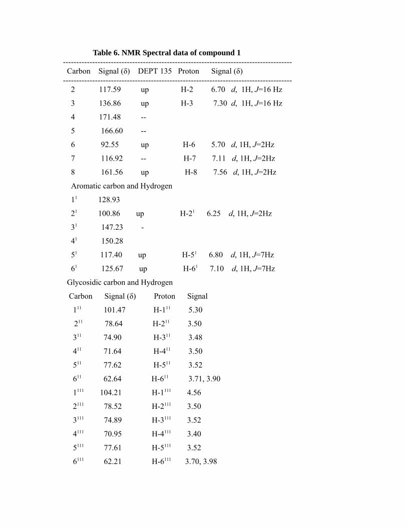
Table 6. NMR Spectral data of compound 1-------------------------------------------------------------------------------------- Carbon Signal (δ) DEPT 135 Proton Signal (δ)-------------------------------------------------------------------------------------- 2 117.59 up H-2 6.70 d, 1H, J=16 Hz
3 136.86 up H-3 7.30 d, 1H, J=16 Hz
4 171.48 --
5 166.60 --
6 92.55 up H-6 5.70 d, 1H, J=2Hz
7 116.92 -- H-7 7.11 d, 1H, J=2Hz
8 161.56 up H-8 7.56 d, 1H, J=2Hz
Aromatic carbon and Hydrogen
11 128.93
21 100.86 up H-21 6.25 d, 1H, J=2Hz
31 147.23 -
41 150.28
51 117.40 up H-51 6.80 d, 1H, J=7Hz
61 125.67 up H-61 7.10 d, 1H, J=7Hz
Glycosidic carbon and Hydrogen
Carbon Signal (δ) Proton Signal
111 101.47 H-111 5.30 211 78.64 H-211 3.50
311 74.90 H-311 3.48
411 71.64 H-411 3.50
511 77.62 H-511 3.52
611 62.64 H-611 3.71, 3.90
1111 104.21 H-1111 4.56
2111 78.52 H-2111 3.50
3111 74.89 H-3111 3.52
4111 70.95 H-4111 3.40
5111 77.61 H-5111 3.52
6111 62.21 H-6111 3.70, 3.98
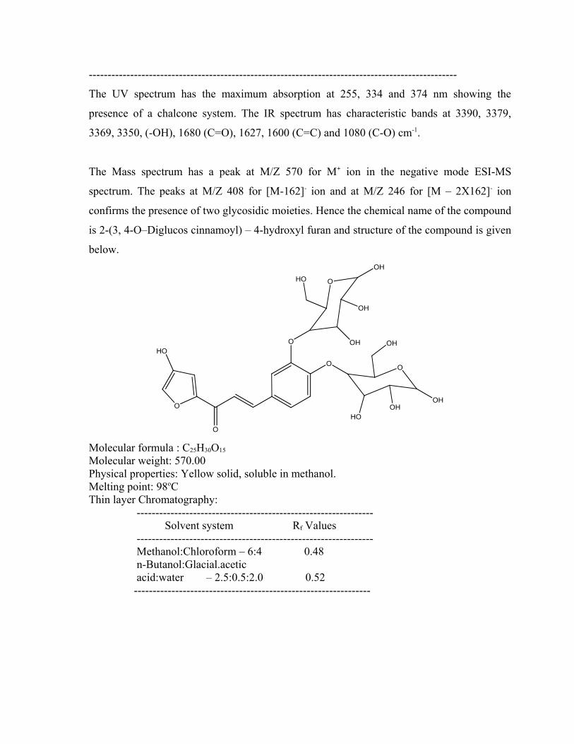
--------------------------------------------------------------------------------------------------
The UV spectrum has the maximum absorption at 255, 334 and 374 nm showing the
presence of a chalcone system. The IR spectrum has characteristic bands at 3390, 3379,
3369, 3350, (-OH), 1680 (C=O), 1627, 1600 (C=C) and 1080 (C-O) cm-1.
The Mass spectrum has a peak at M/Z 570 for M+ ion in the negative mode ESI-MS
spectrum. The peaks at M/Z 408 for [M-162]- ion and at M/Z 246 for [M – 2X162]- ion
confirms the presence of two glycosidic moieties. Hence the chemical name of the compound
is 2-(3, 4-O–Diglucos cinnamoyl) – 4-hydroxyl furan and structure of the compound is given
below.
O
O
O
O
O
HO
OH
OH
HOOH
O
HO
OHOH
OH
Molecular formula : C25H30O15
Molecular weight: 570.00Physical properties: Yellow solid, soluble in methanol.Melting point: 98oCThin layer Chromatography: --------------------------------------------------------------- Solvent system Rf Values --------------------------------------------------------------- Methanol:Chloroform – 6:4 0.48 n-Butanol:Glacial.acetic acid:water – 2.5:0.5:2.0 0.52 ---------------------------------------------------------------
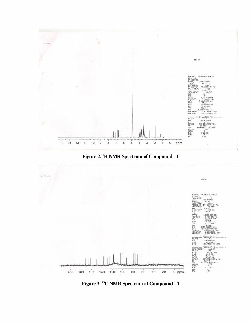
Figure 2. 1H NMR Spectrum of Compound - 1
Figure 3. 13C NMR Spectrum of Compound - 1
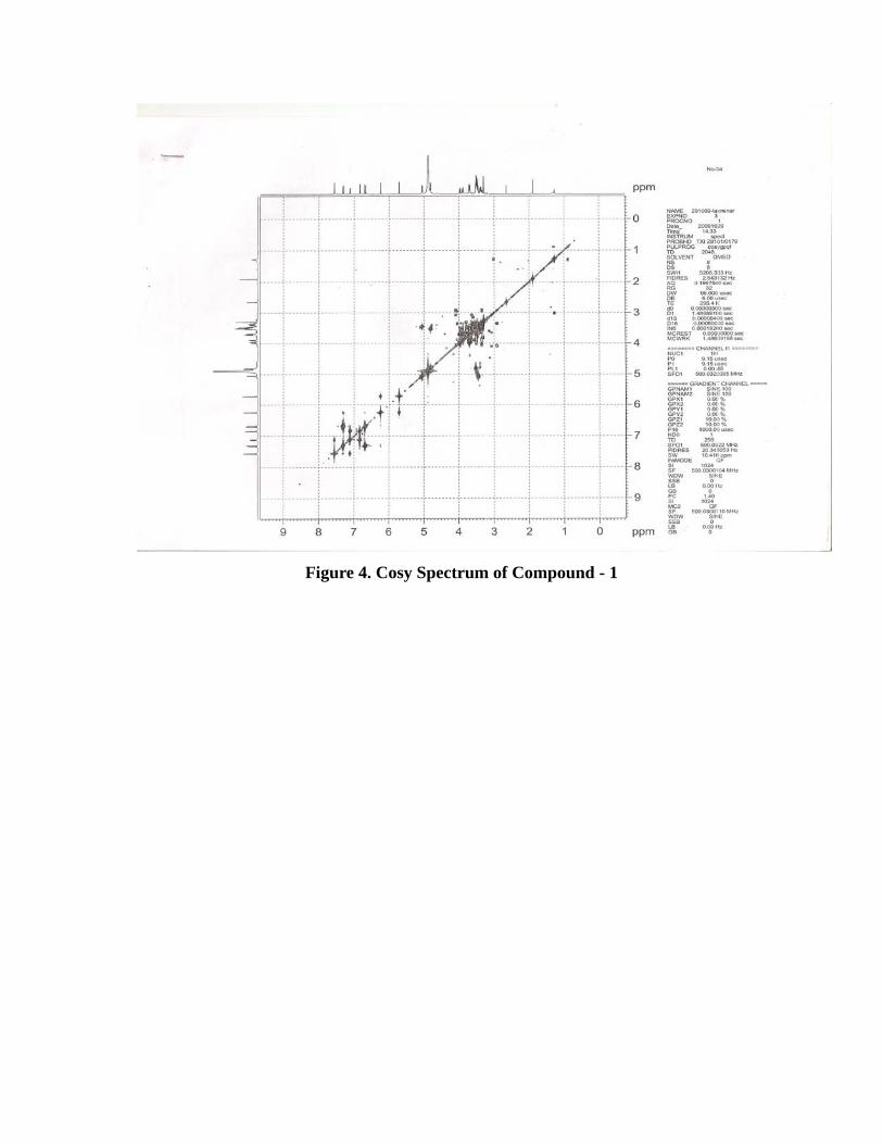
Figure 4. Cosy Spectrum of Compound - 1
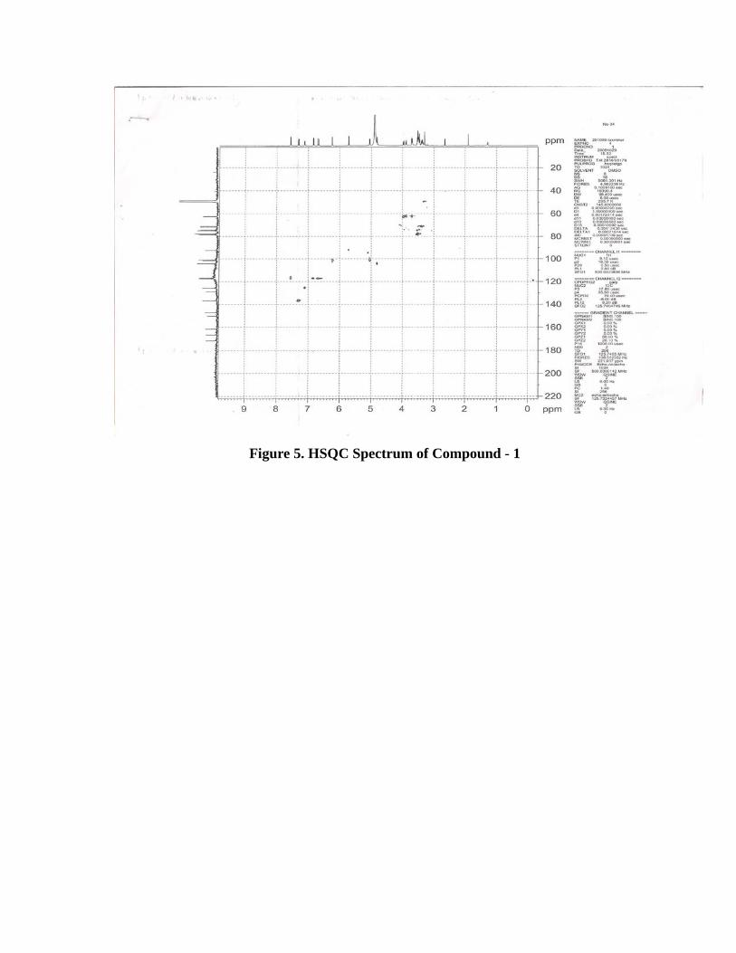
Figure 5. HSQC Spectrum of Compound - 1
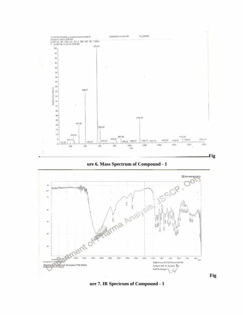
Fig
ure 6. Mass Spectrum of Compound - 1
Fig
ure 7. IR Spectrum of Compound - 1
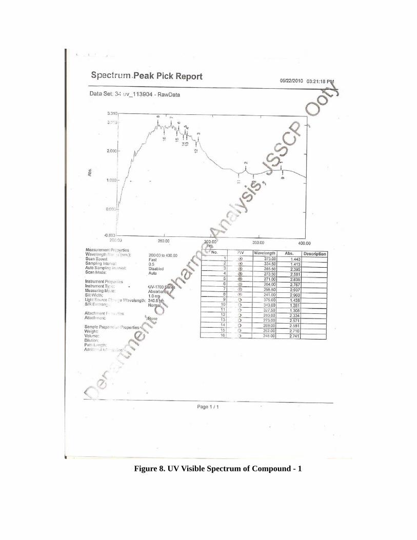
Figure 8. UV Visible Spectrum of Compound - 1
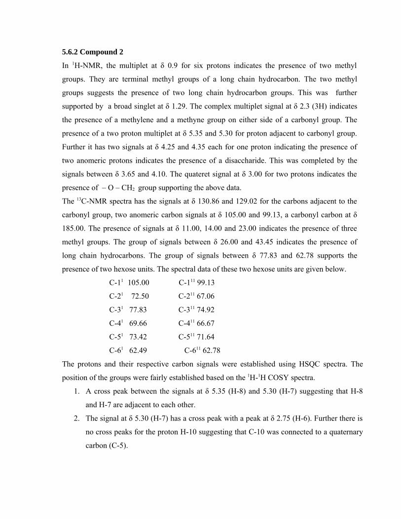
5.6.2 Compound 2
In 1H-NMR, the multiplet at δ 0.9 for six protons indicates the presence of two methyl
groups. They are terminal methyl groups of a long chain hydrocarbon. The two methyl
groups suggests the presence of two long chain hydrocarbon groups. This was further
supported by a broad singlet at δ 1.29. The complex multiplet signal at δ 2.3 (3H) indicates
the presence of a methylene and a methyne group on either side of a carbonyl group. The
presence of a two proton multiplet at δ 5.35 and 5.30 for proton adjacent to carbonyl group.
Further it has two signals at δ 4.25 and 4.35 each for one proton indicating the presence of
two anomeric protons indicates the presence of a disaccharide. This was completed by the
signals between δ 3.65 and 4.10. The quateret signal at δ 3.00 for two protons indicates the
presence of – O – CH2 group supporting the above data.
The 13C-NMR spectra has the signals at δ 130.86 and 129.02 for the carbons adjacent to the
carbonyl group, two anomeric carbon signals at δ 105.00 and 99.13, a carbonyl carbon at δ
185.00. The presence of signals at δ 11.00, 14.00 and 23.00 indicates the presence of three
methyl groups. The group of signals between δ 26.00 and 43.45 indicates the presence of
long chain hydrocarbons. The group of signals between δ 77.83 and 62.78 supports the
presence of two hexose units. The spectral data of these two hexose units are given below.
C-11 105.00 C-111 99.13
C-21 72.50 C-211 67.06
C-31 77.83 C-311 74.92
C-41 69.66 C-411 66.67
C-51 73.42 C-511 71.64
C-61 62.49 C-611 62.78
The protons and their respective carbon signals were established using HSQC spectra. The
position of the groups were fairly established based on the 1H-1H COSY spectra.
1. A cross peak between the signals at δ 5.35 (H-8) and 5.30 (H-7) suggesting that H-8
and H-7 are adjacent to each other.
2. The signal at δ 5.30 (H-7) has a cross peak with a peak at δ 2.75 (H-6). Further there is
no cross peaks for the proton H-10 suggesting that C-10 was connected to a quaternary
carbon (C-5).
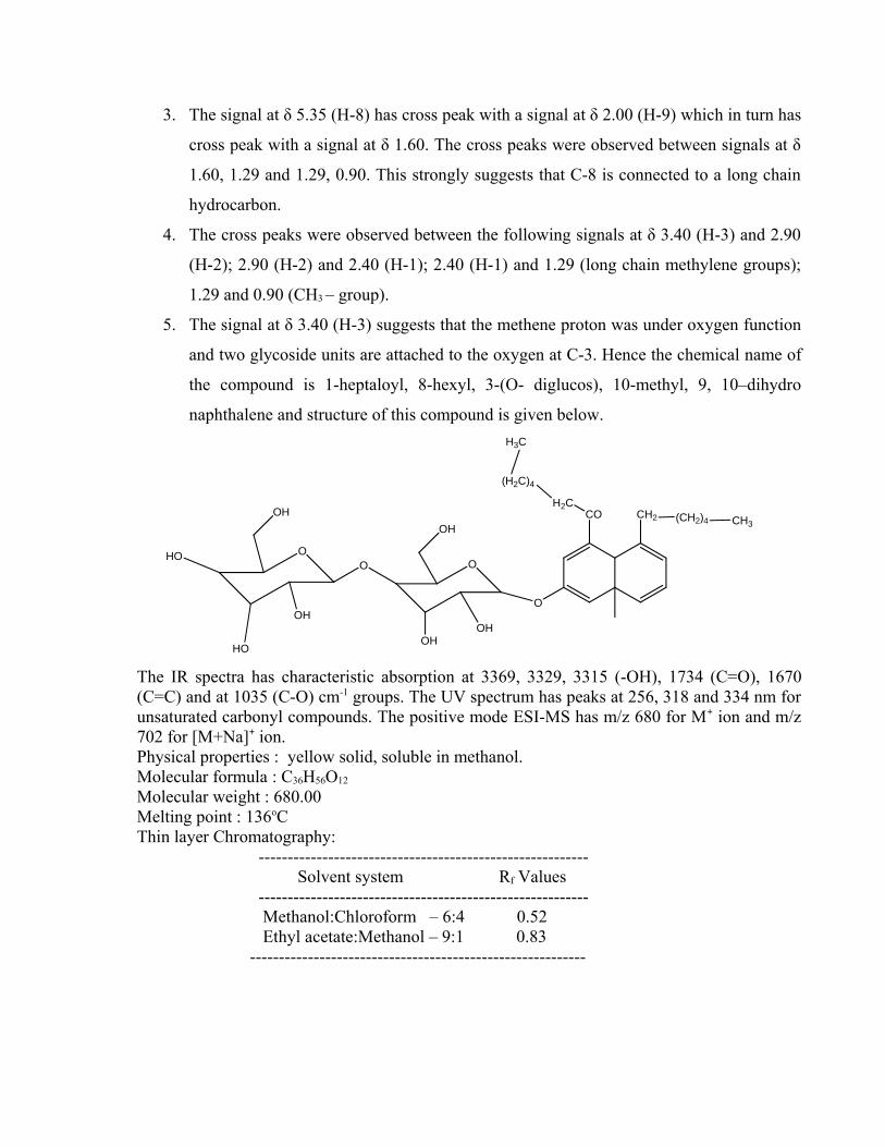
3. The signal at δ 5.35 (H-8) has cross peak with a signal at δ 2.00 (H-9) which in turn has
cross peak with a signal at δ 1.60. The cross peaks were observed between signals at δ
1.60, 1.29 and 1.29, 0.90. This strongly suggests that C-8 is connected to a long chain
hydrocarbon.
4. The cross peaks were observed between the following signals at δ 3.40 (H-3) and 2.90
(H-2); 2.90 (H-2) and 2.40 (H-1); 2.40 (H-1) and 1.29 (long chain methylene groups);
1.29 and 0.90 (CH3 – group).
5. The signal at δ 3.40 (H-3) suggests that the methene proton was under oxygen function
and two glycoside units are attached to the oxygen at C-3. Hence the chemical name of
the compound is 1-heptaloyl, 8-hexyl, 3-(O- diglucos), 10-methyl, 9, 10–dihydro
naphthalene and structure of this compound is given below.
O
CO CH2 (CH2)4 CH3
H2C
(H2C)4
H3C
OOO
OH
HO
HO
OH
OHOH
OH
The IR spectra has characteristic absorption at 3369, 3329, 3315 (-OH), 1734 (C=O), 1670(C=C) and at 1035 (C-O) cm-1 groups. The UV spectrum has peaks at 256, 318 and 334 nm forunsaturated carbonyl compounds. The positive mode ESI-MS has m/z 680 for M+ ion and m/z702 for [M+Na]+ ion.Physical properties : yellow solid, soluble in methanol.Molecular formula : C36H56O12
Molecular weight : 680.00Melting point : 136oC Thin layer Chromatography: ---------------------------------------------------------
Solvent system Rf Values ---------------------------------------------------------
Methanol:Chloroform – 6:4 0.52 Ethyl acetate:Methanol – 9:1 0.83 ----------------------------------------------------------
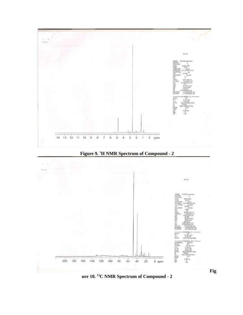
Figure 9. 1H NMR Spectrum of Compound - 2
Figure 10. 13C NMR Spectrum of Compound - 2
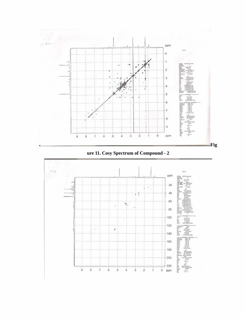
Fig
ure 11. Cosy Spectrum of Compound - 2
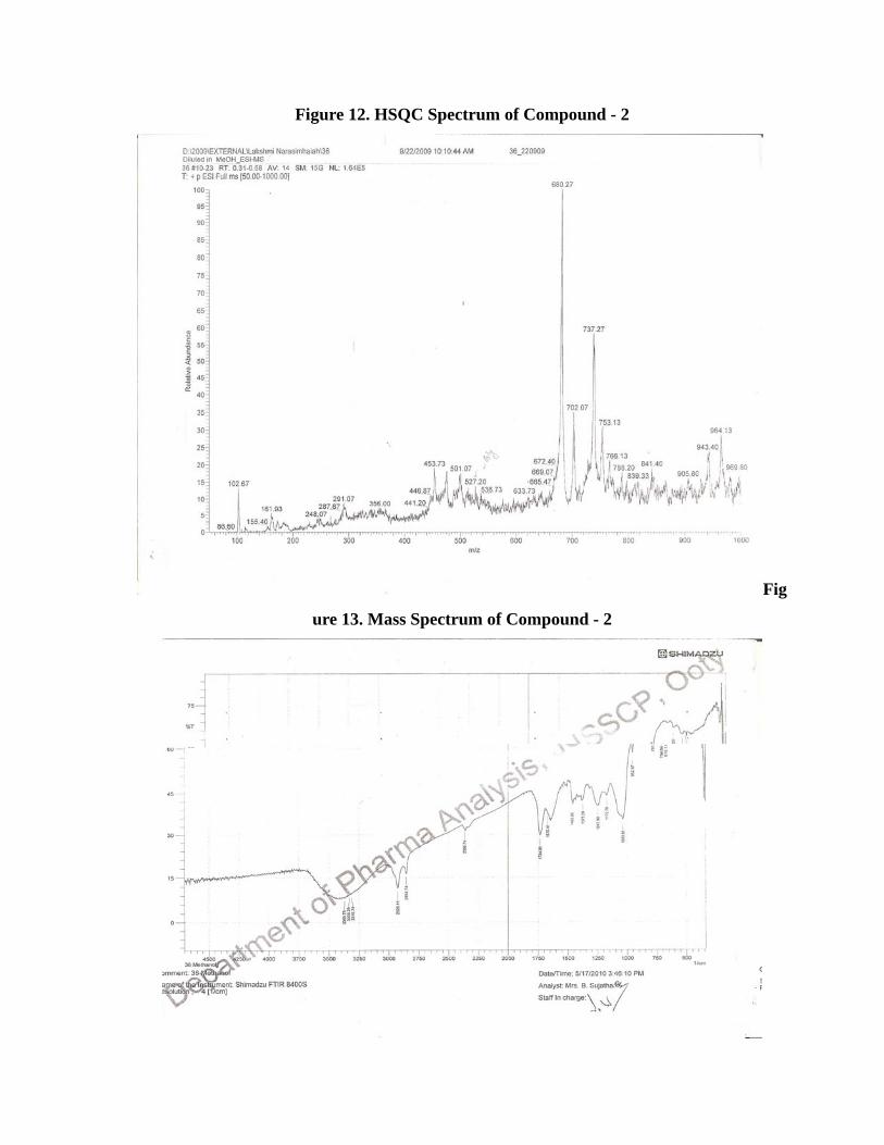
Figure 12. HSQC Spectrum of Compound - 2
Fig
ure 13. Mass Spectrum of Compound - 2
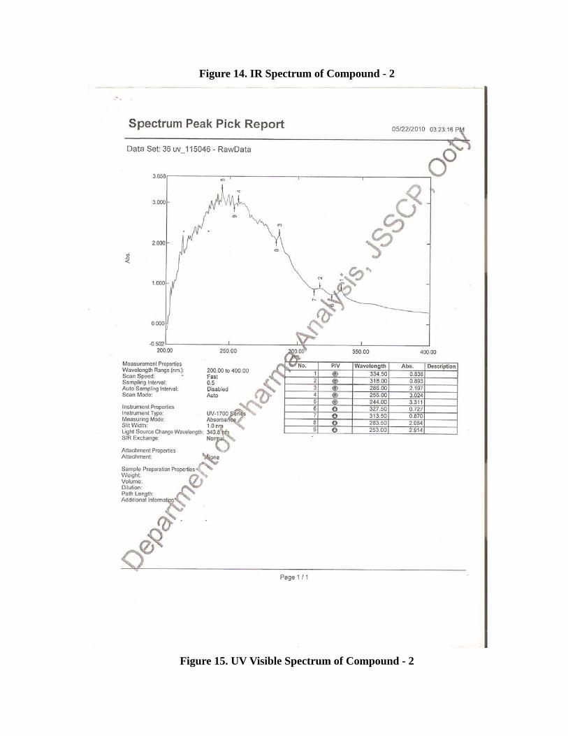
Figure 14. IR Spectrum of Compound - 2
Figure 15. UV Visible Spectrum of Compound - 2
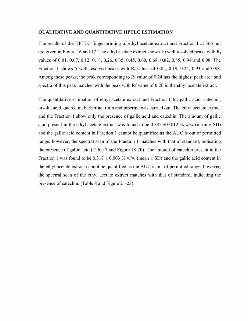
QUALITATIVE AND QUANTITATIVE HPTLC ESTIMATION
The results of the HPTLC finger printing of ethyl acetate extract and Fraction 1 at 366 nm
are given in Figure 16 and 17. The ethyl acetate extract shows 10 well resolved peaks with Rf
values of 0.01, 0.07, 0.12, 0.18, 0.26, 0.35, 0.45, 0.60, 0.68, 0.82, 0.85, 0.94 and 0.98. The
Fraction 1 shows 5 well resolved peaks with Rf values of 0.02, 0.19, 0.24, 0.93 and 0.98.
Among these peaks, the peak corresponding to Rf value of 0.24 has the highest peak area and
spectra of this peak matches with the peak with Rf value of 0.26 in the ethyl acetate extract.
The quantitative estimation of ethyl acetate extract and Fraction 1 for gallic acid, catechin,
ursolic acid, quercetin, berberine, rutin and piperine was carried out. The ethyl acetate extract
and the Fraction 1 show only the presence of gallic acid and catechin. The amount of gallic
acid present in the ethyl acetate extract was found to be 0.385 ± 0.012 % w/w (mean ± SD)
and the gallic acid content in Fraction 1 cannot be quantified as the AUC is out of permitted
range, however, the spectral scan of the Fraction 1 matches with that of standard, indicating
the presence of gallic acid (Table 7 and Figure 18-20). The amount of catechin present in the
Fraction 1 was found to be 0.317 ± 0.003 % w/w (mean ± SD) and the gallic acid content in
the ethyl acetate extract cannot be quantified as the AUC is out of permitted range, however,
the spectral scan of the ethyl acetate extract matches with that of standard, indicating the
presence of catechin. (Table 8 and Figure 21-23).
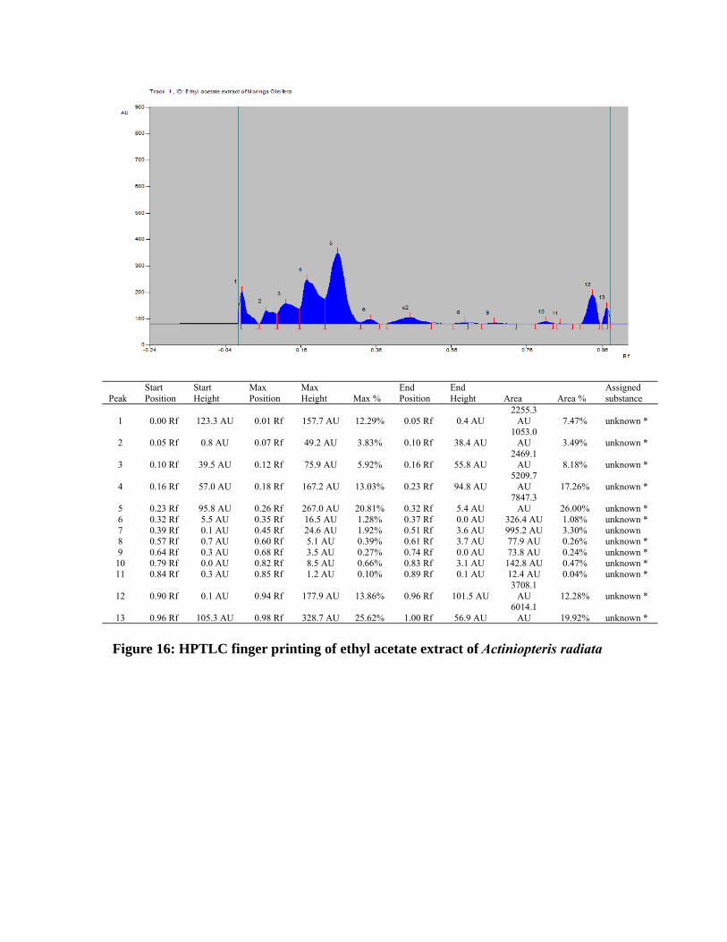
PeakStart Position
Start Height
Max Position
Max Height Max %
End Position
End Height Area Area %
Assigned substance
1 0.00 Rf 123.3 AU 0.01 Rf 157.7 AU 12.29% 0.05 Rf 0.4 AU2255.3
AU 7.47% unknown *
2 0.05 Rf 0.8 AU 0.07 Rf 49.2 AU 3.83% 0.10 Rf 38.4 AU1053.0
AU 3.49% unknown *
3 0.10 Rf 39.5 AU 0.12 Rf 75.9 AU 5.92% 0.16 Rf 55.8 AU2469.1
AU 8.18% unknown *
4 0.16 Rf 57.0 AU 0.18 Rf 167.2 AU 13.03% 0.23 Rf 94.8 AU5209.7
AU 17.26% unknown *
5 0.23 Rf 95.8 AU 0.26 Rf 267.0 AU 20.81% 0.32 Rf 5.4 AU7847.3
AU 26.00% unknown *6 0.32 Rf 5.5 AU 0.35 Rf 16.5 AU 1.28% 0.37 Rf 0.0 AU 326.4 AU 1.08% unknown *7 0.39 Rf 0.1 AU 0.45 Rf 24.6 AU 1.92% 0.51 Rf 3.6 AU 995.2 AU 3.30% unknown8 0.57 Rf 0.7 AU 0.60 Rf 5.1 AU 0.39% 0.61 Rf 3.7 AU 77.9 AU 0.26% unknown *9 0.64 Rf 0.3 AU 0.68 Rf 3.5 AU 0.27% 0.74 Rf 0.0 AU 73.8 AU 0.24% unknown *10 0.79 Rf 0.0 AU 0.82 Rf 8.5 AU 0.66% 0.83 Rf 3.1 AU 142.8 AU 0.47% unknown *11 0.84 Rf 0.3 AU 0.85 Rf 1.2 AU 0.10% 0.89 Rf 0.1 AU 12.4 AU 0.04% unknown *
12 0.90 Rf 0.1 AU 0.94 Rf 177.9 AU 13.86% 0.96 Rf 101.5 AU3708.1
AU 12.28% unknown *
13 0.96 Rf 105.3 AU 0.98 Rf 328.7 AU 25.62% 1.00 Rf 56.9 AU6014.1
AU 19.92% unknown *
Figure 16: HPTLC finger printing of ethyl acetate extract of Actiniopteris radiata
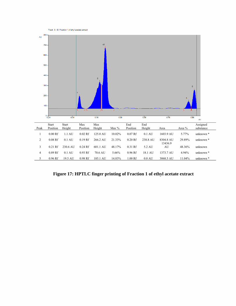
PeakStart Position
Start Height
Max Position
Max Height Max %
End Position
End Height Area Area %
Assigned substance
1 0.00 Rf 1.1 AU 0.02 Rf 125.0 AU 10.02% 0.07 Rf 0.1 AU 1603.9 AU 5.77% unknown *
2 0.08 Rf 0.1 AU 0.19 Rf 266.2 AU 21.33% 0.20 Rf 238.8 AU 8304.8 AU 29.89% unknown *
3 0.21 Rf 230.6 AU 0.24 Rf 601.1 AU 48.17% 0.31 Rf 5.2 AU13436.9
AU 48.36% unknown
4 0.89 Rf 0.1 AU 0.93 Rf 70.6 AU 5.66% 0.96 Rf 18.1 AU 1373.7 AU 4.94% unknown *
5 0.96 Rf 19.5 AU 0.98 Rf 185.1 AU 14.83% 1.00 Rf 0.0 AU 3068.5 AU 11.04% unknown *
Figure 17: HPTLC finger printing of Fraction 1 of ethyl acetate extract
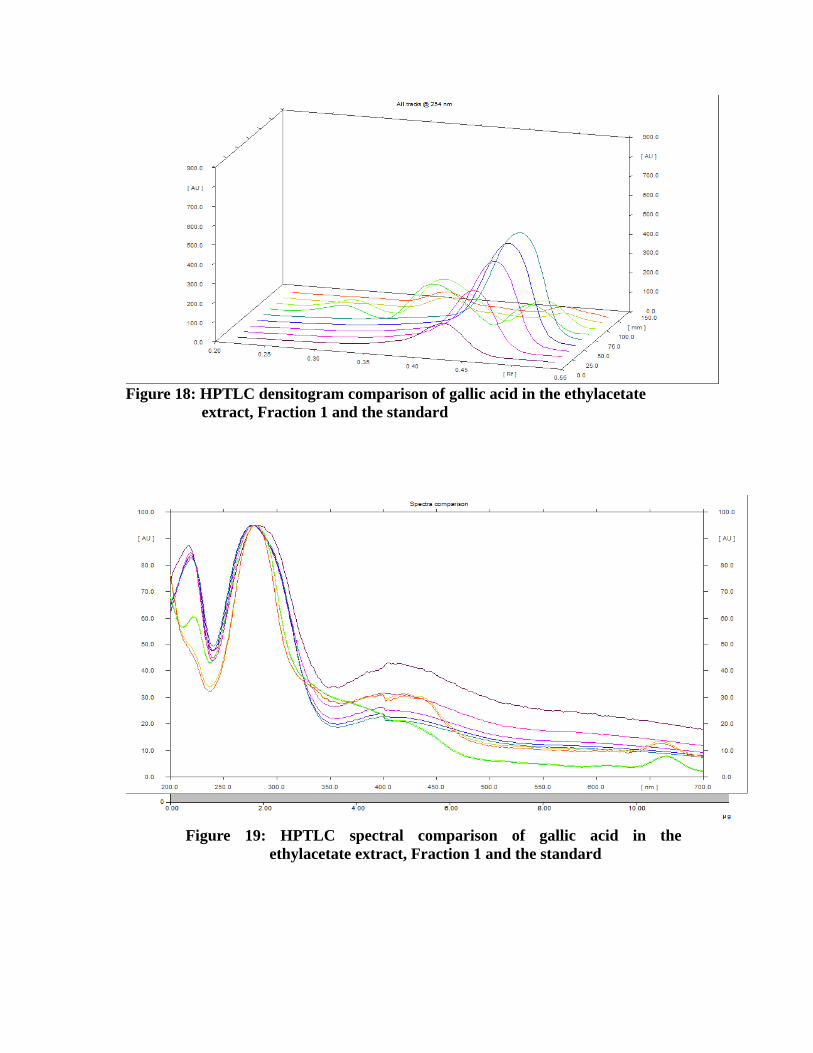
Figure 18: HPTLC densitogram comparison of gallic acid in the ethylacetate extract, Fraction 1 and the standard
Figure 19: HPTLC spectral comparison of gallic acid in the ethylacetate extract, Fraction 1 and the standard

Figure 20: Linear calibration curve of gallic acid standard
Table 7: Quantitative estimation of gallic acid content in ethyl acetate extract and Fraction 1 using regression equation
Track Vial RfAmount Fraction(µg)
AreaCalculatedamount (ng)
Remark % w/wAverage % w/w (mean ± SD)
1 1 0.42 1.000 5821.09 Std Level 12 1 0.45 2.000 11534.16 Std Level 23 1 0.46 4.000 16785.71 Std Level 34 1 0.47 6.000 19933.95 Std Level 45 1 Std Level 56 2 0.38 6858.04 787.25 EA extract 0.393564
0.385 ± 0.0127 2 0.39 6764.97 752.84 EA extract 0.3763618 3 0.38 1764.41 <0.0 g Fraction 1 Out of
permittedrange
Out ofpermitted
range9 3 0.37 1757.5 <0.0 g Fraction 1
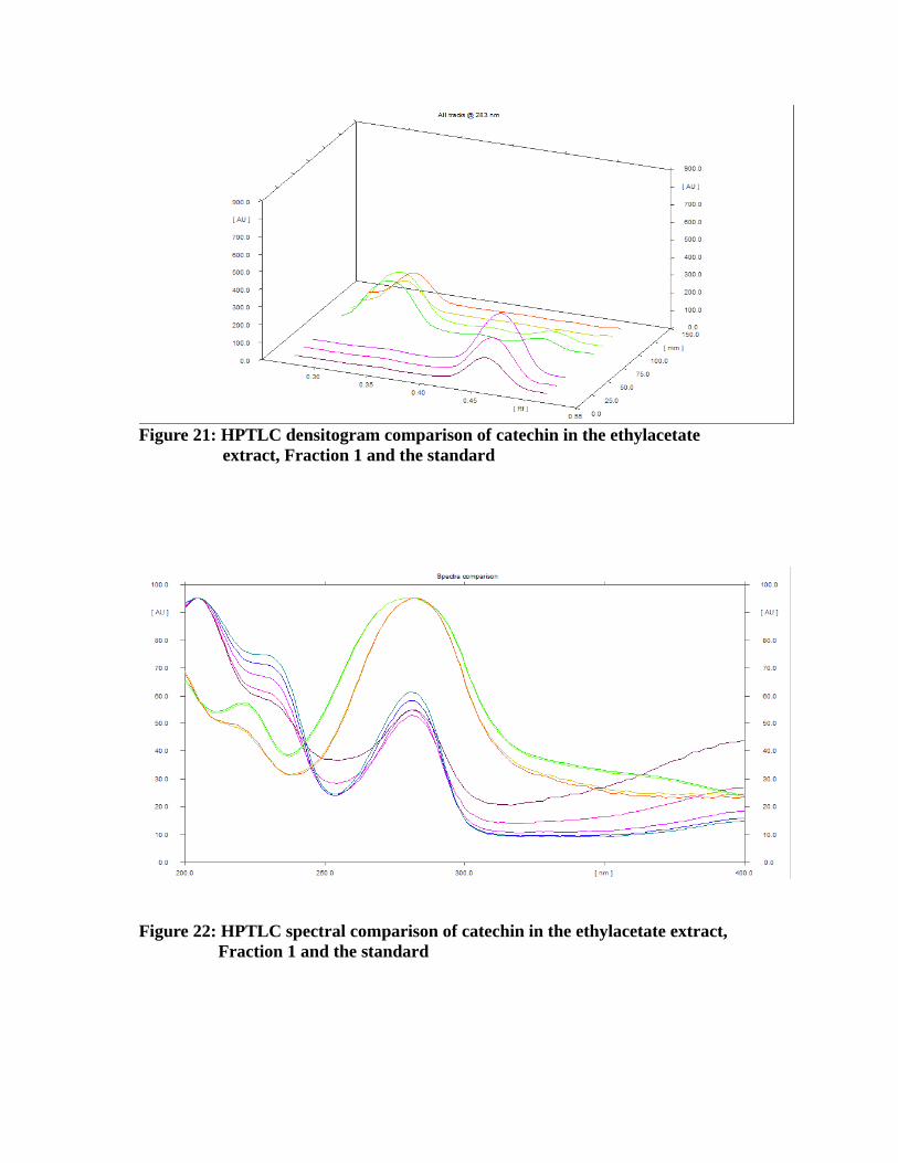
Figure 21: HPTLC densitogram comparison of catechin in the ethylacetate extract, Fraction 1 and the standard
Figure 22: HPTLC spectral comparison of catechin in the ethylacetate extract, Fraction 1 and the standard
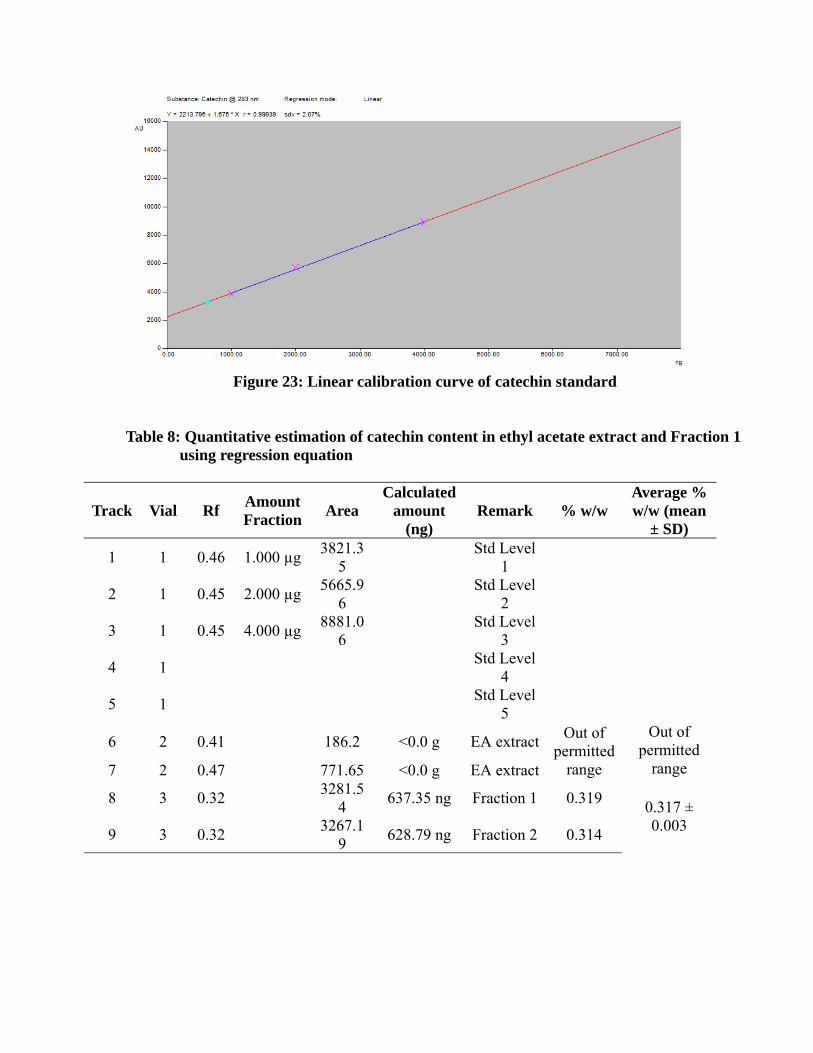
Figure 23: Linear calibration curve of catechin standard
Table 8: Quantitative estimation of catechin content in ethyl acetate extract and Fraction 1using regression equation
Track Vial Rf AmountFraction Area
Calculatedamount
(ng)Remark % w/w
Average %w/w (mean
± SD)
1 1 0.46 1.000 µg 3821.35
Std Level1
2 1 0.45 2.000 µg 5665.96
Std Level2
3 1 0.45 4.000 µg 8881.06
Std Level3
4 1 Std Level4
5 1 Std Level5
6 2 0.41 186.2 <0.0 g EA extract Out ofpermitted
range
Out ofpermitted
range7 2 0.47 771.65 <0.0 g EA extract
8 3 0.32 3281.54 637.35 ng Fraction 1 0.319 0.317 ±
0.0039 3 0.32 3267.19 628.79 ng Fraction 2 0.314
5.8 In vitro antioxidant studies of Actiniopteris radiata
The petroleum ether, chloroform, ethyl acetate and ethanol extracts were screened for in vitro
antioxidant activity. The in vitro antioxidant studies includes DPPH scavenging assay, ABTS
scavenging assay, Nitric oxide assay, Super oxide assay and Deoxyribose assay. Among the
extracts tested for in vitro antioxidant activity, ethanol and ethyl acetate extract exhibited
potent antioxidant activity in DPPH, ABTS, Nitric oxide, Super oxide and Deoxyribose
methods. The values of extracts are compared with the values of standards ascorbic acid and
rutin.
5.8.1 DPPH radical scavenging assay
DPPH generated radical was tested for Actiniopteris radiata along with the rutin and ascorbic
acid. DPPH is a stable free radical. The assay is based on the measurement of the scavenging
ability of antioxidants towards stable radical DPPH.. Actiniopteris radiata extract reduces the
radical to the corresponding hydrazine when it reacts with the hydrogen donors in the
antioxidants. DPPH radicals react with suitable reducing agents, the electrons become paired
off and the solution loses colour stoichiometrically depending on the number of electrons
taken up. The ethanol extract of Actiniopteris radiata has shown potent antioxidant activity
with half inhibition concentration (IC50) of 1.98 ± 0.13 µg/ml.
5.8.2 ABTS radical cation scavenging assay
The ABTS assay is based on the inhibition of the absorbance of the radical cation ABTS+.
The ABTS chemistry involves direct generation of ABTS radical mono cation with no
involvement of any intermediary radical. It is a decolorisation assay, thus the radical cation is
performed prior to addition of antioxidant test system. The extract act either by inhibiting or
scavenging the ABTS+ radicals. The ethanol extract of Actiniopteris radiata has shown
potent antioxidant activity with half inhibition concentration (IC50) of 47.48 ± 0.91 µg/ml.
5.8.3 Superoxide anion radical scavenging assay
In Superoxide anion scavenging activity, superoxide anions damage biomolecules directly or
indirectly by forming H2O2, .OH, peroxy nitrite or singlet oxygen during aging and
pathological events. Superoxide directly initiate lipid peroxidation. The superoxide radical
scavenging activity of Actiniopteris radiata extracts were assayed by the PMS-NADH
system. The superoxide scavenging activity of Actiniopteris radiata extracts were increased
markedly with the increase in concentrations. The ethylacetate extract has shown potent
antioxidant activity with the half inhibition concentration (IC50) was 18.62 ± 3.82 µg/ml.
These results suggested that Actiniopteris radiata extract has a potent superoxide radical
scavenging effects.
5.8.4 Nitric oxide radical scavenging assay
In Nitric oxide scavenging activity, nitric oxide (NO) is a free radical produced in
mammalian cells, involved in the regulation of various physiological processes. The excess
production of NO is associated with several diseases. Nitric oxide is very unstable species
under aerobic condition. It reacts with oxygen to produce stable product nitrate and nitrite
through intermediates NO2, N2O4 and N3O4. The nitrite produced by the incubation of
solution of sodium nitroprusside in standard phosphate buffer at 25oC was reduced by the
Actiniopteris radiata extract. This may be due to the antioxidant principles in the extract
which compete with oxygen to react with nitric oxide and thus inhibits the generation of
nitrite. The ethanol extract has potent antioxidant activity with IC50 value of 109.40 ± 6.06
µg/ml.
5.8.5 Deoxyribose degradation assay
In Deoxyribose method, increasing concentration of antioxidant reduces DNA expression.
The control DNA exhibits both super-coiled and open circular forms. Incorporation of test
compounds damages the super-coiled form and at the same time increases the expression of
open circular form. The test compounds damage the super-coiled form. Further the damage
exerted by H2O2 and FeCl3 was reduced at higher concentrations of Actiniopteris radiata
extract. The ethyl acetate extract of Actiniopteris radiata has shown antioxidant activity with
half inhibition concentration (IC50) of 144.30 ± 8.79 µg/ml. The half inhibition concentration
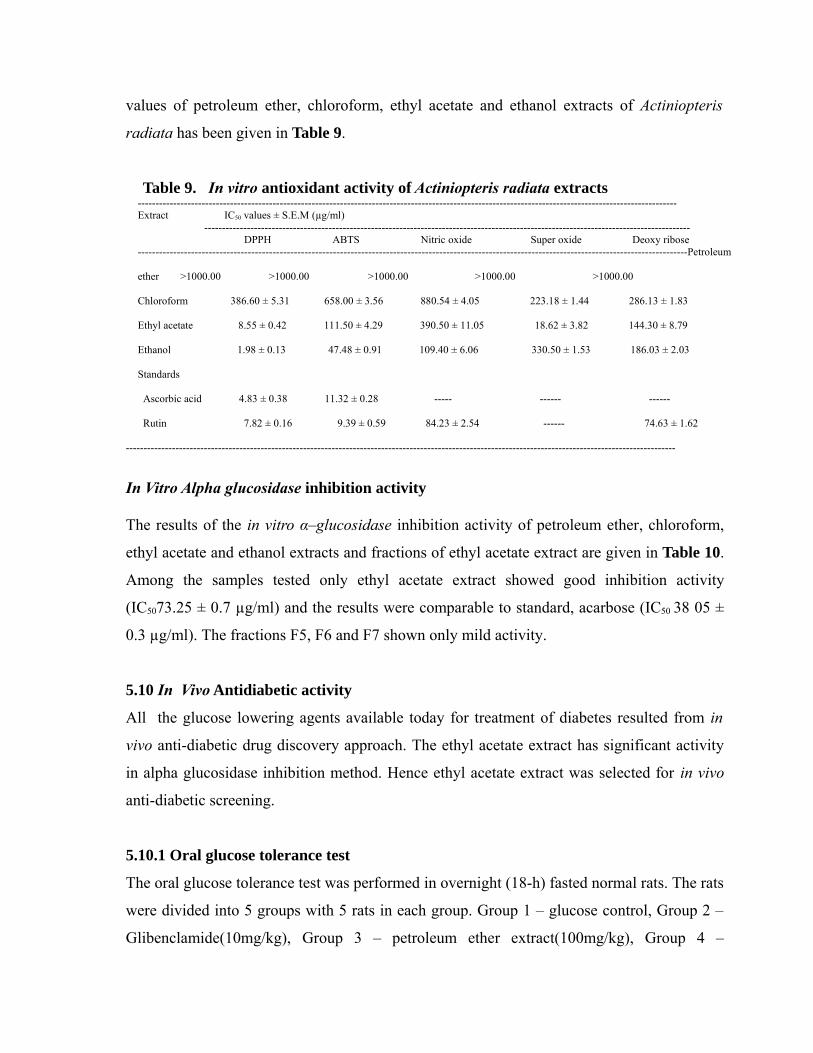
values of petroleum ether, chloroform, ethyl acetate and ethanol extracts of Actiniopteris
radiata has been given in Table 9.
Table 9. In vitro antioxidant activity of Actiniopteris radiata extracts --------------------------------------------------------------------------------------------------------------------------------------------------------Extract IC50 values ± S.E.M (µg/ml) ----------------------------------------------------------------------------------------------------------------------------------------- DPPH ABTS Nitric oxide Super oxide Deoxy ribose-----------------------------------------------------------------------------------------------------------------------------------------------------------Petroleum
ether >1000.00 >1000.00 >1000.00 >1000.00 >1000.00
Chloroform 386.60 ± 5.31 658.00 ± 3.56 880.54 ± 4.05 223.18 ± 1.44 286.13 ± 1.83
Ethyl acetate 8.55 ± 0.42 111.50 ± 4.29 390.50 ± 11.05 18.62 ± 3.82 144.30 ± 8.79
Ethanol 1.98 ± 0.13 47.48 ± 0.91 109.40 ± 6.06 330.50 ± 1.53 186.03 ± 2.03
Standards
Ascorbic acid 4.83 ± 0.38 11.32 ± 0.28 ----- ------ ------
Rutin 7.82 ± 0.16 9.39 ± 0.59 84.23 ± 2.54 ------ 74.63 ± 1.62
-----------------------------------------------------------------------------------------------------------------------------------------------------------
In Vitro Alpha glucosidase inhibition activity
The results of the in vitro α–glucosidase inhibition activity of petroleum ether, chloroform,
ethyl acetate and ethanol extracts and fractions of ethyl acetate extract are given in Table 10.
Among the samples tested only ethyl acetate extract showed good inhibition activity
(IC5073.25 ± 0.7 µg/ml) and the results were comparable to standard, acarbose (IC50 38 05 ±
0.3 µg/ml). The fractions F5, F6 and F7 shown only mild activity.
5.10 In Vivo Antidiabetic activity
All the glucose lowering agents available today for treatment of diabetes resulted from in
vivo anti-diabetic drug discovery approach. The ethyl acetate extract has significant activity
in alpha glucosidase inhibition method. Hence ethyl acetate extract was selected for in vivo
anti-diabetic screening.
5.10.1 Oral glucose tolerance test
The oral glucose tolerance test was performed in overnight (18-h) fasted normal rats. The rats
were divided into 5 groups with 5 rats in each group. Group 1 – glucose control, Group 2 –
Glibenclamide(10mg/kg), Group 3 – petroleum ether extract(100mg/kg), Group 4 –
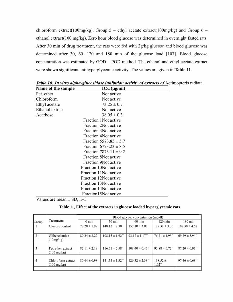
chloroform extract(100mg/kg), Group 5 – ethyl acetate extract(100mg/kg) and Group 6 –
ethanol extract(100 mg/kg). Zero hour blood glucose was determined in overnight fasted rats.
After 30 min of drug treatment, the rats were fed with 2g/kg glucose and blood glucose was
determined after 30, 60, 120 and 180 min of the glucose load [107]. Blood glucose
concentration was estimated by GOD – POD method. The ethanol and ethyl acetate extract
were shown significant antihyperglycemic activity. The values are given in Table 11.
Table 10: In vitro alpha-glucosidase inhibition activity of extracts of Actiniopteris radiata Name of the sample IC50 (µg/ml)Pet. ether Not active Chloroform Not active Ethyl acetate 73.25 ± 0.7 Ethanol extract Not active Acarbose 38.05 ± 0.3
Fraction 1Not activeFraction 2Not activeFraction 3Not activeFraction 4Not activeFraction 5573.85 ± 5.7Fraction 6773.23 ± 8.5Fraction 7873.11 ± 9.2Fraction 8Not activeFraction 9Not active
Fraction 10Not activeFraction 11Not activeFraction 12Not activeFraction 13Not activeFraction 14Not activeFraction15Not active
Values are mean ± SD, n=3
Table 11, Effect of the extracts in glucose loaded hyperglycemic rats.
Group Treatments
Blood glucose concentration (mg/dl)0 min 30 min 60 min 120 min 180 min
1 Glucose control 78.28 ± 1.99 148.12 ± 2.30 157.10 ± 3.88 127.31 ± 3.30 102.30 ± 4.52
2 Glibenclamide(10mg/kg)
80.24 ± 2.22 108.15 ± 1.62** 93.17 ± 1.17** 76.21 ± 1.95** 69.29 ± 3.94**
3 Pet. ether extract (100 mg/kg)
82.11 ± 2.18 116.31 ± 2.58* 108.40 ± 0.46** 95.88 ± 0.72** 87.20 ± 0.91**
4 Chloroform extract(100 mg/kg)
80.64 ± 0.98 141.34 ± 1.32** 126.32 ± 2.38** 118.52 ± 1.62**
97.46 ± 0.68**
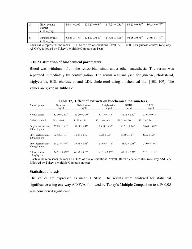
5 Ethyl acetate extract (100 mg/kg)
84.04 ± 2.07 128.20 ± 0.64* 117.28 ± 0.35** 94.25 ± 0.56** 86.24 ± 0.77**
6 Ethanol extract (100 mg/kg)
82.21 ± 1.73 124.32 ± 0.82* 118.45 ± 1.20** 90.25 ± 0.71** 74.04 ± 1.40**
Each value represents the mean ± S.E.M of five observations. *P<0.05, **P<0.001 vs glucose control (one wayANOVA followed by Tukey’s Multiple Comparison Test)
5.10.2 Estimation of biochemical parameters
Blood was withdrawn from the retroorbital sinus under ether anaesthesia. The serum was
separated immediately by centrifugation. The serum was analysed for glucose, cholesterol,
triglyceride, HDL cholesterol and LDL cholesterol using biochemical kits [108, 109]. The
values are given in Table 12.
Table 12, Effect of extracts on biochemical parameters.Animal group S.glucose S.cholesterol S.triglyceride S.HDL S.LDL mg/dl mg/dl mg/dl mg/dl mg/dl
Normal control 82.10 ± 3.62** 65.30 ± 3.10** 63.15 ± 3.46** 52.13 ± 2.60** 23.01 ± 0.48**
Diabetic control 503.18 ± 6.31 86.23 ± 4.31 121.53 ± 3.68 38.71 ± 1.30 33.47 ± 2.36
Ethyl acetate extract 77.98 ± 1.62** 56.11 ± 1.42** 93.59 ± 3.32** 45.31 ± 0.86** 28.63 ± 0.82** 100mg/kg b.w
Ethyl acetate extract 72.92 ± 1.15** 51.48 ± 2.19** 53.06 ± 0.74** 51.80 ± 1.07** 29.62 ± 0.74**
200mg/kg b.w
Ethyl acetate extract 68.33 ± 1.46** 59.53 ± 1.41** 58.69 ± 1.70** 48.92 ± 0.88** 28.07 ± 1.01**
400mg/kg b.w
Glibenclamide 76.31± 0.828** 61.32 ± 3.20** 61.21± 2.30** 46.18 ± 0.72** 22.31 ± 2.51**
10mg/kg b.w Each value represents the mean ± S.E.M of five observations. **P<0.001 vs diabetic control (one way ANOVAfollowed by Tukey’s Multiple Comparison test)
Statistical analysis
The values are expressed as mean ± SEM. The results were analysed for statistical
significance using one-way ANOVA, followed by Tukey’s Multiple Comparison test. P<0.05
was considered significant.
6. DISCUSSION
The present study was undertaken to examine the antidiabetic activity of Actiniopteris
radiata. The effect of extract in diabetes changes in associated complications, biochemical
parameters was also assessed. Wistar rats of either sex were induced diabetic by
streptozotocin. The blood glucose level above 200 mg/dl were considered diabetic and used
for this experiment. The diabetic rats were treated for 7 days with ethyl acetate extract and
glibenclamide. Biochemical parameters were estimated after treatment.
In the glucose loaded hyperglycemic model, the extracts were tested for antihyperglycemic
activity, ethanol and ethyl acetate extracts were exhibited significant antihyperglycemic
activity at a dose level of 100 mg/kg. Chloroform extract has least antihyperglycemic
activity at a dose level of 100 mg/kg. Excessive amount of glucose in the blood induces the
insulin secretion. This secreted insulin will stimulate peripheral glucose consumption and
control the production of glucose through different mechanisms [110]. However, from the
study (glucose control), it was clear that the secreted insulin requires 2-3 h to bring back the
glucose level to normal.
In general, an increase in blood glucose level is usually accompanied by an increase in
plasma cholesterol, triglyceride, LDL levels and a decrease in HDL levels as observed in
diabetic rats [111]. The marked hyperlipidemia (increase in the level of lipid in the body)
that characterizes the diabetic state may be the consequence of the uninhibited actions of
lipolytic hormones on fat depots [112]. The ethyl acetate extract at a dose of 400 mg/kg b.w
has significant activity than lower dose of ethyl acetate extract (100 mg/kg b.w). The
significant reduction in the blood glucose level comparable to that produced by
glibenclamide treatment [113]. Further ethyl acetate extract was purified by column
chromatography that led to isolation of a two new compounds [114]. The flavones are
present in the ethyl acetate extract, it resulted in a decrease in plasma glucose and increase
in insulin levels. The flavones also mimics the effects of insulin [115].
In conclusion, this study has shown that ethyl acetate extract of Actiniopteris radiata has
significant antidiabetic activity. This research supports the inclusion of this plant in
antidiabetic preparations and useful in development of antidiabetic drug.
7. SUMMARY AND CONCLUSION
The traditional herbal medications are become mainstream throughout the world. Since
ancient times, plants have been source of medicines. Ayurveda and other Indian literature
mention the use of plants in treatment of various human diseases. India has about 45000
plant species and among them several thousands have medicinal properties. Plants are
major source of drugs and many of the currently available drugs have been derived
directly or indirectly from them.
Since ancient times, plants have been used in the treatment of diabetes mellitus. There are
many hypoglycemic plants and their active principles varies. Therefore considerable
diversity in the mechanism of action. Some act by increasing the release of insulin and
require a minimum of β-cells to exert their action. Other plant extracts or constituents act
by modifying glucose metabolism. All are important since they are used to treat the
different aspects of diabetes mellitus.
The selected plant Actiniopteris radiata was collected from Nilgiri district, Tamilnadu
and authenticated. The dried plant material was subjected to successive extraction with
petroleum ether, chloroform, ethyl acetate and ethanol by soxhlet method. The extracts
were concentrated under reduced pressure and controlled temperature.
The phytochemical studies of the extracts gave positive test for the flavanoids,
carbohydrates, hydrocarbons, fatty acids, sterols, steroids, steroidal glycoside and
unknown glycoside. Determination of water soluble extract, alcohol soluble extract, total
ash, acid insoluble ash and water soluble ash were carried out. The quantitative
phytochemical estimations total phenol content and total flavonoid content of the extracts
were estimated.
Qualitative and quantitative determinations of ethyl acetate extract and fractions were
done by HPTLC method.
In vitro antioxidant studies DPPH, ABTS, Nitric oxide, Super oxide and Deoxy ribose
were carried out. The ethyl acetate extract shown potent in vitro antioxidant activity. The
ethyl acetate extract of Actiniopteris radiata was selected for in vivo anti-diabetic activity
based on the in vitro antioxidant activity.
In vitro antidiabetic activity was performed by alpha glucosidase inhibition activity. The
successive extracts of Actiniopteris radiata and fractions of ethyl acetate extract were
screened for alpha glucosidase inhibition activity. The results was compared with the
values of standard (acarbose). The ethyl acetate extract has shown significant antidiabetic
activity and fraction 5, 6, 7 shown moderate antidiabetic activity. The ethyl acetate
extract was selected for in vivo antidiabetic activity based on the results of in vitro
antidiabetic activity.
Wistar rats of either sex weighing 180-220 g (6 to 8 weeks) with no prior drug treatment
were used for in vivo anti-diabetic activity. The acute toxicity study of the ethyl acetate
extract was determined according to the O E C D guidelines No.425. Female Wistar rats
weighing 180-220 g (6 to 8 weeks) were used for this study. The test samples in a single
dose of 400 mg/kg b.w, 800 mg/kg b.w and 2 g/kg b.w were given orally. The animals
were observed for 24 hours and monitored for 14 days to record general behaviour and
mortality. No mortality was observed till the end of the study.
Streptozotocin was used to induce diabetes. Streptozotocin 55 mg/kg b.w was
administered intraperitoneally. The rats with blood glucose level above 200 mg/dl were
considered diabetic and used in the experiment.
The oral glucose tolerance test was performed in overnight fasted normal rats. Zero hour
blood sugar was determined in overnight fasted rats. After 30 min of drug treatment, the
rats were fed with 2 g/kg b.w glucose and blood glucose was determined after 30, 60, 120
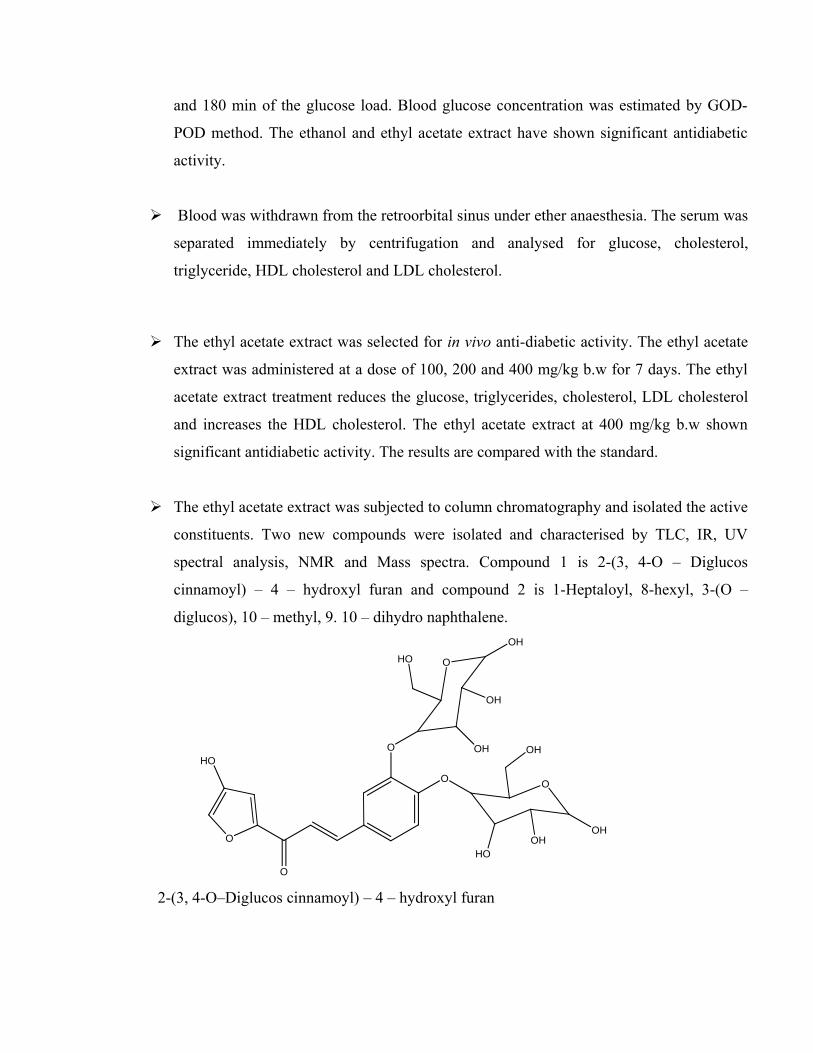
and 180 min of the glucose load. Blood glucose concentration was estimated by GOD-
POD method. The ethanol and ethyl acetate extract have shown significant antidiabetic
activity.
Blood was withdrawn from the retroorbital sinus under ether anaesthesia. The serum was
separated immediately by centrifugation and analysed for glucose, cholesterol,
triglyceride, HDL cholesterol and LDL cholesterol.
The ethyl acetate extract was selected for in vivo anti-diabetic activity. The ethyl acetate
extract was administered at a dose of 100, 200 and 400 mg/kg b.w for 7 days. The ethyl
acetate extract treatment reduces the glucose, triglycerides, cholesterol, LDL cholesterol
and increases the HDL cholesterol. The ethyl acetate extract at 400 mg/kg b.w shown
significant antidiabetic activity. The results are compared with the standard.
The ethyl acetate extract was subjected to column chromatography and isolated the active
constituents. Two new compounds were isolated and characterised by TLC, IR, UV
spectral analysis, NMR and Mass spectra. Compound 1 is 2-(3, 4-O – Diglucos
cinnamoyl) – 4 – hydroxyl furan and compound 2 is 1-Heptaloyl, 8-hexyl, 3-(O –
diglucos), 10 – methyl, 9. 10 – dihydro naphthalene.
O
O
O
O
O
HO
OH
OH
HOOH
O
HO
OHOH
OH
2-(3, 4-O–Diglucos cinnamoyl) – 4 – hydroxyl furan

O
CO CH2 (CH2)4 CH3
H2C
(H2C)4
H3C
OOO
OH
HO
HO
OH
OHOH
OH
1-Heptaloyl, 8-hexyl, 3-(O–diglucos), 10 – methyl, 9. 10 – dihydro naphthalene.
Objectives achieved
1. The potent in vitro antioxidant activity was found in the ethyl acetate extract of
Actiniopteris radiata.
2. The significant antidiabetic activity of ethyl acetate extract was found in alpha
glucosidase inhibition activity.
3. The potent in vivo antidiabetic activity was found in the ethyl acetate extract of
Actiniopteris radiata.
4. Two new compounds were isolated and characterised by TLC, IR, UV spectral analysis,
NMR and Mass spectra. Compound 1 is 2-(3, 4-O–Diglucos cinnamoyl) – 4 – hydroxyl
furan and compound 2 is 1-Heptaloyl, 8-hexyl, 3-(O–diglucos), 10 – methyl, 9. 10–
dihydro naphthalene.
5. The in vitro antidiabetic, in vitro antioxidant and in vivo antidiabetic activities were
performed for the first time for Actiniopteris radiata.
Scope for further research
There is scope for further research in isolating the other phytocnstituents and to carry out the
other biological activities of the extract and its phytoconstituents. There is need to establish
the mechanism of action of other biological activities.
8. REFERENCES
1. Emmanuelle Hay, Marie-Christine Aumond, Sabine Mallet, Vincent Dumontet, Marc
Litaudon, Rondeau and Pascal Richomme. Antioxidant xanthones from Garcinia
vieillardii. J. Nat. Prod. 2004; 67, 707-709.
2. Zhizhen Zhang, Hala N. Elsohly, Xing-Cong Li, Shabana I. Khan, Sheldon E. Broedel,
Jr. Robert E. Rauli, Ronald L. Cihlar and Larry A. Walker. Flavanone glycosides from
Miconia trailii. J. Nat. Prod. 2003; 66, 39-41.
3. Hatem Braham, Zine Mighri, Hichem Ben Jannet, Susan Matthew and Pedro M. Abreu.
Antioxidant phenolic glycosides from Moricandia arvensis. J. Nat. Prod. 2005; 68, 517-
522.
4. Ihsan Calis, Jorg Heilmann, Deniz Tasdemir, Anthony Linden, Chris M. Ireland and
Otto Sticher. flavonoid, iridoid and lignan glycosides from Putoria calabrica. J. Nat.
Prod. 2001; 64, 961-964.
5. Patricia Maximo, Ana Lourenco, Sonia Savluchinske Feio and Jose Carlos Roseiro.
Flavonoids from Ulex airensis and Ulex europaeus ssp.europaeus. J. Nat. Prod. 2002;
65, 175-178.
6. Nour-Eddine Es-Safi, Samira Khlifi, Lucien Kerhoas, Albert Kollmann, Ahmed El
Abbouyi and Paul-Henri Ducrot. Antioxidant constituents of the aerial parts of
Globularia alypum growing in Morocco. J. Nat. Prod. 2005; 68, 1293-1296.
7. Marcy J. Balunas and Douglas Kinghorn A. Drug discovery from medicinal plants. Life
Sci. 2005; 78, 431-441.
8. Bang Yeon Hwang, Hang Sub Kim, Jeong Hyeong Lee, Young Soo Hong, Jai Seup Ro,
Kyong Soon Lee and Jung Joon Lee. Antioxidant benzoylated flavan-3-ol glycoside
from Celastrus orbiculatus. J. Nat. Prod. 2001; 64, 82-84.
9. Jian-Hua Zou, Junshan Yang and Liang Zhou. Acylated flavone C-glycosides from
Trollius ledebouri. J. Nat. Prod. 2004; 67, 664-667.
10. Ramawat K.G. Herbal drugs: Ethnomedicine to modern medicine. 2010; 2-30.
11. Giuseppina Cioffi, Luis Morales Escobar, Alessandra Braca and Nunziatina De
Tommasi. Antioxidant chalcone glycosides and flavanones from Maclura tinctoria. J.
Nat. Prod. 2003; 66, 1061-1064.
12. Yuping Tang, Biao Yu, Jie Hu, Tao Wu and Hongzheng Hui. Three new
homoisoflavanone glycosides from the bulbs of Ornithogalum caudatum. J. Nat. Prod.
2002; 65, 218-220.
13. Habsah Mohamad, Nordin H. Lajis, Faridah Abas, Abdul Manaf Ali, Mohamad
Aspollah Sukari, Hiroe Kikuzaki and Nobuji Nakatani. Antioxidative constituents of
Etlingera elatior. J. Nat. Prod. 2005; 68, 285-288.
14. Leila Zanatta, Ilana Seifriz, Tania Beatriz Creczynski – Pasa, Moacir Geraldo Pizzolatti
and Fatima Regina Mena Barreto Silva. Hypoglycemic effect and antioxidant potential
of kempferol-3, 7-O-(α)-dirhamnoside from Bauhinia forficata leaves. J. Nat. Prod.
2004; 67, 829-832.
15. Kumar A, llavarasan R, Jayachandran T, Decaraman M and Aravindhan P.
Phytochemicals investigation on a tropical plant, Syzygium cumini from Kattupalayam,
Erode district, Tamilnadu, South India. Pak J Nutr. 2009; 8(1), 83-85.
16. Zhen Dan He, Kit-Man Lau, Paul Pui-Hay But, Ren-Wang Jiang, Hui Dong, Shuang-
Cheng Ma, kwok-pui Fung, Wen-Cai Ye and Han-Dong Sun. Antioxidative glycosides
from the leaves of Lingustrum robustum. J. Nat. Prod. 2003; 66, 851-854.
17. Alsuko Itoh, Takao Tanahashi, Naotaka Nagakura, Yukiko Takenaka, Cheng-Chang
Chen. and Jury Pelletier. Flavonoid glycosides from Adina racemosa and their inhibitory
activities on eukaryotic protein synthesis. J. Nat. Prod. 2004; 67, 427-431
18. Boonchoo Sritularak, Kittisak Likhitwitayawuid, Jurgen Conrad, Bernhard Vogler,
Sabine Reeb, Iris Klaiber and Wolfgang Kraus. New flavones from Millettia
erythrocalyx. J. Nat. Prod. 2002; 65, 589-591.
19. Sabu. M. C and Subburaju.T. Effect of Cassia auriculata Linn on serum glucose level,
glucose utilization by isolated rat hemidiaphragm. J Ethnopharmacol. 2002; 80, 203-
206.
20. Bhushan Patwardhan, Ashok D. B. Vaidya and Mukund Chorghade. Ayurveda and
natural products drug discovery. J Nat Prod. 2004; 86, 789-799.
21. Nabil Semmar, Bernard Fenet, Marie-Aleth Lacaille-Dubois, Katia Gluchoff-Fiasson,
Rachid Chemli and Maurice Jay. Two new glycosides from Astragalus caprinus. J. Nat.
Prod. 2001; 64, 656-658.
22. Alan L. Harvey. Medicines from nature: are natural products still relevant to drug
discovery. TiPS. 1999; 20, 196-198.
23. Bnouham Mohamed, Abderrahim Ziyyat, Hassane Mekhfi, Abdelhafid Tahri. and
Abdelkhaleq Legssyer. Medicinal plants with potential antioxidant activity- A review of
ten years of herbal medicine research,(1990-2000). Int J Diabetes and Metabolism.
2006; 14, 1-25.
24. Grover J.K, Yadav S. and Vats V. Medicinal plants of India with anti-diabetic potential.
J Ethnopharmacol. 2002; 81, 81-100.
25. Mark S. Butler. The role of natural product chemistry in drug discovery. J. Nat. Prod.
2004; 67, 2141-2153.
26. Frank E. Koehn and Guy T. Carter. The evolving role of natural products in drug
discovery. Nat. Rev. 2005; 4, 206-220.
27. Agarwal S.S and Paridhavi M. Herbal drug technology. 2007; 1-26.
28. Tanko.Y, Okasha. M. A, Magaji. G. M, Yerima. M, Yaro. A. H, Saleh. M. I. A and
Mohammed. A. Antidiabetic properties of Securinega virosa leaf extract. African J
Biotec. 2008; 7(1), 022-024.
29. Babu. B. H, Shylesh. B. S and Padikkala. J. Antioxidant and hepatoprotective effect of
Acanthus ilicifolius. Fitoterapia. 2001; 72, 272-277.
30. Yoshihiro Mimaki, Kazuki Watanabe, Yuka Ando, Chiseko Sakuma. and Yutaka
Sashida. Flavonol glycosides and steroidal saponins from the leaves of Cestrum
nocturnum and their cytotoxicity. J. Nat. Prod. 2001; 64, 17-22.
31. Grover J.K, Yadav S. and Vats V. Medicinal plants of India with anti-diabetic potential.
J Ethnopharmacol. 2002; 81, 81-100.
32. Ivorra M.D, Paya M. and Villar A. A Review of natural products and plants as potential
antidiabetic drugs. J Ethnopharmacol. 1989; 27, 243-275.
33. Maryam Zahin, Farrukh Aqil and Iqbal Ahmad. The in vitro antioxidant activity and
total phenolic content of four Indian medicinal plants. Int J Pharm Pharm.Sci. 2009; 1,
88-95.
34. Marcy J. Balunas and Douglas Kinghorn A. Drug discovery from medicinal plants. Life
Sci. 2005; 78, 431-441.
35. Papiya Mitra Mazumder, Mamta Farswan and Parcha V. Effect of an isolated active
compound (Cg-1) of Cassia glauca leaf on blood glucose, lipid profile and atherogenic
index in diabetic rats. Ind J Pharmacol. 2009; 41(4), 182-186.
36.Manisha Modak, Priyanjali Dixit, Jayant Londho, Saroj Ghaskadbi and Thomas Paul A.
Devasagayam. Indian herbs and herbal drugs used for the treatment of diabetes. J Clin
Biochem Nutr. 2007; 40(3), 163-173.
37.Mankil Jung, Moonsoo Park, Hyun Chul Lee, Yoon-Ho Kang, Eun Seok Kang and Sang
Ki Kim. Anti-diabetic medicinal plants. Cur Med Chem. 2006; 13, 1203-1218.
38.Wei Jia, Wenyuan Gao and Lida Tang. Antidiabetic herbal drugs officially approved in
China. Phytother. Res. 2003; 17, 1127-1134.
39.Grover J.K, Yadav S. and Vats V. Medicinal plants of India with anti-diabetic potential.
J Ethnopharmacol. 2002; 81, 81-100.
40.Analava Mitra. Anti-diabetic uses of some common herbs in tribal belts of Midnapur
district of Bengal. Ethno-Med. 2007; 1(1), 37-45.
41.Filomena Conforti, Giancarlo A. Statti, Rosa Tundis, Francesco Menichini and Peter
Houghton. Antioxidant activity of methanolic extract of Hypericum triquetrifolium
Turra aerial part. Fitoterapia. 2002; 73, 479-483.
42.Amro. B, Aburjai. T and Al-Khalil. S. Antioxidative and radical scavenging effects of
olive cake extract. Fitoterapia. 2002; 73, 456-461.
43.Shylesh. B. S and Padikkala. J. Antioxidant and anti-inflammatory activity of Emilia
sonchifolia. Fitoterapia. 1999; 70, 275-278.
44.Helle Wangensteen, Anca Miron, Mahiuddin Alamgir, Sultana Rajia, Anne Berit
Samuelsen and Karl Egil Malterud. Antioxidant and 15-lipoxygenase inhibitory activity
of rotenoids, isoflavones and phenolic glycosides from Sarcolobus globosus.
Fitoterapia. 2006; 77, 290-295.
45.Naznin Ara and Hasan Nur. In vitro antioxidant activity of methanolic leaves and flowers
extracts of Lippia alba. Res J Med Med Sci. 2009; 4(1), 107-110.
46.Sampath Kumar, Ashok K. Tiwari, Venkat Reddy. S, Aparna. P, Jagadeeshwar Rao. R,
Zehra Ali. A and Madhusudana Rao. Free radical scavenging and xanthine oxidase
inhibitory constituents from Stereospermum personatum. J. Nat. Prod. 2005; 68, 1615-
1621.
47.Kardosova. A and Machova. E. Antioxidant activity of medicinal plant polysaccharides.
Fitoterapia. 2006; 77, 367-373.
48.Kirana. H, Girish. H. N and Srinivasan. B. P. Aqueous extract of Pterocarpus marsupium
restores glutathione in Type 2 diabetic rats. Int. J. Pharmacol. Biol. Sci. 2009; 3(2),83-
86.
49.Davies K. J. Intracellular proteolytic systems may function as secondary antioxidant
defenses: a hypothesis. J Free Rad Bio Med. 1986; 2, 155-173.
50.Sohal R. S, Allen R. G and Nations C. Oxygen free radicals play a role in cellular
differentiation: an hypothesis. J Free Rad Bio Med. 1986; 2, 175-181.
51.Halliwell B. Can oxidative DNA damage be used as biomarker of cancer risk in humans?
Problems, resolutions and preliminary results from nutritional supplementation studies.
Free Rad Res. 1998; 29, 469-486.
52.Nishikant A. Raut and Naresh J. Gaikwad. Antidiabetic activity of hydro-ethanolic
extract of Cyperus rotundus in alloxan induced diabetes in rats. Fitoterapia. 2006; 77,
585-588.
53.Gutteridge J. M. Lipid peroxidation and antioxidants as biomarkers of tissue damage.
Clin Chem. 1995; 41, 1819-1828.
54.Jeanette Schultz Johansen, Alex K Harris, David J Rychly and Adviye Ergul. Oxidative
stress and the use of antioxidants in diabetes: Linking basic science to clinical practice.
Cardiovasc Diabetol. 2005; 4, 5.
55.Balasubramanyam M, Rema M and Premanand C. Biochemical and molecular
mechanisms of diabetic retinopathy. Cur Sci. 2002; 83 (12), 1506-1514.
56.Zenon Jastrzebski, Oscar J. Medina, Marlen L. Moreno and Shela Gorinstein. In vitro
studies of polyphenol compounds, total antioxidant capacity and other dietary indices in
a mixture of plants. Int J Food Sci Nutr. 2007; 58(7), 531-541.
57.Srinivasan K. and Ramarao P. Animal models in type 2 diabetes research: An overview.
Indian J Med Res. 2007; 125, 451-472.
58.Okpanachi A.O, Adelaiye A.B, Dikko A.A.U, Kabiru M, Mohammed A and Tanko Y.
Evaluation of the effect of Aqueous-methanolic stem bark extract of Acacia polycantha
on blood glucose levels of alloxan induced diabetic wistar rats. Int J Anim Veter Adv.
2010; 2(3), 59-62.
59.Ivorra M.D, Paya M. and Villar A. A Review of natural products and plants as potential
antidiabetic drugs. J Ethnopharmacol. 1989; 27, 243-275.
60.Babu V, Gangadevi T. and Subramoniam A. Antidiabetic activity of ethanol extract of
Cassia kleinii leaf in streptozotocin induced diabetic rats and isolation of an active
fraction and toxicity evaluation of the extract. Ind J Pharmacol. 2003; 35, 290-296.
61.Ishii H, Jirousek M. R, Koya D, Takagi C, Xia P, Cleromont A, Bursell S. E, Kern T. S,
Ballas L. M, Heath W. F and King G. L. Amelioration of vascular dysfunctions in
diabetic rats by an oral PKCb inhibitor. Science.1996; 272, 728-731.
62.Nandhakumar Jothivel, Sethumathi Pudhupalayam Ponnusamy, Malini Appachi,
Sengottuvelu Singaravel, Duraisamy Rasilingam, Karthikeyan Deivasigamani and
Sivakumar Thangavel. Anti-diabetic activity of methanol leaf extract of Costus pictus
D.DON in alloxan induced diabetic rats. J Health Sci. 2007; 53(6), 655-663.
63.Ranjan Chakrabarti and Ramanujam Rajagopalan. Diabetes and insulin resistance
associated disorders: Disease and therapy. Cur Sci. 2002; 83(12), 1533-1538.
64.Julia E. Chrubasik, Basil D. Roufogalis, Hildebert Wagner, Sigrum A. Chrubasik. A
comprehensive review on nettle effect and efficacy profiles, part I: Herba urticae.
Phytomedicine. 2007; 14, 423-435.
65.Kandasamy Murugesh, Veerendra Yeligar, Deepak Kumar Dash. and Pinaki Sengupta.
antidiabetic, antioxidant and antihyperlipidemic status of Heliotropium zeylanicum
extract on Streptozotocin-Induced diabetes in rats. Biol. Pharm. Bull. 2006; 29(11),
2202-2205.
66.Kirtikar K.R and Basu B.D. Indian Medicinal Plants. 2nd edition. 1999; 55-56.
67.Bhambie S. and George C.X. Studies in Pteridophytes. Cur. Sci. 1972; 41(7), 258-260.
68.Bhambie S, Tiwari H.P and George C.X. Chemical Analysis Actiniopteris radiata. Cur.
Sci. 1972; 41(21), 788.
69.Taneja S.C and Tiwari H.P. Chemical constituents of Actiniopteris radiata. Cur. Sci.
1974; 43(23), 749-750.
70.Naik J.B and Jadge D.R. Evaluation of analgesic activity of Actiniopteris radiata. J
Pharmacy Res. 2010; 3(7), 1556-1557.
71.Vivek R. Alagai and Gautam P. Vadnere. In vitro antihistaminic and anticholinergic
activity of Actiniopteris radiata on isolated rat ileum. Int J Pharm Tech Research. 2011;
3(1), 05-07.
72.Taur D. J, Taware S. B, Patil R. N, Patil R. Y and Kharya M. D. Pharmacognostical and
preliminary phytochemical evaluation of Clitoria ternatea leaves. Pharmacog J. 2010;
2(9), 260-265.
73.Dholwani K. K, Saluja A. K, Bhatt S and Chauhan N. (2009). Pharmacognostical
profiling of roots of Salacia prenoides L. Pharmacog. J., 2009; 1(3), 14-24.
74.Atsuko Itoh, Tomoko Kumashiro, Takao Tanahashi, Naotaka Nagakura and Toyoyuki
Nishi. flavonoid glycosides from Rhazya orientalis. J. Nat. Prod. 2002; 65, 352-357.
75.Chien-Chih Chen, Yu-Lin Huang, Chu-Wen Wang. and Jun-Chih Ou. Water-Soluble
Glycosides from Ruta graveolens. J. Nat. Prod. 2001; 64, 990-992.
76.Nabil Semmar, Marie-Aleth Lacaille-Dubois, Katia Gluchoff-Fiasson, Rachid Chemli.
and Maurice Jay. New glycosides from Astragalus caprinus. J. Nat. Prod. 2002; 65,
176-179.
77.Atsuko Itoh, Takao Tanahashi, Naotaka Nagakura. and Toyoyuki Nishi. Flavonoid
glycosides from Rhazya orientalis. J. Nat. Prod. 2001; 63, 262-268.
78.Boonchoo Sritularak, Jurgen Conrad, Bernhard Vogler, Sabine Reeb, Iris Klaiber. and
Wolfgang Kraus. New Flavones from Millettia erythrocalyx. J. Nat. Prod. 2001; 61,
379-386.
79.Rajkumar S and Senthil Kumar N. Isolation, chemical characterization and hypoglycemic
activity of Alangium salvifolium Wang bark in alloxan induced hyperglycemic rats.
IJPSR. 2011; 2(6), 1518-1524.
80.Atsuko Itoh, Tomoko Kumashiro, Takao Tanahashi, Naotaka Nagakura and Toyoyuki
Nishi. flavonoid glycosides from Rhazya orientalis. J. Nat. Prod. 2002; 65, 352-357.
81.Nabil Semmar, Bernard Fenet, Katia Gluchoff-Fiasson, Aurangzeb Hasan. and Maurice
Jay. Four new flavonol glycosides from the leaves of Astragalus caprinus. J. Nat. Prod.
2002; 65, 576-579.
82.Boonchoo Sritularak, Kittisak Likhitwitayawuid, Jurgen Conrad, Bernhard Vogler,
Sabine Reeb, Iris Klaiber and Wolfgang Kraus. New flavones from Millettia
erythrocalyx. J. Nat. Prod. 2002; 65, 589-591.
83.Barriere C, Centeno D, Lebert A, Leroy-Setrin S, Berdague J. L and Talon R. Roles of
superoxide dismutase and catalase of Staphylococcus xylosus in the inhibition of linoleic
acid oxidation. FEMS Microbiol. Lett. 2001; 201, 181-185.
84.Woisky R. and Salatino A. Analysis of propolis: some parameters and procedures for
chemical quality control. J. Apic. Res. 1998; 37, 99-105.
85.Nicholls P. B. The isolation of indole-3- acetyl- 2-O-nzyo-inositol from Zea mays.
Planta. 1967; 72, 258-264.
86.Annie Shirwaikar, Kirti S Prabhu. and Punitha I.S.R. In vitro antioxidant studies of
Sphaeranthus indicus. Indian J Exp Biol. 2006; 44, 993-996.
87.Geethaa Sahgal, Surash Ramanathan, Sreenivasan Sasidharan, Mohd Nizam Mordi,
Sabariah Ismail. and Sharif Mahsufi Mansor. In vitro antioxidant and xanthine oxidase
inhibitory activities of methanolic Swietenia mahagoni seed extracts. Molecules. 2009;
14, 4476-4485.
88.Habbu. P. V., Mahadevan. K. M, Kulkarni. P. V, Daulatsingh. C, Veerapur. V. P and
Shastry. R. A. Adaptogenic and in vitro antioxidant activity of flavanoids and other
fractions of Argyreia speciosa Boj in acute and chronic stress paradigms in rodents. Ind.
J. Exp. Biol. 2010; 48, 53-60.
89.Krishnaraju V. Alluri, Chirravuri V. Rao, Tayi V. N. Rao, Reddy K. N and Golakoti
Trimurtulu. In vitro and In vivo antioxidant activity of Aphanamixis polystachya bark.
American J Infect Dis. 2009; 5(2), 60-67.
90.Gabriel A. Agbor, Joe A. Vinson, Julius E. Oben and Jeanne Y. Ngogang. Comparative
analysis of the in vitro antioxidant activity of white and black pepper. Free Radi Res.
2001; 33, 693-704.
91.Ioana Paduraru, Ofelia Paduraru and Anca Miron. Assessment of antioxidant activity of
Basilici herba aqueous extract in vitro studies. Farmacia, 2008; VI(4), 402-408.
92.Rachh. P. R, Patel. S. R, Hirpara. H. V, Rupareliya. M. T, Rachh. M. R, Bhargava. A. S,
Patel. N. M and Modi. D. C. In vitro evaluation of antioxidant activity of Gymnema
sylvestrer. leaf extract. Rom. J. Biol. – Plant Biol. 2009; 54, 141-148.
93.Nagulendran K.R, Velavan S, Mahesh R. and Hazeena Begum V. In vitro antioxidant
activity and total polyphenolic content of Cyperus rotundus rhizomes. E-J Che. 2007;
4(3), 440-449.
94.Tirtha Ghosh, Tapan Kumar Maity, Mrinmay Das, Anindya Bose and Deepak Kumar
Dash. In vitro antioxidant and hepatoprotective activity of ethanolic extract of Bacopa
monnieri Linn. aerial parts. I J P T. 2007; 6(1), 77-85.
95.Dong Wang, Li-Jun Wang, Feng-Xue Zhu, Ji-Ye Zhu, Xiao Dong Chen, Lei Zou,
Masayoshi Saito and Li-te Li. In vitro and In vivo studies on the antioxidant activities of
the aqueous extracts of Douchi. Food. Che. 2008; 107, 1421-1428.
96.Kaviarasan S, Naik G.H, Gangabhagirathi R, Anuradha C.V. and Priyadarshini K.I. In
vitro studies on antiradical and antioxidant activities of fenugreek seeds. Food. Che.
2007; 103, 31-37.
97.Sudha. M and Gnanamani. A. In vitro studies on evaluation of pro and antioxidant
properties of unspent tannins. Cur. Sci. 2008; 95(1), 87-89.
98.Young-Jun Shim, Ho-Kyung Doo, Se-Young Ahn, Yong-Suk Kim, Je-Kyung Seong, In-
Sun Park and Bon-Hong Min. Inhibitory effect of aqueous extract from the gall of Rhus
chinensis on alpha glucosidase activity and postprandial blood glucose. J
Ethnopharmacol. 2003; 85, 283-287.
99.Lakshminarayana N, Prasad Y.R, Gharat L, Thomas A, Narayana S, Raghuram A,
Srinivasan C.V and Gopalan B. Synthesis and evaluation of some novel
dibenzo[b,d]furan carboxylic acids as potential anti-diabetic agents. Eur J Med Chem.
2010; 45(9), 3709-3718.
100.Hu F, Li X, Zhao L, Feng S and Wang C. Anti-diabetic properties of purified
polysaccharide from Hedysarum polybotrys. Can J Physiol Pharmacol. 2010; 88(1), 64-
72.
101. Vijay S. Patel., Chitra V, Lakshmi Prasanna P. and Krishnaraju V. Hypoglycemic effect
of aqueous extract of Parthenium hysterophorus L. in normal and alloxan induced
diabetic rats. Indian J Pharmacol. 2008; 40(4), 183-185.
102. Akhila Shetty J, Shyamjith, Deepa and Alwar M.C. Acute toxicity studies and
determination of median lethal dose. Cur. Sci 2007; 93 (7), 917-920.
103. Babu V, Gangadevi T. and Subramoniam A. Antidiabetic activity of ethanol extract of
Cassia kleinii leaf in streptozotocin induced diabetic rats and isolation of an active
fraction and toxicity evaluation of the extract. Ind J Pharmacol. 2003; 35, 290-296.
104. Chattopadhyay S, Ramanathan M, Das J. and Bhattacharya S.K. Animal models in
experimental diabetes mellitus. Ind J Exp Biol. 1997; 35, 1141-1145.
105. Frode T.S and Medeiros Y.S. Animal models to test drugs with potential anti-diabetic
activity. J Ethnopharmacol. 2008; 115, 173-183.
106. Feng-Lin Hsu, Min Liu I, Daih-Huang Kuo, Wang-Chuan Chen, Hui-Chen Su. and Juei-
Tang Cheng. Antihyperglycemic effect of puerarin in Streptozotocin-Induced diabetic
rats. J. Nat. Prod. 2003; 66, 788-792.
107. Djomeni Dzeufiet P.D, Tedong L, Asongalem E.A, Dimo T, Sokeng S.D. and
Kamtchouing. Hypoglycaemic effect of methylene chloride/methanol root extract of
Ceiba pentandra in normal and diabetic rats. Ind J Pharmacol. 2006; 38, 194-197.
108. Edwin Jerald E, Joshi S.B and Jain D.C. Antidiabetic activity of flower buds of Michelia
champaca Linn. Ind J Pharmacol, 2008; 40, 256-260.
109. Kolawole O.T, Abiona F.E, Kolawole S.O, Ayankunle A.A and Olaniran O.I. Effect of
Momordica charantia fruit extract on normal and alloxan diabetic rats. Int J Pharmacol.
2011; 7(4), 532-535.
110. Kedar P, Chakrabarti CH. Effect of bittergourd seed and glibenclamide in streptozotocin
induced diabetes mellitus. Ind J Exp Biol. 1982; 20, 232–235.
111. Mitra S. K, Gopumadhavan S, Muralidhar T. S, Anturlikar S. D, Sujatha M. B. Effect of
D-400, a mineraloherbal preparation on lipid profile, glycosylated hemoglobin and
glucose tolerance in streptozotocin induced diabetes in rats. Ind J Exp Biol. 1995; 33,
798–800.
112. Bopanna KN, Kanna J, Sushma G, Balaraman R, Rathod SP. Antidiabetic and
antihyperlipidemic effects of neem seed kernel powder on alloxan diabetic rabbits. Ind
J Pharmacol. 1997; 29, 162–167.
113. Dong-Hyum Kim, Byung-Keun Yang, Sang-Chul Jeong, Jun-Bo Park, Sung-Pill Cho,
Surajit Das, Jong-Won Yun and Chi-Hyun Song. Production of a hypoglycemic,
extracellular polysaccharide from the submerged culture of the mushroom, Phellinus
linteus. Biotech Lett. 2001; 23, 513-517.
114. Cho SY, Park JY, Park EM. Alteration of hepatic antioxidant enzyme activities and lipid
profile in streptozotocin induced diabetic rats by supplementation of dandelion water
extract. Clin Chem Acta. 2002; 317, 109–117.
115. Goutam Brahmachari. Bio-flavonoids with promising anti-diabetic potentials: A critical
survey. Res Signp. 2011; 187-212.
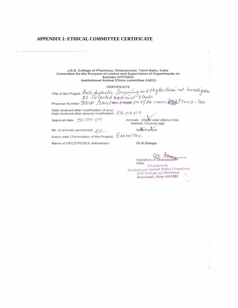
APPENDIX 1: ETHICAL COMMITTEE CERTIFICATE
Related Documents


