Review BDNF and VEGF in the pathogenesis of stress-induced affective diseases: An insight from experimental studies Marta Nowacka, Ewa Obuchowicz Abstract: Stress is known to play an important role in etiology, development and progression of affective diseases. Especially, chronic stress, by initiating changes in the hypothalamic-pituitary-adrenal axis (HPA), neurotransmission and the immune system, acts as a trigger for af- fective diseases. It has been reported that the rise in the concentration of pro-inflammatory cytokines and persistent up-regulation of glucocorticoid expression in the brain and periphery increases the excitotoxic effect on CA3 pyramidal neurons in the hippocampus re- sulting in dendritic atrophy, apoptosis of neurons and possibly inhibition of neurogenesis in adult brain. Stress was observed to disrupt neuroplasticity in the brain, and growing evidence demonstrates its role in the pathomechanism of affective disorders. Experimental studies indicate that a well-known brain-derived neurotrophic factor (BDNF) and vascular endothelial growth factor (VEGF) which have recently focused increasing attention of neuroscientists, promote cell survival, positively modulate neuroplas- ticity and hippocampal neurogenesis. In this paper, we review the alterations in BDNF and VEGF pathways induced by chronic and acute stress, and their relationships with HPAaxis activity. Moreover, behavioral effects evoked in rodents by both above-mentioned factors and the effects consequent to their deficit are presented. Biochemical as well as behavioral findings suggest that BDNF and VEGF play an important role as components of cascade of changes in the pathomechanism of stress-induced affective diseases. Fur- ther studies on the mechanisms regulating their expression in stress conditions are needed to better understand the significance of trophic hypothesis of stress-induced affective diseases. Key words: BDNF, VEGF, animal models of stress, affective diseases Abbrevations: ACTH – adrenocorticotropic hormone, BDNF – brain-derived neurotrophic factor, CMS – chronic mild stress, CNS – central nervous system, CREB – cyclic AMP re- sponse element binding protein, CRH – corticotropin-releasing hormone, ERK – extracellular sigal-regulated kinase, Flt-1 – fms-like-tyrosine kinase, Flk-1 – fetal liver kinase, FRL – Flinders Resistant Line, FSL – Flinders Sensitive Line, GR – glucocorticoid receptor, L-HPA – limbic-hypothalamus- pituitary-adrenal, MAPK – mitogen-activated protein kinase, MR – mineralocorticoid receptor, NMDA – N-methyl-D- aspartic acid, NGF – nerve growth factor, PI3K – phospha- tidylinositol-3-kinase, PLCg – phospholipase C-g, TrkB – tro- pomyosin receptor kinase, VEGF – vascular endothelial growth factor Introduction Experimental studies and clinical observations have indicated that stress plays a significant role in the pathogenesis of mental disorders. Stress not only in- fluences neurotransmission in the central nervous sys- tem (CNS) but also leads to permanent changes in its structure due to the activation of an array of neuroen- docrine and immunological mechanisms [53]. Over- activation of these systems elicits sensitization to stressful stimuli, which promotes development of 535

Welcome message from author
This document is posted to help you gain knowledge. Please leave a comment to let me know what you think about it! Share it to your friends and learn new things together.
Transcript
Review
BDNF and VEGF in the pathogenesis
of stress-induced affective diseases:
An insight from experimental studies
Marta Nowacka, Ewa Obuchowicz
Department of Pharmacology, Medical University of Silesia, Medyków 18, PL 40-752 Katowice, Poland
Correspondence: Ewa Obuchowicz, e-mail: [email protected]
Abstract:
Stress is known to play an important role in etiology, development and progression of affective diseases. Especially, chronic stress, by
initiating changes in the hypothalamic-pituitary-adrenal axis (HPA), neurotransmission and the immune system, acts as a trigger for af-
fective diseases. It has been reported that the rise in the concentration of pro-inflammatory cytokines and persistent up-regulation of
glucocorticoid expression in the brain and periphery increases the excitotoxic effect on CA3 pyramidal neurons in the hippocampus re-
sulting in dendritic atrophy, apoptosis of neurons and possibly inhibition of neurogenesis in adult brain. Stress was observed to disrupt
neuroplasticity in the brain, and growing evidence demonstrates its role in the pathomechanism of affective disorders.
Experimental studies indicate that a well-known brain-derived neurotrophic factor (BDNF) and vascular endothelial growth factor
(VEGF) which have recently focused increasing attention of neuroscientists, promote cell survival, positively modulate neuroplas-
ticity and hippocampal neurogenesis. In this paper, we review the alterations in BDNF and VEGF pathways induced by chronic and
acute stress, and their relationships with HPA axis activity. Moreover, behavioral effects evoked in rodents by both above-mentioned
factors and the effects consequent to their deficit are presented. Biochemical as well as behavioral findings suggest that BDNF and
VEGF play an important role as components of cascade of changes in the pathomechanism of stress-induced affective diseases. Fur-
ther studies on the mechanisms regulating their expression in stress conditions are needed to better understand the significance of
trophic hypothesis of stress-induced affective diseases.
Key words:
BDNF, VEGF, animal models of stress, affective diseases
Abbrevations: ACTH – adrenocorticotropic hormone, BDNF
– brain-derived neurotrophic factor, CMS – chronic mild
stress, CNS – central nervous system, CREB – cyclic AMP re-
sponse element binding protein, CRH – corticotropin-releasing
hormone, ERK – extracellular sigal-regulated kinase, Flt-1 –
fms-like-tyrosine kinase, Flk-1 – fetal liver kinase, FRL –
Flinders Resistant Line, FSL – Flinders Sensitive Line, GR –
glucocorticoid receptor, L-HPA – limbic-hypothalamus-
pituitary-adrenal, MAPK – mitogen-activated protein kinase,
MR – mineralocorticoid receptor, NMDA – N-methyl-D-
aspartic acid, NGF – nerve growth factor, PI3K – phospha-
tidylinositol-3-kinase, PLCg – phospholipase C-g, TrkB – tro-
pomyosin receptor kinase, VEGF – vascular endothelial
growth factor
Introduction
Experimental studies and clinical observations have
indicated that stress plays a significant role in the
pathogenesis of mental disorders. Stress not only in-
fluences neurotransmission in the central nervous sys-
tem (CNS) but also leads to permanent changes in its
structure due to the activation of an array of neuroen-
docrine and immunological mechanisms [53]. Over-
activation of these systems elicits sensitization to
stressful stimuli, which promotes development of
Pharmacological Reports, 2013, 65, 535�546 535
Pharmacological Reports2013, 65, 535�546ISSN 1734-1140
Copyright © 2013by Institute of PharmacologyPolish Academy of Sciences
mental disorders [10]. It has been found that both
acute and chronic stress, as a modulator of central
nervous system function, in combination with genetic
background, often predisposes patients to develop-
ment of mental disorders and in later life can be a fac-
tor that triggers or aggravates disease episodes [9].
It appears that trophic factors play an important
role among agents implicated in the pathogenesis of
mental disorders [23]. Among members of the neuro-
trophin family, brain-derived neurotrophic factor
(BDNF) belongs to the most intensively studied. The
other neurotrophic factor, vascular endothelial growth
factor (VEGF) has recently become a focus of interest
in the context of mental diseases. BDNF plays a key
role in neurogenesis, promotes synaptic plasticity and
neuronal cell survival while its reduced expression
can contribute to structural anomalies and functional
impairment in the central nervous system. VEGF also
participates in the mentioned processes. In the light of
these findings, it can be expected that low levels of
trophic factors, in particular BDNF, can be engaged in
etiology of mental disorders [27, 100].
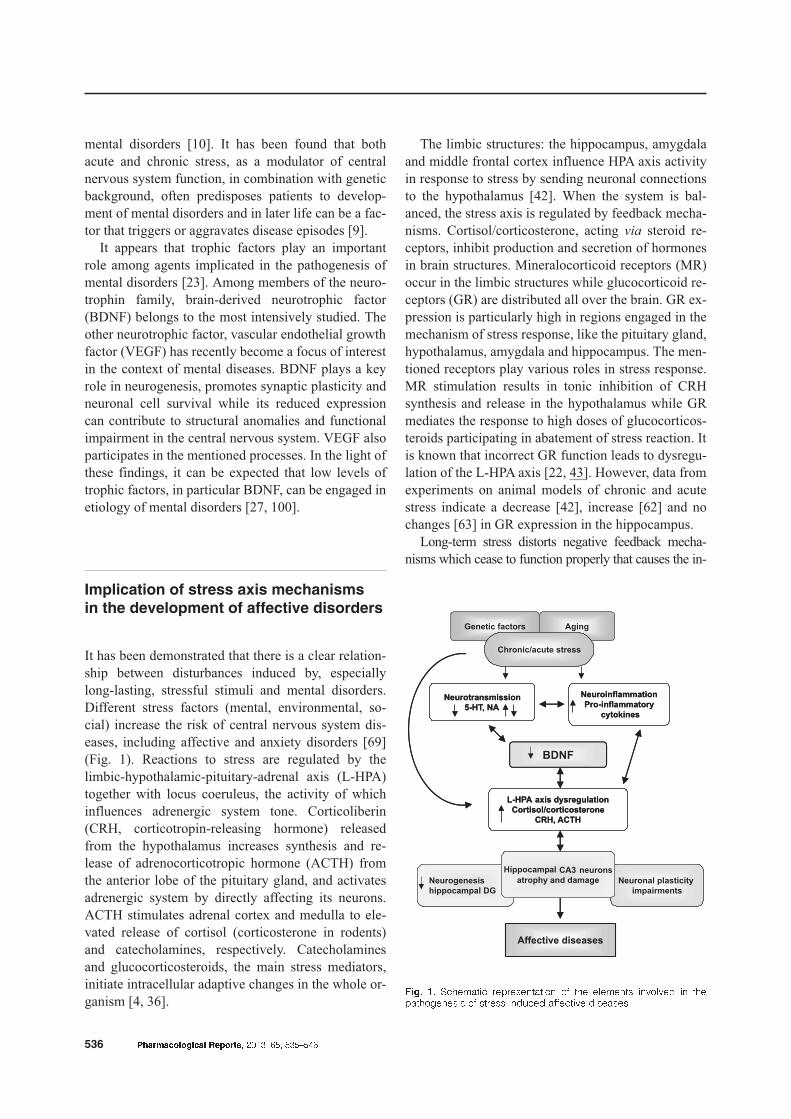
Implication of stress axis mechanisms
in the development of affective disorders
It has been demonstrated that there is a clear relation-
ship between disturbances induced by, especially
long-lasting, stressful stimuli and mental disorders.
Different stress factors (mental, environmental, so-
cial) increase the risk of central nervous system dis-
eases, including affective and anxiety disorders [69]
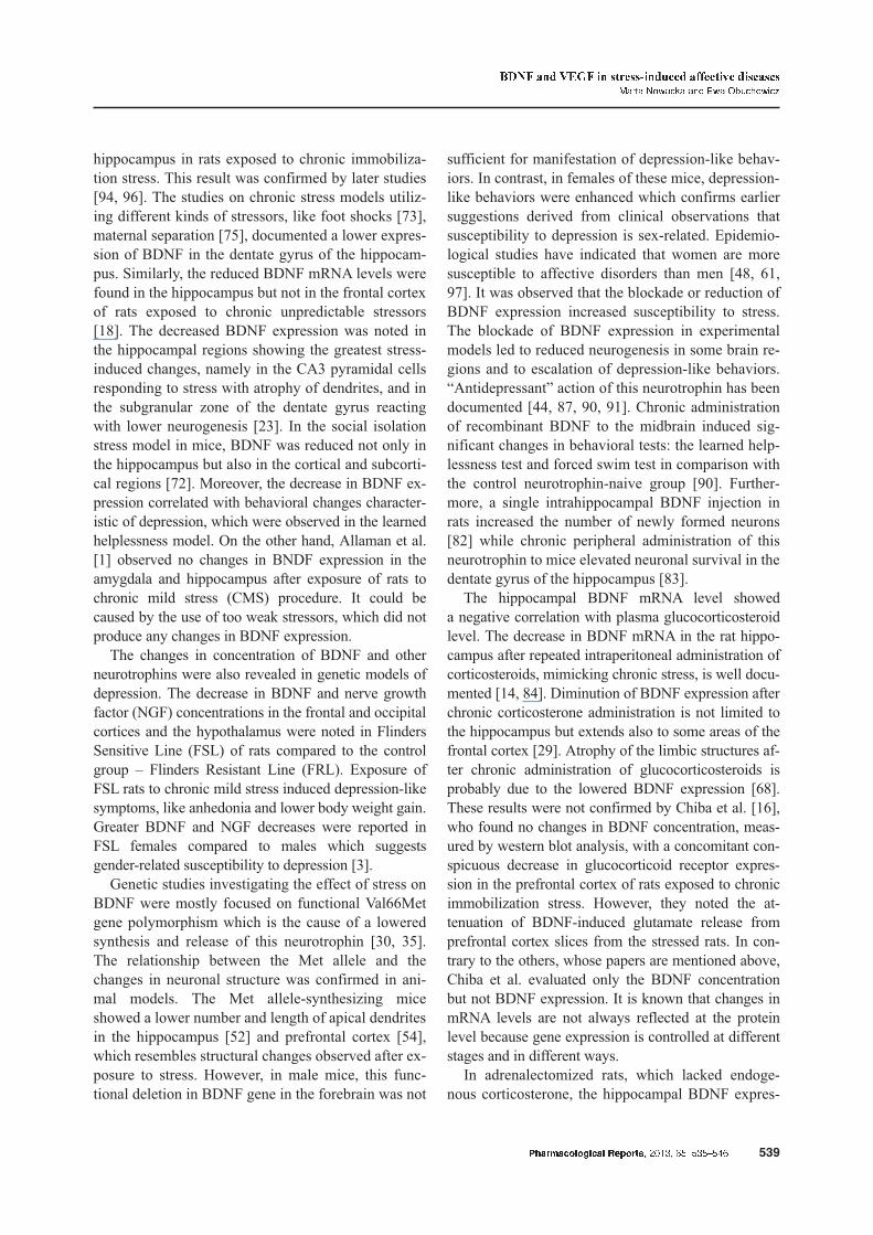
(Fig. 1). Reactions to stress are regulated by the
limbic-hypothalamic-pituitary-adrenal axis (L-HPA)
together with locus coeruleus, the activity of which
influences adrenergic system tone. Corticoliberin
(CRH, corticotropin-releasing hormone) released
from the hypothalamus increases synthesis and re-
lease of adrenocorticotropic hormone (ACTH) from
the anterior lobe of the pituitary gland, and activates
adrenergic system by directly affecting its neurons.
ACTH stimulates adrenal cortex and medulla to ele-
vated release of cortisol (corticosterone in rodents)
and catecholamines, respectively. Catecholamines
and glucocorticosteroids, the main stress mediators,
initiate intracellular adaptive changes in the whole or-
ganism [4, 36].
The limbic structures: the hippocampus, amygdala
and middle frontal cortex influence HPA axis activity
in response to stress by sending neuronal connections
to the hypothalamus [42]. When the system is bal-
anced, the stress axis is regulated by feedback mecha-
nisms. Cortisol/corticosterone, acting via steroid re-
ceptors, inhibit production and secretion of hormones
in brain structures. Mineralocorticoid receptors (MR)
occur in the limbic structures while glucocorticoid re-
ceptors (GR) are distributed all over the brain. GR ex-
pression is particularly high in regions engaged in the
mechanism of stress response, like the pituitary gland,
hypothalamus, amygdala and hippocampus. The men-
tioned receptors play various roles in stress response.
MR stimulation results in tonic inhibition of CRH
synthesis and release in the hypothalamus while GR
mediates the response to high doses of glucocorticos-
teroids participating in abatement of stress reaction. It
is known that incorrect GR function leads to dysregu-
lation of the L-HPA axis [22, 43]. However, data from
experiments on animal models of chronic and acute
stress indicate a decrease [42], increase [62] and no
changes [63] in GR expression in the hippocampus.
Long-term stress distorts negative feedback mecha-
nisms which cease to function properly that causes the in-
536 Pharmacological Reports, 2013, 65, 535�546
Hippocampal
Fig. 1. Schematic representation of the elements involved in thepathogenesis of stress-induced affective diseases
creased release of both CRH and glucocorticosteroids
thus further damaging neurons, particularly in the
CA3 region of the hippocampus [26, 98]. It seems that
only at the initial stage of hyperactivation of neuro-
transmitter systems glucocorticosteroids have protec-
tive effect on hippocampal neurons helping preserve
the balanced response to changing environment [60].
Both the consequences of glucocorticosteroid-induced
changes in the stress axis, activation of neurotransmitter
systems [42] and altered activity of the immune system
[21] play a role in the pathomechanism of mental disor-
ders. The majority of depressed patients show, apart from
hypercortisolemia, the impairment of dexamethasone-
induced inhibition of endogenous cortisol release which
indicates lowering of glucocorticosteroid receptor sensi-
tivity [70]. The increased weight of the adrenal glands
visible in radiographic studies suggests a prolonged and
excessive trophic effect of ACTH on these gland [65].
Depressive syndromes were found to be associated
with hyperactivity of the L-HPA axis, an weakened
feedback inhibition of this axis and in some patients,
with hyperactivity of the adrenergic system [69]. Corti-
costerone treatment or exposure to stressful stimuli were
observed to decrease serotonin (5-HT) level in the brain
and to diminish density of serotonergic 5-HT1A recep-
tors. The impairment of 5-HT transmission, like nor-
adrenergic system failure, can diminish production of
neurotrophic factors due to unfavorable changes in the
second messenger systems in the cell [103].
Stress modulates activity of the immune system. Pro-
inflammatory cytokines released by the immune cells
stimulate L-HPA axis. IL-1 and IL-6 promote CRH re-
lease from the hypothalamus and directly increase ACTH
secretion from the pituitary gland [79, 101]. Moreover,
exposure to stressful events (e.g., open field) increased
pro-inflammatory cytokine release prior to surge of glu-
cocorticosteroids, which suggests that elevated concentra-
tions of these hormones can have protective effect on the
CNS neurons, also in the case of damage produced by the
immune system hyperactivation [19, 102].
Stress-induced morphological changes
in the hippocampus as a consequence
of disordered synaptic plasticity
and neurogenesis
Synaptic plasticity is defined as the ability of the brain
to adapt to environmental changes. Its impairment by,
for instance, a prolonged stress, can provoke morpho-
logical changes in brain structures and functional
deficits of many molecular and cellular mechanisms
[71]. Distortion of neuronal plasticity results mainly
from suppression of neurogenesis, cell atrophy or en-
hanced apoptosis [17].
The hippocampus is the structure which is the main
target of pathogenesis and treatment of affective diseases.
This brain structure is engaged in learning and memory
processes which, at the cellular level, depend on the
modulation of neuronal plasticity [98]. It is believed that
neurogenesis, progressing also in adults, is crucially in-
volved in learning and memory processes. Neurogenesis
occurs mostly in the subgranular zone of the dentate gy-
rus of the hippocampus. Neurogenesis in the adult brain is
a dynamic process of creation, maturation, migration and
integration of new neurons. This process can be up- or
down-regulated by various neuroendocrine, environ-
mental or pharmacological factors [40, 71]. Disruption of
neurogenesis is aggravated by chronic stress, increased
glucocorticoseroid level or advanced age [26].
It is supposed that newly created neurons partici-
pate in the regulation of the changes in the hippocam-
pus induced by anxiety and stress and that they can
prevent development of depression and mediate ac-
tion of antidepressant drugs [24, 78]. According to the
hypothesis referred to as “trophic hypothesis”, stress
disturbs the formation of new neurons, which leads to
hippocampal damage and disease progression while
the action of antidepressant drugs largely consists in
restoration of the proper level of neurogenesis [39].
Stress suppresses hippocampal granular cell prolif-
eration and reduces the number and length of apical
dendrites of pyramidal neurons in the CA3 region,
which are particularly vulnerable to stressful stimuli
[25]. Studies conducted in experimental animal mod-
els demonstrated atrophy and death of pyramidal cells
in the CA3 region of the hippocampus as the result of
exposure to prolonged stress [26, 60]. Similar effect
was noted after glucocorticosteroid treatment at doses
producing similar brain level of these hormones as in
stressful situations [38]. These processes can be
blocked by prior administration of an NMDA (N-me-
thyl-D-aspartic acid) receptor antagonist which sug-
gests the involvement of the enhanced glutamatergic
transmission in the mechanism of destructive action
of stress and glucocorticosteroids on neuronal groups
and neurogenesis [37]. Moreover, exposure to stress
or glucocorticosteroids increases sensitivity of py-
ramidal cells of the CA3 region to such damaging fac-
tors as hypoglycemia or hypoxia [79, 80].
Pharmacological Reports, 2013, 65, 535�546 537
BDNF and VEGF in stress-induced affective diseasesMarta Nowacka and Ewa Obuchowicz
Brain imaging studies in depressed patients demon-
strated a smaller hippocampal volume and altered
shape [11, 55, 77]. At present, two causes have been
proposed to explain these changes: 1) postmortem
studies showed an increased packing density of neu-
ronal and glial cells with concomitant reduction of
neuron soma size in pyramidal regions and the dentate
gyrus of the hippocampus [89] and 2) the hippocam-
pal shrinkage and impairment of its function resulting
in the weaker inhibition of endocrine activity of the
HPA axis can be the result of the suppressed dentate
gyrus cell proliferation or their diminished survival
[47]. Nuclear magnetic resonance volumetric studies
in healthy subjects with familial history of depressive
disorders evidenced a smaller volume of the hippo-
campus, which suggests that genetic factors can con-
tribute to the observed structural changes [15].
Since a positive correlation was shown between the
hippocampus shrinkage and the cognitive deficits and
memory disturbances in depressed patients, it is be-
lieved that these disturbances are a functional conse-
quence of the structural changes in this structure [98].
The effect of stress on brain-derived
neurotrophic factor (BDNF)
Neurotrophic factors (neurotrophins) are a group of
proteins possessing similar structure, synthesized in
peripheral tissues innervated by sensory and sympa-
thetic neurons and in neurons of some brain structures
[7]. The greatest role in the pathogenesis of affective
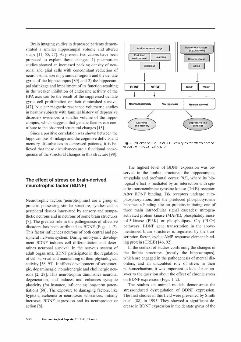
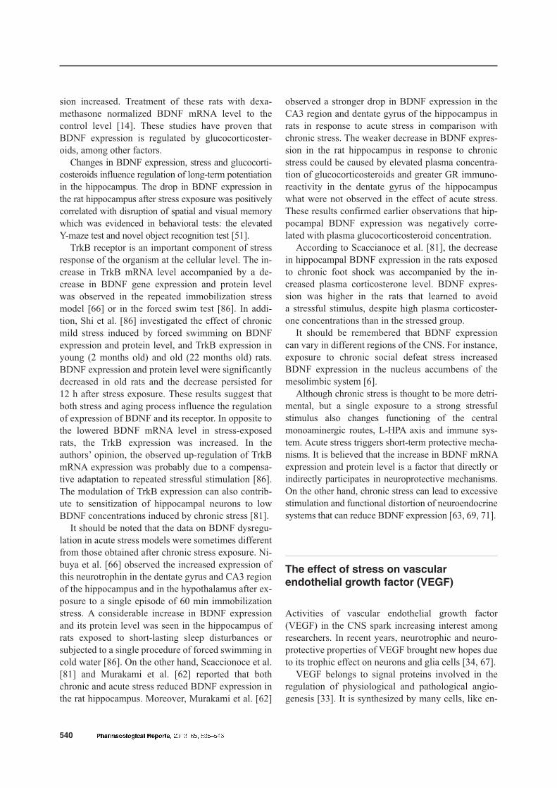
disorders has been attributed to BDNF (Figs. 1, 2).
This factor influences neurons of both central and pe-
ripheral nervous system. During embryonic develop-
ment BDNF induces cell differentiation and deter-
mines neuronal survival. In the nervous system of
adult organisms, BDNF participates in the regulation
of cell survival and maintaining of their physiological
activity [58, 93]. It affects development of serotoner-
gic, dopaminergic, noradrenergic and cholinergic neu-
rons [2, 28]. This neurotrophin diminishes neuronal
degeneration, and induces and enhances synaptic
plasticity (for instance, influencing long-term poten-
tiation) [58]. The exposure to damaging factors, like
hypoxia, ischemia or neurotoxic substances, initially
increases BDNF expression and its neuroprotective
action [8].
The highest level of BDNF expression was ob-
served in the limbic structures: the hippocampus,
amygdala and prefrontal cortex [92], where its bio-
logical effect is mediated by an interaction with spe-
cific transmembrane tyrosine kinase (TrkB) receptor.
After BDNF binding, Trk receptors undergo auto-
phosphorylation, and the produced phosphotyrosine
becomes a binding site for proteins initiating one of
three main intracellular signal cascades: mitogen-
activated protein kinase (MAPK), phosphatidylinosi-
tol-3-kinase (PI3K) or phospholipase C-g (PLCg)
pathways. BDNF gene transcription in the above-
mentioned brain structures is regulated by the tran-
scription factor, cyclic AMP response element bind-
ing protein (CREB) [46, 92].
In the context of studies confirming the changes in
the limbic structures (mostly the hippocampus),
which are engaged in the pathogenesis of mental dis-
orders, and an undoubted role of stress in their
pathomechanism, it was important to look for an an-
swer to the question about the effect of chronic stress
on BDNF expression (Figs. 1, 2).
The studies on animal models demonstrate the
stress-induced dysregulation of BDNF expression.
The first studies in this field were presented by Smith
et al. [88] in 1995. They showed a significant de-
crease in BDNF expression in the dentate gyrus of the
538 Pharmacological Reports, 2013, 65, 535�546
Fig. 2. Influence of BDNF and VEGF activity on the effects depend-ent on the hippocampal function
hippocampus in rats exposed to chronic immobiliza-
tion stress. This result was confirmed by later studies
[94, 96]. The studies on chronic stress models utiliz-
ing different kinds of stressors, like foot shocks [73],
maternal separation [75], documented a lower expres-
sion of BDNF in the dentate gyrus of the hippocam-
pus. Similarly, the reduced BDNF mRNA levels were
found in the hippocampus but not in the frontal cortex
of rats exposed to chronic unpredictable stressors
[18]. The decreased BDNF expression was noted in
the hippocampal regions showing the greatest stress-
induced changes, namely in the CA3 pyramidal cells
responding to stress with atrophy of dendrites, and in
the subgranular zone of the dentate gyrus reacting
with lower neurogenesis [23]. In the social isolation
stress model in mice, BDNF was reduced not only in
the hippocampus but also in the cortical and subcorti-
cal regions [72]. Moreover, the decrease in BDNF ex-
pression correlated with behavioral changes character-
istic of depression, which were observed in the learned
helplessness model. On the other hand, Allaman et al.
[1] observed no changes in BNDF expression in the
amygdala and hippocampus after exposure of rats to
chronic mild stress (CMS) procedure. It could be
caused by the use of too weak stressors, which did not
produce any changes in BDNF expression.
The changes in concentration of BDNF and other
neurotrophins were also revealed in genetic models of
depression. The decrease in BDNF and nerve growth
factor (NGF) concentrations in the frontal and occipital
cortices and the hypothalamus were noted in Flinders
Sensitive Line (FSL) of rats compared to the control
group – Flinders Resistant Line (FRL). Exposure of
FSL rats to chronic mild stress induced depression-like
symptoms, like anhedonia and lower body weight gain.
Greater BDNF and NGF decreases were reported in
FSL females compared to males which suggests
gender-related susceptibility to depression [3].
Genetic studies investigating the effect of stress on
BDNF were mostly focused on functional Val66Met
gene polymorphism which is the cause of a lowered
synthesis and release of this neurotrophin [30, 35].
The relationship between the Met allele and the
changes in neuronal structure was confirmed in ani-
mal models. The Met allele-synthesizing mice
showed a lower number and length of apical dendrites
in the hippocampus [52] and prefrontal cortex [54],
which resembles structural changes observed after ex-
posure to stress. However, in male mice, this func-
tional deletion in BDNF gene in the forebrain was not
sufficient for manifestation of depression-like behav-
iors. In contrast, in females of these mice, depression-
like behaviors were enhanced which confirms earlier
suggestions derived from clinical observations that
susceptibility to depression is sex-related. Epidemio-
logical studies have indicated that women are more
susceptible to affective disorders than men [48, 61,
97]. It was observed that the blockade or reduction of
BDNF expression increased susceptibility to stress.
The blockade of BDNF expression in experimental
models led to reduced neurogenesis in some brain re-
gions and to escalation of depression-like behaviors.
“Antidepressant” action of this neurotrophin has been
documented [44, 87, 90, 91]. Chronic administration
of recombinant BDNF to the midbrain induced sig-
nificant changes in behavioral tests: the learned help-
lessness test and forced swim test in comparison with
the control neurotrophin-naive group [90]. Further-
more, a single intrahippocampal BDNF injection in
rats increased the number of newly formed neurons
[82] while chronic peripheral administration of this
neurotrophin to mice elevated neuronal survival in the
dentate gyrus of the hippocampus [83].
The hippocampal BDNF mRNA level showed
a negative correlation with plasma glucocorticosteroid
level. The decrease in BDNF mRNA in the rat hippo-
campus after repeated intraperitoneal administration of
corticosteroids, mimicking chronic stress, is well docu-
mented [14, 84]. Diminution of BDNF expression after
chronic corticosterone administration is not limited to
the hippocampus but extends also to some areas of the
frontal cortex [29]. Atrophy of the limbic structures af-
ter chronic administration of glucocorticosteroids is
probably due to the lowered BDNF expression [68].
These results were not confirmed by Chiba et al. [16],
who found no changes in BDNF concentration, meas-
ured by western blot analysis, with a concomitant con-
spicuous decrease in glucocorticoid receptor expres-
sion in the prefrontal cortex of rats exposed to chronic
immobilization stress. However, they noted the at-
tenuation of BDNF-induced glutamate release from
prefrontal cortex slices from the stressed rats. In con-
trary to the others, whose papers are mentioned above,
Chiba et al. evaluated only the BDNF concentration
but not BDNF expression. It is known that changes in
mRNA levels are not always reflected at the protein
level because gene expression is controlled at different
stages and in different ways.
In adrenalectomized rats, which lacked endoge-
nous corticosterone, the hippocampal BDNF expres-
Pharmacological Reports, 2013, 65, 535�546 539
BDNF and VEGF in stress-induced affective diseasesMarta Nowacka and Ewa Obuchowicz
sion increased. Treatment of these rats with dexa-
methasone normalized BDNF mRNA level to the
control level [14]. These studies have proven that
BDNF expression is regulated by glucocorticoster-
oids, among other factors.
Changes in BDNF expression, stress and glucocorti-
costeroids influence regulation of long-term potentiation
in the hippocampus. The drop in BDNF expression in
the rat hippocampus after stress exposure was positively
correlated with disruption of spatial and visual memory
which was evidenced in behavioral tests: the elevated
Y-maze test and novel object recognition test [51].
TrkB receptor is an important component of stress
response of the organism at the cellular level. The in-
crease in TrkB mRNA level accompanied by a de-
crease in BDNF gene expression and protein level
was observed in the repeated immobilization stress
model [66] or in the forced swim test [86]. In addi-
tion, Shi et al. [86] investigated the effect of chronic
mild stress induced by forced swimming on BDNF
expression and protein level, and TrkB expression in
young (2 months old) and old (22 months old) rats.
BDNF expression and protein level were significantly
decreased in old rats and the decrease persisted for
12 h after stress exposure. These results suggest that
both stress and aging process influence the regulation
of expression of BDNF and its receptor. In opposite to
the lowered BDNF mRNA level in stress-exposed
rats, the TrkB expression was increased. In the
authors’ opinion, the observed up-regulation of TrkB
mRNA expression was probably due to a compensa-
tive adaptation to repeated stressful stimulation [86].
The modulation of TrkB expression can also contrib-
ute to sensitization of hippocampal neurons to low
BDNF concentrations induced by chronic stress [81].
It should be noted that the data on BDNF dysregu-
lation in acute stress models were sometimes different
from those obtained after chronic stress exposure. Ni-
buya et al. [66] observed the increased expression of
this neurotrophin in the dentate gyrus and CA3 region
of the hippocampus and in the hypothalamus after ex-
posure to a single episode of 60 min immobilization
stress. A considerable increase in BDNF expression
and its protein level was seen in the hippocampus of
rats exposed to short-lasting sleep disturbances or
subjected to a single procedure of forced swimming in
cold water [86]. On the other hand, Scaccionoce et al.
[81] and Murakami et al. [62] reported that both
chronic and acute stress reduced BDNF expression in
the rat hippocampus. Moreover, Murakami et al. [62]
observed a stronger drop in BDNF expression in the
CA3 region and dentate gyrus of the hippocampus in
rats in response to acute stress in comparison with
chronic stress. The weaker decrease in BDNF expres-
sion in the rat hippocampus in response to chronic
stress could be caused by elevated plasma concentra-
tion of glucocorticosteroids and greater GR immuno-
reactivity in the dentate gyrus of the hippocampus
what were not observed in the effect of acute stress.
These results confirmed earlier observations that hip-
pocampal BDNF expression was negatively corre-
lated with plasma glucocorticosteroid concentration.
According to Scaccianoce et al. [81], the decrease
in hippocampal BDNF expression in the rats exposed
to chronic foot shock was accompanied by the in-
creased plasma corticosterone level. BDNF expres-
sion was higher in the rats that learned to avoid
a stressful stimulus, despite high plasma corticoster-
one concentrations than in the stressed group.
It should be remembered that BDNF expression
can vary in different regions of the CNS. For instance,
exposure to chronic social defeat stress increased
BDNF expression in the nucleus accumbens of the
mesolimbic system [6].
Although chronic stress is thought to be more detri-
mental, but a single exposure to a strong stressful
stimulus also changes functioning of the central
monoaminergic routes, L-HPA axis and immune sys-
tem. Acute stress triggers short-term protective mecha-
nisms. It is believed that the increase in BDNF mRNA
expression and protein level is a factor that directly or
indirectly participates in neuroprotective mechanisms.
On the other hand, chronic stress can lead to excessive
stimulation and functional distortion of neuroendocrine
systems that can reduce BDNF expression [63, 69, 71].
The effect of stress on vascular
endothelial growth factor (VEGF)
Activities of vascular endothelial growth factor
(VEGF) in the CNS spark increasing interest among
researchers. In recent years, neurotrophic and neuro-
protective properties of VEGF brought new hopes due
to its trophic effect on neurons and glia cells [34, 67].
VEGF belongs to signal proteins involved in the
regulation of physiological and pathological angio-
genesis [33]. It is synthesized by many cells, like en-
540 Pharmacological Reports, 2013, 65, 535�546
dothelial cells, macrophages, lymphocytes T, smooth
muscle cells, nephrocytes, keratocytes, osteoblasts,
cancer cells, brain cells: astrocytes, neuronal stem
cells. VEGF stimulates proliferation and survival of
endothelial cells and acts as an NO-dependent vasore-
laxant. It influences formation of vascular networks
and increases vascular permeability [13, 20, 32]. In the
brain, VEGF participates in vasculogenesis and angio-
genesis in embryonic life and postnatal period [12].
VEGF acts by binding with specific tyrosine kinase
receptors: VEGFR1 (Flt-1, fms-like-tyrosine kinase)
and VEGFR2 (Flk-1, fetal liver kinase). VEGF inter-
action with the extracellular domain of VEGFR2 trig-
gers phosphorylation of tyrosine residues in the tyro-
sine kinase domain [12, 74]. In neurons and Schwann
cells, VEGFR2 stimulation activates PLCg/MAPK
pathway. In astrocytes and microglia, VEGF activates
MAPK/ERK (ERK, extracellular sigal-regulated ki-
nase) by stimulation of VEGFR2, and PI3K by inter-
action with VEGFR1 [12].
In the brain, apart from the effects on nervous cell
metabolism, VEGF directly stimulates neurogenesis
(Fig. 2). This effect was observed both in in vitro and
in vivo studies [49, 57, 64]. Hippocampal neurogene-
sis regulated by VEGF is stimulated by environmental
enrichment and exercises, learning and antidepressant
drugs, while stress and aging diminishes it. Suppres-
sion of neurogenesis in rats by irradiation causes fear
and depression-like behaviors [31, 99]. In addition,
VEGF activates neurogenesis by stimulating endothe-
lial cells to release neurotrophic factors, e.g., BDNF,
which improves neuronal survival and integration in
the subventricular zone of the dentate gyrus of the
hippocampus. Intracerebral administration of VEGF
stimulated neurogenesis in the subventricular and
subgranular zone of the dentate gyrus of the hippo-
campus [76, 85]. Research results suggest that VEGF
influences neuronal plasticity in the CNS of adult ani-
mals but the mechanism of this action has not been
elucidated so far. It is supposed that VEGF by its neu-
roprotective activity, like BDNF, promotes the hippo-
campus-dependent processes, like learning and mem-
ory [56, 59].
As mentioned above, synaptic plasticity, which is
important for the formation of memory traces, learn-
ing, and neurogenesis in the adult brain, play a signifi-
cant role both in stress response and repeated stress-
induced adaptations. The mechanism responsible for
the relationship between the stress-induced distortion
of synaptic plasticity and suppression of neurogene-
sis, and the depressive behavior has not been fully
elucidated so far. It appears that due to the involve-
ment of VEGF in synaptic plasticity and neurogene-
sis, this factor can be engaged in etiopathogenesis of
mental disorders induced by stress.
The effect of stress on VEGF gene expression and
protein level was investigated in several studies.
VEGF expression in the CA3 region of the hippocam-
pus was studied using an in situ hybridization method
in stress-sensitive and stress-resistant rats. The study
showed that VEGF mRNA level was decreased in the
stress-sensitive group exposed to CMS procedure
compared to the stress-resistant group. The authors
suggested that VEGF down-regulation in the CMS-
sensitive rats could contribute to functional hippo-
campal damage due to a weaker neuroprotective ac-
tion of this factor [5].
Heine et al. [41] demonstrated the down-regulation
of VEGF and its receptor VEGFR2 in the dentate gy-
rus of the hippocampus of rats exposed to chronic
stress procedure lasting 21 days. These rats were ex-
posed to such stressors as immobilization, forced
swimming in cold water, and intermittent social isola-
tion and group housing. The changes in the hippo-
campal VEGF and VEGFR2 expression were investi-
gated using immunocytochemical methods. A smaller
VEGF immunoreactivity compared to the control
group was observed in cytoplasm of astrocytes in the
hilus of the hippocampus and in the granular cell
layer. In stressed rats, VEGFR2 expression was also
lowered in the dentate gyrus and the hilus of the hip-
pocampus. That experiment was performed on the
group of stressed and control unstressed rats and on
the group of rats which were maintained under stan-
dard conditions for 3 weeks following the stress pro-
cedure in order to observe regenerative mechanisms.
In the last group, VEGF and VEGFR2 expression
in the brain regions under study was increased com-
pared to the stress-exposed group. The results of those
studies confirmed the hypothesis that vascularization
is an important component aiding neurogenesis, and
susceptible to a prolonged stress. Chronic stress-in-
duced suppression of hippocampal neurogenesis was
seen mostly in the region vascularized by capillaries.
According to the authors’ opinion, this led to reduc-
tion of blood flow and density of capillaries in the
hippocampus, which resemble vascular disturbances
observed often in depressed patients.
However, further experiments of Bergström et al.
[5] did not show significant differences in VEGF ex-
Pharmacological Reports, 2013, 65, 535�546 541
BDNF and VEGF in stress-induced affective diseasesMarta Nowacka and Ewa Obuchowicz
pression in the CA3 region of the hippocampus be-
tween rats exposed to acute and chronic immobiliza-
tion stress (lasting 1 h and applied once or daily for
a week) and unstressed animals.
The newest studies focusing on the effect of stress
and exercise on VEGF expression, vascular density
and neurogenesis in the hippocampus were conducted
in the chronic unpredictable stress model. Mice were
exposed to such stressors as immobilization, low tem-
perature (4°C), water and food deprivation in the
night, nocturnal light exposure, forced swimming for
15 min and cage rotation. No differences in the hippo-
campal and plasma VEGF concentration were seen
between stressed and unstressed groups. However,
chronic stress induced transient depression-like be-
haviors of mice, and, like in the latter paper, de-
creased vascular density and neurogenesis in the hip-
pocampus. Regular exercise reversed these disadvan-
tageous physiological and behavioral changes but had
no effect on the hippocampal and plasma VEGF ex-
pression in mice [50].
Uysal et al. [95] evaluated the effect of acute foot
shock stress on the hippocampal VEGF concentration
in young female and male rats. Moreover, in order to
examine the effect of acute stress on spatial memory,
behavior of rats was evaluated in the Morris water
maze test. They evidenced that both female and male
rats exposed to foot shocks (of low and high intensity)
showed a higher VEGF expression, improved learning
ability and better spatial memory compared to the con-
trol group. These results suggest that VEGF is directly
implicated in improvement of cognitive functions in
rats. VEGF concentration in females was much lower
than in males, which confirms the earlier observations
that sensitivity to stress is sex-dependent.
It is known that VEGF action on neuroplasticity
and neurogenesis is mediated by VEGFR2 and that
exposure to a prolonged stress or exogenous gluco-
corticosteroids induces neurochemical and behavioral
disruption in rodents [45]. Howell et al. [45] demon-
strated an inhibitory effect of chronic corticosterone
on VEGFR2 expression in the frontal cortex of mice.
They documented a significant decrease in VEGFR2
protein level, as demonstrated by western blot analy-
sis, in the frontal cortex of mice which were treated
with corticosterone for 7 weeks, vs. the control group.
The plasma level of this protein in the stressed group
was also diminished. In contrast, the frontal cortex
and plasma VEGF protein level in the corticosterone-
treated rats was elevated compared to the control
group. These researchers observed GR down-regu-
lation which underlined significance of these recep-
tors for the suppression of VEGFR2 expression in-
duced by chronic corticosterone administration. These
results suggest that VEGF synthesis in the frontal cor-
tex is increased by a feedback mechanism in response
to the inhibition of this signal route, mediated by
VEGFR2 both in the brain and periphery. However, it
cannot be excluded that the increase in the central
VEGF concentration is a response to the lowered pe-
ripheral VEGF level induced by chronic corticoster-
one administration.
Conclusions
Long-term or intense stress, which is considered to be
a significant factor in the pathogenesis of affective
disorders, produces adverse effects on brain structures
leading to their permanent changes [26]. This effect is
largely dependent on excessive activation of the
L-HPA axis and impairment of its crucial regulatory
feedback mechanism. In addition, stress exposure dis-
rupts neuronal plasticity which can result from sup-
pressed neurogenesis, cell atrophy or enhanced apop-
tosis [17, 26]. Although the contribution of the dimin-
ished neurogenesis to the pathomechanism of stress-
induced affective disorders seems probable, its sig-
nificance has not been fully explained, yet. It is diffi-
cult to unequivocally resolve to what extent the re-
duced neurogenesis influences morphological changes
observed in the brains of patients and whether indeed
it promotes development of affective disorders. How-
ever, it does not appear that diminution of the number
of newly formed neurons could be the trigger initiat-
ing the cascade of the observed functional changes in
neuronal networks. Instead, it rather seems that distur-
bances of synaptic plasticity also of other origin than
limited neurogenesis can play a more important role.
Heterogeneity of affective disorders suggests that
they can be caused by different functional anomalies
in neuronal networks underlain by a number of
mechanisms.
BDNF and VEGF are crucially involved in the pro-
cesses of neurogenesis and synaptic plasticity. These
factors are sensitive to stressors, which was con-
firmed by results of the studies on animal models of
542 Pharmacological Reports, 2013, 65, 535�546
stress induced by immobilization, foot shocks, forced
swimming, social isolation, etc., which evidenced
dysregulation of these factors. Moreover, BDNF
alone showed “antidepressant” effect, and a single in-
tracerebral BDNF administration, and VEGF alike,
stimulated neurogenesis in vivo. On the other hand,
stress-induced suppression of BDNF and VEGF ex-
pression evokes in animals behaviors characteristic of
depression which are reversed by antidepressant
drugs or exercise and environmental enrichment that
increase expression of these factors. It was demon-
strated that after stress exposure, BDNF and VEGF
were directly affected by: (i) elevated levels of gluco-
corticosteroids, which produced a stronger effect un-
der stress conditions due to GR dysfunction, (ii) in-
creased activity of monoaminergic systems or (iii) other
detrimental factors, like glutamate. Other significant
factors influencing different BDNF and VEGF ex-
pression in response to stress include sex an age. Old
male and female rats alike showed lower BDNF and
VEGF expression after exposure to stress compared
to young males and females. Thus, the results of a ma-
jority of experimental studies have indicated the im-
plication of BDNF and VEGF in the pathogenesis of
stress-induced affective disorders, which is the basis
for “the neurotrophic hypothesis” of the pathomecha-
nism of these disorders. Of course, this hypothesis
does not abolish “the monoaminergic hypothesis” but
supplements it.
It is difficult to resolve to what extent the stress-
induced reduction in BDNF and VEGF expression di-
rectly contributes to the distortions of synaptic plas-
ticity and neurogenesis in the hippocampus but it is
supposed that structural and morphological changes
in this structure in the patients suffering from affec-
tive disorders are at least partially consequent to the
decreased expression of these factors. Since it is not
known whether these changes are the effect of the dis-
ease or its cause, it is difficult to assess the sequence
of changes, i.e., whether dysregulation of BDNF and
VEGF expression precedes or coincides with the
changes in the hippocampus. The results of a few
studies showing a paradoxical increase in BDNF and
VEGF expression following stress exposure in the
brain of rodents can indicate that other pathways can
also be engaged in the pathogenesis of stress-induced
diseases. However, based on the majority of studies
conducted so far and reviewed in this article, it ap-
pears that BDNF and VEGF fulfill an important but
not a key role in the pathogenesis of these diseases,
and full elucidation of the mechanisms regulating
their expression will contribute to answering still un-
answered questions about the pathomechanism of af-
fective disorders.
References:
1. Allaman I, Papp M, Kraftsik R, Fiumelli H, Magistretti
P, Martin JL: Expression of brain-derived neurotrophic
factor is not modulated by chronic mild stress in the rat
hippocampus and amygdala. Pharmacol Rep, 2008, 60,
1001–1007.
2. Altar CA, Boylan CB, Fritsche M, Jackson C, Hyman C,
Lindsay RM: The neurotrophins NT-4/5 and BDNF aug-
ment serotonin, dopamine and GABAergic systems dur-
ing behaviorally effective infusions to the substantia ni-
gra. Exp Neurol, 1994, 130, 31–40.
3. Angelucci F, Aloe L, Vasquez PJ, Mathe AA: Mapping
the differences in the brain concentration of brain-
derived neurotrophic factor (BDNF) and nerve growth
factor (NGF) in an animal model of depression. Neurore-
port, 2000, 11, 1369–1373.
4. Bale TL: Stress sensitivity and the development of affec-
tive disorders. Horm Behav, 2006, 50, 529–533.
5. Bergström A, Jayatissa MN, Mørk A, Wiborg O:
Stress sensitivity and resilience in the chronic mild stress
rat model of depression; an in situ hybridization study.
Brain Res, 2008, 1196, 41–52.
6. Berton O, McClung CA, Dileone RJ, Krishnan V,
Renthal W, Russo SJ, Graham D et al.: Essential role
of BDNF in the misolimbic dopamine pathway in social
defeat stress. Science, 2006, 311, 868–878.
7. Binder DK: The role of BDNF in epilepsy and other dis-
eases of the mature nervous system. Adv Exp Med Biol,
2004, 548, 34–56.
8. Binder DK, Scharfam HE: Bran-derived neurotrophic
factor. Growth Factors, 2004, 22, 123–131.
9. Binder EB, Nemeroff CB: The CRF system, stress,
depression and anxiety-insights from human genetic
studies. Mol Psychiatry, 2010, 15, 574–588.
10. Blugeot A, Rivat C, Bouvier E, Molet J, Mouchard A,
Zeau B, Bernard C et al.: Vulnerability to depression:
from brain neuroplasticity to identification of biomark-
ers. J Neurosci, 2011, 31, 12889–12899.
11. Bremner JD, Narayan M, Anderson ER, Staib LH, Miller
HL, Charney DS: Hippocampal volume reduction in ma-
jor depression. Am J Psychiatry, 2000, 157, 115–118.
12. Brochington A, Lewist C, Wharton S, Shaw PJ: Vascular
endothelial growth factor and the nervous system. Neu-
ropathol Appl Neurobiol, 2004, 30, 427–446.
13. Carmeliet P, Strokebaum E: Vascular and neuronal effect
of VEGF in the nervous system: implications for neuro-
logical disorders. Cell Dev Biol, 2002, 13, 39–53.
14. Chao HM, Sakai RR, Ma LY, McEwen BS: Adrenal
steroid regulation of neurotrophic factor expression in
the hippocampus. Endocrinology, 1998, 139, 3112–3118.
Pharmacological Reports, 2013, 65, 535�546 543
BDNF and VEGF in stress-induced affective diseasesMarta Nowacka and Ewa Obuchowicz
15. Chen MC, Hamilton JP, Gotlib IH: Decreased hippocam-
pal volume in healthy girls at risk of depression. Arch
Gen Psychiatry, 2010, 67, 270–276.
16. Chiba S, Numakawa T, Ninomiya M, Richards MC,
Wakabayashi C, Kunugi H: Chronic restraint stress
causes anxiety- and depression-like bahaviors, down-
regulates glucocorticoid receptor expression, and attenu-
ates glutamate release induced by brain-derived neuro-
trophic factor in the prefrontal cortex. Prog Neuropsy-
chopharmacol Biol Psychiatry, 2012, 39, 112–119.
17. Christoffel DJ, Golden SA, Russo SJ: Structural and syn-
aptic plasticity in stress-related disorders. Rev Neurosci,
2011, 22, 535–549.
18. Cieœlik K, Sowa-Kuæma M, Ossowska G, Legutko B,
Wolak M, Opoka, Nowak G.: Chronic unpredictable
stress-induced reduction in the hippocampal brain-derived
neurotrophic factor (BDNF) gene expression is antago-
nized by zinc treatment. Pharmacol Rep, 2011, 63,
537–543.
19. Connor TJ, Leonard BE: Depression, stress and immuno-
logical action: the role of cytokines in depressive disor-
ders. Life Sci, 1998, 62, 583–606.
20. Cross MJ, Claesson-Welsh L: FGF and VEGF function
in angiogenesis: signalling pathways, biological re-
sponses and therapeutic inhibition. Trends Pharmacol
Sci, 2001, 22, 201–207.
21. Cuba³a WJ, Godlewska B, Trzonkowski P, Landowski J:
Indicators of the persistent pro-inflammatory activation
of the immune system in depression. Psychiatr Pol, 2006,
40, 431–444.
22. Cuba³a WJ, Landowski J: Serotoninergic system and
limbic-hypothalamic-pituitary-adrenal axis (LHPA axis)
in depression. Psychiatr Pol, 2006, 40, 415–430.
23. Duman RS: Neuronal damage and protection in the pa-
thophysiology and treatment of psychiatric illness: stress
and depression. Dialogues Clin Neurosci, 2009, 11,
239–255
24. Duman RS: Role of neurotrophic factors in the etiology
and treatment of mood disorders. Neuromolecular Med,
2004, 5, 11–25.
25. Duman RS: Synaptic plasticity and mood disorders. Mol
Psychiatry, 2002, 7, S29– S34.
26. Duman RS, Malberg J, Nakaqawa S: Regulation of adult
neurogenesis by psychotropic drugs and stress. J Phar-
macol Exp Ther, 2001, 299, 401–407.
27. Duman RS, Monteggia LM: A neurotrophic model for
stress-related mood disorders. Biol Psychiatry, 2006, 59,
1116–1127.
28. Dwivedi Y: Brain-derived neurotrophic factor: role in de-
pression and suicide. Neuropsychiatr Dis and Treat,
2009, 5, 434–449.
29. Dwivedi Y, Rizavi HS, Pandey GN: Antidepressants re-
verse corticosterone-mediated decrease in BDNF expres-
sion: differental regulation of specific exons by antidepres-
sants and corticosterone. Neurosci, 2006, 139, 1017–1029.
30. Egan MF, Kojima M, Callicot JH, Goldberg TE, Kola-
chana BS, Bertolino A, Zaitsev E et al.: The BDNF
val66met polymorphism affects activity-dependent se-
cretion of BDNF and human memory and hippocampal
function. Cell 2003, 112, 257–269.
31. Fabel K, Fabel K, Tam B, Kaufer D, Baiker A, Simmons
N, Kuo CJ, Palmer TD: VEGF is necessary for exercise-
induced adult hippocampal neurogenesis. Eur J Neuro-
sci, 2003, 18, 2803–2812.
32. Ferrara N, Davis-Smyth T: The biology of vascular en-
dothelial growth factor. Endocr Rev, 1997, 18, 4–25.
33. Folkman J, D’Amore PA: Blood vessel formation: what
is its molecular basis? Cell, 1996, 87, 1153–1155.
34. Fournier NM, Duman RS: Role of vascular endothelial
growth factor in adult hippocampal neurogenesis: impli-
cations for the pathophysiology and treatment of depres-
sion. Behav Brain Res, 2012, 227, 440–449.
35. Frodl T, Schüle C, Schmitt G, Born C, Baghai T, Zill P,
Bottlender R et al.: Association of the brain-derived neu-
rotrophic factor Val66Met polymorphism with reduced
hippocampal volumes in major depression. Arch Gen
Psychiatry, 2007, 64, 410–416.
36. Guilliams TG, Edwards L: Chronic stress and the HPA
axis: clinical assessment and therapeutic considerations.
Standard, 2010, 9, 1–12.
37. Gould E, Cameron HA: Regulation of neuronal birth,
migration and death in the rat dentate gyrus. Deb Neuro-
sci, 1996, 18, 22–35.
38. Gould E, Daniels DC, Cameron HA, McEwen BS:
Expression of adrenal steroid receptors by newly born
cells and pyknotic cells in the dentate gyrus of the post-
natal rat. Mol Cell Neurosci, 1992, 3, 44–48.
39. Gould E, Tanapat P: Stress and hippocampal neurogene-
sis. Biol Psychiatry, 1999, 46, 1472–1479.
40. Hanson ND, Owens MJ, Nemeroff CB: Depression, anti-
depressants, and neurogenesis: a critical reappraisal.
Neuropsychopharmacology, 2011, 36, 2589–2602.
41. Heine VM, Zareno J, Maslam S, Joëls M, Lucassen PJ:
Chronic stress in the adult dentate gyrus reduces cell pro-
liferation near the vasculature and VEGF and Flk-1 pro-
tein expression. Eur J Neurosci, 2005, 21, 1304–1314.
42. Herman JP, Ostrander MM, Mueller NK, Fiqueiredo H:
Limbic system mechanisms of stress regulation: hypo-
thalamo-pituitary-adrenocortical axis. Prog Neuro-
psychopharmacol Biol Psychiatry, 2005, 29, 1201–1213.
43. Holsboer F: The corticosteroid receptor hypothesis of de-
pression. Neuropsychopharmacology, 2000, 23, 477–501.
44. Hoshaw BA, Malberg JE, Lucki I: Central administration
of IGF-I and BDNF leads to long-lasting antidepressant-
like effects. Brain Res, 2005, 1037, 204–208.
45. Howell KR, Kutiyanawalla A, Pillai A: Long-term con-
tinuous corticosterone treatment decreases VEGF
receptor-2 expression in frontal cortex. PloS One, 2011,
6, e20198.
46. Huang EJ, Reichardt LF: Neurotrophins: roles in neu-
ronal development and function. Annu Rev Neurosci,
2001, 24, 677–736.
47. Jedynak P, Jaho³kowski P, Filipkowski RK: Adult neuro-
genesis and depression. Neuropsychiatr Neuropsychol,
2007, 2, 57–65.
48. Kessler RC: The effects of stressful life events on de-
pression. Annu Rev Psychol, 1997, 48, 191–214.
49. Khaibullina AA, Rosenstein JM, Krum JM: Vascular en-
dothelial growth factor promotes neurite maturation in
primary CNS neuronal cultures. Brain Res Dev, 2004,
148, 59–68.
544 Pharmacological Reports, 2013, 65, 535�546
50. Kiuchi T, Lee H, Mikami T: Regular exercise cures
depression-like behavior via VEGF-Flk-1 signaling in
chronicall stressed mice. Neuroscience, 2012, 207,
208–217.
51. Knapman A, Heinzmann JM, Hellweq R, Holsboer F,
Landgraf R, Touma C: Increased stress reactivity is asso-
ciated with cognitive deficits and decreased hippocampal
brain-derived neurotrophic factor in a mouse model of
affective disorders. J Psychiatr Res, 2010, 44, 566–575.
52. Lee Y, Duman RS, Marek GJ: The mGlu2/3 receptor
agonist LY354740 suppresses immobilization stress-
induced increase in rat prefrontal cortical BDNF mRNA
expression. Neurosci Lett, 2006, 398, 328–332.
53. Leonard BE: HPA and immune axes in stress: involve-
ment of the serotonergic sysstem. Neuroimmunomodula-
tion, 2006, 13, 268–276.
54. Liu RJ, Aghajanian GK: Stress blunts serotonin- and
hypocretin evoked EPSCs in prefrontal cortex: role of
corticosterone-mediated apical dendritic atrophy. Proc
Natl Acad Sci USA, 2008, 105, 359–364.
55. MacQueen GM, Campbell S, McEwen BS, Macdonald
K, Amano S, Joffe RT, Nahmias C, Young LT: Course of
illness, hippocampal function, and hippocampal volume
in major depression. Proc Natl Acad Sci USA, 2003,
100, 1387–1392.
56. Manji HK, Moore GJ, Rajkowska G, Chen G: Neuro-
plasticity and cellular resilience in mood disorders. Mol
Psychiatry, 2000, 5, 578–593.
57. Maurer MH, Tripps WK, Feldmann RE Jr, Kuschinsky
W: Expression of vascular endothelial growth factor and
its receptors in rat neural stem cells. Neurosci Lett, 2003,
344, 165–168.
58. McAllister KA: Neurotrophins and neuronal differentia-
tion in the central nervous system. Cell Mol Life Sci
2001, 58, 1054–1060.
59. McCloskey DP, Croll SD, Scharfman HE: Depression of
synaptic transmission by vascular endothelial growth
factor in adult rat hippocampus and evidence for in-
creased efficacy after chronic seizures. J Neurosci, 2005,
25, 8889–8897.
60. McEwen BS, Eiland L, Hunter RG, Miller MM: Stres
and anxiety: structural plasticity and epigenetic regula-
tion as a consequence of stress. Neuropharmacology,
2012, 62, 3–12.
61. Monteggia LM, Luikart B, Barrot M, Theobold D,
Malkovska I, Nef S, Parada LF, Nestler EJ.: Brain-
derived neurotrophic factor conditional knockouts show
gender differences in depression-related behaviors. Biol
Psychiatry, 2007, 61, 187–197.
62. Murakami S, Imbe H, Morikawa Y, Kubo C, Senba E:
Chronic stress, as well as acute stress, reduces BDNF
mRNA expression in the rat hippocampus but less robus-
tly. Neurosci Res, 2005, 53, 129–139.
63. Naert G, Ixart G, Maurice T, Tapia-Arancibia L,
Givalois L: Brain-derived neurotrophic factor and
hypothalamic-pituitary-adrenal axis adaptation processes
in a depressive-like state induced by chronic stress. Mol
Cell Neurosci, 2011, 46, 55–66.
64. Namieciñska M, Marciniak K, Nowak JZ: VEGF as
an angiogenic, neurotrophic, and neuroprotective factor
(Polish). Post Hig Med. Dosw, 2005, 59, 573–583.
65. Nemeroff CB, Krishnan KR, Reed D, Leder R, Beam C,
Dunnick NR: Adrenal gland enlargement in major de-
pression. A computed tomographic study. Arch Gen
Psychiatry, 1992, 49, 384–387.
66. Nibuya M, Takahashi M, Russell DS, Duman RS:
Repeated stress increases catalytic TrkB mRNA in rat
hippocampus. Neurosci Lett, 1999, 267, 81–84.
67. Nowacka MM, Obuchowicz E: Vascular endothelial growth
factor (VEGF) and its role in the central nervous system:
a new element in the neurotrophic hypothesis of antidepres-
sant drug action. Neuropeptides, 2012, 46, 1–10.
68. Numakawa T, Yokomaku D, Richards M, Hori H,
Adachi N, Kunugi H: Functional interactions between
steroid hormones and neurotrophin BDNF. World J Biol
Chem, 2010, 1, 133–143.
69. Palazidou E: The neurobiology of depression. Br Med
Bull, 2012, 101, 127–145.
70. Parker KJ, Schatzberg AF, Lyons DM: Neuroendocrine
aspects of hypercortisolism in major depression. Horm
Behav, 2003, 43, 60–66.
71. Pittenger C, Duman RS: Stress, depression and neuro-
plasticity: a convergence of mechanisms. Neuropsycho-
pharmacology, 2008, 33, 88–109.
72. Pizarro JM, Lumley LA, Medina W, Robison CL, Chang
WE, Alagappan A, Bah MJ et al.: Acute social defeat re-
duces neurotrophin expression in brain cortical and sub-
cortical areas in mice. Brain Res, 2004, 1025, 10–20.
73. Rasmusson AM, Shi L, Duman R: Downregulation of
BDNF mRNA in the hippocampal dentate gyrus after
re-exposure to cues previously associated with foot-
shock. Neuropsychopharmacology, 2002, 27, 133–142.
74. Robinson CJ, Stringer SE: The splice variants of vascu-
lar endothelial growth factor (VEGF) and their receptors.
J Cell Sci, 2001, 11, 853–865.
75. Roceri M, Hendriks W, Racagni G, Ellenbroek BA,
Riva MA: Early maternal deprivation reduces the expres-
sion of BDNF and NMDA receptor subunits in the rat
hippocampus. Mol Psychiatry, 2002, 7, 609–616.
76. Ruiz de Almodovar C, Lambrechts D, Mazzone M,
Carmeliet P: Role and therapeutic potential of VEGF
in the nervous system. Physiol Rev, 2009, 89, 607–648.
77. Saarelainen T, Hendolin P, Lucas G, Koponen E,
Sairanen M, MacDonald E, Agerman K et al.: Activation
of the TrkB neurotrophin receptor is induced by antide-
pressant drugs and is required for antidepressantinduced
behavioral effects. J Neurosci, 2003, 23, 349–357.
78. Santarelli L, Saxe M, Gross C, Surget A, Battaglia F,
Dulawa S, Weisstaub N et al.: Requirement of hippocam-
pal neurogenesis for the behavioral effects of antidepres-
sants. Science, 2003, 301, 805–809.
79. Sapolsky RM: Glucocorticoids and hippocampal atrophy
in neuropsychiatric disorders. Arch Gen Psychiatry,
2000, 57, 925–935.
80. Sapolsky RM: Stress and plasticity in the limbic system.
Neurochem Res, 2003, 28, 1735–1742.
81. Scaccianoce S, Del Bianco P, Caricasole A, Nicoletti F,
Catalani A: Relationship between learning, stress and
hippocampal brain-derived neurotrophic factor. Neuro-
science, 2003, 121, 825–828.
82. Schaaf MJ, de Jong J, de Kloet RR, Vreugdenhil E:
Downregulation of BDNF mRNA and protein in the rat
Pharmacological Reports, 2013, 65, 535�546 545
BDNF and VEGF in stress-induced affective diseasesMarta Nowacka and Ewa Obuchowicz
hippocampus by corticosterone. Brain Res, 1998, 813,
112–120.
83. Scharfman H, Goodman J, Macleod A, Phani S,
Antonelli C, Croll S: Increased neurogenesis and the
ectopic granule cells after intrahippocampal BDNF infu-
sion in adult rats. Exp Neurol, 2005, 192, 348–356.
84. Schmidt HD, Duman RS: Peripheral BDNF produces
antidepressant-like effects in cellular and behavioral
models. Neuropsychopharmacology, 2010, 35, 2378–
2391.
85. Segi-Nishida E, Warner-Schmidt JL, Duman RS: Elec-
troconvulsive seizure and VEGF increase the prolifera-
tion of neural strem-like cells in the rat hippocampus.
Proc Natl Acad Sci USA, 2008, 105, 11352–11357.
86. Shi SS, Shao SH, Yuan BP, Pan F, Li ZL: Acute stres and
chronic stres change brain-derived neurotrophic factor
(BDNF) and tyrosine kinase-coupled receptor (TrkB)
expression in both young and aged rat hippocampus.
Yonsei Med J, 2010, 51, 661–671.
87. Shirayama Y, Chen AC, Nakagawa S, Russell DS,
Duman RS: Brain-derived neurotrophic factor produces
antidepressant effects in behavioral models of depres-
sion. J Neurosci, 2002, 22, 3251–3261.
88. Smith MA, Makino S, Kvetnansky R, Post RM: Stress
and glucocorticoids affect the expression of brain-
derived neurotrophic factor and neurotrophin-3 mRNAs
in the hippocampus. J Neurosci, 1995, 15, 1768–1777.
89. Strockmeier CA, Mahajan GJ, Konick LC, Overholser
JC, Jurjus GJ, Meltzer HY, Uylings HB et al.: Cellular
changes in the postmortem hippocampus in major de-
pression. Biol Psychiatry, 2004, 56, 640–650.
90. Suciak JA, Lewis DR, Wiegand SJ, Lindsay RM:
Antidepressant-like effect of brain-derived neurotrophic
factor (BDNF). Pharmacol Biochem Behav, 1997, 56,
131–137.
91. Taliaz D, Stall N, Dar DE, Zangen A: Knockdown of
brain-derived neurotrophic factor in specific brain sites
precipitates behaviors associated with depression and re-
duces neurogenesis. Mol Psychiatry, 2010, 15, 80–92.
92. Tapia-Arancibia L, Rage F, Givalois L, Arancibia S:
Physiology of BDNF: focus on hypothalamic function.
Front Neuroendocrinol, 2004, 25, 77–107.
93. Thoenen H: Neurotrophins and activity-dependent plas-
ticity. Prog Brain Res, 2000, 128, 183–191.
94. Ueyama T, Kawai Y, Nemoto K, Sekimoto M, Tone S,
Senba E: Immobilization stress reduced the expression
of neurotrophins and their receptors in the rat brain.
Neurosci Res, 1997, 28, 103–110.
95. Uysal N, Sisman AR, Dayi A, Ozbal S, Cetin F, Baykara
B, Aksu I, Tas A et al.: Acute footshock-stress increases
spatial learning-memory and correlates to increases hippo-
campal BDNF and VEGF and cell numbers in adolescent
male and female rats. Neurosci Lett, 2012, 514, 141–146.
96. Vaidya VA, Marek GJ, Aghajanian GK, Duman RS:
5HT2A receptor-mediated regulation of brain-derived
neurotrophic factor mRNA in the hippocampus and the
neocortex. J Neurosci, 1997, 17, 2785–2795.
97. Wagner BM, Compas BE: Gender, instrumentality and
expressivity: moderators of the relation between stress
and psychological symptoms during adolescence. Am
J Commun Psychol, 1990, 18, 383–406.
98. Warner-Schmidt JL, Duman RS: Hippocampal neuro-
genesis: opposing effects of stress and antidepressant
treatment. Hippocampus, 2006, 16, 239–249.
99. Warner-Schmidt JL, Duman RS: VEGF as a potential
target for therapeutic intervention in depression. Curr
Opin Pharmacol, 2008, 8, 14–19.
100. Warner-Schmidt JL, Duman RS: VEGF is an essential me-
diator of the neurogenic and behavioral actions of antide-
pressants. Proc Natl Acad Sci USA, 2007, 104, 4647–4652.
101. Webster JI, Tonelli L, Sternberg EM: Neuroendocrine
regulation of immunity. Annu Rev Immunol, 2002, 20,
125–163.
102. Wichers M, Maes M: The psychoneuroimmuno-
pathophysiology of cytokine induced depression in hu-
mans. Int J Neuropsychopharmacol, 2002, 5, 375–388.
103. Wolkowitz OM, Epel ES, Reus V: Stress hormone-related
psychopathology: pathophysiological and treatment im-
plications. World J Biol Psychiatry, 2001, 2, 115–143.
Received: September 25, 2012; in the revised form:December 14, 2012;accepted: January 8, 2013.
546 Pharmacological Reports, 2013, 65, 535�546
Related Documents