VIROLOGY 166,423-431 (1988) Antibody-Resistant Mutations in Cross-Reactive and Type-Specific Epitopes of Herpes Simplex Virus 1 Glycoprotein B Map in Separate Domains KONSTANTIN G. KOUSOULAS,’ BIN HUO, AND LENORE PEREIRA’ Department of Stomatology, School of Dentistry, and Department of Microbiology and Immunology, School of Medicine, University of California-San Francisco, San Francisco, California 94 143 Received February 2, 1988; accepted June 7, 1988 To characterize the domains of HSV-1 glycoprotein 6 (gB), we isolated mutants resistant to monoclonal antibodies with potent neutralizing activity. Partial nucleotide sequencing of the mutations revealed that gB contains two domains comprising discontinuous and continuous amino acids that bind cross-reactive and type-specific neutralizing antibod- ies. Four mutations in a discontinuous domain, R1435, R233, R1375, and Ri26, contained substitutions of Tyr278 for l-b8, HisZW for Arg,, GlnZT4 for Arg,,,, and Asn 273for Tyr,,,, respectively. Two mutations in a continuous domain, R1392 and R1397, contained substitutions of Thr,, for Ala,, and Thr,, for AsneT, respectively, and overlapped two other type-specific epitopes. Analysis of the nucleotide sequence of strain KOS showed differences from strain F at four residues proximal to the R1392 mutation and one residue proximal to the R1397 mutation, which explains the failure of HSV-1 (F)-specific antibodies to these epitopes to react with KOS. One target site for proteolytic cleavage of gB by cellular enzymes maps at the amino terminus, partially overlapping four HSV-1 -specific epitopes. Q 1988 Academic INTRODUCTION Herpes simplex virus 1 (HSV-1) glycoprotein B (gB) is incorporated into the virion envelope and is a major target for humoral and cellular immune responses (re- viewed by Norrild, 1980; Spear, 1984). gB is essential for production of infectious virus inasmuch as tempera- ture-sensitive (ts) mutants in gB have been isolated that adsorb to cells but fail to penetrate at the nonpermis- sive temperature (Deluca et a/., 1982; Little eta/., 1981; Mansetvigi et al., 1977; Sarmiento et a/., 1979). The nucleotide sequences of gB genes from HSV-1 and -2 have been published (Bzik et a/., 1984a; Pellett et al., 1985b; Stuve et a/., 1987). Recent studies demon- strated the presence of gB homologs in the genomes of other human herpesviruses, including Epstein-Barr virus (Pellett et a/., 1985a), cytomegalovirus (Cranage et a/., 1986), and varicella-zoster virus (Davidson and Scott, 1986; Keller et a/., 1986). The conserved nature of this glycoprotein indicates that gB plays a central role in the life cycle of the herpesviruses. Phenotypes of mutants observed to date suggest that HSV-1 gB is a multifunctional protein. Syncytial (syr-r) loci map in the domain of HSV-1 gB and other genes, suggesting that gB may interact with other pro- teins to mediate cell fusion (Bzik et a/., 1984b; Debroy eta/., 1985; Deluca eta/., 1982; Kousoulas eta/., 1984; ’ Present address: Department of Veterinary Microbiology, Louisi- ana State University, Baton Rouge, LA. * To whom requests for reprints should be addressed. Little and Schaffer, 1981; Pogue-Geile et a/., 1984; Pogue-Geile and Spear, 1987; Ruyechan et al., 1979). Native gB forms dimers and therefore contains cogni- tive sites for itself and other proteins (Chapsal and Per- eira, 1988; Claesson-Welsh and Spear, 1986; Haffey and Spear, 1980; Sarmiento and Spear, 1979). Temp- erature-sensitive mutations in the extracellular region of gB cause defects in processing and alter the confor- mation of the glycoprotein (Deluca et al., 1982; Chap- sal and Pereira, 1988; Haffey and Spear, 1980; Sar- miento and Spear, 1979). A unique class of mutants resistant to neutralizing antibodies to gB has been re- ported (Holland et al., 1983; Kousoulas er al., 1984; Marlin et al., 1986). Two of these mutations have been mapped to a hydrophilic region of the gB molecule (Pel- lett et a/., 198513). In this study, we isolated mutants resistant to neu- tralization by antibodies to gB. Partial nucleotide se- quence analysis showed that four mutations clustered in a discontinuous, type-common domain between res- idues 273 and 298. Two HSV-l-specific mutations mapped in a continuous domain between residues 32 and 47, a region of divergence between HSV-1 and -2 glycoproteins. A subset of epitopes at the amino termi- nus of gB partially overlaps the site of proteolytic cleav- age by cellular enzymes. MATERIALS AND METHODS Reagents Restriction enzymes were obtained from Boehringer- Mannheim Biochemicals (Indianapolis, IN) and New 423 0042-6822/88 $3.00 Copyright Q 1999 by Academbc Press, Inc. All rights of reproduction in any form reserved.

Welcome message from author
This document is posted to help you gain knowledge. Please leave a comment to let me know what you think about it! Share it to your friends and learn new things together.
Transcript
VIROLOGY 166,423-431 (1988)
Antibody-Resistant Mutations in Cross-Reactive and Type-Specific Epitopes of Herpes Simplex Virus 1 Glycoprotein B Map in Separate Domains
KONSTANTIN G. KOUSOULAS,’ BIN HUO, AND LENORE PEREIRA’
Department of Stomatology, School of Dentistry, and Department of Microbiology and Immunology, School of Medicine, University of California-San Francisco, San Francisco, California 94 143
Received February 2, 1988; accepted June 7, 1988
To characterize the domains of HSV-1 glycoprotein 6 (gB), we isolated mutants resistant to monoclonal antibodies with potent neutralizing activity. Partial nucleotide sequencing of the mutations revealed that gB contains two domains comprising discontinuous and continuous amino acids that bind cross-reactive and type-specific neutralizing antibod- ies. Four mutations in a discontinuous domain, R1435, R233, R1375, and Ri26, contained substitutions of Tyr278 for l-b8, HisZW for Arg,, GlnZT4 for Arg,,,, and Asn 273 for Tyr,,,, respectively. Two mutations in a continuous domain, R1392 and R1397, contained substitutions of Thr,, for Ala,, and Thr,, for AsneT, respectively, and overlapped two other type-specific epitopes. Analysis of the nucleotide sequence of strain KOS showed differences from strain F at four residues proximal to the R1392 mutation and one residue proximal to the R1397 mutation, which explains the failure of HSV-1 (F)-specific antibodies to these epitopes to react with KOS. One target site for proteolytic cleavage of gB by cellular enzymes maps at the amino terminus, partially overlapping four HSV-1 -specific epitopes. Q 1988 Academic
INTRODUCTION
Herpes simplex virus 1 (HSV-1) glycoprotein B (gB) is incorporated into the virion envelope and is a major target for humoral and cellular immune responses (re- viewed by Norrild, 1980; Spear, 1984). gB is essential for production of infectious virus inasmuch as tempera- ture-sensitive (ts) mutants in gB have been isolated that adsorb to cells but fail to penetrate at the nonpermis- sive temperature (Deluca et a/., 1982; Little eta/., 1981; Mansetvigi et al., 1977; Sarmiento et a/., 1979). The nucleotide sequences of gB genes from HSV-1 and -2 have been published (Bzik et a/., 1984a; Pellett et al., 1985b; Stuve et a/., 1987). Recent studies demon- strated the presence of gB homologs in the genomes of other human herpesviruses, including Epstein-Barr virus (Pellett et a/., 1985a), cytomegalovirus (Cranage et a/., 1986), and varicella-zoster virus (Davidson and Scott, 1986; Keller et a/., 1986). The conserved nature of this glycoprotein indicates that gB plays a central role in the life cycle of the herpesviruses.
Phenotypes of mutants observed to date suggest that HSV-1 gB is a multifunctional protein. Syncytial (syr-r) loci map in the domain of HSV-1 gB and other genes, suggesting that gB may interact with other pro- teins to mediate cell fusion (Bzik et a/., 1984b; Debroy eta/., 1985; Deluca eta/., 1982; Kousoulas eta/., 1984;
’ Present address: Department of Veterinary Microbiology, Louisi- ana State University, Baton Rouge, LA.
* To whom requests for reprints should be addressed.
Little and Schaffer, 1981; Pogue-Geile et a/., 1984; Pogue-Geile and Spear, 1987; Ruyechan et al., 1979). Native gB forms dimers and therefore contains cogni- tive sites for itself and other proteins (Chapsal and Per- eira, 1988; Claesson-Welsh and Spear, 1986; Haffey and Spear, 1980; Sarmiento and Spear, 1979). Temp- erature-sensitive mutations in the extracellular region of gB cause defects in processing and alter the confor- mation of the glycoprotein (Deluca et al., 1982; Chap- sal and Pereira, 1988; Haffey and Spear, 1980; Sar- miento and Spear, 1979). A unique class of mutants resistant to neutralizing antibodies to gB has been re- ported (Holland et al., 1983; Kousoulas er al., 1984; Marlin et al., 1986). Two of these mutations have been mapped to a hydrophilic region of the gB molecule (Pel- lett et a/., 198513).
In this study, we isolated mutants resistant to neu- tralization by antibodies to gB. Partial nucleotide se- quence analysis showed that four mutations clustered in a discontinuous, type-common domain between res- idues 273 and 298. Two HSV-l-specific mutations mapped in a continuous domain between residues 32 and 47, a region of divergence between HSV-1 and -2 glycoproteins. A subset of epitopes at the amino termi- nus of gB partially overlaps the site of proteolytic cleav- age by cellular enzymes.
MATERIALS AND METHODS Reagents
Restriction enzymes were obtained from Boehringer- Mannheim Biochemicals (Indianapolis, IN) and New
423 0042-6822/88 $3.00 Copyright Q 1999 by Academbc Press, Inc. All rights of reproduction in any form reserved.
424 KOUSOULAS, HUO, AND PEREIRA
England Biolabs (Beverly, MA) and were used accord- ing to the instructions of the suppliers. T4 DNA ligase, Escherichia co/i DNA polymerase I large fragment (Klenow fragment), and calf intestinal alkaline phospha- tase were from Boehringer-Mannheim Biochemicals. Sequencing primers were from New England Biolabs. Deoxy- and dideoxynucleotide triphosphates were pur- chased from Pharmacia P-L Biochemicals (Piscata- way, NJ). Biotin- and avidin-linked antibodies (Vectas- tain ABC Kit) were purchased from Vector Laboratories (Mountain View, CA).
Strains and vectors
Plasmid vectors pUC9 an pUC19 and their host JM83 (Vieira and Messing, 1982) were obtained from Bethesda Research Laboratories (Gaithersburg, MD).
Monoclonal antibodies
Monoclonal antibodies to HSV-1 gB were derived from hybridomas that were produced by fusion of NS- 1 myeloma cells with spleen cells of BALB/c mice im- munized with extracts of HSV-l-infected cells or with immunoaffinity-purified gB, as described previously (Eisenberg et al., 1982; Pereira et al., 1980, 1981). The properties of antibodies used in this study have been published (Chapsal and Pereira, 1988; Kousoulas et a/., 1984).
Viruses, cells, and media
Virus stocks were grown in African green monkey kidney cells (Vero) or in human epidermoid carcinoma No. 2 cells (HEp-2) which were grown in Dulbecco’s minimum essential medium supplemented with 1% fe- tal calf serum. The isolation and properties of HSV-1 (F), HSV-l(KOS), and HSV-2(G) have been published (Ejer- cite et a/., 1968; Kieff et a/., 197 1; Little and Schaffer, 1981). The properties of monoclonal-antibody-resis- tant mutants R233, R126, and R233Bl/R126 and the procedures for their propagation and preparation of their DNA have been reported elsewhere (Kousoulas et al,, 1984; Pellett et al,, 198513). Mutants R1392B, R1397/2, R1375, R1435/8, and R1435/1 1 were de- rived from HSV-1 (F) stocks propagated in the presence of 5 gg/ml of 5-bromo-2-deoxyuridine from 4 to 24 hr postinfection. The mutagenized stocks were enriched for nonreactive mutants by propagation in the pres- ence of antibody, followed by selection of nonreactive viable plaques (white plaques) in a biotin-avidin-en- hanced surface immunoassay (Kousoulas et a/., 1984).
Preparation of radiolabeled infected cell extracts
HEp-2 or Vero cells were infected with 5 PFU of virus per cell and labeled with 50 &i/ml of [35S]methionine
(sp act > 400 Ci/mmol; DuPont-New England Nuclear, Inc.) from 6 to 24 hr postinfection. For immunoprecipi- tation tests, infected eels were extracted with 1% NP- 40 and 1% sodium deoxycholate as described pre- viously (Pereira et al., 1980).
lmmunoassays
Procedures used for immunofluorescence, biotin- avidin-enhanced surface immunoassays, neutraliza- tion tests, immunoprecipitation, polyacrylamide gel electrophoresis, and the transfer of electrophoretically separated proteins to nitrocellulose were described previously (Braun et al., 1983; Kousoulas et al., 1984; Pereira et al., 1980). Conditions used for native poly- acrylamide gels have been described (Cohen et a/., 1986; Chapsal and Pereira, 1988).
Recombinant DNA methods
The panel of DNase I overlapping subclones of the HSV-1 (F) gB gene and conditions used for marker-res- cue experiments have been reported (Kousoulas et a/., 1984). For DNA sequencing, Narl sites were used to subclone Kpnl N fragments of R1392 and R1397 into the Accl site of pUC19, and Aval sites were used to subclone R1375, R1435, and R233 Kpnl N fragments.
Marker rescue
For marker-rescue experiments, intact DNA from nonreactive mutants (white plaque phenotype) was co- transfected onto rabbit skin cells with cloned DNA frag- ments coding for regions of wild-type gB (Kousoulas et al., 1984). Progeny virus was collected, plated at limit- ing dilution, and scored for the appearance of reactive plaques using a biotin-avidin-enhanced surface immu- noassay.
DNA sequencing
All sequencing was performed by the dideoxy tech- nique (Sanger et al., 1977) with pUCl9 as a template as described in detail previously (Pellett et a/., 198513). [a-35S]dATP (sp act 500 &i/mmol, DuPont-New En- gland Nuclear, Inc.) and gradient buffer gels were fixed and dried before autoradiography (Biggin et a/., 1983; Garoff and Ansorge, 1981).
RESULTS
Characterization of monoclonal antibodies to gB
To generate mutants resistant to neutralization, we used a panel of monoclonal antibodies to gB (Chapsal and Pereira, 1988; Kousoulas et al., 1984). Table 1 summarizes the relevant properties of the neutralizing

ANTIBODY-RESISTANT MUTATIONS IN HSV-1 gB 425
TABLE 1
IMMUNOPRECIPITATION REACTIONS OF NEUTRALIZING MONOCLONALANTIBODIESTO HSV-1 (F) gB WITH DIFFERENT CELL TYPES AND VIRUS STRAINS
Epitope$ Monoclonal antibodies
Cells infected with HSV-l(F) Viruses tested *
Denatured Native HSV-1 HSV-2
HEP Vero HEP Vero F KOS G
Discontinuous H126 H233 H1375 H1435
Continuous H1392 H1397 H1396 H1839
- - + + - - + + - - + + - - + + + - + - + - + - + - + - + - + -
+ + + + + + + + + + + + + - - + - - + - - + + -
a Epitopes on gB are defined by reactions of antibodies with sodium dodecyl sulfate-denatured gB (continuous) or native gB dimers (discontinu- ous) (Chapsal and Pereira, in press).
*Viruses propagated in HEp-2 cells.
antibodies used in this study. lmmunoprecipitation tests were done with HEp-2 and Vero cells infected with different virus strains. Immune reactions were also performed with electrophoretically separated polypep- tides transferred to nitrocellulose under native or dena- turing conditions (see Materials and Methods). Epi- topes of H126, H233, H1375, and H1435 antibodies were designated as discontinuous because they failed to detect gB denatured with sodium dodecyl sulfate, reacting only with gB dimers electrophoresed in poly- acrylamide gels and transferred to nitrocellulose under native conditions (Chapsal and Pereira, 1988). In con-
trast, H1392, H1397, H1396, and H1839 antibodies recognized continuous epitopes on gB dimers and on native or denatured monomers. H 1397 and H 1396 an- tibodies were specific for HSV-l(F) whereas H1392 also reacted weakly and H1839 antibody reacted stronglywith strain KOS. All antibodies had neutralizing activity and reacted strongly in the enhanced surface immunoassay used to select nonreactive mutants.
Characterization of antibody-resistant mutants in neutralization tests
To identify the amino acids that participate in neutral- izing epitopes, we first isolated mutants resistant to
TABLE 2
PERCENTAGE NEUTRALIZATION OF MONOCLONAL ANTIBODIES REACTED WITH HSV STRAINS AND ANTIBODY-RESISTANT MUTANTS IN gB”
Monoclonal antibodies
Viruses H126
Type-common
H1375 H1435 H233 H1392
Type-specific
H1396 H1397
HSV-1 (F) 100 100 100 100 53 50 98 HSV-2 (G) 100 100 100 100 0 0 0 HSV-1 (KOS) 100 98 100 100 0 0 0 R126 0 0 100 100 100 100 100 R1375 0 0 100 100 100 100 100 R 143518 37 0 0 100 100 100 100 R1435/1 1 29 0 0 100 100 100 100 R233 100 100 100 0 100 100 100 R126/233 0 0 100 0 100 100 100 R1392 100 100 100 100 0 0 86 R1397 100 100 100 100 0 0 0
a For neutralization tests, 200-300 PFU of virus was reacted with 10 ~1 of mouse ascites for 2 hr at room temperature and then plated on Vero cells. Data are means of three separate tests.
426 KOUSOULAS, HUO, AND PEREIRA
126 233
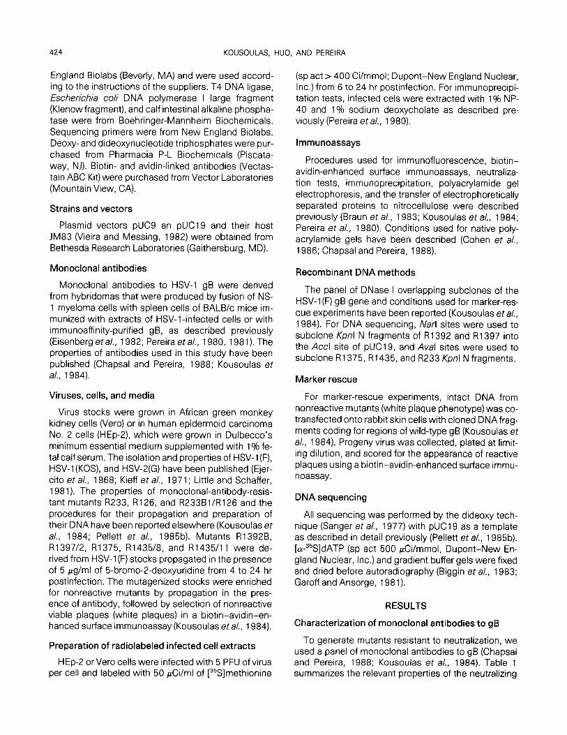
FIG. 1. Autoradiograms of electrophoretically separated polypeptides in immune precipitates, obtained by reacting homologous and heterolo- gous monoclonal antibodies with infected HEp-2 cells labeled with [%]methionine. Strains HSV-l(F), HSV2(G), and HSV-l(KOS) and mutants R1435/8, R1435/11, R126, R233, R126/233, and R1375, resistant to cross-neutralizing antibodies, are shown in top and middle panels. Mutants R13928, R1397, R1392, and R1397/2, resistant to HSV-1 -specific antibodies, are shown in bottom panel. Viruses are listed above each lane and antibodies are indicated above each section. gB + shows the precipitated glycoprotein.
cross-reactive antibodies H233, H1375, and H1435 and HSV-l-specific antibodies H1392 and H1397, as described under Materials and Methods. A mutant re- sistant to H 126 was isolated previously (Kousoulas et a/., 1984). To detect overlapping epitopes, the mutants were also reacted with heterologous antibodies (Table 2). Antibodies to discontinuous, type-common epi- topes neutralized strains HSV-1 (F), HSV-1 (KOS), and HSV-2(G). Cross-reactivity tests showed that mutant R126 was resistant to H1375 antibody and mutant R1375 to H126 antibody, indicating that the epitopes were affected by either mutation. H 1435 antibody neu- tralized R126 and R1375 mutants, but R1435/8 and R1435/1 1 were neutralized only partially by H 126 and were not neutralized by H 1375. These results indicate that the H 126 and H1375 epitopes were affected by the R1435 mutation, but that the H 1435 epitope was not altered by the R126 and R1375 mutations. R233 was neutralized by H126, H1375, and H1435 antibod- ies, and H233 antibody neutralized heterologous mu- tants R126, R1375, and R1435.
Antibodies to HSV-1 specific epitopes also showed complex neutralization patterns. H 1392 and H 1397 an- tibodies neutralized strain F but not KOS, G, or mutants R1392 and R1397 selected with the homologous anti- body. Cross-reactivity tests showed that antibody H1397 almost completely neutralized R1392 but that H1392 antibody failed to neutralize R1397. Although H 1396 did not select resistant mutants, the antibody failed to neutralize either R1392 or R1397. Insofar as the H 1396 epitope was altered by mutations in R1392 and R1397, the epitopes are partially overlapping.
Properties of resistant mutants in immunoprecipitation tests
The next series of experiments was done to deter- mine whether the selected mutants in gB would bind antibodies in immunoprecipitation tests. Antibodies to discontinuous epitopes reacted with HSV-1 (F), HSV- 1 (KOS), and HSV-2(G) but generally failed to react with gB specified by the selected mutant (Fig. 1). Whereas
ANTIBODY-RESISTANT MUTATIONS IN HSV-1 gB 427
mutants R126, R1375, R1435, and recombinant R126/ 233 failed to react with the homologous antibodies, neutralization-resistant mutant R233 was precipitated by H233 antibody. It was of interest that H 1375 anti- body reacted with mutant Rl435/8 but failed to recog- nize R1435/1 1, suggesting that mutations in the inde- pendent plaque isolates differed. Reactions with heter- ologous antibodies paralleled results of neutralization tests suggesting that the epitopes of H126 and H 1375 overlap and that the H233 epitope is separate from those of H126, H 1375, and H1435.
Antibodies to continuous epitopes also generally failed to react with gB specified by the selected mutant; the exception, R1392B, a spontaneous mutant, was precipitated by H 1392 antibody. Although repeated se- lection with antibodies H1396 and H 1839 failed to yield nonreactive mutants, H1839 reacted weakly and H1396 did not react with the heterologous R1392 mu- tant. Inasmuch as H 1839 precipitated strain KOS gB and H 1392 reacted weakly with KOS (data not shown), these epitopes may partially overlap. These results are supported by the finding that H 1839 antibody reacts weakly with R1392 and strongly with other mutants (Fig. 1).
Mapping and nucleotide sequence analyses of mutations that result in neutralization resistance
To map the approximate location of antibody-resis- tant mutations in gB, we performed marker rescue ex- periments by cotransfecting intact mutant DNA and wild-type DNA fragments with staggered deletions in gB, as described previously (Kousoulas et al., 1984). Mutations R233, R1375, and R1435/8 were rescued by plasmids pRB2021 and pRB2094, which overlap by 377 bp. DNA fragments encoding the portion of the gB sequence from amino acids 147 to 454 were cloned and sequenced. The mutation in R1375 was a G to A base change resulting in the substitution of Gln for Arg at residue 274, and the mutation in R1435 was a C to T base change resulting in the substitution of Tyr for His at residue 278 (data not shown). The mutation in R233, a G to A base change, resulted in the substitu- tion of His for Arg at residue 298.
The R1392 and R1397 mutations were rescued with plasmids pBR2085 and pBR2069, which contain DNA fragments overlapping by 180 bp, encoding residues -4 to +57. DNA fragments that encode gB residues -26 to +91 in the amino terminus were cloned and sequenced. The mutation in R1392 was a G to A base change resulting in a substitution of Thr for Ala at resi- due 32, and the R1397 mutation was an A to C base change resulting in the substitution of Thr for Asn at residue 47. The sequencing results are summarized in Table 3.
TABLE 3
NUCLEOTIDE SEQUENCE AND PREDICTED AMINO ACID CHANGES IN CON- TINUOUS AND DISCONTINUOUS EPITOPES OF ANTIBODY-RESISTANT Mu- TANTS IN gB
Epitopes’
Antibody- resistant mutants
Changes in nucleotide sequence
Predicted amino acid
substitutions
Discontinuous
Continuous
R233 CGC + CAC Amg8 -+ Hi% R1375 CGG + CAG Am4 + Glh R1435 CAC + TAC Hb8 + Tyr278 R126/Blb TAC + AX TyrzT3 - Ash
AGC -, ACC SerzB3 + Thrza3 R1392 GCC + ACC Ah + Thrs2 R1397 AAC -P ACC AS%, + Thr4,
a Epitopes on gB are defined by reactions of antibodies with so- dium dodecyl sulfate-denatured gB (continuous) or native gB dimers (discontinuous) (Chapsal and Pereira, in press).
* Mutation previously mapped (Pellet et al., 1985b).
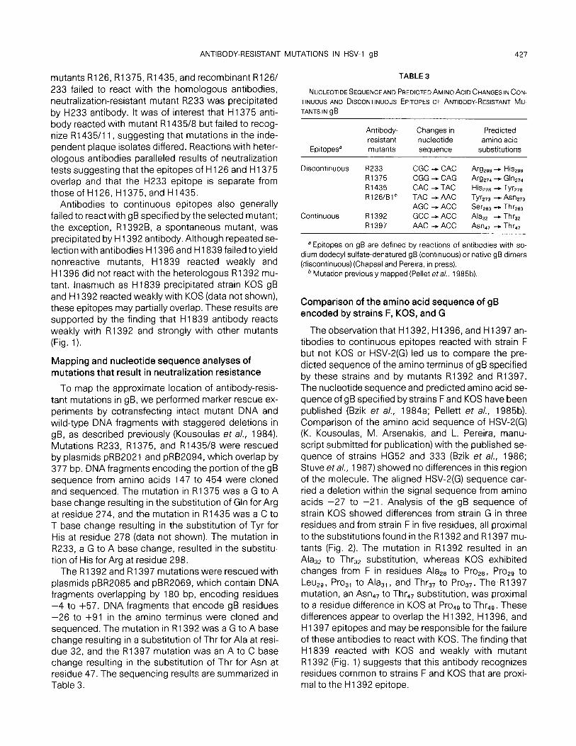
Comparison of the amino acid sequence of gB encoded by strains F, KOS, and G
The observation that H 1392, H 1396, and H 1397 an- tibodies to continuous epitopes reacted with strain F but not KOS or HSV-2(G) led us to compare the pre- dicted sequence of the amino terminus of gB specified by these strains and by mutants R1392 and R1397. The nucleotide sequence and predicted amino acid se- quence of gB specified by strains F and KOS have been published (Bzik et al., 1984a; Pellett et a/., 1985b). Comparison of the amino acid sequence of HSV-2(G) (K. Kousoulas, M. Arsenakis, and L. Pereira, manu- script submitted for publication) with the published se- quence of strains HG52 and 333 (Bzik et a/., 1986; Stuve et a/., 1987) showed no differences in this region of the molecule. The aligned HSV-2(G) sequence car- ried a deletion within the signal sequence from amino acids -27 to -21. Analysis of the gB sequence of strain KOS showed differences from strain G in three residues and from strain F in five residues, all proximal to the substitutions found in the R1392 and R1397 mu- tants (Fig. 2). The mutation in R1392 resulted in an Alas2 to Thr,, substitution, whereas KOS exhibited changes from F in residues Alap to ProZ8, ProZ9 to Leu,, , Pros, to Ala,, , and Thr,, to Pros,. The R1397 mutation, an Asn4, to Thr,, substitution, was proximal to a residue difference in KOS at Prod9 to Thr,,. These differences appear to overlap the H 1392, H 1396, and H 1397 epitopes and may be responsible for the failure of these antibodies to react with KOS. The finding that H1839 reacted with KOS and weakly with mutant R1392 (Fig. 1) suggests that this antibody recognizes residues common to strains F and KOS that are proxi- mal to the H 1392 epitope.
428 KOUSOULAS, HUO. AND PEREIRA
n*“-ll108)
HSV- 1 (F)
IiSV-Z(G)
MS”-1 IXOSI
HSV-l(F)
HSV-2(G)
em-1 (XCS)
HSV-l(F)
HSV.2(0)
SSV-1 ,IOSl
HSV-1 (F)
HSV.2(0)
ss”-l(Kos~
HSV-1 (F)
WV.?(G)
-29 ‘1’ ‘f” .;=ty yP
Met-arggln-gly-ala-ala- .._.. - arg-gly-cys-arg-trp-phe-r;al-;al-~~ala-leu-leu~~-~u-thr-leu~gly I I I IIll I
-1 l l
val-leu-val-ala-ser-a~-ala-pro-s~r~s~r-pro-gly-lhr~pr~gl~val-ala~ala~ala~lhr+ln-ala-ala-asn-gly- I I I I
9/y A I
Ita aa Ill aa 111 IIs VII
pro l=” “I El Pro
I I gly-pro-ala-f~pr~-a~-pro-pro~a~p~~g~y-pro-ala~p~-lhr~g~~~pl~~s-~~~s-~~~lys-
SW er9 pm WI SW lhr Irr a w l r9 Ihr lye
lys-pro-lys-asn-pro-pro-pro-pro-a~g-pro-~la-~l~a~p-asn-ala-thr-val-ala-a~~~-his-ala-~hr-leu- I I II I I
w ghl a* Ihr Pro pro .sp sls Vel
47
arg-glu-his-leu-arg-asp-ile-lys-a~glu-asn-t~r-as~ala-a~phe-tyr-val~s-pro- I I
II@ !JlU VII sls 9ln
FIG. 2. Comparison of the predicted amino acid sequence of the region coding for -29 to +87 residues of gB specified by HSV-l(F) (Pellett er a/., 1985b), HSV-1 (KOS) (Bzik et al., 1984) and HSV-2(G) (Kousoulas et al., manuscript in preparation). Amino acids from -29 to -1 contain the signal sequence. Residues in the KOS sequence (top line) and the G sequence (bottom line) are compared with those of strain F (middle line). Residues that differ are indicated and missing residues are represented by (. . .). Mutations in R1392 and R1397 are also indicated (boxes). N- glycosylation site, shaded box.
Comparison of the amino-terminal sequence of gB from strain G with that of strain F showed differences in 37 residues, rendering the amino terminus of the molecule highly type-specific. Results of this study showing that continuous epitopes of four type-specific antibodies mapped within the amino terminus of gB were supported by transient expression of truncated derivatives (L. Pereira, K. Kousoulas, M. Ali, B. Huo and T. Banks, manuscript in preparation) and by analysis of the immunologic properties of gB made by a HSV-1 x HSV-2 inter-typic recombinant in this gene (Kousou- las et al., manuscript submitted for publication).
Reactivity of antibodies with HSV-1 gB made in Vero cells and HEp-2 cells
We previously reported that HSV-2 gB undergoes cleavage in Vero cells into fragments with greater mo- bilities, and that a HSV-2-specific antibody failed to re- act with cleavage products made in Vero cells (Pereira et a/., 1981, 1982). To determine whether HSV-1 -spe- cific antibodies react similarly, we infected Vero and HEp-2 cells with strain F or G, precipitated extracts with antibodies, and subjected the precipitates to elec- trophoresis in denaturing polyacrylamide gels. Results of these experiments are summarized in Table 1. Type-
specific H 1392, H1396, H 1397, and H 1839 antibodies reacted with HEp-2 cells infected with strain F but not G, and failed to react with extracts of infected Vero cells. The finding that cleavage disrupts the reactivity of four antibodies, and that the epitopes of these anti- bodies are type-specifc and map in the amino terminus of the molecule, indicates that one cleavage site is within the first 50 amino acids of the amino terminus and is partially overlapped by these epitopes.
DISCUSSION
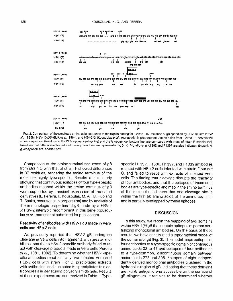
In this study, we report the mapping of two domains within HSV-1 (F) gB that contain epitopes of potent neu- tralizing monoclonal antibodies. On the basis of these results, we have constructed a topographical model of the domains of gB (Fig. 3). The model maps epitopes of four antibodies to a type-specific domain of continuous amino acids 32 to 47 and epitopes of four antibodies to a type-common, discontinuous domain between amino acids 273 and 298. Epitopes of eight indepen- dently derived monoclonal antibodies clustered in the hydrophilic region of gB, indicating that these domains are highly antigenic and accessible on the surface of gB oliogomers. It remains to be determined whether
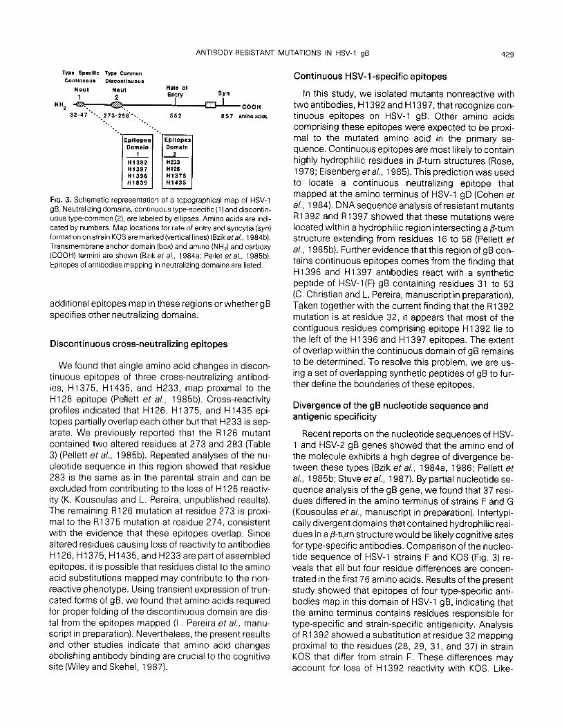
ANTIBODY-RESISTANT MUTATIONS IN HSV-1 gB 429
TYPO Sp*clflc Typa Common Contlnuour Dlscontlnuour
Neut Neut Rate of
1 2 Entry Syn -
coon 552 6 5 7 amino acids
-*.. -. *. --.*
--. -+. Epltopes Epltopes Domain Domain 1
1II
H1392 H233 Ii1397 H126 H1396 H1375 H1839 H1435
FIG. 3. Schematic representation of a topographical map of HSV-1 gB. Neutralizing domains, continuous type-specific (1) and discontin- uous type-common (2). are labeled by ellipses. Amino acids are indi- cated by numbers. Map locations for rate of entry and syncytia (syn) formation on strain KOS are marked (vertical lines) (Bzik era/., 1984b). Transmembrane anchor domain (box) and amino (NH,) and carboxy (COOH) termini are shown (Bzik et al., 1984a; Pellet et a/., 1985b). Epitopes of antibodies mapping in neutralizing domains are listed.
additional epitopes map in these regions orwhether gB specifies other neutralizing domains.
Discontinuous cross-neutralizing epitopes
We found that single amino acid changes in discon- tinuous epitopes of three cross-neutralizing antibod- ies, H1375, H1435, and H233, map proximal to the H 126 epitope (Pellett et al., 1985b). Cross-reactivity profiles indicated that H 126, H 1375, and H 1435 epi- topes partially overlap each other but that H233 is sep- arate. We previously reported that the R126 mutant contained two altered residues at 273 and 283 (Table 3) (Pellett et al., 1985b). Repeated analyses of the nu- cleotide sequence in this region showed that residue 283 is the same as in the parental strain and can be excluded from contributing to the loss of H126 reactiv- ity (K. Kousoulas and L. Pereira, unpublished results). The remaining R126 mutation at residue 273 is proxi- mal to the R1375 mutation at residue 274, consistent with the evidence that these epitopes overlap. Since altered residues causing loss of reactivity to antibodies H 126, H 1375, H 1435, and H233 are part of assembled epitopes, it is possible that residues distal to the amino acid substitutions mapped may contribute to the non- reactive phenotype. Using transient expression of trun- cated forms of gB, we found that amino acids required for proper folding of the discontinuous domain are dis- tal from the epitopes mapped (L. Pereira et a/., manu- script in preparation). Nevertheless, the present results and other studies indicate that amino acid changes abolishing antibody binding are crucial to the cognitive site (Wiley and Skehel, 1987).
Continuous HSV-1 -specific epitopes
In this study, we isolated mutants nonreactive with two antibodies, H 1392 and H 1397, that recognize con- tinuous epitopes on HSV-1 gB. Other amino acids comprising these epitopes were expected to be proxi- mal to the mutated amino acid in the primary se- quence. Continuous epitopes are most likely to contain highly hydrophilic residues in P-turn structures (Rose, 1978; Eisenberg eta/., 1985). This prediction was used to locate a continuous neutralizing epitope that mapped at the amino terminus of HSV-1 gD (Cohen et a/., 1984). DNA sequence analysis of resistant mutants R1392 and R1397 showed that these mutations were located within a hydrophilic region intersecting a P-turn structure extending from residues 16 to 58 (Pellett et a/., 198513). Further evidence that this region of gB con- tains continuous epitopes comes from the finding that H1396 and H 1397 antibodies react with a synthetic peptide of HSV-l(F) gB containing residues 31 to 53 (C. Christian and L. Pereira, manuscript in preparation). Taken together with the current finding that the R1392 mutation is at residue 32, it appears that most of the contiguous residues comprising epitope H1392 lie to the left of the H 1396 and H 1397 epitopes. The extent of overlap within the continuous domain of gB remains to be determined. To resolve this problem, we are us- ing a set of overlapping synthetic peptides of gB to fur- ther define the boundaries of these epitopes.
Divergence of the gB nucleotide sequence and antigenic specificity
Recent reports on the nucleotide sequences of HSV- 1 and HSV-2 gB genes showed that the amino end of the molecule exhibits a high degree of divergence be- tween these types (Bzik et al., 1984a, 1986; Pellett et al., 198513; Stuve et al., 1987). By partial nucleotide se- quence analysis of the gB gene, we found that 37 resi- dues differed in the amino terminus of strains F and G (Kousoulas et al., manuscript in preparation). Intertypi- callydivergent domains that contained hydrophilic resi- dues in a P-turn structure would be likely cognitive sites for type-specific antibodies. Comparison of the nucleo- tide sequence of HSV-1 strains F and KOS (Fig. 3) re- veals that all but four residue differences are concen- trated in the first 76 amino acids. Results of the present study showed that epitopes of four type-specific anti- bodies map in this domain of HSV-1 gB, indicating that the amino terminus contains residues responsible for type-specific and strain-specific antigenicity. Analysis of R1392 showed a substitution at residue 32 mapping proximal to the residues (28, 29, 31, and 37) in strain KOS that differ from strain F. These differences may account for loss of H 1392 reactivity with KOS. Like-
430 KOUSOULAS, HUO, AND PEREIRA
wise, strain KOS exhibits a change at residue 49, proxi- mal to the R1397 mutation at position 37. The failure of H1397 to react with strain KOS indicated that ProJ9 is a participating residue in the H1397 epitope. The ob- servation that H 1392 antibody failed to neutralize the R1397 mutant suggests that this mutation affects the partially overlapping H 1392 epitope. If this is true, then one or more of the amino acids at positions that differ between strains F and KOS may participate in the H 1392, H 1396, and H1397 epitopes. Analyses of addi- tional mutants selected with these antibodies should further define the residues that comprise each epitope.
Proteolytic cleavage of gB in Vero cells
We reported that HSV-2 gB made in Vero cells speci- fied different immunological and electrophoretic prop- erties from gB made in HEp-2 cells, as a result of cleav- age by cellular enzymes (Pereira et a/., 1981, 1982). The observations that cleavage precluded the reactiv- ity of a subset of neutralizing antibodies and that the cleavage fragments were glycosylated suggested that they were derived from the extracellular region of the molecule. In the present study, we found that H1392, H1396, H1397, and H1839 antibodies, whose epi- topes map at the amino terminus of gB, failed to precip- itate the Vero cell products. This result is consistent with the notion that the cleavage sites are located at the amino terminus of the molecule and partially over- lap these epitopes.
ACKNOWLEDGMENTS
These studies were aided in part by Public Health Service Grants DE-08275 from the National Institute of Dental Research and Al- 29592 from the National Institutes of Allergy and Infectious Diseases and by a grant from the Academic Senate Committee on Research Support of the University of California San Francisco. K.G.K. was a postdoctoral trainee in the Public Health Service Training Program in Sexually Transmitted Diseases (P32-AC07234).
REFERENCES
BIGGIN, M. D., GIBSON, T. J., and HONG, G. F. (1983). Buffer gradient gels and 36S label as an aid to rapid DNA sequence determination. Proc. Nat/. Acad. Sci. USA 80,3963-3965.
BRAUN, D. K., PEREIRA, L., NORRILD, B., and ROIZMAN, B. (1983). Appli- cation of denatured, electrophoretically separated, and immobi- lized lysates of herpes simplex virus-infected cells for the detection of monoclonal antibodies and for studies of the properties of viral proteins. J. Viral. 46, 103-l 12.
BZIK, D. J., DEBROY, C., Fox, B., PEDERSON, N. E., and PERSON, S. (1986). The nucleotide sequence of the gB glycoprotein gene of HSV-2 and comparison with the corresponding gene of HSV-1. vi- rology 155, 322-333.
BZIK, D. J., Fox, B. A., DELUCA, N. A., and PERSON, S. (1984s). Nucleo- tide sequence specifying the glycoprotein gene, gB. of herpes sim- plex virus type 1. Virology 133,30 l-3 14.
BZIK, D. J., Fox, B.A., DELUCA, N. A., and PERSON, S. (1984b). Nucleo- tide sequence of the region of the herpes simplex virus type 1 gB glycoprotein gene: Mutations affecting rate of entry and cell fusion. Virology 137, 185-l 90.
CHAPSAL, J. M., and PEREIRA, L. (1988). Characterization of epitopes on native and denatured forms of herpes simplex virus glycopro- tein B. Virology 164,427-434.
CLAESSON-WELSH, L., and SPEAR, P. G. (1986). Oligomerization of her- pes simplex virus glycoprotein B. J. Viral. 60, 803-806.
COHEN, G. H., DIETZSCHOLD, B., PONCE DE LEON, M., LONG, D., GOLUB, E., VARRICHIO, A., PEREIRA, L., and EISENBERG, R.J. (1984). Localiza- tion and synthesis of an antigenic determinant of herpes simplex virus glycoprotein D that stimulates the production of neutralizing antibody. J. Viral. 49, 102-l 08.
COHEN, G. H., ISOLA, V. J., KUHN% J., BERMAN, P. W., and EISENBERG, R. J. (1986). Localization of discontinuous epitopes of herpes sim- plex virus glycoprotein D: Use of a nondenaturing (“native”) gel system of polyacrylamide gel electrophoresis coupled with West- ern blotting. J. Viral. 60, 157-l 66.
CRANAGE, M. P., KOUSARIDES, T., BANKIER, A. T., SATCHWELL, S., WES- TON, K., TOMLINSON, P., BARRELL, B., HART, H., BELL, S. E., MINSON, A. C., and SMITH, G. L. (1986). Identification of the human cyto- megalovirus glycoprotein B gene and induction of neutralizing anti- bodies via its expression in recombinant vaccinia virus. EMBO J. 5,3057-3063.
DAVIDSON, A. J., and Scorr, J. E. (1986). The complete DNA se- quence of varicella-zostervirus. J. Gen. Viral. 67, 1759-l 816.
DEBROY, C., PEDERSON, N., and PERSON, S. (1985). Nucleotide se- quence of a herpes simplex virus type 1 gene that causes cell fu- sion. Virology 145, 36-48.
DELUCA, N., BZIK, D., BOND, V. C., Person, S., and SNIPES, W. (1982). Nucleotide sequences of herpes simplex virus type 1 (HSV-1) affecting virus entry, cell fusion, and production of glycoprotein gB (VP7). Virology 122,411-413.
EISENBERG, R. J., LONG, D., PEREIRA, L., HAMPAR, B., ZWEIG, M., and COHEN, G. H. (1982). Effect of monoclonal antibodies on limited proteolysis of native glycoprotein gD of herpes simplex virus type 1.1. Viral. 41,478-488.
EISENBERG, R. J.. LONG, D., PONCE DE LEON, M., MATTHEWS, J. T., SPEAR, P. G.. GIBSON, M. G., ~SKY, L. A., BERMAN, P., GOLUB, E., and COHEN, G. H. (1985). Localization of epitopes of herpes sim- plex virus type 1 glycoprotein D. J. Viral. 53, 634-644.
EJERCITO, P. M., KIEFF, E. D., and ROIZMAN, B. (1968). Characterization of herpes simplex virus strains differing in their effect on social be- havior of infected cells. J. Gen. Viral. 3, 357-364.
GAROFF, H., and ANSORGE, W. (1981). Improvements of DNA se- quencing gels. Anal. Biochem. 115,450-457.
HAFFEY, M. L., and SPEAR, P. G. (1980). Alterations in glycoprotein gB specified by mutants and their partial revertants in herpes simplex virus type 1 and relationship to other mutant phenotypes. J. Viral. 35,114-l 28.
HOLLAND, T. C., SANDRI-GOLDIN, R. M., HOLLAND, L. E., MARLIN, S. D., LEVINE, M., and GLORIOSO, J. C. (1983). Physical mapping of the mutation in an antigenic variant of herpes simplex virus type 1 by use of an immoreactive plaque assay. J. Viral. 46,649-652.
KELLER, P. M., DAVISON, A. J., LOWE, R. S., BENNET, C. D., and ELLIS, R. W., (1986). Identification and structure of the gene encoding gpll, a major glycoprotein of varicella-zoster virus. Virology 152, 181-191.
KIEFF, E. D., BACHENHEIMER, S. L., and ROIZMAN, B. (1971). Size, com- position, and structure of the deoxyribonucleic acid of herpes sim- plex virus subtypes 1 and 2.1. Viral. 8, 125-l 42.
Kousoul~s, K. G., PELLET-~. P. E., PEREIRA. L., and ROIZMAN. B. (1984). Mutations affecting conformation or sequence of neutralizing epi-
ANTIBODY-RESISTANT MUTATIONS IN HSV-1 gB 431
topes identified by reactivity of viable plaques segregated from syn and fs domains of HSV-1 (F) gB gene. Virology 135,379-394.
LITTLE, S. P., JOFRE, J. T., COURTNEY, R. J., and SCHAFFER, P. A. (1981). A virion-associated glycoprotein essential for infectivity of herpes simplex virus type 1 Virology 115, 149-l 60.
LITTLE, S. P., and SCHAFFER. P. A. (1981). Expression of the syncytial syn phenotype in HSV-1, strain KOS: Genetic and phenotypic stud- ies of mutants in two syn loci. Virology 112, 686-702.
MANSERVIGI, R., SPEAR, P. G., and BUCHAN, A. (1977). Cell fusion in- duced by herpes simplex virus is promoted and suppressed by different viral glycoproteins. Proc. Nat/. Acad. Ski. USA 74, 3913- 3917.
MARLIN, S. D., HIGHLANDER, S. L.. HOLLAND, T. C., LEVINE, M., and GLORIOSO, J. C. (1986). Antigenic variation (mar mutations) in her- pes simplex virus glycoprotein B can induce temperature-depen- dent alterations in gB processing and virus production. J. Viral. 59, 142-153.
NORRILD, B. (1980). Immunochemistry of herpes simplex virus glyco- proteins. Cur. Top. Microbial. lmmunol. 90, 67-l 06.
PELLET, P. E., BIGGIN, M. D., BARRELL, B., and ROIZMAN, B. (1985a). Epstein-Barr virus genome may encode a protein showing sig- nificant amino acid and predicted secondary structure homology with glycoprotein B of herpes simplex virus 1. J. Viral. 56, 807- 813.
PELLET, P. E., Kousoul~s, K. G., PEREIRA, L., and ROIZMAN, B. (1985b). Anatomy of the herpes simplex virus 1 strain F glycoprotein B gene: Primary sequence and predicted protein structure of the wild type and of monoclonal antibody-resistant mutants. J. Viral. 53, 243-253.
PEREIRA, L., DONDERO. D., NORRILD, B., and ROIZMAN, B. (1981). Differential immunologic and electrophoretic properties of glyco- proteins gA and gB of HSV-2 produced in HEp-2 and Vero cells. Proc. Nat/. Acad. Sci. USA 78, 5202-5206.
PEREIRA, L., DONDERO, D., and ROIZMAN, B. (1982). Herpes simplex virus glycoprotein gA/B: Evidence that the infected Vero cell prod- ucts comap and arise by proteolysis. J. Viral. 44, 88-97.
PEREIRA, L., KLASSEN, T., and BARINGER, J. R. (1980). Type common and type specific monoclonal antibody to herpes simplex virus 1. Infect. Immun. 29,724-732.
POGUE-GEILE, K. L., LEE, G. T.-Y., SHAPIRO, S. K., and SPEAR, P. G. (1984). Fine mapping of mutations in the fusion-inducing MP strain of herpes simplex virus type 1. Virology 136, 100-l 09.
POGUE-GEILE. K. L., and SPEAR, P. G. (1987). The single base pair sub- stitution responsible for thesyn phenotype of herpes simplexvirus type 1, strain MP. Virology 157, 67-74.
ROSE, G. D. (1978). Prediction of chain turns in globular proteins on a hydrophobic basis. Nature (London) 272,586-590.
RUYECHAN, T. W., MORSE, L. S., KNIPE, D. M., and ROIZMAN, B. (1979). Molecular genetics of herpes simplex virus. II. Mapping of the ma- jor viral glycoproteins and of the genetic loci specifying the social behavior of infected cells. 1. Viral. 29, 677-697.
SANGER, F., NICKLEN, S., and COULSON, A. R. (1977). DNAsequencing with chain terminating inhibitors. Proc. Nat/. Acad. Sci. USA 74, 5463-5467.
SARMIENTO, M., HAFFEY, M., and SPEAR, P. G. (1979). Membrane pro- teins specified by herpes smplex viruses. Ill. Role of glycoprotein VP7 (B2) in virion infectivity. J. viral. 29, 1 149-l 158.
SARMIENTO, M., and SPEAR, P. G. (1979). Membrane proteins speci- fied by herpes simplex viruses. IV. Conformation of the virion gly- coprotein designated VP7(B2). 1. Viral. 29, 1159-l 167.
SPEAR, P. G. (1984). Glycoproteins specified by herpes simplex vi- ruses. In “The Herpesviruses” (B. Roizman, Ed.), Vol. 3, pp. 315- 356. Plenum, New York.
STUVE, L. L., BROWN-SHIMER, S., PACHL, C., NAJARIAN, R., DINA, D., and BURKE, R. L. (1987). Structure and expression of the herpes sim- plex virus type-2 glycoprotein gB gene. J. Viral. 61,326-335.
VIEIRA. J.. and MESSING, J. (1982). The pUC plasmids, an M13mp7- derived system for insertion mutagenesis and sequencing with synthetic universal primers. Gene 19, 259-268.
WILEY, D. C., and SKEHEL, 1. J. (1987). The structure and function of the hemagglutinin membrane glycoprotein of influenza virus. Annu. Rev. Biochem. 56,365-394.
Related Documents