Citation: Zoratti, C.; Moretti, R.; Rebuzzi, L.; Albergati, I.V.; Di Somma, A.; Decorti, G.; Di Bella, S.; Crocè, L.S.; Giuffrè, M. Antibiotics and Liver Cirrhosis: What the Physicians Need to Know. Antibiotics 2022, 11, 31. https://doi.org/ 10.3390/antibiotics11010031 Academic Editor: Jeffrey Lipman Received: 18 November 2021 Accepted: 24 December 2021 Published: 28 December 2021 Publisher’s Note: MDPI stays neutral with regard to jurisdictional claims in published maps and institutional affil- iations. Copyright: © 2021 by the authors. Licensee MDPI, Basel, Switzerland. This article is an open access article distributed under the terms and conditions of the Creative Commons Attribution (CC BY) license (https:// creativecommons.org/licenses/by/ 4.0/). antibiotics Review Antibiotics and Liver Cirrhosis: What the Physicians Need to Know Caterina Zoratti 1 , Rita Moretti 1 , Lisa Rebuzzi 1 , Irma Valeria Albergati 1 , Antonietta Di Somma 1 , Giuliana Decorti 2 , Stefano Di Bella 1 , Lory Saveria Crocè 1,3 and Mauro Giuffrè 1,3, * 1 Department of Medical, Surgical and Health Sciences, University of Trieste, 34127 Trieste, Italy; [email protected] (C.Z.); [email protected] (R.M.); [email protected] (L.R.); [email protected] (I.V.A.); [email protected] (A.D.S.); [email protected] (S.D.B.); [email protected] (L.S.C.) 2 Institute for Maternal and Child Health-IRCCS Burlo Garofolo, 34137 Trieste, Italy; [email protected] 3 Italian Liver Foundation, 34149 Trieste, Italy * Correspondence: [email protected]; Tel.: +39-0403994044 Abstract: The liver is the primary site of drug metabolism, which can be altered by a variety of diseases affecting the liver parenchyma, especially in patients with liver cirrhosis. The use of antibiotics in patients with cirrhosis is usually a matter of concern for physicians, given the lack of practical knowledge for drug choice and eventual dose adjustments in several clinical scenarios. The aim of the current narrative review is to report, as broadly as possible, basic, and practical knowledge that any physician should have when approaching a patient with liver cirrhosis and an ongoing infection to efficiently choose the best antibiotic therapy. Keywords: antibiotics; liver cirrhosis; ascites; spontaneous bacterial peritonitis; hepatic encephalopathy 1. Introduction The liver is the primary site of drug metabolism. Biotransformation of drugs and xenobiotics into more hydrophilic compounds is necessary for their elimination through the urine and the bile. Most drugs are metabolized by phase I (mainly oxidation, reduction, and hydrolysis, leading to the introduction of functional groups and often changing the biological properties) and phase II (that involves conjugation with an endogenous substance such as glucuronic acid, sulfate, glycine) reactions. These steps are largely dependent on two factors: hepatic blood flow and the metabolic capacity of the liver [1]. Liver cirrhosis is the final common pathological pathway of liver damage arising from many chronic liver diseases. Although there are several causes of liver cirrhosis, some histopathological changes in the liver parenchyma, shared between the various etiologies, include degeneration and necrosis of hepatocytes and the replacement of liver parenchyma by fibrotic tissues, regenerative nodules, and loss of liver function [2]. In liver cirrhosis, impaired drug disposition is due to many pathological changes, including liver cell necrosis, portosystemic shunt, reduction in the concentration of drug- binding proteins, atypical drug volume of distribution, altered drug metabolism and elimination, altered pharmacodynamics, drug interactions, and the frequent association with renal failure [1] as shown in Figure 1. In particular, the impairment of drug metabolism is directly related to the degree of liver dysfunction and, although various tests can be used to predict the severity of liver function (e.g., indocyanine green clearance, Child Pugh or Meld-Na [1]. Of notice, the liver metabolic capacity must be reduced more than 90% to actually require dosage adjustment. However, the use of medications in patients with cirrhosis is usually a matter of concern for physicians, considering the risk of developing an acute Antibiotics 2022, 11, 31. https://doi.org/10.3390/antibiotics11010031 https://www.mdpi.com/journal/antibiotics

Welcome message from author
This document is posted to help you gain knowledge. Please leave a comment to let me know what you think about it! Share it to your friends and learn new things together.
Transcript
�����������������
Citation: Zoratti, C.; Moretti, R.;
Rebuzzi, L.; Albergati, I.V.; Di
Somma, A.; Decorti, G.; Di Bella, S.;
Crocè, L.S.; Giuffrè, M. Antibiotics
and Liver Cirrhosis: What the
Physicians Need to Know. Antibiotics
2022, 11, 31. https://doi.org/
10.3390/antibiotics11010031
Academic Editor: Jeffrey Lipman
Received: 18 November 2021
Accepted: 24 December 2021
Published: 28 December 2021
Publisher’s Note: MDPI stays neutral
with regard to jurisdictional claims in
published maps and institutional affil-
iations.
Copyright: © 2021 by the authors.
Licensee MDPI, Basel, Switzerland.
This article is an open access article
distributed under the terms and
conditions of the Creative Commons
Attribution (CC BY) license (https://
creativecommons.org/licenses/by/
4.0/).
antibiotics
Review
Antibiotics and Liver Cirrhosis: What the Physicians Needto KnowCaterina Zoratti 1, Rita Moretti 1 , Lisa Rebuzzi 1, Irma Valeria Albergati 1, Antonietta Di Somma 1,Giuliana Decorti 2 , Stefano Di Bella 1 , Lory Saveria Crocè 1,3 and Mauro Giuffrè 1,3,*
1 Department of Medical, Surgical and Health Sciences, University of Trieste, 34127 Trieste, Italy;[email protected] (C.Z.); [email protected] (R.M.); [email protected] (L.R.);[email protected] (I.V.A.); [email protected] (A.D.S.);[email protected] (S.D.B.); [email protected] (L.S.C.)
2 Institute for Maternal and Child Health-IRCCS Burlo Garofolo, 34137 Trieste, Italy; [email protected] Italian Liver Foundation, 34149 Trieste, Italy* Correspondence: [email protected]; Tel.: +39-0403994044
Abstract: The liver is the primary site of drug metabolism, which can be altered by a variety ofdiseases affecting the liver parenchyma, especially in patients with liver cirrhosis. The use ofantibiotics in patients with cirrhosis is usually a matter of concern for physicians, given the lack ofpractical knowledge for drug choice and eventual dose adjustments in several clinical scenarios. Theaim of the current narrative review is to report, as broadly as possible, basic, and practical knowledgethat any physician should have when approaching a patient with liver cirrhosis and an ongoinginfection to efficiently choose the best antibiotic therapy.
Keywords: antibiotics; liver cirrhosis; ascites; spontaneous bacterial peritonitis; hepatic encephalopathy
1. Introduction
The liver is the primary site of drug metabolism. Biotransformation of drugs andxenobiotics into more hydrophilic compounds is necessary for their elimination throughthe urine and the bile. Most drugs are metabolized by phase I (mainly oxidation, reduction,and hydrolysis, leading to the introduction of functional groups and often changing thebiological properties) and phase II (that involves conjugation with an endogenous substancesuch as glucuronic acid, sulfate, glycine) reactions. These steps are largely dependent ontwo factors: hepatic blood flow and the metabolic capacity of the liver [1].
Liver cirrhosis is the final common pathological pathway of liver damage arising frommany chronic liver diseases. Although there are several causes of liver cirrhosis, somehistopathological changes in the liver parenchyma, shared between the various etiologies,include degeneration and necrosis of hepatocytes and the replacement of liver parenchymaby fibrotic tissues, regenerative nodules, and loss of liver function [2].
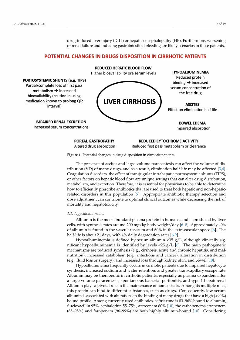
In liver cirrhosis, impaired drug disposition is due to many pathological changes,including liver cell necrosis, portosystemic shunt, reduction in the concentration of drug-binding proteins, atypical drug volume of distribution, altered drug metabolism andelimination, altered pharmacodynamics, drug interactions, and the frequent associationwith renal failure [1] as shown in Figure 1. In particular, the impairment of drug metabolismis directly related to the degree of liver dysfunction and, although various tests can be usedto predict the severity of liver function (e.g., indocyanine green clearance, Child Pugh orMeld-Na [1].
Of notice, the liver metabolic capacity must be reduced more than 90% to actuallyrequire dosage adjustment. However, the use of medications in patients with cirrhosis isusually a matter of concern for physicians, considering the risk of developing an acute
Antibiotics 2022, 11, 31. https://doi.org/10.3390/antibiotics11010031 https://www.mdpi.com/journal/antibiotics
Antibiotics 2022, 11, 31 2 of 19
drug-induced liver injury (DILI) or hepatic encephalopathy (HE). Furthermore, worseningof renal failure and inducing gastrointestinal bleeding are likely scenarios in these patients.
Antibiotics 2022, 11, x FOR PEER REVIEW 2 of 20
usually a matter of concern for physicians, considering the risk of developing an acute drug-induced liver injury (DILI) or hepatic encephalopathy (HE). Furthermore, worsen-ing of renal failure and inducing gastrointestinal bleeding are likely scenarios in these patients.
The presence of ascites and large volume paracentesis can affect the volume of dis-tribution (VD) of many drugs, and as a result, elimination half-life may be affected [3,4]. Coagulation disorders, the effect of transjugular intrahepatic portosystemic shunts (TIPS), or other factors on hepatic blood flow are unique settings that can alter drug distribution, metabolism, and excretion. Therefore, it is essential for physicians to be able to determine how to efficiently prescribe antibiotics that are used to treat both hepatic and non-hepatic-related disorders in this population [5]. Appropriate antibiotic therapy selection and dose adjustment can contribute to optimal clinical outcomes while decreasing the risk of mor-tality and hepatotoxicity.
Figure 1. Potential changes in drug disposition in cirrhotic patients.
1.1. Hypoalbuminemia Albumin is the most abundant plasma protein in humans, and is produced by liver
cells, with synthesis rates around 200 mg/kg body weight/day [6–8]. Approximately 40% of albumin is found in the vascular system and 60% in the extravascular space [6]. The half-life is about 21 days, with 4% daily degradation rates [6,9].
Hypoalbuminemia is defined by serum albumin <35 g/L, although clinically signifi-cant hypoalbuminemia is identified by levels <25 g/L [6]. The main pathogenetic mecha-nisms are reduced synthesis (e.g., cirrhosis, acute and chronic hepatitis, and malnutrition), increased catabolism (e.g., infections and cancer), alteration in distribution (e.g., fluid loss or surgery), and increased loss through kidney, skin, and bowel [10].
Hypoalbuminemia frequently occurs in cirrhotic patients due to impaired hepatocyte synthesis, increased sodium and water retention, and greater transcapillary escape rate. Albumin may be therapeutic in cirrhotic patients, especially as plasma expanders after a large volume paracentesis, spontaneous bacterial peritonitis, and type 1 hepatorenal Al-bumin plays a pivotal role in the maintenance of homeostasis. Among its multiple roles, this protein can bind to different substances, such as drugs. Consequently, low serum al-bumin is associated with alterations in the binding of many drugs that have a high (>90%) bound profile. Among currently used antibiotics, ceftriaxone is 83–96% bound to albumin, flucloxacillin 95%, cephalothin 55–75%, aztreonam 60% [10], the carbapenems ertapenem
Figure 1. Potential changes in drug disposition in cirrhotic patients.
The presence of ascites and large volume paracentesis can affect the volume of dis-tribution (VD) of many drugs, and as a result, elimination half-life may be affected [3,4].Coagulation disorders, the effect of transjugular intrahepatic portosystemic shunts (TIPS),or other factors on hepatic blood flow are unique settings that can alter drug distribution,metabolism, and excretion. Therefore, it is essential for physicians to be able to determinehow to efficiently prescribe antibiotics that are used to treat both hepatic and non-hepatic-related disorders in this population [5]. Appropriate antibiotic therapy selection anddose adjustment can contribute to optimal clinical outcomes while decreasing the risk ofmortality and hepatotoxicity.
1.1. Hypoalbuminemia
Albumin is the most abundant plasma protein in humans, and is produced by livercells, with synthesis rates around 200 mg/kg body weight/day [6–8]. Approximately 40%of albumin is found in the vascular system and 60% in the extravascular space [6]. Thehalf-life is about 21 days, with 4% daily degradation rates [6,9].
Hypoalbuminemia is defined by serum albumin <35 g/L, although clinically sig-nificant hypoalbuminemia is identified by levels <25 g/L [6]. The main pathogeneticmechanisms are reduced synthesis (e.g., cirrhosis, acute and chronic hepatitis, and mal-nutrition), increased catabolism (e.g., infections and cancer), alteration in distribution(e.g., fluid loss or surgery), and increased loss through kidney, skin, and bowel [10].
Hypoalbuminemia frequently occurs in cirrhotic patients due to impaired hepatocytesynthesis, increased sodium and water retention, and greater transcapillary escape rate.Albumin may be therapeutic in cirrhotic patients, especially as plasma expanders aftera large volume paracentesis, spontaneous bacterial peritonitis, and type 1 hepatorenalAlbumin plays a pivotal role in the maintenance of homeostasis. Among its multiple roles,this protein can bind to different substances, such as drugs. Consequently, low serumalbumin is associated with alterations in the binding of many drugs that have a high (>90%)bound profile. Among currently used antibiotics, ceftriaxone is 83–96% bound to albumin,flucloxacillin 95%, cephalothin 55–75%, aztreonam 60% [10], the carbapenems ertapenem(85–95%) and faropenem (96–99%) are both highly albumin-bound [10]. Considering
Antibiotics 2022, 11, 31 3 of 19
glycopeptides, vancomycin is bound to albumin between 30% and 60%, while teicoplaninis 90–95% [10]. A study compared the effectiveness of ertapenem, with other carbapenems,such as meropenem or imipenem, in the presence of hypoalbuminemia against infectionssustained by carbapenems-susceptible Enterobacteriaceae in hospitalized patients [11].
The results showed a higher 30-day mortality incidence with ertapenem compared toimipenem/meropenem [11]. Moreover, using the regression models, it could be inferreda five-fold mortality risk for a subject treated with ertapenem and an albumin level of2 g/dL compared to a subject with normal albumin (4 g/dL), whereas there was no suchassociation among subjects treated with imipenem or meropenem [11].
For highly protein-bound antibacterials, hypoalbuminemia increases the unboundfraction of the administered drug, which is available for distribution and clearance fromthe plasma. Only the unbound drug can distribute into body tissues, exerting its pharma-cological effect and potential toxicity, and be eliminated from the vascular compartment.Hence hypoalbuminemia increases the apparent total VD and the clearance (CL) of thedrug, decreasing the effective concentration in serum over time, which could be a problemwhen facing bacteria with high minimal inhibitory concentration (MIC). Besides takinginto consideration renal impairment and hepatic insufficiency in drug dose adjustments,hypoalbuminemia should also be considered to optimize antibiotic treatment [10].
Moreover, the percentage of antibiotics bound by albumin may also be altered byother clinical scenarios that can occur in cirrhotic patients, such as increased concentra-tion of endogenous molecules with high affinity for albumin (e.g., in uremia), increasedfree fatty acids, or bilirubin levels or administration of other highly protein-bound drugscan displace highly bound antibiotics from their albumin binding sites. The pharmacoki-netic alterations caused by hypoalbuminemia could have significant consequences onthe attainment of pharmacokinetic/pharmacodynamic targets. The higher VD could bea problem for concentration-dependent drugs, such as the aminoglycosides, requiring ahigher loading dose to reach therapeutic levels. On the other hand, faster CL may affect theefficacy of ß-lactams. Penicillins, cephalosporins, carbapenems, and monobactams belongto the ß-lactams family and are time-dependent antibiotics. Their effect depends on thepercentage of time in which serum concentrations of the free drug remain above the MICof the causative pathogen (% fT > MIC). For maximal bactericidal activity, penicillins andmonobactams require at least 50–60% fT > MIC [10], cephalosporines require 60–70%, andcarbapenems 40% fT > MIC [10].
Higher CL and VD due to hypoalbuminemia lead to low drug concentrations, espe-cially for hydrophilic drugs, which may be below fT > MIC targets and thereby ineffective.Therefore, to maintain free drug levels above the MIC, optimizing their pharmacoki-netic/pharmacodynamic target, also given the short half-life of most ß-lactams, continuousor extended infusions of these agents may be used, and have shown to improve 30-dayssurvival in patients with cirrhosis [12].
In conclusion, the outcome of antibacterial therapy depends on achieving therapeuticconcentrations of unbound antibacterial at the target site of infection. Empirical dosingregimens are usually derived from healthy study populations. Therefore, further studiesshould be mandatory to optimize antibacterial dosing in hypoalbuminemic patients, mainlywhen infections are sustained by pathogens with high MIC. In daily clinical practice, in-creased doses of antibiotics could be necessary for achieving the optimal antibacterial target.Loading doses for at least the first 24 h of treatment should then be considered. In theirreview, Ulldemolins et al., suggest prescribing doses 50–100% higher than standard dosesas a loading dose for highly bound drugs in moderate to severe hypoalbuminemia cases incritically ill patients with moderate to severe hypoalbuminemia. Dose adjustment must bedriven by the predicted CL, considering albumin levels and other factors influencing CL(e.g., renal function, hepatic function.) [10]. Strict monitoring of blood levels for antibioticswith a low therapeutic index could also be helpful.
Antibiotics 2022, 11, 31 4 of 19
1.2. Infections and Liver Cirrhosis
Patients with cirrhosis are at increased risk for bacterial infections, one of the mostcommon complications and a significant cause of death in these patients [13], with anoverall incidence ranging between 25% and 40% in patients with decompensated cirrhosis,about five times higher than in the general population [14].
Patients with cirrhosis are more susceptible to infections because they are both in astate of immune dysfunction and of excessive activation of proinflammatory cytokines.The immune dysfunction leads to an altered defense against bacterial agents, associatedwith reduced bacterial clearance, facilitating gut bacterial overgrowth and translocationinduced by increased intestinal permeability [15]. On the other hand, the enormousactivation of proinflammatory cytokines and the increase in the circulating levels of tumornecrosis factor (TNF)-alpha and interleukin (IL)-6 contribute to sepsis-related organ failure.This pro-inflammatory phase is followed by a prolonged “immune-paralysis”, known ascompensatory anti-inflammatory syndrome (CARS), which facilitates secondary infectionsand death [16]. Cumulative infection-related mortality for cirrhotic patients is 43.5% [17],with cirrhotic patients having a 2-fold risk for infection-related death if compared to non-cirrhotic patients, especially taking into consideration the 70% mortality risk in cirrhoticswith septic shock [18]. The main risk factors associated with increased infection prevalenceare severe liver impairment and consequences of clinically significant portal hypertension.
Recent evidence also notes that patients with non-alcoholic fatty liver disease (NAFLD)show propensity for infection, even in the absence of overt cirrhosis [19]. This is probablydue to various mechanism such as the ongoing low grade of inflammatory status, thedeficient function of both hepatic natural killer cells and neutrophils in the contest of insulinresistance [20], vitamin D deficiency [21,22], and an increased intestinal permeability asa consequence of both bowel intestinal overgrowth and tight junction alterations in thesmall bowel epithelium [23,24]. Although there are a large number of theories, the exactpathological reason by which patients with NAFLD are more susceptible to infectionsremains unclear.
The most common type of infections in cirrhotic patients involve spontaneous bacterialperitonitis (SPB) in 25–30% of cases, followed by urinary tract infection (UTI) (20–25%),pneumonia (15–20%), and soft tissue infection (10%) [25–27].
In about 75% of cases, infections are diagnosed at hospital admission and are treatedas community-acquired infections, while in the remaining 25% of cases patients get infectedduring the hospitalizations [28–30].
Bacteria involved in community-acquired infections among patients with liver dis-eases are susceptible to commonly employed antibiotics. In the nosocomial setting, theextensive use of broad-spectrum antibiotics has facilitated the spread of multi-drug resistant(MDR) bacteria, among which the most common are extended-spectrum beta-lactamase(EBLS)-producing Enterobacteriaceae, followed by non-fermentative Gram-negative bacilli(i.e., Pdeudomonas aeruginosa, Stenotrophomonas maltophilia, and Acinetobacter baumanii);carbapenemase-producing Enterobacteriaceae; methicillin-resistant Staphylococcus aureus(MRSA) and vancomycin-susceptible or resistant enterococci (VSE, VRE) [23,31,32]. Conse-quently, the efficacy of empirical antibiotic treatment is decreased in nosocomial infectionscompared to community-acquired and healthcare-associated episodes [31]. Infectionscaused by MDR bacteria are becoming a big challenge to face, considering that they nega-tively impact short-term prognosis, increasing treatment failure, septic shock, and hospitalmortality [23,31,32]. It is foreseeable that infections caused by multi-resistant microor-ganisms will become more common and challenging to manage in the near future. Indeveloping countries urinary UTIs caused by MDR-bacteria are becoming frequent inhospital settings and they represent a significant factor of mortality among patients withliver cirrhosis [33]. UTIs can clinically occur in different forms, from the uncomplicatedcystitis to the severe pyelonephritis than can lead to septic shock, therefore it is importantto prevent these infections, to know how to recognize them promptly and to undertake thecorrect antibiotic strategy [33].
Antibiotics 2022, 11, 31 5 of 19
The epidemiological pattern of MDR bacteria differs among geographical areas, prob-ably due to different prescription policies of antibiotics in different countries, and regularassessment of local epidemiology is recommended.
It is, therefore, crucial to establish an appropriate therapy in patients with cirrhosisand bacterial infection.
2. Ascites and Spontaneous Bacterial Peritonitis
Ascites is the most common complication of liver cirrhosis, with 60% of compensatedpatients developing ascites within ten years from cirrhosis diagnosis [34]. Ascites developand worsen in parallel to the building up of portal hypertension, and it is associated withpoor prognosis and a poor quality of life [35,36].
SBP is defined as the infection of a previously sterile ascitic fluid without any clearintra-abdominal infective origin. In 40–60% of SBP cases, the responsible microorganismcan be identified from ascitic fluid/blood cultures [26,27,35,37,38]. SBP often presents withabdominal pain and fever, eventually followed by gastrointestinal manifestation (nausea,vomit, and diarrhea), hepatic encephalopathy, and renal impairment. SBP diagnosis is basedon the detection of an ascitic fluid polymorphonuclear (PMN) count ≥250 cells/mm3, whichalso represents a recommendation to empirically start antibiotic treatment [37,39,40]. SBPoverall mortality may exceed 90%, however early diagnosis and treatment can drasticallyreduce the risk to approximately 20% [35,41]. SBP pathogenesis appears to be relatedto decreased bacterial clearance associated with structural and functional alterations inintestinal mucosa that often develops in cirrhotic patients [42–45]. Regardless of thepathophysiology behind SBP development, the most common isolated bacteria in SBP areEscherichia coli and Gram-positive cocci (i.e., streptococci and enterococci).
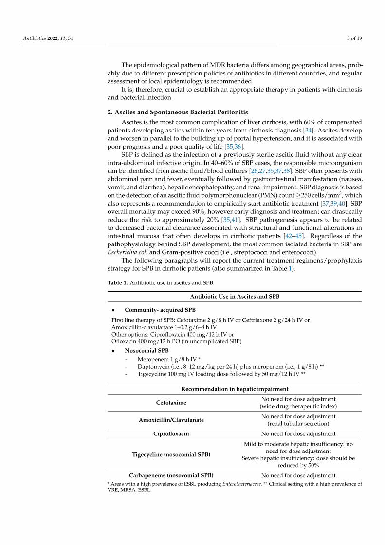
The following paragraphs will report the current treatment regimens/prophylaxisstrategy for SPB in cirrhotic patients (also summarized in Table 1).
Table 1. Antibiotic use in ascites and SPB.
Antibiotic Use in Ascites and SPB
• Community- acquired SPB
First line therapy of SPB: Cefotaxime 2 g/8 h IV or Ceftriaxone 2 g/24 h IV orAmoxicillin-clavulanate 1–0.2 g/6–8 h IVOther options: Ciprofloxacin 400 mg/12 h IV orOfloxacin 400 mg/12 h PO (in uncomplicated SBP)
• Nosocomial SPB
- Meropenem 1 g/8 h IV *- Daptomycin (i.e., 8–12 mg/kg per 24 h) plus meropenem (i.e., 1 g/8 h) **- Tigecycline 100 mg IV loading dose followed by 50 mg/12 h IV **
Recommendation in hepatic impairment
Cefotaxime No need for dose adjustment(wide drug therapeutic index)
Amoxicillin/Clavulanate No need for dose adjustment(renal tubular secretion)
Ciprofloxacin No need for dose adjustment
Tigecycline (nosocomial SPB)
Mild to moderate hepatic insufficiency: noneed for dose adjustment
Severe hepatic insufficiency: dose should bereduced by 50%
Carbapenems (nosocomial SPB) No need for dose adjustment* Areas with a high prevalence of ESBL producing Enterobacteriaceae. ** Clinical setting with a high prevalence ofVRE, MRSA, ESBL.
Antibiotics 2022, 11, 31 6 of 19
2.1. Empirical Antibiotic Therapy and Prophylaxis of Community Acquired PBS
Empirical antibiotic therapy should be prescribed immediately after the diagnosis ofSBP, while ascitic fluid culture results are still pending [37,38]. For example, cefotaxime,a third-generation cephalosporin, has been broadly studied in SBP therapy consideringthat it covers most causative organisms and reaches high ascitic fluid concentrationsduring administration [46,47]. A 2 g dose of cefotaxime every eight hours has been shownto produce an excellent concentration in the ascitic fluid and clinical effectiveness [48].Alternatively, a 2 g dose of ceftriaxone per day [49] has been shown to prevent SPB incirrhotic patients with gastrointestinal hemorrhage [50].
Furthermore, amoxicillin-clavulanate, first given intravenously and then orally, hasshown to have similar results on SBP resolution and mortality compared with cefo-taxime [47] and could be a valid alternative to cefotaxime for the empirical treatmentof bacterial infections in cirrhosis.
Ciprofloxacin can be used for patients who cannot take a cephalosporin (e.g., allergicpatients), although it does not reach the ascitic fluid as well as cefotaxime [51]. In general,ciprofloxacin is given at a dose of 400 mg intravenously twice daily to patients with normalrenal function.
Specific oral agents may be as effective as parenteral therapy in treating uncomplicatedSBP [51,52]. A trial demonstrated comparable outcomes with a short course of intravenousciprofloxacin (200 mg every 12 h for two days) followed by oral ciprofloxacin therapy(500 mg every 12 h for five days) compared with intravenous therapy alone for sevendays [51]. Oral ofloxacin 400 mg twice daily has given similar results as intravenous cefo-taxime in uncomplicated SBP, which means without renal failure, hepatic encephalopathy,gastrointestinal bleeding, ileus, or shock [52].
Primary prophylaxis should be considered in patients with cirrhosis at high riskfor SBP (i.e., low serum albumin and severe liver/renal dysfunction). Moreover, in allpatients with a prior episode of SBP, it is crucial to begin long-term prophylaxis. Interms of secondary prophylaxis data shows that norfloxacin (400 mg/day) could reduceSBP recurrence from 70% to 20% [53] In these settings, secondary prophylaxis should beintroduced immediately after the first episode of acute SBP and should be continued untilliver transplantation or the resolution of ascites [17].
Cefotaxime. Among third-generation cephalosporins, cefotaxime is the first-line ther-apy in community-acquired infections. The liver metabolizes about 40–50% of cefotaximeto an active deacetylated metabolite [54]. Two pharmacokinetic studies in cirrhotic patientsdemonstrated a threefold increase in cefotaxime’s half-life in comparison to healthy sub-jects, while one study did not find any significant difference [55–57]. However, due to thevery high therapeutic index of the antibiotic, despite these pharmacokinetic alterations,dose adjustment is not necessary for patients with hepatic impairment [55].
Amoxicillin-clavulanate. Amoxicillin-clavulanate is eliminated via renal tubularsecretion and does not require dose adjustment in cirrhotic patients.
Ciprofloxacin and Norfloxacin. All fluoroquinolones are poorly bound to proteins.Norfloxacin and ciprofloxacin are eliminated both by the kidney and the liver. Hepaticbiotransformation leads to several metabolites which partially maintain an antibacterialactivity [58]. Studies suggest that, in patients with cirrhosis, there is no need to adjustciprofloxacin doses [3,4]. Although no pharmacokinetic study in patients with hepaticdysfunction has been published for norfloxacin, the drug has been largely employed in theclinics, with excellent tolerability [53].
Trimethoprim/sulfamethoxazole (TMP/SMX). Trimethoprim/sulfamethoxazoleblocks two consecutive steps in the biosynthesis of nucleic acids and proteins that areessential for the persistence of many bacteria in the organism [59]. Two studies comparedthe effectiveness of trimethoprim/sulfamethoxazole versus norfloxacin in the prophylaxisof SBP in cirrhotic patients [60,61]. The results in both studies showed that trimetho-prim/sulfamethoxazole and norfloxacin are equally effective in SPB primary and secondaryprophylaxis, however patients treated with TMP/SMX developed more frequently adverse
Antibiotics 2022, 11, 31 7 of 19
effects [60]. On the other hand, TMP/SMX is an economic drug [62,63] and it is easily avail-able in public health system in many countries, therefore TMP/SMX could be considered avalid alternative to norfloxacin in the contest of SPB prophylaxis.
2.2. Nosocomial SBP Therapy
Nosocomial bacterial infections are a relevant complication in hospitalized [58,64].In particular, nosocomial SBP (NSBP) may arise in up to one-third of hospitalized cir-rhotics with an overall 30-days survival which does not reach 20%, especially in thosepatients where empirical treatments are not started promptly [65]. The most frequentlyisolated MDR bacteria in NSBP are represented by ESBL-producing Enterobacteriaceae,non-fermentable Gram-negative bacilli (such as P. aeruginosa), MRSA, and VRE [66]. Unfor-tunately, current guidelines for treating SBP do not provide a helpful distinction betweencommunity-acquired and nosocomial episodes [65].
Although a 2013 position paper recommended piperacillin/tazobactam or meropenemwith daptomycin for NSBP [23], also confirmed by some field experts [67,68] who alsosuggested the use tigecycline to cover ESBL- producing Enterobacteriaceae [31].
Piperacillin/tazobactam as the risk to be inadequate for patients with life-threateninginfections due to ESBL-producing Enterobacteriaceae [69].
Meropenem has excellent bactericidal activity against ESBL-producing Enterobacteri-aceae, whereas it is poorly active against staphylococci and enterococci [70].
Glycopeptides (e.g., vancomycin, teicoplanin) should be used carefully because ofthe potential risk of nephrotoxicity. In particular, the rate of acute kidney injury is higherin patients with concomitant acute liver failure, which may be related to hemodynamicinstability or the development of hepatorenal syndrome [71].
Daptomycin is a lipopeptide active against MDR Gram-positive pathogens, includingmethicillin-susceptible S. aureus, MRSA and VRE [72]. However, there is a decreasedsusceptibility to daptomycin (DAPR) reported in MRSA [73]. Despite this observation, thecombination of daptomycin/beta-lactams has been proven clinically effective for preventingand treating infections due to DAPR-MRSA strains [74].
Tigecycline. Tigecycline pharmacokinetic properties were assessed in patients withdifferent stages of liver cirrhosis [64], with no significant difference in the pharmacokineticsbetween patients with Child-Pugh class A or B and healthy controls. Nevertheless, patientsin Child-Pugh class C showed half of the clearance compared with controls [64]. Thus,implying that patients with mild to moderate liver cirrhosis do not require dose adjustments,whereas patients with more severe disease should have tigecycline maintenance dosesreduced by 50%.
In conclusion, different therapeutic options are recommended according to the onsetof the infection, third-generation cephalosporins are preferred for community-acquiredinfections, but they do not have adequate microbial coverage for treatment of nosocomialSBP. Cirrhotic patients with nosocomial SBP in a clinical setting with a high prevalence ofVRE, MRSA, ESBL should receive as empirical antibiotic therapy: high dose of daptomycin(i.e., 8–12 mg/kg every 24 h) plus meropenem (i.e., 1 g/8 h) [65,75].
3. Hepatic Encephalopathy
HE is defined as “brain dysfunction caused by liver impairment and/or portosystemicshunting manifesting as a wide spectrum of neurological or psychiatric abnormalitiesranging from subclinical alterations to coma” [76].
This pathological condition is a prevalent complication of portal hypertension andcirrhosis, occurring in 50–70% of patients. Considering signs and symptoms of HE, theclinical spectrum may be highly variable and better defined as a continuum. It affectspatients’ behavior, cognitive sphere, and motor skills. There is an inevitable heterogeneitynot only between different patients but also longitudinally for an individual patient. Thismakes HE a not always so easy condition to diagnose and treat. According to ISHENClassification [77], HE can be classified as in Overt (OHE), clinically evident with disorien-
Antibiotics 2022, 11, 31 8 of 19
tation and asterixis as the most specific signs; and Covert (CHE), not clinically evident butdemonstrable by psychometric and electrophysiological tests. At this time, only OHE isroutinely treated, whereas Minimal HE and CHE, which are not easy to define, have noclear treatment recommendation [78].
Most liver-failure patients develop minimal hepatic encephalopathy (MHE). MHE canprogress to clinical HE, which can evolve to coma and death.
There is a large variability in the onset of MHE and the progression to clinical HE.Most patients remain undiagnosed. Patients may survive for years with progressive
deterioration of neurological function. Nevertheless, MHE reduces the ability to performexecutive functions in daily living, diminishes the quality of life, predisposes to clinical HE,and reduces lifespan. HE is characterized by motor slowness that ends in dysmetric move-ments, progressive cognitive and psychiatric alterations, cerebral edema, and coma [79–81].The urgency of new diagnostic criteria is a strong need because it represents a moleculartsunami inside the brain whenever HE is overcoming.
The two main pathogenic factors traditionally considered for HE are hyperammonemiaand inflammation. Major corrections inside these two historical factors have been recentlydemonstrated. The effects of hyperammonemia on the brain depend on several factors: theconcentration of ammonia; the speed at which the levels of ammonia increase; the durationof hyperammonemia; the period of brain development in which HE occurs. When animalmodels are subjected to acute ammonia intoxication, the concentration of ammonia in thebrain rapidly increases from a basal level of approximately 0.2 µmol g−1 to 1–3 µmol g−1.Regardless of ammonia concentration, the brain’s only way for its removal is the astrocyticglutamine synthesis, presented by spectroscopic measures [82,83] inside HE brains.
In rats, acute liver failure involves selective alterations of specific brain areas (i.e., thecerebellum), with substantial blood-brain barrier leakages, promoting the first steps ofvasogenic edema. Hyperammonemia leads to hyperactivation of N-methyl D-aspartate(NMDA) receptors. They activate the glutamate–nitric oxide–cyclic GMP pathway, produc-ing excessive nitric oxide and cGMP [84]. Both the facts contribute to the animal’s death byexcitotoxic mechanisms. The astrocytic glutamine accumulation induces water symport,which might cause astrocytic swelling and cytotoxic edema [85]. At a later stage, NMDAreceptor activation increases lactate and CBF alterations (increase in the cortex and decreasein the cerebellum), further increasing ICP, contributing to death [86].
Hyperammonemia per se induces neuroinflammation. In cultured microglia, ammoniaupregulates the microglial activation marker (allograft inflammatory factor 1), elevatedin the HE-human brain [87]. Acute liver failure promotes regional neuroinflammation,associated with a consequent modification of cerebral blood flow, more evident in the braincortex but not in the cerebellum [87].
On the contrary, animal models of chronic HE show cerebellar inflammation, witha concomitant increase of tumor necrosis factor and an intracranial elevation of Il-2, Il-6,and Il-8 [88]. Moreover, microglial activation is an overwhelming mechanism because itsactivation is not only an intrinsic one but might be regulated by the blood-brain barrierleakages, through which pro-inflammatory cytokines can pass, or by direct infiltration ofblood-immune cells. Blood cytokines may also stimulate receptors on endothelial cells andtrigger the release of inflammatory factors into the brain [89].
The traditional role of brain edema in HE is under debate, with recent functional MRI(fMRI) studies. The conventional vision of the vasogenic edema as the beginner of the neu-rological alterations is defeated by animal models of HE undergoing fMRI. They show anunexpected increase in the apparent diffusion coefficient (just the opposite of what is com-monly detected in cytotoxic edema). They offer motor and executive alterations in the absenceof brain edema a [90]. Magnetoencephalography studies evidenced an altered synchronizationof the thalamocortical coupling in HE animal and human HE models [91,92]. Resting-statefMRI studies show that MHE patients have a selective default-mode thalamus-basal ganglia-frontal cortex, with substantial decreased functional connection alterations [93]. Therefore,
Antibiotics 2022, 11, 31 9 of 19
brain edema is considered just a final part of the sequence of events that occur in theHE brain.
The most essential accepted pathogenic mechanism of HE is the severe impairmentof neurotransmitters exerted by ammonia. Independently by its concentrations, ammoniainhibits postsynaptic chloride extrusion [94] and the postsynaptic effect of glutamate re-ceptors [95]. Rat hippocampal models showed inhibition of glutamate uptake capacity byblood samples of human HE patients, and the inhibition is directly related to the ammoniaconcentrations [96]. Moreover, ammonia inhibited the high-affinity glutamate uptake intosynaptosomes of rat astrocytes [97]. This situation is furthermore confirmed by the revealof a significant loss of glutamate transporter (GLT-1, dispersed all around the brain andGLAST, localized in the cerebellum) in astrocytes and (EAAC-1) in neurons of cerebellar ratsections [98]. The drastic reduction of GLT-1 and GLAST expression determined an increaseof extracellular glutamate release, with a consequent downregulation of the AMPA/kainatereceptors, as a rapid response to acute liver failure; chronic liver failure directly damagedNMDA neural receptors [99,100]. It has even been suggested that the demonstrated de-crease of AMPA/kainate receptors implies an overt hyper-expression of NMDA receptorsin acute phases, which might benefit from the memantine supplementation [101–104].Finally, it has been argued that acute NMDA hyper-expression can determine a possi-ble downregulation of dopamine inside the basal ganglia [105]. It has been proposedthat ammonia modifies GABA agonists to their GABA-A ligands, defining the so-calledbarbiturate–like action of HE, and definitively affects GABA uptake [106]. More importantis the possible effect of ammonia on GABA different receptor types. Ammonia acts on thepost-synaptic GABA-benzodiazepine receptor complex and on the PTBR receptors, whichare localized all around the body but are strongly localized on the astrocytic mitochon-dria. There is a constant increment of their densities all around the astrocytes during HEstatus, increasing the PTBR mRNA in HE experimental condition. Exposure of culturedglioma cells to PTBR ligands results in astrocyte proliferation and mitochondrial swellingas observed in HE [107–109]. The deposition of heavy metals, in particular manganese,inside the globus pallidus in chronic liver failure, contributes to the alterations of astrocyticfunctions inside the basal ganglia, interrupting several motor refinement networks, leadingto some movement alteration observed in chronic liver patients [110,111]. PTBR activationassociated with manganese exposure results in increased allopreg-nanolone synthesis, apotent GABA-A receptor agonist [111]. Together with all these alterations, a constantincrease of the serotonin metabolite 5-hydroxy indole acetic acid (5-HIAA) has been foundin the cerebral spinal fluid of all the chronic liver failure patients, with an increased brainserotonin degradation and a synaptic deficit of it.
The serotonin synaptic deficit determines a constellation of neuropsychiatric symp-toms such as altered sleep patterns, depression, and personality changes [112]. On theother hand, homovanillic acid, as dopamine metabolite, is increased in HE, probably due tothe accumulation of manganese, as above reported, and to an increment of dopamine oxi-dation [113]. Histamine is another target for HE; rats models show an increase of histamineup to 6 months post-liver failure, including a loss of histamine in H3 receptor sites [114,115],determining a loss of circadian rhythms, and electrical brain alterations, which might resem-ble the human hepatic coma [115]. Finally, brain extracts from animal HE models containaltered beta-endorphin levels, and their brains demonstrate region-selective alterations ofthe mu and delta-opioid receptor sites [116,117].
Antibiotic Use in Hepatic Encephalopathy
As stated above, multiple components play a role, alone or in combination, in thepathophysiology of HE. It is important to remember that HE is a reversible condition, andthe correct treatment positively influences patients’ survival, the possibility of recurrence,and the quality of life. HE treatment varies according to the severity of presentation andthe goals of therapy. Most treatments are based on manipulating the intestinal microbiota.Therefore, antibiotics that act on the gut represent a key treatment strategy. It is essential
Antibiotics 2022, 11, 31 10 of 19
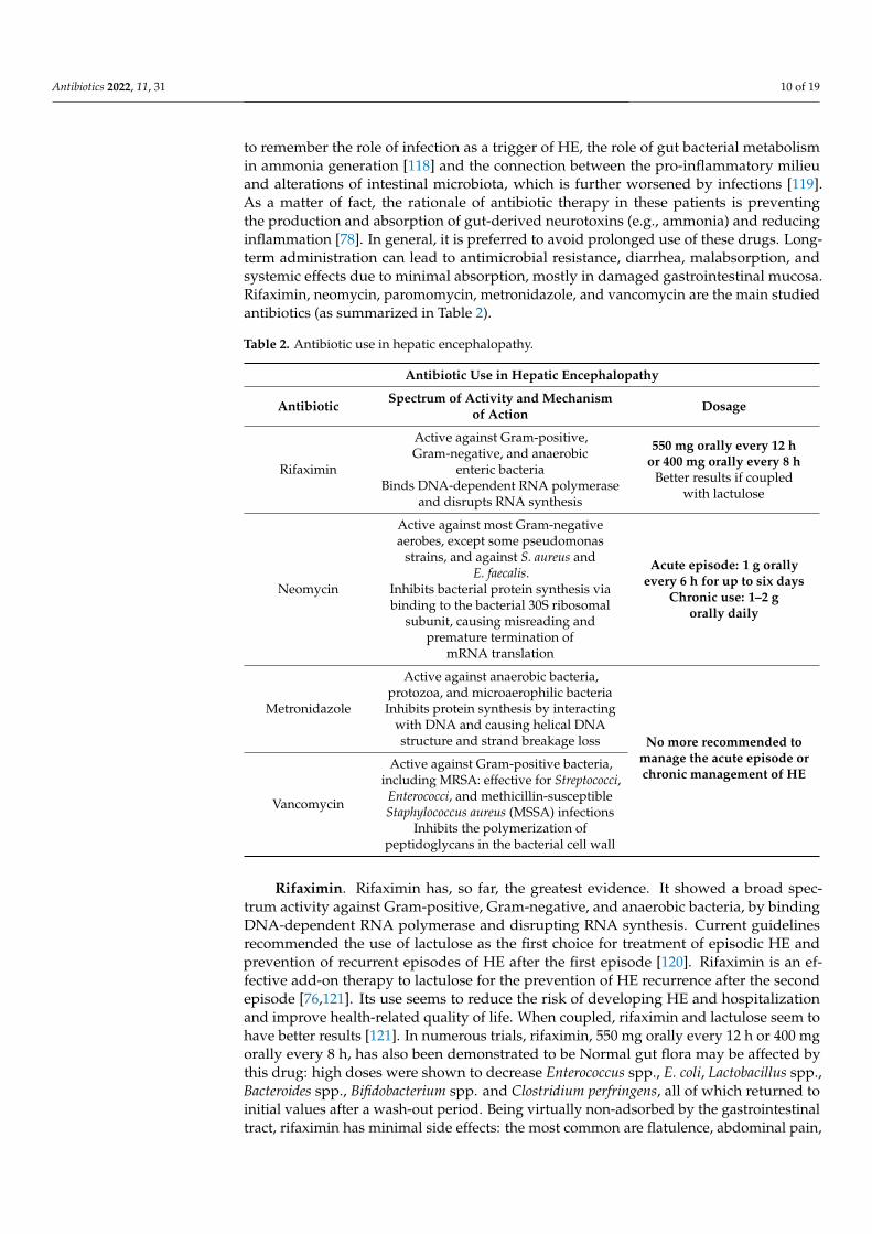
to remember the role of infection as a trigger of HE, the role of gut bacterial metabolismin ammonia generation [118] and the connection between the pro-inflammatory milieuand alterations of intestinal microbiota, which is further worsened by infections [119].As a matter of fact, the rationale of antibiotic therapy in these patients is preventingthe production and absorption of gut-derived neurotoxins (e.g., ammonia) and reducinginflammation [78]. In general, it is preferred to avoid prolonged use of these drugs. Long-term administration can lead to antimicrobial resistance, diarrhea, malabsorption, andsystemic effects due to minimal absorption, mostly in damaged gastrointestinal mucosa.Rifaximin, neomycin, paromomycin, metronidazole, and vancomycin are the main studiedantibiotics (as summarized in Table 2).
Table 2. Antibiotic use in hepatic encephalopathy.
Antibiotic Use in Hepatic Encephalopathy
Antibiotic Spectrum of Activity and Mechanismof Action Dosage
Rifaximin
Active against Gram-positive,Gram-negative, and anaerobic
enteric bacteriaBinds DNA-dependent RNA polymerase
and disrupts RNA synthesis
550 mg orally every 12 hor 400 mg orally every 8 h
Better results if coupledwith lactulose
Neomycin
Active against most Gram-negativeaerobes, except some pseudomonas
strains, and against S. aureus andE. faecalis.
Inhibits bacterial protein synthesis viabinding to the bacterial 30S ribosomal
subunit, causing misreading andpremature termination of
mRNA translation
Acute episode: 1 g orallyevery 6 h for up to six days
Chronic use: 1–2 gorally daily
Metronidazole
Active against anaerobic bacteria,protozoa, and microaerophilic bacteriaInhibits protein synthesis by interacting
with DNA and causing helical DNAstructure and strand breakage loss No more recommended to
manage the acute episode orchronic management of HE
Vancomycin
Active against Gram-positive bacteria,including MRSA: effective for Streptococci,Enterococci, and methicillin-susceptibleStaphylococcus aureus (MSSA) infections
Inhibits the polymerization ofpeptidoglycans in the bacterial cell wall
Rifaximin. Rifaximin has, so far, the greatest evidence. It showed a broad spec-trum activity against Gram-positive, Gram-negative, and anaerobic bacteria, by bindingDNA-dependent RNA polymerase and disrupting RNA synthesis. Current guidelinesrecommended the use of lactulose as the first choice for treatment of episodic HE andprevention of recurrent episodes of HE after the first episode [120]. Rifaximin is an ef-fective add-on therapy to lactulose for the prevention of HE recurrence after the secondepisode [76,121]. Its use seems to reduce the risk of developing HE and hospitalizationand improve health-related quality of life. When coupled, rifaximin and lactulose seem tohave better results [121]. In numerous trials, rifaximin, 550 mg orally every 12 h or 400 mgorally every 8 h, has also been demonstrated to be Normal gut flora may be affected bythis drug: high doses were shown to decrease Enterococcus spp., E. coli, Lactobacillus spp.,Bacteroides spp., Bifidobacterium spp. and Clostridium perfringens, all of which returned toinitial values after a wash-out period. Being virtually non-adsorbed by the gastrointestinaltract, rifaximin has minimal side effects: the most common are flatulence, abdominal pain,
Antibiotics 2022, 11, 31 11 of 19
headaches, constipation, nausea, and vomiting. Furthermore, no reported drug interactionsmake it relatively safe.
Aminoglycosides (neomycin and paromomycin). Neomycin is effective against mostGram-negative aerobes, except some Pseudomonas strains, and against S. aureus andEnterococcus faecalis. It inhibits bacterial protein synthesis via binding to the bacterial 30Sribosomal subunit, causing misreading and premature termination of mRNA translation.Neomycin is usually orally administered, 1 g every six hours for up to six days in an acuteepisode of overt HE and 1–2 g daily for chronic use [78]. Although neomycin is poorlyabsorbed from the gastrointestinal tract, ototoxicity and nephrotoxicity may occur. Dueto these side effects and lack of demonstrated clinical benefit, neomycin clinical use isnot recommended.
The use of aminoglycosides should therefore be reserved for patients with severebacterial infections that cannot be treated with other antibiotics.
Paromomycin has also been utilized but the evidence for it is limited and therefore itis not approved for the treatment of HE [78,122].
Metronidazole. Metronidazole inhibits protein synthesis by interacting with DNA andcausing helical DNA structure and strand breakage loss. It acts against anaerobic bacteria,protozoa, and microaerophilic bacteria. Because of its prolonged rate of elimination inHE patients, increased risk for irreversible peripheral neurotoxicity, and limited studieson the efficacy, metronidazole is no longer recommended to manage the acute episode orchronic management of HE [78,123]. Disulfiram-like reactions in patients drinking ethanolor taking ethanol-containing medications may occur too [123].
Vancomycin. It inhibits the polymerization of peptidoglycans in bacterial cellularwalls. It is prevalently used to treat/prevent bacterial infections caused by Gram-positivebacteria, including MRSA. It is effective against enterococci, streptococci, and methicillin-susceptible S. aureus (MSSA) infections. This drug may be safer for managing an acute HEepisode, but in the face of an increased prevalence of VRE and other bacterial resistanceand limited studies, vancomycin is not recommended [31,78].
4. Renal Impairment
In cirrhotic patients, decompensated hepatic failure leads to renal vasoconstriction andsubsequent renal failure, which reduces renal elimination of agents with primary renal clear-ance, leading to increased serum drug concentrations [3,5,124,125]. Nephrotoxicity is one ofthe most common adverse drug reactions (6.8%), and most adverse drug reactions occur inpatients with advanced cirrhosis (Child-Pugh C), often with renal impairment [5]. Anotheraspect to highlight is that patients with cirrhosis have reduced effective renal plasma flowand glomerular filtration rates, even in the absence of ascites [126,127]. Moreover, patientswith liver cirrhosis have low serum creatinine concentrations (due to impaired synthesis ofcreatinine and reduced muscle mass), which may indicate that actual glomerular filtrationrates cannot be estimated from serum creatinine levels [128,129].
Furthermore, cirrhotics have decreased, thus drugs with renal elimination and narrowtherapeutic range should be dosed with extreme caution in these patients [126]. Severalantibiotics have shown diminished renal elimination, such as in the case of ofloxacin,ampicillin, aminoglycosides, and vancomycin.
Ofloxacin. The metabolism is affected by renal dysfunction in patients with ascites.Of notice, ofloxacin penetration into the ascitic fluid is excellent with effective therapeuticconcentrations [130,131]. Lower renal excretion in cirrhosis has been proven for ofloxacin,and dose adjustments are warranted [5,126].
Ampicillin. For drugs such as ampicillin, pharmacokinetic parameters are unchangedin cirrhosis, but dose reductions are recommended for patients with renal impairment [132].Aminoglycosides and vancomycin. Serum levels of aminoglycosides must be monitoredclosely in these patients to decrease the risk of renal failure [133]. Intravenous (IV) van-comycin can cause increased toxicity in patients with liver failure [1]. The combination
Antibiotics 2022, 11, 31 12 of 19
of aminoglycosides and IV vancomycin is generally contraindicated given their relativelyhigh risk of inducing nephrotoxicity and renal failure [1,5,134].
Infection treatment in patients with liver cirrhosis also includes strategies to preventacute kidney injury (AKI). According to the International Ascites Club and the Acute Dialy-sis Quality Initiative, AKI in liver cirrhosis is defined by an increase in serum concentrationby 0.3 mg/dL in less than 48 h or by a 50% increase from stable baseline concentrationsin the previous six months, without taking into consideration the final serum creatininelevel [135,136]. AKI usually develops in 27–34% of patients with cirrhosis following in-fections [25]. Other risk factors for infection-induced AKI in cirrhosis include advancedliver disease [137], preexisting kidney disease hypovolemia, low cardiac [25], unresolvedinfection [137].
In cirrhotics with SBP, the administration of human albumin drastically reduces theincidence of AKI and improves overall survival [138,139]: in particular, albumin (1.5 g/kgwithin six hours of SBP diagnosis, followed by 1 g/kg on day 3) plus intravenous an-tibiotics reduced incidence of renal impairment from 33% to 10% and mortality from29% to 10% [138]. However, albumin infusion appears to be more effective in those pa-tients with baseline bilirubin ≥ 4 mg/dL, creatinine ≥ 1 mg/dL, or blood urea nitrogen≥ 30 mg/dL [39]. In addition, to actively prevent AKI, all potentially nephrotoxic drugs,such as non-steroidal anti-inflammatory drugs (NSAIDs), vasodilators, and aminoglyco-sides, should be avoided [135].
Hepatorenal Syndrome
As already stated, patients with hepatorenal syndrome are at higher risk of reducedantibiotics renal clearance, thus needing dose reductions [125]. In particular, during infec-tion, an acute kidney injury that does not respond to albumin infusion is considered ashepatorenal syndrome [140]. Infections, such as SBP, can precipitate active liver decompen-sation manifestations or may promote the occurrence of new decompensating symptoms,and are the major precipitants of acute-on-chronic liver failure (ACLF) in Western coun-tries [14,141–143].
5. Transjugular Intrahepatic Portosystemic Shunts (TIPS)
Transjugular intrahepatic portosystemic shunts (TIPS) are placed to manage compli-cations from clinically significant portal hypertension [5]. Portosystemic shunting is theleading cause of altered drug metabolism in liver cirrhosis [1].
Among the potential drug handling changes in cirrhosis are the reduced hepaticblood flow, lower first-pass extraction, and portosystemic shunting that determine a higherbioavailability and serum levels for most drugs [3,5,58]. Drugs with an ordinarily highrate of liver first-pass extraction will typically have reduced bioavailability. However, dueto decreased hepatic blood flow and presence of porto-systemic shunts, these drugs havehigher bioavailability and serum concentrations often require dose reductions [5].
The presence of portosystemic shunts, including TIPS, may lead to increased bioavail-ability of some drugs. This could be a problem for some drugs that can prolong the QTcinterval, especially considering that cirrhotics patients have baseline QTc interval pro-longation (due to the portosystemic shunting of cardioactive substances derived fromthe splanchnic circulation) [144]. Among antibiotics, fluoroquinolones may prolong QTcintervals in patients with TIPS [145], and appropriate care should be taken.
The macrolide antibiotics erythromycin and clarithromycin are also known to prolongthe QTc interval; in their review, Vuppalanchi et al., observed that after a 7-day courseof erythromycin, cirrhotic patients with a TIPS developed a significantly more significantprolongation in their QTc interval (180 ± 68 ms) compared with both the cirrhotic patientswithout TIPS (31 ± 10 ms) and with the healthy controls (38 ± 3 ms) (p = 0.03) [145]. Thesefindings should prompt caution when prescribing medications known to prolong QTc incirrhotic patients who have undergone a TIPS procedure, especially those patients who arebeing prescribed a fluoroquinolone for SBP treatment or prophylaxis [5].
Antibiotics 2022, 11, 31 13 of 19
6. Conclusions
In conclusion, bacterial infections are one of the most frequent complications and asignificant cause of death in cirrhotic patients, and therefore it is crucial to know how to useantibiotics correctly. The safe use of medications in patients with chronic liver disease is anongoing challenge, and this is especially true in patients with cirrhosis, in whom significantchallenges can occur in the metabolism and handling of a large number of drugs. In general,in order to decide drug dosing in liver failure, three essential factors need to be considered(1) pharmacokinetic alterations of drugs, (2) pharmacodynamic alteration of drugs, and(3) increased susceptibility of patients to adverse events, particularly hepatotoxicity. Inpatients with cirrhosis, the drug dosing should be individualized depending on severalfactors, including renal function and nutritional status, adherence, and drug interaction [1].
In general, most drugs can be used safely in cirrhosis, including potentially hepatotoxicones, but lower doses or reduced dosing frequency are often recommended due to alteredpharmacokinetics. Drugs that can precipitate renal failure, gastrointestinal bleeding, SBP,and encephalopathy should be identified and avoided [5].
Moreover, whenever possible, measuring drug levels in the blood and monitoringadverse events frequently should be done. Early diagnosis and appropriate antibiotic treat-ment are the first steps in the management of cirrhotic patients with infection. While third-generation cephalosporins remain the gold-standard strategy in most community-acquiredinfections, the empirical treatment of nosocomial and healthcare-associated infectionsshould be adapted to the local epidemiological pattern of antibiotic resistance [146] oftenrequires the use of broad-spectrum antibiotics, such as piperacillin-tazobactam, carbapen-ems or tigecycline. Moreover, prompt de-escalation of antibiotic treatment is recommendedto prevent the development of antibiotic resistance [14].
We have attempted to summarize current knowledge and translate it into relevantand practical recommendations through this narrative review. However, the current andincreasing spread of MDR bacteria, especially in the nosocomial setting, represents asignificant challenge in the correct management of cirrhotic patients.
Funding: This research received no external funding.
Institutional Review Board Statement: Not applicable.
Informed Consent Statement: Not applicable.
Conflicts of Interest: The authors declare no conflict of interest.
References1. Amarapurkar, D.N. Prescribing Medications in Patients with Decompensated Liver Cirrhosis. Int. J. Hepatol. 2011, 2011, 519526.
[CrossRef] [PubMed]2. Zhou, W.C.; Zhang, Q.B.; Qiao, L. Pathogenesis of liver cirrhosis. World J. Gastroenterol. 2014, 20, 7312–7324. [CrossRef]3. Verbeeck, R.K. Pharmacokinetics and dosage adjustment in patients with hepatic dysfunction. Eur. J. Clin. Pharmacol. 2008,
64, 1147–1161. [CrossRef]4. Elbekai, R.; Korashy, H.; El-Kadi, A. The Effect of Liver Cirrhosis on the Regulation and Expression of Drug Metabolizing
Enzymes. Curr. Drug Metab. 2004, 5, 157–167. [CrossRef]5. Lewis, J.H.; Stine, J. Review article: Prescribing medications in patients with cirrhosis—A practical guide. Aliment. Pharmacol.
Ther. 2013, 37, 1132–1156. [CrossRef]6. Gatta, A.; Verardo, A.; Bolognesi, M. Hypoalbuminemia. Intern. Emerg. Med. 2012, 7 (Suppl. 3), S193–S199. [CrossRef]7. Franch-Arcas, G. The meaning of hypoalbuminaemia in clinical practice. Clin. Nutr. 2001, 20, 265–269. [CrossRef] [PubMed]8. Nicholson, J.; Wolmarans, M.; Park, G. The role of albumin in critical illness. Br. J. Anaesth. 2000, 85, 599–610. [CrossRef]9. Rothschild, M.A.; Oratz, M.; Schreiber, S.S. Serum albumin. Hepatology 1988, 8, 385–401. [CrossRef] [PubMed]10. Ulldemolins, M.; Roberts, J.A.; Roberts, J.A.; Rello, J.; Paterson, D.L.; Lipman, J. The Effects of Hypoalbuminaemia on Optimizing
Antibacterial Dosing in Critically Ill Patients. Clin. Pharmacokinet. 2011, 50, 99–110. [CrossRef]11. Zusman, O.; Farbman, L.; Tredler, Z.; Daitch, V.; Lador, A.; Leibovici, L.; Paul, M. Association between hypoalbuminemia and
mortality among subjects treated with ertapenem versus other carbapenems: Prospective cohort study. Clin. Microbiol. Infect.2014, 21, 54–58. [CrossRef]

Antibiotics 2022, 11, 31 14 of 19
12. Bartoletti, M.; Giannella, M.; Lewis, R.E.; Caraceni, P.; Tedeschi, S.; Paul, M.; Schramm, C.; Bruns, T.; Merli, M.; Cobos-Trigueros,N.; et al. Extended Infusion of β-Lactams for Bloodstream Infection in Patients with Liver Cirrhosis: An Observational MulticenterStudy. Clin. Infect. Dis. 2019, 69, 1731–1739. [CrossRef] [PubMed]
13. Bartoletti, M.; Giannella, M.; Lewis, R.; Caraceni, P.; Tedeschi, S.; Paul, M.; Schramm, C.; Bruns, T.; Merli, M.; Cobos-Trigueros, N.;et al. A prospective multicentre study of the epidemiology and outcomes of bloodstream infection in cirrhotic patients. Clin.Microbiol. Infect. 2018, 24, 546.e1–546.e8. [CrossRef]
14. Fernández, J.; Acevedo, J. New antibiotic strategies in patients with cirrhosis and bacterial infection. Expert Rev. Gastroenterol.Hepatol. 2015, 9, 1495–1500. [CrossRef] [PubMed]
15. Wiest, R.; Garcia-Tsao, G. Bacterial translocation (BT) in cirrhosis. Hepatology 2005, 41, 422–433. [CrossRef]16. Bone, R.C.; Grodzin, C.J.; Balk, R.A. Sepsis: A New Hypothesis for Pathogenesis of the Disease Process. Chest 1997, 112, 235–243.
[CrossRef]17. Pleguezuelo, M.; Benitez, J.M.; Jurado, J.; Montero, J.L.; De La Mata, M. Diagnosis and management of bacterial infections in
decompensated cirrhosis. World J. Hepatol. 2013, 5, 16–25. [CrossRef] [PubMed]18. Plessier, A.; Denninger, M.-H.; Consigny, Y.; Pessione, F.; Francoz, C.; Durand, F.; Francque, S.; Bezeaud, A.; Chauvelot-Moachon,
L.; Lebrec, D.; et al. Coagulation disorders in patients with cirrhosis and severe sepsis. Liver Int. 2003, 23, 440–448. [CrossRef]19. Adenote, A.; Dumic, I.; Madrid, C.; Barusya, C.; Nordstrom, C.W.; Prada, L.R. NAFLD and Infection, a Nuanced Relationship.
Can. J. Gastroenterol. Hepatol. 2021, 2021, 5556354. [CrossRef]20. Alba-Loureiro, T.; Munhoz, C.; Martins, J.; Cerchiaro, G.; Scavone, C.; Curi, R.; Sannomiya, P. Neutrophil function and metabolism
in individuals with diabetes mellitus. Braz. J. Med. Biol. Res. 2007, 40, 1037–1044. [CrossRef] [PubMed]21. Targher, G.; Bertolini, L.; Scala, L.; Cigolini, M.; Zenari, L.; Falezza, G.; Arcaro, G. Associations between serum 25-hydroxyvitamin
D3 concentrations and liver histology in patients with non-alcoholic fatty liver disease. Nutr. Metab. Cardiovasc. Dis. 2007,17, 517–524. [CrossRef] [PubMed]
22. Lee, S.M.; Jun, D.W.; Cho, Y.K.; Jang, K.S. Vitamin D deficiency in non-alcoholic fatty liver disease: The chicken or the egg?Clin. Nutr. 2015, 36, 191–197. [CrossRef] [PubMed]
23. Jalan, R.; Fernandez, J.; Wiest, R.; Schnabl, B.; Moreau, R.; Angeli, P.; Stadlbauer, V.; Gustot, T.; Bernardi, M.; Canton, R.; et al.Bacterial infections in cirrhosis: A position statement based on the EASL Special Conference 2013. J. Hepatol. 2014, 60, 1310–1324.[CrossRef] [PubMed]
24. Miele, L.; Valenza, V.; La Torre, G.; Montalto, M.; Cammarota, G.; Ricci, R.; Mascianà, R.; Forgione, A.; Gabrieli, M.L.; Perotti,G.; et al. Increased intestinal permeability and tight junction alterations in nonalcoholic fatty liver disease. Hepatology 2009,49, 1877–1887. [CrossRef]
25. Bunchorntavakul, C.; Chavalitdhamrong, D. Bacterial infections other than spontaneous bacterial peritonitis in cirrhosis. World J.Hepatol. 2012, 4, 158–168. [CrossRef] [PubMed]
26. Fernández, J.; Navasa, M.; Gómez, J.; Colmenero, J.; Vila, J.; Arroyo, V.; Rodés, J. Bacterial infections in cirrhosis: Epidemiologicalchanges with invasive procedures and norfloxacin prophylaxis. Hepatology 2002, 35, 140–148. [CrossRef] [PubMed]
27. Caly, W.R.; Strauss, E. A prospective study of bacterial infections in patients with cirrhosis. J. Hepatol. 1993, 8, 353–358. [CrossRef]28. Fernández, J.; Prado, V.; Trebicka, J.; Amoros, A.; Gustot, T.; Wiest, R.; Deulofeu, C.; Garcia, E.; Acevedo, J.; Fuhrmann, V.; et al.
Multidrug-resistant bacterial infections in patients with decompensated cirrhosis and with acute-on-chronic liver failure inEurope. J. Hepatol. 2018, 70, 398–411. [CrossRef]
29. Piano, S.; Singh, V.; Caraceni, P.; Maiwall, R.; Alessandria, C.; Fernandez, J.; Soares, E.C.; Kim, D.J.; Kim, S.E.; Marino, M.; et al.Epidemiology and Effects of Bacterial Infections in Patients with Cirrhosis Worldwide. Gastroenterology 2019, 156, 1368–1380.e10.[CrossRef]
30. Bajaj, J.S.; O’Leary, J.G.; Reddy, K.R.; Wong, F.; Olson, J.C.; Subramanian, R.M.; Brown, G.; Noble, N.A.; Thacker, L.R.; Kamath, P.S.;et al. Second infections independently increase mortality in hospitalized patients with cirrhosis: The north american consortiumfor the study of end-stage liver disease (NACSELD) experience. Hepatology 2012, 56, 2328–2335. [CrossRef]
31. Fernández, J.; Acevedo, J.; Castro, M.; Garcia, O.; Rodriguez-Lope, C.; Roca, D.; Pavesi, M.; Solà, E.; Moreira, L.; Silva, A.; et al.Prevalence and risk factors of infections by multiresistant bacteria in cirrhosis: A prospective study. Hepatology 2012, 55, 1551–1561.[CrossRef] [PubMed]
32. Fernández, J.; Gustot, T. Management of bacterial infections in cirrhosis. J. Hepatol. 2012, 56, S1–S12. [CrossRef]33. Milovanovic, T.; Dumic, I.; Velickovic, J.; Lalosevic, M.S.; Nikolic, V.; Palibrk, I. Epidemiology and risk factors for multi-drug
resistant hospital-acquired urinary tract infection in patients with liver cirrhosis: Single center experience in Serbia. BMC Infect. Dis.2019, 19, 141. [CrossRef] [PubMed]
34. Ginés, P.; Quintero, E.; Arroyo, V.; Terés, J.; Bruguera, M.; Rimola, A.; Caballería, J.; Rodés, J.; Rozman, C. Compensated cirrhosis:Natural history and prognostic factors. Hepatology 1987, 7, 122–128. [CrossRef]
35. Tandon, P.; Garcia-Tsao, G. Bacterial infections, sepsis, and multiorgan failure in cirrhosis. Semin. Liver Dis. 2008, 28, 26–42.[CrossRef]
36. Guevara, M.; Cárdenas, A.; Uriz, J.; Ginès, P. Prognosis in patients with cirrhosis and ascites. In Ascites and Renal Dysfunction inLiver Disease: Pathogenesis, Diagnosis and Treatment; Ginès, P., Arroyo, V., Rodés, J., Schrier, R.W., Eds.; Blackwell: Malden, MA,USA, 2005; pp. 260–270.
Antibiotics 2022, 11, 31 15 of 19
37. Rimola, A.; García-Tsao, G.; Navasa, M.; Piddock, L.J.; Planas, R.; Bernard, B.; Inadomi, J.M. Diagnosis, treatment and prophylaxisof spontaneous bacterial peritonitis: A consensus document. J. Hepatol. 2000, 32, 142–153. [CrossRef]
38. Wong, F.; Bernardi, M.; Balk, R.; Christman, B.; Moreau, R.; Garcia-Tsao, G.; Patch, D.; Soriano, G.; Hoefs, J.; Navasa, M. Sepsis incirrhosis: Report on the 7th meeting of the International Ascites Club. Gut 2005, 54, 718–725. [CrossRef]
39. European Association for the Study of the Liver. EASL clinical practice guidelines on the management of ascites, spontaneousbacterial peritonitis, and hepatorenal syndrome in cirrhosis. J. Hepatol. 2010, 53, 397–417. [CrossRef]
40. Runyon, B.A. Management of adult patients with ascites due to cirrhosis: An update. Hepatology 2009, 49, 2087–2107. [CrossRef]41. Garcia-Tsao, G. Current management of the complications of cirrhosis and portal hypertension: Variceal hemorrhage, ascites, and
spontaneous bacterial peritonitis. Gastroenterology 2001, 120, 726–748. [CrossRef]42. Guarner-Argente, C.; Sánchez, E.; Vidal, S.; Roman, E.; Concepción, M.; Poca, M.; Sanchez, D.; Juárez, C.; Soriano, G.; Guarner, C.
Toll-like receptor 4 D299G polymorphism and the incidence of infections in cirrhotic patients. Aliment. Pharmacol. Ther. 2010,31, 1192–1199. [CrossRef]
43. Chang, C.-S.; Chen, G.-H.; Lien, H.-C.; Yeh, H.-Z. Small intestine dysmotility and bacterial overgrowth in cirrhotic patients withspontaneous bacterial peritonitis. Hepatology 1998, 28, 1187–1190. [CrossRef] [PubMed]
44. Giuffrè, M.; Campigotto, M.; Campisciano, G.; Comar, M.; Crocè, L.S. A story of liver and gut microbes: How does the intestinalflora affect liver disease? A review of the literature. Am. J. Physiol. Liver Physiol. 2020, 318, G889–G906. [CrossRef] [PubMed]
45. Giuffrè, M.; Moretti, R.; Campisciano, G.; Da Silveira, A.B.M.; Monda, V.M.; Comar, M.; Di Bella, S.; Antonello, R.M.; Luzzati, R.;Crocè, L.S. You Talking to Me? Says the Enteric Nervous System (ENS) to the Microbe. How Intestinal Microbes Interact with theENS. J. Clin. Med. 2020, 9, 3705. [CrossRef] [PubMed]
46. Felisart, J.; Rimola, A.; Arroyo, V.; Perez-Ayuso, R.M.; Quintero, E.; Ginès, P.; Rodes, J. Cefotaxime is more effective than isampicillin-tobramycin in cirrhotics with severe infections. Hepatology 1985, 5, 457–462. [CrossRef] [PubMed]
47. Ricart, E.; Soriano, G.; Novella, M.T.; Ortiz, J.; Sàbat, M.; Kolle, L.; Sola-Vera, J.; Miñana, J.; Dedéu, J.M.; Gómez, C.; et al.Amoxicillin-clavulanic acid versus cefotaxime in the therapy of bacterial infections in cirrhotic patients. J. Hepatol. 2000,32, 596–602. [CrossRef]
48. Dalmau, D.; Layrargues, G.P.; Fenyves, D.; Willems, B.; Turgeon, F.; Turgeon, P. Cefotaxime, Desacetyl-Cefotaxime, andBactericidal Activity in Spontaneous Bacterial Peritonitis. J. Infect. Dis. 1999, 180, 1597–1602. [CrossRef] [PubMed]
49. Mazer, L.; Tapper, E.B.; Piatkowski, G.; Lai, M. The need for antibiotic stewardship and treatment standardization in the care ofcirrhotic patients with spontaneous bacterial peritonitis—A retrospective cohort study examining the effect of ceftriaxone dosing.F1000Research 2014, 3, 57. [CrossRef]
50. Fernández, J.; del Arbol, L.R.; Gómez, C.; Durandez, R.; Serradilla, R.; Guarner, C.; Planas, R.; Arroyo, V.; Navasa, M. Norfloxacinvs. Ceftriaxone in the Prophylaxis of Infections in Patients with Advanced Cirrhosis and Hemorrhage. Gastroenterology 2006,131, 1049–1056. [CrossRef] [PubMed]
51. Terg, R.; Cobas, S.; Fassio, E.; Landeira, G.; Ríos, B.; Vasen, W.; Abecasis, R.; Ríos, H.; Guevara, M. Oral ciprofloxacin after a shortcourse of intravenous ciprofloxacin in the treatment of spontaneous bacterial peritonitis: Results of a multicenter, randomizedstudy. J. Hepatol. 2000, 33, 564–569. [CrossRef]
52. Navasa, M.; Follo, A.; Llovet, J.M.; Clemente, G.; Vargas, V.; Rimola, A.; Marco, F.; Guarner, C.; Forne, M.; Planas, R.; et al. Ran-domized, comparative study of oral ofloxacin versus intravenous cefotaxime in spontaneous bacterial peritonitis. Gastroenterology1996, 111, 1011–1017. [CrossRef]
53. Ginès, P.; Rimola, A.; Planas, R.; Vargas, V.; Marco, F.; Almela, M.; Forne, M.; Miranda, M.L.; Llach, J.; Salmerón, J.M.; et al.Norfloxacin prevents spontaneous bacterial peritonitis recurrence in cirrhosis: Results of a double-blind, placebo-controlled trial.Hepatology 1990, 12, 716–724. [CrossRef]
54. Lefrock, J.L.; Prince, R.A.; Left, R.D. Mechanism of Action, Antimicrobial Activity, Pharmacology, Adverse Effects, and ClinicalEfficacy of Cefotaxime. Pharmacother. J. Hum. Pharmacol. Drug Ther. 1982, 2, 174–184. [CrossRef] [PubMed]
55. Ko, R.J.; Sattler, F.R.; Nichols, S.; Akriviadis, E.; Runyon, B.; Appleman, M.; Cohen, J.L.; Koda, R.T. Pharmacokinetics of cefotaximeand desacetylcefotaxime in patients with liver disease. Antimicrob. Agents Chemother. 1991, 35, 1376–1380. [CrossRef] [PubMed]
56. Graninger, W.; Uihlein, M.; Ferenci, P.; Moser, C.; Georgopoulos, A. Cefotaxime and desacetyl-cefotaxime blood levels in hepaticdysfunction. J. Antimicrob. Chemother. 1984, 14, 143–146. [CrossRef]
57. Hary, L.; Andrejak, M.; Leleu, S.; Orfila, J.; Capron, J.P. The pharmacokinetics of ceftriaxone and cefotaxime in cirrhotic patientswith ascites. Eur. J. Clin. Pharmacol. 1989, 36, 613–616. [CrossRef] [PubMed]
58. Westphal, J.-F.; Brogard, J.-M. Drug Administration in Chronic Liver Disease. Drug Saf. 1997, 17, 47–73. [CrossRef]59. Kalkut, G. Sulfonamides and trimethoprim. Cancer Investig. 1998, 16, 612–615. [CrossRef]60. Lontos, S.; Shelton, E.; Angus, P.W.; Vaughan, R.; Roberts, S.K.; Gordon, A.; Gow, P.J. A randomized controlled study of
trimethoprim-sulfamethoxazoleversusnorfloxacin for the prevention of infection in cirrhotic patients. J. Dig. Dis. 2014, 15, 260–267.[CrossRef]
61. Alvarez, R.F.; De Mattos, A.A.; Corrêa, E.B.D.; Cotrim, H.P.; Nascimento, T.V.S.B. Trimethoprim-sulfamethoxazole versusnorfloxacin in the prophylaxis of spontaneous bacterial peritonitis in cirrhosis. Arq. Gastroenterol. 2005, 42, 256–262. [CrossRef][PubMed]
62. Inadomi, J.; Sonnenberg, A. Cost-analysis of prophylactic antibiotics in spontaneous bacterial peritonitis. Gastroenterology 1997,113, 1289–1294. [CrossRef] [PubMed]
Antibiotics 2022, 11, 31 16 of 19
63. Singh, N.; Gayowski, T.; Yu, V.L.; Wagener, M.M. Trimethoprim-Sulfamethoxazole for the Prevention of Spontaneous BacterialPeritonitis in Cirrhosis. Ann. Intern. Med. 1995, 122, 595–598. [CrossRef]
64. Korth-Bradley, J.M.; Baird-Bellaire, S.J.; Patat, A.A.; Troy, S.M.; Böhmer, G.M.; Gleiter, C.H.; Buecheler, R.; Morgan, M.Y.Pharmacokinetics and Safety of a Single Intravenous Dose of the Antibiotic Tigecycline in Patients with Cirrhosis. J. Clin.Pharmacol. 2011, 51, 93–101. [CrossRef]
65. Fiore, M.; Maraolo, A.E.; Gentile, I.; Borgia, G.; Leone, S.; Sansone, P.; Passavanti, M.B.; Aurilio, C.; Pace, M.C. Nosocomialspontaneous bacterial peritonitis antibiotic treatment in the era of multi-drug resistance pathogens: A systematic review. World J.Gastroenterol. 2017, 23, 4654–4660. [CrossRef] [PubMed]
66. Fernandez, J.; Arroyo, V. Bacterial infections in cirrhosis: A growing problem with significant implications. Clin. Liver Dis. 2013,2, 102–105. [CrossRef]
67. Dever, J.B.; Sheikh, M.Y. Review article: Spontaneous bacterial peritonitis—Bacteriology, diagnosis, treatment, risk factors andprevention. Aliment. Pharmacol. Ther. 2015, 41, 1116–1131. [CrossRef]
68. Solà, E.; Solé, C.; Ginès, P. Management of uninfected and infected ascites in cirrhosis. Liver Int. 2016, 36, 109–115. [CrossRef][PubMed]
69. Retamar, P.; López-Cerero, L.; Muniain, M.A.; Pascual, Á.; Rodríguez-Baño, J.; The ESBL-REIPI/GEIH Group. Impact of the MICof Piperacillin-Tazobactam on the Outcome of Patients with Bacteremia Due to Extended-Spectrum-β-Lactamase-ProducingEscherichia coli. Antimicrob. Agents Chemother. 2013, 57, 3402–3404. [CrossRef] [PubMed]
70. Edwards, J.R. Meropenem: A microbiological overview. J. Antimicrob. Chemother. 1995, 36, 1–17. [CrossRef]71. Shirali, A.; Pazhayattil, G.S. Drug-induced impairment of renal function. Int. J. Nephrol. Renov. Dis. 2014, 7, 457–468. [CrossRef]
[PubMed]72. Eisenstein, B.I.; Oleson, F.B., Jr.; Baltz, R.H. Daptomycin: From the Mountain to the Clinic, with Essential Help from Francis Tally,
MD. Clin. Infect. Dis. 2010, 50, S10–S15. [CrossRef] [PubMed]73. Fiore, M.; Andreana, L.; Leone, S. Treatment of spontaneous bacterial peritonitis: Beyond the current international guidelines.
Liver Int. 2016, 36, 918. [CrossRef] [PubMed]74. Renzoni, A.M.; Kelley, W.L.; Rosato, R.R.; Martinez-Moral, M.-P.; Roch, M.; Fatouraei, M.; Haeusser, D.P.; Margolin, W.; Fenn, S.;
Turner, R.D.; et al. Molecular Bases Determining Daptomycin Resistance-Mediated Resensitization to β-Lactams (Seesaw Effect)in Methicillin-Resistant Staphylococcus aureus. Antimicrob. Agents Chemother. 2017, 61, e01634-16. [CrossRef]
75. Fiore, M.; Andreana, L. The Possible Role of Anti-Methicillin-Resistant Staphylococcus Aureus Antimicrobial Agents in Sponta-neous Bacterial Peritonitis. Infect. Dis. Rep. 2015, 7, 91. [CrossRef]
76. Vilstrup, H.; Amodio, P.; Bajaj, J.; Cordoba, J.; Ferenci, P.; Mullen, K.D.; Weissenborn, K.; Wong, P.; Talwalkar, J.A.; Conjee-varam,H.S.; et al. Hepatic Encephalopathy in Chronic Liver Disease: 2014 Practice Guideline by the European Association for the Studyof the Liver and the American Association for the Study of Liver Diseases. J. Hepatol. 2014, 61, 642–659. [CrossRef] [PubMed]
77. Bajaj, J.S.; Cordoba, J.; Mullen, K.D.; Amodio, P.; Shawcross, D.; Butterworth, R.F.; Morgan, M.Y. Review article: The designof clinical trials in hepatic encephalopathy—An International Society for Hepatic Encephalopathy and Nitrogen Metabolism(ISHEN) consensus statement. Aliment. Pharmacol. Ther. 2011, 33, 739–747. [CrossRef]
78. Patidar, K.R.; Bajaj, J.S. Antibiotics for the treatment of hepatic encephalopathy. Metab. Brain Dis. 2013, 28, 307–312. [CrossRef]79. Moretti, R.; Gazzin, S.; Crocè, L.S.; Baso, B.; Masutti, F.; Bedogni, G.; Tiribelli, C. Rapid identification system of frontal dysfunction
in subclinical hepatic encephalopathy. Ann. Hepatol. 2016, 15, 559–567. [PubMed]80. Kircheis, G.; Wettstein, M.; Timmermann, L.; Schnitzler, A.; Häussinger, D. Critical flicker frequency for quantification of
low-grade hepatic encephalopathy. Hepatology 2002, 35, 357–366. [CrossRef]81. Córdoba, J. New assessment of hepatic encephalopathy. J. Hepatol. 2011, 54, 1030–1040. [CrossRef]82. Lockwood, A.H.; Yap, E.W.H.; Wong, W.-H. Cerebral Ammonia Metabolism in Patients with Severe Liver Disease and Minimal
Hepatic Encephalopathy. J. Cereb. Blood Flow Metab. 1991, 11, 337–341. [CrossRef]83. Lockwood, A.H.; Weissenborn, K.; Butterworths, R.F. An image of the brain in patients with liver disease. Curr. Opin. Neurol.
1997, 10, 525–533. [CrossRef]84. Hermenegildo, C.; Marcaida, G.; Montoliu, C.; Grisolia, S.; Miñana, M.-D.; Felipo, V. NMDA Receptor antagonists prevent acute
ammonia toxicity in mice. Neurochem. Res. 1996, 21, 1237–1244. [CrossRef] [PubMed]85. Shawcross, D.; Davies, N.A.; Williams, R.; Jalan, R. Systemic inflammatory response exacerbates the neuropsychological effects of
induced hyperammonemia in cirrhosis. J. Hepatol. 2004, 40, 247–254. [CrossRef] [PubMed]86. Jalan, R.; Damink, S.W.O.; Hayes, P.C.; Deutz, N.; Lee, A. Pathogenesis of intracranial hypertension in acute liver failure:
Inflammation, ammonia and cerebral blood flow. J. Hepatol. 2004, 41, 613–620. [CrossRef] [PubMed]87. Zemtsova, I.; Görg, B.; Keitel, V.; Bidmon, H.-J.; Schrör, K.; Häussinger, D. Microglia activation in hepatic encephalopathy in rats
and humans. Hepatology 2011, 54, 204–215. [CrossRef] [PubMed]88. Montoliu, C.; Piedrafita, B.; Serra, M.A.; del Olmo, J.A.; Urios, A.; Rodrigo, J.M.; Felipo, V. IL-6 and IL-18 in Blood May
Discriminate Cirrhotic Patients with and without Minimal Hepatic Encephalopathy. J. Clin. Gastroenterol. 2009, 43, 272–279.[CrossRef]
89. Cauli, O.; Rodrigo, R.; Piedrafita, B.; Llansola, M.; Mansouri, M.T.; Felipo, V. Neuroinflammation contributes to hypokinesia inrats with hepatic encephalopathy: Ibuprofen restores its motor activity. J. Neurosci. Res. 2009, 87, 1369–1374. [CrossRef] [PubMed]

Antibiotics 2022, 11, 31 17 of 19
90. Córdoba, J.; Alonso, J.; Rovira, A.; Jacas, C.; Sanpedro, F.; Castells, L.; Vargas, V.; Margarit, C.; Kulisewsky, J.; Esteban, R.; et al.The development of low-grade cerebral edema in cirrhosis is supported by the evolution of 1H-magnetic resonance abnormalitiesafter liver transplantation. J. Hepatol. 2001, 35, 598–604. [CrossRef]
91. Timmermann, L.; Gross, J.; Butz, M.; Kircheis, G.; Häussinger, D.; Schnitzler, A. Mini-asterixis in hepatic encephalopathy inducedby pathologic thalamo-motor-cortical coupling. Neurology 2003, 61, 689–692. [CrossRef] [PubMed]
92. Felipo, V.; Ordoño, J.F.; Urios, A.; El Mlili, N.; Giménez-Garzó, C.; Aguado, C.; González-Lopez, O.; Giner-Duran, R.; Serra, M.A.;Wassel, A.; et al. Patients with minimal hepatic encephalopathy show impaired mismatch negativity correlating with reducedperformance in attention tests. Hepatology 2012, 55, 530–539. [CrossRef]
93. Qi, R.; Xu, Q.; Zhang, L.J.; Zhong, J.; Zheng, G.; Wu, S.; Zhang, Z.; Liao, W.; Zhong, Y.; Ni, L.; et al. Structural and FunctionalAbnormalities of Default Mode Network in Minimal Hepatic Encephalopathy: A Study Combining DTI and fMRI. PLoS ONE2012, 7, e41376. [CrossRef]
94. Raabe, W. Synaptic transmission in ammonia intoxication. Neurochem. Pathol. 1987, 6, 145–166. [CrossRef]95. Szerb, J.C.; Butterworth, R.F. Effect of ammonium ions on synaptic transmission in the mammalian central nervous system. Prog.
Neurobiol. 1992, 39, 135–153. [CrossRef]96. Schmidt, W.; Wolf, G.; Meier, M.; Reum, T. Hepatic encephalopathy influences high-affinity uptake of transmitter glutamate and
aspartate into the hippocampal formation. Metab. Brain Dis. 1990, 5, 19–31. [CrossRef]97. Chan, H.; Hazell, A.S.; Desjardins, P.; Butterworth, R.F. Effects of ammonia on glutamate transporter (GLAST) protein and mRNA
in cultured rat cortical astrocytes. Neurochem. Int. 2000, 37, 243–248. [CrossRef]98. Suárez, I.; Bodega, G.; Fernández, B. Modulation of glutamate transporters (GLAST, GLT-1 and EAAC1) in the rat cerebellum
following portocaval anastomosis. Brain Res. 2000, 859, 293–302. [CrossRef]99. Michalak, A.; Butterworth, R.F. Selective loss of binding sites for the glutamate receptor ligands [3H]kainate and (S)-[3H]5-
fluorowillardiine in the brains of rats with acute liver failure. Hepatology 1997, 25, 631–635. [CrossRef]100. Onyekwere, C.A.; Ogbera, A.O.; Hameed, L. Chronic liver disease and hepatic encephalopathy: Clinical profile and outcomes.
Niger. J. Clin. Pract. 2011, 14, 181–185. [CrossRef] [PubMed]101. Vogels, B.A.; Maas, M.A.; Daalhuisen, J.; Quack, G.; Chamuleau, R.A. Memantine, a noncompetitive NMDA receptor antagonist
improves hyperammonemia-induced encephalopathy and acute hepatic encephalopathy in rats. Hepatology 1997, 25, 820–827.[CrossRef]
102. Borkowska, H.D.; Oja, S.S.; Hilgier, W.; Saransaari, P.; Albrecht, J. Effect of acute hepatic encephalopathy on [3H]dopamine releasefrom rat cerebral cortex and striatum in vitro: Role of Ca2+. Acta Neurobiol. Exp. 2000, 60, 1–7.
103. Hermenegildo, C.; Monfort, P.; Felipo, V. Activation ofN-methyl-D-aspartate receptors in rat brainin vivo following acuteammonia intoxication: Characterization byin vivo brain microdialysis. Hepatology 2000, 31, 709–715. [CrossRef]
104. Felipo, V. Hepatic encephalopathy: Effects of liver failure on brain function. Nat. Rev. Neurosci. 2013, 14, 851–858. [CrossRef][PubMed]
105. Hilgier, W.; Zielinska, M.; Borkowska, H.; Gadamski, R.; Walski, M.; Oja, S.; Saransaari, P.; Albrecht, J. Changes in the extracellularprofiles of neuroactive amino acids in the rat striatum at the asymptomatic stage of hepatic failure. J. Neurosci. Res. 1999, 56,76–84. [CrossRef]
106. Bender, A.S.; Norenberg, M.D. Effect of ammonia on GABA uptake and release in cultured astrocytes. Neurochem. Int. 2000, 36,389–395. [CrossRef]
107. Butterworth, R.F. The astrocytic (“peripheral-type”) benzodiazepine receptor: Role in the pathogenesis of portal-systemicencephalopathy. Neurochem. Int. 2000, 36, 411–416. [CrossRef]
108. Desjardins, P.; Bandeira, P.; Rao, V.R.; Ledoux, S.; Butterworth, R.F. Increased expression of the peripheral-type benzodiazepinereceptor-isoquinoline carboxamide binding protein mRNA in brain following portacaval anastomosis. Brain Res. 1997, 758,255–258. [CrossRef]
109. Itzhak, Y.; Roig-Cantisano, A.; Dombro, R.S.; Norenberg, M.D. Acute liver failure and hyperammonemia increase peripheral-typebenzodiazepine receptor binding and pregnenolone synthesis in mouse brain. Brain Res. 1995, 705, 345–348. [CrossRef]
110. Malecki, E.A.; Devenyi, A.G.; Barron, T.F.; Mosher, T.; Eslinger, P.; Flaherty-Craig, C.V.; Rossaro, L. Iron and manganese homeosta-sis in chronic liver disease: Relationship to pallidal T1-weighted magnetic resonance signal hyperintensity. Neurotoxicology 1999,20, 647–652.
111. Hazell, A.S.; Desjardins, P.; Butterworth, R.F. Increased expression of glyceraldehyde-3-phosphate dehydrogenase in culturedastrocytes following exposure to manganese. Neurochem. Int. 1999, 35, 11–17. [CrossRef]
112. Bengtsson, F.; Bugge, M.; Johansen, K.H.; Butterworth, R.F. Brain Tryptophan Hydroxylation in the Portacaval Shunted Rat: AHypothesis for the Regulation of Serotonin Turnover In Vivo. J. Neurochem. 1991, 56, 1069–1074. [CrossRef]
113. Mousseau, D.D.; Perney, P.; Layrargues, G.P.; Butterworth, R.F. Selective loss of pallidal dopamine D2 receptor density in hepaticencephalopathy. Neurosci. Lett. 1993, 162, 192–196. [CrossRef]
114. Lozeva, V.; Valjakka, A.; Lecklin, A.; Olkkonen, H.; Hippelainen, M.; Itkonen, M.; Plumed, C.; Tuomisto, L. Effects of the histamineH1receptor blocker, pyrilamine, on spontaneous locomotor activity of rats with long-term portacaval anastomosis. Hepatology2000, 31, 336–344. [CrossRef]
115. Lozeva, V.; Lecrlin, A.; Tuomisto, L.; Butterworth, R. Induction of H-1 and H-3 histaminergic receptor subtypes in autopsied braintissue from cirrhotic patients: Role in the pathogenesis of portal-systemic encephalopathy? Hepatology 2000, 32, 221A.
Antibiotics 2022, 11, 31 18 of 19
116. Thornton, J.R.; Losowsky, M.S. Plasma methionine enkephalin concentration and prognosis in primary biliary cirrhosis. BMJ1988, 297, 1241–1242. [CrossRef] [PubMed]
117. de Waele, J.; Audet, R.M.; Leong, D.K.; Butterworth, R.F. Portacaval anastomosis induces region-selective alterations of theendogenous opioid system in the rat brain. Hepatology 1996, 24, 895–901. [CrossRef] [PubMed]
118. Bustamante, J.; Rimola, A.; Ventura, P.-J.; Navasa, M.; Cirera, I.; Reggiardo, V.; Rodés, J. Prognostic significance of hepaticencephalopathy in patients with cirrhosis. J. Hepatol. 1999, 30, 890–895. [CrossRef]
119. Bajaj, J.S. The role of microbiota in hepatic encephalopathy. Gut Microbes 2014, 5, 397–403. [CrossRef]120. Sharma, B.C.; Sharma, P.; Agrawal, A.; Sarin, S.K. Secondary Prophylaxis of Hepatic Encephalopathy: An Open-Label Randomized
Controlled Trial of Lactulose Versus Placebo. Gastroenterology 2009, 137, 885–891.e1. [CrossRef]121. Montagnese, S.; Russo, F.P.; Amodio, P.; Burra, P.; Gasbarrini, A.; Loguercio, C.; Marchesini, G.; Merli, M.; Ponziani, F.R.; Riggio,
O.; et al. Hepatic encephalopathy 2018: A clinical practice guideline by the Italian Association for the Study of the Liver (AISF).Dig. Liver Dis. 2019, 51, 190–205. [CrossRef]
122. Berk, D.P.; Chalmers, T. Deafness Complicating Antibiotic Therapy of Hepatic Encephalopathy. Ann. Intern. Med. 1970, 73, 393.[CrossRef]
123. Loft, S.; Sonne, J.; Døssing, M.; Andreasen, P.B. Metronidazole Pharmacokinetics in Patients with Hepatic Encephalopathy. Scand.J. Gastroenterol. 1987, 22, 117–123. [CrossRef] [PubMed]
124. Rodighiero, V. Effects of Liver Disease on Pharmacokinetics. Clin. Pharmacokinet. 1999, 37, 399–431. [CrossRef] [PubMed]125. Halilovic, J.; Heintz, B.H. Antibiotic dosing in cirrhosis. Am. J. Health Pharm. 2014, 71, 1621–1634. [CrossRef] [PubMed]126. Delcò, F.; Tchambaz, L.; Schlienger, R.; Drewe, J.; Krähenbühl, S. Dose Adjustment in Patients with Liver Disease. Drug Saf. 2005,
28, 529–545. [CrossRef]127. Al-Kareemy, E.; Sobh, M.; Muhammad, A.; Mostafa, M.; Saber, R. Renal dysfunction in liver cirrhosis: Renal duplex Doppler US
vs. scintigraphy for early identification. Clin. Radiol. 1998, 53, 44–48. [CrossRef]128. Krahenbuhl, S. Carnitine metabolism in patients with chronic liver disease. Hepatology 1997, 25, 148–153. [CrossRef]129. Takabatake, T.; Ohta, H.; Ishida, Y.-I.; Hara, H.; Ushiogi, Y.; Hattori, N. Low Serum Creatinine Levels in Severe Hepatic Disease.
Arch. Intern. Med. 1988, 148, 1313–1315. [CrossRef]130. Montay, G.; Gaillot, J. Pharmacokinetics of fluoroquinolones in hepatic failure. J. Antimicrob. Chemother. 1990, 26, 61–67. [CrossRef]131. Sambatakou, H.; Giamarellos-Bourboulis, E.J.; Galanakis, N.; Giamarellou, H. Pharmacokinetics of fluoroquinolones in uncom-
pensated cirrhosis: The significance of penetration in the ascitic fluid. Int. J. Antimicrob. Agents 2001, 18, 441–444. [CrossRef]132. Lewis, G.P.; Jusko, W.J. Pharmacokinetics of ampicillin in cirrhosis. Clin. Pharmacol. Ther. 1975, 18, 475–484. [CrossRef] [PubMed]133. Acevedo, J.; Prado, V.; Fernández, J. Changing Options for Prevention and Treatment of Infections in Cirrhosis. Curr. Treat.
Options Gastroenterol. 2014, 12, 256–267. [CrossRef]134. Pauly, D.J.; Musa, D.M.; Lestico, M.R.; Lindstrom, M.J.; Hetsko, C.M. Risk of nephrotoxicity with combination vancomycin-
aminoglycoside antibiotic therapy. Pharmacother. J. Hum. Pharmacol. Drug Ther. 1990, 10, 378–382.135. Angeli, P.; Gines, P.; Wong, F.; Bernardi, M.; Boyer, T.D.; Gerbes, A.L.; Moreau, R.; Jalan, R.; Sarin, S.K.; Piano, S.; et al. Diagnosis
and management of acute kidney injury in patients with cirrhosis: Revised consensus recommendations of the International Clubof Ascites. Gut 2015, 64, 531–537. [CrossRef] [PubMed]
136. Wong, F.; Nadim, M.K.; Kellum, J.A.; Salerno, F.; Bellomo, R.; Gerbes, A.; Angeli, P.; Moreau, R.; Davenport, A.; Jalan, R.; et al.Working Party proposal for a revised classification system of renal dysfunction in patients with cirrhosis. Gut 2011, 60, 702–709.[CrossRef] [PubMed]
137. Fasolato, S.; Angeli, P.; Dallagnese, L.; Maresio, G.; Zola, E.; Mazza, E.; Salinas, F.; Donà, S.; Fagiuoli, S.; Sticca, A.; et al. Renalfailure and bacterial infections in patients with cirrhosis: Epidemiology and clinical features. Hepatology 2007, 45, 223–229.[CrossRef]
138. Sort, P.; Navasa, M.; Arroyo, V.; Aldeguer, X.; Planas, R.; Ruiz-Del-Arbol, L.; Castells, L.; Vargas, V.; Soriano, G.; Guevara, M.;et al. Effect of Intravenous Albumin on Renal Impairment and Mortality in Patients with Cirrhosis and Spontaneous BacterialPeritonitis. N. Engl. J. Med. 1999, 341, 403–409. [CrossRef]
139. Piano, S.; Tonon, M.; Angeli, P. Changes in the epidemiology and management of bacterial infections in cirrhosis. Clin. Mol.Hepatol. 2021, 27, 437–445. [CrossRef]
140. Bunchorntavakul, C.; Chamroonkul, N.; Chavalitdhamrong, D. Bacterial infections in cirrhosis: A critical review and practicalguidance. World J. Hepatol. 2016, 8, 307–321. [CrossRef]
141. Cheong, H.S.; Kang, C.; Lee, J.A.; Moon, S.Y.; Joung, M.K.; Chung, D.R.; Koh, K.C.; Lee, N.Y.; Song, J.; Peck, K.R. ClinicalSignificance and Outcome of Nosocomial Acquisition of Spontaneous Bacterial Peritonitis in Patients with Liver Cirrhosis.Clin. Infect. Dis. 2009, 48, 1230–1236. [CrossRef] [PubMed]
142. Song, K.-H.; Jeon, J.H.; Park, W.B.; Park, S.-W.; BIN Kim, H.; Oh, M.-D.; Lee, H.-S.; Kim, N.J.; Choe, K.W. Clinical outcomes ofspontaneous bacterial peritonitis due to extended-spectrum beta-lactamase-producing Escherichia coli and Klebsiellaspecies: Aretrospective matched case-control study. BMC Infect. Dis. 2009, 9, 41. [CrossRef] [PubMed]
143. Heo, J.; Seo, Y.S.; Yim, H.J.; Hahn, T.; Park, S.H.; Ahn, S.H.; Park, J.Y.; Park, J.Y.; Kim, M.Y.; Park, S.K.; et al. Clinical Features andPrognosis of Spontaneous Bacterial Peritonitis in Korean Patients with Liver Cirrhosis: A Multicenter Retrospective Study. GutLiver 2009, 3, 197–204. [CrossRef] [PubMed]
Antibiotics 2022, 11, 31 19 of 19
144. Trevisani, F.; Merli, M.; Savelli, F.; Valeriano, V.; Zambruni, A.; Riggio, O.; Caraceni, P.; Domenicali, M.; Bernardi, M. QT intervalin patients with non-cirrhotic portal hypertension and in cirrhotic patients treated with transjugular intrahepatic porto-systemicshunt. J. Hepatol. 2003, 38, 461–467. [CrossRef]
145. Vuppalanchi, R.; Juluri, R.; Ghabril, M.; Kim, S.; Thong, N.; Gorski, J.C.; Chalasani, N.; Hall, S.D. Drug-induced QT Prolongationin Cirrhotic Patients with Transjugular Intrahepatic Portosystemic Shunt. J. Clin. Gastroenterol. 2011, 45, 638–642. [CrossRef]
146. Ripoll, C.; Groszmann, R.; Garcia–Tsao, G.; Grace, N.; Burroughs, A.; Planas, R.; Escorsell, A.; Garcia-Pagan, J.C.; Makuch, R.;Patch, D.; et al. Hepatic Venous Pressure Gradient Predicts Clinical Decompensation in Patients with Compensated Cirrhosis.Gastroenterology 2007, 133, 481–488. [CrossRef] [PubMed]
Related Documents