Antibacterial peptides - key players in host defense at epithelial surfaces Eliasson, Mette 2010 Link to publication Citation for published version (APA): Eliasson, M. (2010). Antibacterial peptides - key players in host defense at epithelial surfaces. Lund University: Faculty of Medicine. Total number of authors: 1 General rights Unless other specific re-use rights are stated the following general rights apply: Copyright and moral rights for the publications made accessible in the public portal are retained by the authors and/or other copyright owners and it is a condition of accessing publications that users recognise and abide by the legal requirements associated with these rights. • Users may download and print one copy of any publication from the public portal for the purpose of private study or research. • You may not further distribute the material or use it for any profit-making activity or commercial gain • You may freely distribute the URL identifying the publication in the public portal Read more about Creative commons licenses: https://creativecommons.org/licenses/ Take down policy If you believe that this document breaches copyright please contact us providing details, and we will remove access to the work immediately and investigate your claim.

Welcome message from author
This document is posted to help you gain knowledge. Please leave a comment to let me know what you think about it! Share it to your friends and learn new things together.
Transcript
LUND UNIVERSITY
PO Box 117221 00 Lund+46 46-222 00 00
Antibacterial peptides - key players in host defense at epithelial surfaces
Eliasson, Mette
2010
Link to publication
Citation for published version (APA):Eliasson, M. (2010). Antibacterial peptides - key players in host defense at epithelial surfaces. Lund University:Faculty of Medicine.
Total number of authors:1
General rightsUnless other specific re-use rights are stated the following general rights apply:Copyright and moral rights for the publications made accessible in the public portal are retained by the authorsand/or other copyright owners and it is a condition of accessing publications that users recognise and abide by thelegal requirements associated with these rights. • Users may download and print one copy of any publication from the public portal for the purpose of private studyor research. • You may not further distribute the material or use it for any profit-making activity or commercial gain • You may freely distribute the URL identifying the publication in the public portal
Read more about Creative commons licenses: https://creativecommons.org/licenses/Take down policyIf you believe that this document breaches copyright please contact us providing details, and we will removeaccess to the work immediately and investigate your claim.
Antibacterial peptides
– key players in host defense at epithelial surfaces
Mette Eliasson
Inst för Kliniska Vetenskaper, Lund Avd för Lungmedicin och Allergologi
Medicinska Fakulteten, Lunds Universitet
Akademisk avhandling
som, med vederbörligt tillstånd från Medicinska Fakulteten vid Lunds Universitet för avläggande av doktorsexamen i Medicinsk Vetenskap, kommer att
offentligen försvaras i Lundmarksalen, Astronomihuset, Sölvegatan 27, 29e januari, 2010, kl 09.15
Fakultetsopponent
Docent Johan Bylund Institutionen för Medicin
Avd för Reumatologi och Inflammationsforskning Sahlgrenska Akademin, Göteborgs Universtitet
Organization Document nameLUND UNIVERSITY DOCTORAL DISSERTATION
Date of issue
Sponsoring organization
Author(s)
Title and subtitle
Abstract
Key words:
Classification system and/or index termes (if any):
Supplementary bibliographical information: Language
ISSN and key title: ISBN
Recipient’s notes Number of pages Price
Security classification
DO
KU
MEN
TD
ATAB
LAD
enl
SIS
61
41 2
1
Distribution by (name and address)I, the undersigned, being the copyright owner of the abstract of the above-mentioned dissertation, hereby grantto all reference sources permission to publish and disseminate the abstract of the above-mentioned dissertation.
Signature ____________________________________ Date_______________________
Department of Clinical Sciences, LundDivision of Respiratory Medicine andAllergologyFaculty of Medicine, Lund, Sweden
January 29, 2010
Mette Eliasson
Antibacterial peptides - key players in host defense at epithelial surfaces
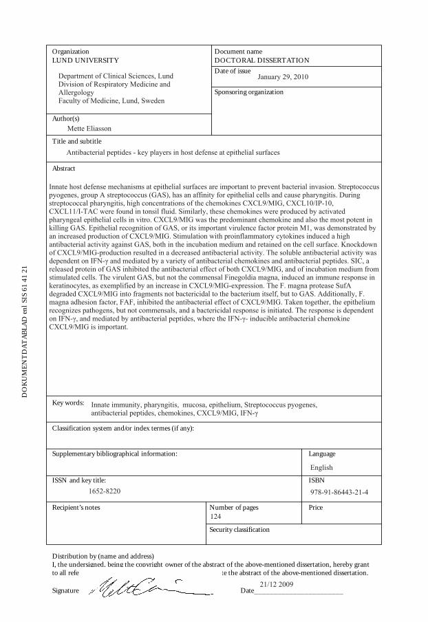
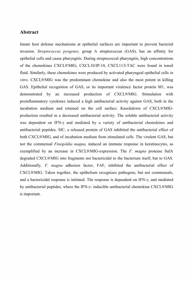
Innate host defense mechanisms at epithelial surfaces are important to prevent bacterial invasion. Streptococcuspyogenes, group A streptococcus (GAS), has an affinity for epithelial cells and cause pharyngitis. Duringstreptococcal pharyngitis, high concentrations of the chemokines CXCL9/MIG, CXCL10/IP-10,CXCL11/I-TAC were found in tonsil fluid. Similarly, these chemokines were produced by activatedpharyngeal epithelial cells in vitro. CXCL9/MIG was the predominant chemokine and also the most potent inkilling GAS. Epithelial recognition of GAS, or its important virulence factor protein M1, was demonstrated byan increased production of CXCL9/MIG. Stimulation with proinflammatory cytokines induced a highantibacterial activity against GAS, both in the incubation medium and retained on the cell surface. Knockdownof CXCL9/MIG-production resulted in a decreased antibacterial activity. The soluble antibacterial activity wasdependent on IFN-γ and mediated by a variety of antibacterial chemokines and antibacterial peptides. SIC, areleased protein of GAS inhibited the antibacterial effect of both CXCL9/MIG, and of incubation medium fromstimulated cells. The virulent GAS, but not the commensal Finegoldia magna, induced an immune response inkeratinocytes, as exemplified by an increase in CXCL9/MIG-expression. The F. magna protease SufAdegraded CXCL9/MIG into fragments not bactericidal to the bacterium itself, but to GAS. Additionally, F.magna adhesion factor, FAF, inhibited the antibacterial effect of CXCL9/MIG. Taken together, the epitheliumrecognizes pathogens, but not commensals, and a bactericidal response is initiated. The response is dependenton IFN-γ, and mediated by antibacterial peptides, where the IFN-γ- inducible antibacterial chemokineCXCL9/MIG is important.
Innate immunity, pharyngitis, mucosa, epithelium, Streptococcus pyogenes,antibacterial peptides, chemokines, CXCL9/MIG, IFN-γ
English
1652-8220 978-91-86443-21-4
21/12 2009
124
Antibacterial peptides
– key players in host defense at epithelial surfaces
Mette Eliasson
Department of Clinical Sciences, Lund Division of Respiratory Medicine and Allergology
Faculty of Medicine Lund University, Sweden
Lund 2010
Mette Eliasson
Department of Clinical Sciences, Lund Division of Respiratory Medicine and Allergology Lund University Biomedical Center, B14, Tornavägen 10, 221 84 Lund, Sweden E-mail: [email protected] Phone: + 46 (0)70 5331967 Fax: +46 (0)46 157756
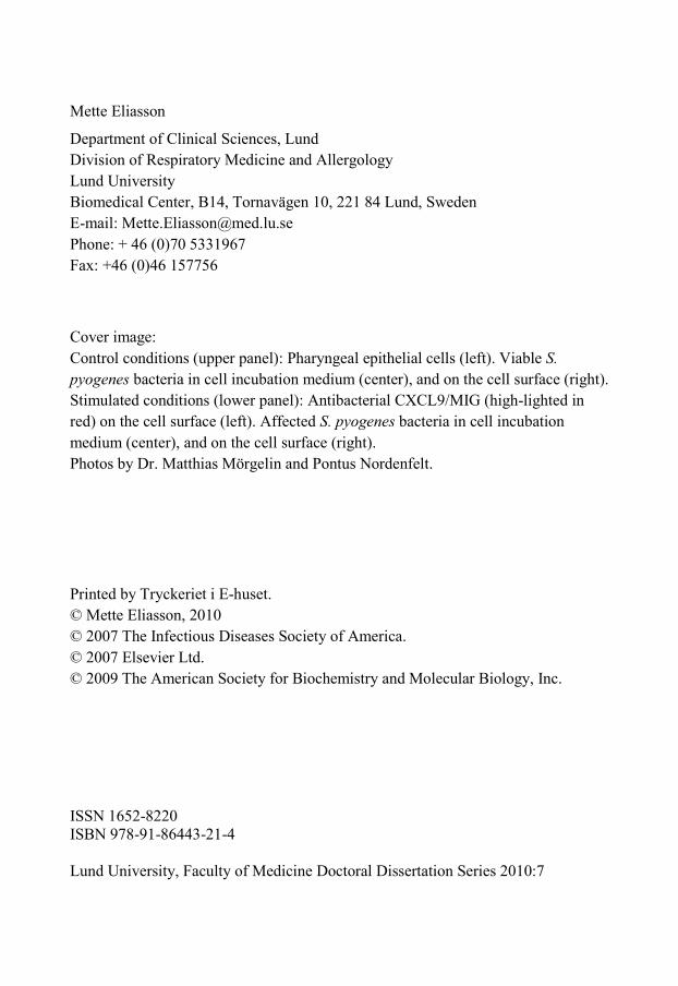
Cover image: Control conditions (upper panel): Pharyngeal epithelial cells (left). Viable S. pyogenes bacteria in cell incubation medium (center), and on the cell surface (right). Stimulated conditions (lower panel): Antibacterial CXCL9/MIG (high-lighted in red) on the cell surface (left). Affected S. pyogenes bacteria in cell incubation medium (center), and on the cell surface (right). Photos by Dr. Matthias Mörgelin and Pontus Nordenfelt. Printed by Tryckeriet i E-huset. © Mette Eliasson, 2010 © 2007 The Infectious Diseases Society of America. © 2007 Elsevier Ltd. © 2009 The American Society for Biochemistry and Molecular Biology, Inc. ISSN 1652-8220 ISBN 978-91-86443-21-4 Lund University, Faculty of Medicine Doctoral Dissertation Series 2010:7
Abstract
Innate host defense mechanisms at epithelial surfaces are important to prevent bacterial
invasion. Streptococcus pyogenes, group A streptococcus (GAS), has an affinity for
epithelial cells and cause pharyngitis. During streptococcal pharyngitis, high concentrations
of the chemokines CXCL9/MIG, CXCL10/IP-10, CXCL11/I-TAC were found in tonsil
fluid. Similarly, these chemokines were produced by activated pharyngeal epithelial cells in
vitro. CXCL9/MIG was the predominant chemokine and also the most potent in killing
GAS. Epithelial recognition of GAS, or its important virulence factor protein M1, was
demonstrated by an increased production of CXCL9/MIG. Stimulation with
proinflammatory cytokines induced a high antibacterial activity against GAS, both in the
incubation medium and retained on the cell surface. Knockdown of CXCL9/MIG-
production resulted in a decreased antibacterial activity. The soluble antibacterial activity
was dependent on IFN-γ and mediated by a variety of antibacterial chemokines and
antibacterial peptides. SIC, a released protein of GAS inhibited the antibacterial effect of
both CXCL9/MIG, and of incubation medium from stimulated cells. The virulent GAS, but
not the commensal Finegoldia magna, induced an immune response in keratinocytes, as
exemplified by an increase in CXCL9/MIG-expression. The F. magna protease SufA
degraded CXCL9/MIG into fragments not bactericidal to the bacterium itself, but to GAS.
Additionally, F. magna adhesion factor, FAF, inhibited the antibacterial effect of
CXCL9/MIG. Taken together, the epithelium recognizes pathogens, but not commensals,
and a bactericidal response is initiated. The response is dependent on IFN-γ, and mediated
by antibacterial peptides, where the IFN-γ- inducible antibacterial chemokine CXCL9/MIG
is important.

Antibacterial peptides – key players in host defense at epithelial surfaces
Contents
List of papers ...................................................................................................................................... - 8 -
Abbreviations ..................................................................................................................................... - 9 -
Introduction ...................................................................................................................................... - 11 -
PAMPs and PRRs ............................................................................................................................ - 11 -
Innate immunity at epithelial surfaces .............................................................................................. - 12 -
Cytokines .................................................................................................................................... - 14 -
Chemokines ................................................................................................................................ - 18 -
CC chemokines ...................................................................................................................... - 20 -
ELR-positive CXC chemokines ............................................................................................. - 20 -
IFN-γ-inducible ELR-negative CXC chemokines ................................................................. - 20 -
Interactions with glycosaminoglycans (GAGs) ..................................................................... - 21 -
Antibacterial peptides (APs) ....................................................................................................... - 22 -
Dual actions - antibacterial chemokines and chemotactic APs ................................................... - 24 -
Antibacterial redundancy among chemokines and peptides ........................................................ - 26 -
Oxidative innate immune defenses ............................................................................................. - 27 -
Streptococcus pyogenes, group A streptococcus (GAS)................................................................... - 28 -
M protein and hyaluronic acid capsule........................................................................................ - 28 -
SIC and SpeB .............................................................................................................................. - 29 -
Finegoldia magna ............................................................................................................................ - 30 -
SufA ............................................................................................................................................ - 31 -
FAF ............................................................................................................................................. - 31 -
Bacterial protection against antibacterial chemokines and peptides ................................................. - 32 -
Results and discussion ...................................................................................................................... - 33 -
Conclusions ...................................................................................................................................... - 40 -
Acknowledgements .......................................................................................................................... - 41 -
Populärvetenskaplig sammanfattning på svenska ............................................................................ - 43 -
References ........................................................................................................................................ - 46 -
Mette Eliasson
- 8 -
List of papers
This thesis is based on the following papers, which will be referred to in the text by their
roman numerals (I-IV):
Paper I
Arne Egesten, Mette Eliasson, Helena M Johansson, Anders I Olin. Matthias Mörgelin, Andrea
Mueller, James E Pease, Inga-Maria Frick, Lars Björck. The CXC chemokine MIG/CXCL9 is
important in innate immunity against Streptococcus pyogenes. The Journal of Infectious
Diseases.2007 Mar 1;195(5):684-93
Paper II
Mette Eliasson, Inga-Maria Frick, Mattias Collin, Ole E Sørensen, Lars Björck, Arne Egesten. M1
protein of Streptococcus pyogenes increases the production of the antibacterial CXC chemokine
MIG/CXCL9 in pharyngeal epithelial cells. Microbial Pathogenesis. 2007 Nov-Dec;43(5-6):224-33
Paper III
Christofer Karlsson*, Mette Eliasson*, Anders I Olin, Matthias Mörgelin, Anna Karlsson, Martin
Malmström, Arne Egesten, Inga-Maria Frick. SufA of the opportunistic pathogen Finegoldia magna
modulates the action of the antibacterial chemokine MIG/CXCL9, promoting bacterial survival during
epithelial inflammation. The Journal of Biological Chemistry. 2009 Oct 23;284(43):29499-508
* both authors contributed equally to this work
Paper IV
Mette Eliasson, Anders I Olin, Matthias Mörgelin, Mattias Collin, Arne Egesten. Interferon-γ Induces
a Peptide-Mediated Bactericidal Response in Human Pharyngeal Epithelial Cells. Manuscript
Antibacterial peptides – key players in host defense at epithelial surfaces
- 9 -
Abbreviations
AP Antibacterial peptide
CTL Cytotoxic T lymphocyte
FAF F. magna adhesion factor
GAGs Glucosaminoglycans
GAS Group A streptococcus
GRO-(α/β/γ) Growth-related oncogene (α/β/γ)
I-TAC Interferon-inducible T-cell alpha chemoattractant
IFN Interferon
IL Interleukin
IP-10 Interferon-inducible protein 10
LPS Lipopolysaccharide
LTA Lipoteichoic acid
MAPK Mitogen-activated protein kinase
MIG Monokine induced by interferon gamma
NADPH Nicotinamide adenine dinucleotide phosphate
NFκB Nuclear factor kappa B
NK Natural killer
PAMP Pathogen-associated molecular pattern
PG Peptidoglycan
PRR Pattern-recognition receptor
RNS Reactive nitrogen species
ROS Reactive oxygen species
SIC Streptococcal inhibitor of complement
Mette Eliasson
- 10 -
SLPI Secretory leukocyte protease inhibitor
Stat Signal transducer and activator of transcription
SufA Subtilase of Finegoldia magna
Th T helper
TNF Tumor necrosis factor
TLR Toll-like receptor
Antibacterial peptides – key players in host defense at epithelial surfaces
- 11 -
Introduction
The epithelial linings that cover the mucosal surfaces of the upper airways and the skin are
under constant exposure to microbes. Bacteria of the skin- and mucosal microbiota are
usually beneficial to the host and can provide protection by competing with pathogens for
nutrients and space. Pathogenic bacteria express virulence factors such as adherence
molecules, enzymes and toxins that enable them to colonize and invade epithelial surfaces.
The host is therefore dependent on robust protection against pathogens.
The innate immune system provides a first line of defense against invading microbial
pathogens. It is an evolutionary conserved system and, in contrast to the adaptive system
which is restricted to vertebrates, genes involved are represented in vertebrates,
invertebrates, and also in plants. It is older than the adaptive immune system, works
unspecifically to invading pathogens, and does not generate immunologic memory (Lien &
Ingalls, 2002; Medzhitov & Janeway, 2000). It takes three to five days for the adaptive
system to reach sufficient number of produced and differentiated effector lymphocytes. In
contrast, the innate immunity mechanisms are activated immediately upon microbial
recognition (Bartlett et al., 2008; Hippenstiel et al., 2006; Kawai & Akira, 2009; Medzhitov
& Janeway, 2000).
PAMPs and PRRs
The innate immune recognition of microorganisms is based on the detection of highly
conserved structures called pathogen-associated molecular patterns (PAMPs). PAMPs are
usually essential for the survival and pathogenicity of the microorganism and are shared by
entire classes of pathogens (Medzhitov & Janeway, 2000). LPS (lipopolysaccharide), PG
(peptidoglycan), LTA (lipoteichoic acid), and DNA are examples of common bacterial
PAMPs. PAMPs are recognized by pattern-recognition receptors (PRRs) that have been
selected over the course of evolution (Iwasaki & Medzhitov, 2004; Lien & Ingalls, 2002;
Medzhitov, 2001; Pasare & Medzhitov, 2005). PRRs are present on cells involved in the
initial microbial recognition, including epithelial cells, dendritic cells (DCs),
monocytes/macrophages, neutrophils, and natural killer (NK) cells (Iwasaki & Medzhitov,
Mette Eliasson
- 12 -
2004). In addition to PAMPs, endogenous molecules from stressed or damaged host cells
may also be recognized by cells of the innate immunity (Matzinger, 2002). Through
activation of NF-κB and MAPK signaling pathways, genes for a variety of effector
molecules are transcribed, resulting in production of e.g. antibacterial peptides (APs),
cytokines and chemokines (Akira et al., 2001; Bartlett et al., 2008; Hippenstiel et al., 2006;
Kawai & Akira, 2005; Medzhitov, 2001).
The Toll-like receptors (TLRs) are the best characterized PRRs in mammalian species, and
they are the mammalian homologues of Toll receptors (Rock et al., 1998), originally
identified in the fruit fly, Drosophila melanogaster (Anderson et al., 1985; Lemaitre et al.,
1996). The Toll receptors are evolutionary conserved and are found in plants, insects,
worms and vertebrates (Iwasaki & Medzhitov, 2004; Lien & Ingalls, 2002; Medzhitov,
2001; Pasare & Medzhitov, 2005). Invasive bacteria that are not recognized by TLRs can be
recognized via either NOD1 or NOD2, members of the cytosolic recognition family NOD
(nucleotide-binding oligomerization domain ) that detects bacterial cell wall constituents
present in the cytosol of host cells (Kawai & Akira, 2009).
Innate immunity at epithelial surfaces
The innate immune responses that takes place in the mucosa and in the skin involve a
variety of mechanisms and mediators, such as epithelial cells, DCs,
monocytes/macrophages, neutrophils, complement, NK cells, cytokines, chemokines, APs,
and the oxidative immune defense.
The epithelium – Inhaled microbes can be removed mechanically from the airways by ciliary
movement and through coughing. Airway and oro-pharyngeal epithelium is also protected
by mucus that constitutes a physical barrier protecting the epithelial surface by direct
binding of microorganisms. IgA, present in mucus, saliva and sweat, inhibits bacterial
binding to epithelial cells. The skin is further protected against microorganisms by its low
water content, acidic pH, and antibacterial lipids. Antibacterial chemokines and peptides are
constitutively produced in the skin- and airway-epithelium and can associate with mucins,
Antibacterial peptides – key players in host defense at epithelial surfaces
- 13 -
which place them in an ideal protective position against incoming microbes (Bartlett et al.,
2008; Felgentreff et al., 2006). The epithelium not only forms a physical barrier, but is also
important in the initial recognition of invading pathogens at epithelial surfaces. Upon
recognition, the epithelium responds by producing inflammatory mediators such as
cytokines, APs, and chemokines. The constitutive and induced production of antibacterial
components emphasize the important role of epithelial cells in innate immunity, not only
combating early and later phases of infections, but also preventing infection from
developing in the first place (Boman, 2003; Hiemstra, 2001). The epithelium-derived
molecules also have important roles in activating and directing cells of the adaptive immune
system.
DCs – are phagocytosing and antigen-presenting cells (APCs). They are present in
peripheral lymphoid tissue, such as the tonsils, and skin (Langerhans´ cells) where they
constantly scan the surroundings, and present endogenous or exogenous antigens on their
surfaces. Their major function is to activate T helper (Th) cells (Iwasaki & Medzhitov,
2004).
Monocytes/macrophages – are long-lived phagocytosing cells and APCs. They are present
in blood (monocytes) and in tissues (macrophages). Upon microbial stimuli, they produce
bactericidal reactive oxygen and nitrogen species (ROS/RNS). Macrophages are an
important source of cytokines that trigger the recruitment and activation of inflammatory
cells.
Neutrophils – are highly specialized but short-lived phagocytosing cells. They circulate in
the blood and are recruited to the mucosa and skin upon a bacterial infection. They kill
phagocytosed bacteria by releasing their granular content, consisting of a broad range of
antimicrobial agents such as peptides and enzymes. An important feature of the neutrophils
is their ability to mount a powerful oxidative burst in response to microbial and endogenous
stimuli. Besides affecting the phagocytosed pathogens, ROS can also kill microbes in
surrounding tissues (Rada & Leto, 2008).
Mette Eliasson
- 14 -
NK cells – Under resting conditions, NK cells are found in blood and lymphatic vessels and
they reside primarily in the spleen and in secondary lymphoid organs, such as the tonsils.
NK cells contain granules with cytotoxic molecules, used for killing tumor cells or virus-
infected host cells. NK cells interact with DCs in order to generate an appropriate immune
response in both the lymph node and in the inflamed skin (Andoniou et al., 2008). During
the early innate immune response, activated NK cells are rapid producers of IFN-γ in
response to various exogenous and endogenous stimuli (Billiau & Matthys, 2009;
Schoenborn & Wilson, 2007).
Cytokines
During the initial response to infection, recognition of pathogens by PRRs on cells of the
innate immune system results in a simultaneous production of cytokines and chemokines.
Cytokines regulate the chemokine production either as antagonists or synergists, thus
directing the type of immune response. Recruited innate immune cells amplify the release of
cytokines and chemokines, thus sustaining the innate immune response that eventually
results in recruitment of cells belonging to the adaptive immune response (Charo &
Ransohoff, 2006; Gouwy et al., 2005; Luster, 2002).
IL-1β and TNF-α – are the two most important cytokines in the early immune response.
They are produced by a variety of cells, but Th cells and monocytes/macrophages are the
main producers (Gouwy et al., 2005; Strieter et al., 2002; Veckman et al., 2003). Other
sources of IL-1β and TNF-α are keratinocytes and DCs (Giustizieri et al., 2001; Kupper,
1990). IL-1β- and TNF-α-signaling result in the activation of NF-κB and an enhanced
transcription of genes mediating innate immune responses (Lien & Ingalls, 2002; Strieter et
al., 2002).
Interferons (IFNs) – The interferons are major regulators of the innate immune system and
are produced in response to a variety of stimuli. The type I IFNs (IFN-α, IFN-β) have
antiviral activity and are induced by most human cells after exposure to viruses, double-
stranded viral RNA, bacteria, and PAMPs (Kawai & Akira, 2005; Li et al., 2009; Smith et
al., 2005). IFN-γ is the only type II interferons and is produced in response to cytokines,
Antibacterial peptides – key players in host defense at epithelial surfaces
- 15 -
primarily by NK cells (Li et al., 2009) and to a lesser extent by other cells, such as Th1
cells, cytotoxic T lymphocytes (CTLs) (Billiau & Matthys, 2009) and mast cells (Marshall,
2004).
Important contributions of IFN-γ to the immune response include, priming of macrophages
to produce proinflammatory cytokines such as TNF-α and IL-12, inducing the production of
potent antimicrobials such as APs and reactive oxygen and nitrogen species in phagocytes.
IFN-γ also induces the production of APs and chemokines in epithelial cells. Thus, IFN-γ
not only optimizes cell-mediated effector functions of the innate immune system but also
acts as a regulator of the adaptive immune response to bacterial agents (Billiau & Matthys,
2009; Schoenborn & Wilson, 2007).
The IFNs signal predominantly through the Jak (Janus kinase)-Stat pathway (Kawai &
Akira, 2005). The IFN-γ receptor contains two chains, IFN-γR1and IFN-γR2. Functionally,
IFN-γ acts as a homodimer and upon binding the receptor Jak1, Jak 2, and Stat1 become
phosphorylated. Stat1 translocates to the nucleus where it binds to the IFN-γ-activation
sequence in the promoter region of target genes (Kawai & Akira, 2005; Li et al., 2009;
Ohmori et al., 1997; Pestka et al., 2004). IFN-γ and TNF-α synergize to induce the
expression of many gene products during inflammation. The synergy is dependent upon
Stat1 and NF-κB. Motifs for these transcription factors are present in the promoter regions
of many inflammatory genes, such as genes coding for chemokines (Mahalingam et al.,
2001; Ohmori et al., 1997).
Cytokines direct the immune response
The innate and adaptive immune systems are two interdependent parts of a single integrated
immune system and the response to pathogens requires the coordinated action of both
systems (Luster, 2002). The move from innate to adaptive immune responses is, among
other things, dependent on epithelial-derived products such as cytokines and chemokines.
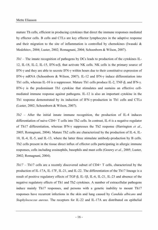
Naive CD4+ T cells are activated by DCs, and can differentiate into various effector
lineages, including Th1, Th2, Th17, and regulatory T cells. The cell development diverges
rapidly after antigen priming and is directed by specific cytokines (Figure 1). This results in
Mette Eliasson
- 16 -
mature Th cells, efficient in producing cytokines that direct the immune responses mediated
by effector cells. B cells and CTLs are key effector lymphocytes in the adaptive response
and their migration to the site of inflammation is controlled by chemokines (Iwasaki &
Medzhitov, 2004; Luster, 2002; Romagnani, 2004; Schoenborn & Wilson, 2007).
Th1 – The innate recognition of pathogens by DCs leads to production of the cytokines IL-
12, IL-18, IL-2, IL-15, IFN-α/β, that activate NK cells. NK cells is the primary source of
IFN-γ and they are able to secrete IFN-γ within hours due to their constitutive expression of
IFN-γ mRNA (Schoenborn & Wilson, 2007). IL-12 and IFN-γ induce differentiation into
Th1 cells, whereas IL-10 is a suppressor. Mature Th1 cells produce IL-2, TNF-β, and IFN-γ.
IFN-γ is the predominant Th1 cytokine that stimulates and sustains an effective cell-
mediated immune response against pathogens. IL-12 is also an important cytokine in the
Th1 response demonstrated by its induction of IFN-γ-production in Th1 cells and CTLs
(Luster, 2002; Schoenborn & Wilson, 2007).
Th2 – After the initial innate immune recognition, the production of IL-4 induces
differentiation of naive CD4+ T cells into Th2 cells. In contrast, IL-4 is a negative regulator
of Th17 differentiation, whereas IFN-γ suppresses the Th2 response (Harrington et al.,
2005; Romagnani, 2004). Mature Th2 cells are characterized by the production of IL-6, IL-
10, IL-4, IL-5, and IL-13, where the latter three stimulate antibody-production by B cells.
Th2 cells present in the tissue direct influx of effector cells participating in allergic immune
responses, cells including eosinophils, basophils and mast cells (Gouwy et al., 2005; Luster,
2002; Romagnani, 2004).
Th17 – Th17 cells are a recently discovered subset of CD4+ T cells, characterized by the
production of IL-17A, IL-17F, IL-21, and IL-22. The differentiation of the Th17 lineage is a
result of positive regulatory effects of TGF-β, IL-1β, IL-6, IL-21, IL-23 and absence of the
negative regulatory effects of Th1 and Th2 cytokines. A number of extracellular pathogens
induce mainly Th17 responses, and persons with a genetic inability to mount Th17
responses have recurrent infections in the skin and lung caused by Candida albicans and
Staphylococcus aureus. The receptors for IL-22 and IL-17A are distributed on epithelial
Antibacterial peptides – key players in host defense at epithelial surfaces
- 17 -
cells of the skin and airways and activation induces the expression of several host defense
genes, including human beta defensin (hBD)-2, Duox2, mucin, CXCL1/GRO-α,
CXCL9/MIG, and CXCL10/IP-10. IL-22, IL-17A, IL-17F, and TNF-α can act both
synergistically and additively to enhance the production of APs. Furthermore, IL-17A and
IL-17F also synergize with TNF-α. IL-21 acts in an autocrine loop amplifying the Th17 cell
response and its own synthesis. In addition, IL-21 stimulates IFN-γ production of both Th1
cells and CTLs. The effects of the Th17-produced cytokines and the distribution of their
receptors, imply that Th17 cells may be important during bacterial infections at epithelial
surfaces.
Figure 1. An overview of the differentiation of naive CD4+ cells
into Th cells, and the cytokines involved.
MHC-IINaive CD4+
Th1
IFN-γ
IFN-γ, IL-2
IL-4, IL-5, IL-6, IL-10, IL-13
Th17
IL-21
Th2
IL-4
IFN-γ
DC/Macrophage
NK cell
IFN-γCTLs
+ IL-12, IFN-γ, IL-21
B cells Antibodies
+ IL-4, IL-5, IL-13
IL-17A, IL-17F, IL-21, IL-22
Act
ivat
ion
of t
he a
dapt
ive
imm
une
syst
em
Cell-mediated response
Humoral response
Mette Eliasson
- 18 -
Chemokines
Chemokines constitute a large family of cytokines with chemotactic properties. All
chemokines regulate trafficking of leukocytes, during both health and disease (Baggiolini et
al., 1994; Billiau & Matthys, 2009; Luster, 1998).
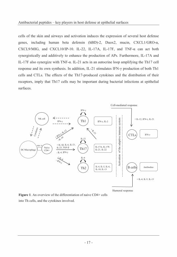
Chemokines have a relatively low level of sequence identity, but their three-dimensional
structures, functions, or receptor-binding profiles, are strikingly similar. In the N-terminal
region, three antiparallel β-sheets are connected by disulphide bridges, a structure that is
important for binding and activation of the corresponding chemokine receptor. The C-
terminal region consists of an amphipathic α-helix, where one half of the circular structure is
composed of nonpolar hydrophobic amino acids and the other side contains charged amino
acids, thereby being hydrophilic (Baggiolini, 1998; Baggiolini, 2001; Luster, 2002; Turnbull
et al., 2001), (Figure 2).
Chemokines are small GAG-binding proteins consisting of roughly 70-130 amino acids.
More than 50 chemokines have thus far been identified in humans (Baggiolini et al., 1994;
Baggiolini, 1998; Baggiolini, 2001; Luster, 1998). The majority are cationic proteins (pIs of
~9) at physiological pH due to a relatively high content of the amino acids lysine and
arginine.
Chemokines have been divided into four families, according to the arrangement of
conserved N-terminal cysteine motifs, C, CC, CXC, and CX3C, where X is a non-conserved
amino acid residue. The CXC and CC chemokines represent the largest families. The CXC
Figure 2. The predicted structure of the
chemokine CXCL9/MIG. β-sheets in
yellow, α-helix in purple.
C
ELRCXC
C
N
C C
CXC
C
N
CC
CC
C
N
C
Antibacterial peptides – key players in host defense at epithelial surfaces
- 19 -
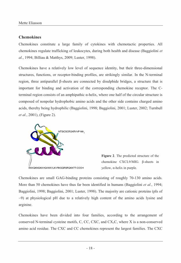
chemokines can be divided into ELR-positive and ELR-negative CXC chemokines, based on
the presence or absence of a glutamic acid-leucine-arginin (ELR) motif, preceding the first
two cysteines in the N-terminal region (Baggiolini, 1998; Cole et al., 2001; Luster, 1998;
Luster, 2002), (Figure 3).
Figure 3. Schematic drawing of a CC chemokine (left), an ELR-positive CXC chemokine (center),
and an ELR-negative CXC chemokine (right).
Receptor activation – Several chemokines can share the same receptor and several different
receptors may bind the same ligand. The receptors can be restricted to a certain type of cell
and to a cell state (activated and/or differentiated), and can be constitutively expressed,
induced but also downregulated (Luster, 1998). Ten receptors for CC chemokines (CCR1-
10), and six receptors for the CXC chemokines (CXCR1-6) have to date been characterized
in humans (Luster, 1998; Murphy, 2002). Binding of a chemokine to its corresponding
specific G-protein-coupled cell-surface receptor leads to cellular activation, an increase in
intracellular calcium, and results in cellular responses (Luster, 1998; Murphy, 2002).
The presence of receptors on neurons, epithelial cells, and endothelial cells indicates
additional roles for chemokines (Luster, 1998). In addition to leukocyte chemotaxis, some
chemokines modulate angiogenesis, tumor growth, and inhibit stem-cell proliferation.
Depending on the context, chemokines have different effects. For example, the ELR-
negative CXC chemokine CXCL10/IP-10, inhibits neovascularization, tumor growth and
metastasis, whereas the ELR-positive CXC chemokines, CXCL8/IL-8 and CXCL6/GCP-2,
promote angiogenesis and tumor metastasis (Gijsbers et al., 2005; Luster, 1998).
Mette Eliasson
- 20 -
CC chemokines
Several members of the CC chemokine family, for example CCL20/MIP-3α and
CCL28/MEC, are produced by epithelial cells (Sauty et al., 1999). The CC chemokine
receptors (CCRs) are preferentially expressed on activated lymphocytes of the Th2
phenotype, but also on eosinophils and basophils (Hoover et al., 2002; Lazarus et al., 2003;
Luster, 1998; Luster, 2002).
ELR-positive CXC chemokines
The most studied member of this group is CXCL8/IL-8. It is produced by epithelial cells
and neutrophils which thereby become activated in response to CXC8/IL-8 in an auto-
/paracrine manner (Baggiolini, 2001; Sauty et al., 1999). Other members of this chemokine
family are CXCL1/2/3 (GRO-α/β/γ), which are produced by epithelial cells (Baggiolini,
1998; Luster, 1998). The ELR-positive CXC chemokines predominantly activate and recruit
neutrophils through their receptors CXCR1 (CXCL8/IL-8) and CXCR2 (CXCL8/IL-8,
CXCL1/2/3 (GRO-α/β/γ)). These receptors are also expressed on endothelial cells (Murphy,
2002).
IFN-γ-inducible ELR-negative CXC chemokines
Members of this family are the three closely related chemokines CXCL9/MIG, CXCL10/IP-
10, and CXCL11/I-TAC (Cole et al., 1998; Farber, 1997; Loetscher et al., 1996). Upon IFN-
γ-stimulation, these chemokines are produced by a variety of cells but predominantly by
epithelial cells and monocytes (Cole et al., 1998; Egesten et al., 2007; Farber, 1993; Farber,
1997; Karlsson et al., 2009; Loetscher et al., 1996; Sauty et al., 1999). Their receptor
CXCR3 is expressed on Th1 cells, NK cells, DCs, mast cells and endothelial cells
(Baggiolini, 1998; Loetscher et al., 1996; Luster, 2002; Murphy, 2002).
IFN-γ is important for the optimal induction of CXCL9/MIG, but in the absence of IFN-γ,
IFN-α/β can mediate production (Mahalingam et al., 2001). IL-1β and TNF-α can synergize
with IFN-γ and enhance the production of CXCL9/MIG, CXCL10/IP-10, and CXCL11/I-
TAC (Egesten et al., 2007; Lauw et al., 2000; Sauty et al., 1999).
Antibacterial peptides – key players in host defense at epithelial surfaces
- 21 -
Interactions with glycosaminoglycans (GAGs)
Heparansulfate, heparin, chondroitinsulfate, and dermatansulfate are GAGs found on cell
surfaces and within extracellular matrix (Hoogewerf et al., 1997). GAGs can be soluble or
surface-bound and are composed of polysaccharide-chains attached to a protein core.
Immobilized GAGs are essential for the biological activity of most chemokines (Johnson et
al., 2004; Proudfoot et al., 2003; Shaw et al., 2004).
Most chemokines are highly cationic peptides that interact with the negatively charged
GAGs (Luster et al., 1995; Prydz & Dalen, 2000). However, anionic chemokines also bind
to GAGs, suggesting that the interaction is specific and not just electrostatic. GAG-binding
sequences of chemokines and the corresponding motifs on GAGs have been identified,
thereby regulating the interactions (Turnbull et al., 2001). The preference for individual
GAGs can vary between chemokines and certain chemokines can discriminate between
GAGs (Handel et al., 2005). GAG-binding may involve or induce oligomerization of the
chemokines, suggesting that GAG-binding specificity may be a feature of chemokines that
need to form oligomers in order to be active (Czaplewski et al., 1999; Proudfoot et al.,
2003; Swaminathan et al., 2003; Zhang et al., 1994).
Oligomerization on GAGs establishes a local chemotactic gradient that efficiently attracts
leukocytes (Hoogewerf et al., 1997; Luster, 1998; Proudfoot et al., 2003). The
oligomerization on GAGs is also likely to be important on mucosal surfaces where
antibacterial chemokines can be highly concentrated and form a bactericidal barrier against
colonizing microbes (Egesten et al., 2007; Luster, 1998). In addition, oligomerization of
chemokines on GAGs on epithelial and endothelial cell surfaces may be a requirement in
order to avoid being washed away from the local site under flow conditions (Proudfoot et
al., 2003). Some chemokines are able to interact with receptors as monomers (Rajarathnam
et al., 1994) and it is suggested that these are chemokines being produced within the
extravascular space, which is not exposed to flow (Proudfoot et al., 2003).
Mette Eliasson
- 22 -
Antibacterial peptides (APs)
Under basal conditions, some APs are constitutively expressed by skin- and airway
epithelia, and binding to GAGs can retain them on epithelial surfaces. The production of
APs by epithelial cells and cells of the submucosal tissue, can also be induced in response to
microbial pathogens, proinflammatory cytokines, growth factors, and injury (Bartlett et al.,
2008; Elias, 2007; Roupe et al., 2009; Sorensen et al., 2006).
APs have broad-spectrum effects on Gram-positive bacteria, Gram-negative bacteria, and
also show activities against fungi and enveloped viruses. An important aspect is that they are
active against bacteria resistant to conventional antibiotics (e.g. methicillin-resistant S.
aureus (MRSA)) (Chambers, 2005). Despite the broad antimicrobial efficiency, bacteria of
the human microbiota are relatively resistant to APs (Boman, 2000; Ge et al., 1999;
Karlsson et al., 2009). Because of their multifunctional properties, these peptides are
commonly termed antimicrobial peptides (AMPs). However, since fungi seldom cause
survival problems for mammals, the driving force behind their evolution has probably been
the antibacterial function. For this reason, Boman proposed that this family of peptides
should be referred to as antibacterial peptides (APs) (Boman, 2003).
An important characteristic of APs is their combined cationic and hydrophobic properties,
resulting in amphipathicity (Peschel & Sahl, 2006). The main target of APs is negatively
charged bacterial membranes, to which APs are attracted electrostatically. The APs attain an
amphipathic helical conformation, allowing them to insert into the hydrophobic face of the
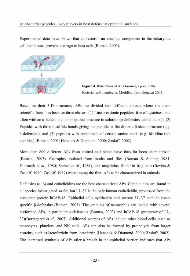
phospholipid bilayer (Tossi et al., 2000), (Figure 4)(Brogden, 2005). It is unclear if the pore-
forming activity is the primary killing mechanism, however, APs interacting with the
membrane cause a reduced membrane potential, a disturbed osmotic balance, an increased
pressure within the membrane, events resulting in lysis of the bacteria (Boman, 2003;
Peschel & Sahl, 2006). In addition, many APs can neutralize the effect of LPS (Rosenfeld et
al., 2006) and some APs can act intracellularly, killing bacteria by interfering with DNA and
protein synthesis (Boman, 2003).
Antibacterial peptides – key players in host defense at epithelial surfaces
- 23 -
Experimental data have shown that cholesterol, an essential component in the eukaryotic
cell membrane, prevents damage to host cells (Boman, 2003).
Based on their 3-D structures, APs are divided into different classes where the main
scientific focus has been on three classes. (1) Linear cationic peptides, free of cysteines, and
often with an α-helical and amphipathic structure in solution (α-defensins, cathelicidins). (2)
Peptides with three disulfide bonds giving the peptides a flat dimeric β-sheet structure (e.g.
β-defensins), and (3) peptides with enrichment of certain amino acids (e.g. histidine-rich
peptides) (Boman, 2003; Hancock & Diamond, 2000; Zasloff, 2002).
More than 800 different APs from animal and plants have thus far been characterized
(Boman, 2003). Cecropins, isolated from moths and flies (Boman & Steiner, 1981;
Hultmark et al., 1980; Steiner et al., 1981), and magainins, found in frog skin (Bevins &
Zasloff, 1990; Zasloff, 1987) were among the first APs to be characterized in animals.
Defensins (α, β) and cathelicidins are the best characterized APs. Cathelicidins are found in
all species investigated so far, but LL-37 is the only human cathelicidin, processed from the
precursor protein hCAP-18. Epithelial cells synthesize and secrete LL-37 and the tissue
specific β-defensins (Boman, 2003). The granules of neutrophils are loaded with several
preformed APs, in particular α-defensins (Boman, 2003) and hCAP-18 (precursor of LL-
37)(Borregaard et al., 2007). Additional sources of APs include other blood cells, such as
monocytes, platelets, and NK cells. APs can also be formed by proteolytis from larger
proteins, such as lactoferricin from lactoferrin (Hancock & Diamond, 2000; Zasloff, 2002).
The increased synthesis of APs after a breach in the epithelial barrier, indicates that APs
Figure 4. Illustration of APs forming a pore in the
bacterial cell membrane. Modified from Brogden 2005.

Mette Eliasson
- 24 -
may have a role during epithelial repair, protecting the underlying tissue from pathogens
until the epithelial barrier is restored (Hiemstra, 2001; Roupe et al., 2009; Sorensen et al.,
2006).
The importance of APs has been questioned and there are some considerations to take into
account when discussing their in vivo relevance. Most, but not all APs show decreased
activity in the presence of salt at levels present in plasma. In cystic fibrosis, the salt
concentration is increased, resulting in an environment that does not support optimal
functioning of APs with recurrent bacterial infections as a consequence (mainly caused by
Pseudomonas aeruginosa). In addition, most APs are inhibited by various substances such
as plasma proteins, and soluble GAGs. However, there are many in vivo examples
demonstrating important roles of APs in bacterial infections. Individuals suffering from
Kostmann´s syndrome are born with only few neutrophils and acquire severe periodontitis
and airway infections. Recent investigations have revealed that hCAP-18/LL-37 was
missing in both neutrophils and saliva (Putsep et al., 2002). Mice are normally highly
sensitive to infection with Salmonella typhimurium but became resistant when transfected
with the human enteric defensin hBD-5 (Boman, 2003).
Dual actions - antibacterial chemokines and chemotactic APs
APs present in humans at low (nanomolar) concentrations may exhibit other functions,
including chemotaxis. During inflammation, certain chemokines reach antibacterial
concentrations (micromolar), levels that are beyond what is required for chemotactic
activity, and may act as APs. Amphipathic molecules with a high positive charge density
are generally potent antimicrobials (Cole et al., 2001). Most chemokines are positively
charged at neutral pH, and Yang et al demonstrated that no chemokine with a pI lower than
9.0 was antibacterial against E. coli, indicating that cationicity is an important feature for
antibacterial chemokines (Yang et al., 2003). Additionally, the topological amphipathic
Antibacterial peptides – key players in host defense at epithelial surfaces
- 25 -
design, with clusters of hydrophobic and cationic amino acids organized in discrete surface
areas, is an important structural characteristic for antibacterial chemokines (Yang et al.,
2003; Zasloff, 2002). These properties make it possible for the peptides to associate with,
and disrupt bacterial membranes. However, there are examples of both linear and anionic
peptides that possess antibacterial activity (Boman, 2003).
CC chemokines – Antibacterial activity has been ascribed to several CC chemokines.
CCL20/MIP-3α is constitutively produced by many cells and is upregulated by
proinflammatory stimuli such as LPS, IFN-γ, TNF-α, IL-1β (Eliasson et al., 2007; Schutyser
et al., 2003). High production of this antibacterial chemokine by epithelial cells during
mucosal inflammation points to direct antibacterial roles for CCL20/MIP-3α (Starner et al.,
2003; Yang et al., 2003). Mucosa-associated epithelial chemokine (MEC), CCL28/MEC is
produced constitutively in salivary glands and high concentrations are found in saliva.
CCL28/MEC is effective against some strains of Gram-positive and Gram-negative bacteria,
as well as C. albicans. This indicates that MEC/CCL28 has dual roles in mucosal immunity,
both as a chemoattractant and also as a broad-spectrum AP when secreted into low-salt body
fluids such as saliva (Hieshima et al., 2003).
ELR-positive CXC chemokines – Antibacterial activity in the C-terminal region of
epithelium and neutrophil-derived CXCL8/IL-8 has been reported (Bjorstad et al., 2005;
Yount et al., 2007). CXCL1/2/3 (GRO-α/β/γ) are expressed by a variety of myeloid and
epithelial cells after stimulation with proinflammatory cytokines (Becker et al., 1994;
Eliasson et al., 2007) and have antibacterial activity (Yang et al., 2003). CXCL6/GCP-2 is
expressed by macrophages and epithelial cells and its antibacterial effect against Gram-
positive and Gram-negative pathogens indicates that CXCL6/GCP-2 is important during
mucosal infections (Linge et al., 2008).
IFN-γ-inducible ELR-negative CXC chemokines – CXCL9/MIG, CXCL10/IP-10 and
CXCL11/I-TAC are IFN-γ inducible chemokines. High production is seen in pharynx and
airway epithelial cells and production is increased by IL-1β and TNF-α (Egesten et al.,
2007; Sauty et al., 1999). All three have a broad antibacterial spectrum (Cole et al., 2001;
Mette Eliasson
- 26 -
Crawford et al., 2009; Egesten et al., 2007; Karlsson et al., 2009). The antibacterial activity
of CXCL9/MIG has been mapped to the C-terminal region (Egesten et al., 2007) which is
cationic, amphipathic and has a predicted α-helical structure, all features typical of APs.
CXCL9/MIG has a higher antibacterial activity than CXCL10/IP-10 and CXCL11/I-TAC
(Egesten et al., 2007), and this might be explained by the more extended α-helical C-
terminal of CXCL9/MIG. CXCL10/IP-10 and CXCL11/I-TAC have higher affinity for the
CXCR3 receptor, thus these chemokines might be more important in regulating leukocyte
trafficking while CXCL9/MIG has evolved towards an antibacterial function.
Several of the classical APs have chemotactic properties. For example, LL-37 can induce
calcium mobilization in, and is chemotactic for monocytes, neutrophils and CTLs (De et al.,
2000). Humanα -defensin-1, and human β-defensin-2 are chemoattractive for
monocytes/macrophages (Presicce et al., 2009; Territo et al., 1989) and CTLs (Chertov et
al., 1996).
Antibacterial redundancy among chemokines and peptides
It is important to note that, in vivo, APs do not exist alone but rather as a cocktail of multiple
APs, each with overlapping spectra of activity. Their mode of action differs and by
cooperatively attacking different targets in bacteria, one AP could unmask vulnerable
structures for another AP.
In the airways, the presence of multiple factors increases the antibacterial potency of airway
surface liquid (ASL). This was demonstrated by the synergistic actions of lactoferrin,
lysozyme, and SLPI, some of the most abundant antibacterial components present in the
airways (Singh et al., 2000; Travis et al., 1999). Synergistic effects of lysozyme and
lactoferrin with other APs, e.g. LL-37, hBD-2, have also been confirmed (Bals et al., 1998).
Cole et al found that the microbicidal activity of nasal fluid is dependent on synergistic and
additive interactions between lysozyme, lactoferrin, and APs (Cole et al., 1999).
Antibacterial peptides – key players in host defense at epithelial surfaces
- 27 -
Oxidative innate immune defenses
In addition to the cell- and peptide-mediated responses of the innate immune system,
phagocytes deliver microbicidal oxygen- and nitrogen-derivatives that target cellular
components in bacteria, such as lipids, proteins and nucleotide bases (Fang, 2004; Hampton
et al., 1998).
In humans, the Nox (NADPH oxidase) family includes seven oxidase genes, Nox 1-5 and
Duox 1-2 (Kuwano et al., 2006). The Nox/Duox family delivers antimicrobial reactive
oxygen species (ROS), including superoxide and hydrogen peroxide (Bartlett et al., 2008;
Rada & Leto, 2008). In phagocytes, hydrogen peroxide is converted to the microbicidal
oxidant hypochlorous acid by myeloperoxidase (MPO). Hypobromous and hypothiocyanous
acids are other potent MPO products (Winterbourn, 2008).
Inducible nitric oxide synthase (iNOS) generates reactive nitrogen species (RNS) derived
from nitric oxide. Since nitric oxide readily reacts with superoxide to form the microbicidal
peroxynitrite, there is a clear overlap between the two systems (Winterbourn, 2008). The
Nox and the iNOS are the two most important pathways of the phagocyte-mediated
microbial clearance (Fang, 2004; Winterbourn, 2008).
Recent studies suggest that iNOS, as well as several of the Nox/Duox family members, are
expressed at high levels in epithelial cells and may be involved in innate immune functions
at mucosal surfaces (Leto & Geiszt, 2006; Rada & Leto, 2008). Airway epithelial cells
produce extracellular hydrogen peroxide, and the concurrent presence of lactoperoxidase
(LPO) results in generation of bactericidal reactive oxidants, e.g. hypothiocyanite (Bartlett
et al., 2008; Leto & Geiszt, 2006; Moskwa et al., 2007; Rada & Leto, 2008). Nox/Duox
expression in epithelial cells is enhanced in response to microbes and proinflammatory
cytokines (Rada & Leto, 2008; Sansonetti, 2004). For example, Duox2-expression, and
Nox2-expression in airway epithelial cells (Harper et al., 2005), and Nox1-expression in
colon epithelial cells (Kuwano et al., 2006), is highly induced by IFN-γ.

Mette Eliasson
- 28 -
Streptococcus pyogenes, group A streptococcus (GAS)
Streptococcus pyogenes is a Gram-positive bacterium causing common infections such as
impetigo and pharyngitis (Bisno & Stevens, 1996; Cunningham, 2000). Pharyngitis can be
caused by a variety of bacteria and viruses, but GAS is the most common causative
bacterium (Bisno, 1996; Fischetti, 1989). GAS can also cause severe clinical conditions
such as invasive skin- and soft tissue infections, and septicemia (Bisno & Stevens, 1996;
Bisno et al., 2003; Cunningham, 2000; Nitsche-Schmitz et al., 2007; Nowak, 1994; Stevens,
2000).
M protein and hyaluronic acid capsule
The cell membrane-anchored M protein appears as hair-like projections on the surface of the
streptococcal cell wall and is composed of two polypeptide chains complexed in an α-
helical coiled coil configuration that traverses the cell wall (Phillips et al., 1981). M protein
is released from the streptococcal cell wall spontaneously (Åkesson et al., 1994) but also
after proteolytic cleavage by the streptococcal cysteine protease SpeB (Berge & Björck,
1995). The initial step in colonization of the pharynx or the skin is adherence to epithelial
cells (Cunningham, 2000). M protein facilitates bacterial adhesion to epithelial cells (Ellen
& Gibbons, 1972) by binding to GAGs (Frick et al., 2003b), mucins (Ryan et al., 2001), and
fibronectin (Cunningham, 2000; Nitsche-Schmitz et al., 2007).
In order to invade deeper tissues, GAS has evolved numerous adhesins, including M
proteins, and the hyaluronic acid capsule covering the bacterium. The adhesins bind
extracellular matrix proteins and plasma proteins (Nitsche-Schmitz et al., 2007; Okada et
al., 1994). In addition, GAS produces proteases and other enzymes that degrade tissue, thus
providing access to nutrients and enhancing bacterial invasion (Nitsche-Schmitz et al.,
2007). M protein contains fibrinogen- , albumin- and IgG-binding regions that facilitate
bacterial persistence in infected tissue (Herwald et al., 2004; Åkesson et al., 1994). A
prerequisite for survival in tissues is the ability of pathogens to resist being killed by
phagocytosis. The hyaluronic acid capsule contributes to antiphagocytotic properties of
GAS (Comstock & Kasper, 2006; Fischetti, 1989; Foley & Wood, 1959; Moses et al., 1997;
Wessels & Bronze, 1994). In addition, different studies suggest that M protein-expressing
Antibacterial peptides – key players in host defense at epithelial surfaces
- 29 -
GAS can both resist being phagocytosed by neutrophils, and, in the case of phagocytosis
occuring, survive in the phagosome by inhibiting its maturation (Fischetti, 1989; Foley &
Wood, 1959; Moses et al., 1997; Staali et al., 2003; Staali et al., 2006).
The N-terminal part of the M protein is highly variable, and as a consequence there are more
than 100 different serotypes of GAS (Bisno et al., 2003). Several different M protein
serotypes have been isolated from severe, invasive disease. However, the dominating strain
has been of the M1 serotype (Davies et al., 1996; Holm, 1996). In addition, Davies et al
demonstrated that an increased incidence of invasive disease parallels an increased
proportion of infections caused by the M1 serotype (Davies et al., 1996).
GAS not only adheres to epithelial cells but can also become internalized by epithelial and
cells of lymphoid tissues (Dombek et al., 1999; LaPenta et al., 1994; Molinari et al., 1997;
Osterlund & Engstrand, 1995). Internalization may lead to persistence and a carrier state
(Cunningham, 2000), as indicated by studies demonstrating viable intracellular GAS
harbored in tonsils from patients with recurrent tonsillitis (Cunningham, 2000; Fischetti,
1989; Osterlund & Engstrand, 1997; Osterlund et al., 1997). Internalized bacteria are able
to survive antibiotics that act extracellularly, such as penicillins. Approximately 30 % of
penicillin-treated children have surviving GAS isolated from the nasopharynx after
treatment (Gerber, 1994; Osterlund & Engstrand, 1995). The role of M1 protein in
internalization is not clear. Cue et al demonstrated that M1 acts as an invasin, when
compared with M1-negative isogenic mutant (Cue et al., 1998). In contrast, other groups did
not observe a difference between M1+ and M1- GAS in the uptake of bacteria by lymphoid
tissue (Hyland et al., 2009; LaPenta et al., 1994). However, kinetic studies by LaPenta et al
demonstrated that the internalization of M1+ GAS by epithelial cells was more efficient than
that of M1- (LaPenta et al., 1994).
SIC and SpeB
Protein SIC (streptococcal inhibitor of complement) was originally isolated from the
growth medium of GAS (M1 strain), and was found to inhibit the host-protective cytolytic
function of the complement cascade in vitro (Akesson et al., 1996). The protein is
Mette Eliasson
- 30 -
associated with the cell wall but is released from the bacterial surface by proteolytic
cleavage (Berge & Björck, 1995). SpeB is an extracellular cysteine protease and contributes
to the virulence of GAS by its degrading of human immunoglobulins, fibronectin, and
components of the extracellular matrix (Bisno et al., 2003; Collin & Olsen, 2001;
Schmidtchen et al., 2001). Both SIC and SpeB have been shown to inhibit or inactivate APs.
SIC block the antibacterial effect through binding inhibition (Egesten et al., 2007; Fernie-
King et al., 2002; Fernie-King et al., 2004; Frick et al., 2003a), whereas SpeB actively
degrades the APs (Egesten et al., 2009; Karlsson et al., 2009).
Finegoldia magna
The human microbiota consists of a diverse population of resident microorganisms
(commensals) that may become pathogenic in response to an impaired epithelial barrier
(opportunists). Finegoldia magna, formerly known as Peptostreptococcus magnus, is a
Gram-positive anaerobic coccus. F. magna bacteria are commensals of the mouth, the
upper respiratory and gastrointestinal tract, the skin and soft tissues, and the female
genitourinary system, sites where they may also cause opportunistic infections (Murdoch,
1998). Acute and chronic skin wounds harbor a diverse microbiota and in 70-80% of both
infected and noninfected wounds, anaerobic organisms are found. F. magna is a commonly
isolated species, indicating that it is among the most virulent of the anaerobic bacteria
(Bowler & Davies, 1999; Stephens et al., 2003; Wall et al., 2002).
It has been documented that Gram-positive anaerobic cocci and their metabolites interfere
with wound healing by inhibiting fibroblast proliferation, inhibiting growth of keratinocytes
and consequently reepithelialization (Stephens et al., 2003; Wall et al., 2002). F. magna
bacteria produce enzymes interfering with components of the extracellular matrix (Karlsson
et al., 2007), thus disturbing matrix remodeling, and proteolytically active strains of F.
magna are associated with infections causing tissue destruction (Krepel et al., 1992).
Antibacterial peptides – key players in host defense at epithelial surfaces
- 31 -
SufA
The recently discovered SufA (Subtilase of Finegoldia magna) from F. magna, is a
subtilisin-like serine protease, associated with the bacterial surface but also released during
growth (Karlsson et al., 2007). SufA interferes with innate immunity by cleaving
antibacterial chemokines and peptides, a feature that may promote colonization and survival
of F. magna (Karlsson et al., 2007; Karlsson et al., 2009). Cysteine-rich members of the
defensins seem to be protected from degradation. On the other hand, the antibacterial α-
helical structure of the CXC chemokine CXCL9/MIG (Egesten et al., 2007) and the linear
α-helical AP LL-37 (Bals & Wilson, 2003), which both lack cysteines, are degraded and
inactivated (Karlsson et al., 2007; Karlsson et al., 2009).
FAF
F. magna adhesion factor (FAF) enables the binding of bacteria to the basement membrane
between the epidermis and dermis, by interacting with the basement membrane protein BM-
40 (Frick et al., 2008). FAF is a surface protein but is also released by SufA (Karlsson et al.,
2009) and can bind to and inhibit the effect of antibacterial chemokines and peptides (Frick
et al., 2008; Karlsson et al., 2009). FAF-expressing F. magna strains are more resistant to
APs. Interestingly, FAF shows no affinity for the AP α-defensin and this peptide is not
bactericidal against F. magna (Frick et al., 2008).
Mette Eliasson
- 32 -
Bacterial protection against antibacterial chemokines and peptides
The bacterial surface is negatively charged and the cationic feature of most antibacterial
peptides and chemokines is believed to have evolved as a consequence of the long interplay
during evolution. However, pathogenic bacteria have invented mechanisms to overcome the
AP-mediated first line of defense. (Otto, 2009; Peschel & Sahl, 2006).
Strategies from bacteria to escape being killed by the APs have resulted in different
mechanisms, including a change in the AP target to make it less susceptible. For example,
many Gram-positive pathogens, such as GAS and S. aureus can modify teichoic acids with
D-alanine, and decrease the net negative charge making the target less accessible for the
cationic APs (Kristian et al., 2005; Peschel et al., 1999; Peschel & Sahl, 2006).
Another mechanism aims at destroying or inhibiting APs, preventing them from reaching
the cytoplasmic membrane. Secreted bacterial proteases and proteins can efficiently digest
and inactivate or inhibit APs. This is exemplified by SpeB and protein SIC from GAS, and
SufA and protein FAF from F. magna (Egesten et al., 2009; Fernie-King et al., 2002;
Fernie-King et al., 2004; Frick et al., 2003a; Karlsson et al., 2007; Karlsson et al., 2009;
Schmidtchen et al., 2002). However, APs that are less susceptible to digestion by bacterial
proteases have evolved, and the presence of multiple disulfide bridges in the structure seems
to render them more resistant (Egesten et al., 2009; Peschel & Sahl, 2006). Another
important observation is that bacterial proteases can degrade proteoglycans, thereby
releasing GAGs that can bind and inactivate APs. These soluble GAGs can be found in
clinical conditions such as chronic wounds (Baranska-Rybak et al., 2006; Schmidtchen et
al., 2001).
Antibacterial peptides – key players in host defense at epithelial surfaces
- 33 -
Results and discussion
Paper I and II
The ELR negative CXC chemokines CXCL9/MIG, CXCL10/IP-10, and CXCL11/I-TAC
are produced by a variety of cells both during health and disease, and were recently shown
to be antibacterial (Cole et al., 2001). In Paper I, the three chemokines where detected at
high concentrations in tonsil fluid from patients suffering from streptococcal pharyngitis.
CXCL9/MIG was found at the highest concentration, exceeding what is required for
chemotactic activity (Xanthou et al., 2003). Since the three chemokines can be produced by
a variety of cells at mucosal surfaces, the source of the chemokines found in tonsil fluid
could not be attributed to a single cell-type. Sauty et al have shown that airway epithelial
cells produce the three chemokines in vitro. IFN-γ is necessary for the transcription, and
TNF-α enhances their production (Sauty et al., 1999). In vitro, we demonstrated that
pharyngeal epithelial cells can produce the chemokines at high concentrations, either after
stimulation with IFN-γ alone (Paper II), or in combination with TNF-α (Paper I). The levels
of production in vitro were similar to what was seen in tonsil fluid, CXCL9/MIG being the
predominant chemokine, followed by CXCL10/IP-10 and CXCL11/I-TAC.
− Epithelial recognition
The initial step in the innate immune response against bacteria is the recognition of PAMPs
by PRRs on host cells. This recognition results in the production of cytokines, chemokines
and APs.
LTA, PG (Bisno et al., 2003; Schroder et al., 2003), and protein M1 of GAS (Pahlman et
al., 2006), respectively, stimulates elaboration of cytokines via TLR2 on monocytes. In
addition, GAS stimulates the production of the chemokines CXCL8/IL-8, CXCL9/MIG, and
CXCL10/IP-10 in macrophages (Veckman et al., 2003). LPS and LTA, respectively,
increases the production of the chemokines hCAP-18/LL-37, and CXCL8/IL-8 in epithelial
Mette Eliasson
- 34 -
cells from the upper respiratory tract (Nell et al., 2004), and the production of the APs hBD-
2 and hBD-3 in skin- and airway epithelia is induced in response to a variety of microbial
pathogens as well as proinflammatory stimuli (Bartlett et al., 2008; Sorensen et al., 2005).
In Paper I and II, we stimulated pharyngeal epithelial cells with IFN-γ in combination with
protein M1 or heat-killed GAS. The CXCL9/MIG production was dose-dependently
increased in the presence of M1 and GAS, respectively. There was no difference in the
response to M1-abscent mutant strains of GAS, demonstrating a broad epithelial recognition
of GAS-derived PAMPs. The epithelial cells sense the bacteria and respond fast as indicated
by an increase in CXCL9/MIG-mRNA already after three hours of stimulation with M1.
The upregulation of CXCL8/IL-8 and IL-6 expression in epithelial cells in response to GAS
is mediated through NF-κB and MAPK signaling pathways (Tsai et al., 2006). In Paper II,
rapid phosphorylations of both p38 MAPK and NF-κB were seen after addition of M1
protein irrespectively of pre-incubation with IFN-γ. This demonstrates a constitutive, and
not an IFN-γ-induced recognition of M1 protein. The enhanced CXCL9/MIG production
after stimulation with IFN-γ was shown to involve NF-κB, as demonstrated by the decreased
production after inhibition of NF-kB activation. Furthermore, microarrays revealed that, in
response to IFN-γ and protein M1, genes coding for several antibacterial CC and CXC
chemokines, e.g. CCL20/MIP-3α, CXCL9/MIG, CXCL10/IP-10, CXCL11/I-TAC, and
CXCL1/GRO-α, CXCL2/GRO-β, and CXCL3/GRO-γ, were upregulated in pharyngeal
epithelial cells.
– Antibacterial activity against GAS
CXCL9/MIG, CXCL10/IP-10, and CXCL11/I-TAC all showed a dose-dependent
antibacterial activity against GAS, CXCL9/MIG being the most potent. Two regions with
high isoelectric points (pIs) were identified in CXCL9/MIG, one in the N-terminal and one
in the C-terminal part of the molecule. The latter region is cationic and amphipathic and has
a predicted α-helical structure, all typical features of peptide-sequences exhibiting
Antibacterial peptides – key players in host defense at epithelial surfaces
- 35 -
antibacterial activity. A synthetic peptide from this region showed antibacterial activity
comparable to that of intact CXCL9/MIG. Electron microscopy demonstrated gold-labelled
CXCL9/MIG associated with the bacterial cell wall. Disruption of the bacterial cell wall and
leakage of bacterial contents after incubation with CXCL9/MIG, demonstrates that
CXCL9/MIG is bactericidal rather than bacteriostatic, and that the killing, at least in part,
involves membrane-disintegration (Paper I).
The physiological relevancy of APs is often discussed due to that the antibacterial effect of
most APs in vitro is lost in 150 mmol l-1 salt, found in plasma. However, considering that the
concentration of salt in oral fluid is 5 mmol l-1 (Aps & Martens, 2005), and that we could
demonstrate antibacterial activity of the chemokines in 150 mmol l-1 salt, our results indicate
that the antibacterial effect of the chemokines during streptococcal pharyngitis is highly
relevant (Paper I).
CXCL9/MIG was found both secreted into the incubation medium and also on the epithelial
cell surface (Paper I and II). In Paper I, I show that GAS was efficiently killed on the
surface of pharyngeal epithelial cells stimulated with a combination of IFN-γ and TNF-α.
CXCL9/MIG bound heparin with high affinity, a property shared by many APs. It has been
proposed that antibacterial chemokines, forming oligomers when bound to GAGs, could
serve as a concentrated antibacterial gradient at epithelial surfaces, thereby reducing the
possibilities for bacteria to adhere (Luster, 1998). Knock-down of CXCL9/MIG by siRNA
resulted in a reduced killing of GAS on the stimulated cell surface, implying an important
antibacterial role of CXCL9/MIG at epithelial surfaces. We could also demonstrate an
antibacterial activity in the incubation medium from pharyngeal epithelial cells stimulated
with a combination of IFN-γ and TNF-α.
− Defense mechanisms of GAS
Bacteria and APs have evolved concomitantly for millions of years, hence bacteria have
evolved mechanisms to defend themselves against APs. Protein SIC, released from GAS
during growth, binds APs. In Paper I, SIC bound to CXCL9/MIG, CXCL10/IP-10, and
CXCL11/I-TAC with high affinity, resulting in neutralization of the antibacterial activity. In
Mette Eliasson
- 36 -
contrast, there was no interaction with the CXC chemokines CXCL1/GRO-α and
CXCL8/IL-8. Interestingly, pre-incubation with SIC did not interfere with the chemotactic
activity of CXCL9/MIG, CXCL10/IP-10, and CXCL11/I-TAC. Thus, if the antibacterial
activity of APs becomes neutralized by factors released by the bacteria, there may be other
ways for the host to overcome bacteria, for example by chemotactic recruitment of CTLs. In
addition to neutralizing the effect of single APs, SIC dose-dependently reduced the
antibacterial activity in incubation medium after stimulation of epithelial cells with a
combination of IFN-γ and TNF-α. Protein SIC may explain the high frequency of GAS
infections caused by strains of the M1 serotype. The unique variation in SIC between
different M1 strains (Stockbauer et al., 1998), and the ability to interfere with APs may
facilitate the invasion at epithelial surfaces rich in APs. Previous studies show that a SIC-
negative mutant M1 strain was less efficient in colonizing the mouse mucosal surface in the
throat after intranasal infection (Lukomski et al., 2000). We could demonstrate that SIC and
heparin compete for the same binding site in CXCL9/MIG, and this attribute of SIC may
reduce the ability of APs to bind to GAGs at epithelial surfaces.
Paper III
Bacteria of the human microbiota are generally regarded as beneficical to the host.
Finegoldia magna is a commensal, but can cause opportunistic infections in the upper
respiratory tract, in the skin and in soft tissues, locations where the virulent pathogen GAS
often causes infection.
Keratinocytes produce various cytokines and APs in response to microbes, cytokines
(Albanesi et al., 2000; Schroder & Harder, 2006; Sorensen et al., 2005) or injury (Nickoloff
& Naidu, 1994; Roupe et al., 2009; Sorensen et al., 2006). In this study, IFN-γ-stimulated
keratinocytes produced CXCL9/MIG and presence of GAS enhanced the transcription.
However, different F. magna strains did not enhance the transcription, suggesting that this
commensal is not inducing an immune response in epithelial cells. Similarly, Veckman et al
demonstrated that CXCL9/MIG-production was observed in GAS-stimulated macrophages,
Antibacterial peptides – key players in host defense at epithelial surfaces
- 37 -
but not after stimulation with the nonpathogenic Lactobacillus rhamnosus (Veckman et al.,
2003).
The protease SufA, produced by F. magna, has previously been shown to cleave and
inactivate APs, such as LL-37 and CXCL9/MIG (Karlsson et al., 2007). In the present
study, cleaved CXCL9/MIG was detected in supernatants from IFN-γ-stimulated
keratinocytes infected with the SufA-expressing strains 505 and ALB8. No cleavage
occurred when stimulating with the isogenic mutant CK05 that lacks SufA. Cleavage of
CXCL9/MIG was a fast process and already after five minutes, multiple fragments had been
generated. Although of various length, all fragments contained the previously identified
bactericidal region of CXCL9/MIG, located in the putative amphipathic α-helix. SufA-
generated CXCL9/MIG-fragments killed GAS efficiently, while F. magna 505 bacteria
were unaffected. Both bacterial strains were highly sensitive to intact CXCL9/MIG.
Interestingly, the ability of cleaved CXCL9/MIG to activate its receptor CXCR3 was
retained.
The F. magna surface protein FAF was cleaved off and released from the bacterial cell
surface by SufA. Previously, FAF has been shown to inhibit the antibacterial effect of the
antibacterial peptide LL-37 (Frick et al., 2008) and in the present study we could
demonstrate that FAF bound CXCL9/MIG with high affinity, and dose-dependently blocked
the antibacterial activity of CXCL9/MIG.
Paper IV
After recognition of bacteria, one of the earliest events in the innate immune response is the
production of cytokines. IL-1β, TNF-α, and IFN-γ are three important proinflammatory
cytokines that are induced in response to infections caused by bacteria, for example GAS
(Goldmann et al., 2007; Muller-Alouf et al., 1997)
In Paper I, stimulation of pharyngeal epithelial cells, with a combination of IFN-γ and TNF-
α, generated an antibacterial response against GAS. In this study, I compared the cytokine-
Mette Eliasson
- 38 -
induced antibacterial response against GAS by stimulating pharyngeal epithelial cells with
different combinations of IFN-γ, TNF-α, and IL-1β. Incubation media from cells stimulated
with IFN-γ, alone or in combination with TNF-α showed high antibacterial activity. The
combination of IFN-γ and IL-1β was less efficient, whereas no antibacterial effect was seen
in the absence of IFN-γ. Previous studies have shown that the innate response in mice after
intranasal infection with GAS, was dominated by interferon-responsive genes, and resulted
in enhanced expression of CXCL10/IP-10 and production of CXCL9/MIG. GAS spread
more readily from nasal lymphoid tissue to peripheral lymph nodes in IFN-γ knock-out mice
(Hyland et al., 2009). In another study, GAS-infected mice treated with anti-IFN-γ died
from the infection, and mice unable to produce IFN-γ, were more susceptible to lethal
infection (Raeder et al., 2000).
In Paper II, microarrays revealed that many genes coding antibacterial chemokines and
peptides were expressed and upregulated after exposing pharyngeal epithelial cells to IFN-γ.
In this study, early findings indicated that the antibacterial effect was mediated by peptides
and that antibacterial chemokines and/or APs could be important components. The threshold
in the antibacterial effect seen after 24 hours of stimulation with IFN-γ, suggests that
antibacterial chemokines and/or peptides accumulate in the cell incubation medium and
reach a critical concentration over time. Electron microscopy images demonstrated a
disrupted bacterial cell wall, and leakage of bacterial content, indicating that the bactericidal
effect is mediated by membrane-disrupting actors. This also indicates that the effect of the
cell incubation medium is bactericidal, not bacteriostatic. In Paper I, we demonstrated that
protein SIC neutralized the antibacterial activity of incubation medium from pharyngeal
epithelial cells stimulated with a combination of IFN-γ and TNF-α. In the present study,
preincubation of medium from IFN-γ-stimulated cells with SIC led to a dose-dependent
reduction of the antibacterial activity. This is yet another indication that the antibacterial
activity present in the cell-incubation medium is peptide-mediated. Interestingly, in Paper I,
we showed that SIC did not bind to the ELR positive CXC-chemokines CXCL1/GRO-α and
CXCL8/IL-8, indicating a specificity where SIC may preferentially bind and inhibit
chemokines that are efficient in killing GAS.
Antibacterial peptides – key players in host defense at epithelial surfaces
- 39 -
Characterization of antibacterial components in the cell incubation medium using LC-
MS/MS and ELISAs displayed presence of several antibacterial chemokines and
peptides/proteins, such as CXCL1/2/3 (GRO-α/β/γ), CXCL9/MIG, CXCL10/IP-10,
CXCL11/I-TAC, CCL20/MIP-3α, NGAL, lysozyme, and SLPI. High antibacterial effect of
several peptides against GAS, as exemplified by low MBC90-values, was demonstrated. Of
importance was the detection of lysozyme in the cell incubation medium, an antibacterial
protein that can improve the bactericidal potency by synergistic interactions with other APs
(Singh et al., 2000). In Paper I, CXCL9/MIG was an important component of the
bactericidal response on the surface of pharyngeal epithelial cells. In this study,
CXCL9/MIG was important for the antibacterial activity in IFN-γ-stimulated cell incubation
medium. This was demonstrated by a decreased killing of GAS after inhibiting the
production of CXCL9/MIG in pharyngeal epithelial cells.
Recent research suggests that reactive oxygen and nitrogen species are important in the
bactericidal defense at airway epithelial surfaces and that the response is enhanced by IFN-γ
(Harper et al., 2005; Moskwa et al., 2007; Rada & Leto, 2008). However, I found no
reduction in the bactericidal activity of IFN-γ-stimulated cell incubation medium after
inhibition of either the oxidase-system or the NO-synthase. In addition, the in vitro model I
used, consisting of mono-layered epithelial cells, lacks peroxidases, such as, lactoperoxidase
(LPO) that could convert ROS and RNS to highly bactericidal products. This indicates that
ROS and RNS have no or minor roles in mediating the bactericidal effect seen in this study.
Mette Eliasson
- 40 -
Conclusions
− The epithelium not only serves as a physical barrier, but is important in innate
recognition of pathogenic bacteria that may result in bactericidal responses.
− The epithelial response to pathogens affects the transcription, translation, and
release of multifunctional peptides that are both antibacterial and chemotactic.
Bactericidal activity can be found both on the surface of epithelial cells and also
released to the environment, representing a powerful local host defense mechanism
against colonizing and invading pathogenic bacteria.
− Inflammatory mediators direct the epithelial response. The cytokine IFN-γ induces
an antibacterial response in epithelial cells, demonstrating its importance,
orchestrating the first line of defense against pathogenic bacteria at epithelial
surfaces.
− During evolution, pathogenic bacteria have evolved defense mechanisms in order
to circumvent peptide-mediated host defenses, for example by release of AP-
neutralizing proteins and AP-degrading enzymes.
− The ability of F. magna to avoid recognition by epithelial cells, and, as a
consequence, the antibacterial response including APs, may serve to create an
ecological niche for bacteria of the human microbiota during pathogen-induced
immune responses.
Antibacterial peptides – key players in host defense at epithelial surfaces
- 41 -
Acknowledgements
Thank you
– Arne Egesten, my supervisor, for your time, trust and encouragement. Thank you for
sharing your impressive knowledge, you are truly a human encyclopedia. I have really
appreciated sharing room with you, your good sense of humor and your weird stories about
migrated relatives.
– Mattias Collin for sharing your valuable time and for your enthusiasm in trouble-
shooting. I have enjoyed our brain-storming meetings.
– Ole Sørensen, my co-supervisor, and Inga-Maria Frick for teaching me melt curves and
growth curves, for great know-how in science and for good collaboration.
– Lars Björck for your generosity and comfortable leadership, contributing to an inspiring
workplace with lots of laughs.
– Pia for being a good friend, for sharing the same high-quality-sense of humor, for
assistance in the lab and for reminding me of “how a good mother ought to behave”.
– Maria B and Matthias M for great EM-pictures. –Anders O and Johan M for
proteomics. – Anita for “being there” with invaluable help, advice and After Eight.
– All present and former B14-persons, – Andreas, Anneli, Artur, Axel, Daniel, Emma,
Fredrik, Gopinath, Hans, Heiko, Ingbritt, Ingrid, Jakob, Jill, Jörgen, Karin, Kristofer,
Lisa, Magnus O, Magnus R, Malgorzata, Malin, Marta, Martina, Mikael, Mina,
Monica, Mukesh, Praween, Rolf, Sara S, Sara K.S, Silla, Sofie, Sonja, Tobba, Torsten,
Ulla, Victoria and Wasen. Thank you all for contributing to a great atmosphere with your
positive, helpful, and interesting personalities!

Mette Eliasson
- 42 -
– Oonagh and Barbara for friendship, for being endless sources of inspiration (and KI-
gossip), and for your advice in science. – Anna and Maria A for dinners, talking, and
encouragement.
– Jakob and Bo for unforgettable entertainment at the Christmas party 2007!
– Markus for being a great off-pist companion, and accompanied by Adam, Pontus, Erik
and Christofer, thanks for many laughs and fruitful conversations about travels, skiing,
Zlatan, movies, Innate immunity, music, Zlatan, skiing, movies, and….. Zlatan.…
Drinking coffee with you has been well-invested time!
– Former PhD students Sara, Helena and Björn for great times at B14, and thanks Björn
for all laughs while juggling and playing table-tennis.
– Jessica, Ullis, Johan, Björn, and Martin for many good years in Linköping and for
making them so unpredictable!
– Good old friends for being good and old, and for coping with my absence these last
months, I promise to return to Planet Social in February. Thank you Anna, Paula, Linda for
your unexplainable interest in [strĕp'tə-kŏk'əs pī'ə-jĕn'es] during my friday-seminar-
rehearsals.
– My family for being who you are: caring, easy-going and fun to spend time with!
– Ella for making me focus on Curious George instead of science, for letting me sleep until
late in the mornings, and for being such a lovely little thing!
– Simon for your patience, your infinite source of energy, your love, and for lots of fun
during these years. Thank you also for Wednesday-cakes, and for being the maid of my
dreams!
Antibacterial peptides – key players in host defense at epithelial surfaces
- 43 -
Populärvetenskaplig sammanfattning på svenska
Människans övre luftvägar och hud är utsatta för sjukdomsframkallande bakterier.
Epitelceller utgör en barriär, epitel, som gör det svårt för bakterierna att ta sig in i kroppen.
Epitelet har även visat sig ha en viktig roll i det inledande, ospecifika immunsförsvaret. Vid
närvaro av bakterier och/eller om det uppstår en skada på epitelet, inleds ett inflammatoriskt
svar i epitelcellerna som resulterar i produktion av inflammationsdrivande ämnen, t.ex.
cytokiner och kemokiner. Cytokiner är små ämnen som ansvarar för kommunikationen
mellan immunförsvarets celler. Kemokiner är cytokiner som aktiverar och sätter vita
blodkroppar i rörelse. Kemokiner produceras av en mängd olika celltyper, i både normala
tillstånd och under sjukdom. Vissa kemokiner kan döda bakterier och epitelet tillverkar även
andra små proteiner som kan döda bakterier som kallas antibakteriella peptider (AP). Dessa
antibakteriella ämnen fungerar som kroppsegna antibiotika. Effekten är snabb och bakterier
kan avdödas innan det specifika immunförsvaret med aktivering av vita blodkroppar och
produktion av antikroppar hunnit komma igång. Den bakteriedödande egenskapen, i
kombination med att de tillverkade ämnena även aktiverar och dirigerar vita blodkroppar,
visar epitelets viktiga roll i det inledande immunförsvaret mot bakterier.
Halsfluss kan orsakas av bakterien Streptococcus pyogenes, grupp A streptokock (GAS).
GAS kan även orsaka mer allvarliga sjukdomar som angriper hud och mjuka vävnader och
om bakterierna kommer ut i blodomloppet kan det leda till livshotande tillstånd. Epitelets
försvarsmekanismer är därför av kritisk betydelse för att begränsa och eliminera
bakteriernas spridning. Ofarliga bakterier i vår normalflora kan bli sjukdomsframkallande
som följd av en epitelskada, virusinfektion, eller hos personer med nedsatt immunförsvar.
Normalflore-bakterien Finegoldia magna kan under sådana förhållanden bland annat orsaka
infektioner i hud och mjuka vävnader .
Sammanfattning av delarbete I, II, IV
CXCL9/MIG, CXCL10/IP-10 och CXCL11/I-TAC är kemokiner vars produktion är
beroende av IFN-γ och förstärks av TNF-α. IFN-γ och TNF-α är cytokiner som tillverkas i
kroppen vid infektion..
Mette Eliasson
- 44 -
I arbete I kunde vi mäta höga nivåer av de tre kemokinerna, framförallt CXCL9/MIG, i
tonsillvätska från patienter med streptokock-halsfluss. Dessa nivåer var mycket högre än de
som krävs för att rekrytera vita blodkroppar till infektionsplatsen. I en modell av
streptokock-halsfluss såg vi att epitelceller, motsvarande de i halsen/tonsillerna, tillverkade
de tre kemokinerna efter stimulering med IFN-γ och TNF-α. Epitelcellerna kunde även
känna närvaro av GAS och dess ytproteiner, med ökad tillverkning av de tre kemokinerna
som följd (Arbete I och II).
GAS som placerades på ytan av celler som stimulerats med IFN-γ och TNF-α eller i
lösningen som cellerna stimulerats i avdödades effektivt (Arbete I och IV). Om jag
stimularade celler med TNF-α utan IFN-γ närvarande överlevde GAS (Arbete IV). Detta
tyder på att cytokinen IFN-γ har en viktig roll i det epiteliala immunsvaret mot bakterier.
Eftersom CXCL9/MIG var den kemokin som tillverkades i störst mängd, användes
syntetiskt framställt CXCL9/MIG i avdödningsförsök mot GAS. CXCL9/MIG var effektiv i
avdödningen och när jag blockerade tillverkningen av CXCL9/MIG i epitelceller, fann jag
att bakteriavdödningen minskade avsevärt (Arbete I och IV). Analys av den antibakteriella
lösningen visade att många antibakteriella kemokiner och peptider fanns i höga
koncentrationer. Dessa kemokiner och AP användes i avdödningsförsök där jag fann att de
dödade GAS effektivt (Arbete IV). GAS kunde öka sin egen överlevnad genom att frisätta
ett protein, SIC, som hämmade både CXCL9/MIG och den avdödande aktiviteten i
lösningen som celler stimulerats i (Arbete I och IV).
Resultaten visar att epitel i övre luftvägarna kan känna igen grupp A streptokocker och dess
ytproteiner med ökad antibakteriell aktivitet som följd. GAS kan i sin tur försvara sig mot
det antibakteriella svaret genom att frisätta proteinet SIC. Cytokinen IFN-γ måste vara
närvarande för att epitelcellernas svar ska bli antibakteriellt. Antibakteriella peptider och
kemokiner är ansvariga för en stor del av bakterieavdödningen, där ffa den IFN-γ-beroende
kemokinen CXCL9/MIG är viktig.
Antibacterial peptides – key players in host defense at epithelial surfaces
- 45 -
Sammanfattning av delarbete III
Epitelceller i huden reagerade inte på närvaro av normalflore-bakterien Finegoldia magna. I
motsats reagerade de på den sjukdomsframkallande bakterien GAS med hög avläsning av
CXCL9/MIG-genen som följd. Intakt CXCL9/MIG dödade F. magna och GAS, men F.
magna frisatte ett protein (SufA) som kunde klyva CXCL9/MIG. De resulterande
fragmenten kunde inte längre döda F. magna men var fortfarande effektiva mot GAS. FAF,
ett F. magna-protein som är viktigt för bakteriens vidhäftande till vävnader, frisattes från
bakterien med hjälp av SufA och hämmade den avdödande effekten av CXCL9/MIG.
Resultaten visar att F. magna, i motsats till GAS, inte startar ett immunsvar i epitelceller i
huden. Försvarsmekanismerna hos F. magna kan förklara hur normalflore-bakterier kan
överleva i hud och på slemhinnor trots närvaro av bakteriedödande ämnen som tillverkats i
det immunsvar som väckts av en sjukdomsframkallande bakterie som t.ex. GAS.
Mette Eliasson
- 46 -
References
Akesson, P., Sjoholm, A. G. & Bjorck, L. (1996). Protein SIC, a novel extracellular protein of Streptococcus pyogenes interfering with complement function. J Biol Chem 271, 1081-1088. Akira, S., Takeda, K. & Kaisho, T. (2001). Toll-like receptors: critical proteins linking innate and acquired immunity. Nat Immunol 2, 675-680. Albanesi, C., Scarponi, C., Sebastiani, S., Cavani, A., Federici, M., De Pita, O., Puddu, P. & Girolomoni, G. (2000). IL-4 enhances keratinocyte expression of CXCR3 agonistic chemokines. J Immunol 165, 1395-1402. Anderson, K. V., Jurgens, G. & Nusslein-Volhard, C. (1985). Establishment of dorsal-ventral polarity in the Drosophila embryo: genetic studies on the role of the Toll gene product. Cell 42, 779-789. Andoniou, C. E., Coudert, J. D. & Degli-Esposti, M. A. (2008). Killers and beyond: NK-cell-mediated control of immune responses. Eur J Immunol 38, 2938-2942. Aps, J. K. & Martens, L. C. (2005). Review: The physiology of saliva and transfer of drugs into saliva. Forensic Sci Int 150, 119-131. Baggiolini, M., Dewald, B. & Moser, B. (1994). Interleukin-8 and related chemotactic cytokines--CXC and CC chemokines. Adv Immunol 55, 97-179. Baggiolini, M. (1998). Chemokines and leukocyte traffic. Nature 392, 565-568. Baggiolini, M. (2001). Chemokines in pathology and medicine. J Intern Med 250, 91-104. Bals, R., Wang, X., Zasloff, M. & Wilson, J. M. (1998). The peptide antibiotic LL-37/hCAP-18 is expressed in epithelia of the human lung where it has broad antimicrobial activity at the airway surface. Proc Natl Acad Sci U S A 95, 9541-9546. Bals, R. & Wilson, J. M. (2003). Cathelicidins--a family of multifunctional antimicrobial peptides. Cell Mol Life Sci 60, 711-720. Baranska-Rybak, W., Sonesson, A., Nowicki, R. & Schmidtchen, A. (2006). Glycosaminoglycans inhibit the antibacterial activity of LL-37 in biological fluids. J Antimicrob Chemother 57, 260-265. Bartlett, J. A., Fischer, A. J. & McCray, P. B., Jr. (2008). Innate immune functions of the airway epithelium. Contrib Microbiol 15, 147-163. Becker, S., Quay, J., Koren, H. S. & Haskill, J. S. (1994). Constitutive and stimulated MCP-1, GRO alpha, beta, and gamma expression in human airway epithelium and bronchoalveolar macrophages. Am J Physiol 266, L278-286. Berge, A. & Björck, L. (1995). Streptococcal cysteine proteinase releases biologically active fragments of streptococcal surface proteins. J Biol Chem 270, 9862-9867. Bevins, C. L. & Zasloff, M. (1990). Peptides from frog skin. Annu Rev Biochem 59, 395-414.
Antibacterial peptides – key players in host defense at epithelial surfaces
- 47 -
Billiau, A. & Matthys, P. (2009). Interferon-gamma: a historical perspective. Cytokine Growth Factor Rev 20, 97-113. Bisno, A. L. (1996). Acute pharyngitis: etiology and diagnosis. Pediatrics 97, 949-954. Bisno, A. L. & Stevens, D. L. (1996). Streptococcal infections of skin and soft tissues. N Engl J Med 334, 240-245. Bisno, A. L., Brito, M. O. & Collins, C. M. (2003). Molecular basis of group A streptococcal virulence. Lancet Infect Dis 3, 191-200. Bjorstad, A., Fu, H., Karlsson, A., Dahlgren, C. & Bylund, J. (2005). Interleukin-8-derived peptide has antibacterial activity. Antimicrob Agents Chemother 49, 3889-3895. Boman, H. G. & Steiner, H. (1981). Humoral immunity in Cecropia pupae. Curr Top Microbiol Immunol 94-95, 75-91. Boman, H. G. (2000). Innate immunity and the normal microflora. Immunol Rev 173, 5-16. Boman, H. G. (2003). Antibacterial peptides: basic facts and emerging concepts. J Intern Med 254, 197-215. Borregaard, N., Sorensen, O. E. & Theilgaard-Monch, K. (2007). Neutrophil granules: a library of innate immunity proteins. Trends Immunol 28, 340-345. Bowler, P. G. & Davies, B. J. (1999). The microbiology of infected and noninfected leg ulcers. Int J Dermatol 38, 573-578. Brogden, K. A. (2005). Antimicrobial peptides: pore formers or metabolic inhibitors in bacteria? Nat Rev Microbiol 3, 238-250. Chambers, H. F. (2005). Community-associated MRSA--resistance and virulence converge. N Engl J Med 352, 1485-1487. Charo, I. F. & Ransohoff, R. M. (2006). The many roles of chemokines and chemokine receptors in inflammation. N Engl J Med 354, 610-621. Chertov, O., Michiel, D. F., Xu, L., Wang, J. M., Tani, K., Murphy, W. J., Longo, D. L., Taub, D. D. & Oppenheim, J. J. (1996). Identification of defensin-1, defensin-2, and CAP37/azurocidin as T-cell chemoattractant proteins released from interleukin-8-stimulated neutrophils. J Biol Chem 271, 2935-2940. Cole, A. M., Dewan, P. & Ganz, T. (1999). Innate antimicrobial activity of nasal secretions. Infect Immun 67, 3267-3275. Cole, A. M., Ganz, T., Liese, A. M., Burdick, M. D., Liu, L. & Strieter, R. M. (2001). Cutting edge: IFN-inducible ELR- CXC chemokines display defensin-like antimicrobial activity. J Immunol 167, 623-627.
Mette Eliasson
- 48 -
Cole, K. E., Strick, C. A., Paradis, T. J. & other authors (1998). Interferon-inducible T cell alpha chemoattractant (I-TAC): a novel non-ELR CXC chemokine with potent activity on activated T cells through selective high affinity binding to CXCR3. J Exp Med 187, 2009-2021. Collin, M. & Olsen, A. (2001). Effect of SpeB and EndoS from Streptococcus pyogenes on human immunoglobulins. Infect Immun 69, 7187-7189. Comstock, L. E. & Kasper, D. L. (2006). Bacterial glycans: key mediators of diverse host immune responses. Cell 126, 847-850. Crawford, M. A., Zhu, Y., Green, C. S. & other authors (2009). Antimicrobial effects of interferon-inducible CXC chemokines against Bacillus anthracis spores and bacilli. Infect Immun 77, 1664-1678. Cue, D., Dombek, P. E., Lam, H. & Cleary, P. P. (1998). Streptococcus pyogenes serotype M1 encodes multiple pathways for entry into human epithelial cells. Infect Immun 66, 4593-4601. Cunningham, M. W. (2000). Pathogenesis of group A streptococcal infections. Clin Microbiol Rev 13, 470-511. Czaplewski, L. G., McKeating, J., Craven, C. J. & other authors (1999). Identification of amino acid residues critical for aggregation of human CC chemokines macrophage inflammatory protein (MIP)-1alpha, MIP-1beta, and RANTES. Characterization of active disaggregated chemokine variants. J Biol Chem 274, 16077-16084. Davies, H. D., McGeer, A., Schwartz, B., Green, K., Cann, D., Simor, A. E. & Low, D. E. (1996). Invasive group A streptococcal infections in Ontario, Canada. Ontario Group A Streptococcal Study Group. N Engl J Med 335, 547-554. De, Y., Chen, Q., Schmidt, A. P., Anderson, G. M., Wang, J. M., Wooters, J., Oppenheim, J. J. & Chertov, O. (2000). LL-37, the neutrophil granule- and epithelial cell-derived cathelicidin, utilizes formyl peptide receptor-like 1 (FPRL1) as a receptor to chemoattract human peripheral blood neutrophils, monocytes, and T cells. J Exp Med 192, 1069-1074. Dombek, P. E., Cue, D., Sedgewick, J., Lam, H., Ruschkowski, S., Finlay, B. B. & Cleary, P. P. (1999). High-frequency intracellular invasion of epithelial cells by serotype M1 group A streptococci: M1 protein-mediated invasion and cytoskeletal rearrangements. Mol Microbiol 31, 859-870. Egesten, A., Eliasson, M., Johansson, H. M., Olin, A. I., Morgelin, M., Mueller, A., Pease, J. E., Frick, I. M. & Bjorck, L. (2007). The CXC chemokine MIG/CXCL9 is important in innate immunity against Streptococcus pyogenes. J Infect Dis 195, 684-693. Egesten, A., Olin, A. I., Linge, H. M., Yadav, M., Morgelin, M., Karlsson, A. & Collin, M. (2009). SpeB of Streptococcus pyogenes differentially modulates antibacterial and receptor activating properties of human chemokines. PLoS One 4, e4769. Elias, P. M. (2007). The skin barrier as an innate immune element. Semin Immunopathol 29, 3-14. Eliasson, M., Frick, I. M., Collin, M., Sorensen, O. E., Bjorck, L. & Egesten, A. (2007). M1 protein of Streptococcus pyogenes increases production of the antibacterial CXC chemokine MIG/CXCL9 in pharyngeal epithelial cells. Microb Pathog 43, 224-233.
Antibacterial peptides – key players in host defense at epithelial surfaces
- 49 -
Ellen, R. P. & Gibbons, R. J. (1972). M protein-associated adherence of Streptococcus pyogenes to epithelial surfaces: prerequisite for virulence. Infect Immun 5, 826-830. Fang, F. C. (2004). Antimicrobial reactive oxygen and nitrogen species: concepts and controversies. Nat Rev Microbiol 2, 820-832. Farber, J. M. (1993). HuMig: a new human member of the chemokine family of cytokines. Biochem Biophys Res Commun 192, 223-230. Farber, J. M. (1997). Mig and IP-10: CXC chemokines that target lymphocytes. J Leukoc Biol 61, 246-257. Felgentreff, K., Beisswenger, C., Griese, M., Gulder, T., Bringmann, G. & Bals, R. (2006). The antimicrobial peptide cathelicidin interacts with airway mucus. Peptides 27, 3100-3106. Fernie-King, B. A., Seilly, D. J., Davies, A. & Lachmann, P. J. (2002). Streptococcal inhibitor of complement inhibits two additional components of the mucosal innate immune system: secretory leukocyte proteinase inhibitor and lysozyme. Infect Immun 70, 4908-4916. Fernie-King, B. A., Seilly, D. J. & Lachmann, P. J. (2004). The interaction of streptococcal inhibitor of complement (SIC) and its proteolytic fragments with the human beta defensins. Immunology 111, 444-452. Fischetti, V. A. (1989). Streptococcal M protein: molecular design and biological behavior. Clin Microbiol Rev 2, 285-314. Foley, M. J. & Wood, W. B., Jr. (1959). Studies on the pathogenicity of group A streptococci. II. The antiphagocytic effects of the M protein and the capsular gel. J Exp Med 110, 617-628. Frick, I. M., Akesson, P., Rasmussen, M., Schmidtchen, A. & Bjorck, L. (2003a). SIC, a secreted protein of Streptococcus pyogenes that inactivates antibacterial peptides. J Biol Chem 278, 16561-16566. Frick, I. M., Schmidtchen, A. & Sjöbring, U. (2003b). Interactions between M proteins of Streptococcus pyogenes and glycosaminoglycans promote bacterial adhesion to host cells. Eur J Biochem 270, 2303-2311. Frick, I. M., Karlsson, C., Morgelin, M., Olin, A. I., Janjusevic, R., Hammarstrom, C., Holst, E., de Chateau, M. & Bjorck, L. (2008). Identification of a novel protein promoting the colonization and survival of Finegoldia magna, a bacterial commensal and opportunistic pathogen. Mol Microbiol 70, 695-708. Ge, Y., MacDonald, D. L., Holroyd, K. J., Thornsberry, C., Wexler, H. & Zasloff, M. (1999). In vitro antibacterial properties of pexiganan, an analog of magainin. Antimicrob Agents Chemother 43, 782-788. Gerber, M. A. (1994). Treatment failures and carriers: perception or problems? Pediatr Infect Dis J 13, 576-579.
Mette Eliasson
- 50 -
Gijsbers, K., Gouwy, M., Struyf, S. & other authors (2005). GCP-2/CXCL6 synergizes with other endothelial cell-derived chemokines in neutrophil mobilization and is associated with angiogenesis in gastrointestinal tumors. Exp Cell Res 303, 331-342. Giustizieri, M. L., Mascia, F., Frezzolini, A., De Pita, O., Chinni, L. M., Giannetti, A., Girolomoni, G. & Pastore, S. (2001). Keratinocytes from patients with atopic dermatitis and psoriasis show a distinct chemokine production profile in response to T cell-derived cytokines. J Allergy Clin Immunol 107, 871-877. Goldmann, O., von Kockritz-Blickwede, M., Holtje, C., Chhatwal, G. S., Geffers, R. & Medina, E. (2007). Transcriptome analysis of murine macrophages in response to infection with Streptococcus pyogenes reveals an unusual activation program. Infect Immun 75, 4148-4157. Gouwy, M., Struyf, S., Proost, P. & Van Damme, J. (2005). Synergy in cytokine and chemokine networks amplifies the inflammatory response. Cytokine Growth Factor Rev 16, 561-580. Hampton, M. B., Kettle, A. J. & Winterbourn, C. C. (1998). Inside the neutrophil phagosome: oxidants, myeloperoxidase, and bacterial killing. Blood 92, 3007-3017. Hancock, R. E. & Diamond, G. (2000). The role of cationic antimicrobial peptides in innate host defences. Trends Microbiol 8, 402-410. Handel, T. M., Johnson, Z., Crown, S. E., Lau, E. K. & Proudfoot, A. E. (2005). Regulation of protein function by glycosaminoglycans--as exemplified by chemokines. Annu Rev Biochem 74, 385-410. Harper, R. W., Xu, C., Eiserich, J. P., Chen, Y., Kao, C. Y., Thai, P., Setiadi, H. & Wu, R. (2005). Differential regulation of dual NADPH oxidases/peroxidases, Duox1 and Duox2, by Th1 and Th2 cytokines in respiratory tract epithelium. FEBS Lett 579, 4911-4917. Harrington, L. E., Hatton, R. D., Mangan, P. R., Turner, H., Murphy, T. L., Murphy, K. M. & Weaver, C. T. (2005). Interleukin 17-producing CD4+ effector T cells develop via a lineage distinct from the T helper type 1 and 2 lineages. Nat Immunol 6, 1123-1132. Herwald, H., Cramer, H., Mörgelin, M., Russell, W., Sollenberg, U., Norrby-Teglund, A., Flodgaard, H., Lindbom, L. & Björck, L. (2004). M protein, a classical bacterial virulence determinant, forms complexes with fibrinogen that induce vascular leakage. Cell 116, 367-379. Hiemstra, P. S. (2001). Epithelial antimicrobial peptides and proteins: their role in host defence and inflammation. Paediatr Respir Rev 2, 306-310. Hieshima, K., Ohtani, H., Shibano, M. & other authors (2003). CCL28 has dual roles in mucosal immunity as a chemokine with broad-spectrum antimicrobial activity. J Immunol 170, 1452-1461. Hippenstiel, S., Opitz, B., Schmeck, B. & Suttorp, N. (2006). Lung epithelium as a sentinel and effector system in pneumonia--molecular mechanisms of pathogen recognition and signal transduction. Respir Res 7, 97. Holm, S. E. (1996). Invasive group A streptococcal infections. N Engl J Med 335, 590-591.
Antibacterial peptides – key players in host defense at epithelial surfaces
- 51 -
Hoogewerf, A. J., Kuschert, G. S., Proudfoot, A. E., Borlat, F., Clark-Lewis, I., Power, C. A. & Wells, T. N. (1997). Glycosaminoglycans mediate cell surface oligomerization of chemokines. Biochemistry 36, 13570-13578. Hoover, D. M., Boulegue, C., Yang, D., Oppenheim, J. J., Tucker, K., Lu, W. & Lubkowski, J. (2002). The structure of human macrophage inflammatory protein-3alpha /CCL20. Linking antimicrobial and CC chemokine receptor-6-binding activities with human beta-defensins. J Biol Chem 277, 37647-37654. Hultmark, D., Steiner, H., Rasmuson, T. & Boman, H. G. (1980). Insect immunity. Purification and properties of three inducible bactericidal proteins from hemolymph of immunized pupae of Hyalophora cecropia. Eur J Biochem 106, 7-16. Hyland, K. A., Brennan, R., Olmsted, S. B., Rojas, E., Murphy, E., Wang, B. & Cleary, P. P. (2009). The early interferon response of nasal-associated lymphoid tissue to Streptococcus pyogenes infection. FEMS Immunol Med Microbiol 55, 422-431. Iwasaki, A. & Medzhitov, R. (2004). Toll-like receptor control of the adaptive immune responses. Nat Immunol 5, 987-995. Johnson, Z., Kosco-Vilbois, M. H., Herren, S. & other authors (2004). Interference with heparin binding and oligomerization creates a novel anti-inflammatory strategy targeting the chemokine system. J Immunol 173, 5776-5785. Karlsson, C., Andersson, M. L., Collin, M., Schmidtchen, A., Bjorck, L. & Frick, I. M. (2007). SufA--a novel subtilisin-like serine proteinase of Finegoldia magna. Microbiology 153, 4208-4218. Karlsson, C., Eliasson, M., Olin, A. I., Morgelin, M., Karlsson, A., Malmsten, M., Egesten, A. & Frick, I. M. (2009). SufA of the opportunistic pathogen Finegoldia magna modulates actions of the antibacterial chemokine MIG/CXCL9, promoting bacterial survival during epithelial inflammation. J Biol Chem. Kawai, T. & Akira, S. (2005). Toll-like receptor downstream signaling. Arthritis Res Ther 7, 12-19. Kawai, T. & Akira, S. (2009). The roles of TLRs, RLRs and NLRs in pathogen recognition. Int Immunol 21, 317-337. Krepel, C. J., Gohr, C. M., Walker, A. P., Farmer, S. G. & Edmiston, C. E. (1992). Enzymatically active Peptostreptococcus magnus: association with site of infection. J Clin Microbiol 30, 2330-2334. Kristian, S. A., Datta, V., Weidenmaier, C., Kansal, R., Fedtke, I., Peschel, A., Gallo, R. L. & Nizet, V. (2005). D-alanylation of teichoic acids promotes group a streptococcus antimicrobial peptide resistance, neutrophil survival, and epithelial cell invasion. J Bacteriol 187, 6719-6725. Kupper, T. S. (1990). Immune and inflammatory processes in cutaneous tissues. Mechanisms and speculations. J Clin Invest 86, 1783-1789.
Mette Eliasson
- 52 -
Kuwano, Y., Kawahara, T., Yamamoto, H., Teshima-Kondo, S., Tominaga, K., Masuda, K., Kishi, K., Morita, K. & Rokutan, K. (2006). Interferon-gamma activates transcription of NADPH oxidase 1 gene and upregulates production of superoxide anion by human large intestinal epithelial cells. Am J Physiol Cell Physiol 290, C433-443. LaPenta, D., Rubens, C., Chi, E. & Cleary, P. P. (1994). Group A streptococci efficiently invade human respiratory epithelial cells. Proc Natl Acad Sci U S A 91, 12115-12119. Lauw, F. N., Simpson, A. J., Prins, J. M., van Deventer, S. J., Chaowagul, W., White, N. J. & van der Poll, T. (2000). The CXC chemokines gamma interferon (IFN-gamma)-inducible protein 10 and monokine induced by IFN-gamma are released during severe melioidosis. Infect Immun 68, 3888-3893. Lazarus, N. H., Kunkel, E. J., Johnston, B., Wilson, E., Youngman, K. R. & Butcher, E. C. (2003). A common mucosal chemokine (mucosae-associated epithelial chemokine/CCL28) selectively attracts IgA plasmablasts. J Immunol 170, 3799-3805. Lemaitre, B., Nicolas, E., Michaut, L., Reichhart, J. M. & Hoffmann, J. A. (1996). The dorsoventral regulatory gene cassette spatzle/Toll/cactus controls the potent antifungal response in Drosophila adults. Cell 86, 973-983. Leto, T. L. & Geiszt, M. (2006). Role of Nox family NADPH oxidases in host defense. Antioxid Redox Signal 8, 1549-1561. Li, M., Liu, X., Zhou, Y. & Su, S. B. (2009). Interferon-lambdas: the modulators of antivirus, antitumor, and immune responses. J Leukoc Biol 86, 23-32. Lien, E. & Ingalls, R. R. (2002). Toll-like receptors. Crit Care Med 30, S1-11. Linge, H. M., Collin, M., Nordenfelt, P., Morgelin, M., Malmsten, M. & Egesten, A. (2008). The human CXC chemokine granulocyte chemotactic protein 2 (GCP-2)/CXCL6 possesses membrane-disrupting properties and is antibacterial. Antimicrob Agents Chemother 52, 2599-2607. Loetscher, M., Gerber, B., Loetscher, P., Jones, S. A., Piali, L., Clark-Lewis, I., Baggiolini, M. & Moser, B. (1996). Chemokine receptor specific for IP10 and mig: structure, function, and expression in activated T-lymphocytes. J Exp Med 184, 963-969. Lukomski, S., Hoe, N. P., Abdi, I., Rurangirwa, J., Kordari, P., Liu, M., Dou, S. J., Adams, G. G. & Musser, J. M. (2000). Nonpolar inactivation of the hypervariable streptococcal inhibitor of complement gene (sic) in serotype M1 Streptococcus pyogenes significantly decreases mouse mucosal colonization. Infect Immun 68, 535-542. Luster, A. D., Greenberg, S. M. & Leder, P. (1995). The IP-10 chemokine binds to a specific cell surface heparan sulfate site shared with platelet factor 4 and inhibits endothelial cell proliferation. J Exp Med 182, 219-231. Luster, A. D. (1998). Chemokines--chemotactic cytokines that mediate inflammation. N Engl J Med 338, 436-445.
Antibacterial peptides – key players in host defense at epithelial surfaces
- 53 -
Luster, A. D. (2002). The role of chemokines in linking innate and adaptive immunity. Curr Opin Immunol 14, 129-135. Mahalingam, S., Chaudhri, G., Tan, C. L., John, A., Foster, P. S. & Karupiah, G. (2001). Transcription of the interferon gamma (IFN-gamma )-inducible chemokine Mig in IFN-gamma-deficient mice. J Biol Chem 276, 7568-7574. Marshall, J. S. (2004). Mast-cell responses to pathogens. Nat Rev Immunol 4, 787-799. Matzinger, P. (2002). The danger model: a renewed sense of self. Science 296, 301-305. Medzhitov, R. & Janeway, C., Jr. (2000). Innate immunity. N Engl J Med 343, 338-344. Medzhitov, R. (2001). Toll-like receptors and innate immunity. Nat Rev Immunol 1, 135-145. Molinari, G., Talay, S. R., Valentin-Weigand, P., Rohde, M. & Chhatwal, G. S. (1997). The fibronectin-binding protein of Streptococcus pyogenes, SfbI, is involved in the internalization of group A streptococci by epithelial cells. Infect Immun 65, 1357-1363. Moses, A. E., Wessels, M. R., Zalcman, K., Alberti, S., Natanson-Yaron, S., Menes, T. & Hanski, E. (1997). Relative contributions of hyaluronic acid capsule and M protein to virulence in a mucoid strain of the group A Streptococcus. Infect Immun 65, 64-71. Moskwa, P., Lorentzen, D., Excoffon, K. J., Zabner, J., McCray, P. B., Jr., Nauseef, W. M., Dupuy, C. & Banfi, B. (2007). A novel host defense system of airways is defective in cystic fibrosis. Am J Respir Crit Care Med 175, 174-183. Muller-Alouf, H., Capron, M., Alouf, J. E., Geoffroy, C., Gerlach, D., Ozegowski, J. H., Fitting, C. & Cavaillon, J. M. (1997). Cytokine profile of human peripheral blood mononucleated cells stimulated with a novel streptococcal superantigen, SPEA, SPEC and group A streptococcal cells. Adv Exp Med Biol 418, 929-931. Murdoch, D. A. (1998). Gram-positive anaerobic cocci. Clin Microbiol Rev 11, 81-120. Murphy, P. M. (2002). International Union of Pharmacology. XXX. Update on chemokine receptor nomenclature. Pharmacol Rev 54, 227-229. Nell, M. J., Tjabringa, G. S., Vonk, M. J., Hiemstra, P. S. & Grote, J. J. (2004). Bacterial products increase expression of the human cathelicidin hCAP-18/LL-37 in cultured human sinus epithelial cells. FEMS Immunol Med Microbiol 42, 225-231. Nickoloff, B. J. & Naidu, Y. (1994). Perturbation of epidermal barrier function correlates with initiation of cytokine cascade in human skin. J Am Acad Dermatol 30, 535-546. Nitsche-Schmitz, D. P., Rohde, M. & Chhatwal, G. S. (2007). Invasion mechanisms of Gram-positive pathogenic cocci. Thromb Haemost 98, 488-496. Nowak, R. (1994). Flesh-eating bacteria: not new, but still worrisome. Science 264, 1665.
Mette Eliasson
- 54 -
Ohmori, Y., Schreiber, R. D. & Hamilton, T. A. (1997). Synergy between interferon-gamma and tumor necrosis factor-alpha in transcriptional activation is mediated by cooperation between signal transducer and activator of transcription 1 and nuclear factor kappaB. J Biol Chem 272, 14899-14907. Okada, N., Pentland, A. P., Falk, P. & Caparon, M. G. (1994). M protein and protein F act as important determinants of cell-specific tropism of Streptococcus pyogenes in skin tissue. J Clin Invest 94, 965-977. Osterlund, A. & Engstrand, L. (1995). Intracellular penetration and survival of Streptococcus pyogenes in respiratory epithelial cells in vitro. Acta Otolaryngol 115, 685-688. Osterlund, A. & Engstrand, L. (1997). An intracellular sanctuary for Streptococcus pyogenes in human tonsillar epithelium--studies of asymptomatic carriers and in vitro cultured biopsies. Acta Otolaryngol 117, 883-888. Osterlund, A., Popa, R., Nikkila, T., Scheynius, A. & Engstrand, L. (1997). Intracellular reservoir of Streptococcus pyogenes in vivo: a possible explanation for recurrent pharyngotonsillitis. Laryngoscope 107, 640-647. Otto, M. (2009). Bacterial sensing of antimicrobial peptides. Contrib Microbiol 16, 136-149. Pahlman, L. I., Morgelin, M., Eckert, J. & other authors (2006). Streptococcal M protein: a multipotent and powerful inducer of inflammation. J Immunol 177, 1221-1228. Pasare, C. & Medzhitov, R. (2005). Toll-like receptors: linking innate and adaptive immunity. Adv Exp Med Biol 560, 11-18. Peschel, A., Otto, M., Jack, R. W., Kalbacher, H., Jung, G. & Gotz, F. (1999). Inactivation of the dlt operon in Staphylococcus aureus confers sensitivity to defensins, protegrins, and other antimicrobial peptides. J Biol Chem 274, 8405-8410. Peschel, A. & Sahl, H. G. (2006). The co-evolution of host cationic antimicrobial peptides and microbial resistance. Nat Rev Microbiol 4, 529-536. Pestka, S., Krause, C. D. & Walter, M. R. (2004). Interferons, interferon-like cytokines, and their receptors. Immunol Rev 202, 8-32. Phillips, G. N., Jr., Flicker, P. F., Cohen, C., Manjula, B. N. & Fischetti, V. A. (1981). Streptococcal M protein: alpha-helical coiled-coil structure and arrangement on the cell surface. Proc Natl Acad Sci U S A 78, 4689-4693. Presicce, P., Giannelli, S., Taddeo, A., Villa, M. L. & Della Bella, S. (2009). Human defensins activate monocyte-derived dendritic cells, promote the production of proinflammatory cytokines, and up-regulate the surface expression of CD91. J Leukoc Biol 86, 941-948. Proudfoot, A. E., Handel, T. M., Johnson, Z., Lau, E. K., LiWang, P., Clark-Lewis, I., Borlat, F., Wells, T. N. & Kosco-Vilbois, M. H. (2003). Glycosaminoglycan binding and oligomerization are essential for the in vivo activity of certain chemokines. Proc Natl Acad Sci U S A 100, 1885-1890.
Antibacterial peptides – key players in host defense at epithelial surfaces
- 55 -
Prydz, K. & Dalen, K. T. (2000). Synthesis and sorting of proteoglycans. J Cell Sci 113 Pt 2, 193-205. Putsep, K., Carlsson, G., Boman, H. G. & Andersson, M. (2002). Deficiency of antibacterial peptides in patients with morbus Kostmann: an observation study. Lancet 360, 1144-1149. Rada, B. & Leto, T. L. (2008). Oxidative innate immune defenses by Nox/Duox family NADPH oxidases. Contrib Microbiol 15, 164-187. Raeder, R. H., Barker-Merrill, L., Lester, T., Boyle, M. D. & Metzger, D. W. (2000). A pivotal role for interferon-gamma in protection against group A streptococcal skin infection. J Infect Dis 181, 639-645. Rajarathnam, K., Sykes, B. D., Kay, C. M., Dewald, B., Geiser, T., Baggiolini, M. & Clark-Lewis, I. (1994). Neutrophil activation by monomeric interleukin-8. Science 264, 90-92. Rock, F. L., Hardiman, G., Timans, J. C., Kastelein, R. A. & Bazan, J. F. (1998). A family of human receptors structurally related to Drosophila Toll. Proc Natl Acad Sci U S A 95, 588-593. Romagnani, S. (2004). Immunologic influences on allergy and the TH1/TH2 balance. J Allergy Clin Immunol 113, 395-400. Rosenfeld, Y., Papo, N. & Shai, Y. (2006). Endotoxin (lipopolysaccharide) neutralization by innate immunity host-defense peptides. Peptide properties and plausible modes of action. J Biol Chem 281, 1636-1643. Roupe, K. M., Nybo, M., Sjobring, U., Alberius, P., Schmidtchen, A. & Sorensen, O. E. (2009). Injury Is a Major Inducer of Epidermal Innate Immune Responses during Wound Healing. J Invest Dermatol. Ryan, P. A., Pancholi, V. & Fischetti, V. A. (2001). Group A streptococci bind to mucin and human pharyngeal cells through sialic acid-containing receptors. Infect Immun 69, 7402-7412. Sansonetti, P. J. (2004). War and peace at mucosal surfaces. Nat Rev Immunol 4, 953-964. Sauty, A., Dziejman, M., Taha, R. A., Iarossi, A. S., Neote, K., Garcia-Zepeda, E. A., Hamid, Q. & Luster, A. D. (1999). The T cell-specific CXC chemokines IP-10, Mig, and I-TAC are expressed by activated human bronchial epithelial cells. J Immunol 162, 3549-3558. Schmidtchen, A., Frick, I. M. & Bjorck, L. (2001). Dermatan sulphate is released by proteinases of common pathogenic bacteria and inactivates antibacterial alpha-defensin. Mol Microbiol 39, 708-713. Schmidtchen, A., Frick, I. M., Andersson, E., Tapper, H. & Bjorck, L. (2002). Proteinases of common pathogenic bacteria degrade and inactivate the antibacterial peptide LL-37. Mol Microbiol 46, 157-168. Schoenborn, J. R. & Wilson, C. B. (2007). Regulation of interferon-gamma during innate and adaptive immune responses. Adv Immunol 96, 41-101.
Mette Eliasson
- 56 -
Schroder, J. M. & Harder, J. (2006). Antimicrobial skin peptides and proteins. Cell Mol Life Sci 63, 469-486. Schroder, N. W., Morath, S., Alexander, C., Hamann, L., Hartung, T., Zahringer, U., Gobel, U. B., Weber, J. R. & Schumann, R. R. (2003). Lipoteichoic acid (LTA) of Streptococcus pneumoniae and Staphylococcus aureus activates immune cells via Toll-like receptor (TLR)-2, lipopolysaccharide-binding protein (LBP), and CD14, whereas TLR-4 and MD-2 are not involved. J Biol Chem 278, 15587-15594. Schutyser, E., Struyf, S. & Van Damme, J. (2003). The CC chemokine CCL20 and its receptor CCR6. Cytokine Growth Factor Rev 14, 409-426. Shaw, J. P., Johnson, Z., Borlat, F., Zwahlen, C., Kungl, A., Roulin, K., Harrenga, A., Wells, T. N. & Proudfoot, A. E. (2004). The X-ray structure of RANTES: heparin-derived disaccharides allows the rational design of chemokine inhibitors. Structure 12, 2081-2093. Singh, P. K., Tack, B. F., McCray, P. B., Jr. & Welsh, M. J. (2000). Synergistic and additive killing by antimicrobial factors found in human airway surface liquid. Am J Physiol Lung Cell Mol Physiol 279, L799-805. Smith, P. L., Lombardi, G. & Foster, G. R. (2005). Type I interferons and the innate immune response--more than just antiviral cytokines. Mol Immunol 42, 869-877. Sorensen, O. E., Thapa, D. R., Rosenthal, A., Liu, L., Roberts, A. A. & Ganz, T. (2005). Differential regulation of beta-defensin expression in human skin by microbial stimuli. J Immunol 174, 4870-4879. Sorensen, O. E., Thapa, D. R., Roupe, K. M., Valore, E. V., Sjobring, U., Roberts, A. A., Schmidtchen, A. & Ganz, T. (2006). Injury-induced innate immune response in human skin mediated by transactivation of the epidermal growth factor receptor. J Clin Invest 116, 1878-1885. Staali, L., Morgelin, M., Bjorck, L. & Tapper, H. (2003). Streptococcus pyogenes expressing M and M-like surface proteins are phagocytosed but survive inside human neutrophils. Cell Microbiol 5, 253-265. Staali, L., Bauer, S., Morgelin, M., Bjorck, L. & Tapper, H. (2006). Streptococcus pyogenes bacteria modulate membrane traffic in human neutrophils and selectively inhibit azurophilic granule fusion with phagosomes. Cell Microbiol 8, 690-703. Starner, T. D., Barker, C. K., Jia, H. P., Kang, Y. & McCray, P. B., Jr. (2003). CCL20 is an inducible product of human airway epithelia with innate immune properties. Am J Respir Cell Mol Biol 29, 627-633. Steiner, H., Hultmark, D., Engstrom, A., Bennich, H. & Boman, H. G. (1981). Sequence and specificity of two antibacterial proteins involved in insect immunity. Nature 292, 246-248. Stephens, P., Wall, I. B., Wilson, M. J., Hill, K. E., Davies, C. E., Hill, C. M., Harding, K. G. & Thomas, D. W. (2003). Anaerobic cocci populating the deep tissues of chronic wounds impair cellular wound healing responses in vitro. Br J Dermatol 148, 456-466.
Antibacterial peptides – key players in host defense at epithelial surfaces
- 57 -
Stevens, D. L. (2000). Streptococcal toxic shock syndrome associated with necrotizing fasciitis. Annu Rev Med 51, 271-288. Stockbauer, K. E., Grigsby, D., Pan, X., Fu, Y. X., Mejia, L. M., Cravioto, A. & Musser, J. M. (1998). Hypervariability generated by natural selection in an extracellular complement-inhibiting protein of serotype M1 strains of group A Streptococcus. Proc Natl Acad Sci U S A 95, 3128-3133. Strieter, R. M., Belperio, J. A. & Keane, M. P. (2002). Cytokines in innate host defense in the lung. J Clin Invest 109, 699-705. Swaminathan, G. J., Holloway, D. E., Colvin, R. A., Campanella, G. K., Papageorgiou, A. C., Luster, A. D. & Acharya, K. R. (2003). Crystal structures of oligomeric forms of the IP-10/CXCL10 chemokine. Structure 11, 521-532. Territo, M. C., Ganz, T., Selsted, M. E. & Lehrer, R. (1989). Monocyte-chemotactic activity of defensins from human neutrophils. J Clin Invest 84, 2017-2020. Tossi, A., Sandri, L. & Giangaspero, A. (2000). Amphipathic, alpha-helical antimicrobial peptides. Biopolymers 55, 4-30. Travis, S. M., Conway, B. A., Zabner, J., Smith, J. J., Anderson, N. N., Singh, P. K., Greenberg, E. P. & Welsh, M. J. (1999). Activity of abundant antimicrobials of the human airway. Am J Respir Cell Mol Biol 20, 872-879. Tsai, P. J., Chen, Y. H., Hsueh, C. H., Hsieh, H. C., Liu, Y. H., Wu, J. J. & Tsou, C. C. (2006). Streptococcus pyogenes induces epithelial inflammatory responses through NF-kappaB/MAPK signaling pathways. Microbes Infect 8, 1440-1449. Turnbull, J., Powell, A. & Guimond, S. (2001). Heparan sulfate: decoding a dynamic multifunctional cell regulator. Trends Cell Biol 11, 75-82. Wall, I. B., Davies, C. E., Hill, K. E., Wilson, M. J., Stephens, P., Harding, K. G. & Thomas, D. W. (2002). Potential role of anaerobic cocci in impaired human wound healing. Wound Repair Regen 10, 346-353. Veckman, V., Miettinen, M., Matikainen, S., Lande, R., Giacomini, E., Coccia, E. M. & Julkunen, I. (2003). Lactobacilli and streptococci induce inflammatory chemokine production in human macrophages that stimulates Th1 cell chemotaxis. J Leukoc Biol 74, 395-402. Wessels, M. R. & Bronze, M. S. (1994). Critical role of the group A streptococcal capsule in pharyngeal colonization and infection in mice. Proc Natl Acad Sci U S A 91, 12238-12242. Winterbourn, C. C. (2008). Reconciling the chemistry and biology of reactive oxygen species. Nat Chem Biol 4, 278-286. Xanthou, G., Williams, T. J. & Pease, J. E. (2003). Molecular characterization of the chemokine receptor CXCR3: evidence for the involvement of distinct extracellular domains in a multi-step model of ligand binding and receptor activation. Eur J Immunol 33, 2927-2936.
Mette Eliasson
- 58 -
Yang, D., Chen, Q., Hoover, D. M., Staley, P., Tucker, K. D., Lubkowski, J. & Oppenheim, J. J. (2003). Many chemokines including CCL20/MIP-3a display antimicrobial activity. J Leukoc Biol 74, 448-455. Yount, N. Y., Waring, A. J., Gank, K. D., Welch, W. H., Kupferwasser, D. & Yeaman, M. R. (2007). Structural correlates of antimicrobial efficacy in IL-8 and related human kinocidins. Biochim Biophys Acta 1768, 598-608. Zasloff, M. (1987). Magainins, a class of antimicrobial peptides from Xenopus skin: isolation, characterization of two active forms, and partial cDNA sequence of a precursor. Proc Natl Acad Sci U S A 84, 5449-5453. Zasloff, M. (2002). Antimicrobial peptides of multicellular organisms. Nature 415, 389-395. Zhang, X., Chen, L., Bancroft, D. P., Lai, C. K. & Maione, T. E. (1994). Crystal structure of recombinant human platelet factor 4. Biochemistry 33, 8361-8366. Åkesson, P., Schmidt, K. H., Cooney, J. & Björck, L. (1994). M1 protein and protein H: IgGFc- and albumin-binding streptococcal surface proteins encoded by adjacent genes. Biochem J 300 ( Pt 3), 877-886.
Related Documents


![Antibacterial peptides from thermophilic bacteria · 2018. 7. 7. · International Journal of Engineering Research & Science (IJOER) ISSN: [2395-6992] [Vol-3, Issue-5, May- 2017]](https://static.cupdf.com/doc/110x72/60d36490cbdb95070b2eee5f/antibacterial-peptides-from-thermophilic-bacteria-2018-7-7-international-journal.jpg)