ANTIBACTERIAL ACTIVITIES OF BOTH COMBINED AND INDIVIDUAL MEDICINAL PLANTS EXTRACTS TRADITIONALLY USED TO TREAT PNEUMONIA By IMMACULATE MHANGO Submitted in fulfilment of the requirements for the Degree of Master of Biomedical Technology in the Department of Medicinal Laboratory Science in the Faculty of Health Sciences at the Nelson Mandela Metropolitan University April 2017 Supervisor: Prof. Nanette Smith

Welcome message from author
This document is posted to help you gain knowledge. Please leave a comment to let me know what you think about it! Share it to your friends and learn new things together.
Transcript
ANTIBACTERIAL ACTIVITIES OF BOTH COMBINED AND INDIVIDUAL MEDICINAL
PLANTS EXTRACTS TRADITIONALLY USED TO TREAT PNEUMONIA
By
IMMACULATE MHANGO
Submitted in fulfilment of the requirements for the
Degree of Master of Biomedical Technology
in the Department of Medicinal Laboratory Science in the Faculty
of Health Sciences at the Nelson Mandela Metropolitan University
April 2017
Supervisor: Prof. Nanette Smith

i
DECLARATION
I, Immaculate Mhango, s214305864, I hereby declare that this dissertation is my own
work. As required by research rules and conduct, I also declare that I have fully cited and
referenced all material and results that are not original of this work. It is being submitted
for the fulfilment for the Degree Master of Biomedical Technology at the University of
Nelson Mandela Metropolitan University. It has not previously been submitted for
assessment or completion of any postgraduate qualification to another University or for
another qualification.
Signature:
Date: 16th March 2017

ii
ACKNOWLEDGEMENTS
Firstly, I would like to thank and praise Lord Jesus for seeing me through this journey,
without God’s strength, good health, wisdom direction and knowledge this study could
have not been possible.
I would like to express my deepest appreciation to my supervisor Professor Nanette Smith
for your selfless help, kindness, patience and a chance given to me. Your dedication and
passion to your field of research is so inspiring and adorable. Under your wings I have
learnt value and meaning of hard work, perseverance and commitment.
Mrs. Lindsey Beyleveld your invaluable assistance for supplies and materials for
completion of practical part of this project is highly acknowledged.
I would also like to thank all the staff members at the Department of Biomedical sciences
at NMMU for their kind assistance, guidance and motivation.
Many thanks to the Research Capacity Development Department for the financial aid.
My colleague Thokozani Kamawamba thank you for your support, encouragement at all
times you just pass by to see how I was doing, and for sharing your research experience
with me.
To my friends and colleagues, I thank you for your support and understanding. You have
been there with me on each and every step on this journey; holding my hand when I
couldn’t walk, giving me courage when I was about to give up, providing helping hands
when I was in need. I just ask GOD to remember each one of you provide for you what
your heart desperately desires.
To my lovely sister Agness Mhango and darling grandmother Anifa Tewesa thank you for
your endless prayers, support and encouragement throughout this journey.
Lastly, I want to acknowledge Mrs Mankhamba senior for helping in identifying and
assessing plant materials obtained from Malawi.
iii
ABSTRACT
Pneumonia is one of the five major leading causes of death in children under-fives years
and the elderly worldwide. Antibiotics used for its treatment are less potent due to bacteria
development of bacteria resistant to antibiotics. This has led to a surge in search of novel
drugs. There are already some drugs in clinical use that have natural products and
derivatives such as quinine, morphine, vincristine, and taxol among others. The healing
value of medicinal plants has been well accepted since Stone Age across the globe. This
plant therapy has been prescribed and prepared independently or in combination. The
following plants: Terminalia sericea, Warburgia salutaris, Dodonea angustofolia,
Eucalyptus camaldulensis, Ballota africana, Kigelia africana and Acorus gramineus.
These plants are most commonly used for treatment of pneumonia and other ailment,
were studied to validate their antimicrobial activity based on scientific determination.
The primary aim of this study was to evaluate the efficacy of these plants against bacteria
pneumonia pathogens. Seven medicinal plants, independently and in combinations were
relatively analysed for their antimicrobial activity against Staphylococcus aureus,
Streptococcus pyogenes, and Klebsiella pneumoniae. Ground plant material of roots,
bark and leafs were prepared with acetone, ethanol and distilled water. Dimethyl sulfoxide
(10 &100%) was used as a reconstitution solvent and ciprofloxacin (10 %) as a positive
control. The antimicrobial efficacy was determined using agar well diffusion and microtiter
plate methods. Interaction between plants was evaluated by calculating fraction inhibitory
concentration index (∑FIC). Noteworthy activity for individual studies with all test
organisms was observed with T. sericea. However, highest ZOI (30 mm) was observed
for B. africana ethanol extract for S. pyogenes. Weak microbial activity was noted in W.
salutaris and D. angustofolia extracts with all test organisms.
Good antimicrobial activity was observed in combination studies with all organisms. The
potency of different plant combinations varied with highest ZOI observed with B. africana
and W. salutaris ranging from 33-35 mm, conversely ZOI of 35 mm was also noted for S.
aureus in B. africana and E. camaldulensis ethanol extract. Noteworthy antimicrobial
activity was observed in T. sericea and D. angustofolia against all test pathogens. weak
antimicrobial activity with highest MICs was observed in combinations where W. salutaris

iv
was involved. After calculating ∑FICs, strongest synergistic effect was displayed for W.
salutaris and D. angustofolia against all test organisms (lowest ∑FICs 0.0491). Most plant
extract combinations, displayed either synergistic, additive or indifferent effect, with few
demonstrating antagonistic interactions. Significant antagonism effect was noted for S.
pyogenes with T. sericea ethanol extract ∑FIC value of 15.51.
Based on results of this study use of plants in combination increase antimicrobial efficacy.
The antimicrobial activities; synergistic and additive effects observed adds credibility in
the use of plant combination for therapeutic value in treatment of pneumonia. Future
studies are recommended to identify and isolate specific active compounds involved in
plant combination interactions. The importance of combination studies for possible
development of new antimicrobials that can succumb bacterial resistance need to be
highlighted.
v
TABLE OF CONTENT
DECLARATION ................................................................................................................ i
ACKNOWLEDGEMENTS ................................................................................................ii
ABSTRACT ..................................................................................................................... iii
TABLE OF CONTENT ..................................................................................................... v
LIST OF TABLES ............................................................................................................ix
LIST OF FIGURES .......................................................................................................... x
LIST OF ABBREVIATIONS .............................................................................................xi
CHAPTER ONE ............................................................................................................ 13
INTRODUCTION ......................................................................................................................13
1.1 Background ..................................................................................................................13
1.2 Pneumonia Prevalence ............................................................................................15
1.3.1 The specific objectives of this study:..........................................................................16
1.4 Significance of the research .......................................................................................17
CHAPTER TWO ............................................................................................................ 19
LITERATURE REVIEW .............................................................................................................19
2.1 Pneumonia .............................................................................................................19
2.1.1 Classification of Pneumonia ..............................................................................20
2.1.1.1 Bronchopneumonia ..........................................................................................20
2.1.1.2 Lobar pneumonia ...............................................................................................21
2.1.2 Clinical presentation ............................................................................................21
2.1.3 Pathogenesis .......................................................................................................21
2.1.4 Atypical pneumonia .............................................................................................22
2.1.5 Non- infective pneumonias ..................................................................................23
2.1.6 Pneumonia diagnosis .........................................................................................24
2.1.6.1 Laboratory analysis ..........................................................................................24
2.1.7 General Signs and Symptoms of Pneumonia .....................................................25
2.1.8 Treatment, prevention and management of bacterial pneumonia.........................27
2.2 General Review of Traditional Medicine ................................................................30
2.2.1 Traditional Herbal Medicine .................................................................................31
2.2.2 Medicinal plants ...................................................................................................32
2.2.3 Medicinal plants secondary metabolites ...............................................................33
2.2.3.1 Flavonoids ........................................................................................................34
2.2.3.2 Coumarins .......................................................................................................36
vi
2.2.3.3 Lignans .....................................................................................................36
2.2.3.4 Quinones .....................................................................................................37
2.2.3.5 Terpenoids ..................................................................................................37
2.2.3.6 Cardiac Glycosides ....................................................................................38
2.2.3.7 Alkaloids ....................................................................................................39
2.2.3.8 Saponins .........................................................................................................40
2.2.3.9 Tannins ...........................................................................................................41
2.2.4 Methods for extraction of medicinal plants .......................................................41
2.2.4.1 Maceration ......................................................................................................41
2.2.4.2 Sonification assisted solvent extraction ...........................................................42
2.2.4.3 Percolation .................................................................................................42
2.2.4.4 Soxhlet extraction ........................................................................................43
2.2.4.5 Steam distillation .........................................................................................43
2.2.4.6 Infusion ........................................................................................................43
2.2.4.7 Digestion .....................................................................................................43
2.2.4.8 Decoction ....................................................................................................43
2.2.5 Combination Studies ......................................................................................44
2.2.6 Medicinal plants selected for this study ...........................................................45
2.2.6.1 Salutaris warburgia (Cannelaceae) ...............................................................46
2.2.6.2 Terminalia sericea (Combritaceae)................................................................47
2.2.6.3. Ballota africana (Lamiaceae) .......................................................................49
2.2.6.4 Kigelia africana (Bignoniaceae) .....................................................................50
2.2.6.5 Dodonaea angustofolia (Sapindaceae) .........................................................52
2.2.6.6 Eucalyptus camaldulensis (Myrtaceae) .......................................................54
2.2.6.7 Acorus gramineus (Acoraceae) .......................................................................55
2.3 Classification of antimicrobial therapy.................................................................56
2.3.1 Mechanisms of Antimicrobials .......................................................................57
2.3.2 Action mode of antimicrobial resistance .......................................................61
2.4 Microorganisms ..........................................................................................................63
2.4.1 Staphylococcus aureus ........................................................................................64
2.4.2 Streptococcus pyogenes...................................................................................65
2.4.3 Klebsiella pneumoniae ......................................................................................66
CHAPTER THREE ........................................................................................................ 67
ANTIMICROBIAL EVALUATION FOR PLANT USED TO TREAT BACTERIAL PNEUMONIA
..............................................................................................................................................67

vii
3.1 Introduction ..............................................................................................................67
3.2 Materials and methods .............................................................................................68
3.2.1 Plant sample selection and collection ....................................................................68
3.2.2 Plant preparation .................................................................................................68
3.2.3 Plant Extraction Methods .....................................................................................68
3.3 Antimicrobial assays ...............................................................................................69
3.3.1 Panel of bacterial strains.....................................................................................69
3.3.2 Media and culture preparation ...........................................................................69
3.3.2 Antimicrobial assessment using Agar well diffusion method ...............................70
3.3.3 Minimum inhibition concentration .........................................................................71
3.3.4 Combination studies ...........................................................................................73
3.3.5 Fractional inhibitory concentration (FIC) determination .......................................75
CHAPTER FOUR .......................................................................................................... 77
RESULTS ..............................................................................................................................77
4.1 Introduction ..............................................................................................................77
4.2 Extraction results ......................................................................................................77
4.2.1 One-way ANOVA with post-hoc Tukey HSD Test Calculator ...............................77
4.2.2 Conclusion from Anova ........................................................................................79
4.3 Antimicrobial screening ............................................................................................81
4.3.1 Staphylococcus aureus ........................................................................................81
4.3.2 Streptococcus pyogenes ....................................................................................83
4.3.3 Klebsiella pneumoniae ........................................................................................83
4.4 Evaluation of plant extracts bioactivity in combination studies ..................................84
4.5 Evaluation of Minimum Inhibitory Concentration (MIC) Using Microdilution Method .....89
4.5.1 MIC values of combination studies ............................................................................91
4.6 Fractional inhibitory concentration in 1:1 combination ................................................94
CHAPTER FIVE .......................................................................................................... 100
DISCUSSIONS .................................................................................................................... 100
5.1 Introduction .............................................................................................................. 100
5.2 Susceptibility testing of individual plants ................................................................ 101
5.3 Combination studies ................................................................................................. 108
CHAPTER SIX ............................................................................................................ 115
SUMMARY, CONCLUSIONS AND RECOMMENDATIONS ................................................ 115
6.1 Summary ................................................................................................................... 115

viii
6.2 Conclusion ................................................................................................................. 117
6.3 Recommendations ..................................................................................................... 119
References .................................................................................................................. 120
ix
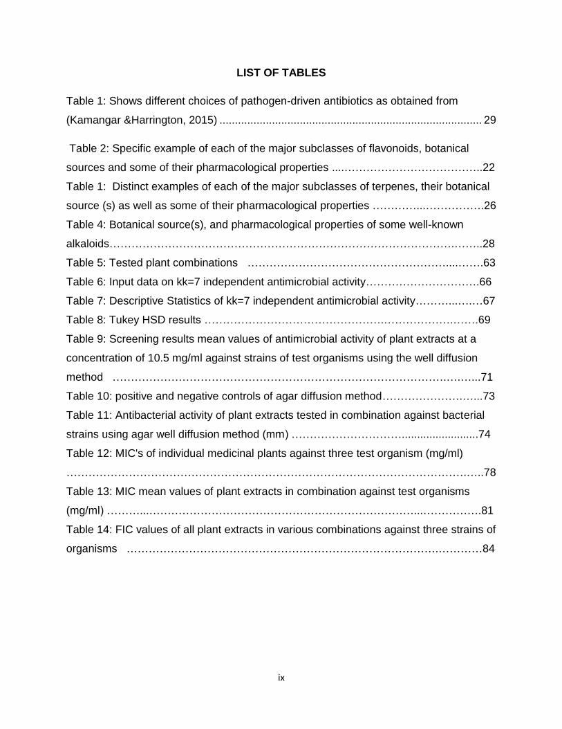
LIST OF TABLES
Table 1: Shows different choices of pathogen-driven antibiotics as obtained from
(Kamangar &Harrington, 2015) ..................................................................................... 29
Table 2: Specific example of each of the major subclasses of flavonoids, botanical
sources and some of their pharmacological properties ....………………………………..22
Table 1: Distinct examples of each of the major subclasses of terpenes, their botanical
source (s) as well as some of their pharmacological properties …………...…………….26
Table 4: Botanical source(s), and pharmacological properties of some well-known
alkaloids………………………………………………………………………………….……..28
Table 5: Tested plant combinations ………………………………………………....…….63
Table 6: Input data on kk=7 independent antimicrobial activity………………………….66
Table 7: Descriptive Statistics of kk=7 independent antimicrobial activity………...….…67
Table 8: Tukey HSD results ………………………………………….……………….…….69
Table 9: Screening results mean values of antimicrobial activity of plant extracts at a
concentration of 10.5 mg/ml against strains of test organisms using the well diffusion
method ……………………………………………………………………………….….…...71
Table 10: positive and negative controls of agar diffusion method………………….…...73
Table 11: Antibacterial activity of plant extracts tested in combination against bacterial
strains using agar well diffusion method (mm) ………………………….........................74
Table 12: MIC's of individual medicinal plants against three test organism (mg/ml)
……………………………………………………………………………………………….…..78
Table 13: MIC mean values of plant extracts in combination against test organisms
(mg/ml) ………...………………………………………………………………...…………….81
Table 14: FIC values of all plant extracts in various combinations against three strains of
organisms ………………………………………………………………………….…………84

x
LIST OF FIGURES
Figure 1: An image of Warburgia salutaris .................................................................... 46
Figure 2: An image of leaves and fruits of Terminalia sericea Taken from (Lembede,
2014) ............................................................................................................................. 48
Figure 3: A picture showing a description of Ballota africana leaves (Photo: I. Mhango)
...................................................................................................................................... 50
Figure 4: An image of Kigelia africana taken from Kumbula Indegenous nursery ......... 51
Figure 5: The fruits of Dodonaea angustofolia. Taken from (Plantbook, 2016). ............ 53
Figure 6: An image showing Eucalyptus camaldulensis. As taken from (Lucidcentral,
2016). ............................................................................................................................ 54
Figure 7: An image of Acorus gramineus. Taken from: (grasses on Pinterest) ............. 55
Figure 8 = (a) ZOIs of ethanol extracts combination against S. pyogenes plate; (b) =
ZOIs of acetone extracts combinations against S. aureus; (c) = ZOIs of acetone extracts
combinations against K. pneumoniae (d) = ZOIs of acetone extracts combinations
against S. pyogenes. ..................................................................................................... 87
Figure 9: (a) = ZOIs of aqueous extracts combination against K. pneumoniae (b) = ZOIs
of ethanol extracts combinations against S. aureus. ..................................................... 88

xi
LIST OF ABBREVIATIONS
ACIP – Advisory committee in immunization practices
AIDS - Acquired immunodeficiency syndrome
ANOVA- Analysis of Variance
ATCC - American Type Culture Collection
BAL – Broncho alveolar lavage
BOOP – Bronchiolitis obliterans organizing pneumonia
BPM – Beats per minute
C – Carbon
CFU - Colony Forming Units
CNS – Central nervous system
CT – Computed tomography
DMSO - Dimethyl Sulfoxide
DNA- Deoxyribonucleic acid
TMP – Thymidine monophosphate
FIC - Fractional Inhibitory Concentration
H1N1 – Influenzae A virus/ swine flu
Hib - Haemophilus influenzae type b
HIV - Human immunodeficiency virus
HPLC High - performance liquid chromatography
HPTLC High - performance thin-layer chromatography
INT - p-iodonitrotetrazolium-chloride/violet
KHz – Kilohertz
Mg - Milligram
MH Mueller - Hinton agar
MHB Muelle r- Hinton broth
MIC - Minimum Inhibitory Concentration
Min - Minutes
Ml - Millilitre
mm - Millimetre
mRNA - Messenger ribonucleic acid
MRSA - Methicillin Resistant Staphylococcus aureus
NMMU - Nelson Mandela Metropolitan University
PABA - Para-aminobenzoic acid
PBP - Penicillin binding protein
PCV13 – Pneumococcal conjugate vaccine
PGE2 – Prostaglandin E2
PH - Power of hydrogen
PPSV23 – Pneumococcal polysaccharide vaccine

xii
RNA - Ribonucleic acid
RSV – Respiratory syncytial virus
Spp – Species
THF - Tetrahydrofolic acid
tRNA - Transfer RNA
TSB – Trypticase soy broth
UNICEF - United Nations Children’s Fund
UV - Ultra Violet
WBC - White blood cell
WBC/μL - White blood cell per microliter
WHO - World Health Organisation
ZOI – Zone of inhibition
Β – Beta
μL - Microliter
μl- Micro-litre
μm – Micrometre
13
CHAPTER ONE
INTRODUCTION
1.1 Background
Many prescription drugs, are derived from natural products and their compounds. The
use and discovery of medicinal plants have progressed for years with a close analysis
on their special properties. Medicinal plants have more than one chemical compound,
and advanced research has been conducted on some of them, to extract individual
components and identify the unique properties they contain (Fawzi, 2013). More than
two thirds of the anticancer drugs approved between the 1940s and 2006 are either
natural products, or were produced based on the knowledge obtained from natural
products (Newman & Cragg, 2006). Other familiar examples of plant derived medicines
include quinine, morphine, codeine, colchicine, atropine, reserpine, digoxin, taxol and
vincristine (B E Van Wyk & Wink, 2004). Compounds like alkaloids, poly phenols,
glycosides, terpenes, terpenoids, oils and many more have specific roles in living
organisms.
Currently antimicrobial agents are globally one of the most valued therapeutic agents
used in treatment of infectious diseases. Van Vuuren (2010) in his PhD thesis explained
that the treatment of infectious diseases focuses on cure rather than easing the
symptoms or only on pharmacological management of the disease. Plants can produce
exceedingly hundred thousand molecules; not all of them have bacterial potential, but
some have significant activity against pathogens. The magnitude of natural resources
is not the same as existing antimicrobials but stills provides some extent of hope
(Khurram et al., 2009). This has led to an increase in the search of such antimicrobials
in recent years. This increase can be observed in the number of articles published on
the antimicrobial activities of medicinal plants during 1966 – 1994 which amount to 115,
while the number of articles on the same subject appearing during 1995 – 2004 period
are 307; that is more than a two fold increase in just one decade, showing the growth
of Interest in the search of antimicrobials of natural origin (Khurram et al., 2009; Ríos
& Recio, 2005).
14
Substantial evidence from recent studies highlight increased resistance to commonly
used antibiotics and informs us that our protection against these organisms is running
out at an increased rate. In order to overcome this challenge there is a need to develop
new antimicrobials, which requires continued research on indigenous plants. Relying
on natural products such as plants and natural substances can therefore be good
alternatives. If these compounds are not potent as conventional antimicrobials they can
be used as adjuncts, enhancing mechanism of their therapeutic effectiveness (Khurram
et al., 2009). Herbalist, prescribe the use of singular plant and at times a combination,
in attempts to improve potency for treatment of respiratory infection. During the 20th
century, attempts were focused on one-target, the one drug which was key to finding a
single chemical entity being able to inhibit a distinct molecular target (Keith, Borisy, &
Stockwell, 2005). This led to the isolation of some successful single molecular
compounds from various plant species for example, Artemisinin from Artemisia annua
L. and quinine from Chinchona spp. (Talika, 2012).
In order to reduce the emergence of antibiotic resistance, strict infection control
measurements as well as logical and appropriate use of antibiotics have been
established (Tseng, Ke, & Chang, 2014). Many studies have been conducted that
emphasize the use of drug combinations to reduce resistance, toxicity and side effects,
and at the same time maximize therapeutic advantage with high efficacy and
bioavailability. Moreover, many synthetic drugs available in pharmacies, and
administered to patients in hospitals possess a variety of chemical compounds that
have useful biological functions. The use of combination therapy is based on the
knowledge that many diseases have multi-casual aetiology and a complex
pathophysiology. The complexity of treating multi- drug resistance infections has led to
an enormous search for novel and effective antibiotics, especially
structures/compounds originating from natural products (Wagner & Ulrich-merzenich,
2009). Compound and molecule identification through phytochemical analysis and
microbial activities has played an important role as lead components for the
development of new antimicrobials (Drewes, 2012; Frum & Viljoen, 2006). Studies
have shown that the plant extracts in combination of two or more are exhibiting effective

15
antimicrobial activity against a wide range of microorganisms including drug resistant
bacteria (Prakash et al., 2006).
According to the World Health Organisation (WHO) (2003), 80% of the total population
in some Asian and African countries presently use herbal medicine for some aspects
of primary health care. New strategies are being implemented in botany, information
systems, regulations, chemistry, and clinical trials which are present to ensure that
traditional medicine is improved globally. Pneumonia is one of the leading causes of
death in developing countries especially in children and older people with debilitating
conditions. Bacterial infections contribute disproportionately to pneumonia mortality in
developing countries. Although bacterial infections account for not more than 50% of
cases of pneumonia, they cause nearly 70% of deaths that are caused by pneumonia
(WHO & UNICEF, 2009). Resistance of microorganisms to antibiotics that are used to
treat pneumonia has been reported to make it difficult to treat pneumonia.
1.2 Pneumonia Prevalence
Globally, lung infections and gastrointestinal infections are responsible for most death
in developing countries. Respiratory diseases are regarded as a common cause of
death in developed nations, accounting for about 14% of deaths in both sex (Cross &
Underwood, 2013). Pneumonia is an opportunistic disease for HIV/AIDS patients and
individuals with other compromised health conditions. Pneumonia is regarded as a
common cause of death overall, and the most common fatal hospital acquired infection.
“Pneumonia remains the leading infectious cause of death among children under five,
killing 2,500 children a day” (Campbell et al., 2013).
In 2008, pneumonia manifested itself in approximately 156 million children, where 151
were from developing countries and 5 million from developed countries. In 2015,
pneumonia accounted for 16 % of deaths of children under five years, killing 920 136
children (WHO, 2016). In developing countries it still remains the leading cause of
death with rates being highest in children under five, adults above 75 years and the
chronically ill (Kabra, Lodha, & Pandey, 2010). In 2010, pneumonia resulted in 1.3
million deaths worldwide, of which 18% of all deaths were of children under five years,

16
and 95% of this occurred in developing countries (L. Liu et al., 2012). It is estimated
that pneumonia affects 450 million people per year globally, 7% of population, and
results in about 4 million deaths (Ruuskanen, Lahti, Jennings, & Murdoch, 2011).
Children in developing countries are nearly 18 times more likely to die before the age
of five than children in high-income countries due to pneumonia and other acute
infections. Pneumonia is the single largest contribution to child mortality, accounting
close to 28-34% of all under five globally and responsible for death of more than 2
million children under five annually (Tong, 2013).
Pneumonia affects children and families all over, but is widespread in South Asia and
sub-Saharan Africa (WHO, 2016). The larger part of death of children mortality caused
by pneumonia affect the poor due to high exposure rates to risk factors associated
with developing acute respiratory infections such as overcrowding, poor
environmental conditions, malnutrition as well as limited access to curative health
sciences. Preventive measures to reduce pneumonia are available, which aim at
reducing indoor air pollution, promoting adequate nutrition and increasing
immunisation rates with vaccines that prevent children and adults >65yrs from
developing infections that cause pneumonia (Ronald, 2005; WHO & UNICEF, 2009).
1.3 Aim and specific objectives of the study
The main aim of the study is to evaluate antimicrobial activity of the following medicinal
plants Kigelia Africana, Ballota Africana, Dudonea Angustofolia, Warbugia Salutaris,
Termialia Sericea, Acorus Calamus, and Eucalyptus camldulensis by testing the
activity of individual plant extract preparations against microbial isolates that cause
pneumonia.
1.3.1 The specific objectives of this study:
1. To collect and to identify the medicinal plants for the study.
2. To prepare and extract the plants materials separately and in some combined
preparations using a variety of solvent, like distilled water, ethanol, dimethyl
ether and acetone.
3. To test the antimicrobial sensitivity of the plant extracts and combined
preparations by agar well diffusion.
17
4. To perform interactive plant combination studies to determine the efficacy
when plants are used together.
5. To determine the minimum inhibition concentration (MIC) of both combined
and individual plant extracts against bacteria isolates.
6. To determine the fractional inhibitory concentration index (∑FIC) of the plant
extracts in the combined preparations.
1.4 Significance of the research
Medicinal plants can be used as alternatives for antibiotics and chemotherapeutic
agents in certain circumstances. Many infections are difficult to treat because of
resistance that emerge in the organisms against the antibiotics. One way of
overcoming this problem is discovering new compounds to use that are not based on
existing synthetic antimicrobial agents and those that are viable and exhibiting no
resistance. Combinations studies of plant extracts have been included in this study to
help in discovering new alternatives that can be used to overcome drug resistance
development to known antibiotics. For example vaccine for Streptococcus
pneumoniae and influenza infections have already been discovered and are
administered in hospitals (Ashby et al., 2012). Considering that disease-causing
agents may develop resistance if a single plant is used for the treatment of specific
disease, the use of plant in combinations is preferred, since there is minimal chance
of developing resistance and can be used for long time.
Synergistic or polyvalent effect from interactions of natural compounds will be useful
in discovering new chemical constituents that can be used for developing novel
antimicrobials hence overcoming the surge of resistance. The use of medicinal plants
in combinations has an advantage of having their polyphenols that act as antioxidant
and free radical scavengers that remove harmful metabolite and toxins from the body
hence boosting the immunity. Although people from rural areas, and some traditional
healers, believe that medicinal plants are more effective in treating infectious
conditions than synthetic antibiotics, it is still very important to evaluate with scientific
experiments. Another significance of medicinal plant is that it can cure more than one
ailment, as seen in literatures, where one plant has numerous medicinal benefits,

18
hence advantageous over synthetic drugs. Fresh and dry parts of studied plants can
be used for preparation of herbal drugs, herbal processed products and traditional
herbal drugs. Availability and cost effectiveness of medicinal plants can benefit less
privileged people and those living in remote areas far from health facilities
(Mohlakoana, 2010; Suliman, 2011). This study will help scientists to validate the
usage of these plants.
19
CHAPTER TWO
LITERATURE REVIEW
2.1 Pneumonia
Globally, pneumonia is a number one killer disease affecting children under the age of
five years. Out of an estimated 9 million child deaths as resorted in 2007, about 20%
or 1.8 million deaths were due to pneumonia. WHO (2015) and UNICEF (2009)
reported that child mortality due to pneumonia is as a result of malnutrition, poverty and
inadequate access to the health care. The burden that pneumonia impose on families
and the health system in developing countries aggravate unfairness completely
exposing children who are poor, hungry and living in remote areas.
Pneumonia infection has different pathogenic causes which results in unpredictable
and varying antibiotic treatment selections. These pathogens have developed
resistance to antibiotics such as the B-lactams, macrolides, vancomycin and
fluroquilones. Global prevalence of drug resistance streptococcal pneumoniae has
increased. This has resulted in the establishment of risk factors in individuals who are
on B–lactams treatment within the previous three months. The highly affected
individuals include; the alcoholics, those with simultaneous presence of two chronic or
immunosuppressive diseases. Resistance displayed by methicillin resistance
Staphylococcus aureus is also a major concern leading to massive search of medicinal
plant for possible antimicrobial effect (Kamanga, 2013).
Pneumonia is a lung infection that is caused by either bacteria, viruses or fungi.
Indeed pneumonia infection is mainly characterized by inflammation of the lungs,
primarily the alveoli (Cross & Underwood, 2013; Reid, Roberts, & MacDuff, 2011). It
arises when from weak immune system, pre-existing illness and when the body fails
to filter out microorganisms from the air that we breathe in. Pneumonia refers to
pneumonitis (lung inflammation) that is usually due to either infection or non-
infectious, that has additional feature of pulmonary consolidation (Ashby et al., 2012).
There are a number of ways in which pneumonia has been discussed; Stegman and
Branger (2005) describe pneumonia as inflammation of the lung parenchyma
characterized by consolidation of the affected part with the alveolar air spaces being

20
filled with exudates and inflammatory cells fibrin. Underwood (2013) stipulated that
pneumonia is usually due to an infection affecting distal airways and alveoli with the
formation of an inflammatory exudate.
Depending on how and where it was acquired, pneumonia can be classified as either
community acquired-aspirations, headache associated, hospital acquired or
ventilator-associated pneumonia. Pneumonia can also be classified based on the
affected area of the lung namely; lobar pneumonia, bronchial pneumonia, and acute
interstitial pneumonia, clinical circumstances and causative agents (McLukcie, 2009).
From Underwood (2013), pneumonia can be classified into four major pathological
classes.
2.1.1 Classification of Pneumonia
2.1.1.1 Bronchopneumonia
Bronchopneumonia is commonly manifested at old age, infancy and on patients with
debilitating diseases such as cancer, chronic renal failure, cardiac failure, or
cerebrovascular accidents (Reid et al., 2011). It also occurs as a manifestation of
secondary infection in viral conditions like influenza and measles. Moreover,
bronchopneumonia can co-exist with other respiratory tract infections in patients with
acute bronchitis, chronic obstructive airways diseases or cystic fibrosis (Underwood,
2013). In other occasions when there is failure in cleaning the respiratory secretions,
as in post- operative period, it creates a liability to the development of
bronchopneumonia. Bacteria known to cause bronchopneumonia include
Staphylococcus aureus, Streptococci Pyogenes and Haemophilus influenza
(Cheesebrough, 2006).
Bronchopneumonia has a distinguishing patchy distribution, centred on inflamed
bronchioles and bronchi with succeeding spread to surrounding alveoli (Cross &
Underwood, 2013; Kamanga, 2013). Areas of the affected lung tend to be bottom
lateral and bilateral, and appear focally grey or red at post-mortem with filled bronchi
and histological diagnosis. The affected lung also shows atypical acute inflammation
with exudation in the bronchi and adjacent alveolar spaces. There is a chance of an
21
inflammation resolution when antibiotic and physiotherapy is administered (Cross &
Underwood, 2013; Reid et al., 2011).
2.1.1.2 Lobar pneumonia
Lobar pneumonia is relatively unfamiliar in infancy and old ages, but normally
pneumococcal pneumonia affects healthy adults in between 20-50 years of age.
However, lobar pneumonia caused by Klebsiella quintessentially affects the elderly,
diabetics or alcoholics (Cross & Underwood, 2009, 2013). 90 % of lobar pneumonia
is due to Streptococcus Pneumoniae (pneumococci). Pneumococci may spread from
the lungs into the pleural cavity or pericardium and cause abscesses (Kamanga,
2013). The infection of the pleural space (known as pleurisy) is very painful with
striking changes that occur in the alveoli where pleural exudate is common (O’Grady,
Torzillo, Frawley, & Chang, 2014). Lobar pneumonia is characterized by diffuse
inflammation that affects an entire lobe or even two lobes of the affected lung (Cross
& Underwood, 2013).
2.1.2 Clinical presentation
The patient shows symptoms of high fever that can be over 40 0C, cough and
production of sputum. The sputum appears purulent and sometimes with flecks of
blood referred as rusty sputum (Cross & Underwood, 2013). The chest signs range
from dullness to percussions with bronchial breathing, as the lung becomes
consolidated.
2.1.3 Pathogenesis
The pathology of lobar pneumonia undergoes sequential combined phases in which
the alveoli in the lobe are evenly affected (Reid et al., 2011). The pathological phases
of lobar pneumonia can be summarized as follows:
Phase 1: Congestion: This can last for 24 hours and represents the outpouring of
protein rich exudate into alveolar spaces, with venous congestion. Gross appearance
of the lung is heavy, oedematous and red in colour.
Phase 2: Red hepatisation: This usually lasts for 2 to 5 days. At this phase, there is
massive accumulation of polymorphs, lymphocytes and macrophages in the alveolar

22
spaces. The red cells are also extraverted from the distended capillaries. The
overlying pleura bear a fibrinous exudate where the lung is red, solid and airless with
consistency resembling liver.
Phase 3: Grey hepatisation: This phase last a few days (4 to 8 days) and it
constitutes further accumulation of fibrin with destruction of white and red blood cells.
The lung is now grey brown and solid.
Phase 4: Resolution: This takes place at about 8 to 10 days in untreated cases and
represents the resorption of exudate and enzymatic digestion on inflammatory debris,
with preservation of underlying alveolus wall architecture (Cross & Underwood, 2013;
Reid et al., 2011).
2.1.4 Atypical pneumonia
Other organisms rather than well-known organisms cause atypical pneumonia. It is
sub-classified according to the host, thus either immunocompromised, suppressed or
non-immunosuppressed hosts (Denis & Et-al, 2014; Reid et al., 2011).
In non-immunosuppressed host, pneumonia may be due to viruses like influenza,
RSV, adenovirus and mycoplasma (Augenbraun, 2014; Cross & Underwood, 2013).
The clinical cause of viral pneumonia depends on the severity and extent of the
diseases. In fatal cases, the lungs appear heavy, red and consolidated in adult
respiratory syndrome. Mycoplasma pneumonia is caused by Mycoplasma
pneumoniae and often affects individuals less than 40 years of age. It usually causes
low grade pneumonia with interstitial inflammation and less exudation (Cross &
Underwood, 2013; Kamangar & Harrington, 2015).
In non-immunosuppressed host, pneumonia can be caused by bacillus, Legionella
pneumophilia that result in legionnaires diseases. It mostly appears in middle-aged
and older adults, smokers and those with chronic illness and weak immune system.
The condition worsens within the first 4 to 6 days and reach resolution next 4 to 5
days, but it takes time for symptoms to completely go away (Denis & Et-al, 2014).
Patient infected by legionnaires diseases, may be previously well, although a
proportion have underlying chronic illness like heart failure or carcinoma. Symptoms
23
include cough, dyspnoea and chest pains with systematic features like headache,
confusion, nausea, vomiting and diarrhoea (Cross & Underwood, 2013).
Immunosuppressed host may be correlative, as it occurs in patients at the extreme
age, diabetics, malnourished, those on high dose steroid therapy, undergoing
chemotherapy for malignancy, immunosuppression for transplantation and those with
HIV/AIDS infection (Cross & Underwood, 2013; Denis & Et-al, 2014; Kamangar &
Harrington, 2015). Patients with immunosuppression are prone to opportunistic non-
pathogenic infections and they present clinical features such as onset fever, shortness
of breath, cough and pulmonary infiltrates (Augenbraun, 2014). Common opportunistic
agents include pneumocystis jirovecii, fungi (candida and aspergillus); viruses
(cytomegaloviruses, herpes simplex and varicella zoster) and HIV lung disease (Cross
& Underwood, 2013; Reid et al., 2011).
2.1.5 Non- infective pneumonias
Non-infective pneumonias are described as occurrence of non-contagious
pneumonia. Below is brief information on some causes and clinical features of
non-infective pneumonias:
a) Cryptogenic organizing pneumonia is the idiopathic form of organizing
pneumonia (formerly called bronchiolitis obliterans organizing pneumonia
(BOOP)). This is a type of diffuse interstitial lung disease that affects the distal
bronchioles, respiratory bronchioles, alveolar ducts, and alveolar walls (Cross
& Underwood, 2013; Kamangar & Harrington, 2015). The primary area of injury
is within the alveolar wall. This clinical syndrome is characterized by mild
systemic upset with possible cough, low-grade fever and breathlessness.
Radiology diagnostic shows evidence of focal lung consolidation (Cross &
Underwood, 2009, 2013).
b) Aspiration pneumonia also known as anaerobic pneumonia is an inflammation
of the lung and bronchial tubes that occur when food, vomitus or fluid is
aspirated into the lung, resulting in secondary inflammation and consolidation
(Dock, Boskey, & Brian Wu, 2015).

24
c) Lipid pneumonia is a lung inflammation that develops when vacuoles of lipid
are ingested by foreign-body giant cells that might result in interstitial fibrosis.
Lipid pneumonia may be endogenous, associated with airway obstruction
causing distal collections of a foamy macrophages and giant cells, alternatively
lipid pneumonia may be exogenous as pertaining to aspiration of materials
containing lipid content like paraffin or oily node drops (Cross & Underwood,
2013).
d) Eosinophilic pneumonia which is also referred as Loffler’s syndrome is
characterised by numerous eosinophils in the interstitium and alveoli (Cross &
Underwood, 2009). Acute eosinophilic pneumonia is usually idiopathic and is
associated with blood eosinophilia. The lung shows extensive infiltration with
eosinophils and presence of organizing exudates which may go on to give rise
to finding fibrosis (Cross & Underwood, 2013; Reid et al., 2011).
2.1.6 Pneumonia diagnosis
Different tests and procedures are performed to come up with the correct diagnosis
so that proper management and treatment is provided. The following methods and
tests may not be useful for diagnostic purposes but are useful for classifying illness
severity and site-of-care or admission decisions: laboratory tests, trans-tracheal
aspiration, chest radiotherapy, chest CT scanning, chest ultrasonography,
bronchoscopy with BAL thoracentesis and other specific test (Kamangar & Harrington,
2015)
2.1.6.1 Laboratory analysis
In cases where culturing blood is considered, blood samples should be obtained
before the administration of antibiotics. These cultures require a minimum of 24 hours
to incubate and grow. When blood cultures are positive, they correlate well with the
microbiologic agent causing the pneumonia. Unfortunately, blood cultures show poor
sensitivity in pneumonia where findings are positive in approximately 40% of reported
cases (Kamangar & Harrington, 2015). Even in pneumococcal pneumonia, the results
are often negative. Their yield may be higher in patients with severe pneumonia
infection. These findings probably have minimal clinical effect in treating bacterial
pneumonia since the use of blood cultures only rarely dictates a change in empiric
25
antibiotics. Cell blood count analysis may show leukopenia <4000 white blood cell
count per microliter or leucocytosis >12000 wbc/ul, which may be an inauspicious
clinical sign of impeding sepsis (Kamanga, 2013). The white blood cell (WBC) count
should be more than 25 per low-power field in non-immunosuppressed patients, and
there is presence of neutrophils in patient’s sputum smear (Reid et al., 2011). Other
laboratory test includes biochemistries such as C-reactive protein, urea, electrolytes,
liver function test and pulse oximetry (Kamanga, 2013; Kamangar & Harrington,
2015). Capsular serotyping is used by doing Quelling reaction in detecting
streptococcus pneumoniae (Cheesbrough, 2006).
2.1.7 General Signs and Symptoms of Pneumonia
The following are the signs and symptoms exhibited by patients suffering from
pneumonia:
Cough up mucus from the lungs, rusty or tinged with blood
Fever typically >38.8 0C
Difficulty in breathing
Shaking and teeth chattering chills
Severe chest pains that worsens when coughing or breathe in
Fast heart beat
Cough which result in tachypnea (>18 respiration/min) and tachycardia >100
bpm or bradycardia <60 bpm.
Tiredness and weakness
Central cyanosis
Nausea, vomiting and diarrhoea
Children may experience headache, loss of appetite and may develop
wheezing.
Young infants may suffer convulsions, unconsciousness, hypothermia < 35°C,
lethargy and feeding problems.
In adult 70 year or older they have altered mental status with no recognized
cause (Cross & Underwood, 2013; Kamangar & Harrington, 2015; Reid et al.,
2011; WHO, 2010).

26
Symptoms caused by bacteria and viruses are often the same, but viral symptoms are
gradual and not severe.
In general, the organisms causing pneumonia include:
Bacteria: Streptococcus pneumoniae, Klebsiella pneumoniae, Haemophilus
influenzae, Staphylococcus aureus, Streptococcus pyogenes, Neisseria
meningitidis, Moraxella catarrhis, Legionella pneumophilia, Mycoplasma
pneumoniae and Chlamydophilia pneumoniae.
Viruses: Respiratory syncytial virus, rhinovirus, herpes simplex and severe
acute respiratory syndrome.
Fungi: Pneumocystis jirovecii
And various chemicals (Cheesebrough, 2006; Cross & Underwood, 2013):
Physical findings in bacterial pneumonia may include the following:
Adventitious breath sounds, such as rales/crackles, rhonchi, or wheezes
Decreased intensity of breath sounds
Egophony
Whispering pectoriloquy
Dullness to percussion
Tracheal deviation
Lymphadenopathy
Pleural friction rub (Kamangar & Harrington, 2015).
Examination findings that may indicate a specific aetiology for consideration are as
follows:
Bradycardia may indicate a Legionella aetiology.
Periodontal disease may suggest an anaerobic and/or polymicrobial
infection.
Bullous myringitis may indicate Mycoplasma pneumoniae infection.
Physical evidence of risk for aspiration may include a decreased gag
reflex.
27
Cutaneous nodules, especially in the setting of central nervous system
(CNS) findings may suggestion Nocardia infection (Kamangar & et al,
2015).
2.1.8 Treatment, prevention and management of bacterial pneumonia
The following personal health practices help in preventing the spread of bacterial
pneumonia:
Health habits, good hygiene practice like frequent hand cleaning and isolation
of patients with multiple resistant respiratory tract pathogens helps prevent
pneumonia (Tong, 2013).
Hand washing in-between patient contacts, sneezing or sneezing into an elbow
or sleeve instead of hands, avoid interacting with the sick, getting proper
nutrition and having enough rest (WHO & UNICEF, 2009).
Refrain from smoking and other pollutants, at the same time reducing indoor
and outdoor indoor air pollution, and become familiarizes about warning signs
to identify infection, specifically a cough, fast breathing or difficult breathing
(Chang, Ooi, Perera, & Grimwood, 2013; WHO & UNICEF, 2009).
For breastfeeding mothers, during the first six months breastfeeding is advised
to critically prevent pneumonia, since breast milk contains a nourishing supply
of nutrients, antioxidants, hormones and antibodies a child needs for growth
and development (Marie B.Coyle, 2005).
Vaccination is one of the key method that is preferred as a preventive measure (Marie
B.Coyle, 2005). Many vaccines can prevent infection by bacteria or viruses that may
cause pneumonia. Some of these vaccines are namely; pneumococcal conjugate,
Haemophilus influenza type-b (Hib), pertussis (whooping cough), varicella
(chickenpox), measles, seasonal and 2009 H1N1 influenza (flu) vaccines (Kamangar
& Harrington, 2015; WHO & UNICEF, 2009). The advisory committee in immunization
practices (ACIP) in 2015 gave a recommendation on the pneumococcal
polysaccharide vaccine (PPSV23) and the pneumococcal conjugate vaccine (PCV13).
Immunocompetent adult aged 65yrs and order, who have not previously received
28
pneumococcal vaccine: A dose of PPSV23 should be given 1yr or more following a
dose of PCV13, these two vaccines should not be co-administered (Kamangar &
Harrington, 2015; WHO & UNICEF, 2009).
In treatment, the ACIP currently recommend that a dose of PCV13 be followed by a
dose of PPSV23 in persons aged 2 years or older who are at high risk for
pneumococcal diseases because of underlying medical conditions (Kamangar &
Harrington, 2015). Children with immunocompromising conditions, functional or
anatomical asplenia should receive a second dose of PPSV23 5years after the first
PPSV23 dose (Kamangar & Harrington, 2015). The ACIP on October 12, 2012,
published the updated recommendations for pneumococcal vaccination on high risk
adults; which propose use of Prevnar 13 in addition to the previously Pneumovax 23
for adults aged 19 years and older with immunocompromising conditions (Chang et
al., 2013; Kamangar & Harrington, 2015; WHO & UNICEF, 2009). The purposes of
bacterial pneumonia pharmacotherapy are to eliminate the infection, reduce morbidity,
and prevent complications. Treatment of pneumonia depends largely on the practical
use of antibiotic regimes directed against potential pathogens as determined by the
setting in which the infection took place and the potential for exposure to
multidrug-resistant organisms (Kamangar & Harrington, 2015). Table 1 presents first
and second-line antibiotic choices for specific organisms that cause bacterial
pneumonia.

29
Table 2: Shows different choices of pathogen-driven antibiotics as obtained from
(Kamangar &Harrington, 2015)
Streptococcus pneumoniae
Penicillin
susceptible Penicillin G, amoxicillin
Macrolide, cephalosporin
(oral or parenteral),
clindamycin, doxycycline,
respiratory fluoroquinolone
(MIC < 2 mcg/mL)
Penicillin resistant Agents chosen on the basis
of sensitivity
Vancomycin, linezolid, high-
dose amoxicillin (3 g/d with
MIC ≤4 mcg/mL
(MIC ≥2 mcg/mL)
Staphylococcus aureus
Methicillin
susceptible Anti-staphylococcal penicillin Cefazolin, clindamycin
Methicillin resistant Vancomycin, linezolid Trimethoprim-
sulfamethoxazole
Haemophilus influenza
Non–beta-
lactamase
producing
Amoxicillin
Fluoroquinolone,
doxycycline, azithromycin,
clarithromycin
Beta-lactamase
producing
Second- or third-generation
cephalosporin,
amoxicillin/clavulanate
Fluoroquinolone,
doxycycline, azithromycin,
clarithromycin
Mycoplasma
pneumoniae Macrolide, tetracycline Fluoroquinolone
Chlamydophila
pneumoniae Macrolide, tetracycline Fluoroquinolone

30
Legionella species Fluoroquinolone,
azithromycin Doxycycline
Chlamydophila
psittaci Tetracycline Macrolide
Coxiella burnetii Tetracycline Macrolide
Francisella tularensis Doxycycline Gentamicin, streptomycin
Yersinia pestis Streptomycin, gentamicin Doxycycline, fluoroquinolone
Bacillus
anthracis(inhalational)
Ciprofloxacin, levofloxacin,
doxycycline
Other fluoroquinolones,
beta-lactam (if susceptible),
rifampin, clindamycin,
chloramphenicol
Enterobacteriaceae Third-generation
cephalosporin, carbapenem
Beta-lactam/beta-lactamase
inhibitor, fluoroquinolone
Pseudomonas
aeruginosa
Antipseudomonal beta-
lactam plus ciprofloxacin,
levofloxacin, or
aminoglycoside
Aminoglycoside plus
ciprofloxacin or levofloxacin
Bordetella pertussis Macrolide Trimethoprim-
sulfamethoxazole
Anaerobe (aspiration) Beta-lactam/beta-lactamase
inhibitor, clindamycin Carbapenem
MIC = Minimal inhibitory concentration.
2.2 General Review of Traditional Medicine
Approximately 80 % of the world’s population rely on traditional medicine. Traditional
medicine is regarded as traditional herbal medicine when the material being used for
treatment originates from plant material. According to the WHO, traditional medicine

31
relates to health practices, approaches, knowledge, and beliefs incorporating plant,
animal, and mineral-based medicines, spiritual therapies, manual techniques, and
exercises, applied singularly or in combination to treat, distinguish, control illness and
maintain wellness (WHO, 2003). As seen from literature, studies on herbal medicines
they are described as plant medicine, phytomedicine, pharmacognosy and natural
products. “Natural products” usually refer to by-product processed or derived from living
organisms, including plants, animals, insects, microorganisms and marine organisms.
This explains why traditional herbal medicine is only a small part of a more cohesive
and holistic health care (W. J. H. Liu, 2011; WHO, 2003).
2.2.1 Traditional Herbal Medicine
Plant-based system progresses to play an important role in health care and their use
by different cultures has been extensively documented. A survey of pure plant-derived
compounds used as drugs in countries having WHO-traditional medicine centres show
that of the 122 compounds identified, 80 % were used for related ethnomedical
purposes and derived from 94 species. For example, “khellin from Ammi visnaga (L)
Lamk led to the development of chromolyn (in the form of sodium chromoglycate) as a
bronchodilator and galegine from Galega officinalis L. which was the model for the
synthesis of metformin and other bisguanidine-type antidiabetic drugs. Also,
papaverine from Papaver somniferum which formed the basis for verapamil is used for
the treatment of hypertension. The latter plant is better known for being the source of
painkillers such as morphine and codeine. The best example of ethnomedicine's role
in guiding drug discovery and development is that of the antimalarial drugs, particularly
quinine and artemisinin” (Cragg & Newman, 2013).
African traditional medicine is the oldest medicine regime culturally referred as the
cradle of mankind. Although ethnobotanical literature is well documented, little is
known of its scientific information like efficacy, phytochemistry, on indigenous
medicinally used plants. From (1997-2008) a number of discovery has surface of the
chemistry and biological activity of plants used in traditional healing. South Africa owns
a distinctive and varied botanical heritage with over 30, 000 plant species of which
approximately 3000 species are therapeutically used. Despite South African unique
botanical heritage, it also has a cultural diversity with traditional healing being integral

32
to each ethnic group. It is known to be rich in flora endemic and diversity. Although a
number of publications has focused on the isolation and identification of bioactive
compounds it’s important to consider complexity of plants. regarding that single
compound may not be responsible for the observed activity, but preferably a
combination of compounds either major or minor working together in an additive or
synergistic manner (SF van Vuuren, 2008, 2010).
2.2.2 Medicinal plants
Currently, medicinal plants are widely used for primary healthcare in the developing
countries, and is regarded as a possible source of important bioactive compounds. The
conduction of ethnobacterial studies are required to unveil locally vital medicinal plant
species and document popular knowledge, which is under threat of being lost. Some
of the ethnobotanical studies, it resulted in discovery of digoxin which was extracted
from Digitalis Pupurea, a plant used by European populations for its positive
cardiovascular effects and recogutum of anticancer etoposide and teniposide extracted
from Pidophyllum peltatum (Benarba et al., 2015).
Medicinal plants carry substances that can play a role in treatment purposes or which
can be used as precursors for the synthesis of important drugs. Studies on plant use
as the source of drugs and dietary supplements have accelerated in recent years. Out
of this studies plants have been found to contain in-vitro antimicrobial property because
of the presence of wide variety of secondary metabolites. With reference to the past
events, pharmacological screening of compounds of natural or synthetic origin has
been the source of countless therapeutic agent. Erratic evaluation instrument in
discovering new biological active molecules, has been most productive in the area of
antibiotics (Dhanalakshmi, Dhivya, & Manimegalai, 2013).
Globally the scientists are inspecting the possibilities of deploying or uncovering
pharmacologically active compounds from medicinal plants. For instance, screening of
medicinal plants for their phytochemicals, antioxidant, anticancer and antimicrobial
activities is the paramount review for finding out successful phytochemically active
theory. Many investigations of this nature were concerned with the study of aqueous or
solvent extracts of plant fraction and testing them individually for particular
33
pharmacological activities like, antibacterial, hepatoprotective, hypoglycaemic and
hypolipidemic activities. Recent studies has shown that the plant extracts in
combinations of two or more are revealing effective antimicrobial activity against many
organisms including drug resistance bacteria’s (Natchimuthu Karmegam, Jayakumar,
& Karuppusamy, 2012).
2.2.3 Medicinal plants secondary metabolites
Medicinal herbs continue to contribute significantly to modern prescription drugs by
providing lead compounds upon which the synthesis of novel drugs can be developed.
“The American Society of Pharmacognosy defines pharmacognosy as ‘‘the study of the
physical, chemical, biochemical and biological properties of drugs, drug substances, or
potential drugs or drug substances of natural origin as well as the search for new drugs
from natural sources’’ (Balunas & Kinghorn, 2005). Approximately 60 % of the
anticancer drugs and 75% of the anti-infectious disease drugs approved from 1981-
2002, could be traced to natural origins. Moreover, 61% of all new chemical existence
introduced worldwide as drugs during the same period were inspired by natural
products. The use and search for drugs, and dietary supplements derived from plants
have accelerated up to date. Pharmacologists, microbiologists, biochemist, botanists,
and natural-products chemists globally are currently investigating medicinal herbs for
phytochemicals and lead compounds that could be developed for treatment of different
diseases (Bashar & Omar, 2011). Plants synthesize a wide range of organic
compounds that are traditionally categorized as primary, secondary metabolites,
despite the fact that the exact boundaries between the two groups can in some case to
a certain extent not be clear. Primary metabolites are compounds that have essential
roles to do with photosynthesis, respiration, growth and development like phytosterols,
acyllipids, nucleotides, amino acids and organic acids. Secondary metabolites are other
phytochemicals, many of which accumulate in surprisingly high concentration in some
species, these are structurally diverse and many are distributed among a very limited
number of species (Crozier, Clifford, & Ashihara, 2006).
Despite secondary metabolites being disregarded, the function in plants is now
attracting attention as some appear to have a key role protecting plants from herbivores
and microbial infection. They may act as attractants for pollinations and seed-

34
dispersing animals, as ellolopathic agents, UV protectants and signal molecules in the
formation of nitrogen-fixing root nodules in legumes. Secondary metabolites are also
of interest, with the fact that they are used as dyes, fibres, waxes, glues, oils, flavouring
agents, drugs and perfumes. They are viewed as possible sources of novel drugs,
antibiotics and herbicides. Based on their biosynthesis origin they are divided into three
major groups i) flavonoids and allied phenolic and polyphenolic compounds, ii)
terpenoids and iii) nitrogen- containing alkaloids and Sulphur containing compounds
(Crozier et al., 2006). Below are some of the secondary herbal metabolites which are
going to be discussed briefly as follows:
2.2.3.1 Flavonoids
Flavonoids are polyphenolic compounds comprising of 15 carbons, with two aromatic
rings by a three carbon bridge. They are the most prevalent out of the many phenolic,
and are present through-out the plant domain. Principle subclasses of flavonoid are
flavones, flavan-3-ols, isoflavones, flavoners and anthocynanidins. The flavonoids
groups in relation to minor components of the diet are dihydrofravonols, flavan-3, 4
diols, coumarins, chacones, dihydrochalconers and aurones. Solubility of flavonoids
depend on their existing forms (Crozier et al., 2006).
Table 2: Specific example of each of the major subclasses of flavonoids, botanical
sources and some of their pharmacological properties
Medical
Properties
Botanical Sources Examples Flavonoids
Anti- inflammatory
analgesic
Fruits of various
citrus trees
Naringenin,
hesperetin
Flavanones
Anti-tumour activity Generally, in
herbaceous families,
Apigenin, luteolin Flavones
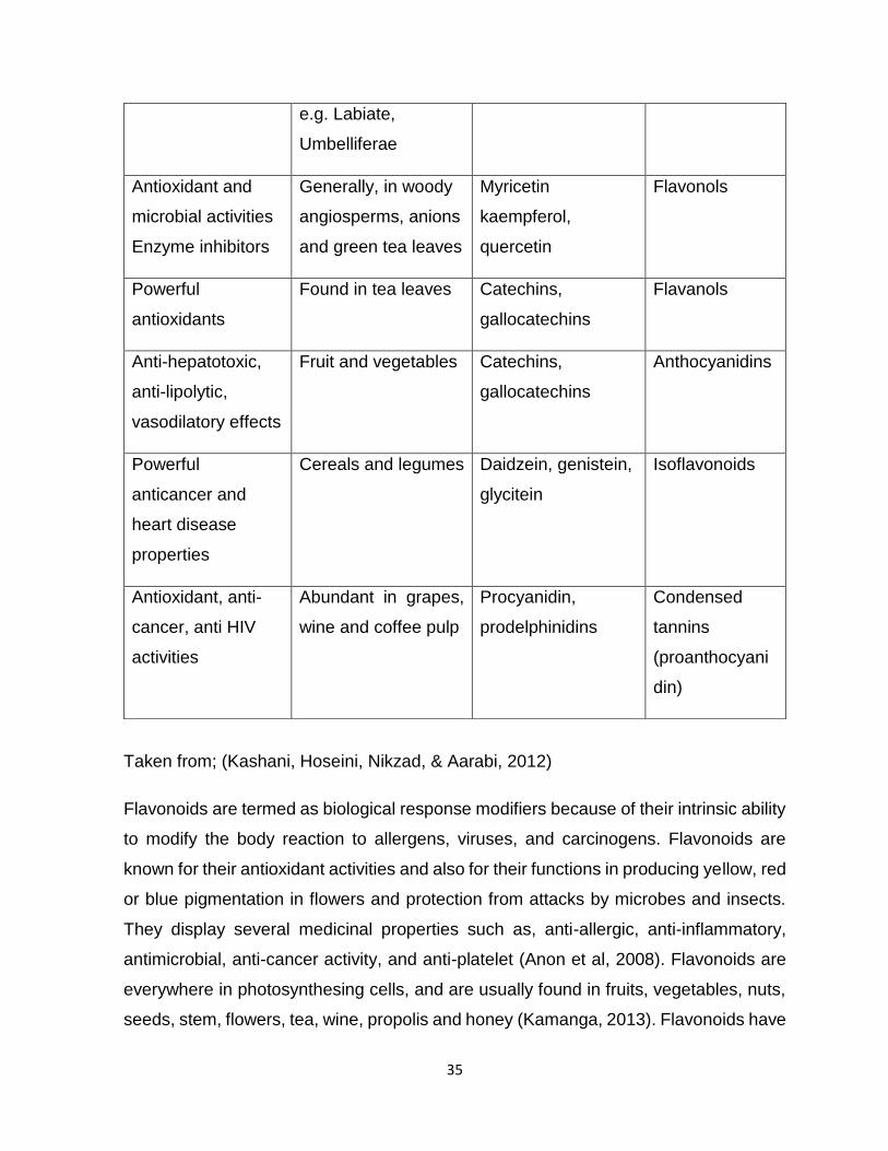
35
Taken from; (Kashani, Hoseini, Nikzad, & Aarabi, 2012)
Flavonoids are termed as biological response modifiers because of their intrinsic ability
to modify the body reaction to allergens, viruses, and carcinogens. Flavonoids are
known for their antioxidant activities and also for their functions in producing yellow, red
or blue pigmentation in flowers and protection from attacks by microbes and insects.
They display several medicinal properties such as, anti-allergic, anti-inflammatory,
antimicrobial, anti-cancer activity, and anti-platelet (Anon et al, 2008). Flavonoids are
everywhere in photosynthesing cells, and are usually found in fruits, vegetables, nuts,
seeds, stem, flowers, tea, wine, propolis and honey (Kamanga, 2013). Flavonoids have
e.g. Labiate,
Umbelliferae
Antioxidant and
microbial activities
Enzyme inhibitors
Generally, in woody
angiosperms, anions
and green tea leaves
Myricetin
kaempferol,
quercetin
Flavonols
Powerful
antioxidants
Found in tea leaves Catechins,
gallocatechins
Flavanols
Anti-hepatotoxic,
anti-lipolytic,
vasodilatory effects
Fruit and vegetables Catechins,
gallocatechins
Anthocyanidins
Powerful
anticancer and
heart disease
properties
Cereals and legumes Daidzein, genistein,
glycitein
Isoflavonoids
Antioxidant, anti-
cancer, anti HIV
activities
Abundant in grapes,
wine and coffee pulp
Procyanidin,
prodelphinidins
Condensed
tannins
(proanthocyani
din)

36
the capacity to strongly inhibit the topoisomerase enzyme that are responsible for DNA
cleavage in replication, which result to mutations of DNA that often leads to natural
acute leukaemia. Their antimicrobial activity of flavonoids is based on their ability to
bind in a covalent complex with bacterial cell wall and interrupt the cellular activity,
hence making them target for antimicrobial effect (Nkosi, 2013). Their antiviral function
has been revealed with HIV as well as herpes simplex virus (W. J. H. Liu, 2011).
2.2.3.2 Coumarins
Coumarins are a group of 1-benzopyron derivatives that are normally present in higher
plants. They serve as growth inhibitors (anti-auxins) and defence compounds in plants.
Coumarins are present in nearly every plant family, but found in large volume in
Legumioseae (bean family), Rutecae (citrus family) and umbelliferae families (also
known Apiaceae, parsley-fenenl family) (W. J. H. Liu, 2011). Coumarins were found to
have many-biological activities including anti-HIV, antitumor, antihypertension, anti-
arrhythmia, anti-inflammation, anti-osteoporosis, pain relief, and prevention of asthma
and antisepsis. Coumarin derivatives are extensively used as anticoagulants for the
treatment of excessive or undesirable blood clotting because of their competitive
bonding to vitamin K reductase and vitamin K epoxide reductase, which are vital to
blood clotting. 7-hydroxyl coumarins are used to absorb ultraviolet (UV) rays in
sunscreen cosmetics and for synthesis of anticancer drugs (Kamanga, 2013; W. J. H.
Liu, 2011).
2.2.3.3 Lignans
Lignans are found in flax seeds, pumpkin seeds, rye, soya beans, broccoli, some
barriers, and in herbs like Magnolia officinalis, schizardia, and chinensis, and
Podophyllum peltunum. Lignans are widely studied for their possible anti-cancer
properties, and their effect on cancer preventions was approved by a number of initial
studies in humans and animals. They are one of the major classes of oestrogens like
chemicals called photoestrogeners. They are efficient on binding to oestrogen
receptors and interfering with the cancer promoting effects of oestrogen on breast
tissue, hence inhibiting breast growth, prostate and colon cancer. Etoposide is a
podophyllotoxin derivatives now used to treat lung cancer, testicular cancer, and acute
lymphocytic leukaemia. Lignans are also well-known as good antioxidant, potent
37
antioxidant scavenge free radicals that can damage tissue and are thought to play a
role in the treatment of many diseases like inflammation (W. J. H. Liu, 2011).
2.2.3.4 Quinones
Quinones occur as pigment in bacteria, fungi and some higher plants. Their derivatives
have been isolated from plants and animals like Juglone in unripe walnuts, Spinulosin
from Penecillium spinuhsum, arnebinone and arnebifuranone from Aenebia euchroma,
tanchinone derivatives from Salvia miltiorrhiz, and sennoside A-D from palmatum.
Quinones have various biological activities such as antimicrobacterial, antitumor,
inhibition of PGE2, biosynthesis, and anti-cardiovascular diseases (W. J. H. Liu, 2011).
Quinones are known for their ability to target cell wall polypeptides, surface exposed
adhesions and membrane bound enzymes. Coenzymes Q10 is benzoquinone
derivatives used for cardiovascular diseases, hypertension, and cancer in clinics.
Vitamin K compound like K1 and K2 belong to naphthoquinones. Capable of improving
blood coagulation, thereby used for the treatment of natal bleeding(W. J. H. Liu, 2011;
Nkosi, 2013).
2.2.3.5 Terpenoids
Terpenoids, also known as isoprenoids constitute the largest group of herbal secondary
metabolites, they play a role in defence, wound scaling and thermotolerance of plants
as well as in the pollination of seed crops. They are also accountable for the flavour of
fruits, the fragrance of the flowers and the quality of agricultural products (Kashani et
al., 2012). Most terpenoids are optically active, hydrophobic and readily dissolves in
liposoluble solvents. Terpenoids are further classified into many classes namely,
hemiterenoids (C 5), monoterpenoids (C 10), sesquiterpenoids (C 15), diterpenoids (C
20), sesterterpenoids (C 25), triterpenoids (C 30), tetraterpenoids (C 40) carotenoids,
and polyterpenoids (C 5n) based on the number of carbon atoms in the same manner
as in isoprene. Both triterpenoids and steroids are normally present in plants in the form
of saponins. Sesquiterpernoids exhibit a wide range of biological activities and a
number of them have displayed remarkable antimicrobial activity (Kamanga, 2013; W.
J. H. Liu, 2011).

38
Table 3: Distinct examples of each of the major subclasses of terpenes, their
botanical source (s) as well as some of their pharmacological properties
Medical
Properties
Botanical
Sources
Examples Terpenes
Analgesic and
anti-
inflammatory
activities
Essential oils of
some Pinus Spp
and coniferous
woods
Camphor,
limonen
Monoterpenes (C10)
Antibacterial,
antifungal,
antimalarial,
mulluscicidal
Essential oils of
many plant
species
Bisabolol,
Ngaione
Hymenoxin,
Santonin
Sesquiterpenes(C15)
Anti-
hypertensive
Anti-cancer
activities
Gymnosperm
woods (Larix spp)
Taxus (brevifolia)
Forskolin, Phorbo
esters,Taxol
(Paclitaxel)
Diterpenes (C20)
Ant-
inflammatory
Hemolytic
properties
Bark of the birch
Betulaalba, Larix,
Picea, Pinus,
Fagus, Quercus
spp
Betulin
(Pentacyclic
triterpene)
Phytosterols β-
Sitosterol and
campesterol
β-Carotene
Antioxidant
activity
Vegetables such
as carrots and
pumpkin
β-Carotene Tetraterpenes (C40)
Carotenoids
Taken from: (Kashani et al., 2012)
2.2.3.6 Cardiac Glycosides
Glycosides are active compounds made up of two components, a carbohydrate
component identified as the glycone and a non-carbohydrate component known as the
39
aglycone. Medicinally important glycosides are made up of anthraquinone glycosides,
coumarin glycosides and steroidal (cardiac) glycosides (Kashani et al., 2012). Cardiac
glycosides also known as cardenolides are named after the effect of their group
compounds on the heart. Mostly cardiac glycosides are toxic and have many
pharmacological activities such as oleandrin (the toxic component from the leaves the
leaves of Oleander), and digitoxin one the component of the heart drug digitalis. These
natural compounds the cardiac glycosides are used for the treatment of cardiac failure
in clinic for congestive heart failure and cardiac arrhythmia. Some cardiac glycosides
are used as arrowhead poisons in hunting (Anon et al, 2008; W. J. H. Liu, 2011).
2.2.3.7 Alkaloids
Alkaloids are well known for their varied pharmacological activities. A lot of synthetic
drugs are developed from naturally occurring alkaloids or their synthetic analogues.
They are classified further based on major classes these are; pyrrolidine (e.g., occur in
betonicine from white horehound); pyridine (e.g., gentianine from gentian); piperidine
(e.g., occur in plants such as pelletierine); pyrrolizidine common in Senecio; and many
more (Kamanga, 2013).
Alkaloids have following biological activities such as, (e.g., berberine), anti-malaria
(e.g., quinine), analgesia (e.g., morphine), anaesthesia (e.g., cocaine), anticancer (e.g.,
vincristine), cardiant (e.g., dl-demethylcoclaurine), antihypertension (e.g., reserpine),
cholinomimeric action (e.g., galatamine), relieving cough (e.g., codeine), spasmolysis
(e.g., atropine), vasodilatation (e.g., vincamine), anti-arrhythmia (e.g., quinidine), and
anti-asthma (e.g., ephedrine). For instance the morphine alkaloids are powerful pain
relievers and narcotics, and vincristine, isolated from vinsa rosea (Catharansus roseus)
is one of the viable anti-leukemic in use today (Kamanga, 2013; W. J. H. Liu, 2011).
Alkaloids are a huge class of nitrogen, containing secondary metabolites of plants,
microbes and mammals. From 1806 when morphine was discovered from the opium
poppy, Papaver somniferum, more than 10,000 alkaloids have been purified and
identified from natural resources (W. J. H. Liu, 2011). Alkaloids have different effect on
prokaryotic DNA, and have ability to interact within prokaryotic DNA, hence making

40
them potential candidate for antimicrobial study in discovery of new antibiotics (Nkosi,
2013).
Table 4: Botanical source(s), and pharmacological properties of some well-known
alkaloids
Alkaloids Examples Botanical Sources Medical
Properties
Opium alkaloids Morphine, heroin Papaver Somniferum
(Opium poppy)
Analgesics (pain
relievers and
narcotics
Belladonna
alkaloids
Cocaine, atropine,
scopolamine,
hyoscyamine
Datura species, Atropa
belladonna,
Hyoscyamus niger
(henbane).
Anti-cholinergic
(local anaesthetics)
and stimulants
Cinchona
alkaloids
Quinine, quinidine Cinchona species Antimalarial,
antiarrhythmic
Activities
Cantharansus
alkaloids (Vinca)
Vinblastine,
Vincristine
Catharanthus roseus
(Madagascar rosy
periwinkle)
Anti-cancer
(antileukemic)
Activity
Rauwolfia
alkaloids
Reserpine Rauwolfia Species Anti-hypertensive
activity
Taken from: (Kashani et al., 2012)
2.2.3.8 Saponins
Saponins are found in different plant parts; leaves, stems, roots bulbs, flowers and
fruits. Saponis are characterized by their bitter taste and their ability to haemolyse red
blood cells. Medically they are used as expectorant, epilepsy, emetic and as a
treatment for excessive salivation, chlorosis and migraines’. In Ayurvedic medicine
saponins are used as treatment of eczema, psoriasis and for removing freckles. In
41
human diet they are known to be vital in controlling cholesterol, while as for digitalis-
type saponins they strengthen the heart muscle causing the heart to pump more
efficiently. Saponins are known as plants immune system activity, they act as an
antibiotic to protect the plant against microbes and fungus. In animas saponins inhibit
cancer tumour growth, especially in lung and blood cancers without affecting normal
cells (Anon et al, 2008).
2.2.3.9 Tannins
Tannins occur as two major types – the hydrolysable tannins and the non-hydrolysable
(condensed) tannins. Hydrolysable tannins are compounds which are made up of one
or more sugars bonded to phenolic acid molecules. The phenolic acids are either gallic
acid or ellagic acids. However, condensed tannins or proanthocyanidins are made up
of two or more flavonoid units which decompose into anthocyanidins when treated with
acids at high temperature. Cola tannins are examples of proanthocyanidins. Tannins
have antiseptic effects which are usually used pharmaceutically to cure intestinal
disorders such as diarrhoea and dysentery (Anon et al, 2008; Kamanga, 2013).
2.2.4 Methods for extraction of medicinal plants
All the substances in the universe, including plants, are composed of chemical
compounds. In order to study herbal medicine, the major bioactive chemical
components should first be known. Biochemical, biological, or pharmacological studies
can be performed scientifically only after the biological compounds in herbs are
correctly extracted, isolated, and identified. Chemical studies of herbal medicines
provide fundamental substances for further studies of biological and pharmacological
activity. During the earlier decades of the 1800s, chemical studies in plants could only
be performed on active compounds that were highly concentrated and isolated into a
relatively pure form by techniques such as distillation or extraction with water, acid,
base, or alcohol. Their structures were mainly determined by chemical degradation and
proven by synthesis in an unambiguous manner. Scientists were unable to determine
the stereochemistry of compounds (W. J. H. Liu, 2011). Some of the methods include:
2.2.4.1 Maceration
In this process, the whole coarsely powered crude drug is placed in a stoppered
container with solvent, it is left at room temperature for 3 days or weeks depending on
42
material and solvent, with continuous agitation till the point the soluble matter dissolves.
The mixtures are strained, the marc (the damp solid material) is pressed, and the
mixture of liquids are classified by filtration after standing. The maceration method is
easy but time consuming and less efficient. It is suitable for thermolabile compound
(Kashani et al., 2012; W. J. H. Liu, 2011; Swami Handa, Singh Khanuja, Longo, & Dutt
Rakesh, 2008).
2.2.4.2 Sonification assisted solvent extraction
Sonification assisted solvent extraction is a modified maceration whereby ultrasound is
utilized to improve the extraction efficiency. It is same as maceration only that the
container is placed in ultrasound bath, whereby ultrasound transfers the mechanical
power onto the plant cells, resulting in breakdown of cell walls and increase of
solubilisation of metabolites in the solvents. The length, frequency and temperature of
sonification are the main factors affecting the extraction yields. This technique is easy
and efficient commonly used in the lab. Ultrasound frequency ranging from 20 KHz to
2000 KHz is normally used, it increases the permeability of cell walls and produces
cavitation. Known disadvantages of this procedure though minimal, is deleterious effect
of ultrasound energy on active constituents of medicinal plants through formation of
free radicals therefore resulting in undesirable changes in drug molecule (W. J. H. Liu,
2011; Swami Handa et al., 2008).
2.2.4.3 Percolation
The percolator is a cylindrical or conical container made from glass or metal with a tap
at the bottom. Ground plant material is added first until the whole plant material has
submerged. The percolator is selected with the cover and allowed to soak for 24hrs,
after this period the liquid is slowly flow out of the bottom of the percolator with a certain
flow by adjusting the switch. Fresh solvent is continuously added on top of plant
material, the process continues until recovered residues of extraction reduces (W. J.
H. Liu, 2011; Swami Handa et al., 2008). Factors influencing the extraction yields are,
percolation late, temperature of solvent and solvent used. In this process hot solvent is
refluxed through herbal material. Usually, it has an advantage of providing high yields,
it is quick and uses less solvent, but one disadvantage is that decomposition may occur
due to heat (Kashani et al., 2012).
43
2.2.4.4 Soxhlet extraction
In this procedure finely pulverized powder is placed in a porous bag or thimble of strong
filter pores placed in one chamber of the soxhlet apparatus. Then extracting solvent in
a flask is heated, and its vapours condense in a condenser. The condensed extract
drips into the thimble containing the crude drug, and extracts it by contact, the process
continues and is carried out until a drop of solvent from siphon tube doesn’t leave
residue when evaporated. It has an advantage over other extraction methods, since
large amounts of crude drug can be extracted with much smaller solvent. The
commercially available soxhlet instrument is composed of an extraction chamber with
reflux condenser and a collecting flask (W. J. H. Liu, 2011; Swami Handa et al., 2008).
2.2.4.5 Steam distillation
There is special apparatus for distilling volatile oil which is immiscible with water. In this
process a vapour mixture of essential oil and water is produced by heating the plant
material immersed in water. Vapour mixture is condensed and distillate is separated
into two or three immiscible layers. Stem distillation is an effective method in
preparation of fragrance and flavouring water insoluble natural products (W. J. H. Liu,
2011; Swami Handa et al., 2008).
2.2.4.6 Infusion
Fresh infusion are prepared by macerating the crude drug for a short period of time
with cold or boiling water and filtered through a filter. These are dilute solutions of the
readily soluble constituents of the raw drugs, suitable for aromatic drugs to prevent
volatile oil to evaporate at other temperatures (Swami Handa et al., 2008).
2.2.4.7 Digestion
Digestion is a form of maceration with slight warming during the extraction process. It
is accompanied by moderately increased temperature in order to avoid altering active
ingredients in the use of menstruum. The common used temperatures are between
35-40 0C which might rise to 50 0C. Usually this process is used in tougher plants parts
or those that contain poorly soluble substances (Swami Handa et al., 2008)
2.2.4.8 Decoction
This involves boiling of crude drugs in a specific volumes and definite times depending
on the plant or the active ingredient to extract. It is then cooled and strained. The
44
decoction process is actually used for active ingredients that do not change with
temperature. The decoction are prepared for immediate use and should not be stored
for more than 24 hours (Swami Handa et al., 2008).
2.2.5 Combination Studies
Synergy has been defined as an outcome of the combination action of constituents or
parts of plants being greater than expected from a collection of individual plants. From
reported views and reviews, it was deduced that synergy play part in the medicinal
effects of plant extracts and is considered to be one of the greatest assets of
phytotherapy. Based on knowledge obtained from herbalist, it is always assumed that
extract from whole plant have higher effect over single isolated constituents and it is
even acclaimed that combined herbs have synergistic effects (Barnes, 1999).
Combination therapy is popular in antimicrobial chemotherapy and is usually used
commercially available drugs. An example of commercial drug is Augmentin which is
used to treat infections such as sinusitis, pneumonia, ear infections, bronchitis, urinary
tract and skin infections. Herbalists often use combinations of plants to treat or cure
diseases. An example from ethnobotanical literature in the combined administration of
Salvia species and Leonidas leonurus to treat different kinds of infections (Kamatou,
Viljoen, van Vuuren, & van Zyl, 2006).
Barnes (1999), described synergy as a kind of moderation of activity between
ingredients that is either a potentiation of beneficial effects or an attenuation of
undesirable effects. So often the term synergy is frequently used not accurately and
intends to include all kinds of interactions between constituents and a single plant
extract and a mixture of multiple components of herbal plants. It is defining that
interaction between ingredients will take place. However, it is not easy to decide
whether the effect is truly synergic or solely addictive which question precise evidence.
Antagonism is as vital as synergy since one of the basic principles of herbalism is that
the toxicity of plant extracts is minimal than in single constituents (Barnes, 1999;
Williamson, 2001).
45
Herbalists, traditionally use multi-herb preparations which makes the evaluation of
additive or synergistic effects even more difficult. However, it is still possible to test the
effect of an individual herbal extract or in combination that gives an indication of synergy
or antagonism with no real evidence as to which constituents are interacting. In
synergy, polyvalent action is sometimes used to outline the effect of various active
constituents acting in combination, in harmony and possibly innate and to describe the
total effect as synergistic (Barnes, 1999).
The application of combination therapy has gained a wider acceptance, mostly in the
infectious disease treatment. For example, WHO has advised pharmaceutical
companies to stop encouraging the use of Artemisia derivatives in monotherapy against
malarial infection. Alternately, artemisim combination therapy should be promoted not
only because of its 95 % against malarial parasite (specifically Plasmodium falciparum)
but also due to its restrain to resistance. A non-interactive or indifferent effect
represents an expected linear response when two agents are combined and show
either an additive nor antagonistic effect. Antagonism explains the scenario where two
or more agents in combination have an overall activity which is less than the sum of
their individual effects. For clarification, words such as synergism, additives, indifferent
and antagonism will be used in this paper to describe a variety of interactions as
adapted from (SF Van Vuuren et al., 2011).
2.2.6 Medicinal plants selected for this study
In this study, indigenous medicinal plants were selected and tested for their
antimicrobial activity against some of the bacteria that cause pneumonia. The criterion
for selection was based on traditional knowledge, previous studies, as well as history
of folk-use in the treatment of infectious diseases (B.E. Van Wyk & Gericke, 2000; B.
Wyk, Outtshoom, & Gericke, 1997).
The following indigenous medicinal plants from South Africa and Malawi were selected
for study: Kigelia africana, Ballota africana, Dudonea angustofolia, Warbugia salutaris,
Terminalia sericea, Acorus calamus, Eucalyptus camldulensis.

46
2.2.6.1 Salutaris warburgia (Cannelaceae)
This is portrayed as a universal remedy of Africa and a member of cinnamon family
(cannelaceae). It is known by the following vernacular names, amazwecehlayo fever
tree (E), pepper-bark (E), pepper- bark tree (E), pepper-leaf tree (E), pepper-root tree
(E), koorsboom (A), peperbasboom (A), peperblaarboom (A), sterkbos (A) ZULU
amazwecehlabayo, isibaha, isibhaha and mlombwa. It’s a forest tree which grows
southwards as far as Kwazulu-Natal, eastern and northern Gauteng and across
Swaziland and in Malawi. Its growth habitat is forest and kloofs. W. salutaris has been
used in curing fever, respiratory disorders, toothaches, dermatological disorders,
sinusitis, stomach ailments, pains, malaria, venereal disease and cancer. The pepper
like, bitter stems and root bark are used to cure many ailments. Smoked or expectorant
salutaris are used diversely as a remedy for common colds (Grace, Prendergast, Jager,
& van Staden, 2003; Leonard & Viljoen, 2015; Maroyi, 2013; Sprague, n.d.; B.E. Van
Wyk & Gericke, 2000).
Figure 1: An image of Warburgia salutaris
Taken from (SANBI, 2016).

47
The powdered bark is most often used for various herbal formulations. Sometimes, the
leaves, roots and stalk are also used. Dried and grounded Warbugia salutaris is most
often prepared as a decoction or infusion and is to be taken orally, inhalation, snuff (to
clear the sinuses), or consumed as tea. Usually, monotherapy treatments are
administered but polyherbal treatments have also been explained in literature. For
example, Warbugia salutaris, mixed with fat and at times with leaves of Hibiscus
surattensis L stalks are applied topically to treat genital sores and inflammation. The
bark of W. salutaris is used together with Artemisia afra leaves and Acorus calamus
loot as an infective agent. When mixed with approximately 5 ml water, W. salutaris bark
powder is taken for a dry cough, or is mixed with leaves of Cannabis sativa L. and
smoked as herbal remedy for cough (Grace et al., 2003; Leonard & Viljoen, 2015;
Maroyi, 2014; B.E. Van Wyk & Gericke, 2000).
W. salutaris has a number of major chemical constituents which include; polygodial,
isopolygodialor, isoditadeonal, warbuganal, muzigadial, 3-Hydroxymuzigadial,
sesquoterpenes and flavonoids. From previous studies, W. salutaris have been tested
for antifungal properties of various secondary metabolites and activity was noted
against most yeast and filamentous fungi. Rabe and van Staden (1997) also reported
minimal activity against S. aureus (MIC 0.5 mg/ml) S. epidermidis (2.0 mg/dl) and
bacillus subtilis (0.5 md/dl) as well as E. coli. Antibacterial activities were also reported
in by Sprague (n.d.) on organisms such as Staphylococcus aureus, Pseudomonas
aeruginosa, Candida utilis, Bacillus subtilis (Grace et al., 2003; Leonard & Viljoen,
2015; Rabe & Van Staden, 1997; Sprague, n.d.).
2.2.6.2 Terminalia sericea (Combritaceae)
Terminalia Sericea also known as silver cluster leaf is endemic to Tanzania, Angola,
Namibia, Zimbabwe, Botswana, democratic republic of Congo and South Africa. It
grows well in open mixed woodland on sandy oils.

48
Figure 2: An image of leaves and fruits of Terminalia sericea Taken from (Lembede,
2014)
In traditional medicine, both the roots and leaves are boiled in water and the infusion
is taken. This has been used as a remedy for stomach ailments and for treating
bilharzia, diarrhoea and pneumonia (B. Wyk et al., 1997).
The bark is used against diabetes and to dress wounds. In case of bleeding, a paste is
made by cooking the leaves in water and placing them on wounds. The bitter root
decoction is used as a remedy to treat infected eye, diarrhoea and colic, to treat
infertility and venereal diseases (syphilis and gonorrhoea). Parts of plants are also used
to treat dysentery, wounds and menorrhagia, but in Tanzania, it is used in the treatment
of fever, hypertension and bacterial infections (Talika, 2012).
T. sericea contain phytochemical constituents namely; tannins, triterpenoid saponins
and flavonoids are found in the leaves, bark and roots. The triterpenoid saponin consist
49
of sericoside, arjunglucoside and sericic acid. Anolignan B, termilignan B and arjunic
acid are some of the phytochemicals that have been recently isolated in T. Sericea,
some of these phytochemicals are identified to have antimicrobial activity (Lembede,
2014). From previous studies Terminalia sericea have demonstrated antimicrobial
activity against Proteus vulgaris and Proteus mirabilis (Cock & van Vuuren, 2010).
From a study done by Nkwanyana (2013), T. sericea aqueous extract displayed activity
on B. cereus, E. faecalis, P. vulgaris, S. typhimurium S. flexneri and S. aureus.
Antifungal properties had also been confirmed in some investigation done.
2.2.6.3. Ballota africana (Lamiaceae)
Ballota Africana is found in South Africa and extends from the southern part of Namibia
down to the west coast and cape peninsula, throughout the little Karoo and further
along to the Eastern Cape and the Free State. A recipe for a cough is recommended
in which boiled brown sugar, cloves, lemon juice and water are mixed with a few
springs of B. africana (Roberts, 1990). Traditionally, use of Ballota Africana for
treatment of haemorrhoids by making brandy tincture is still famous today.
The Khoi often used the leaves in combination with fresh Salvia and dry B. africana is
said to be good for colds, influenza, asthma, bronchitis, colic, typhoid fever, hysteria
and over-excitement, snakebite, hoarseness, heart trouble, insomnia headaches, liver
problems, piles and as a foot-bath for arthritis (Ben Erik Van Wyk, Van Oudtshoorn, &
Gericke, 1997). Microchemical tests done by some researchers indicated the
presence of saponins and tannins and have reported insufficient information in the
scientific literature concerning the secondary chemistry of this species (“Ballota
africana herba,” n.d.). In a study done by Cock and van Vuuren (2014) Ballota africana
showed potency on Klebsiella pneumoniae.

50
Figure 3: A picture showing a description of Ballota africana leaves (Photo: I. Mhango)
2.2.6.4 Kigelia africana (Bignoniaceae)
Kigelia africana (syn. Kigelia pinnata, Kigelia aethiopica) is commonly referred to as
sausage or cucumber tree because of its huge sausage or cucumber-like fruit and
belongs to the Bignoniaceae family. Due to its extended existence, it has vernacular
names in many languages: Rawuya (Hausa, Nigeria); Uturubein (Igbo, Nigeria);
Pandoro, Iyan (Yoruba, Nigeria); Balmkheera Hindi (India); Bechi (Nupe, Nigeria);
Mwegea (Swahili: Kenya, Tanzania); Mvunguti (Malawi); Umfongothi (Zulu, South
Africa) and Ebie (Igala, Nigeria) (Atawodi & Olowoniyi, 2015).
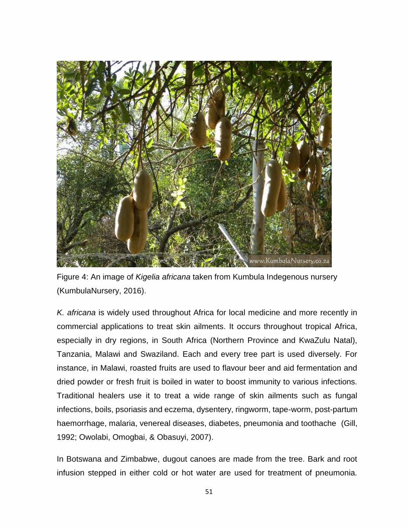
51
Figure 4: An image of Kigelia africana taken from Kumbula Indegenous nursery
(KumbulaNursery, 2016).
K. africana is widely used throughout Africa for local medicine and more recently in
commercial applications to treat skin ailments. It occurs throughout tropical Africa,
especially in dry regions, in South Africa (Northern Province and KwaZulu Natal),
Tanzania, Malawi and Swaziland. Each and every tree part is used diversely. For
instance, in Malawi, roasted fruits are used to flavour beer and aid fermentation and
dried powder or fresh fruit is boiled in water to boost immunity to various infections.
Traditional healers use it to treat a wide range of skin ailments such as fungal
infections, boils, psoriasis and eczema, dysentery, ringworm, tape-worm, post-partum
haemorrhage, malaria, venereal diseases, diabetes, pneumonia and toothache (Gill,
1992; Owolabi, Omogbai, & Obasuyi, 2007).
In Botswana and Zimbabwe, dugout canoes are made from the tree. Bark and root
infusion stepped in either cold or hot water are used for treatment of pneumonia.
52
Kigelia africana contains naphthoquinones and iridols dihydroiscocon marines in
extracts of the bark phenylopropanoids caffeic acid, P-coumaric acid and ferulic acid,
sterols and flavonoids (Joffe, 2003). The ethanolic extract of K. africana exhibit
antibacterial and antifungal activity against S. aureus and C. albicans but not against
the strains of E. coli and P. aeruginosa. However, the aqueous extract on the same
study demonstrated no activity indicating that ethanol is a better extracting solvent
(Owolabi et al., 2007).
K. africana extracts have showed significant growth inhibitory activity towards K.
pneumoniae (Cock & van Vuuren, 2014). Moreover, K. africana showed the presence
of glycosides, phenolic compounds (hydrolysable tannins), alkaloids, flavonoid and
reducing sugar in all extracts while cardiac glycoside was noted only in chloroform
extract (Abdulkadir, Adedokun, & John, 2015). It has been reported that the extracts
from many species of bignoniaceae contain secondary metabolites such as saponins,
tannins, flavonoids, quinones, alkaloids, anthralene derivatives, reducing sugars,
glycosides, carbohydrates, querletin, kaempferol, α-sitosterol, terpenes, steroids and
coumarins and their derivatives (Azu, 2013).
2.2.6.5 Dodonaea angustofolia (Sapindaceae)
D. angustofolia grows in variety of habitats ranging from arid, semi-arid to high rainfall
regions and in frost-hardly regions. It is found in a wide strip along coast from
Namaqualand through the Western Cape, Eastern Cape to KwaZulu Natal as well as
further north in Mozambique and Zambia. It also occurs naturally in Saudi Arabia,
Australia and New Zealand. D. Angustofolia is widely used for colds, influenza,
stomach complaints and measles. Other early uses of the plant included treatment of
pneumonia, tuberculosis and skin rashes and it can be combined with viscum capense
(B E Wyk, Endtsshoon, & Gericke, 2009).

53
For a sore throat and oral thrush, it is used as a gargle. The khoi-khoi used a concoction
of the root for the colds and influenza whilst in Namaqualand the green leave were
boiled slowly, then left to steep, strained, and the extract was used for influenza, colds,
and induce sweating (Harris, 2015). It is used to relieve coughs and the congested
feeling typical of influenza, croup and diphtheria. The leave extracts of D. angustofolia
is considered to alleviate stomach ailments and fever, as a mild purgative for
rheumatism, sore throat and haemorrhoids (Shuizen et al., 2011).
In a study powdered plant material of Dodonaea Angustofolia demonstrated the
presence of many secondary metabolites, such as tannins, alkaloids, phytosteriods,
saponnins and polyphenols(Al-baker, Ahmed, Hanash, & Al-hazmi, 2014) . From an
investigation on antimicrobial activity of selected Eastern Cape medicinal plants D.
Angustofolia displayed antimicrobial activity against B. cereus (Mohlakoana, 2010).
Figure 5: The fruits of Dodonaea angustofolia. Taken from (Plantbook, 2016).
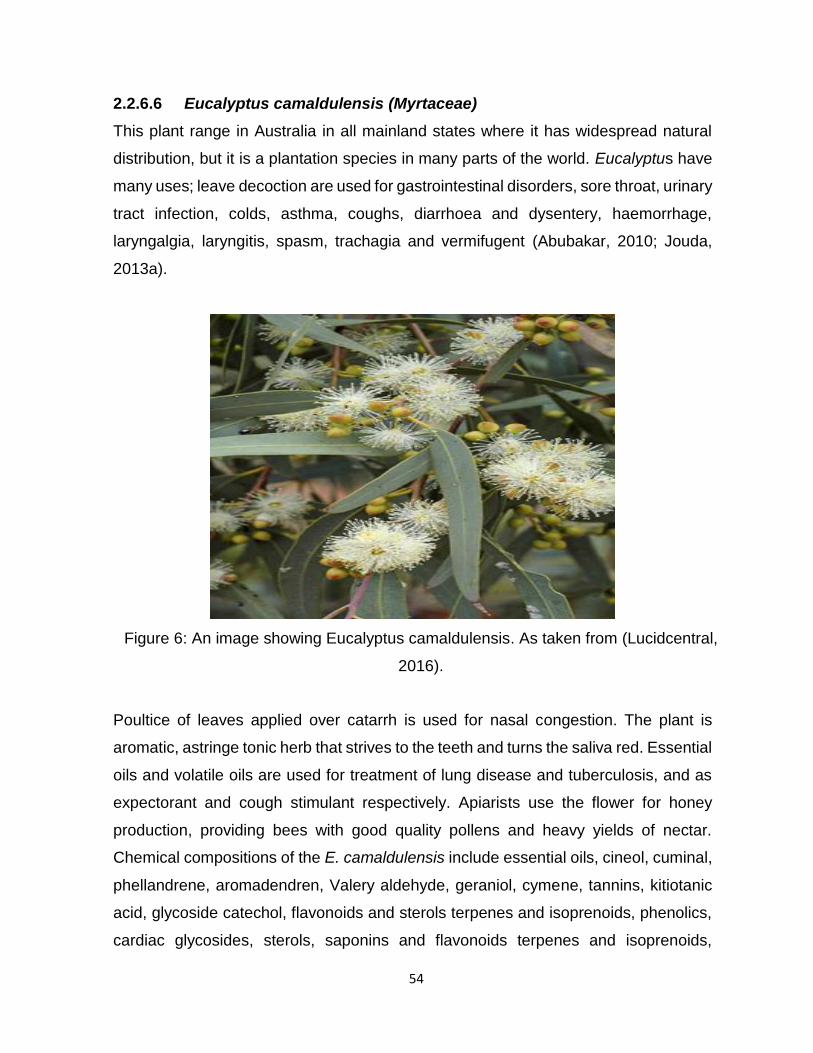
54
2.2.6.6 Eucalyptus camaldulensis (Myrtaceae)
This plant range in Australia in all mainland states where it has widespread natural
distribution, but it is a plantation species in many parts of the world. Eucalyptus have
many uses; leave decoction are used for gastrointestinal disorders, sore throat, urinary
tract infection, colds, asthma, coughs, diarrhoea and dysentery, haemorrhage,
laryngalgia, laryngitis, spasm, trachagia and vermifugent (Abubakar, 2010; Jouda,
2013a).
Poultice of leaves applied over catarrh is used for nasal congestion. The plant is
aromatic, astringe tonic herb that strives to the teeth and turns the saliva red. Essential
oils and volatile oils are used for treatment of lung disease and tuberculosis, and as
expectorant and cough stimulant respectively. Apiarists use the flower for honey
production, providing bees with good quality pollens and heavy yields of nectar.
Chemical compositions of the E. camaldulensis include essential oils, cineol, cuminal,
phellandrene, aromadendren, Valery aldehyde, geraniol, cymene, tannins, kitiotanic
acid, glycoside catechol, flavonoids and sterols terpenes and isoprenoids, phenolics,
cardiac glycosides, sterols, saponins and flavonoids terpenes and isoprenoids,
Figure 6: An image showing Eucalyptus camaldulensis. As taken from (Lucidcentral,
2016).

55
phenolics, cardiac glycosides, sterols, saponins and flavonoids (Abubakar, 2010;
Grbovc et al., 2010). E. camaldulensis aqueous and acetone extract when tested
against Escherichia coli, Staphylococcus aureus, Salmonella typhi, Proteus mirabilis
and Klebsiella pneumoniae they demonstrated antibacterial activity, and highest was
observed in acetone extract (Abubakar, 2010). It was noted that ethanolic leaf extract
of E. camaldulensis had marked fungicidal effect against clinical dermatophytic fungal
isolates; Microsporium gypseum and Trichophyton mentagrophytes (Jouda, 2013a).
2.2.6.7 Acorus gramineus (Acoraceae)
Acorus gramineus is indigenous to Asia, but has been introduced across Europe,
Australia, New Guinea, South Africa, Reunion and North America. The dry rhizome
of A. gramineus, called Acori graminei rhizome has been used largely in Korean
traditional medicine (Lee, Yun, & Hwang, Byung KookLee, 2004).
Figure 7: An image of Acorus gramineus. Taken from: (grasses on Pinterest)
56
It has common names like grass-leaved sweet rush, Japanese sweet flag, dwarf sweet
flag (R. Balakumbahan, 2011). The plants root has medicinal uses like: antifungal,
antibacterial, antiperiodic, antirheumatic, antispasmodic, aromatic, cardiac,
carminative, diaphoretic, emmenagogue, febrifuge, sedative, stimulant, stomachic,
tonic and vermifuge It is also powdered and applied to bleeding gums, digestive
problems (gastralgia and diarrhoea), cough, bronchial asthma, neurasthenia,
depression, epilepsy, body parasites, dermatosis and haemorrhoids.
The root contains an essential oil consisting of asarone and asaryl aldehyde, and bitter
glucoside acorin. Asarone increases the hypnotic effect of barbiturates and ethanol
lowers blood pressure and has antibacterial activity against Staphylococcus aureus,
streptococci and mycobacterium. The whole plant is anodyne, antiperiodic,
antispasmodic, digestive, diaphoretic, diuretic, expectorant, sedative, stimulant,
stomachic, sudorific, tonic, vermifuge. The dried root repels insects likely as an
insecticidal and is used in Vietnam to kill lice, bugs and fleas. Antifungal activity of
beta-asarone from rhizomes of Acorus gramineus was recently demonstrated in a
study whereby a number of fungal under study were inhibited (Lee et al., 2004).
2.3 Classification of antimicrobial therapy
In an effort to battle the various forms of diseases that have continued to invade
humans from ancient times up to present, variety of antimicrobials have been
developed against pathogens for these diseases. Antimicrobials are substances that
kill or inhibit the growth of microorganisms, which refers to all agents that act against
microbial organisms; bacteria (antibacterial), viruses (antiviral), fungi (antifungal) and
protozoa (antiprotozoal) (Cheesbrough, 2006; Michigan State University (institution),
2011). This is not synonymous with antibiotics, “antibiotic” refers to substances
produced by microorganisms that act against another microorganism. Some vital
antibiotics include culture extracts and filtrates of fungi such as penicillium,
amphotericin B, griseofulvin and cephalosporium, and bacteria such as
aminoglycosides (Streptomyces, kanamycin, amikacin, gentamicin and tobramycin)
tetracycline’s, polymyxins, rifampicin, fucidin, chloramphenicol, erythromycin,
clindamycin, vancomycin and Bacillus species (bacitracin
57
by B. licheniformis or B. subtilis, polymyxin by B. polymyxa and gramicidin by B.
brevis). Therefore, antibiotics does not include antimicrobial substances that originate
from synthetic derivatives (sulphonamides and quinolones), or semisynthetic
(methicillin and amoxicillin), or those which come from plants (quercetin and alkaloids)
or animals (lysozyme). Some broadly antifungal agents include nystatin, flucytosine
and imidazole agents (Cheesbrough, 2006; Michigan State University (institution),
2011).
Because of the re-occurring resistance of pathogenic microorganisms to antibiotics,
and their side effects has resulted to active investigation of other sources of
antimicrobials such as medicinal plants for their potential antimicrobial activity. Plants
produce secondary metabolites (phytochemicals), which have potential as
antibacterial when used individually and in combination or potentiators of other
antibacterial agents. Since phytochemicals usually act through different mechanisms
rather than the conventional antibiotics, they could therefore be of use in treatment of
resistant bacteria (Chinyama, 2009; Frum & Viljoen, 2006).
2.3.1 Mechanisms of Antimicrobials
In order to understand the mechanism of bacterial resistance, bacterial physiology,
pharmacology and antimicrobial drugs and molecular biology of infectious agents
need to be understood. In this study, some of these processes will be briefly discussed
to gain an insight of resistance concept. Broad spectrum is related to antimicrobials
with activity against a wide range of gram positive and negative organisms like
tetracycline’s, aminoglycosides and chloramphenicol. Sometimes, antimicrobials are
used in combination to treat mixed infections, prevent drug resistance from
developing. They can also be used to treat infection when organism is not known, or
to obtain greater antimicrobial effect from two bactericidal drugs activity together
(synergistic effect) (Cheesbrough, 2006; Michigan State University (institution), 2011).
Antimicrobials are mostly categorized according to their primary functions. However,
they are sometimes classified according to their function, thus to say agents that kill
microbes at usual dosages are called bactericidal, and those that inhibit their growth
are bacteriostatic (Cheesebrough, 2006). Different ways on how antimicrobials act on

58
bacteria based on structure and degree of affinity to certain target sites within bacteria
cells are as follows:
a) By inhibiting cell wall formation: This mechanism leads to cell wall lysis.
Examples include; penicillin’s, cephalosporin’s, carbapemarins and
vancomycin’s. Since human and animal cells have no cell walls, drugs that
target cell walls selectively kill or inhibit bacterial organisms since this structure
is critical for the life and survival of bacterial species. Bacteria drugs in this
group disrupts normal balance between cell wall and its degradation, hence
exposing cell wall to destruction by losing out inclusion or allowing water to flow
into the cell causing it to burst (Cheesebrough, 2006; Kamanga, 2013;
Michigan State University (institution), 2011; Nkosi, 2013).
Antimicrobial agents that interfere with cell wall synthesis are often bactericidal.
Activity of bet-lactam antimicrobials on gram-negative bacteria is through
passage of this drugs into the cell through porin channels in the outer
membrane. In susceptible cells, beta-lactam molecules bind to penicillin
binding proteins (PBPs) enzyme involved in cell wall production. The
attachment of the beta-lactam molecules to the PBPs, located on the surface
of the cytoplasmic membrane, obstruct the process, resulting in weakened or
defective cell walls that leads to cell lysis and death. Since gram-positive
bacteria do not possess an outer membrane, beta lactam antimicrobials diffuse
through cell wall and the following steps are similar to those in gram negative
(Marie B.Coyle, 2005).
b) By inhibition of cytoplasmic membrane function or damaging bacterial cell
membrane, leading to loss of cell contains and cell death. Cell membranes in
a cell they act as a vital barrier that isolate and control the intra and extra cellular
flow of substances into and outside living cell. Any possible damage to cell
membrane could result in leakage of important cell contains (solutes)
responsible for the cells survival. The action of this this class of antibiotic are
frequently poorly selective and can often be toxic for systematic use in
mammalian host, as some of this structures can be found in both eukaryotic

59
and prokaryotic cells (Cheesebrough, 2006; Marie B.Coyle, 2005; Michigan
State University (institution), 2011). As a result, most clinical usage is limited to
relevant applications. Example of drugs that fall under this category are,
polymixins, amphotericin B and colistin (Cheesebrough, 2006; Michigan State
University (institution), 2011). Drug like gramicidin is known for disrupting the
bacterial cytoplasmic membrane by forming a channel through the membrane
that result in damaging its integrity, antifungal drugs polyeners they are known
to have similar mechanism (Kamanga, 2013; Nkosi, 2013).
c) By interfering protein synthesis as a result arresting bacterial growth and
also cause further damage leading to cell death by binding to the machinery
that builds proteins. Enzymes and cellular structures are essentially made of
proteins, and their synthesis is a basic process necessary for the multiplication
and survival of all bacterial cells. Several type of antibacterial agents’ target
bacteria protein synthesis by binding to either 30s 0r 50s sub units of the
intracellular ribosomes. This process result in the disruption of the normal
cellular metabolism. Tetracycline’s (tetracycline, minocycline and doxycycline)
bind to the 30S subunit of the ribosome and block the attachment of transfer
RNA (tRNA). Therefore, new amino acids cannot be added to the growing
protein chain, as synthesis of protein is inhibited. The action of tetracycline’s is
bacteriostatic (Cheesebrough, 2006; Kamanga, 2013; Marie B.Coyle, 2005;
Michigan State University (institution), 2011).
Aminoglycosides (e.g. gentamicin, tobramycin, amikacin, and streptomycin)
also bind to the 30S ribosomal subunit and they block protein synthesis in two
possible ways. First they bind to 30S subunit of the ribosome and prevent it
from attaching to messenger RNA (mRNA), secondly, the presence of the
aminoglycoside on the ribosome may cause misreading of the mRNA. “This
leads to the insertion of the wrong amino acid into the protein or interference
with the ability of amino acids to connect with one another. These activities
often occur simultaneously and the overall effect is bactericidal” (Marie
B.Coyle, 2005). Inhibition of protein synthesis by binding to the 50S ribosomal
60
subunit happens when macrolides (erythromycin, azithromycin and
clarithromycin) and lincosamides (clindamycin) attach to the 50S ribosomal
subunit triggering termination of the growing protein chain and inhibition of
protein production. Chloramphenicol binds to the 50S subunit of the ribosome
and interferes with binding of amino acids to the growing protein. Antimicrobial
agents that interferes protein synthesis in this way are bacteriostatic (Marie
B.Coyle, 2005).
d) By inhibiting the production of nucleic acids, hence preventing bacteria
from reproducing. Nucleic acids production is important for the synthesis of
DNA, some antibiotic they work by binding to vital components for DNA and
RNA synthesis, causing interference in normal cellular process, eventually
compromising bacterial multiplication and survival. In order for nucleic acid to
be produced it requires tetrahydrofolic acid, which is normally reduced to
tetrahydrofolic acid by enzymes dihydrofolate reductase (DHFR). THR is
primarily important in thymidine monophosphate (dTMP) synthesis, which are
substrates for DNA production. Anti-folate is known to inhibit nucleic acid by
inhibiting DHFR enzymes. The antimicrobial action happens when antifolates
which are DHFR analogy competitively and irreversibly bind with DHFR at a
greater affinity than DHF. Leading in inhibition of the supply of THF and
obstruction of DNA synthesis, as a result disrupting cellular growth.
Trimethoprim works by inhibiting dihydrofolate reductase interfering with
nucleic acid production by inhibiting purine production (Cheesebrough, 2006;
Kamanga, 2013; Michigan State University (institution), 2011; Nkosi, 2013).
Fluoroquinolones (e.g. nalidixic acid, ciprofloxacin, levofloxacin and
gemifloxacin) interfere with DNA synthesis by blocking the enzyme DNA
gyrase, which helps to wind and unwind DNA during DNA replication. The
enzyme binds to DNA and initiate double stranded breaks that allow the DNA
to unwind. Fluoroquinolones bind to the DNA gyrase-DNA complex and permit
the broken DNA strands to be released into the cell, which leads apoptosis.
61
Rifampicin binds to DNA-dependent RNA polymerase, which blocks the
production of RNA and result in apoptosis (Marie B.Coyle, 2005).
e) By inhibition of other metabolic pathways or processes some antibiotics
they act on selected cellular processes necessary for the survival of bacterial
pathogens. For example, sulphonamides and trimethoprim disrupt the folic acid
pathway which is imperative for bacteria to produce precursors important for
DNA synthesis. For many organisms’ para-amino-benzoic acid (PABA) is an
essential metabolite and is involved in the synthesis of folic acid, important
precursor for nucleic acids synthesis. Sulphonamides are structural analogs of
PABA and compete with PABA for the enzyme dihydropteroate synthetase.
Trimethoprim acts on the folic acid synthesis pathway at a point after the
sulphonamides. It inhibits the enzyme dihyrofolate reductase. Trimethoprim
and sulphonamides can be used separately or together. When used together
they produce a sequential blocking of the folic acid synthesis pathway and have
a synergistic effect. Both trimethoprim and the sulphonamides are
bacteriostatic (Cheesbrough, 2006; Kamanga, 2013; Nkwanyana, 2013).
2.3.2 Action mode of antimicrobial resistance
Modification of active site of the target results in the inefficiency on binding of the
drugs. Bacteria they change the patterns and other essential compartment of bacterial
cell, which are used as binding site for antimicrobials. One target for interposition, at
the active site for instance penicillin-binding protein (PBP). Beta-lactam antibiotics
they alter penicillin-binding proteins in order to hinder antibiotics access to the
enlarging peptidoglycan. For example, the mode of action for the high level resistance
of staph A to PBP2 to a variant that is designated PBP2a. As another option, may be
loss of the binding proteins that have a greater affinity for the antibiotics (Winn, Allen,
Janda, & Koneman, 2006). With literature revelation, a lot of natural products do exist,
which are specialised to overcome resistance microorganisms for example changes
of genetic origin are associated with resistance to aminoglycosides, lincomycin and
erythromycin (Cheesebrough, 2006; Wagner & Ulrich-merzenich, 2009). Changes in
the affinity of receptors for other antibiotics such as DNA gyrase and quinolones or
62
ribosomes and aminoglycosides are also important. Despite modification of active site
the second most important mechanism is the change in receptors for antibiotic
attachment to critical structures whereby there’s alteration of normal binding proteins
on the cell membrane of gram positive bacteria to binding proteins with reduced affinity
for antibiotics (Winn et al., 2006).
Efflux of antibiotic from the cell by hindering antibiotics from penetrating into the
bacteria cell or after penetration to eject the accumulated dry out of bacteria cell
(Wagner & Ulrich-merzenich, 2009). Some bacteria they actively remove antimicrobial
agent from the bacterial cell so that intracellular concentration of antimicrobial agents
won’t reach suitable amount to exert their effective antimicrobial activity. The energy-
dependent efflux mechanism is a prime defence for bacteria against tetracycline’s and
macrolides, these groups interfere with protein synthesis at the level of ribosome.
Relatively as with staphylococci against the quinolones, removal of antibiotics is
resistance mechanism which actually interfere with DNA gyrase (Marie B.Coyle,
2005). The resistance of gram negative bacilli to many antibiotics was originally
attributed to poor entry of the antibiotic into the bacterial cell because of size,
hydrophobicity and charge. The efflux of these antibiotics from bacterial cells is vital,
since the pumps can excrete chemical compounds, including antimicrobial agents,
into extracellular medium directly, whereby their re-entry is prohibited (Cheesbrough,
2006; Michigan State University (institution), 2011; Winn et al., 2006).
Direct destruction or modification of the antibiotics by enzymes produced by the
microorganisms, thus enzymes destroy or inactivate antimicrobials. Genes that code
for the production of resistance enzymes are carried by plasmids and they can be
moved from one species to another. An individual plasmid possesses genes that code
resistance to several antimicrobial drugs like streptomycin, chloramphenicol,
tetracycline’s and sulphonamides. Plasmids of the nature are called resistance (R)
factors, but other plasmids they carry genes that code for resistance to penicillin.
Examples of enzymes that causes resistance are Beta lactamases which destroy the
beta lactam ring that forms part of the structure of penicillin and cephalosporin. These
organisms, staphylococcus aureus, haemophilus influenzae and Neisseria
gonorrhoea are associated with Beta-lactamase production. Certain gram negative

63
bacteria equally produce acetylating, adenylating, and phosphorylating enzymes that
have ability of inactivating aminoglycosides, like chloramphenicol is inactivated by the
enzymes acetyltransferase. Some enzymes in gram negative bacteria that inactivate
streptomycin and kanamycin they are widely distributed making these antibiotics
vulnerable to common clinical use. A minor resistance for tetracycline is enzymatic
inactivation. Resistance to macrolides and lincosamides in gram positive bacteria may
be yielded due to several methods; alone or in combination-enzymatic inactivation,
alteration of ribosomal target or active efflux (Cheesbrough, 2006; Marie B.Coyle,
2005; Wagner & Ulrich-merzenich, 2009; Winn et al., 2006).
Bypass of a metabolic block imposed by antimicrobial agent like trimethoprim-
sulfamethoxazole and enterococci also by bacteria altering the permeability of their
cell membrane, making impossible or difficult for antimicrobial to enter bacteria cell.
This kind of resistance have been observed in bacteria resistance to polymyxins and
tetracycline’s, but the cell wall of streptococci form a natural barrier to aminoglycosides
(Cheesebrough, 2006; Wagner & Ulrich-merzenich, 2009). Auxotrophic mutants that
use thymine for growth may bypass the activity of trimethoprim and sulphonamides in-
vitro making use of available substrate and alternative pathway. Important clinical
example of bypass process is the ability of naturally occurring strains of enterococcus
species to use compounds like folic acid growth in vivo in the presence of trimethoprim
– sulfamexazole. Strains of enterococci may appear susceptible in-vitro but they don’t
respond to chemotherapy in-vivo. In order to check the in-vitro result of resistant,
there’s a need to add folic acid to the testing media. For this reason hereof enterococci
should not be used for susceptible testing against trimethoprim (Marie B.Coyle, 2005;
Michigan State University (institution), 2011; Wagner & Ulrich-merzenich, 2009; Winn
et al., 2006).
2.4 Microorganisms
Organisms selected to for this study were clinical strain of; Klebsiella pneumoniae,
Streptococcus pyogenes, and ATCC 43300 strain of Staphylococcus aureus.
64
2.4.1 Staphylococcus aureus
Staphylococcus aureus is one of the important human pathogen among the
staphylococci species. S. aureus is gram positive coccus, uniform in size, can occur
in groups, singly and also in pairs, tetrads and short chains. Predominantly it appears
in grape like structures and is non- motile, non-capsulated, non-spore-forming.
Commonly found in the external environment like, on the intertriginous skin folds,
mucous membranes, perineum, the axillae, vagina, and the intestine of humans and
other animals. Common specimens from which S. aureus is isolated are; sputum,
blood, urine, faeces, vomit, pus and anterior nasal perineal swabs which are
analysed by microscopy and culture (Cheesebrough, 2006; Kamanga, 2013; Winn
et al., 2006).
S. aureus grow well aerobically and in carbon dioxide enriched atmosphere
(facultative anaerobes), expect for S. aureus sub-species anaerobius S.
saprophyticus. Desirable temperature range for growth is 10-420c with an optimum
of 35-370c. On blood agar and chocolate, it produces yellow to cream or
occasionally white 1-2 mm in diameter colonies. When grown aerobically they are
beta- haemolytic, on MacConkey smaller colonies are seen after overnight
incubation. Biochemically S. aureus are non -lactose fermenter, coagulase positive,
DNases positive and catalase positive (Cheesbrough, 2006; Winn et al., 2006).
S. aureus is known to cause a number of infection processes ranging from benign
infections to life threatening systemic illness. It causes conjunctivitis in new-born,
septicaemia, endocarditis, osteomyelitis, mastitis, food poison, toxic shock
syndrome, scalded skin syndrome, and antibiotic associated enteritis
(Cheesebrough, 2006). Skin infection include simple folliculitis and impetigo,
furuncles and carbuncles which involves subcutaneous tissues and causing
systemic symptoms, such as fever. In postsurgical infections it suffices as a
breeding ground for the development of systematic infections. Pneumonia and
empyema is also experienced, which is categorized into community acquired,
bronchopneumonia which is manifested in elderly individuals and is associated with
viral pneumonia as a predisposing factor, and also nosocomial pneumonia due to

65
S. aureus which happens in hospital because of cross infection of obstructive
pulmonary diseases, intubation, and aspiration (Cheesbrough, 2006; Winn et al.,
2006).
S. aureus produce enzyme and toxins which are termed virulence factors which
believed to contribute to their ability to cause infection. Initially, penicillin was the
preferred drug for S. aureus treatment, but due to acquisition of plasmid borne
genetic elements coding for Beta lactamases production has led to emergence of
resistant to penicillin. For the mean time over 80% of S. aureus isolates are resistant
to penicillin, as a result semisynthetic penicillinase-resistant penicillin’s have
become the drug of choice for treatment of infections. The latest mode of resistant
is acquired from the presence of an altered penicillin-binding protein (PBP 2a) that
come up from acquisition of a chromosomal gene called mecA. Other drugs that
have developed resistance to S. aureus are glycopeptide agent vancomycin
(Kenneth & Ray, 2004; Winn et al., 2006).
2.4.2 Streptococcus pyogenes
Streptococcus pyogenes consist of Lancefield group A, Beta haemolytic
streptococci, which is among the most prevalent of human bacterial infections and
their common reservoirs in nature are the skin and mucus membranes. Just like
Staphylococci aureus, streptococcal group A also produce several other products
that contribute to virulence. S. pyogenes is a gram positive cocci and non-motile,
they can grow both aerobically and anaerobically. Specimens normally found are
pus, throat swab, or blood depending on site of infection, and blood samples are
also used for antistreptolysin O antibody (Kenneth & Ray, 2004).
S. pyogenes grow well on blood agar whose colonies look grey white or colourless,
dry or shinny and usually irregular in arteria. They are beta-haemolysis which is
markedly when incubated anaerobically. S. pyogenes does not grow on MacConkey
(Cheesbrough, 2006). Group A streptococci cause’s streptococcal pharyngitis,
impetigo, erysipelas, cellulitis, puerperal sepsis, acute rheumatic fever, acute
glomerulonephritis and ear infections. Oral penicillin V or intramuscular benzathine
penicillin G, is currently recommended for streptococcal pharyngitis and other

66
infection however, erythromycin and cotrimoxazole is an alternative to those who
are allergic to penicillin. Resistance to macrolides has been reported globally and
less than 5% in U.S have demonstrated to be resistant to erythromycin (Winn et al.,
2006).
2.4.3 Klebsiella pneumoniae
Klebsiella pneumoniae are gram negative, non-motile, encapsulated,
non-sporulating, facultative, aerobic shaped rod bacterium that is normally found in
the human gastro-intestinal tract (oropharynx). This colonization may prove to be
the origin of lung infections that generally manifested in patients with chronic
conditions such as alcoholism, diabetes mellitus, and chronic obstructive pulmonary
diseases. The pneumonia is prone to destruction with extensive necrosis and
haemorrhage, leading to production of sputum which is thick, mucoid and brick red,
or thin and currant jelly-like appearances. When cultured on blood agar or
MacConkey they produce large and usually mucoid colonies, also when cultured on
broth a stingy type of growth is produced. Specimens collected depends on site of
infection and includes urine, sputum, pus, and infected tissue. Most strains are
lactose fermenters and urease test is positive after 18-24hrs (Cheesbrough, 2006;
Kenneth & Ray, 2004; Ryan & Ray, 2004; Winn et al., 2006).
An adhesion to a mucosal surface is often the first step in the development of an
infection. A survey of the presence of Klebsiella in urban residents, hospital
personnel, and newly admitted patients showed that 30-37% of individuals carried
Klebsiella, including a 29-35% faecal carriage and a 3-4% throat carriage. Strains
of K. pneumoniae and K. oxytoca which have not acquired any resistance are
determined as naturally resistant to ampicillin and carboxypenicillin but susceptible
to other beta-lactam antibiotics. This is due to the production of a chromosomal
penicillinase which is inhibited by clavulanic acid. K. pneumoniae also causes
enteritis and meningitis, urinary tract infection, wound infections and peronitis, and
septicaemia (Cheesbrough, 2006; Kenneth & Ray, 2004; Winn et al., 2006).

67
CHAPTER THREE
ANTIMICROBIAL EVALUATION FOR PLANT USED TO TREAT BACTERIAL
PNEUMONIA
3.1 Introduction
Medicinal plants are used worldwide for primary health care which is usually
administered by herbalist or a local inhabitant with herbal knowledge. As such in
developing countries and South Africa inclusive, up to present medicinal plants, forms
a cornerstone of health care. Traditional medicine use has been known as a source of
biologically important active compound or as an element in complex mixture of
comprehensive constituents (Suliman, 2011).
currently, due to an increase of bacterial resistance there has been a dramatic increase
in the demand for medicinal plants. This resulted in an urgent need to develop new
antibiotics and immune modulating compounds with a broad range chemical
formulation and different mechanism of action (Mohlakoana, 2010).
This chapter discusses the materials and methods that were used in this study to meet
objectives. As observed by Chinyama (2009) revitalized interest in traditional
pharmacopeias’ has encouraged researchers not to focus only on determining scientific
logic but also to concentrate on discovering new compounds with pharmaceutical
value. The plants being investigated in this study were selected based on their reported
ethnobotanical use and ethnopharmacological studies. In this study the antimicrobial
evaluation of medicinal plants was done by testing them against a number of pathogens
associated with pneumonia infections.
68
3.2 Materials and methods
3.2.1 Plant sample selection and collection
Seven plants that are used traditionally to treat pneumonia and related conditions were
selected based on indigenous ethnopharmacological reports from the available
literature and herbal knowledge usage (B.E. Van Wyk & Gericke, 2000; B E Van Wyk
& Wink, 2004; B. Wyk et al., 1997). The plant materials used in this study consisted of
Kigelia africana (bark), Ballota africana (leaves), Dudonea angustofolia (leaves),
Warbugia salutaris (bark), Terminalia sericea (roots), Acorus gramineus (roots), and
Eucalyptus camldulensis (leaves). Three plants namely K. africana, T. sericea and W.
salutaris were collected were collected from Malawi to be precise central region in
Lilongwe rural area, Msalu village which was later identified at Chitedze Research
Station. The remaining four plants were collected from different places in Port Elizabeth
Eastern Cape Province of South Africa.
3.2.2 Plant preparation
Fresh collected plant material of roots, leaves and bark were washed with tap water a
number of times to remove dirt. The roots and bark were further cut into small pieces
to enhance drying; the plant materials were oven dried at 40 °C until absolutely dry.
There after the dried plant material were pulverized into fine and course powder
depending on its property with mortar and pestle, and the powder was kept in zip lock
bags and airtight containers until required for further processing (Mohlakoana, 2010;
Nkwanyana, 2013).
3.2.3 Plant Extraction Methods
Aqueous solvent, (preferably hot distilled water) and two organic solvents namely
(ethanol and acetone) were used for extraction for each plant under study investigation.
2 g of powdered plant material was weighed and subjected to extraction, with 45 ml of
extracting solvent in in polyester centrifuge tubes. The mixtures were vigorously shaken
with a vortex for 5 minutes and left overnight while being heated to 300c for the thorough
dissolving of the plant compounds. Tubes were centrifuged at 3500 ×g for 5 min and
the supernatant was filtered using Wattman No.1 filter paper before being transferred
into pre-weighed glass containers. The procedure was repeated twice using the same
amount of a solvent in each tube.
69
The beakers with aqueous extract supernatants were then placed into 40 °C water-bath
to allow evaporation of the extractant so that the plant extract/residues remain. The
organic solvent was removed by evaporation under a stream of air in a fume hood at
room temperature to quantify extraction effectiveness and comparison of different plant
species. After evaporation, the beakers containing the residues were weighed and were
re-dissolved in 10% and 100% dimethyl sulfoxide (DMSO). The amount of DMSO
added to reconstitute dry extract was dependent on an individual extract. All the plants
were able to dissolve in 10% DMSO except B. africana, D. angustofolia, A. gramineus
extracted with acetone and ethanol, which were dissolved with 100% DMSO.
Concentrations were then standardized between the different plants and then it was
kept in refrigerator at 4 °C till further use for the antimicrobial screening assay. The
method of extraction was adopted from (Eloff, 2000; Eloff JN, 2004; Kamanga, 2013)
3.3 Antimicrobial assays
3.3.1 Panel of bacterial strains
Strains of organisms tested during the present study are some of the predominant
causes of pneumonia. Bacteria strains used in this study were provided by the Faculty
of Health Science, Medical Laboratory Sciences department, microbiology laboratory
at Nelson Mandela Metropolitan University. Three organisms were used for the study,
the strains included, streptococcus pyogenes, Klebsiella pneumoniae and American
Type Culture Collection (ATCC) of Staphylococcus aureus 43300.
3.3.2 Media and culture preparation
Nutrient agar and broth were prepared by adding 1 L of distilled water to 30 g of TSB
powder. The mixture was dissolved and autoclaved for 15 minutes at 121 °C. Muller
Hinton agars were prepared by dissolving 38 g powdered of agar in 1 litre of distilled
water. The mixture was boiled on Bunsen burner to dissolve and thereafter it was
autoclaved for 15 minutes at 121 °C. Shortly before solidifying, the liquid agar was
poured into 90 mm petri dishes to a depth of 4mm, the plate was allowed to dry and
stored in fridge at 2.2°C. Mueller-Hinton agar plate was preferred for use in sensitivity
because it gives good result in batch-to-batch reproducibility, lack in sulphonamide,
trimethoprim and tetracycline inhibitor, and the growth of many bacteria pathogen gives

70
satisfactory result. The agar should have a PH of 7.2-7.4 at room temperature and the
agar surface should be left to dry without droplet of moisture (Cheesbrough, 2006;
Motlhatlego, 2014; Saad, Sadia, Asmani, & Yusuf, 2014).
Bacterial strains were sub cultured in laboratory to get colonies/new strains of these
bacteria, and nutrient agar and Muller Hinton was used for the initial sub-culturing for
all organisms. The microorganisms were incubated at 37°C for 24 hrs and 32 hrs for
streptococcus pyogenes. The bacterial strains were preserved and maintained on
nutrient agar plates and Muller Hinton agar plates at 2°C - 8°C for short-term storage
(Cheesebrough, 2006). Single colonies of each bacterium were also respectively
transferred into McCartney bottles each with 10 ml Oxoid Mueller-Hinton Broth (MHB).
These bottles were then incubated in a shaking incubator overnight at 37°C, but for the
viability maintenance of the bacterial strains, every 30 days the organisms were sub-
cultured (Motlhatlego, 2014).
3.3.2 Antimicrobial assessment using Agar well diffusion method
Antibacterial activity of the plants under investigation were tested using Agar well
diffusion method. Stock cultures were maintained at 4 °C on Nutrient and Muller-Hinton.
In order to come up with active cultures for experiments, organisms were prepared by
transferring a loopful of culture to 5 ml of nutrient broth and incubated at 37 °C for 24
hours. Few isolated colonies (3-5) were selected and suspended in 5 ml sterile
physiological saline using sterile wire loop or disposable swab. (Jouda, 2013b;
Kamanga, 2013).
The turbidity of the bacterial suspension was adjusted and visually compared to a 0.5
McFarland turbidity standard. Turbidity was also verified using spectrophotometric
comparison with a 0.5 McFarland standard to ensure that the suspension contained
approximately 1 to 2 x 10⁸ CFU/ml (Hariprasath, 2010; Kamanga, 2013).
An inoculum suspension was swabbed uniformly to solidified 25 ml Mueller-Hinton agar
(MH) for bacteria, and the inoculum was allowed to dry for 5 minutes. Holes of 6 mm in
diameter were made in the seeded agar using glass Pasteur pipettes. Aliquots of 100
μl from each plant crude extract (10.5 mg/ml) was added into each well on the seeded
71
medium and allowed to stand on the bench for 15-20 minutes for proper diffusion and
thereafter incubated at 37 °C aerobically for 18 hours. Control wells were maintained
by introducing extract solvents (10% & 100% DMSO) as negative controls into the well.
Antimicrobial paper discs of streptomycin (S10), chloramphenicol (C30), Penicillin
(G10), Tetracycline (30), Ciprofloxacin (CIP5), Amoxicillin (A10) were used as positive
control. After 18 hours of incubation, the plates were monitored making sure that the
growth was consistent. After incubation the growth inhibition zone were quantified by
measuring the diameter of the zone of inhibition in mm, inhibition > 8 mm were
considered to be sensitive. The interpretation of inhibition zone results, were reported
as susceptible, intermediate, or resistant to the organisms tested. The experiments
were done in triplicates to make sure the results are reproducible, reliable and valid.
the mean clear zones was calculated and values were compiled (Jouda, 2013a;
Wizemann, Olsen, & Choffnes, 2013).
3.3.3 Minimum inhibition concentration
The minimum inhibitory concentration (MIC) is defined as the lowest concentration of
an antimicrobial that completely inhibits the growth of an organism under specific
condition. After initial screening by agar diffusion assay which aided in identification of
an extract with antibiotic activity, the micro-dilution bioassay for MIC was performed.
This method is based on the principle of exposing test organism to a series of dilutions
of test antimicrobial/substance (Suliman, 2011). There are a number of ways in which
MIC can be determined like agar dilution, the broth dilution method, and microplate or
microdilution method. The microplate is regarded as a “sensitive” and a “quick” method
specifically developed to obtain MIC values for plant extracts against micro-organisms.
This method was developed by Eloff (1998).
The well microtiter plate method is preferred since:
It is not expensive, and approximately 30 times more sensitive than any other
method found in literature.
it requires small quantities of test substance.
It has ability to screen large number of samples.
72
Different sample of micro-organisms, controls can be run on the same plate at
the same time with samples.
reasonable time is spent which makes it a quick and efficient method (Eloff,
1998).
100 μl of Muller Hinton broth was added from second row of the 96-well microtiter plate.
200 μl of each dissolved plant extract (10.5mg/ml) was then added on the first rows
and diluted with Muller Hinton broth in the other wells. These were serially diluted row
by row and 100 μl of the mixture was discarded from the last row, thus leaving each
diluted well with a volume of 100 μl. The same procedure was carried out for
ciprofloxacin (positive control), during which 100 μl of 2.5 mg/ml of dissolved
ciprofloxacin was serially added in triplicate. 100 μL of each bacterial suspension in
suitable growth medium (nutrient broth) was then added to all the wells except the last
column, which served as the sterile control (containing 200 μL of nutrient broth). A few
organic extracts could not be re-dissolved adequately in 10% DMSO, and were
therefore dissolved in 100% (DMSO). Wells containing bacterial suspensions and
growth medium, as well as wells containing 10% and 100% DMSO, bacteria
suspensions and growth medium, were used as negative control. The final volume in
each well was 200 μl. The microtiter plates were incubated at 37°C overnight at moist
conditions, to prevent evaporation and drying of volatile extracts (Nkwanyana, 2013;
Suliman, 2011).
Following incubation of microtiter plates, microbial MIC was detected by using a 0.04%
solution of p-iodonitrotetrazolium (INT). INT chloride was prepared by weighing 0.08 g
of INT of and added to 200 ml of distilled water to make up the required stock solution.
INT (50 µl) of 0.2 mg/ml of (INT) violet (Sigma) was to all microtiter plates wells to detect
microbial growth. The INT was used as the bacterial growth inhibition indicator where
by INT changes to pink purple or red colour in the presence of actively metabolizing
organisms or colourless appearance representing inhibition of bacteria growth. Each
microtiter plate was investigated for microbial growth as soon as efficient colour change
took place for each test organism’s corresponding culture control. To achieve efficient
colour change, MIC plates were incubated at to 37°C for 30 minutes or more
depending on the time it took for their culture controls to indicate growth. This was done

73
in order to confirm the presence of microbial growth in the absence of antimicrobials
(Nkwanyana, 2013; Talika, 2012).
The MIC value was reported as the lowest concentration of extract having no indication
of microbial growth. The concentration of the extract in each well ranged from 10.5
mg/mL in the top row, to 0.048 mg/mL in the last row of wells. In cases where there
was no detection of growth for a specific plant sample (i.e. MIC value ≤ 0.048), the
extract was then further diluted before repeating the MIC assay. Each extract was
tested in triplicate against each microorganism to test for reproducibility of results.
3.3.4 Combination studies
The healing value of synergistic interaction has been known for long period of time, and
many traditional medicine treatments are based on the fact that combinations
treatments might be able to produce the desired results (Williamson, 2001). Many
herbalists rely on a routine where single plant is used for treatment but often, various
plants parts or species are combined to achieve favourable results (Shealy, 1998). The
conceptualization of antimicrobial synergy is based on the belief that when drugs are
used in combination; efficacy increases, toxicity is minimized, side effects decreases,
bioavailability increase, and allow minimum dosage use at the same time it reduces the
progress of antimicrobial resistance (Van Vuuren & Viljoen, 2011).
Three methods are usually used in plant combination studies to determine presence of
synergy, antagonism, indifference or additive interactions. These methods are
chequerboard method, time-kill curves and disk diffusion methods. Qualitative
evaluation of plant interactions the results are obtained by with killing curves and disk
diffusion studies, whereas quantitative assessment is expressed by microtiter-plate
method using FIC and isolobogram assays. Microtiter plate was preferred method for
this current study (Suliman, 2011).
In this study all seven medicinal plants were involved in combination studies. These
plants were combined in 1.1 combination to come up with 21 different plant
combinations. All these 21 plant combinations were extracted with ethanol, acetone
and sterile distilled water. Despite evaluation of single plant conducted, interactive
antimicrobial plant combination studies were done for all 21 combinations. Just like
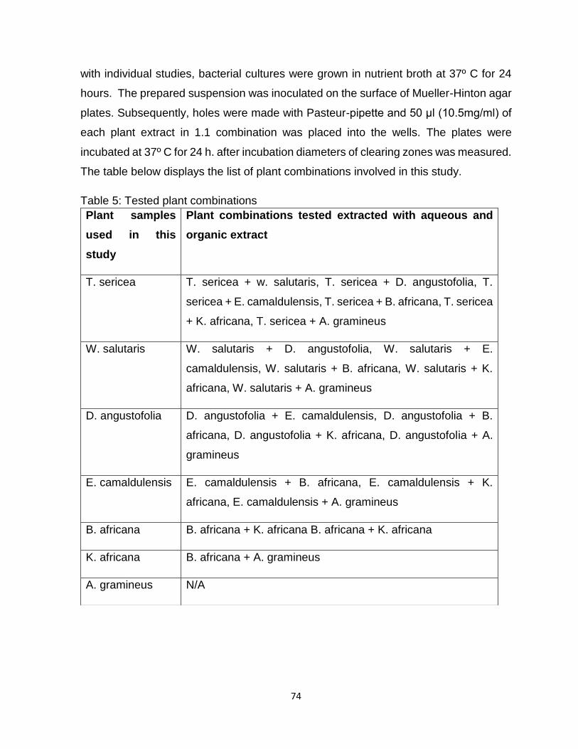
74
with individual studies, bacterial cultures were grown in nutrient broth at 37º C for 24
hours. The prepared suspension was inoculated on the surface of Mueller-Hinton agar
plates. Subsequently, holes were made with Pasteur-pipette and 50 μl (10.5mg/ml) of
each plant extract in 1.1 combination was placed into the wells. The plates were
incubated at 37º C for 24 h. after incubation diameters of clearing zones was measured.
The table below displays the list of plant combinations involved in this study.
Table 5: Tested plant combinations
Plant samples
used in this
study
Plant combinations tested extracted with aqueous and
organic extract
T. sericea T. sericea + w. salutaris, T. sericea + D. angustofolia, T.
sericea + E. camaldulensis, T. sericea + B. africana, T. sericea
+ K. africana, T. sericea + A. gramineus
W. salutaris W. salutaris + D. angustofolia, W. salutaris + E.
camaldulensis, W. salutaris + B. africana, W. salutaris + K.
africana, W. salutaris + A. gramineus
D. angustofolia D. angustofolia + E. camaldulensis, D. angustofolia + B.
africana, D. angustofolia + K. africana, D. angustofolia + A.
gramineus
E. camaldulensis E. camaldulensis + B. africana, E. camaldulensis + K.
africana, E. camaldulensis + A. gramineus
B. africana B. africana + K. africana B. africana + K. africana
K. africana B. africana + A. gramineus
A. gramineus N/A
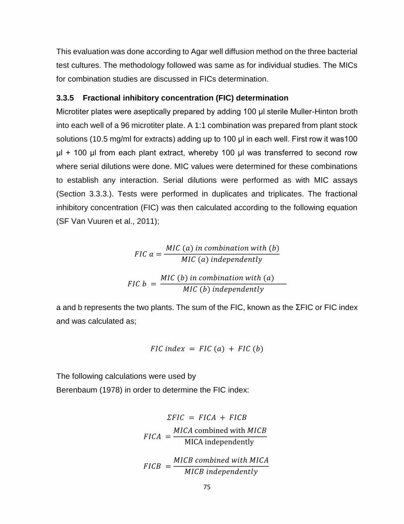
75
This evaluation was done according to Agar well diffusion method on the three bacterial
test cultures. The methodology followed was same as for individual studies. The MICs
for combination studies are discussed in FICs determination.
3.3.5 Fractional inhibitory concentration (FIC) determination
Microtiter plates were aseptically prepared by adding 100 μl sterile Muller-Hinton broth
into each well of a 96 microtiter plate. A 1:1 combination was prepared from plant stock
solutions (10.5 mg/ml for extracts) adding up to 100 μl in each well. First row it was100
μl + 100 μl from each plant extract, whereby 100 μl was transferred to second row
where serial dilutions were done. MIC values were determined for these combinations
to establish any interaction. Serial dilutions were performed as with MIC assays
(Section 3.3.3.). Tests were performed in duplicates and triplicates. The fractional
inhibitory concentration (FIC) was then calculated according to the following equation
(SF Van Vuuren et al., 2011);
𝐹𝐼𝐶 𝑎 = 𝑀𝐼𝐶 (𝑎) 𝑖𝑛 𝑐𝑜𝑚𝑏𝑖𝑛𝑎𝑡𝑖𝑜𝑛 𝑤𝑖𝑡ℎ (𝑏)
𝑀𝐼𝐶 (𝑎) 𝑖𝑛𝑑𝑒𝑝𝑒𝑛𝑑𝑒𝑛𝑡𝑙𝑦
𝐹𝐼𝐶 𝑏 = 𝑀𝐼𝐶 (𝑏) 𝑖𝑛 𝑐𝑜𝑚𝑏𝑖𝑛𝑎𝑡𝑖𝑜𝑛 𝑤𝑖𝑡ℎ (𝑎)
𝑀𝐼𝐶 (𝑏) 𝑖𝑛𝑑𝑒𝑝𝑒𝑛𝑑𝑒𝑛𝑡𝑙𝑦
a and b represents the two plants. The sum of the FIC, known as the ƩFIC or FIC index
and was calculated as;
𝐹𝐼𝐶 𝑖𝑛𝑑𝑒𝑥 = 𝐹𝐼𝐶 (𝑎) + 𝐹𝐼𝐶 (𝑏)
The following calculations were used by
Berenbaum (1978) in order to determine the FIC index:
𝛴𝐹𝐼𝐶 = 𝐹𝐼𝐶𝐴 + 𝐹𝐼𝐶𝐵
𝐹𝐼𝐶𝐴 =𝑀𝐼𝐶𝐴 combined with 𝑀𝐼𝐶𝐵
MICA independently
𝐹𝐼𝐶𝐵 =𝑀𝐼𝐶𝐵 𝑐𝑜𝑚𝑏𝑖𝑛𝑒𝑑 𝑤𝑖𝑡ℎ 𝑀𝐼𝐶𝐴
𝑀𝐼𝐶𝐵 𝑖𝑛𝑑𝑒𝑝𝑒𝑛𝑑𝑒𝑛𝑡𝑙𝑦

76
As projected by previous researchers, in order to determine plant interaction, if a
combination is synergistic, additive, non-interactive or antagonistic, the methods by
Berenbaum (1978) have been adjusted by Van Vuuren and Viljoen (2011), where each
ratio (represented by an isobologram data point) was interpreted as having synergistic
(≤ 0.5), additive (> 0.5 - 1.0), non-interactive (> 1.0 - ≤ 4.0) or antagonistic (> 4.0)
interaction (Table 5). The interpretation by Van Vuuren and Viljoen (2011) is a more
conservative analysis of interpreting synergism.
During the current study, these calculations has been used, bringing into account the
plant extracts forming part of each combination, but not their actual amount in the
interaction. The FIC index Schelz (2006), was used to determine the correlation
between the two plants and may be classified as either synergistic (≤ 0.5), additive (>
0.5-1.0), indifferent (> 1.0-4.0) or antagonistic (> 4.0). Conventional antimicrobials were
included in all repetitions as controls and tests were undertaken in triplicate. In order to
measure the activities of combinations, FIC indices were calculated for the 1:1
combination since isobologram studies were not done. The ΣFIC was calculated from
MIC values obtained from plants investigated independently (Chapter 3, Section 3.3.3)
and for each combination. The ΣFIC expresses the interaction of each plant extract in
combination as a fraction of the effect when it is used independently (Van Vuuren and
Viljoen, 2011). The MIC results for the combined plant extracts (where applicable) were
recorded in triplicate as put in chapter 4, against each of the three test organisms. The
average MIC result for each combination was tabulated, along with their sum fractional
inhibitory concentration (ΣFIC). A difference of no more than one dilution factor was
accepted within these repetitions.
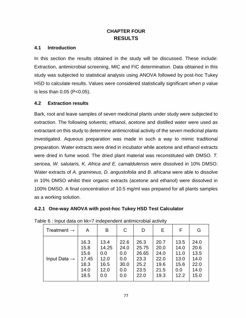
77
CHAPTER FOUR RESULTS
4.1 Introduction
In this section the results obtained in the study will be discussed. These include:
Extraction, antimicrobial screening, MIC and FIC determination. Data obtained in this
study was subjected to statistical analysis using ANOVA followed by post-hoc Tukey
HSD to calculate results. Values were considered statistically significant when p value
is less than 0.05 (P<0.05).
4.2 Extraction results
Bark, root and leave samples of seven medicinal plants under study were subjected to
extraction. The following solvents; ethanol, acetone and distilled water were used as
extractant on this study to determine antimicrobial activity of the seven medicinal plants
investigated. Aqueous preparation was made in such a way to mimic traditional
preparation. Water extracts were dried in incubator while acetone and ethanol extracts
were dried in fume wood. The dried plant material was reconstituted with DMSO. T.
sericea, W. salutaris, K. Africa and E. camaldulensis were dissolved in 10% DMSO.
Water extracts of A. gramineus, D. angustofolia and B. africana were able to dissolve
in 10% DMSO whilst their organic extracts (acetone and ethanol) were dissolved in
100% DMSO. A final concentration of 10.5 mg/ml was prepared for all plants samples
as a working solution.
4.2.1 One-way ANOVA with post-hoc Tukey HSD Test Calculator
Table 6 : Input data on kk=7 independent antimicrobial activity
Treatment → A B C D E F G
Input Data →
16.3 15.8 15.6 17.45 18.3 14.0 18.5
13.4 14.25 0.0 12.0 16.5 12.0 0.0
22.6 24.0 0.0 0.0 30.0 0.0 0.0
26.3 25.75 26.65 23.3 25.2 23.5 22.0
20.7 20.0 24.0 22.0 19.6 21.5 19.3
13.5 14.0 11.0 13.0 15.6 0.0 12.2
24.0 20.6 13.5 14.0 22.0 14.0 15.0
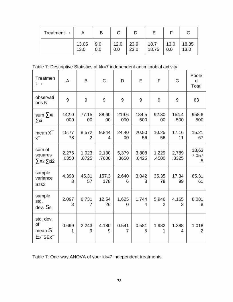
78
Treatment → A B C D E F G
13.05 13.0
9.0 0.0
12.0 0.0
23.9 23.0
18.7 18.75
13.0 0.0
18.35 13.0
Table 7: Descriptive Statistics of kk=7 independent antimicrobial activity
Treatment →
A B C D E F G Poole
d Total
observations N
9 9 9 9 9 9 9 63
sum ∑xi
∑xi
142.0000
77.1500
88.6000
219.6000
184.5500
92.3000
154.4500
958.6500
mean x¯x¯
15.7778
8.5722
9.8444
24.4000
20.5056
10.2556
17.1611
15.2167
sum of squares
∑x2i∑xi2
2,275.6350
1,023.8725
2,130.7600
5,379.3650
3,808.6425
1,229.4500
2,789.3325
18,637.057
5
sample variance
s2s2
4.3988
45.3157
157.3178
2.6406
3.0428
35.3578
17.3499
65.3161
sample std.
dev. Ss
2.0973
6.7317
12.5426
1.6250
1.7444
5.9462
4.1653
8.0818
std. dev. of
mean SEx¯SEx¯
0.6991
2.2439
4.1809
0.5417
0.5815
1.9821
1.3884
1.0182
Table 7: One-way ANOVA of your kk=7 independent treatments
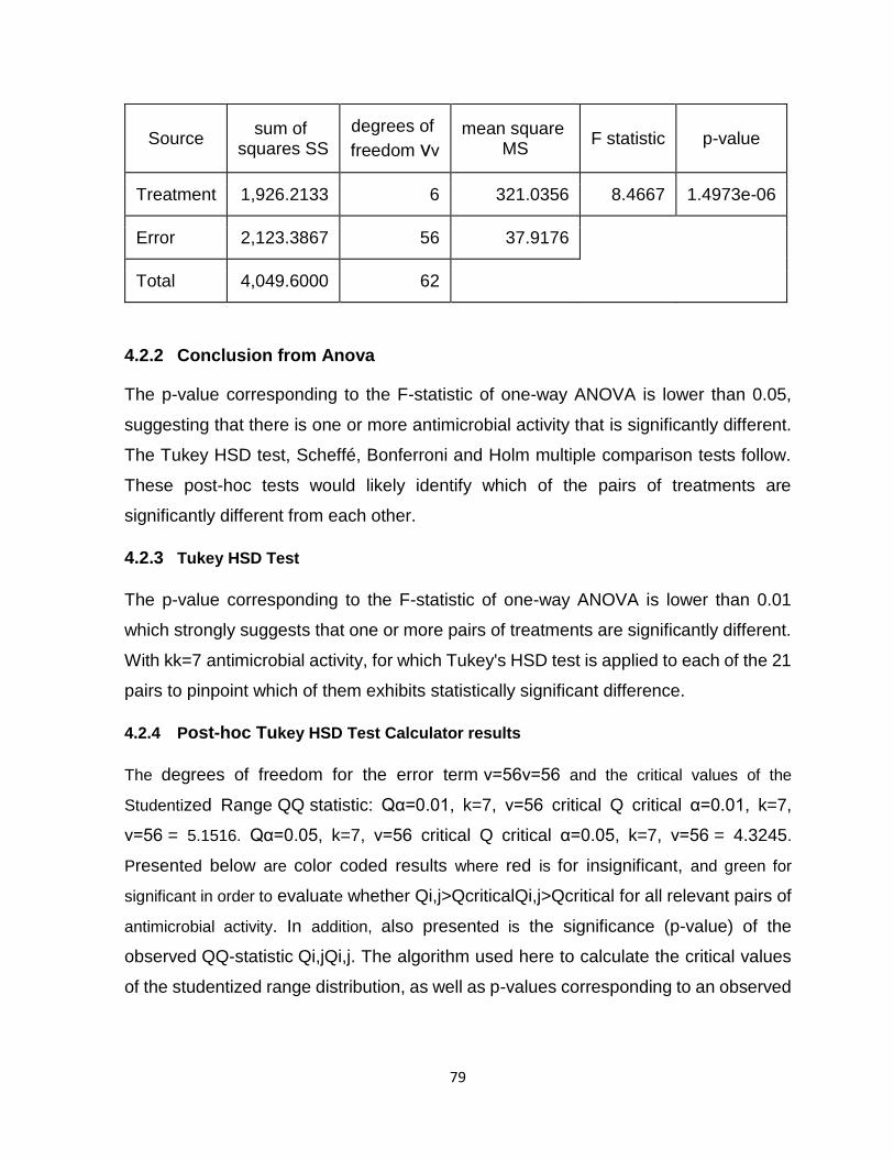
79
Source sum of
squares SS
degrees of
freedom νν
mean square MS
F statistic p-value
Treatment 1,926.2133 6 321.0356 8.4667 1.4973e-06
Error 2,123.3867 56 37.9176
Total 4,049.6000 62
4.2.2 Conclusion from Anova
The p-value corresponding to the F-statistic of one-way ANOVA is lower than 0.05,
suggesting that there is one or more antimicrobial activity that is significantly different.
The Tukey HSD test, Scheffé, Bonferroni and Holm multiple comparison tests follow.
These post-hoc tests would likely identify which of the pairs of treatments are
significantly different from each other.
4.2.3 Tukey HSD Test
The p-value corresponding to the F-statistic of one-way ANOVA is lower than 0.01
which strongly suggests that one or more pairs of treatments are significantly different.
With kk=7 antimicrobial activity, for which Tukey's HSD test is applied to each of the 21
pairs to pinpoint which of them exhibits statistically significant difference.
4.2.4 Post-hoc Tukey HSD Test Calculator results
The degrees of freedom for the error term ν=56ν=56 and the critical values of the
Studentized Range QQ statistic: Qα=0.01, k=7, ν=56 critical Q critical α=0.01, k=7,
ν=56 = 5.1516. Qα=0.05, k=7, ν=56 critical Q critical α=0.05, k=7, ν=56 = 4.3245.
Presented below are color coded results where red is for insignificant, and green for
significant in order to evaluate whether Qi,j>QcriticalQi,j>Qcritical for all relevant pairs of
antimicrobial activity. In addition, also presented is the significance (p-value) of the
observed QQ-statistic Qi,jQi,j. The algorithm used here to calculate the critical values
of the studentized range distribution, as well as p-values corresponding to an observed
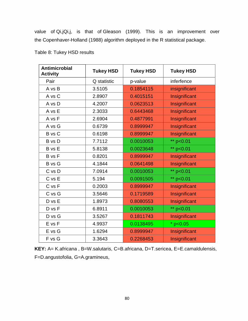
80
value of Qi,jQi,j, is that of Gleason (1999). This is an improvement over
the Copenhaver-Holland (1988) algorithm deployed in the R statistical package.
Table 8: Tukey HSD results
Antimicrobial Activity
Tukey HSD Tukey HSD Tukey HSD
Pair Q statistic p-value inferfence
A vs B 3.5105 0.1854115 insignificant
A vs C 2.8907 0.4015151 Insignificant
A vs D 4.2007 0.0623513 Insignificant
A vs E 2.3033 0.6443468 Insignificant
A vs F 2.6904 0.4877991 Insignificant
A vs G 0.6739 0.8999947 Insignificant
B vs C 0.6198 0.8999947 Insignificant
B vs D 7.7112 0.0010053 ** p<0.01
B vs E 5.8138 0.0023648 ** p<0.01
B vs F 0.8201 0.8999947 Insignificant
B vs G 4.1844 0.0641498 Insignificant
C vs D 7.0914 0.0010053 ** p<0.01
C vs E 5.194 0.0091505 ** p<0.01
C vs F 0.2003 0.8999947 Insignificant
C vs G 3.5646 0.1719589 Insignificant
D vs E 1.8973 0.8080553 Insignificant
D vs F 6.8911 0.0010053 ** p<0.01
D vs G 3.5267 0.1811743 Insignificant
E vs F 4.9937 0.0138495 * p<0.05
E vs G 1.6294 0.8999947 Insignificant
F vs G 3.3643 0.2268453 Insignificant
KEY: A= K.africana , B=W.salutaris, C=B.africana, D=T.sericea, E=E.camaldulensis,
F=D.angustofolia, G=A.gramineus,
81
4.3 Antimicrobial screening
Warburgia salutaris, Eucalyptus camaldulensis, Ballota africana, Kigelia africana,
Terminalia sericea, Acorus gramineus, and Dodonea angustofolia were tested against
three organisms. Plant extracts were screened against strains of Klebsiella
pneumoniae and Staphylococcus aureus ATCC 43300 and results obtained displayed
significant antimicrobial activity. Test strains used in this study are known to cause
bacterial pneumonia, and were tested against these plants extracts to find their
scientific antibiotic effectiveness in pneumonia treatment as speculated.
4.3.1 Staphylococcus aureus
Plant extracts tested against Staphylococcus aureus demonstrated different ranges of
results based on extraction solvent used as shown in T able 5. K. Africana displayed
sensitivity results with little variation in zone of inhibition when tested with all extraction
solvents: 16.3 mm, 15.8 mm, 15.6 mm, water ethanol and acetone respectively.
W. salutaris and B. africana demonstrated sensitivity with S. aureus on water and
ethanol extracts as follows; 13.4 mm on water and 14.25 mm on ethanol and the latter
22.6 mm on water and 24 mm on ethanol respectively with no zone of inhibition (ZOI)
on acetone extracts of both plants.
S. aureus was highly susceptible to all three of the Terminalia sericea extracts as it
showed an inhibition zone of 25.75 mm on ethanol, 26.65 mm on acetone, and 26.3
mm on aqueous extract.
Aqueous, acetone and ethanolic extracts of E. camaldulensis were tested for their
antibacterial activity, by showing various degrees of activity (24 mm Acetone, 20 mm
Ethanol and 20.7 mm with water extracts water).

82
Table 9: Screening results mean values of antimicrobial activity of plant extracts at a
concentration of 10.5 mg/ml against strains of test organisms using the well diffusion
method
Plant extracts
S. aureus ATCC 43300 S. pyogenes K. pneumoniae
H2O E A H2O E A H2O E A
K. Africana 16.3±1.
9 15.8±1.5
15.6±1.8
17.5±2.1
18.3±2.5
14±2.1 18.5±1.4
5 13.05±3.
1 13±3.5
W. salutaris 13.4±1.
5 14.25±1.
3 - 12±1.0
16.5±0.5
12±0.2 - 9±0.8 -
B. Africana 22.6±4.
1 24±4.3 - - 30±1.0 - - 12±2.0 -
T. sericea 26.3±2.
0 25.7±0.9
26.6±1.8
23.3±2.0
25.2±1.2
23.5±1.0
22±1.9 23.9±1.0 -
E.camaldulensis
20.7±1.7
20±1.5 24±2.1 22±2.0 19.6±1.
6 21.5±1.
4 19.3±2.6 18.7±3.4
18.7±2.9
D. angustofolia 13.5±2.
3 14±1.3 11±0.6 13±1.7
15.6±1.1
- 12.2±1.8 13±1.5 -
A. gramineus 24±4.0 20.6±3.4 13.5±1.
5 14±2.5 22±3.3 14±3.4 15±1.3 18.4±3.7 13±1.0
CIP5 28±0.9 10±0.98 35±0.26
C30 25±1.4 - 14±0.75
T30 22±0.97 - -
PG10 9±0.72 21±0.66 -
A10 - 25±1.02 12±0.75
S10 11±0.58 - 19±0.30
DMSO 0% - - -
DMSO 100% - - -
Key: E = Ethanol extracts; A= Acetone extract; H2O = water/aqueous extract; (-) = No
growth inhibition; Numerical figures = representing bacteria zones of inhibition
measured in millimeter. Ciprofloxacin (CIP5), chloramphenicol (C30), Tetracycline (30),
Amoxicillin (A10), Penicillin (G10), Streptomycin (S10), DMSO = Dimethyl-sulfoxide;
(-) = No clearing
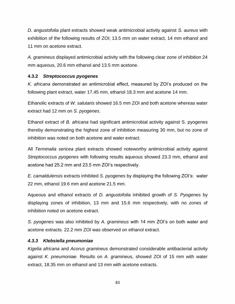
83
D. angustofolia plant extracts showed weak antimicrobial activity against S. aureus with
exhibition of the following results of ZOI; 13.5 mm on water extract, 14 mm ethanol and
11 mm on acetone extract.
A. gramineus displayed antimicrobial activity with the following clear zone of inhibition 24
mm aqueous, 20.6 mm ethanol and 13.5 mm acetone.
4.3.2 Streptococcus pyogenes
K. africana demonstrated an antimicrobial effect, measured by ZOI’s produced on the
following plant extract, water 17.45 mm, ethanol 18.3 mm and acetone 14 mm.
Ethanolic extracts of W. salutaris showed 16.5 mm ZOI and both acetone whereas water
extract had 12 mm on S. pyogenes.
Ethanol extract of B. africana had significant antimicrobial activity against S. pyogenes
thereby demonstrating the highest zone of inhibition measuring 30 mm, but no zone of
inhibition was noted on both acetone and water extract.
All Terminalia sericea plant extracts showed noteworthy antimicrobial activity against
Streptococcus pyogenes with following results aqueous showed 23.3 mm, ethanol and
acetone had 25.2 mm and 23.5 mm ZOI’s respectively.
E. camaldulensis extracts inhibited S. pyogenes by displaying the following ZOI’s: water
22 mm, ethanol 19.6 mm and acetone 21.5 mm.
Aqueous and ethanol extracts of D. angustofolia inhibited growth of S. Pyogenes by
displaying zones of inhibition, 13 mm and 15.6 mm respectively, with no zones of
inhibition noted on acetone extract.
S. pyogenes was also inhibited by A. gramineus with 14 mm ZOI’s on both water and
acetone extracts. 22.2 mm ZOI was observed on ethanol extract.
4.3.3 Klebsiella pneumoniae
Kigelia africana and Acorus gramineus demonstrated considerable antibacterial activity
against K. pneumoniae. Results on A. gramineus, showed ZOI of 15 mm with water
extract, 18.35 mm on ethanol and 13 mm with acetone extracts.

84
K. africana showed various degrees of activity, and highest observed on water extract
18.5, 13.05 mm with ethanol and 13 mm on acetone extract.
Both aqueous and acetone extract of W. salutaris and B. africana were not effective
against K. pneumoniae except ethanol extract which displayed ZOI of 9 mm with W.
salutaris and 12 mm with B. africana
Like the other two organisms tested K. pneumoniae was highly susceptible to T. sericea
with average diameter zone of inhibitions of 23.9 mm on ethanolic extract, 23 mm acetone
extract and 22 mm on aqueous extract.
E. camaldulensis also displayed significant anti-Klebsiella activity with zones of inhibition
of 19.3 mm on water, 18.7 mm ethanol and 18.75 mm on acetone extract.
D. angustofolia displayed minimal antimicrobial activity against K. pneumoniae with ZOI
of 12.2 mm on water extract and 13 mm ethanol and no inhibition was seen with acetone
extract.
Table 10: positive and negative controls of agar diffusion method
Test organism CIP5 C30 T30 PG10 A10 S10 DMSO
10%
DMSO
100%
S. aureus 28 25 22 9 - 11 - -
S. pyogenes 10 - - 21 -25 - - -
K. pneumoniae 35 14 - - 12 19 - -
Key: Ciprofloxacin (CIP5), chloramphenicol (C30), Tetracycline (30), Amoxicillin (A10),
Penicillin (G10), Streptomycin (S10), DMSO = Dimethyl- sulfoxide; (-) = No clearing
The paper diffusion disc produced the following results (table 3) in diffusion assay. No
zones on inhibition was observed around wells where DMSO (10 & 100%) was placed
4.4 Evaluation of plant extracts bioactivity in combination studies
The crude extracts of seven medicinal plants were combined in 1.1 ratios and screened
for their antimicrobial activity against three bacterial pathogens. Ethanol, acetone and
aqueous extracts of all plants studied exhibited broad spectrum of antimicrobial activity.
Plant combinations studies revealed different degrees of antimicrobial activity with some
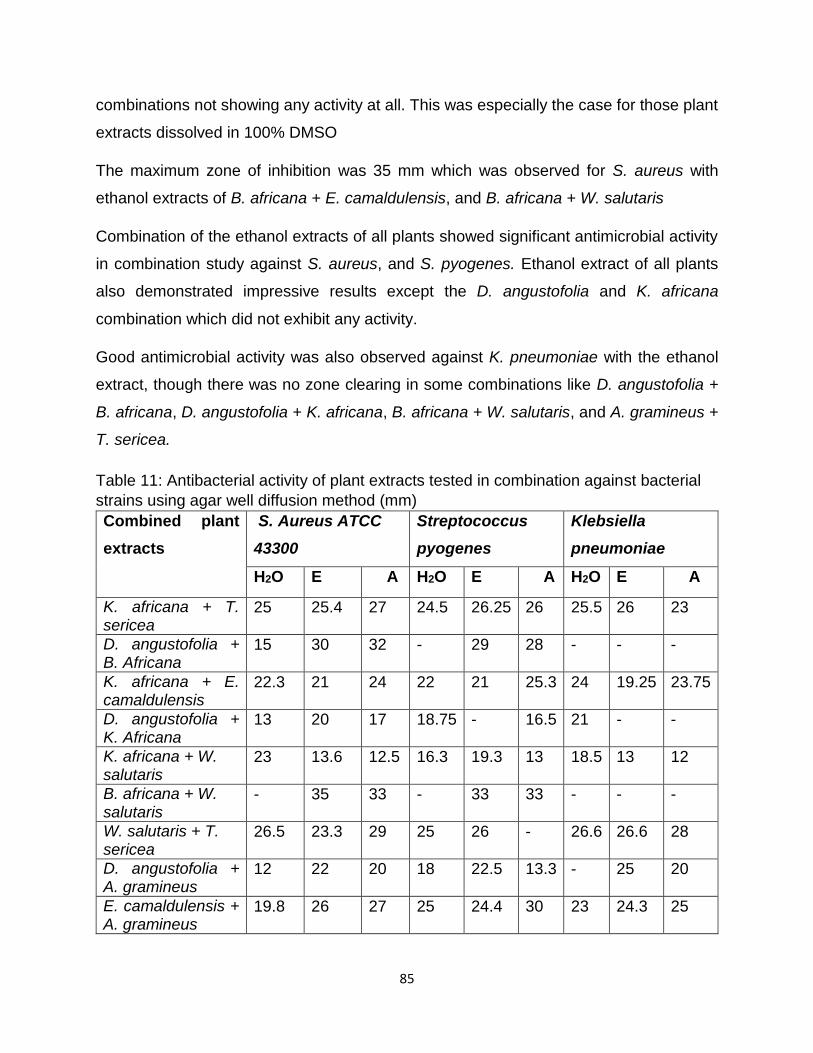
85
combinations not showing any activity at all. This was especially the case for those plant
extracts dissolved in 100% DMSO
The maximum zone of inhibition was 35 mm which was observed for S. aureus with
ethanol extracts of B. africana + E. camaldulensis, and B. africana + W. salutaris
Combination of the ethanol extracts of all plants showed significant antimicrobial activity
in combination study against S. aureus, and S. pyogenes. Ethanol extract of all plants
also demonstrated impressive results except the D. angustofolia and K. africana
combination which did not exhibit any activity.
Good antimicrobial activity was also observed against K. pneumoniae with the ethanol
extract, though there was no zone clearing in some combinations like D. angustofolia +
B. africana, D. angustofolia + K. africana, B. africana + W. salutaris, and A. gramineus +
T. sericea.
Table 11: Antibacterial activity of plant extracts tested in combination against bacterial
strains using agar well diffusion method (mm)
Combined plant
extracts
S. Aureus ATCC
43300
Streptococcus
pyogenes
Klebsiella
pneumoniae
H2O E A H2O E A H2O E A
K. africana + T. sericea
25 25.4 27 24.5 26.25 26 25.5 26 23
D. angustofolia + B. Africana
15 30 32 - 29 28 - - -
K. africana + E. camaldulensis
22.3 21 24 22 21 25.3 24 19.25 23.75
D. angustofolia + K. Africana
13 20 17 18.75 - 16.5 21 - -
K. africana + W. salutaris
23 13.6 12.5 16.3 19.3 13 18.5 13 12
B. africana + W. salutaris
- 35 33 - 33 33 - - -
W. salutaris + T. sericea
26.5 23.3 29 25 26 - 26.6 26.6 28
D. angustofolia + A. gramineus
12 22 20 18 22.5 13.3 - 25 20
E. camaldulensis + A. gramineus
19.8 26 27 25 24.4 30 23 24.3 25
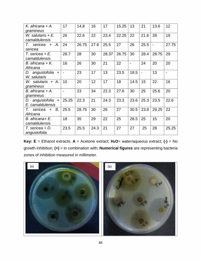
86
K. africana + A. gramineus
17 14.8 16 17 15.25 13 21 13.6 12
W. salutaris + E. camaldulensis
26 22.6 22 23.4 22.25 22 21.6 28 19
T. sericea + A. sericea
24 26.75 27.6 25.5 27 26 25.5 - 27.75
T. sericea + E. camaldulensis
28.7 28 30 28.37 26.75 30 28.4 28.75 29
B. africana + K. Africana
16 26 30 21 22 - 24 20 20
D. angustofolia + W. salutaris
- 23 17 13 23.5 18.5 - 13 -
W. salutaris + A. gramineus
10 20 12 17 18 14.5 15 22 16
B. africana + A. gramineus
- 23 34 22.3 27.6 30 25 25.6 20
D. angustofolia + E. camaldulensis
25.25 22.3 21 24.3 23.3 23.6 25.3 23.5 22.6
T. sericea + B. Africana
25.5 28.75 30 26 27 30.5 23.8 29.25 22
B. africana+ E. camaldulensis
18 35 29 22 25 28.5 25 15 20
T. sericea + D. angustofolia
23.5 25.5 24.3 21 27 27 25 28 25.25
Key: E = Ethanol extracts; A = Acetone extract; H2O= water/aqueous extract; (-) = No
growth inhibition; (+) = in combination with; Numerical figures are representing bacteria
zones of inhibition measured in millimeter.
(a) (b)

87
A
susceptibility evaluation of S. aureus with all combination of plant extract fractions
displayed notable interactions particularly with ethanol and acetone plant extracts, since
sensitivity was observed in all 21 combinations. However, not all aqueous extract
combination exhibits antimicrobial activity against the bacteria tested. As indicated in
Table 11, B. africana + A. gramineus, D. angustofolia + W. salutaris, and B. africana and
W. salutaris did not inhibit bacterial growth.
All plant extract combination displayed significant activity against S. pyogenes. The
results from activity evaluations of ethanolic extract showed inhibitory potential in all
combinations with the exception of D. angustofolia and K. africana. The highest activity
was observed with ethanol and acetone extracts of B. africana and W. salutaris with ZOI
of 33 mm, followed by T. Sericea and E. camaldulensis and also E. camaldulensis and A.
gramineus acetone extracts with Z0I of 30mm; lowest activity was observed with water
extracts of W. salutaris and D. angustofolia with ZI of 13 mm.
(c) (d)
Figure 8 = (a) ZOIs of ethanol extracts combination against S. pyogenes plate; (b)
= ZOIs of acetone extracts combinations against S. aureus; (c) = ZOIs of acetone
extracts combinations against K. pneumoniae (d) = ZOIs of acetone extracts
combinations against S. pyogenes.

88
Klebsiella pneumoniae also exhibited pronounced antimicrobial activity with all plant
extract combinations, with a few of them as shown in the Table 11. D. angustofolia + B.
africana, B. africana + W. salutaris did not show any antimicrobial activity with any crude
plant extract. The D. Angustofolia and K. africana combination, demonstrated
antimicrobial activity with only water extract showing a ZOI of 21 mm. Conversely, the A.
gramineus and D. angustofolia combination exhibited antimicrobial activity in ethanol and
acetone crude extract, unlike water extract which did not inhibit growth of K. pneumoniae.
The A. gramineus and T. sericea combination had a ZOI of 25.5 mm and 27.75 mm on
aqueous and acetone respectively against K. pneumoniae, with no activity on ethanol
extract. The combination of D. angustofolia + W. salutaris showed antimicrobial activity
with only ethanol extract for K. pneumoniae.
Figure 9: (a) = ZOIs of aqueous extracts combination against K. pneumoniae (b) =
ZOIs of ethanol extracts combinations against S. aureus.
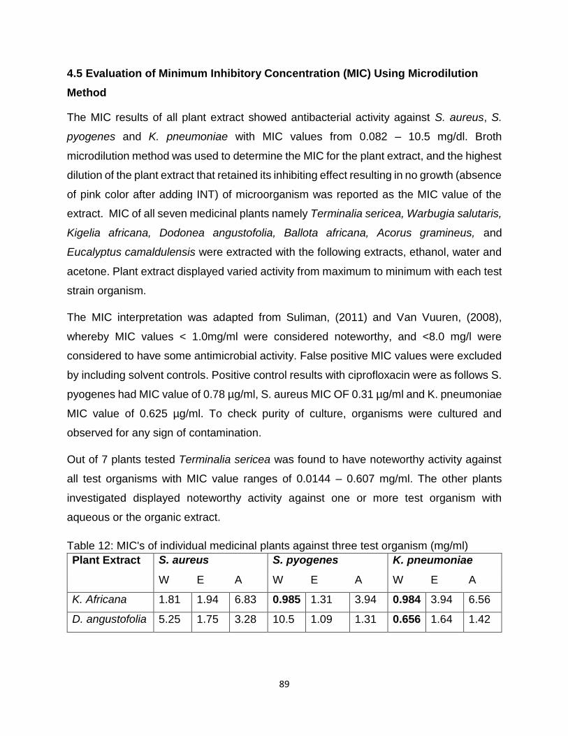
89
4.5 Evaluation of Minimum Inhibitory Concentration (MIC) Using Microdilution
Method
The MIC results of all plant extract showed antibacterial activity against S. aureus, S.
pyogenes and K. pneumoniae with MIC values from 0.082 – 10.5 mg/dl. Broth
microdilution method was used to determine the MIC for the plant extract, and the highest
dilution of the plant extract that retained its inhibiting effect resulting in no growth (absence
of pink color after adding INT) of microorganism was reported as the MIC value of the
extract. MIC of all seven medicinal plants namely Terminalia sericea, Warbugia salutaris,
Kigelia africana, Dodonea angustofolia, Ballota africana, Acorus gramineus, and
Eucalyptus camaldulensis were extracted with the following extracts, ethanol, water and
acetone. Plant extract displayed varied activity from maximum to minimum with each test
strain organism.
The MIC interpretation was adapted from Suliman, (2011) and Van Vuuren, (2008),
whereby MIC values < 1.0mg/ml were considered noteworthy, and <8.0 mg/l were
considered to have some antimicrobial activity. False positive MIC values were excluded
by including solvent controls. Positive control results with ciprofloxacin were as follows S.
pyogenes had MIC value of 0.78 µg/ml, S. aureus MIC OF 0.31 µg/ml and K. pneumoniae
MIC value of 0.625 µg/ml. To check purity of culture, organisms were cultured and
observed for any sign of contamination.
Out of 7 plants tested Terminalia sericea was found to have noteworthy activity against
all test organisms with MIC value ranges of 0.0144 – 0.607 mg/ml. The other plants
investigated displayed noteworthy activity against one or more test organism with
aqueous or the organic extract.
Table 12: MIC's of individual medicinal plants against three test organism (mg/ml)
Plant Extract
S. aureus S. pyogenes K. pneumoniae
W E A W E A W E A
K. Africana 1.81 1.94 6.83 0.985 1.31 3.94 0.984 3.94 6.56
D. angustofolia 5.25 1.75 3.28 10.5 1.09 1.31 0.656 1.64 1.42

90
E.
camaldulensis
1.31 2.63 1.31 1.31 0.656 0.328 0.739 1.31 1.37
B. Africana 3.94 0.76 1.31 1.48 0.164 0.492 2.30 2.63 1.31
W. salutaris 5.25 2.66 4.38 7.88 3.5 5.25 1.72 7.22 3.06
A. gramineus 5.25 0.739 1.31 2.63 1.31 1.97 1.64 2.63 2.63
T. Sericea 0.607 0.328 0.492 0.191 0.0144 0.267 0.369 0.547 0.191
Key: E= Ethanol extracts; A= Acetone extract; W = water/aqueous extract; Numerical
Figures are representing concentration of plant extracts at which it was able to inhibit
bacterial growth in mg/ml; Noteworthy activity values are indicated in bold.
Minimum antimicrobial activity was observed with Dodonea angustofolia and Warburgia
salutaris with the highest MIC values of 10.5 and 7.8 mg/ml respectively with water extract
on S. pyogenes. In contrast, Dodonea angustofolia water extract on K. pneumoniae
demonstrated noteworthy activity with MIC values of 0.656mg/ml, as well as water extract
of Warbugia salutaris which displayed a good MIC of 1.72mg/ml. The highest
antimicrobial activity observed against S. aureus with an ethanol extract of Dodonea
angustofolia was MIC value of 1.75mg/ml and 2.66mg/ml obtained with Warbugia
salutaris.
The Ballota africana Ethanol extract had the overall strongest antibacterial activity, having
MIC values of 0.164 as shown in Table 12 against S. pyogenes. The activity was observed
against S. aureus with the water extract having a MIC of 3.94 mg/ml. The aqueous and
organic extracts of Ballota africana also had microbial activity against K. pneumoniae with
MIC ranges 1.31 – 2.63 mg/ml.
The aqueous extract of Kigelia africana showed noteworthy MIC values of 0.985 and
0.984 mg/ml against S. pyogenes and K. pneumoniae respectively. The same organisms
exhibited weakest activity against the acetone extract of Kigelia africana as displayed in
Table 12 with MIC values ranging from 6.56 – 6.85 mg/ml.
The organic extracts of Eucalyptus camaldulensis showed noteworthy activity with
organic extract against S. pyogenes, specifically the acetone extract which had a MIC
value of 0.328 mg/ml and ethanol had a MIC of 0.656 mg/dl. With the aqueous extract of

91
Eucalyptus camaldulensis, noteworthy activity was observed with MIC value of
0.739mg/ml for K. pneumoniae. Antibacterial activity was also observed against S. aureus
with all plant extracts with MIC values ranging from 1.31 – 2.63 mg/ml.
The ethanol extract of Acorus gramineus ethanol extract also had noteworthy activity
against S. aureus with a MIC value of 0.739 mg/ml. All A. gramineus plant extracts
exhibited antibacterial activity against all test organisms with MIC values ranging from a
minimum of 0.739mg/ml and maximum of 5.25 mg/ml. It is important to take note that
each plant extract in this study was tested in triplicate against test organisms, and mean
value was calculated to come up with final MIC value, moreover, within these tests a rerun
difference of no more than one dilution factor was accepted.
Controls used in the MIC assays; antimicrobial control ciprofloxacin was used at initial
concentration of 2.5 mg/ml. This conventional antimicrobial drug was used to serve as a
positive control, where it was added to each MIC assay to validate antimicrobial
susceptibility of the test organisms.
Some organic extracts like B. africana, D. angustofolia and A. gramineus could not be re-
dissolved adequately with 10 % DMSO as a result they were dissolved in 100% DMSO.
Since all plant extracts were dissolved in 10 and 100% DMSO, these solvents were added
to each MIC assay as a solvent control in order to verify that inhibitory activity observed
were from plant extract. In order to check the viability of tested organisms streak plate
was prepared to determine purity of culture by identifying single colonies.
4.5.1 MIC values of combination studies
As before, MIC assays were performed independently to determine antimicrobial activity
of plants being investigated. The MIC values of the combined plant extracts MIC were
determined by broth dilution, and the highest dilution of the plant extract that retained its
inhibitory effect with no growth of microorganism was recorded as the MIC value. The
antibacterial activities of extract in combination of the plants displayed different degrees
of MIC result as shown in Table 13. Results displayed a variety of pharmacological
activity, from synergistic, additive, indifferent to antagonistic against the bacterial strains
under investigation. Ciprofloxacin was used as the positive control in this study and 10%
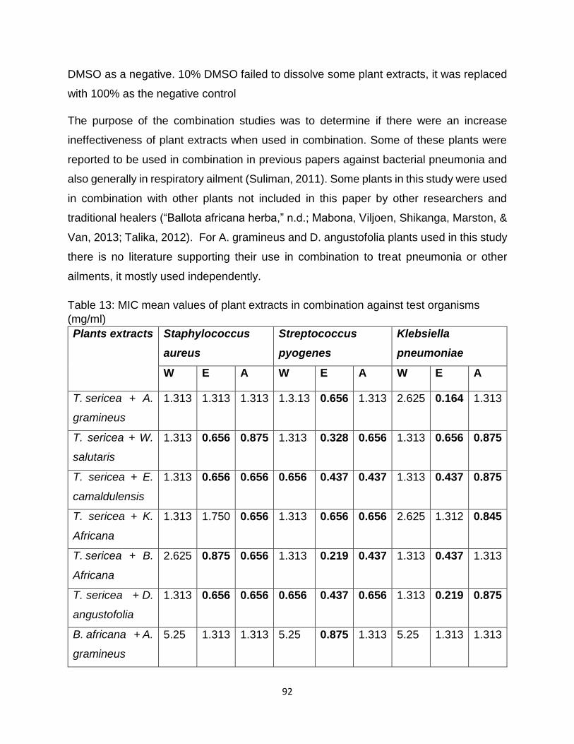
92
DMSO as a negative. 10% DMSO failed to dissolve some plant extracts, it was replaced
with 100% as the negative control
The purpose of the combination studies was to determine if there were an increase
ineffectiveness of plant extracts when used in combination. Some of these plants were
reported to be used in combination in previous papers against bacterial pneumonia and
also generally in respiratory ailment (Suliman, 2011). Some plants in this study were used
in combination with other plants not included in this paper by other researchers and
traditional healers (“Ballota africana herba,” n.d.; Mabona, Viljoen, Shikanga, Marston, &
Van, 2013; Talika, 2012). For A. gramineus and D. angustofolia plants used in this study
there is no literature supporting their use in combination to treat pneumonia or other
ailments, it mostly used independently.
Table 13: MIC mean values of plant extracts in combination against test organisms
(mg/ml)
Plants extracts Staphylococcus
aureus
Streptococcus
pyogenes
Klebsiella
pneumoniae
W E A W E A W E A
T. sericea + A.
gramineus
1.313 1.313 1.313 1.3.13 0.656 1.313 2.625 0.164 1.313
T. sericea + W.
salutaris
1.313 0.656 0.875 1.313 0.328 0.656 1.313 0.656 0.875
T. sericea + E.
camaldulensis
1.313 0.656 0.656 0.656 0.437 0.437 1.313 0.437 0.875
T. sericea + K.
Africana
1.313 1.750 0.656 1.313 0.656 0.656 2.625 1.312 0.845
T. sericea + B.
Africana
2.625 0.875 0.656 1.313 0.219 0.437 1.313 0.437 1.313
T. sericea + D.
angustofolia
1.313 0.656 0.656 0.656 0.437 0.656 1.313 0.219 0.875
B. africana + A.
gramineus
5.25 1.313 1.313 5.25 0.875 1.313 5.25 1.313 1.313

93
B. africana + D.
angustofolia
5.25 0.656 2.625 5.25 0.875 1.313 1.313 0.656 1.313
B. africana + E.
camaldulensis
1.313 1.313 2.625 0.875 1.751 1.313 1.313 2.625 0.609
B. africana +
K. Africana
2.625 2.625 1.313 1.313 2.206 0.875 2.625 2.625 2.625
B. africana + W.
salutaris
5.25 2.625 1.313 5.25 1.313 2.625 2.625 2.625 1.75
K. africana + E.
camaldulensis
2.625 2.625 0.328 0.875 2.625 0.656 1.313 2.625 5.25
K. africana +
W. salutaris
5.25 7.0 5.25 2.625 5.2 10.5 6.563 7.0 10.5
K. africana + A.
gramineus
2.625 5.25 2.625 2.625 1.313 2.625 5.25 5.25 5.25
K. africana + D.
angustofolia
2.625 1.313 1.313 2.625 2.625 0.328 2.625 2.625 0.875
D. angustofolia
+ A. gramineus
10.5 1.313 1.313 10.5 1.313 1.313 0.656 0.656 0.656
D. angustofolia
+ E.
camaldulensis
2.625 0.41 2.625 1.313 1.313 1.313 1.313 0.825 0.492
W. salutaris +D.
angustofolia
1.313 1.750 0.875 5.25 0.875 1.313 5.25 0.656 1.313
W. salutaris +
A. gramineus
5.25 2.625 2.625 5.25 2.625 5.25 5.25 1.313 2.625
W. salutaris +E.
camaldulensis
2.625 2.625 2.625 2.625 1.750 2.625 1.313 2.625 3.5
A. gramineus +
E.
camaldulensis
2.625 1.751 1.313 5.25 1.313 2.625 0.656 1.313 0.656
94
Key: E= Ethanol extracts; A= Acetone extract; W = water/aqueous extract; Numerical
figures are representing concentration of combined plant extracts at which it was able to
inhibit bacterial growth in mg/ml; noteworthy activity values are indicated in bold.
The organic extracts in the combination studies had the highest MIC values in comparison
to aqueous extract as shown with values in Table 9. The noteworthy MIC values were
observed against all microorganisms under study with plant combination of T. sericea +
E. camaldulensis and T. sericea + D. angustofolia and T. sericea + A. gramineus with
ranges from 0.164 – 0.875 mg/ml.
Strong to weak antibacterial activity was observed with mixtures of the following plants;
K. africana + W. salutaris, B. africana + W. salutaris, K. africana + A. gramineus, and also
A. gramineus + W. salutaris. Their MIC values ranged from 1.313 – 10.5 mg/ml. Aqueous
extracts of a number of combinations demonstrated very weak antimicrobial activity. For
instance, the water extract of D. angustofolia + A. gramineus against S. aureus and S.
pyogenes had MIC value of 10.5 mg/dl, the same MIC value was also observed on K.
africana + W. salutaris acetone extract against S. pyogenes. Generally organic extracts
exhibited good antibacterial activity compared to aqueous extract with all test organisms
investigated.
4.6 Fractional inhibitory concentration in 1:1 combination
The ƩFIC values of seven plants which were extracted by distilled water, acetone and
ethanol and tested against three pneumonia pathogens which were in 1:1 combination
are given in Table 14.

95
Table 14: FIC values of all plant extracts in various combinations against three strains of
organisms
Plant Staphylococcus Aureus Streptococcus Pyogenes Klebsiella Pneumoniae
H2o Eth Ace H2o Eth Ace H2O Eth Ace
T+A 1.207 2.86 1.835 3.687 23.05 2.792 4.3505 0.181 3.585
T+W 1.207 1.1235 0.973 3.521 11.4 1.291 2.161 0.649 2.434
T+E 1.582 1.125 0.917 1.968 15.51 1.485 2.668 0.567 2.61
T+K 1.444 3.119 0.715 4.104 23 1.312 4.491 1.366 2.967
T+B 2.501 1.911 0.917 3.881 8.273 1.263 2.065 0.483 3.937
T+D 1.207 1.188 0.767 1.749 15.38 1.478 2.031 0.774 2.599
B+A 1.167 1.753 1 2.774 3.002 1.668 2.742 0.5 0.75
B+D 1.167 0.619 1.4 2.024 3.069 1.835 1.286 0.328 0.963
B+E 0.667 1.114 2.004 0.63 6.675 3.335 1.174 1.5 0.445
B+K 1.058 2.404 0.596 1.11 7.567 1 1.905 0.832 1.2
B+W 1.166 2.221 0.65 2.107 4.193 2.92 3.337 1.816 5.723
K+E 1.725 1.176 0.149 0.778 3 1.084 1.556 1.333 2.316
K+W 1.951 3.12 0.984 1.5 2.728 2.333 5.243 1.374 2.442
K+A 0.975 4.905 2 1.833 1 1 4.268 1.665 2.852
K+D 1.95 0.714 0.296 1.458 2.205 0.167 3.334 1.134 0.375
D+A 2 1.264 0.7 2.5 1.103 0.834 0.52 0.325 0.356
D+E 1.25 0.195 1.4 0.563 1.603 2.5 1.889 0.567 0.359
W+D 0.25 0.829 0.467 0.583 0.527 0.625 11.05 0.0491 0.677
W+A 1 2.276 1.3 1.333 1.375 1.833 3.127 0.341 0.929
W+E 1.25 0.993 1.3 1.167 1.584 4.25 1.27 1.182 1.852
E+A 1.25 1.518 1 3.01 1.5 4.666 0.644 0.751 0.364
Key: Eth = Ethanol extracts; Ace = Acetone extract; H2O = water/aqueous extract; B =
Ballota africana; A = Acorus gramineus; W = Warburgia salutaris, E = Eucalyptus
camaldulensis; D = Dodonea angustofolia; K = Kigelia africana; T = Terminalia sericea;
Numerical Figures are representing the calculated FIC’s from individual and combined
MIC’s; synergistic and additive activity values are indicated in bold.
Out of the 21 combinations of plants tested in this study against the pathogens, varied
results were obtained with different combinations and with different plant extracts.
Noteworthy antimicrobial activity with synergy was noted with some combinations, while

96
some combinations were showing additive interactions, as well as indifferent and
antagonistic interactions.
The combination of the ethanol extract of T. sericea and A. gramineus showed noteworthy
activity which proved to be synergistic against K. pneumoniae with ƩFIC values = 0.181.
The remaining results with this combination showed indifferent results with all extracts in
all three organisms.
When combined, T. sericea + W. salutaris showed broad spectrum antimicrobial activity,
though additive interactions were only portrayed with ethanol extract against K.
pneumoniae and acetone extract against S. aureus. The rest of organic extracts of this
combination displayed indifferent interactions to all three organisms except S. aureus with
ethanol extract which demonstrated antagonistic interactions.
Noteworthy antimicrobial activity was reported with the ethanol combination of T. sericea
and E. camaldulensis for K. pneumoniae with ƩFIC value of .567, and acetone extract for
S. aureus with calculated ƩFIC of 0.917, both depicting additive interactions. The rest of
the results displayed indifferent values in all extracts against all organisms, excluding
ethanol extract on S. pyogenes which had antagonistic interactions (ƩFIC value = 15.51).
0ut of T. sericea and K. africana plant combination extracts, additive interactions were
observed with acetone extracts for S. aureus with ƩFIV value of 0.715, but no synergy
was observed on this combination. The remaining extracts of this combination had non-
interactive antimicrobial activity with ƩFIC values ranging from 1.312 – 3.119, but water
and ethanol extract demonstrated antagonistic reactions with ƩFIC range value =
4.104-23.
The combined effect of T. sericea and B. africana ethanol extract showed synergistic
effect on K. pneumoniae. Acetone extract on S. aureus demonstrated additive
interactions. All aqueous and acetone extracts of this combination when tested for K.
pneumoniae and S. pyogenes demonstrated indifferent activity, same as ethanol extract
on S. aureus and S. pyogenes.
Ethanol extract of T. sericea and D. angustofolia combination for K. pneumoniae and S.
aureus acetone extract showed additive reactions with ƩFIC value = 0.774 and 0.767

97
respectively, but the rest of plant extracts displayed indifferent antimicrobial interactions
expect ethanol extract on S. pyogenes which had antagonistic interactions with ƩFIC
value =15.38.
Both plant combinations of B. africana + A. gramineus and B. africana + K. africana
displayed additive interactions with ƩFIV value ranging 0.596 – 1.0 with ethanol and
acetone extracts on S. aureus, S. pyogenes as well as K. pneumoniae. Moreover, K.
pneumoniae demonstrated synergistic interactions with A. gramineus and B. africana
ethanol extract with ƩFIC value of 0.5. Both combination also showed indifferent
interactions against all test organisms, with the rest of plant extracts.
When testing B. africana and D. angustofolia against all bacteria test strains, ethanol
extract on K. pneumoniae proved to be synergistic with ƩFIC value of 0.328 (Table 14).
Additive interaction was observed with ethanol extract combination for S. aureus and
acetone extract for K. pneumoniae respectively.
Worth noting antibacterial activity was reported with Ballota africana and Eucalyptus
camaldulensis combination acetone extract on K. pneumoniae. Aqueous extract of the
same combinations displayed additive interactions against S. aureus and S. pyogenes.
Ethanol extracts showed antagonistic interactions with ƩFIC value = 6.675 against S.
pyogenes but the rest of extract combinations had indifferent interactions with test
pathogens.
These combinations; Ballota africana and Warburgia salutaris, Kigelia africana and
Warbugia salutaris, portrayed additive interactions with acetone extract on S. aureus. The
rest of the results were indifferent plant with all three organisms (Table 14).
The combination of Kigelia and Eucalyptus showed noteworthy antimicrobial activity with
synergistic interaction with acetone extract on S. aureus and ƩFIC value was reported,
while water extract of this combination demonstrated additive interaction on S. Pyogenes,
but the rest of results showed indifferent interactions with all organisms.
On combining the water extracts of K. africana and A. gramineus, good antimicrobial
activity was observed which proved to be additive against both S. aureus and S. pyogenes
with ƩFIC values of 0.975 and 1.0 respectively. Ethanol extract combinations against S.
98
pyogenes also demonstrated additive interactions but the rest of extracts demonstrated
indifferent interactions.
Both organic extracts of the Kigelia africana and Dodonea angustofolia combination
displayed strong antibacterial activity against S. aureus with ƩFIC values of 0.296
(synergistic) and 0.714 (additive) for acetone extract and ethanol extract respectively.
Synergistic interactions were also observed with acetone extract against S. pyogenes
with ƩFIC value of 0.167 and 0.375 with K. pneumoniae, the rest of extracts in all tested
pathogens had indifferent interactions.
K. pneumoniae was strongly inhibited by all extract of the Dodonea angustofolia and
Acorus gramineus combination. The water extract demonstrated an additive interaction
(ƩFIC value = 0.52). Acetone and ethanol plant extracts had synergistic interactions with
ƩFIC values of 0.325 and 0.356 respectively. S. aureus and S. pyogenes both displayed
additive interactions with acetone extracts but the rest showed indifferent interactions with
these organisms.
The Combination of D. angustofolia and E. camaldulensis showed noteworthy
antibacterial activity against pneumonia pathogens in this study. Synergistic interactions
were observed with ethanol and acetone extract of this combination. Against S. aureus
and K. pneumoniae with ƩFIC values of 0.195 and 0.359 respectively. Additive
interactions were observed for S. pyogenes and K. pneumoniae with ethanol extract of
the combination of D. angustofolia and E. camaldulensis but the rest of plant extracts
demonstrated indifferent interactions with all three organisms (Table 14).
The most interesting synergistic interactions at the ƩFIC value of 0.0491 with Warburgia
salutaris and Dodonea angustofolia was detected against K. pneumoniae. Synergistic
interaction was also noted with water and acetone extract against S. aureus with ƩFIC
values of 0.25 and 0.467 respectively. Additive interactions were observed for Warburgia
salutaris and Dodonea angustofolia combination with ethanol extract against S. aureus
and S. pyogenes, water extract for S. pyogenes and acetone extract against K.
pneumoniae. Antagonistic interactions were observed with water extract of this
combination against S. aureus.

99
A combination of Warburgia salutaris and Acorus gramineus showed major antibacterial
activity. For the ethanol extract when screened against K. pneumoniae, synergistic
interaction was observed with ƩFIC value of 0.341. An additive interaction was noted for
S. aureus with water extract and K. pneumoniae with acetone extract. However, the rest
of the extracts displayed indifferent interactions.
The Combination of Warburgia salutaris and Eucalyptus camaldulensis exhibited
antibacterial activity and a ƩFIC value of 0.993 was found with ethanol extract against S.
aureus. But the remaining plant extracts displayed indifferent interactions with all
pneumonia test pathogens.
When testing the acetone extracts of Eucalyptus camaldulensis and Acorus gramineus
against K. pneumoniae, synergistic interaction was depicted with ƩFIC value = 0.364.
However, ethanol and water extracts displayed additive interactions against K.
pneumoniae. Additive interaction was also observed with acetone extract against S.
aureus. All the plant extract combinations against the three test organism displayed
results which all fall within the non-interactive range.
100
CHAPTER FIVE
DISCUSSIONS
5.1 Introduction
Seven medicinal plants namely Terminalia sericea, Warburgia salutaris, Ballota africana,
Kigelia africana, Acorus gramineus, Dodonea angustofolia, and Eucalyptus
camaldulensis were used in this study. As discussed in chapter three, these plants were
extracted with aqueous and organic solvents, which were eventually re-dissolved in 10%
and 100% DMSO. Thereafter they were analysed for their antimicrobial activity against
pneumonia pathogens precisely staphylococcus aureus, Streptococcus pyogenes, and
Klebsiella pneumonia. Information related to extract reaction on tested organism for
antimicrobial activity which was reported in chapter 4 will be further discussed in this
chapter in relation to previous studies where these plants or similar ones were used.
Some of the plants used in this study have interesting records that are locally vital and
widely used but little or nothing has been recorded about them in scientific literature. As
expressed in chapter 3, the following methods: agar well diffusion, microtiter dilution
method were used for the current study to come up with plant extract antimicrobial
efficacy.
The purpose of the study was to determine the prospective antimicrobial activity of the
plants under study, which are traditionally used to treat pneumonia. With the widespread
problem of bacterial resistance development to antibiotics, there is an urge to look for
new potential antimicrobials(Van Vuuren, 2010). Results obtained in this study showed
that organic plant extracts relatively had higher inhibitory antimicrobial activity when
compared with aqueous extract. As related to previous antimicrobial studies, Bamuamba
et al, in agreement with van Vuuren and Naidoo, Kerinki and Nyiroge, Ncube et al., (as
cited in Talika,2012) suggested that in scenarios where water was used for extraction it
showed poor or no inhibitory activity, since it is too polar to efficiently dissolve organic
elements derived from plant components. In chapter two each plant was discussed based
on their chemical constituency, medicinal use, and originality value of the incident and
possible new uses that haven’t been provided in harmony with list of critical literature
references.

101
Agar well diffusion assay which was preferred method in this study for testing
antimicrobial activity for plant extract showed interesting zones of inhibition and poor
inhibitions with some of the plant extracts.
5.2 Susceptibility testing of individual plants
The aqueous and organic extracts of Terminalia sericea showed very good antimicrobial
activity against all test organism with zones of inhibition ranging from 22-27.75 mm. In all
three test organisms, organic extracts had a slightly large inhibition zone compared to
aqueous extracts. S. aureus demonstrated very good susceptibility activity among the
three organisms with Terminalia sericea. MIC values obtained for this plant also
demonstrated noteworthy antimicrobial activity with all test bacteria strains, eventually S.
pyogenes reporting high MIC values. Terminalia sericea was the strongest /or potent
inhibitor of all seven plants under study with each and every extract against all tested
organisms. MIC values ranging from 0.0144-0.607 mg/ml were reported after calculating
mean values from triplicates results at a concentration of extracts from 0.0821 to 10.5
mg/ml.
Eyhrqust et al., Mushi and Mbwambo, Eldeen et al: a, Eldeen et al: b (as cited in
Lembede, 2014) reported that both aqueous and organic decoction extracts of T. sericea
displayed antimicrobial activity against S. aureus, S. epidermidis, S. pyogenes, E.
aerogenes, B. subtlis, M. luteus, E. coli, K. pneumonia and Sacini specia. Apart from the
organisms mentioned above its antifungal activity against Candida albicans, Aspergillus
niger, Aspergillus fumigates, Cryptococcus neorformans, Microsporium canis, and
Sporothrix schenckii (Eldeen et al & Fyhrqustet et al as cited in Lambede, 2014) described
the potentiation of T. sericea antimicrobial activity to be due to presence of
triperpenesaponins, tannins, flavonoids and aligananon B.
In previous study, Cock and Vuuren (2014) about African plants with anti-Klebsiella
activity for the treatment and prevention of ion Ankylosing Spondylitis, Terminalia sericea
methanol and water extracts were potent inhibitors of K. pneumoniae with ZOIs of 13±0
and 12.0±1.0 mm respectively. Unlike other plants that were used in that study T. sericea
were amongst the most potent inhibitors of K. pneumoniae growth. Terminalia sericea
has been extensively studied by many researchers especially for conditions associated

102
with diarrhea and respiratory ailments (Suliman 2010). Mabira et al, (2013) reported that
T. sericea display mostly worthy noting broad spectrum antimicrobial efficacy against skin
conditions related organisms used in the study, supporting its folkloric use for
dermatological related ailments.
As discussed by Cock and Vuuren (2014), anti-Proteus activity of some medicinal plants,
such as Terminalia sericea were evaluated, and displayed outstanding antibacterial
activity with inhibition zones >10 mm. It was also reported that T. sericea and W. salutaris
have sesuiterponoids (including polygidal and tannins) which are known to have
antibacterial activity. Kwanyama (2013) also concurred with the results for the current
study, since noteworthy activity results to six pathogens with MIC range 0.04-1.00 mg/ml
were reported. Talika (2010) also reported the antimicrobial activity of Terminalia sericea
exhibited in bark extracts against S. aureus. Therefore, based on results obtained from
previous and current study we can easily stipulate that, almost all plant parts of Terminalia
sericea have some antibacterial activity against Pneumonia causing pathogens.
Eucalyptus camaldulensis, showed good antimicrobial activity having highest inhibition
zone of 24mm with acetone extracts but overall all three organism displayed good
antibacterial activity with ZOI ranging from18.7 – 24mm. A worthy noting activity with E.
camaldulensis, MIC value of 0.325 mg/dl against S. Pyogenes with acetone extract was
observed. Based on results obtained for the present study it indicated that E.
camaldulensis can work better in some organisms at higher dosages as an antimicrobial
agent in treatment of bacterial pneumonia, because extracts displayed different
antibacterial activity with different organisms.
Behbahan and Yadzi (2013,) found E. camaldulensis ethanol and aqueous extract to
exhibit good antimicrobial activity against S. aureus with ZOIs average diameter of 20.2
mm at (20 mg/ml) 22 mm at (40 mg/ml) 25.1 mm (60 mg/ml) 26.7 mm at (80 mg/ml), whilst
15 mm, 19 mm, 21 mm, and 24.5 mm were also observed at the same working
concentration as above. When tested against S. aureus for MIC determination ethanol
extract demonstrated half down equal MIC value, and least MIC values were reported on
remaining organisms involved in the study. In a previous study Ayepola and Adeniyi
(2008) revealed that the methanol extracts, dichloromethane fraction and the methanol
103
residue showed varied degrees of bacteria inhibition on S. aureus, B. subtlis, P.
aeruginosa, K. pneumoniae and Y. enterocolitica. The MIC of methanol extract and
dichloromethane fraction which ranged from between 0.04-0.079 mg/ml correlated to
current study which also demonstrated good antimicrobial activity with some extracts.
Jouda (2013), observed antibacterial activity of E. camaldulensis ethanol extract against
S. aureus with ZOI of 13 mm, which was corresponding to current study which reported
20 mm with same organism. As to aqueous extract results were discordant with current
study, Jouda (2013) observed ZOI of 8 mm and 20.7 mm was reported in present study.
Lowest MIC with ethanol extract was from 6.25 mg/ml according to Jouda (2013), 2.63
mg/ml with the same extract against S. aureus. This conformity of result can be due to
varied concentration used, growing conditions, extraction methods and so many
parameters.
Kigelia africana extracts had antibacterial activity against all three organisms involved in
this study with ZOIs ranging from 13 – 18.5 mm and interesting MIC values, which showed
noteworthy antimicrobial activity of 0.985 mg/ml and 0.984 for S. pyogenes and K.
pneumonia with aqueous extract: supporting its use traditionally for the treatment of
different ailment. In a study Abdulkadir (2015), discovered that K. africana chloroform,
methane and aqueous extracts displayed pronounced activity against S. aureus,
compared to K. pneumoniae and other organisms involved in the study. Based on that
research K. africana was found to be rich in flavonoids and phenolic compounds which
are assumed to be responsible for their antimicrobial activity and antioxidant potential.
In another study Owalabi and Omogbai (2007), reported that K. africana at 20mg/ml
showed inhibitory zone of 15.0 ± 0.95 mm with ethanolic extract, which is in alignment
with present study which had 15.6 – 16.3 mm with all extracts against S. aureus. In that
same study antifungal activity was observed for Candida albicans with 20.25 ± 4.60 mm
ZOI, but the rest of organisms like E. coli and P. aeruginosa did not show any susceptible
activity with K. africana. MIC values of 6.25mg/ml and 7.92 mg/ml were observed on S.
aureus and Candida albicans respectively, while in the current study MIC values ranging
from 0.984-6.83 mg/ml were reported with all three extracts against all test pathogens.
104
Naidoo and van Vuuren (2013), examined antimicrobial activity of K. africana in bacterial
and fungal pathogens, and found broad spectrum activity with MIC value ranging from
0.25 – 16 mg/ml with organic and aqueous extract.
It has been noted that aqueous and organic of (methanol and acetone) K. africana
demonstrated good antimicrobial activity in the following organisms, E. coli, S. aureus,
MRSA, S. flexneri and activity was observed with S. typhi. MIC values ranging from 0.139
– 5.54 mg/ml with Kigelia africana, which corresponds to the present study whereby MIC
values of 0.984 – 6.56 mg/ml were reported, hence concurring that the use of Kigelia
africana for potential antimicrobial effect (Nkosi, 2013).
Acorus gramineus in this study demonstrated good antimicrobial activity against all three
organisms with both water and organic extracts. Ethanol extract displayed good results
compared to aqueous extracts, and noteworthy activity was observed with ethanol extract
against S. aureus whilst the rest of MIC results indicate good antibacterial activity. Lee et
al., (as cited in Asha and Ganjewala (2009), reported A. graminieus rhizome (methanol
extract) to have antimicrobial and antifungal properties particularly against
phytopathogenic fungi, due to the presence of α-asarone and asaronaldehyde
compounds. Arm (n.d) also reported A. gramineus to have antibacterial activity against
S. aureus, Streptococci and Mycobacterium which is similar to the present study whereby
S. aureus and S. pyogenes growth were inhibited. Based on information encountered in
search of literature, few scientific studies have been reported by researchers about A.
gramineus, compared to Acorus calamus which has been studied intensively. Initially
Acorus calamus was planned to be used for this current study, but due to difficulty in
accessing it in Eastern Cape and near provinces the hybrid species Acorus gramineus
was used instead.
According to results obtained in this study acetone and aqueous extracts of D.
angustofolia showed antimicrobial activity with ZOI ranging from 11 – 15.6 mm, while
ethanol extract of Dodonea angustofolia did not inhibit K. pneumoniae and S. pyogenes.
This observation was probably due to polarity of plant extracts since it was dissolved in
100% DMSO and the particles were unable to diffuse in Muller Hinton agar, as compared
with MIC assay where all extracts showed antibacterial activity with all organisms. Worth
105
noting antimicrobial activity was observed with water extract against K. pneumoniae, and
yet same aqueous extract had weak antimicrobial activity for S. aureus and S. pyogenes
with MIC value 5.25 and 10.5 mg/ml respectively. Organic extracts of D. angustofolia
showed some good antimicrobial activity with all bacterial pathogens.
According to Mohlakoana (2010), D. angustofolia exhibit some antimicrobial activity
against B. cereus but other organisms like S. aureus, K. pneumoniae which were also
involved in the study do not show any activity which is in contrary to the current study
where antimicrobial activity was reported to these organisms. Gemeda et al (2011),
reported that D. angustofolia had bioactive compounds responsible for their antibacterial
activity. Water solvents were reported to have slight antibacterial prospective, followed by
hexane extractant, yet dichloromethane, acetone and methanol extractant they portion
out resemblance in bioactive compounds on bio-autographs, and extracted the highest
number of antibacterial compounds with variety of polarities.
Ballota africana displayed good antibacterial activity with all three organisms and
noteworthy activity was observed with organic extracts against S. pyogenes with ZOI 30
mm, but water and acetone extracts of B. africana did not show any antibacterial activity
with S. pyogenes and K. pneumoniae. S. aureus were inhibited by both water and ethanol
extract of the plant. Surprisingly MIC values showed noteworthy antimicrobial activity of
0.164mg/ml and 0.492 mg/ml with ethanol and acetone extract for S. pyogenes. Good
antibacterial activity was also seen on water extract with MIC of 1.48 mg/ml. B. africana
demonstrated a worthy noting activity with ethanol extract which had 0.768 mg/ml, and
acetone and water extract also demonstrated good antimicrobial activity. K. pneumonia
which was only inhibited by ethanol extract on sensitivity test, in MIC assay B. africana
revealed best antimicrobial activity with values ranging from 1.31 – 2.30 mg/ml with all
extracts. Discrepancies due to concentration of extract solvent, in this case were caused
DMSO dilution, because just like D. angustofolia and A. gramineus organic extracts, B.
africana organic extracts couldn’t be dissolved in 10% DMSO as a result 100% DMSO
was used instead, which explains the possibility of polar compounds or molecules which
were not able to diffuse in solid agar media and yet were able to show inhibitory activity
with microtiter technique. In this current study it was observed that results obtained
106
through agar well diffusion for antimicrobial sensitivity and microtiter dilution method for
MIC, gave incongruent and confusing result when organic plant extracts dissolved with
100% DMSO were used against test organisms. Results obtained from other plants
extracts in the same study like Acorus gramineus and Dodonea angustofolia whose
organic extracts were also dissolved in 100% DMSO have proved that correlation of
methodologies in determining plant antimicrobial activity doesn’t give inconsistent results
all the time. As explained by Chinyama (2009), several variables impact outcome of
results, like environment, climatic conditions, choice of plant extracts, extraction method,
antimicrobial test method, and test organisms. Furthermore, factors such as technique,
culture medium, strain of bacteria used, and age of agar plate, plant source, whether used
dried or fresh, quality of extract tested can be attributed. The explanation of possible
causes of changes was concluded by saying that there’s no standard method for
expressing the antibacterial test result (Chinyama, 2009).
B. africana leaf methanol extract was found to be less potent inhibitor of K. pneumoniae
in a study where South African plants with anti-Klebsiella activity for the treatment and
prevention of ankylosing spondylitis were being investigated. Aqueous and methanol leaf
extract of B. africana exhibited potent antimicrobial activity with MIC value normally below
700 mg/ml, which was contrary to the lower efficacy seen in disc diffusion screening. It
was further suggested that antimicrobial compounds in these plants might be either large
or low polarity and diffuse less freely in the agar gel (Cock & van Vuuren, 2014).
In previous study B. africana had some good antimicrobial activity against S. Pyogenes
with range of MIC results from 3.125 – 12.5 mg/ml with the following extracts: petroleum
ether, dichloromethane ether, water and ethanol. K. pneumoniae had MIC value ranging
from 1.56 - >12.5 mg/ml with the same extracts above, and S. aureus had noteworthy
activity with 80% ethanol and dichloromethane ether with MIC value of 0.39 mg/ml but
water and petroleum ether displayed poor MIC value which is in alignment with current
study whereby good antibacterial activity was observed with organic extract compared to
aqueous extract (Motlhatlego, 2014).
Out of the seven plants tested in this present study Warbugia salutaris was found to have
weak antimicrobial activity against K. pneumoniae, since ZOI was only observed on

107
ethanol extract, while water and acetone extract did not display any activity. S. pyogenes
surprisingly was inhibited by all Warbugia salutaris extracts and strong inhibition was
recorded on ethanol extract. Antimicrobial activity was also displayed on A. aureus by W.
salutaris water and ethanol extract. Conversely, all three test pathogens were reported to
be inhibited with W. salutaris with microdilution method having MIC values ranging from
1.72 – 7.22 mg/ml.
In a previous study Jager (2003) found that W. salutaris aqueous extract exhibited good
antimicrobial activity against gram positive bacteria (S. aureus ATCC 6538, Bacillus
subtlis ATCC 6633) and methanol extract showed activity against E. coli. Rabe and van
Staden (as cited in Vuuren, 2008), stated that Warbugia salutaris have a compound called
muzigadial, which demonstrated antibacterial activity against S. aureus, B. subtlis, E. coli,
S. epidermidis and M. luteus with MIC values ranging from 12.5 – 100 mg/ml. the same
applies to the current study whereby antimicrobial activity against three test strain of
organisms was observed by the same plant with three different extracts.
W. salutaris was also involved in dermatological relevance plants with medicinal values
research where it was found to have noteworthy antimicrobial activity with MIC values
ranging in between 0.03 > 16 mg/mi against organisms that were involved in that study.
Both bark and leaf parts of Warburgia salutaris extracts were used in that study reported
antimicrobial worthy noting activities especially observed with bark extract compared to
leaf extract (Mabona et al., 2013).These results were in agreement with Rabe and Van
Staden (1997), report of noteworthy activity for W. Salutaris with methanol extract against
S. aureus, contrary to results reported by these authors and the current study as well.
Leonard and Viljoen (2015), agree with Steenkamp et-al (2005) who observed no
antibacterial activity for the aqueous and methanol extracts of W. salutaris against S.
epidermidis and S. aureus. Results obtained in these studies displayed very good
antimicrobial activity compared to results obtained in this current study which justify the
need for further scientific validation of this plant for clinical application. The biological
activity of W. salutaris based on previous studies conducted, was found to be attributed
to the drimane, sesquiterpernoids, including polygodial, warbuganal, muzigadial, muka
108
dial, and ugandensial, flavonoid and miscellaneous compounds present (Leonard &
Viljoen, 2015).
5.3 Combination studies
In-vitro interactions between extracts of A. gramineus, W. salutaris, T. sericea, D.
angustofolia, K. africana, B. africana and E. camaldulensis were determined using agar
well diffusion assay and microdilution method.
As shown in table 3, K. africana and T. sericea demonstrated some antimicrobial activity
with all extracts, and ZOI ranging from 23-27 mm which was not far from K. africana and
E. camaldulensis which had 19.25-25.3 mm. Their MIC’s exhibited noteworthy
antimicrobial activity for S. aureus and K. pneumoniae with acetone extract, but S.
pyogenes showed noteworthy activity with MIC of 0.656 mg/ml with both acetone and
ethanol with K. africana and T. sericea combination. In contrast K. africana and E.
camaldulensis showed noteworthy antimicrobial activity against S. aureus with acetone
extracts as well as on S. pyogenes with acetone and water extracts. The rest of plant
extracts also displayed some antimicrobial activity with all three test bacterial pathogens.
Since results for some combinations were equal or equivalent to single plant tested FIC’s
was done in order to calculate interactions between these plant combinations. T. sericea
and K. africana only displayed additive outcome for S. aureus with acetone extract
whereby FIC value of 0.767 was reported. The rest displayed indifferent antagonistic
interactions which explains that these plants, especially T. sericea works better when
used individually, pertaining to ZOI and MIC values obtained on individual plant.
T. sericea leaf extract according to Talika (2012), inhibited microorganisms in combination
with L. javanica, C. molle at a 1:1:1 ratio against C. neorformans, K. pneumoniae, M.
catarrhalis, S. smigematis and S. aureus with ∑FIC range of 0.64 – 4.83 reported.
K. africana and E. camaldulensis combination demonstrated noteworthy antimicrobial
activity with acetone extract against S. aureus and S. pyogenes, but aqueous extract had
noteworthy activity with S. pyogenes as well. After calculating ∑FIC, synergistic
interactions ware only observed with acetone extract against S. aureus, while aqueous

109
extract displayed additive interaction against S. pyogenes, the rest had non-interactive
interactions.
Combination of Dodonea salutaris + Ballota africana and Ballota africana + Warburgia
salutaris in sensitivity testing did not show any antimicrobial activity against K.
pneumoniae, same with aqueous extract which did not display any activity against S.
pyogenes with both combinations. Only aqueous extract of Dodonea and Ballota had
some antimicrobial activity, while Ballota and Warbugia aqueous extract did not display
any activity.
MIC value of Ballota africana and Dodonea angustofolia combination demonstrated
noteworthy antimicrobial activity with ethanol extract in all three organisms while the
remaining extracts showed good antimicrobial activity as well. Ballota africana and
Warbugia salutaris had microbial activity with all organisms with MIC values ranging from
1.313 – 5.25 mg/ml. ƩFIC’s showed synergistic interactions with Ballota africana and
Dodonea angustofolia ethanol extract for K. pneumoniae, and additive interactions were
observed with ethanol for S. aureus and acetone for K. pneumoniae. Ballota africana and
Warbugia salutaris combination had additive interaction with acetone extract for S. aureus
but the rest of the extracts had indifference reaction.
B. africana is traditionally used in combination with Sativa species to treat measles and
fevers in “khoi khoi” and “san” remedy, while in other circumstances it is combined with
Valeriana capensisor or Stachys hispida for treatment of insomnia or nervous stress
(“Ballota africana herba,” n.d.).
The combination of Warburgia salutaris and Dodonea angustofolia interacted
synergistically against all three organisms at least with one or two plant extract; for S.
aureus water and acetone displayed synergy interactions; for S. pyogenes acetone
extract and for K. pneumoniae ethanol extract showed synergy interactions. While some
remaining extracts in the combination displayed additive interactions, antagonistic
interaction was highly observed with water extract for K. pneumoniae. Warbugia salutaris
and Dodonea angustofolia combination was more potent inhibitor against all organisms
involved in this study than all other plant combination interaction. On sensitivity testing

110
however, aqueous extract on both S. aureus and K. pneumoniae did not show any
antibacterial activity, as well as acetone extract on K. pneumoniae.
Kigelia africana and Warburgia salutaris, Kigelia africana and Acorus gramineus
combinations also showed some inhibition based on zones of inhibition ranging from
12-23 mm and 12-15.2 mm respectively, and MIC’s result also demonstrated good
antimicrobial activity. ƩFIC result of Kigelia africana and Warbugia salutaris acetone
extract displayed additive interaction for S. aureus, but rest of the ƩFIC’s values showed
indifferent and antagonistic results. Kigelia africana and Acorus gramineus had ƩFIC
values of 0.975-1 indicating additive interaction with water extract for S. aureus and S.
pyogenes as well as ethanol extract for S. pyogenes. Rest of results with all plant extracts
for the three organisms displayed indifferent interactions.
Eucalyptus camaldulensis and Acorus gramineus had good antimicrobial activity,
sensitivity showed ZOI ranging from 19.8-27 mm. Noteworthy activity on MIC’s was
observed with water and acetone extract against K. pneumoniae with reported MIC value
of 0.656 mg/ml, and the other extracts also had effective antimicrobial activity with all
three bacterial pathogens. The calculated ƩFIC as revealed in table 5, depicted
synergistic interaction with acetone extract on K. pneumoniae and additive interaction
was observed with water and acetone extract for K. pneumoniae as well as acetone
extract for S. aureus.
Terminalia sericea and Eucalyptus camaldulensis, Terminalia sericea and Dodonea
angustofolia, Terminalia sericea and Ballota africana showed good antibacterial activity
against all three bacterial pathogens with ZOI ranging from 21-35 mm and MIC values
from 0.219-2.625 mg/ml. Noteworthy antibacterial activity was observed with organic
extracts of all this three plant combinations with tested organisms, while MIC values for
aqueous extract were potent with the weakest MIC value of 2.625 mg/ml reported. S.
aureus and K. pneumoniae showed additive interactions with acetone and ethanol
extracts respectively, except Terminalia sericea and Ballota africana combination ethanol
extract which displayed synergistic interactions. Water extract of all these three
combinations displayed indifferent interaction with test organisms (ƩFIC value of
1.3132-2.265).
111
Dodonea angustofolia and Acorus gramineus, Terminalia sericea and Acorus gramineus,
and Ballota africana and Acorus gramineus demonstrated good antimicrobial activity with
all three test pathogens although aqueous extract did not show any activity with Acorus
gramineus and Dodonea angustofolia against K. pneumoniae also Ballota africana and
Acorus gramineus on S. aureus.
Terminalia sericea and Acorus gramineus aqueous extract did not show any inhibition for
K. pneumoniae as well. Dodonea angustofolia and Acorus gramineus exerted noteworthy
antibacterial activity effects against K. pneumoniae with all extracts but some good
antibacterial activity was also observed on S. aureus and S. pyogenes. Similar results
were seen with calculated ƩFIC which showed synergistic interactions with organic
extracts for K. pneumoniae and additive interactions were observed with aqueous extract
with the same organism.
Additive interaction with Dodonea angustofolia and Acorus gramineus was also displayed
with ethanol extracts against S. aureus and S. pyogenes, while the rest of extracts
showed indifferent effects with the same organisms. Ballota africana and Acorus
gramineus had noteworthy antibacterial activity against S. pyogenes with ethanol extract,
but synergistic interactions were obtained against K. pneumoniae and additive
interactions were seen with acetone extract against S. aureus and K. pneumoniae. The
MIC values of Terminalia sericea and Acorus gramineus as presented in table 5, showed
good antibacterial activity against three test organisms with MIC values ranging from
0.164 – 2.625 mg/ml. synergistic interactions were obtained against K. pneumoniae with
ethanol extract (ƩFIC value of 0.181). S. aureus and K. pneumoniae exhibited
antagonistic interaction with aqueous extract, and indifferent interaction were observed
with the rest of the plant extracts.
Ballota africana and Kigelia africana combination showed highest microbial susceptibility
with test organism which was in congruity with Warbugia salutaris and Eucalyptus
camaldulensis having equivalent inhibition patterns. Ballota africana and Kigelia africana
did not had any inhibition against K. pneumoniae with water extract. Ballota africana and
Kigelia africana noteworthy antimicrobial activity was seen with acetone extract against
112
S. pyogenes, whereby mean MIC value ranging from 1.313 – 2.625 mg/ml were obtained
with the rest of extracts combination against all three test organism.
In polyherbal preparation, ADD- 199, K. africana was used in combination with three other
plants: Maytenus senegalensis, Annona senegalensis, and Lannea welwitschii. This
combination was found to have antidiabetic and antioxidant activities (Atawodi &
Olowoniyi, 2015). In another study K. africana was combined with Hypoxis
hemerocallidea and tested against C. albicans, U. urealytcum, O. ureolytica, G. vaginalis,
N. gonorrhoea and good antimicrobial results were displayed with MIC values ranging
from 0.75 - >16 mg/ml with ∑FICs of 0.47 – 7.88. In the same study K. africana combined
with P. africanum, O. engleri and S. seratuloides yielded MIC range of 0.75 - >16mg/ml
and ∑FIC value of 0.31- 32 against the mentioned sexually transmitted infectious agents
(van Vuuren & Naidoo, 2013).
Warbugia salutaris and Eucalyptus camaldulensis combination was also active in
inhibiting all pathogens tested with mean MIC value ranging from 1.313 – 3.5 mg/ml and
was considered good antimicrobial inhibitor. ƩFIC values calculated from the combination
of Ballota africana and Kigelia africana, Warbugia salutaris and Eucalyptus camaldulensis
against all pathogens tested ranged from 0.596 – 7.567, and from 0.993 – 4.25
respectively. As shown with results above Ballota africana and Kigelia africana
combination had additive interactions for S. aureus with acetone extract, K. pneumoniae
with ethanol extract, and for S. pyogenes with acetone extract. Warbugia salutaris and
Eucalyptus camaldulensis combination ethanol extract also displayed additive
interactions for S. aureus. Antagonistic interactions were observed with Warbugia
salutaris and Eucalyptus camaldulensis acetone extract for S. pyogenes, and Ballota
africana and Kigelia africana ethanol extract for S. pyogenes as well.
Sensitivity test for Acorus gramineus and Warbugia salutaris combination showed some
good antimicrobial activity with organic extract compared to aqueous extract. The
combination showed great antimicrobial activity against all three test organism with range
of mean MIC value of 1.313 – 5.25 mg/ml. ƩFIC calculated from Acorus gramineus and
Warbugia salutaris combination against test organism in this study ranged from 0.341 –
3.127.

113
This indicate that this combination had the synergistic interaction for K. pneumoniae with
ethanol extract. Two pathogens namely K. pneumoniae and S. aureus showed additive
interactions with acetone (ƩFIC 0.929) and water (ƩFIC 1.0) extracts of this combination
respectively. The rest of the extracts demonstrated indifferent interactions against
organisms tested.
W. salutaris has been reported by Hutchings (as cited in Mabona, 2013) that when
combined with Hibiscus surattensis it is a useful treatment for sores and skin imitations.
W. salutaris is smoked with Cannabis sativa for upper respiratory tract infections. Bark or
root powder is known to be smoked with other plants and the smoke is inhaled for
expectorant and/or dry cough treatment (Leonard & Viljoen, 2015).
When combined Dodonea angustofolia and Eucalyptus camaldulensis extracts showed
some good antimicrobial activity against pneumonia pathogens with ZOI ranging from 21
– 25 mm. Noteworthy antibacterial activity was observed with organic extracts (acetone
and ethanol) for K. pneumoniae, and with ethanol extract for S. aureus. ƩFIC value of
0.195 and 0.359 were noted for S. aureus with ethanol extract and K. pneumoniae with
acetone extract respectively indicating the synergistic interactions. An additive interaction
was seen for S. pyogenes and K. pneumoniae with ethanol extract. The rest of the
Dodonea Eucalyptus extracts results displayed indifferent interaction with the pathogens
tested, no antagonistic interactions was observed.
S. aureus was inhibited with all Kigelia africana and Dodonea angustofolia extracts. S.
pyogenes growth was inhibited with water and acetone extract, whilst K. pneumoniae was
inhibited by only water extract. The MIC values observed as shown in table 5, combination
of Kigelia africana and Dodonea angustofolia showed good antimicrobial activity with all
three organisms even with those extracts combination which did not show any activity in
initial sensitivity testing. Noteworthy and actually best antimicrobial activities were seen
for this combination against S. pyogenes with acetone extract with mean MIC value of
0.328 mg/ml and also for K. pneumoniae MIC value 0.875 mg/ml. But rest of the extracts
in this combination can be classified as good inhibitors to all test organisms with highest
MIC reported as 2.625mg/ml. ƩFIC calculated showed synergistic interaction for all the
pathogens tested with acetone extract ranging from 0.167 – 0.375. An additive interaction

114
was noted against S. aureus with ethanol extract with a ƩFIC value of 0.714. Indifferent
interaction of the combination was displayed with the rest of the pathogens tested.
The combination of Ballota africana and Eucalyptus camaldulensis combination showed
generally good antimicrobial activity against the test organism with ZOI ranging from 18
– 35 mm. Noteworthy antibacterial activity was seen against S. pyogenes and K.
pneumoniae with ethanol and acetone extract respectively. ƩFIC values for the
combination against K. pneumoniae displayed synergistic interaction with acetone
extract. Based on ƩFIC values S. aureus and S. pyogenes tested with aqueous extract,
additive interactions were observed with this combination. Indifferent interactions were
seen against test organism with the rest of the plant extract, with exception of acetone
extract against S. pyogenes which exhibited antagonistic interaction.
The antimicrobial activity of Terminalia sericea and Warbugia salutaris in combination
was most effective with ZOI ranging from 23.3 – 29 mm, though acetone extract did not
show any activity against S. pyogenes. The best activity was observed with all pathogens
tested with organic extract (ethanol and acetone) of this combination at mean MIC value
range of 0.328 – 0.875 mg/ml. All organisms tested had some antimicrobial activity with
aqueous extracts and MIC of 1.313 mg/ml was reported in all of them. This combination
showed additive interactions for two pathogens namely S. aureus and K. pneumoniae
with acetone and ethanol extracts registering ƩFIC values 0.973 and 0.649 respectively.
Indifferent interaction was observed with the rest of the test organisms except from
ethanol extract against S. pyogenes which displayed antagonistic interaction.
In medicinal plants combinations, synergism mechanism may be attributed to a number
of factors such as: complex multi-target effects, pharmacokinetic or physiochemical
properties, neutralization principles or even therapeutic approaches (Van Vuuren et al.,
2011). Although only few of these plants were reported to be used in combination, majority
hasn’t been reported yet.
115
CHAPTER SIX
SUMMARY, CONCLUSIONS AND RECOMMENDATIONS
6.1 Summary
The study had a number of objectives that were centred in identifying and processing
indigenous medicinal plants that were used in investigating their in-vitro antimicrobial
activity against three pneumonia pathogens in individual and combination studies. The
main objectives of this work were achieved and reported.
In chapter one, the parameters, aims and objectives of the study were introduced.
Pneumonia being one of the clinically important infectious disease, formed the centre of
this study. Pneumonia has been reported as one of the leading causes of mortality and
morbidity worldwide. Moreover, pneumonia is known to affect mostly the under-five,
elderly, immunosuppressed or immune-compromised individuals. Different interventions,
management and treatment have been put in place. Recently, the insurgency of bacterial
resistance to antimicrobials due to inappropriate use of antimicrobials, lack of control
measures in hospitals and lack of trained laboratory personnel to perform sensitivity
testing. Furthermore, bacterial resistance can be caused by unavailability of guidelines
on selection of drugs and information on drug resistance that is not communicated to
medical practioners/clinicians (Cheesebrough, 2006).
For centuries, medicinal plants have been used as remedies for various human ailments
(Kamanga, 2013). However, use of medicinal plants known to be of native or traditional
background, yet remarkable numbers of conventional/synthetic drugs are of plant origin.
The purpose of this study was to find antimicrobial efficacy of selected indigenous plants
used independently and in combinations for bacterial pneumonia treatment. Based on
folk knowledge and literature, the following plants; Terminalia Sericea, Dodonea
anguistifolia, Acorus gramineus, Eucalyptus camaldulensis, Kigelia africana, Ballota
africana and Warbugia salutaris were reported to treat pneumonia. In the current study,
these plants were used individually and in combination against the test organisms
namely; S. aureus, S. pyogenes and K. pneumoniae. Signs, symptoms, diagnosis,
treatment, management and prevention of pneumonia infections were discussed in
chapter two. The chemical composition and properties of individual sample materials for
116
both plants and organisms were presented. Mode of action of antibiotics was also
scrutinized to draw a picture of the possible ways on how medicinal plants work. A number
of extraction methods and chemical compounds that were isolated from previous studies
were listed and discussed. Active plant compounds which were discussed in this study
are known to be the reason for their antimicrobial activity. However, some of them were
not present in the plants used in this study.
Chapter three focuses on materials and methods used to conduct and implement this
study. The organisms and materials used for culture and sensitivity studies were provided
by NMMU. Plant samples were collected from Nsalu village, Lilongwe in Malawi and Port
Elizabeth, South Africa. Extraction solvents, apparatus, equipment and materials were
provided by NMMU’s School of Biomedical Sciences, Microbiology department.
Antimicrobial assays used to analyse the medicinal plants under study were introduced.
Step-wisely, plant preparation, extraction methods and solvents and reconstitution
solvents were described. 10% and 100% DMSO reconstitution solvent and INT colour
indicator for bacterial growth where a pink or purple colour change indicated no bacterial
inhibition in microtiter plate dilution method. Techniques such as ƩFIC for calculating
interaction in combination studies were introduced.
Chapter four presents the experimental results obtained after performing different tests
on individual and combined plant extracts. Results obtained from calculating the mean
values of zone of inhibition (ZOI) in mm and minimum inhibition concentration (MIC) in
mg/ml were reported. According to results, organic extracts had relatively good
antimicrobial activity and larger ZOIs as compared to aqueous extracts. MIC mean values
were calculated for individual and combined plant extracts and then used to determine
the ƩFIC values. Based on the sets of calculated values, plant interactions were
interpreted as either synergism, additive, indifferent or antagonistic interactions. Plant
extracts had varying antimicrobial effects. For instance, plants such as Terminalia
sericea, Ballota africana and Eucalyptus camaldulensis displayed good antimicrobial
activity for the individual studies. Dodonea angustofolia and Warbugia salutaris which had
small ZOIs in individual studies, showed good antimicrobial activity in combination
studies. Combination of D. angustofolia and W. salutaris had significant ƩFIC value for all
117
test organisms ranging from 0.0491- 0.829 which indicated synergistic and additives
interactions, except water extract tested for K. pneumoniae had an antagonistic
interaction with ƩFIC of 11.05. A number of plants demonstrated additive reaction on
either water, ethanol or acetone extract on combinations such as Terminalia sericea and
Kigelia africana, Kigelia africana and Acorus gramineus, Ballota africana and Warburgia
salutaris and Warbugia salutaris and Eucalyptus camaldulensis. However, the rest of their
plant extract combination showed indifferent and antagonistic interactions. All medicinal
plants involved in this study showed some antibacterial activity for all the three test
organisms. It was also noted that organic extracts had greater effect in inhibiting bacterial
growth than water extract. This is justified by Jouda (2013) where organic solvent is best
for extracting active compounds from plants in relation to distilled water.
6.2 Conclusion
The purpose of this study was to validate use of indigenous medicinal plants in individual
and combination studies for treatment of bacterial pneumonia. The study substantiated
the reasoning that the use of combined medicinal plants enhances antimicrobial activity.
The results obtained from this study justify the use of plants in combinations, since results
displaying synergism and additive interactions on plants were less effective when used
individually.
The study evaluated the antimicrobial activities of seven indigenous medicinal plants
against three test organisms when used individually and in combination. Organic extracts
displayed good antimicrobial activity compared to aqueous extract pertaining to ZOIs
measured and MICs on both individual and combination studies on all test organisms.
Despite outstanding activities with organic extracts, aqueous extracts displayed some
good activity in some individual and combined plant extracts which agrees with traditional
use in form of infusion and decoction (Suliman, 2011).
The current antimicrobial study validates use of medicinal plants for treatment of
pneumonia. Terminalia sericea aqueous and organic extracts displayed noteworthy
antimicrobial activity against all three test organism on individual studies with ZOIs
ranging from 22-27.75 mm, and MICs of 0.0144-0.607 mg/ml. But the highest ZOI
(35 mm) was observed with Ballota africana ethanol extract for S. pyogenes. Other plants
118
had noteworthy activity with either one or two extracts against tested organism, while
others against at least one of the tested three organisms. Warbugia salutaris on the
other hand displayed weak antimicrobial activities against all three tested organisms with
both well diffusion and microtiter plate for all extracts.
On combination studies, a number of plant combinations displayed good antimicrobial
activity with large ZOIs and lowest MICs. T. sericea and E. camaldulensis extracts had
noteworthy antimicrobial activity with all three test pathogens with ZOI of 26.75-30 mm.
Nevertheless, highest ZOIs was shown in B. africana and W. salutaris organic extract
combination for S. aureus and S. pyogenes ranging from 33-35 mm, although 35 mm was
also observed in B. africana and E. camaldulensis ethanol extract against S. aureus.
Lowest MICs in combinations was observed with T. sericea and A. gramineus ethanol
extract against K. pneumoniae. However, T. sericea and E. camaldulensis, B. africana
and T. sericea, T. sericea and D. angustofolia, T. sericea and W. salutaris had noteworthy
antimicrobial activity for all tested organisms with noted dominance on organic extracts.
Significant plant interactions against test pathogens were noted for W. salutaris and D.
angustofolia combination with ∑FICs values ranging from 0.049-0.829 for all extracts,
except 11.05 which was calculated for K. pneumoniae with aqueous extract where
antagonistic interaction was observed. Antagonistic effect was noted in T. sericea and E.
camaldulensis ethanol combination with ∑FIC value of 15.51. Many combinations had
synergism or additive interaction with either one or two extracts against one of the test
organism. Synergism mechanism may be attributed to complex multi-target effects,
pharmacokinetic or physiochemical properties, neutralization principles or even
therapeutic approaches (Van Vuuren et al., 2011). Although only few of these plants were
reported to be used in combination, majority hasn’t been reported yet.
This study validates the use of medicinal plants in combination to improve efficacy in
treatment of pneumonia. Plants used in this study can therefore be used as a primary
health care for pneumonia management. Medicinal plants can be used in preference to
conventitional drugs, since they are able to treat more than one ailment which is more
advantegous compared to conventional drugs which target specific conditions. Based
119
on availability and cost effectiveness of medicinal plants, it can benefit less privileged
people and those living in remote areas far from health facilities.
6.3 Recommendations
The study has paveway as a platform for further analysis of intricate uses of medicinal
plants in combination. However, scientific validation of the use of these plants in
combination has been established creating room for future studies in the treatment of
pneumonia as well as for formulating new antimicrobials. Medicinal plants have been
observed to contain a complex of bioactive compounds, which prevent growth of
organisms through various mechanisms than that of conventional antibiotics, hence
posing a remarkable use in treating resistance pathogens. Results obtained from this
work might be enough to serve as a platform for further studies in isolating and identifying
active compounds, since varying activities of medicinal plants are actually related to
variery contest of their active compounds. Also, toxicity studies to be performed to
determine the safety of possible adverse effect in administrating this medicinal plants.
In-vivo clinical trials should be carried out in future studies, to find us possible use of these
plant extracts in the treatment of bacterial pneumonia since what happen in-vitro does not
exactly portray what happens in in-vivo. In order to determine plant interactions, future
studies should address the mechanism of action of synergistic interactions, which will
include receptor site modification, enzymatic degradation, and accumulation of antibiotics
within bacteria cell, decreased outer membrane permeability and efflux pumps.
120
References
Abdulkadir, M. N., Adedokun, A., & John, E. (2015). Phytochemical composition and
antimicrobial evaluation of Kigelia africana LAM, 5(1), 14–17.
Abubakar, E. M. M. (2010). Antibacterial potential of crude leaf extracts of Eucalyptus
camaldulensis against some pathogenic bacteria. African Journal of Plant Science,
4(6), 202–209. Retrieved from
http://www.academicjournals.org/article/article1380118383_Abubakar.pdf\nhttp://ov
idsp.ovid.com/ovidweb.cgi?T=JS&CSC=Y&NEWS=N&PAGE=fulltext&D=cagh&AN
=20143000276\nhttp://oxfordsfx.hosted.exlibrisgroup.com/oxford?sid=OVID:caghdb
&id=pmid:&id=doi:&issn=1996-0
Al-baker, S. M., Ahmed, F., Hanash, S. H., & Al-hazmi, A. A. (2014). Prevalence and
Evaluation of Antimicrobial Activity of Dodonaea viscosa Extract and Antibacterial
Agents against salmonella Spp . Isolated from Poultry, 2, 901–908.
Anon et al. (2008). The history of medicinal plants.
Ashby, B., Turkington, C., Assad, U., El-Masri, I., Porhomayon, J., & El-Solh, A. A.
(2012). The encylopedia of infectious diseases. New York Facts on File P USA
Pneumonia Immunization in Older Adults Review of Vaccine Effectiveness and
Strategies Clinical Interventions in Aging 7 Pp, 242 SRC-, 453–461. JOUR.
Atawodi, S., & Olowoniyi, O. (2015). Pharmacological and Therapeutic Activities of
Kigelia africana (Lam.) Benth. Annual Research & Review in Biology, 5(1), 1–17.
http://doi.org/10.9734/ARRB/2015/8632
Augenbraun, M. et-al. (2014). Pneumonia in Immunocompromised Patients: Overview,
Causes of Pneumonia, HIV/AIDS. Retrieved April 20, 2016, from
http://emedicine.medscape.com/article/807846-overview#a5
Azu, O. O. (2013). The sausage plant ( Kigelia africana ): Have we finally discovered a
male sperm booster ? Journal of Medecine Plant Research, 7(15), 903–910.
http://doi.org/10.5897/JMPR12.0746
Ballota africana herba. (n.d.).

121
Balunas, M. J., & Kinghorn, A. D. (2005). Drug discovery from medicinal plants. Life
Sciences, 78(5), 431–441. http://doi.org/10.1016/j.lfs.2005.09.012
Barnes, J. (1999). A close look at synergy and polyvalent action in medicinalplants,
(1185), 3–14.
Bashar, S., & Omar, S. (2011). Traditional System, Ethics, Safety, Efficacy, and
Regulatory Issues.
Benarba, B., Belabid, L., Righi, K., Elouissi, M., Khaldi, A., & Hamimed, A. (2015).
Ethnobotanical study of medicinal plants used by traditional healers in Mascara (
North West of Algeria ). Journal of Ethnopharmacology, 175, 626–637.
http://doi.org/10.1016/j.jep.2015.09.030
Campbell, H., el Arifeen, S., Hazir, T., O’Kelly, J., Bryce, J., Rudan, I., & Qazi, S. A.
(2013). Measuring Coverage in MNCH: Challenges in Monitoring the Proportion of
Young Children with Pneumonia Who Receive Antibiotic Treatment. PLoS
Medicine, 10(5), e1001421. http://doi.org/10.1371/journal.pmed.1001421
Chang, A. B., Ooi, M. H., Perera, D., & Grimwood, K. (2013). Improving the Diagnosis,
Management, and Outcomes of Children with Pneumonia: Where are the Gaps?
Frontiers in Pediatrics, 1(October), 29. http://doi.org/10.3389/fped.2013.00029
Cheesbrough, M. (2006). District laboratory practice in tropical countries. District
laboratory practice in tropical countries Part 2 (Vol. Part 1).
Cheesebrough, M. (2006). Medical laboratory manual for tropical countries. BOOK.
Chinyama, R. F. (2009). BIOLOGICAL ACTIVITIES OF MEDICINAL PLANTS
TRADITIONALLY USED TO TREAT SEPTICAEMIA IN THE EASTERN CAPE ,.
Nelson Mandela Metropolitan University.
Cock, I. E., & van Vuuren, S. F. (2010). Anti-Proteus Activity of Some South African
Medicinal Plants : Their Potential for the Prevention of Rheumatoid Arthritis.
Cock, I. E., & van Vuuren, S. F. (2014). The potential of selected South African plants
with anti-Klebsiella activity for the treatment and prevent ion of Ankylosing
122
spondylitis. Inflammopharmacology, 23(1), 21–35. http://doi.org/10.1007/s10787-
014-0222-z
Cragg, G. M., & Newman, D. J. (2013). Natural products: A continuing source of novel
drug leads. Biochimica et Biophysica Acta - General Subjects, 1830(6), 3670–3695.
http://doi.org/10.1016/j.bbagen.2013.02.008
Cross, S. S., & Underwood, J. C. E. (2009). General and Systematic Pathology. book,
Elsevier Health Sciences UK.
Cross, S. S., & Underwood, J. C. E. (2013). Underwood’s Pathology: A Clinical
Approach. book, Churchill Livingstone.
Crozier, A., Clifford, M. N., & Ashihara, H. (2006). Plant secondary metabolites:
Occurrence, structure and role in the human diet. Oxford OX42DQ, UK: Blackwell
publishing limited. Retrieved from
http://onlinelibrary.wiley.com/book/10.1002/9780470988558
Denis, H., & Et-al. (2014). Atypical pneumonia: MedlinePlus Medical Encyclopedia.
Retrieved April 19, 2016, from
https://www.nlm.nih.gov/medlineplus/ency/article/000079.htm
Dhanalakshmi, D., Dhivya, R., & Manimegalai, K. (2013). Antibacterial activity of
selected Medicinal plants from South India, 5(April), 63–68.
Dock, E., Boskey, E., & Brian Wu. (2015). Aspiration Pneumonia: Overview, Causes &
Symptoms. Retrieved April 20, 2016, from
http://www.healthline.com/health/aspiration-pneumonia#Overview1
Drewes, S. E. (2012). Natural products research in South Africa: 1890-2010. South
African Journal of Science, 108(5–6), 1–8. http://doi.org/10.4102/sajs.v108i5/6.765
Eloff, J. N. (1998). A sensitive and quick microplate method to determine the minimal
inhibitory concentration of plant extracts for bacteria. Planta Medica, 64(8), 711–
713. http://doi.org/10.1055/s-2006-957563
Eloff, J. N. (2000). On expressing the antibacterial activity of plant extracts - A small first
123
step in applying scientific knowledge to rural primary health care. South African
Journal of Science.
Eloff JN. (2004). Quantification the bioactivity of plant extracts during screening and
bioassay guided fractionation. Phytomedicine : International Journal of
Phytotherapy and Phytopharmacology, 11(4), 370–371.
http://doi.org/10.1078/0944711041495218
Extraction methods. (2016). Retrieved from http://www.medicinalplants-
pharmacognosy.com/pharmacognosy-s-topics/extraction-methods/
Fawzi, M. M. (2013). Plants.Traditional Medicines in Africa: An Appraisal of Ten Potent
African Medicinal plants. EvidenceBased Complementary and Alternative Medicine
Hindawi Journals, 0–1. JOUR.
Frum, Y., & Viljoen, A. M. (2006). In vitro 5-Lipoxygenase and Anti-Oxidant Activities of
South African Medicinal Plants. Skin Pharmacology and Physiology, 1(6), 329–335.
http://doi.org/10.1159/000095253
Gill, L. s. (1992). Ethnomedical uses of plants in Nigeria. University of Benin Press.
Grace, O. M., Prendergast, H. D. V, Jager, A. K., & van Staden, J. (2003). Bark
medicines used in traditional healthcare in KwaZulu-Natal, South Africa: An
inventory. South African Journal of Botany, 69(3), 301–363.
http://doi.org/10.1016/S0254-6299(15)30318-5
Grbovc, S., Orcic, D., Couladis, M., Jovin, E., Burgarin, D., Balog, K., & Mimica-Dukic,
N. (2010). Variation of essential oil composition of eucalypyus camaldulensis
(Mystaceace) from the Montengero coastline. (Vol. 2015 SRC). BOOK.
Hariprasath, L. (2010). Volume : I : Issue-3 : Nov-Dec -2010 IN-VITRO
ANTIBACTERIAL ACTIVITY OF AQUEOUS AND ETHANOLIC EXTRACTS OF
ACORUS CALAMUS Department of Physiology , Sri Lakshmi Narayana Institute of
Medical Sciences Osudu , Department of Physiology , Dr . ALM . PG . IBMS , (3),
1072–1075.
Harris, S. (2015). Dudonea viscosa Jacq var, angustifolia (L.F) Benth. Retrieved from

124
Karoo Desert National Botanical Garden. (Vol. 2015 SRC). BOOK.
Joffe, P. (2003). Kigelia Africana (Lam) Benth. Retrieved from plantzafrica. (Vol. 2015
SRC). BOOK.
Jouda, M. M. (2013a). The Antibacterial Effect of Some Medicinal Plant Extracts and
their Synergistic Effect with Antibiotic and Non-antibiotic Drugs.
Jouda, M. M. (2013b). The Antibacterial Effect of Some Medicinal Plant Extracts and
their Synergistic Effect with Antibiotic and Non-antibiotic Drugs In the name of Allah
, the Beneficent , the Merciful.
Kabra, S. K., Lodha, R., & Pandey, R. M. (2010). Antibiotics for community-acquired
pneumonia in children. The Cochrane Database of Systematic Reviews, (3),
CD004874. JOUR. Retrieved from
http://www.ncbi.nlm.nih.gov/sites/entrez?Db=pubmed&DbFrom=pubmed&Cmd=Lin
k&LinkName=pubmed_pubmed&LinkReadableName=Related
Articles&IdsFromResult=20238334&ordinalpos=3&itool=EntrezSystem2.PEntrez.P
ubmed.Pubmed_ResultsPanel.Pubmed_RVDocSum
Kamanga, M. C. (2013). BIOLOGICAL ACTIVITIES OF MEDICINAL PLANTS
TRADITIONALLY USED IN THE EASTERN CAPE TO TREAT PNEUMONIA.
Nelson Mandela Metropolitan University.
Kamangar, N., & Harrington, A. (2015). Bacterial Pneumonia Workup: Approach
Considerations, Routine Laboratory Tests, Blood Studies. Retrieved April 20, 2016,
from http://emedicine.medscape.com/article/300157-workup
Kamatou, G. P. P., Viljoen, A. M., van Vuuren, S. F., & van Zyl, R. L. (2006). In vitro
evidence of antimicrobial synergy between Salvia chamelaeagnea and Leonotis
leonurus. South African Journal of Botany, 72(4), 634–636.
http://doi.org/10.1016/j.sajb.2006.03.011
Kashani, H. H., Hoseini, E. S., Nikzad, H., & Aarabi, M. H. (2012). Pharmacological
properties of medicinal herbs by focus on secondary metabolites. Life Science
Journal, 9(1), 509–520. http://doi.org/9(1):509-520]. (ISSN: 1097-8135).
125
Keith, C. T., Borisy, A. A., & Stockwell, B. R. (2005). Multicomponent therapeutics for
networked systems. Nature Reviews. Drug Discovery, 4(1), 71–8.
http://doi.org/10.1038/nrd1609
Kenneth, J. R., & Ray, C. G. (2004). Sherris Medical Microbiology. Vasa.
http://doi.org/10.1036/0838585299
Khurram, M., Khan, M. A., Hameed, A., Abbas, N., Qayum, A., & Inayat, H. (2009).
Antibacterial activities of Dodonaea viscosa using contact bioautography technique.
Molecules, 14(3), 1332–1341. http://doi.org/10.3390/molecules14031332
KumbulaNursery. (2016). Kigelia africana | Kumbula Indigenous Nursery. Retrieved
November 21, 2016, from http://kumbulanursery.co.za/plants/kigelia-africana
Lee, J. Y., Yun, B.-S., & Hwang, Byung KookLee, J. Y. (2004). Antifungal activity of
beta-asarone from rhizomes of Acorus gramineus. Journal of Agricultural and Food
Chemistry, 52, 776–780. http://doi.org/10.1021/jf035204o
Lembede, B. W. (2014). Effect of Dietary Terminalia Sericea Aqueous Leaf Extracts on
High-Fructose Diet Fed Growing Wistar Rats. Witwatesrand.
Leonard, C. M., & Viljoen, A. M. (2015). Warburgia : A comprehensive review of the
botany , traditional uses and phytochemistry. Journal of Ethnopharmacology, 165,
260–285. http://doi.org/10.1016/j.jep.2015.02.021
Liu, L., Johnson, H. L., Cousens, S., Perin, J., Scott, S., Lawn, J. E., … Black, R. E.
(2012). Global, regional, and national causes of child mortality: an updated
systematic analysis for 2010 with time trends since 2000. Lancet (London,
England), 379(9832), 2151–2161. JOUR.
Liu, W. J. H. (2011). Traditional Herbal Medicine Research Methods: Identification,
Analysis, Bioassay, and Pharmaceutical and Clinical Studies.
http://doi.org/10.1002/9780470921340
Lucidcentral. (2016). Factsheet - Eucalyptus camaldulensis. Retrieved November 21,
2016, from http://keys.lucidcentral.org/keys/v3/scotia/key/Plants and Fungi of south
western NSW/Media/Html/Eucalyptus_camaldulensis.htm
126
Mabona, U., Viljoen, A., Shikanga, E., Marston, A., & Van, S. (2013). Antimicrobial
activity of southern African medicinal plants with dermatological relevance : From
an ethnopharmacological screening approach , to combination studies and the
isolation of a bioactive compound. Journal of Ethnopharmacology, 148(1), 45–55.
http://doi.org/10.1016/j.jep.2013.03.056
Marie B.Coyle. (2005). Manual of antimicrobial susceptibility testing. Manual of
antimicrobial susceptibility testing. http://doi.org/10.1007/s13398-014-0173-7.2
Maroyi, A. (2013). Warburgia salutaris (Bertol. f.) Chiov.: A multi-use ethnomedicinal
plant species. Journal of Medicinal Plants Research, 7(2), 53–60.
http://doi.org/10.5897/JMPR12.1019
Maroyi, A. (2014). The genus Warburgia: a review of its traditional uses and
pharmacology. Pharmaceutical Biology, 52(3), 378–91.
http://doi.org/10.3109/13880209.2013.837935
McLukcie, A. (2009). Resipiratory diseases and its management. Springer 51. JOUR.
Michigan State University (institution). (2011). Major Action Modes of Antimicrobial
Drugs. Pharmacology Module, 6–16. Retrieved from
http://amrls.cvm.msu.edu/pharmacology/antimicrobials/antimicrobials-an-
introduction
Mohlakoana, K. (2010). ANTIMICROBIAL ACTIVITY OF SELECTED EASTERN CAPE
MEDICINAL PLANTS. Nelson Mandela Metropolitan University.
Motlhatlego, K. E. (2014). Evaluation of plants used in African Traditional Medicine for
asthma and related conditions. University of Kwazulu-Natal.
Natchimuthu Karmegam, Jayakumar, M., & Karuppusamy, S. (2012). Synergistic
Antibacterial Activity of Four Medicinal Plants Collected from Dharapuram Taluk of
Tiruppur District, South India. Science Alert.
http://doi.org/10.1017/CBO9781107415324.004
Newman, D. J., & Cragg, G. M. (2006). Natural products as sources of new drugs over
the last 25 years. Journal of Natural Products, 70(3), 461–477. JOUR. Retrieved
127
from
http://www.ncbi.nlm.nih.gov/sites/entrez?Db=pubmed&DbFrom=pubmed&Cmd=Lin
k&LinkName=pubmed_pubmed&LinkReadableName=Related
Articles&IdsFromResult=17309302&ordinalpos=3&itool=EntrezSystem2.PEntrez.P
ubmed.Pubmed_ResultsPanel.Pubmed_RVDocSum
Nkosi, T. J. (2013). ANTIMICROBIAL ACTIVITIES OF THREE MEDICINAL PLANTS
AGAINST SELECTED DIARRHEAGENIC PATHOGENS. Nelson Mandela
Metropoltian University.
Nkwanyana, M. N. (2013). Univeristy of Zululand Investigation of Plants Used
Traditionally in the Maputaland Area Homesteads. Zululand.
O’Grady, K.-A. F., Torzillo, P. J., Frawley, K., & Chang, A. B. (2014). The radiological
diagnosis of pneumonia in children. Pneumonia: A Peer Reviewed Open Access
Journal, 5(November), 38–51. http://doi.org/10.15172/pneu.2014.5/482
Owolabi, O. J., Omogbai, E. K. I., & Obasuyi, O. (2007). Antifungal and antibacterial
activities of the ethanolic and aqueous extract of Kigelia africana (Bignoniaceae)
stem bark. African Journal of Biotechnology, 6(14), 1677–1680.
http://doi.org/10.4314/ajb.v6i14.57749
Plantbook. (2016). Dodonaea angustifolia | Sand Olive | PLANTBOOK. Retrieved
November 21, 2016, from http://www.plantbook.co.za/dodonaea-angustifolia/
R. Balakumbahan, K. R. and K. K. (2011). Acorus Calamus: an Overview. International
Journal of Biomedical Research, 2(10), 518–529.
http://doi.org/10.7439/ijbr.v2i10.174
Rabe, T., & Van Staden, J. (1997). Antibacterial activity of South African plants used for
medicinal purposes. Journal of Ethnopharmacology, 56(1), 81–87.
http://doi.org/10.1016/S0378-8741(96)01515-2
Reid, R., Roberts, F., & MacDuff, E. (2011). Pathology Illustrated (7th ed.). book,
Elsevier Health Sciences UK.
Ríos, J. L., & Recio, M. C. (2005). Medicinal plants and antimicrobial activity. Journal of

128
Ethnopharmacology, 100(1–2), 80–84. http://doi.org/10.1016/j.jep.2005.04.025
Roberts, M. (1990). Indegenous healing plants. BOOK.
Ronald, B. G. (2005). Chest medine: essentials of pulmonary and critical care medicine.
P 353 Philadelphia PA Lippincott Williams and Wilkins. JOUR.
Ruuskanen, O., Lahti, E., Jennings, L. C., & Murdoch, D. R. (2011). Viral pneumonia.
Lancet (London, England), 377(9773), 1264–1275. JOUR. Retrieved from
http://www.ncbi.nlm.nih.gov/sites/entrez?Db=pubmed&DbFrom=pubmed&Cmd=Lin
k&LinkName=pubmed_pubmed&LinkReadableName=Related
Articles&IdsFromResult=21435708&ordinalpos=3&itool=EntrezSystem2.PEntrez.P
ubmed.Pubmed_ResultsPanel.Pubmed_RVDocSum
Ryan, K. J., & Ray, C. G. (2004). Sherris Medical Microbiology. BOOK.
Saad, R., Sadia, S., Asmani, F., & Yusuf, E. (2014). DETERMINATION OF MINIMUM
INHIBITORY CONCENTRATION UTILIZING MICROTITREPLATE BIOASSAY
FOR THREE MALAYSIAN HERBAL MEDICINES, (February 2016).
SANBI. (2016). Threatened Species Programme | SANBI Red List of South African
Plants. Retrieved November 21, 2016, from
http://redlist.sanbi.org/species.php?species=925-2
Shuizen, G., Mayer, N. M., Steenkamp, Y., Keith, M., Report, N. O., Singh, V., & Aneja,
S. (2011). of South African Plants. Pretoria Southern Africa Botanical Diversity 41
SABONET Pneumonia Management in the Developing World Peadiatric
Respiratory Review, 12 SRC-, 52–59. JOUR.
Sprague, W. (n.d.). Warburgia salutaris cortex. Journal of Ethnopharmacology.
Suliman, S. (2011). Antimicrobial interactions of Artemisia afra used in African
traditional medicine Sajida Suliman. Witwatersrand.
Swami Handa, S., Singh Khanuja, S. P., Longo, G., & Dutt Rakesh, D. (2008).
Extraction techniques of medicinal plants. Extraction Technologies for Medicinal
and Aromatic pltans. Retrieved from

129
http://medcontent.metapress.com/index/A65RM03P4874243N.pdf
Talika, Y. (2012). An ethnopharmacological study of plants used for treating respiratory
infections in rural Maputaland. University of Zululand.
Tong, N. (2013). Background Paper 6.22 Pneumonia. Retrieved from
http://www.who.int/medicines/areas/priority_medicines/BP6_22Pneumo.pdf
Tseng, S. H., Ke, Y. F., & Chang, F. Y. (2014). National action plan to combat
antimicrobial resistance in Taiwan. Journal of Microbiology, Immunology and
Infection, 47(3), 167–170. http://doi.org/10.1016/j.jmii.2014.04.001
van Vuuren, S. (2008). Antimicrobial activity of South African medicinal plants. Journal
of Ethnopharmacology, 119, 462–472. http://doi.org/10.1016/j.jep.2008.05.038
van Vuuren, S. (2010). Chapter 1 Overview of the use of aromatic plants and their
essential oils to treat microbial infections . Witwatersrand.
Van Vuuren, S., Viljoen, A. A., Vuuren, S. Van, Viljoen, A. A., Africa, S., & Africa, S.
(2011). Plant-based antimicrobial studies - methods and approaches to study the
interaction between natural products. Planta Med., 77, 1168–1182.
Van Wyk, B. E., & Gericke, N. (2000). Peoples plants: A guide to useful plants of South
Africa. Briza Publication. JOUR.
Van Wyk, B. E., Van Oudtshoorn, B., & Gericke, N. (1997). Medicinal Plants of South
Africa (2000th ed.). Pretoria.
Van Wyk, B. E., & Wink, M. (2004). Medicinal plants of the world. Pretoria Briza
Publications. JOUR.
Vuuren, S. van, & Naidoo, D. (2013). Plants traditionally used individually and in
combination to treat sexually transmitted infections in northern Maputaland , South
Africa : Antimicrobial activity and cytotoxicity, (March 2016).
http://doi.org/10.1016/j.jep.2013.07.018
Wagner, H., & Ulrich-merzenich, G. (2009). Synergy research : Approaching a new
generation of phytopharmaceuticals, 16, 97–110.

130
http://doi.org/10.1016/j.phymed.2008.12.018
WHO. (2003). WHO. . Traditional medicine. (Vol. 2015 SRC). BOOK.
WHO. (2010). WHO. . Pneumonia disease. (Vol. 2015 SRC). BOOK.
WHO. (2016). Pneumonia. Retrieved November 20, 2016, from
http://www.who.int/mediacentre/factsheets/fs331/en/
WHO, & UNICEF. (2009). Global Action Plan for Prevention and Control of Pneumonia (
GAPP ) Technical Consensus statement. Bulletin of the World Health Organization,
86(5), 1–23. http://doi.org/10.2471/BLT.08.053348
Williamson, E. M. (2001). Synergy and other interactions in phytomedicines, 8(5), 401–
409.
Winn, W. C., Allen, S. D., Janda, W. M., & Koneman, E. W. (2006). Color atlas and
textbook of diagnostic microbiology. (6th editio). Estados Unidos: Lippincott
Williams & Wilkins.
Wizemann, T., Olsen, L. a, & Choffnes, E. R. (2013). The Science and Applications of
Microbial Genomics:: Workshop Summary.
http://doi.org/10.1371/image.pcbi.v01.i07.
Wyk, B. E., Endtsshoon, B., & Gericke, N. (2009). Medicinal plants of South Africa.
Pretoria South Africa Briza Publication. JOUR.
Wyk, B., Outtshoom, B., & Gericke, N. (1997). Medicinal plants of Southern Africa.
Pretoria Briza Publications. JOUR.
Related Documents


