Antiangiogenic Synergism of Integrin-Targeted Fumagillin Nanoparticles and Atorvastatin in Atherosclerosis Patrick M. Winter, PhD * , Shelton D. Caruthers, PhD *,† , Huiying Zhang * , Todd A. Williams * , Samuel A. Wickline, MD FACC * , and Gregory M. Lanza, MD PhD FACC * *Washington University, St. Louis, MO †Philips Healthcare, Andover, MA Abstract OBJECTIVE—Studies were performed to develop a prolonged antiangiogenesis therapy regimen based on theranostic α ν β 3 –targeted nanoparticles. BACKGROUND—Antiangiogenesis therapy may normalize atherosclerotic plaque vasculature and promote plaque stabilization.α ν β 3 –targeted paramagnetic nanoparticles can quantify atherosclerotic angiogenesis and incorporate fumagillin to elicit acute antiangiogenic effects. METHODS—In the first experiment, hyperlipidemic rabbits received α ν β 3 –targeted fumagillin nanoparticles (0, 30, or 90 μg/kg) with either a continued high fat diet or conversion to standard chow. The antiangiogenic response was followed for 4 weeks by MR molecular imaging with α ν β 3 – targeted paramagnetic nanoparticles. In a second 8-week study, atherosclerotic rabbits received atorvastatin (0 or 44 mg/kg diet) alone or with α ν β 3 –targeted fumagillin nanoparticles (only week 0 vs. weeks 0 and 4), and angiogenesis was monitored with MR molecular imaging. Histology was performed to determine the location of bound nanoparticles and to correlate the level of MRI enhancement with the density of angiogenic vessels. RESULTS—α ν β 3 –targeted fumagillin nanoparticles reduced the neovascular signal by 50 to 75% at 1-week, and maintained this effect for 3 weeks regardless of diet and drug dose. In the second study, atherosclerotic rabbits receiving statin alone had no antineovascular benefit over 8 weeks. α ν β 3 –targeted fumagillin nanoparticles decreased aortic angiogenesis for 3 weeks as in study one and readministration on week 4 reproduced the 3-week antineovascular response with no carry over benefit. However, atorvastatin and two doses of α ν β 3– targeted fumagillin nanoparticles (0 and 4 weeks) achieved marked and sustainable antiangiogenesis. Microscopic studies corroborated the high correlation between MR signal and neovessel counts and confirmed that the α ν β 3 -targeted nanoparticles were constrained to the vasculature of the aortic adventia. CONCLUSION—MR molecular imaging with α ν β 3 –targeted paramagnetic nanoparticles demonstrated that the acute antiangiogenic effects of α ν β 3 –targeted fumagillin nanoparticles could Correspondence: Dr. Gregory M. Lanza, MD PhD or Dr. Patrick M. Winter, PhD, Washington University School of Medicine, 4320 Forest Park Avenue, CORTEX Building, Suite 101, Campus Box 8125, St. Louis, MO 63108, Tele: 314-454-8635, Fax: 314-454-5265, Email: [email protected] or [email protected], Web: http://cmrl.wustl.edu. Disclosures Dr. Winter is a consultant to Kereos Inc. (St. Louis, MO). Dr. Caruthers is an employee of Philips Medical Systems (Andover, MA). Dr. Wickline receives equipment support from Philips Healthcare (Andover, MA). Drs. Wickline and Lanza are founders and stockholders of Kereos Inc. (St. Louis, MO). Publisher's Disclaimer: This is a PDF file of an unedited manuscript that has been accepted for publication. As a service to our customers we are providing this early version of the manuscript. The manuscript will undergo copyediting, typesetting, and review of the resulting proof before it is published in its final citable form. Please note that during the production process errors may be discovered which could affect the content, and all legal disclaimers that apply to the journal pertain. NIH Public Access Author Manuscript JACC Cardiovasc Imaging. Author manuscript; available in PMC 2009 September 1. Published in final edited form as: JACC Cardiovasc Imaging. 2008 September ; 1(5): 624–634. doi:10.1016/j.jcmg.2008.06.003. NIH-PA Author Manuscript NIH-PA Author Manuscript NIH-PA Author Manuscript

Welcome message from author
This document is posted to help you gain knowledge. Please leave a comment to let me know what you think about it! Share it to your friends and learn new things together.
Transcript
Antiangiogenic Synergism of Integrin-Targeted FumagillinNanoparticles and Atorvastatin in Atherosclerosis
Patrick M. Winter, PhD*, Shelton D. Caruthers, PhD*,†, Huiying Zhang*, Todd A. Williams*,Samuel A. Wickline, MD FACC*, and Gregory M. Lanza, MD PhD FACC*
*Washington University, St. Louis, MO
†Philips Healthcare, Andover, MA
AbstractOBJECTIVE—Studies were performed to develop a prolonged antiangiogenesis therapy regimenbased on theranostic ανβ3–targeted nanoparticles.
BACKGROUND—Antiangiogenesis therapy may normalize atherosclerotic plaque vasculature andpromote plaque stabilization.ανβ3–targeted paramagnetic nanoparticles can quantify atheroscleroticangiogenesis and incorporate fumagillin to elicit acute antiangiogenic effects.
METHODS—In the first experiment, hyperlipidemic rabbits received ανβ3–targeted fumagillinnanoparticles (0, 30, or 90 μg/kg) with either a continued high fat diet or conversion to standardchow. The antiangiogenic response was followed for 4 weeks by MR molecular imaging with ανβ3–targeted paramagnetic nanoparticles. In a second 8-week study, atherosclerotic rabbits receivedatorvastatin (0 or 44 mg/kg diet) alone or with ανβ3–targeted fumagillin nanoparticles (only week 0vs. weeks 0 and 4), and angiogenesis was monitored with MR molecular imaging. Histology wasperformed to determine the location of bound nanoparticles and to correlate the level of MRIenhancement with the density of angiogenic vessels.
RESULTS—ανβ3–targeted fumagillin nanoparticles reduced the neovascular signal by 50 to 75%at 1-week, and maintained this effect for 3 weeks regardless of diet and drug dose. In the secondstudy, atherosclerotic rabbits receiving statin alone had no antineovascular benefit over 8 weeks.ανβ3–targeted fumagillin nanoparticles decreased aortic angiogenesis for 3 weeks as in study oneand readministration on week 4 reproduced the 3-week antineovascular response with no carry overbenefit. However, atorvastatin and two doses of ανβ3–targeted fumagillin nanoparticles (0 and 4weeks) achieved marked and sustainable antiangiogenesis. Microscopic studies corroborated the highcorrelation between MR signal and neovessel counts and confirmed that the ανβ3-targetednanoparticles were constrained to the vasculature of the aortic adventia.
CONCLUSION—MR molecular imaging with ανβ3–targeted paramagnetic nanoparticlesdemonstrated that the acute antiangiogenic effects of ανβ3–targeted fumagillin nanoparticles could
Correspondence: Dr. Gregory M. Lanza, MD PhD or Dr. Patrick M. Winter, PhD, Washington University School of Medicine, 4320Forest Park Avenue, CORTEX Building, Suite 101, Campus Box 8125, St. Louis, MO 63108, Tele: 314-454-8635, Fax: 314-454-5265,Email: [email protected] or [email protected], Web: http://cmrl.wustl.edu.Disclosures Dr. Winter is a consultant to Kereos Inc. (St. Louis, MO). Dr. Caruthers is an employee of Philips Medical Systems (Andover,MA). Dr. Wickline receives equipment support from Philips Healthcare (Andover, MA). Drs. Wickline and Lanza are founders andstockholders of Kereos Inc. (St. Louis, MO).Publisher's Disclaimer: This is a PDF file of an unedited manuscript that has been accepted for publication. As a service to our customerswe are providing this early version of the manuscript. The manuscript will undergo copyediting, typesetting, and review of the resultingproof before it is published in its final citable form. Please note that during the production process errors may be discovered which couldaffect the content, and all legal disclaimers that apply to the journal pertain.
NIH Public AccessAuthor ManuscriptJACC Cardiovasc Imaging. Author manuscript; available in PMC 2009 September 1.
Published in final edited form as:JACC Cardiovasc Imaging. 2008 September ; 1(5): 624–634. doi:10.1016/j.jcmg.2008.06.003.
NIH
-PA Author Manuscript
NIH
-PA Author Manuscript
NIH
-PA Author Manuscript
be prolonged when combined with atorvastatin, representing a potential strategy to evaluateantiangiogenic treatment and plaque stability.
KeywordsAngiogenesis; Atherosclerosis; Molecular Imaging; Fumagillin; Nanoparticle
Atherosclerotic plaque progresses from an early atheromatous lesion to a thin-cappedvulnerable plaque through aggressive inflammatory and immune responses, comprisingmacrophage infiltration with necrotic core enlargement, neovascular expansion of the vasavasorum, and intraplaque hemorrhage (1-3). Increased plaque angiogenesis, driven by hypoxia(4), proangiogenic growth factors (5) and oxidative stress (6), portends unstable vasculardisease (1,2). Angiogenesis is correlated with plaque rupture (1) and is associated with themorphological features of vulnerable atheromas including macrophage infiltrated fibrous caps(1), lipid-rich cores (7) and thin-cap shoulders (1). The preponderance of data fromexperimental models and human pathological samples indicate that plaque neovasculaturecould serve as a molecular imaging biomarker of atherosclerotic severity and cardiovasculardisease risk.
Traditional therapies, such as HMG-CoA reductase inhibitors, have been proven to reducecardiovascular risk (8), but their benefits may exceed the primary lipid-lowering effects. Thepleiotropic benefits of statins have been attributed to antioxidant effects (9) diminishedleukocyte-endothelial cell adhesion (10), attenuated macrophage activation and cytokinerelease (11), increased endothelial nitric oxide activity (12) or atherosclerotic plaquestabilization (13). Pathological data from excised carotid arteries of patients treated for threemonths with statins have revealed a reduction in microvascular density, which was proposedas an explanation for the additional benefit of statins (14). Others have asserted that directevidence of decreased intraplaque angiogenesis attributable to statins and clinical improvementis lacking, and a normalized vasculature achieved through neovascular pruning may be lessprone to intraplaque hemorrhage and promote plaque stabilization (3,15).
Three anti-VEGF drugs have been approved for use in specific cancers, but these drugs areexpensive and have significant adverse effects including proteinuria, hypertension, thrombosisand intestinal perforation (16). While this risk-benefit profile is acceptable in the context ofcancer patient survival, the efficacy of these or similar drugs for the chronic treatment ofpatients with atherosclerosis is less clear. A potent, clinically translatable antiangiogenicstrategy that provides direct monitoring and treatment is needed for the chronic managementof atherosclerotic patients.
We have shown that ανβ3–targeted paramagnetic nanoparticles can be used to quantifyangiogenesis in atherosclerosis (17) and with the incorporation of fumagillin, this theranosticagent can deliver an acute antiangiogenic effect (18). The overarching hypothesis of thesestudies was to determine whether the acute benefits of ανβ3–targeted fumagillin nanoparticlescould be incorporated into a clinically translatable regimen for testing the potential benefits oflong-term antiangiogenesis therapy. Accordingly, the first objective of these studies was todefine the antiangiogenic pharmacodynamics of a single ανβ3–targeted fumagillin nanoparticledosage. Then secondly, to determine if the antiangiogenic effects of ανβ3–targeted fumagillinnanoparticles used acutely could be sustained with oral atorvastatin.
Winter et al. Page 2
JACC Cardiovasc Imaging. Author manuscript; available in PMC 2009 September 1.
NIH
-PA Author Manuscript
NIH
-PA Author Manuscript
NIH
-PA Author Manuscript
METHODSανβ3–Targeted Nanoparticle Synthesis
ανβ3–Targeted nanoparticles were prepared similar to previous reports (17-19). Allnanoparticle emulsions comprised 20% (v/v) perfluorooctylbromide (Exfluor, Inc., RoundRock, TX), 1.3 to 2% (w/v) of a surfactant co-mixture, 1.7% (w/v) glycerin and water for thebalance. The surfactant co-mixture for the paramagnetic nanoparticles included 69.9 mole%lecithin (Avanti Polar Lipids, Inc., Alabaster, AL), 0.1 mole% peptidomimetic ανβ3–integrinantagonist (US Patent 6,322,770) conjugated to PEG2000-phosphatidylethanolamine (AvantiPolar Lipids, Inc., Alabaster, AL) and 30 mole% gadolinium diethylene-triamine-pentaaceticacid-bis-oleate (Gateway Chemical Technologies, St. Louis, MO). The fumagillin (Sigma, St.Louis, MO) was included at the proportionate expense of lecitin in the surfactant at twodifferent levels corresponding to 30 μg or 90 μg of drug per ml of emulsion. ανβ3-Targetedrhodamine nanoparticles incorporated 0.1 mol% rhodamine phosphatidylethanolamine in thesurfactant mix at the expense of lecithin for fluorescent microscopy studies.
The ανβ3–integrin antagonist used is a quinalone nonpeptide developed by Bristol-MyersSquibb Medical Imaging (US patent 6,511,648), initially reported as the 111In-DOTAconjugate RP748 and cyan 5.5 homologue TA145 (20). The ανβ3–ligand has a 15-foldpreference for the Mn2+ activated receptor (21 nmol/l) (20) and an IC50 for ανβ5, α5β1, and GPIIbIIIa >10 μM (Bristol-Myers Squibb Medical Imaging, Billerica, MA, unpublished data).Nanoparticles have an IC50 of 50pM for the Mn2+ activated ανβ3–integrin (Kereos, Inc., St.Louis, MO, unpublished data).
Experimental DesignAll animal care and experimental protocols were in accordance with Washington Universityguidelines. New Zealand White rabbits (n=37, 10-12 weeks old, Charles River Laboratories,Wilmington, MA) were fed a 0.25% cholesterol diet (Purina Mills, St. Louis, MO) for 100days. In one study, animals either continued on the high cholesterol diet or were converted tostandard chow after the targeted fumagillin treatment; however, the serum cholesterol levelsremained highly elevated over the course of the study in all animals (1374 ± 67 mg/dl)independent of treatment. Therefore, in the second study, the high cholesterol diet wascontinued with or without atorvastatin alone or in combination with nanoparticle treatment.
STUDY ONE—The hypothesis tested in study one was whether a single pulsed dose ofανβ3–targeted fumagillin nanoparticles could persistently suppress angiogenesis in the aorticwall. Animals were randomly assigned to one of six treatment groups:
1. ανβ3–targeted, paramagnetic nanoparticles without fumagillin and 0.0% cholesterol(n=6)
2. ανβ3–targeted, paramagnetic nanoparticles without fumagillin and 0.25% cholesterol(n=8)
3. ανβ3–targeted, paramagnetic nanoparticles with fumagillin (30 μg/kg body weight)and 0.0% cholesterol (n=7)
4. ανβ3–targeted, paramagnetic nanoparticles with fumagillin (30 μg/kg body weight)and 0.25% cholesterol (n=6)
5. ανβ3–targeted, paramagnetic nanoparticles with fumagillin (90 μg/kg body weight)and 0.0% cholesterol (n=5)
6. ανβ3–targeted, paramagnetic nanoparticles with fumagillin (90 μg/kg body weight)and 0.25% cholesterol (n=5)
Winter et al. Page 3
JACC Cardiovasc Imaging. Author manuscript; available in PMC 2009 September 1.
NIH
-PA Author Manuscript
NIH
-PA Author Manuscript
NIH
-PA Author Manuscript
Therapeutic or control nanoparticles were administered IV on week 0 and MR molecularimaging of angiogenesis was repeated on weeks 1, 2, 3 and 4 using ανβ3-targeted, paramagneticnanoparticles (no drug).
STUDY TWO—The hypothesis tested in study two was whether combinational therapy withανβ3–targeted fumagillin nanoparticles and atorvastatin could sustain the antiangiogenic effectobserved in study one. Rabbits were maintained on the 0.25% cholesterol diet throughout thestudy, with a portion of the animals receiving feed supplemented with atorvastatin (44 mg/kg).The following treatment combinations were studied:
1. ανβ3–targeted, paramagnetic nanoparticles without fumagillin and withoutatorvastatin (n=5)
2. ανβ3–targeted, paramagnetic nanoparticles without fumagillin and with atorvastatin(n=8)
3. ανβ3–targeted, paramagnetic nanoparticles with fumagillin (30 μg/kg body weight,baseline and week 4) without atorvastatin (n=5)
4. ανβ3–targeted, paramagnetic nanoparticles with fumagillin (30 μg/kg body weight,baseline) with atorvastatin (n=8)
5. ανβ3–targeted, paramagnetic nanoparticles with fumagillin (30 μg/kg body weight,baseline and week 4) with atorvastatin (n=8)
Aortic neovasculature signal was followed for 8 weeks with serial MR molecular imaging atbaseline and on weeks 1, 2, 4, 6 and 8. Prior to the last imaging session, blood was drawn toassess electrolytes, liver function, and hematology to assess the chronic effects of the high-lipid diet and experimental treatments. All samples were analyzed by Washington UniversityDepartment of Comparative Medicine.
HISTOLOGY—A separate cohort of atherosclerotic animals (n=7) was utilized forhistological determination of ανβ3–targeted nanoparticle binding. In one animal, ανβ3–targetedrhodamine nanoparticles were injected and allowed to circulate for 3 h. Fifteen minutes priorto sacrifice, fluorescein isothiocyanate (FITC)-labeled lectin was administered intra-arterially.Unbound nanoparticles and lectin were flushed from the vasculature by saline perfusion. Theaorta was excised, frozen in OCT medium, sectioned, counterstained with DAPI, and examinedwith fluorescence microscopy. Adjacent sections were stained with hematoxylin and eosin forlight microscopy.
In the remaining animals, MR molecular imaging of angiogenesis was performed with ανβ3–targeted paramagnetic nanoparticles, followed by sacrifice and histological measurement ofmicrovessel density. Formalin-fixed samples were paraffin-embedded, sectioned and stainedfor the expression of ανβ3-integrin (LM-609, Chemicon International, Inc.) and PECAM-1(Chemicon International, Inc.) (18). LM609-positive microvessel density was measured indigitized images from three independent, full transverse sections per animal.
MR molecular imaging of angiogenesisανβ3–Targeted paramagnetic nanoparticles (1 ml/kg) were injected into the marginal ear vein.Animals were imaged before and three hours after injection with a high-resolution, 2D, T1-weighted, fat suppressed turbo spin echo sequence (250 μm by 250 μm resolution, 4 mm thickslices, TR/TE = 380/11 ms, 90° flip angle, SPIR fat suppression, turbo factor = 4, 6 signalaverages, 12.5 minute scan time) using a clinical 1.5 T scanner (Philips Medical Systems,Andover, MA) and a quadrature birdcage coil. Three imaging stacks were acquired coveringthe descending thoracic aorta (10 slices), the transverse aorta (5 slices) and the ascending
Winter et al. Page 4
JACC Cardiovasc Imaging. Author manuscript; available in PMC 2009 September 1.
NIH
-PA Author Manuscript
NIH
-PA Author Manuscript
NIH
-PA Author Manuscript
thoracic aorta (5 slices). Saturation bands were positioned superior and inferior to the imagingstacks to suppress signals from in-flowing blood.
MR Image Analysis. MRI signal enhancement from the aortic wall was quantified using acustom, semiautomated segmentation program previously described (17,18). Briefly, the aorticlumen was defined in each 2D image with a seeded region-growing algorithm. The aortic wallwas defined by dilation of the luminal mask followed by an automated threshold to obtain aconsistent and objective region-of-interest (ROI) encompassing the entire aortic wall (Fig. 1),which was evaluated by visual inspection. Image intensity was normalized across animals andtime points to a fiduciary marker (test tube with 25 μmol/L gadolinium diethylene-triamine-pentaacetic acid in saline) placed within the field of view. The percent enhancement in the MRsignal was calculated slice-by-slice in the 3-hour post injection images relative to the averagepreinjection MR signal, providing an unbiased integrated measurement of contrastenhancement in the aortic wall. The aortic contrast data represents the mean enhancement ofall properly segmented slices.
Statistical AnalysisAll data were analyzed using general linear models (i.e., regression, ANOVA and ANOCOV),and group means for model effects were separated using least-significant differences (p<0.05,SAS Inc., Cary, NC) and reported as mean ± standard error of the mean.
RESULTSDuration of Antiangiogenic Effect After Treatment With ανβ3–Integrin Targeted FumagillinNanoparticles
At baseline, the average percent signal enhancement from ανβ3–targeted paramagneticnanoparticles did not differ between treatment groups and ranged between 20% and 25% (Fig.1). Consistent with previous reports, ανβ3-targeted paramagnetic contrast was diffuselydistributed across and within slices and the enhancement calculated represented the averageof all aortic voxels rather than a thresholded subset (17,18). Targeted characterization ofανβ3-integrin expression reveals that early MR neovascular signal enhancement did not differbetween fat-fed animals switched to standard chow and those continuing on the cholesterol-enriched diet over the four-week study, which reflects the marked serum cholesterol and fattylivers observed in all animals. For clarity of presentation, these treatment groups were pooled(i.e, control).
In the top panel of Figure 2, the overall effects of ανβ3–targeted fumagillin nanoparticles onaortic neovascular contrast as a function of post treatment diet are presented. At week one,neovascular contrast among animals receiving ανβ3–targeted fumagillin nanoparticles wasdecreased by approximately 50% to 75% relative to the control animals (p<0.05). Thedecreased neovascular signal persisted through week 2. On week 3, aortic angiogenesisremained less than the control (p<0.05) in both dietary groups, but the difference was smaller,indicating that the effect of fumagillin was waning. By week four, there was no difference inthe MR neovasculature signal between control and fumagillin treated rabbits regardless of diet.No change (p>0.05) in antiangiogenic response profile was found by increasing the fumagillindose from 30 μg/ml to 90 μg/ml over the four-week period (Fig. 2). Although no difference inantiangiogenic response was appreciated between drug loading levels, the higher doseexhibited a trend toward longer effectiveness when compared to its own signal enhancementat baseline.
Winter et al. Page 5
JACC Cardiovasc Imaging. Author manuscript; available in PMC 2009 September 1.
NIH
-PA Author Manuscript
NIH
-PA Author Manuscript
NIH
-PA Author Manuscript
Antiangiogenic Synergism of ανβ3–Targeted Fumagillin Nanoparticles and AtorvastatinConsistent with the earlier four-week experiment, the MR signal enhancement from ανβ3–targeted paramagnetic nanoparticles averaged between 20% and 25% at baseline (Fig. 3). Theneovascular contrast enhancement among hyperlipidemic control rabbits was constant over theeight-week study. Therapy with atorvastatin did not impact the MR angiogenic signal at thesetime points. The top panel of Figure 3 shows that ανβ3–targeted fumagillin nanoparticleselicited the same acute, antiangiogenic response as observed previously in this and in ourprevious report (18). The first dosage of ανβ3–targeted fumagillin nanoparticles decreased theangiogenic signal for two weeks followed by a return to the baseline level after four weeks.The second administration of ανβ3–targeted fumagillin nanoparticles mirrored theantiangiogenic effects observed after the first dosage, suggesting that the antineovascularresponse was not influenced by the preceding drug treatment.
The antiangiogenic effects of atorvastatin alone and in combination with ανβ3–targetedfumagillin nanoparticles given once (baseline) or twice (baseline and week 4) are presented inthe lower panel of Figure 3. Atorvastatin alone did not effect the MR signal enhancement fromthe aortic neovasculature over the eight-week study. Administration of dietary atorvastatin anda single dose of ανβ3–targeted fumagillin nanoparticles at baseline produced no change in theantiangiogenic response pattern. In addition, continued dietary atorvastatin followingfumagillin treatment during the first four-week period did not elicit an antiangiogenic effectduring weeks 4 to 8.
Serial treatment with ανβ3–targeted fumagillin nanoparticles at baseline and week 4 inconjunction with dietary atorvastatin resulted in the expected pharmacodynamic four-weekcyclic pattern observed previously (see Fig. 2). However, the antiangiogenic response to thesecond dosage of ανβ3–targeted fumagillin nanoparticles was sustained by atorvastatin ratherthan returning to baseline levels. MR neovascular signals on weeks six and eight after thesecond fumagillin treatment remained at less than half of the baseline contrast levels. Whileatorvastatin alone exerted no significant effect on plaque angiogenesis in this short study, it isclear that dietary statin therapy can sustain the acute antineovascular benefits of ανβ3–targetedfumagillin. The additive effects of fumagillin or statin alone are far less than the synergisticeffect of both together. In the ANOVA, this is reflected by the interaction term(statin*fumagillin) p<0.0001, signifying a nonparallel response.
Clinical PathologyBlood samples drawn at the end of the 8-week study were assessed for hematology, electrolyteand liver function (Fig. 4) and compared to laboratory reference ranges published by theUniversity of Minnesota Research Animal Resources program. Leukocyte, hemoglobin,hematocrit and electrolyte (sodium, potassium, and chloride) values were within the normalrange for rabbits and did not differ among treatment groups (p>0.05). Platelet count averageswere similar (p>0.05) across treatments (~ 370,000) and elevated somewhat versus the reportednormal upper limit for rabbits (270,000), which is considerably lower than the normal limit forother mammalian species.
Alkaline phosphatase (AlkP) was two to three-fold higher than the normal upper limit for alltreatment groups, which was numerically highest in the control animals but not statisticallydifferent (p>0.05). Gamma glutamyltransferase (GGT) was similar among treatment groups(p>0.05), but like AlkP, it was numerically highest in the control rabbits. Since a publishednormal range for GGT in rabbits was unavailable, the normal range for other nonhumanmammals was used. Aspartate aminotransferase (AST) was well within the normal limits anddid not differ among treatment groups (p>0.05). Alanine aminotransferase (ALT) wasincreased (p<0.05) in rabbits receiving two doses of fumagillin and dietary statin compared to
Winter et al. Page 6
JACC Cardiovasc Imaging. Author manuscript; available in PMC 2009 September 1.
NIH
-PA Author Manuscript
NIH
-PA Author Manuscript
NIH
-PA Author Manuscript
the normal range and the other treatment groups. Interestingly, the statin only group alsoshowed slightly elevated ALT relative to the control and fumagillin alone groups. While dietarystatin combined with a single dose of ανβ3–targeted fumagillin nanoparticles elevated ALThigher than either drug alone, the level was not beyond the normal range. Clinical pathologyresponses were uncorrelated with serum cholesterol level.
The increased AlkP and GGT values and the frank yellow discoloration of rabbit livers notedon necropsy suggested a marked fatty liver pathology without concomitant hepatobiliarydisease. The increase in ALT in relation to AST also points to mild, subclinical hepatic insultby statins alone, which are known to induce liver enzyme release, and in combination with theperfluorocarbon nanoparticles, which are predominantly cleared by the liver reticularendothelial cells (21).
HistologyFluorescence microscopy confirmed that ανβ3–targeted rhodamine nanoparticles weredistributed in the aortic adventia and were colocalized with FITC-lectin bound to vascularendothelium (Fig. 5). ανβ3–targeted rhodamine nanoparticles were not observed in theextravascular regions of the adventia nor within plaque.
The microvascular density followed a logarithmic relationship (R2=0.84) with the MRI signalenhancement of ανβ3-targeted paramagnetic nanoparticles (Fig. 6). Neovascular signalenhancement increased monotonically at higher advential microvessel counts and declinedrapidly as neovessel counts decreased.
DISCUSSIONIn the present study, we have demonstrated the concept of a prolonged antiangiogenic regimenwith potential for clinical translation using a nanomedicine-based strategy, which offers MRmolecular imaging for patient stratification and monitoring and targeted drug delivery. UsingMR molecular imaging with ανβ3–targeted paramagnetic nanoparticles, the pharmacodynamicantiangiogenic effectiveness of single low dose of ανβ3–targeted fumagillin nanoparticles wasfound to persist for three weeks regardless of drug loading after which continued inflammationled to a recrudescence of the neovasculature to baseline levels. Atorvastatin alone did notinduce neovascular changes detectable by MR molecular imaging with ανβ3–targetedparamagnetic nanoparticles over 8 weeks of study; however, the combination of targetedfumagillin nanoparticles and atorvastatin synergistically sustained the antiangiogenic effect.Microscopic studies corroborated the high correlation between MR signal and neovessel countsand reconfirmed that the ανβ3-targeted nanoparticles were constrained to the vasculature of theaortic adventia.
The integrin-targeted nanoparticles incorporated very low dosages of fumagillin, a mycotoxinproduced by Aspergillus fumigatus that suppresses angiogenesis by inhibition of methionineaminopeptidase 2 (MetAP2) (22). MetAP2 is responsible for cleavage of the NH2-terminalmethionine residue from nascent proteins (23) and is up-regulated during cellular proliferation(24). TNP-470 is a water-soluble form of fumagillin, which shares the active ovalicin core andanalogously inhibits proliferating endothelial cells (i.e., angiogenesis) with little effect on non-endothelial cell types (25). TNP-470 has been studied in human clinical cancer trials andanecdotal cases of disease remission, regression or stabilization have been reported (26,27).Unfortunately, TNP-470 exhibited significant side effects at dosages required for therapeuticeffects (60 to 100 mg/kg, serially), including sudden, moderately severe symptoms ofneurotoxicity. In the current nanoparticle application, the equivalent dosage of fumagillin (theparent compound) was 10,000-fold less than the total drug dose used in a prior murineApoE -/- atherosclerotic model (28), yet still achieved remarkable endpoint efficacy. Similarly,
Winter et al. Page 7
JACC Cardiovasc Imaging. Author manuscript; available in PMC 2009 September 1.
NIH
-PA Author Manuscript
NIH
-PA Author Manuscript
NIH
-PA Author Manuscript
the total gadolinium dose used in these experiments is about 100 times lower than that approvedfor clinical contrast imaging, which should diminish the potential for nephrogenic systemicfibrosis as reported in patients with advanced renal disease or liver transplantation (29).
Ligand-directed perfluorocarbon nanoparticles are a theranostic (i.e., therapeutic anddiagnostic) platform technology targeted in this case to proliferating endothelial cells. Thetargeted ανβ3–integrin is a heterodimeric transmembrane glycoprotein that is differentially up-regulated in proliferating versus quiescent endothelial cells but also expressed by numerouscell types prominently represented in atherosclerotic plaques, including endothelial cells (30,31), macrophages (32), platelets (33), lymphocytes (33) and smooth muscle cells (34). In thisstudy, fluorescence microscopy demonstrated that perfluorocarbon nanoparticles (250 nm)were confined to endothelial targets within the advential microvasculature at the time ofimaging.
The potential role of antiangiogenic therapy in the treatment of atherosclerosis has garneredincreasing scientific discussion both pro (15,35) and con (2,36). While there is generalagreement that angiogenesis is a prominent feature of vulnerable and ruptured plaques versusmore stable fibrocalcific lesions, questions regarding the causative relationship and clinicalrelevance of antiangiogenic therapeutic strategies in cardiovascular disease have been raised.In this work, we have shown how a theranostic nanomedicine approach can be interwoven withstandard clinical practice to provide a sustained antiangiogenic regimen. Moreover,noninvasive MR longitudinal monitoring of atherosclerotic disease with ανβ3–targetedparamagnetic particles alone or enhanced with MR intraplaque hemorrhage assessment (37)offers an attractive, quantitative approach to continued medical management, particularly inasymptomatic patients.
Limitations of StudyThe utility of the New Zealand White atherosclerotic rabbit model derives from thedevelopment of an early-expanded vasa vasorum and neovasculature in response to the lipidinfiltrated aortic wall. Unfortunately, many features of human vulnerable plaque are lackingincluding intraplaque hemorrhage, fibrous cap thinning and plaque rupture. Therefore,important questions surrounding the hypothesis that antiangiogenic treatment will prevent orreduce the incidence of intraplaque hemorrhage as a result of neovessel pruning is poorlyaddressed in this animal model. Moreover, this model does not develop MRI visible lesions,which precludes the use of MRI with endogenous contrast (37,38) for monitoring plaque sizeand/or composition, while other animal models involving balloon injury, de-endothelialationand genetic modifications more closely mimic certain pathology features of vulnerable plaques,such as large lipid cores. None of the models compatible with nanoparticle research fullyembrace the morphologic complexity and biochemical interplay of the human atheroscleroticplaque.
Additionally, hyperlipidemic rabbits suffer increased morbidity beyond 150 days on chroniccholesterol diet, which precluded determining the long-term duration of reduced angiogenesisachieved in the group receiving fumagillin (2x) and statin. Finally, one might reasonablyspeculate that a single dose of fumagillin following a month of statin treatment could yield asimilar sustained reduction of atherosclerotic angiogenesis, which would be more desirablefrom a patient safety, compliance, and healthcare cost perspectives.
Although we did not repeat competitive binding studies in these experiments due to logisticconstraints, we have demonstrated previously that these ανβ3–targeted nanoparticle bindspecifically to the neovascular endothelium with high affinity and avidity (17). Thereproducibility of the present and previous MR imaging results and the targeted antiangiogeniceffects over multiple cohorts of rabbits, together with the “washout” studies showing
Winter et al. Page 8
JACC Cardiovasc Imaging. Author manuscript; available in PMC 2009 September 1.
NIH
-PA Author Manuscript
NIH
-PA Author Manuscript
NIH
-PA Author Manuscript
recrudescence without continued therapy followed by efficacy upon retreatment corroboratethe robustness of this agent for diagnostic imaging and targeted drug delivery in vivo.
ConclusionsOthers have postulated that antiangiogenesis therapy may normalize atherosclerotic plaquevasculature through neovascular pruning, which could diminish intraplaque hemorrhagefrequency and promote plaque stabilization. The overarching hypothesis of these studies wasto determine whether the acute antiangiogenic effects of ανβ3–targeted fumagillinnanoparticles could be incorporated into a clinically translatable regimen for testing thepotential benefits of long-term antineovascular therapy. MR molecular imaging with ανβ3–targeted paramagnetic nanoparticles was used to noninvasively demonstrate that a singleminute dose of ανβ3–targeted fumagillin nanoparticles decreased aortic angiogenesis for threeweeks and this effect was prolonged with the addition of atorvastatin. This theranosticnanomedicine approach could translate into a clinically relevant strategy to evaluate prolongedantiangiogenic treatment and atherosclerotic plaque stability.
AcknowledgementsWe extend sincere appreciation to Grace Hu for analytical support, to Ralph Fuhrhop and Elizabeth Lacy forformulation chemistry, to John Allen and Cordelia Caradine for their assistance with the rabbit model, and to MichaelScott for program management support.
Sources of Funding This work was supported in part by the National Cancer Institute, National Heart Lung and BloodInstitute and the National Institute for Biomedical Imaging and Bioengineering (HL-78631, HL-73646, N01-CO-37007, N01-CO-27031-16, CA-119342 and EB-01704) and Philips Healthcare and Philips Research.
References1. Moreno PR, Purushothaman KR, Fuster V, et al. Plaque neovascularization is increased in ruptured
atherosclerotic lesions of human aorta: Implications for plaque vulnerability. Circulation2004;110:2032–2038. [PubMed: 15451780]
2. Moreno PR, Purushothaman KR, Sirol M, Levy AP, Fuster V. Neovascularization in humanatherosclerosis. Circulation 2006;113:2245–2252. [PubMed: 16684874]
3. Virmani R, Kolodgie FD, Burke AP, et al. Atherosclerotic plaque progression and vulnerability torupture: Angiogenesis as a source of intraplaque hemorrhage. Arterioscler Thromb Vasc Biol2005;25:2054–2061. [PubMed: 16037567]
4. Bjornheden T, Levin M, Evaldsson M, Wiklund O. Evidence of hypoxic areas within the arterial wallin vivo. Arterioscler Thromb Vasc Biol 1999;19:870–876. [PubMed: 10195911]
5. Boyle JJ, Wilson B, Bicknell R, Harrower S, Weissberg PL, Fan TP. Expression of angiogenic factorthymidine phosphorylase and angiogenesis in human atherosclerosis. J Pathol 2000;192:234–42.[PubMed: 11004701]
6. Khatri J, Johnson C, Magid R, et al. Vascular oxidant stress enhances progression and angiogenesisof experimental atheroma. Circulation 2004;109:520–525. [PubMed: 14744973]
7. de Boer OJ, van der Wal AC, Teeling P, Becker AE. Leucocyte recruitment in rupture prone regionsof liid-rich plaques: a prominent role for neovascularization? Cardiovasc Res 1999;41:443–449.[PubMed: 10341843]
8. Pasternak RC, Smith SC Jr, Bairey-Merz CN, Grundy SM, Cleeman JI, Lenfant C. ACC/AHA/NHLBIclinical advisory on the use and safety of statins. Circulation 2002;106:1024–8. [PubMed: 12186811]
9. Girona J, La Ville AE, Sola R, Plana N, Masana L. Simvastatin decreases aldehyde production derivedfrom lipoprotein oxidation. Am J Cardiol 1999;83:846–51. [PubMed: 10190397]
10. Kimura M, Kurose I, Russell J, Granger DN. Effects of fluvastatin on leukocyte-endothelial celladhesion in hypercholesterolemic rats. Arterioscler Thromb Vasc Biol 1997;17:1521–6. [PubMed:9301630]
Winter et al. Page 9
JACC Cardiovasc Imaging. Author manuscript; available in PMC 2009 September 1.
NIH
-PA Author Manuscript
NIH
-PA Author Manuscript
NIH
-PA Author Manuscript
11. Verhoeven BA, Moll FL, Koekkoek JA, et al. Statin treatment is not associated with consistentalterations in inflammatory status of carotid atherosclerotic plaques: a retrospective study in 378patients undergoing carotid endarterectomy. Stroke 2006;37:2054–60. [PubMed: 16809559]
12. Laufs U, La Fata V, Plutzky J, Liao JK. Upregulation of endothelial nitric oxide synthase by HMGCoA reductase inhibitors. Circulation 1998;97:1129–1135. [PubMed: 9537338]
13. Sukhova GK, Williams JK, Libby P. Statins reduce inflammation in atheroma of nonhuman primatesindependent of effects on serum cholesterol. Arterioscler Thromb Vasc Biol 2002;22:1452–1458.[PubMed: 12231565]
14. Koutouzis M, Nomikos A, Nikolidakis S, et al. Statin treated patients have reduced intraplaqueangiogenesis in carotid endarterectomy specimens. Atherosclerosis 2007;192:457–63. [PubMed:17335827]
15. Kolodgie FD, Narula J, Yuan C, Burke AP, Finn AV, Virmani R. Elimination of neoangiogenesis forplaque stabilization: is there a role for local drug therapy? J Am Coll Cardiol 2007;49:2093–101.[PubMed: 17531658]
16. Kamba T, McDonald DM. Mechanisms of adverse effects of anti-VEGF therapy for cancer. Br JCancer 2007;96:1788–95. [PubMed: 17519900]
17. Winter PM, Morawski AM, Caruthers SD, et al. Molecular imaging of angiogenesis in early-stageatherosclerosis with alpha(v)beta3-integrin-targeted nanoparticles. Circulation 2003;108:2270–4.[PubMed: 14557370]
18. Winter P, Neubauer A, Caruthers S, et al. Endothelial alpha(nu)beta(3)-Integrin targeted fumagillinnanoparticles inhibit angiogenesis in atherosclerosis. Arterioscler Thromb Vasc Biol 2006;26:2103–2109. [PubMed: 16825592]
19. Winter PM, Schmieder AH, Caruthers SD, et al. Minute dosages of alpha(nu)beta(3)-targetedfumagillin nanoparticles impair Vx-2 tumor angiogenesis and development in rabbits. FASEB J.2008In Press
20. Sadeghi MM, Krassilnikova S, Zhang J, et al. Detection of injury-induced vascular remodeling bytargeting activated alphavbeta3 integrin in vivo. Circulation 2004;110:84–90. [PubMed: 15210600]
21. Hu G, Lijowski M, Zhang H, et al. Imaging of Vx-2 rabbit tumors with alpha(nu)beta3-integrin-targeted 111In nanoparticles. Int J Cancer 2007;120:1951–7. [PubMed: 17278104]
22. Liu S, Widom J, Kemp CW, Crews CM, Clardy J. Structure of human methionine aminopeptidase-2complexed with fumagillin. Science 1998;282:1324–7. [PubMed: 9812898]
23. Arfin SM, Kendall RL, Hall L, et al. Eukaryotic methionyl aminopeptidases: two classes of cobalt-dependent enzymes. Proc Natl Acad Sci U S A 1995;92:7714–8. [PubMed: 7644482]
24. Wang J, Lou P, Henkin J. Selective inhibition of endothelial cell proliferation by fumagillin is notdue to differential expression of methionine aminopeptidases. J Cell Biochem 2000;77:465–73.[PubMed: 10760954]
25. Griffith EC, Su Z, Niwayama S, Ramsay CA, Chang YH, Liu JO. Molecular recognition ofangiogenesis inhibitors fumagillin and ovalicin by methionine aminopeptidase 2. Proc Natl Acad SciU S A 1998;95:15183–8. [PubMed: 9860943]
26. Bhargava P, Marshall JL, Rizvi N, et al. A Phase I and pharmacokinetic study of TNP-470administered weekly to patients with advanced cancer. Clin Cancer Res 1999;5:1989–95. [PubMed:10473076]
27. Kudelka AP, Verschraegen CF, Loyer E. Complete remission of metastatic cervical cancer with theangiogenesis inhibitor TNP-470. N Engl J Med 1998;338:991–2. [PubMed: 9527612]
28. Moulton KS, Heller E, Konerding MA, Flynn E, Palinski W, Folkman J. Angiogenesis inhibitorsendostatin or TNP-470 reduce intimal neovascularization and plaque growth in apolipoprotein E-deficient mice. Circulation 1999;99:1653–5. [PubMed: 10190871]
29. Broome DR. Nephrogenic systemic fibrosis associated with gadolinium based contrast agents: Asummary of the medical literature reporting. Eur J Radiol. 2008
30. Cheresh DA. Integrins in thrombosis, wound healing and cancer. Biochem Soc Trans 1991;19:835–8. [PubMed: 1794568]
31. Friedlander M, Theesfeld CL, Sugita M, et al. Involvement of integrins alpha v beta 3 and alpha vbeta 5 in ocular neovascular diseases. Proc Natl Acad Sci U S A 1996;93:9764–9. [PubMed: 8790405]
Winter et al. Page 10
JACC Cardiovasc Imaging. Author manuscript; available in PMC 2009 September 1.
NIH
-PA Author Manuscript
NIH
-PA Author Manuscript
NIH
-PA Author Manuscript
32. De Nichilo M, Burns G. Granulocyte-macrophage and macrophage colony-stimulating factorsdifferentially regulate alpha v integrin expression on cultured human macrophages. Proc Natl AcadSci U S A 1993;90:2517–2521. [PubMed: 7681600]
33. Helluin O, Chan C, Vilaire G, Mousa S, DeGrado WF, Bennett JS. The activation state of alphavbeta3 regulates platelet and lymphocyte adhesion to intact and thrombin-cleaved osteopontin. J Biol Chem2000;275:18337–43. [PubMed: 10751402]
34. Itoh H, Nelson P, Mureebe L, Horowitz A, Kent K. The role of integrins in saphenous vein vascularsmooth muscle cell migration. J Vasc Surg 1997;25:1061–9. [PubMed: 9201167]
35. Doyle B, Caplice N. Plaque Neovascularization and Antiangiogenic Therapy for Atherosclerosis. JAm Coll Cardiol 2007;49:2073–2080. [PubMed: 17531655]
36. Khurana R, Simons M, Martin JF, Zachary IC. Role of angiogenesis in cardiovascular disease: Acritical appraisal. Circulation 2005;112:1813–1824. [PubMed: 16172288]
37. Takaya N, Yuan C, Chu B, et al. Presence of intraplaque hemorrhage stimulates progression of carotidatherosclerotic plaques: a high-resolution magnetic resonance imaging study. Circulation2005;111:2768–75. [PubMed: 15911695]
38. Raman SV, Winner MW, Tran T, et al. In vivo atherosclerotic plaque characterization using magneticsusceptibility distinguishes symptom-producing plaques. J Am Coll Cardiol Img 2008;1:49–57.
Selected AbbreviationsAlkP
Alkaline phosphatase
ALT Alanine aminotransferase
AST Aspartate aminotransferase
GGT Gamma glutamyltransferase
IC50 half maximal inhibitory concentration
MetAP2 methionine aminopeptidase 2
MR magnetic resonance
Winter et al. Page 11
JACC Cardiovasc Imaging. Author manuscript; available in PMC 2009 September 1.
NIH
-PA Author Manuscript
NIH
-PA Author Manuscript
NIH
-PA Author Manuscript
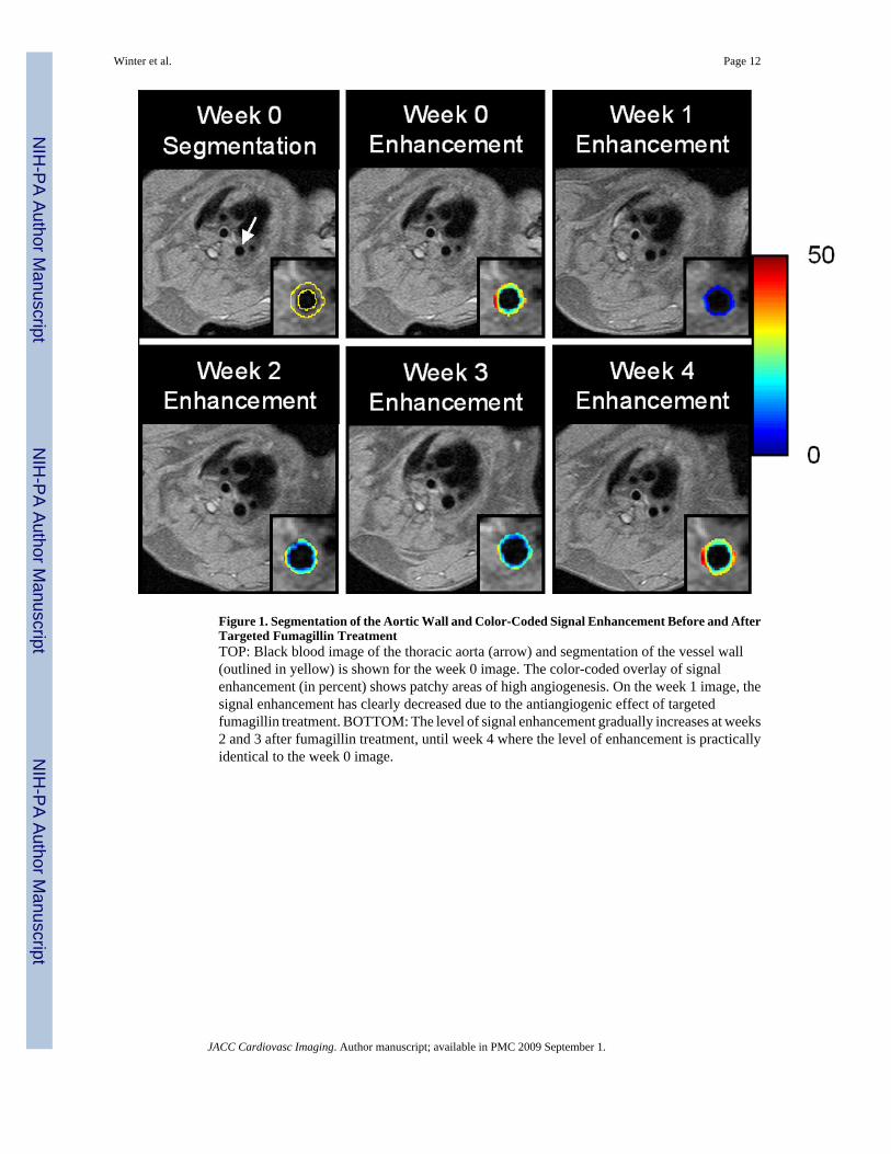
Figure 1. Segmentation of the Aortic Wall and Color-Coded Signal Enhancement Before and AfterTargeted Fumagillin TreatmentTOP: Black blood image of the thoracic aorta (arrow) and segmentation of the vessel wall(outlined in yellow) is shown for the week 0 image. The color-coded overlay of signalenhancement (in percent) shows patchy areas of high angiogenesis. On the week 1 image, thesignal enhancement has clearly decreased due to the antiangiogenic effect of targetedfumagillin treatment. BOTTOM: The level of signal enhancement gradually increases at weeks2 and 3 after fumagillin treatment, until week 4 where the level of enhancement is practicallyidentical to the week 0 image.
Winter et al. Page 12
JACC Cardiovasc Imaging. Author manuscript; available in PMC 2009 September 1.
NIH
-PA Author Manuscript
NIH
-PA Author Manuscript
NIH
-PA Author Manuscript
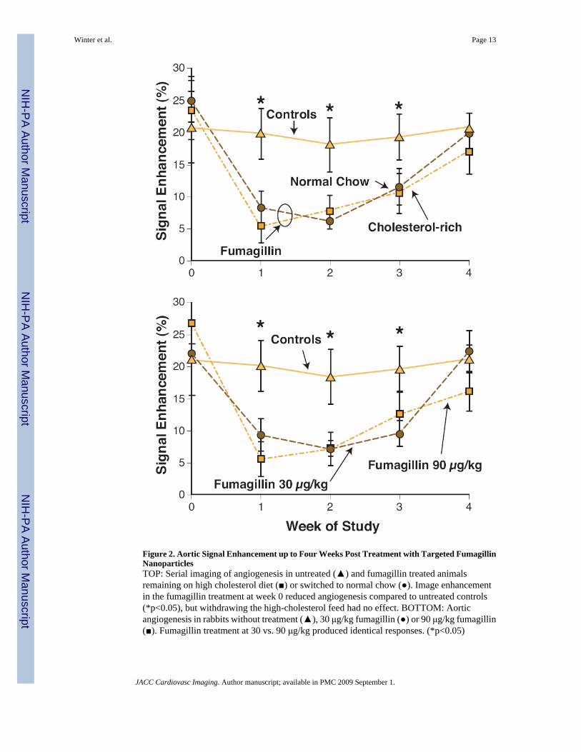
Figure 2. Aortic Signal Enhancement up to Four Weeks Post Treatment with Targeted FumagillinNanoparticlesTOP: Serial imaging of angiogenesis in untreated (▲) and fumagillin treated animalsremaining on high cholesterol diet (■) or switched to normal chow (●). Image enhancementin the fumagillin treatment at week 0 reduced angiogenesis compared to untreated controls(*p<0.05), but withdrawing the high-cholesterol feed had no effect. BOTTOM: Aorticangiogenesis in rabbits without treatment (▲), 30 μg/kg fumagillin (●) or 90 μg/kg fumagillin(■). Fumagillin treatment at 30 vs. 90 μg/kg produced identical responses. (*p<0.05)
Winter et al. Page 13
JACC Cardiovasc Imaging. Author manuscript; available in PMC 2009 September 1.
NIH
-PA Author Manuscript
NIH
-PA Author Manuscript
NIH
-PA Author Manuscript
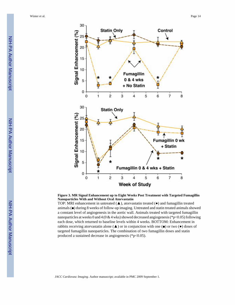
Figure 3. MR Signal Enhancement up to Eight Weeks Post Treatment with Targeted FumagillinNanoparticles With and Without Oral AtorvastatinTOP: MRI enhancement in untreated (▲), atorvastatin treated (●) and fumagillin treatedanimals (■) during 8 weeks of follow-up imaging. Untreated and statin treated animals showeda constant level of angiogenesis in the aortic wall. Animals treated with targeted fumagillinnanoparticles at weeks 0 and 4 (0 & 4 wks) showed decreased angiogenesis (*p<0.05) followingeach dose, which returned to baseline levels within 4 weeks. BOTTOM: Enhancement inrabbits receiving atorvastatin alone (▲) or in conjunction with one (■) or two (●) doses oftargeted fumagillin nanoparticles. The combination of two fumagillin doses and statinproduced a sustained decrease in angiogenesis (*p<0.05).
Winter et al. Page 14
JACC Cardiovasc Imaging. Author manuscript; available in PMC 2009 September 1.
NIH
-PA Author Manuscript
NIH
-PA Author Manuscript
NIH
-PA Author Manuscript
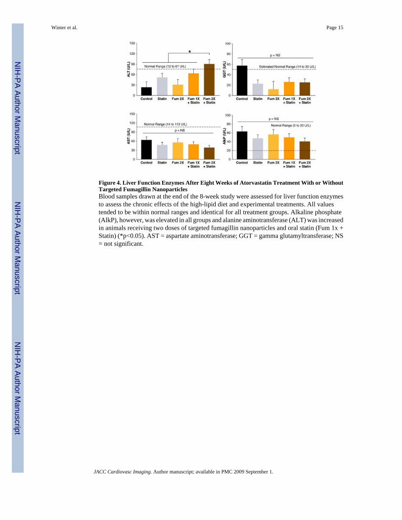
Figure 4. Liver Function Enzymes After Eight Weeks of Atorvastatin Treatment With or WithoutTargeted Fumagillin NanoparticlesBlood samples drawn at the end of the 8-week study were assessed for liver function enzymesto assess the chronic effects of the high-lipid diet and experimental treatments. All valuestended to be within normal ranges and identical for all treatment groups. Alkaline phosphate(AlkP), however, was elevated in all groups and alanine aminotransferase (ALT) was increasedin animals receiving two doses of targeted fumagillin nanoparticles and oral statin (Fum 1x +Statin) (*p<0.05). AST = aspartate aminotransferase; GGT = gamma glutamyltransferase; NS= not significant.
Winter et al. Page 15
JACC Cardiovasc Imaging. Author manuscript; available in PMC 2009 September 1.
NIH
-PA Author Manuscript
NIH
-PA Author Manuscript
NIH
-PA Author Manuscript
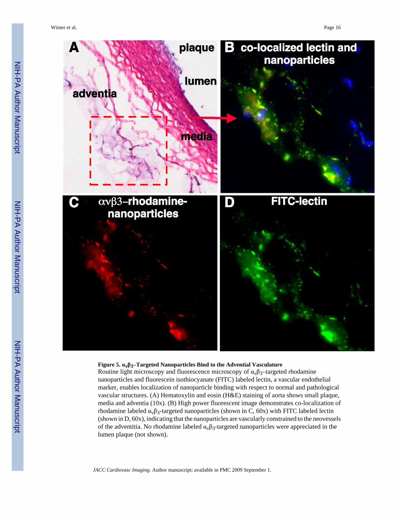
Figure 5. ανβ3–Targeted Nanoparticles Bind to the Advential VasculatureRoutine light microscopy and fluorescence microscopy of ανβ3–targeted rhodaminenanoparticles and fluorescein isothiocyanate (FITC) labeled lectin, a vascular endothelialmarker, enables localization of nanoparticle binding with respect to normal and pathologicalvascular structures. (A) Hematoxylin and eosin (H&E) staining of aorta shows small plaque,media and adventia (10x). (B) High power fluorescent image demonstrates co-localization ofrhodamine labeled ανβ3-targeted nanoparticles (shown in C, 60x) with FITC labeled lectin(shown in D, 60x), indicating that the nanoparticles are vascularly constrained to the neovesselsof the adventitia. No rhodamine labeled ανβ3-targeted nanoparticles were appreciated in thelumen plaque (not shown).
Winter et al. Page 16
JACC Cardiovasc Imaging. Author manuscript; available in PMC 2009 September 1.
NIH
-PA Author Manuscript
NIH
-PA Author Manuscript
NIH
-PA Author Manuscript
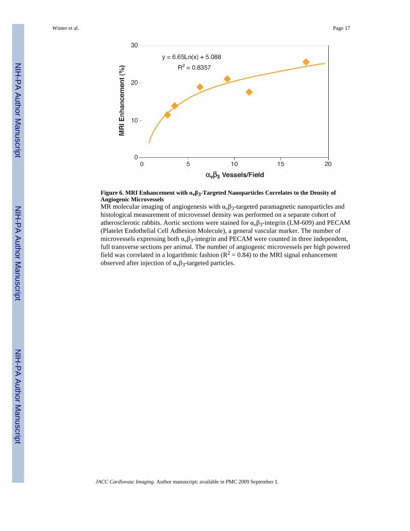
Figure 6. MRI Enhancement with ανβ3-Targeted Nanoparticles Correlates to the Density ofAngiogenic MicrovesselsMR molecular imaging of angiogenesis with ανβ3-targeted paramagnetic nanoparticles andhistological measurement of microvessel density was performed on a separate cohort ofatherosclerotic rabbits. Aortic sections were stained for ανβ3-integrin (LM-609) and PECAM(Platelet Endothelial Cell Adhesion Molecule), a general vascular marker. The number ofmicrovessels expressing both ανβ3-integrin and PECAM were counted in three independent,full transverse sections per animal. The number of angiogenic microvessels per high poweredfield was correlated in a logarithmic fashion (R2 = 0.84) to the MRI signal enhancementobserved after injection of ανβ3-targeted particles.
Winter et al. Page 17
JACC Cardiovasc Imaging. Author manuscript; available in PMC 2009 September 1.
NIH
-PA Author Manuscript
NIH
-PA Author Manuscript
NIH
-PA Author Manuscript
Related Documents