JOURNAL OF VIROLOGY, Jan. 2010, p. 856–866 Vol. 84, No. 2 0022-538X/10/$12.00 doi:10.1128/JVI.00692-09 Copyright © 2010, American Society for Microbiology. All Rights Reserved. Antiangiogenic Arming of an Oncolytic Vaccinia Virus Enhances Antitumor Efficacy in Renal Cell Cancer Models † Kilian Guse, 1,2 Marta Sloniecka, 1,2 Iulia Diaconu, 1,2 Kathryn Ottolino-Perry, 3 Nan Tang, 3 Calvin Ng, 3 Fabrice Le Boeuf, 4 John C. Bell, 4 J. Andrea McCart, 3 Ari Ristima ¨ki, 2 ‡ Sari Pesonen, 1,2 Vincenzo Cerullo, 1,2 and Akseli Hemminki 1,2 * Cancer Gene Therapy Group, Molecular Cancer Biology Program, and Transplantation Laboratory, Haartman Institute, and Finnish Institute for Molecular Medicine, University of Helsinki, Helsinki, Finland 1 ; HUSLAB, Helsinki University Central Hospital, Helsinki, Finland 2 ; Toronto General Research Institute, Toronto, Canada 3 ; and Centre for Cancer Therapeutics, Ottawa Health Research Institute, Ottawa, Canada 4 Received 3 April 2009/Accepted 22 October 2009 Oncolytic vaccinia viruses have shown compelling results in preclinical cancer models and promising preliminary safety and antitumor activity in early clinical trials. However, to facilitate systemic application it would be useful to improve tumor targeting and antitumor efficacy further. Here we report the generation of vvdd-VEGFR-1-Ig, a targeted and armed oncolytic vaccinia virus. Tumor targeting was achieved by deletion of genes for thymidine kinase and vaccinia virus growth factor, which are necessary for replication in normal but not in cancer cells. Given the high vascularization typical of kidney cancers, we armed the virus with the soluble vascular endothelial growth factor (VEGF) receptor 1 protein for an antiangiogenic effect. Systemic application of high doses of vvdd-VEGFR-1-Ig resulted in cytokine induction in an immunocompromised mouse model. Upon histopathological analysis, splenic extramedullary hematopoiesis was seen in all virus-injected mice and was more pronounced in the vvdd-VEGFR-1-Ig group. Analysis of the innate immune response after intrave- nous virus injection revealed high transient and dose-dependent cytokine elevations. When medium and low doses were used for intratumoral or intravenous injection, vvdd-VEGFR-1-Ig exhibited a stronger antitumor effect than the unarmed control. Furthermore, expression of VEGFR-1-Ig was confirmed, and a concurrent antiangiogenic effect was seen. In an immunocompetent model, systemic vvdd-VEGFR-1-Ig exhibited superior antitumor efficacy compared to the unarmed control virus. In conclusion, the targeted and armed vvdd- VEGFR-1-Ig has promising anticancer activity in renal cell cancer models. Extramedullary hematopoiesis may be a sensitive indicator of vaccinia virus effects in mice. In 2002 renal cell cancer accounted for more than 200,000 cases and 100,000 deaths worldwide (33). Unfortunately, che- motherapy, radiotherapy, and immunotherapy yield low re- sponse rates (9, 17) in this cancer type. Thus, prognosis for patients is poor, especially when the disease is metastatic, as median survival is only 8 months (19). Although recently ap- proved drugs, such as sorafenib, sunitinib, temsirolimus, and bevacizumab, have provided additional tools for treatment of renal cell cancer (7), they are usually not curative, and thus new treatment approaches are needed. Oncolytic vaccinia viruses are promising agents for cancer treatment and have shown compelling results in preclinical tumor models (40, 42, 45). Moreover, good safety and prelim- inary evidence of antitumor efficacy were seen in phase 1 clinical trials (22, 26, 32). Vaccinia virus has a strong oncolytic effect due to its fast replication cycle (45) and a high innate tropism to cancer tissue (34). Tumor targeting can be further improved by deleting vaccinia virus genes that are necessary for replication in normal cells but not in cancer cells. For example, deletions of either thymidine kinase (TK) or vaccinia virus growth factor (VGF) or both have been shown to reduce pathogenicity compared to wild-type virus (3, 5, 27). To en- hance antitumor potency, oncolytic vaccinia viruses can be armed with therapeutic transgenes, such as immunostimulatory fac- tors (26) or suicide genes (14, 16, 35). With regard to kidney cancer, an arming approach with antiangiogenenic molecules seems logical, considering the high vascularization character- istic of renal tumors (20). Vascular endothelial growth factor (VEGF) is a major player in tumor angiogenesis and is highly expressed in renal cell cancers (29). VEGF binds to the fms-like-tyrosine kinase re- ceptor (flt-1 or VEGFR-1) and kinase domain region receptor (KDR or VEGFR-2) with high affinity (13). The soluble vas- cular endothelial growth factor receptor 1-Ig fusion protein (VEGFR-1-Ig) used in this study is derived from the mem- brane-bound VEGFR-1 and binds human and murine VEGF without inducing vascular endothelial cell mitogenesis (31). Blocking VEGF with this or closely related molecules has been shown to inhibit tumor growth in several cancer models (18, 21, 25, 39). Although tumor cell selective replication can be enhanced by deletion of TK and/or VGF to reduce pathogenicity (3, 5, * Corresponding author. Mailing address: Cancer Gene Therapy Group, Biomedicum Helsinki, Haartmaninkatu 8, 00290 Helsinki, Fin- land. Phone: 358-9-1912 5464. Fax: 358-9-1912 5465. E-mail: akseli .hemminki@helsinki.fi. † Supplemental material for this article may be found at http://jvi .asm.org/. ‡ Present address: Department of Pathology, HUSLAB, and Haart- man Institute, Genome-Scale Biology Research Program, University of Helsinki, Helsinki, Finland, and Department of Pathology, Institute of Diagnostics, University of Oulu and Oulu University Hospital, Oulu, Finland. Published ahead of print on 11 November 2009. 856

Welcome message from author
This document is posted to help you gain knowledge. Please leave a comment to let me know what you think about it! Share it to your friends and learn new things together.
Transcript
JOURNAL OF VIROLOGY, Jan. 2010, p. 856–866 Vol. 84, No. 20022-538X/10/$12.00 doi:10.1128/JVI.00692-09Copyright © 2010, American Society for Microbiology. All Rights Reserved.
Antiangiogenic Arming of an Oncolytic Vaccinia Virus EnhancesAntitumor Efficacy in Renal Cell Cancer Models�†
Kilian Guse,1,2 Marta Sloniecka,1,2 Iulia Diaconu,1,2 Kathryn Ottolino-Perry,3 Nan Tang,3 Calvin Ng,3Fabrice Le Boeuf,4 John C. Bell,4 J. Andrea McCart,3 Ari Ristimaki,2‡ Sari Pesonen,1,2
Vincenzo Cerullo,1,2 and Akseli Hemminki1,2*Cancer Gene Therapy Group, Molecular Cancer Biology Program, and Transplantation Laboratory, Haartman Institute, and
Finnish Institute for Molecular Medicine, University of Helsinki, Helsinki, Finland1; HUSLAB, Helsinki University Central Hospital,Helsinki, Finland2; Toronto General Research Institute, Toronto, Canada3; and Centre for Cancer Therapeutics,
Ottawa Health Research Institute, Ottawa, Canada4
Received 3 April 2009/Accepted 22 October 2009
Oncolytic vaccinia viruses have shown compelling results in preclinical cancer models and promisingpreliminary safety and antitumor activity in early clinical trials. However, to facilitate systemic application itwould be useful to improve tumor targeting and antitumor efficacy further. Here we report the generation ofvvdd-VEGFR-1-Ig, a targeted and armed oncolytic vaccinia virus. Tumor targeting was achieved by deletion ofgenes for thymidine kinase and vaccinia virus growth factor, which are necessary for replication in normal butnot in cancer cells. Given the high vascularization typical of kidney cancers, we armed the virus with the solublevascular endothelial growth factor (VEGF) receptor 1 protein for an antiangiogenic effect. Systemic applicationof high doses of vvdd-VEGFR-1-Ig resulted in cytokine induction in an immunocompromised mouse model.Upon histopathological analysis, splenic extramedullary hematopoiesis was seen in all virus-injected mice andwas more pronounced in the vvdd-VEGFR-1-Ig group. Analysis of the innate immune response after intrave-nous virus injection revealed high transient and dose-dependent cytokine elevations. When medium and lowdoses were used for intratumoral or intravenous injection, vvdd-VEGFR-1-Ig exhibited a stronger antitumoreffect than the unarmed control. Furthermore, expression of VEGFR-1-Ig was confirmed, and a concurrentantiangiogenic effect was seen. In an immunocompetent model, systemic vvdd-VEGFR-1-Ig exhibited superiorantitumor efficacy compared to the unarmed control virus. In conclusion, the targeted and armed vvdd-VEGFR-1-Ig has promising anticancer activity in renal cell cancer models. Extramedullary hematopoiesis maybe a sensitive indicator of vaccinia virus effects in mice.
In 2002 renal cell cancer accounted for more than 200,000cases and 100,000 deaths worldwide (33). Unfortunately, che-motherapy, radiotherapy, and immunotherapy yield low re-sponse rates (9, 17) in this cancer type. Thus, prognosis forpatients is poor, especially when the disease is metastatic, asmedian survival is only 8 months (19). Although recently ap-proved drugs, such as sorafenib, sunitinib, temsirolimus, andbevacizumab, have provided additional tools for treatment ofrenal cell cancer (7), they are usually not curative, and thusnew treatment approaches are needed.
Oncolytic vaccinia viruses are promising agents for cancertreatment and have shown compelling results in preclinicaltumor models (40, 42, 45). Moreover, good safety and prelim-inary evidence of antitumor efficacy were seen in phase 1clinical trials (22, 26, 32). Vaccinia virus has a strong oncolytic
effect due to its fast replication cycle (45) and a high innatetropism to cancer tissue (34). Tumor targeting can be furtherimproved by deleting vaccinia virus genes that are necessaryfor replication in normal cells but not in cancer cells. Forexample, deletions of either thymidine kinase (TK) or vacciniavirus growth factor (VGF) or both have been shown to reducepathogenicity compared to wild-type virus (3, 5, 27). To en-hance antitumor potency, oncolytic vaccinia viruses can be armedwith therapeutic transgenes, such as immunostimulatory fac-tors (26) or suicide genes (14, 16, 35). With regard to kidneycancer, an arming approach with antiangiogenenic moleculesseems logical, considering the high vascularization character-istic of renal tumors (20).
Vascular endothelial growth factor (VEGF) is a major playerin tumor angiogenesis and is highly expressed in renal cellcancers (29). VEGF binds to the fms-like-tyrosine kinase re-ceptor (flt-1 or VEGFR-1) and kinase domain region receptor(KDR or VEGFR-2) with high affinity (13). The soluble vas-cular endothelial growth factor receptor 1-Ig fusion protein(VEGFR-1-Ig) used in this study is derived from the mem-brane-bound VEGFR-1 and binds human and murine VEGFwithout inducing vascular endothelial cell mitogenesis (31).Blocking VEGF with this or closely related molecules has beenshown to inhibit tumor growth in several cancer models (18, 21,25, 39).
Although tumor cell selective replication can be enhancedby deletion of TK and/or VGF to reduce pathogenicity (3, 5,
* Corresponding author. Mailing address: Cancer Gene TherapyGroup, Biomedicum Helsinki, Haartmaninkatu 8, 00290 Helsinki, Fin-land. Phone: 358-9-1912 5464. Fax: 358-9-1912 5465. E-mail: [email protected].
† Supplemental material for this article may be found at http://jvi.asm.org/.
‡ Present address: Department of Pathology, HUSLAB, and Haart-man Institute, Genome-Scale Biology Research Program, Universityof Helsinki, Helsinki, Finland, and Department of Pathology, Instituteof Diagnostics, University of Oulu and Oulu University Hospital,Oulu, Finland.
� Published ahead of print on 11 November 2009.
856
27), high doses of attenuated vaccinia virus may increase serumcytokine concentrations which parallel the onset of toxic events,as seen with other viral vectors (2, 38). The potential “early”toxicity associated with oncolytic vaccinia viruses has not beencompletely elucidated heretofore (36, 46).
Given the high vascularization of renal cell cancers and thepressing need to generate new antitumor agents with increasedsafety and efficacy, we hypothesized that an oncolytic vacciniavirus targeted by TK and VGF deletions and armed withVEGFR-1-Ig would exhibit enhanced antitumor efficacy due toits antiangiogenic properties in renal cell cancer models com-pared to a nonarmed control virus, allowing reduction of thetreatment dose.
MATERIALS AND METHODS
Cell lines. Human clear cell renal cancer cell lines 786-O, ACHN, and 769-Pwere obtained from the American Type Culture Collection (Manassas, VA) andmaintained under recommended conditions. Renca, a murine kidney cancer cellline, was a kind gift from A. Scarzello (National Cancer Institute, NationalInstitutes of Health, Frederick, MD) and was cultured in RPMI medium sup-plemented with 10% fetal calf serum (FCS) and 1% L-glutamine and penicillin-streptomycin. Pooled human umbilical vein cells (HUVEC; Clonetics endothe-lial cell systems) were purchased from Lonza (Basel, Switzerland) and keptunder the recommended conditions. Vero cells (African green monkey kidneyepithelial cells) and CV-1 cells (African green monkey kidney fibroblasts) wereobtained from the American Type Culture Collection and maintained under therecommended conditions.
Viruses. All vaccinia viruses used in this study are of the Western Reservestrain with disrupted TK and VGF genes for enhanced cancer cell specificity. Forgeneration of vvdd-VEGFR-1-Ig, the luciferase gene was cloned into pSC65 (akind gift from Bernie Moss, National Institutes of Health, Bethesda, MD) (6)under the control of the PE/L promoter, and lacZ (which is under the control ofthe P7.5 promoter) was replaced with VEGFR-1-Ig (VEGF receptor 1 fused tothe Fc tail of human IgG antibody; kindly provided by K. Alitalo, University ofHelsinki, Helsinki, Finland), resulting in pSC65-luc-VEGFR-1-Ig. This shuttleplasmid was cotransfected with vvdd-GFP (27) to CV-1 cells. Successfully re-combined viruses, in which the luciferase gene and VEGFR-1-Ig replaced GFP inthe TK locus, were selected by picking plaques that were positive for luciferaseand negative for green fluorescent protein (GFP). Viruses were amplified onVero cells and purified over a sucrose cushion, and titers were determined witha standard plaque assay on Vero cells as described previously (10). PFU virustiters (PFU/ml) determined by plaque assay have a certain variability and areestimates of the true number of viruses (8). The presence of the inserted geneswas verified by PCR, and the expression of VEGFR-1-Ig by vvdd-VEGFR-1-Ig-infected cells was confirmed by Western blotting.
UV light inactivation of viruses was done as described before (44). Briefly,viruses were suspended in 10 �g/ml psoralen in Hanks balanced salt solutioncontaining 0.1% bovine serum albumin. The suspension was incubated for 10min at room temperature and then irradiated in a CL-1000 UV cross-linker(UVP, Cambridge, United Kingdom) with UV-A light (365 nm) for 3 min. A5-day plaque assay was used to confirm lack of replication competent virus.
Marker gene transfer assay. Cells on 24-well plates were infected with differ-ent concentrations of virus suspended in growth medium containing 2% FCS.Thirty minutes later, cells were washed and incubated in complete growth me-dium for 4 h. Cells were lysed, and luciferase activity was measured according tothe manufacturer’s manual (luciferase assay system; Promega, Madison, WI).
in vitro cytotoxicity assay. Cells on 96-well plates were infected with differentconcentrations of virus suspended in growth medium containing 2% FCS. Onehour later, cells were washed and incubated in growth medium containing 5%FCS for 72 h. Cell viability was then analyzed using MTS [3-(4,5-dimethylthiazol-2-yl)-5-(3-carboxymethoxyphenyl)-2-(4-sulfophenyl)-2H-tetrazolium] (Cell Titer96 AQueous One Solution proliferation assay; Promega).
Animal experiments. Animal experiments were approved by the ExperimentalAnimal Committee of the University of Helsinki and the Provincial Governmentof Southern Finland and the Animal Care Committee of the University HealthNetwork, Ottawa, Canada.
Mice were purchased from Taconic (Ejby, Denmark, and Hudson, NY) at theage of 4 to 5 weeks and housed under standard conditions with food and waterad libitum.
For the immunodeficient models, 5 � 106 786-O cells were injected subcuta-neously into flanks of nude Naval Medical Research Institute (NMRI) mice.When tumors reached the size of approximately 5 by 5 mm, virus was injectedeither intratumorally or intravenously. For bioluminescence imaging, mice wereinjected intraperitoneally with 4.5 mg of D-luciferin dissolved in phosphate-buffered saline (PBS), and after 10 min images were captured using the IVISimaging series 100 system (Xenogen, Alameda, CA). VEGFR-1-Ig concentrationin mouse serum was determined with a human IgG enzyme-linked immunosor-bent assay (ELISA) kit (Immunology Consultants Laboratory, Newberg, OR).
For the immunocompetent model, 1 � 106 Renca cells were injected subcu-taneously on shaved flanks of BALB/c mice. Virus was injected intravenouslywhen tumors reached the size of approximately 5 by 5 mm. Imaging was done asdescribed above.
To assess cytokine concentrations, mice were injected intravenously with virus,and blood samples were taken 6 h, 12 h, and 24 h postinjection. A fluorescence-activated cell sorting array with collected blood serum was performed for inter-leukin 6 (IL-6), tumor necrosis factor (TNF), monocyte chemoattractant protein1 (MCP-1), gamma interferon (IFN-�), macrophage inflammatory protein 1�(MIP-1�, or CCL3), KC (CXCL1), and regulated upon activation normal T-cell-expressed and secreted protein (RANTES; cytometric bead array mouse flexsets; BD Biosciences Pharmingen, Franklin Lakes, NJ).
Histopathology and immunofluorescence staining. Organs of animals werefixed in 10% neutral-buffered formalin. Following fixation, organs were trimmed,embedded in paraffin, sectioned (4 to 5 �m), stained with hematoxylin-eosin, andevaluated by a pathologist.
Four- to 5-�m cryosections of frozen tumors were prepared and fixed inacetone for 10 min at �20°C. Sections were incubated with normal donkey serumfor 15 min and then reacted with primary polyclonal rabbit anti-von Willebrandfactor (1:200 dilution; DakoCytomation, Denmark) overnight. After washingwith PBS, sections were incubated with Alexa Fluor 488-labeled secondary an-tibody (1:250 dilution; Molecular Probes, Invitrogen, Carlsbad, CA) for 30 min.Sections were fixed in 4% paraformaldehyde and mounted with Vectashieldmounting medium (Vector Laboratories, Burlingame, CA). Representative pic-tures of areas of the tumors with the highest microvessel density were capturedat 20� magnification.
Statistical analysis. Tumor sizes as a function of time were compared byMann-Whitney test (SPSS 16.0; SPSS Inc., Chicago, IL), and P values of �0.05were considered statistically significant. A single preplanned comparison of meantumor volume was done by using a two-tailed t test.
RESULTS
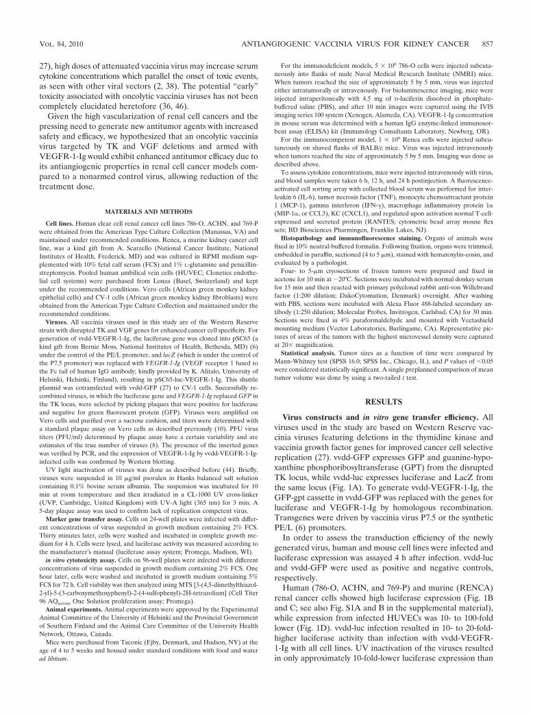
Virus constructs and in vitro gene transfer efficiency. Allviruses used in the study are based on Western Reserve vac-cinia viruses featuring deletions in the thymidine kinase andvaccinia growth factor genes for improved cancer cell selectivereplication (27). vvdd-GFP expresses GFP and guanine-hypo-xanthine phosphoribosyltransferase (GPT) from the disruptedTK locus, while vvdd-luc expresses luciferase and LacZ fromthe same locus (Fig. 1A). To generate vvdd-VEGFR-1-Ig, theGFP-gpt cassette in vvdd-GFP was replaced with the genes forluciferase and VEGFR-1-Ig by homologous recombination.Transgenes were driven by vaccinia virus P7.5 or the syntheticPE/L (6) promoters.
In order to assess the transduction efficiency of the newlygenerated virus, human and mouse cell lines were infected andluciferase expression was assayed 4 h after infection. vvdd-lucand vvdd-GFP were used as positive and negative controls,respectively.
Human (786-O, ACHN, and 769-P) and murine (RENCA)renal cancer cells showed high luciferase expression (Fig. 1Band C; see also Fig. S1A and B in the supplemental material),while expression from infected HUVECs was 10- to 100-foldlower (Fig. 1D). vvdd-luc infection resulted in 10- to 20-fold-higher luciferase activity than infection with vvdd-VEGFR-1-Ig with all cell lines. UV inactivation of the viruses resultedin only approximately 10-fold-lower luciferase expression than
VOL. 84, 2010 ANTIANGIOGENIC VACCINIA VIRUS FOR KIDNEY CANCER 857
with respective noninactivated viruses, suggesting activity ofthe PE/L promoter even in the absence of replication.
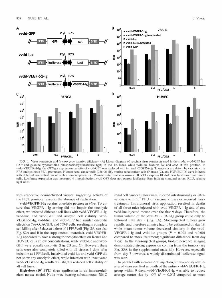
vvdd-VEGFR-1-Ig retains oncolytic potency in vitro. To en-sure that VEGFR-1-Ig arming did not impair the oncolyticeffect, we infected different cell lines with vvdd-VEGFR-1-Ig,vvdd-luc, and vvdd-GFP and assayed cell viability. vvdd-VEGFR-1-Ig, vvdd-luc, and vvdd-GFP had similar oncolyticeffects on 786-O, ACHN, and 769-P cells, resulting in completecell killing after 3 days at a dose of 1 PFU/cell (Fig. 2A; see alsoFig. S2A and B in the supplemental material). vvdd-VEGFR-1-Ig appeared to have a stronger oncolytic effect on Renca andHUVEC cells at low concentrations, while vvdd-luc and vvdd-GFP were equally oncolytic (Fig. 2B and C). However, thesecells were also completely killed with all viruses 3 days afterinfection at 1 PFU/cell. Inactivated vvdd-luc and vvdd-GFP didnot show any oncolytic effect, while infection with inactivatedvvdd-VEGFR-1-Ig resulted in slightly reduced cell viability ofall cell lines.
High-dose (107 PFU) virus application in an immunodefi-cient mouse model. Nude mice bearing subcutaneous 786-O
renal cell cancer tumors were injected intratumorally or intra-venously with 107 PFU of vaccinia viruses or received mocktreatment. Intratumoral virus application resulted in deathsof all three mice injected with vvdd-VEGFR-1-Ig and of onevvdd-luc-injected mouse over the first 9 days. Therefore, thetumor volume of the vvdd-VEGFR-1-Ig group could only befollowed until day 9 (Fig. 3A). Mock-injected tumors grewrapidly, and therefore all mice had to be euthanized on day 19,while mean tumor volume decreased similarly in the vvdd-VEGFR-1-Ig and vvdd-luc groups (P � 0.003 and �0.001compared to mock treatment; significant difference from day7 on). In the virus-injected groups, bioluminescence imagingdemonstrated strong expression coming from the tumors (seeFig. S3A in the supplemental material). However, especiallyfrom day 7 onwards, a widely disseminated luciferase signalwas seen.
In parallel with intratumoral injection, intravenously admin-istered virus resulted in death of the entire vvdd-VEGFR-1-Iggroup within 9 days. vvdd-VEGFR-1-Ig was able to reduceaverage tumor size by 40% (P � 0.002 compared to mock
FIG. 1. Virus constructs and in vitro gene transfer efficiency. (A) Linear diagram of vaccinia virus constructs used in the study. vvdd-GFP hasGFP and guanine-hypoxanthine phosphoribosyltransferase (gpt) in the TK locus, while vvdd-luc features luc and lacZ at this position. Invvdd-VEGFR-1-Ig, the GFP-gpt expression cassette of vvdd-GFP was replaced with luc and VEGFR-1-Ig. Transgenes are driven by vaccinia virusP7.5 and synthetic PE/L promoters. Human renal cancer cells (786-O) (B), murine renal cancer cells (Renca) (C), and HUVEC (D) were infectedwith different concentrations of replication-competent or UV-inactivated vaccinia viruses. HUVECs express 100-fold less luciferase than tumorcells. Luciferase expression was measured 4 h postinfection. vvdd-GFP does not express luciferase. Bars indicate standard errors. RLU, relativelight units.
858 GUSE ET AL. J. VIROL.
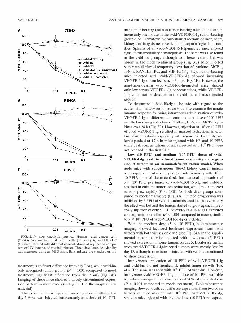
treatment; significant difference from day 7 on), while vvdd-luconly abrogated tumor growth (P � 0.001 compared to mocktreatment; significant difference from day 7 on) (Fig. 3B).Imaging of these mice showed a widely disseminated expres-sion pattern in most mice (see Fig. S3B in the supplementalmaterial).
The experiment was repeated, and organs were collected onday 3.Virus was injected intravenously at a dose of 107 PFU
into tumor-bearing and non-tumor-bearing mice. In this exper-iment only one mouse in the vvdd-VEFGR-1-Ig tumor-bearinggroup died. Hematoxylin-eosin-stained sections of liver, heart,kidney, and lung tissues revealed no histopathologic abnormal-ities. Spleens of all vvdd-VEGFR-1-Ig-injected mice showedsigns of extramedullary hematopoiesis. The same was also foundin the vvdd-luc group, although to a lesser extent, but wasabsent in the mock treatment group (Fig. 3C). Mice injectedwith virus displayed temporary elevation of cytokines MCP-1,IFN-�, RANTES, KC, and MIP-1� (Fig. 3D). Tumor-bearingmice injected with vvdd-VEGFR-1-Ig showed increasingVEGFR-1-Ig serum levels over 3 days (Fig. 3E). However, thenon-tumor-bearing vvdd-VEGFR-1-Ig-injected mice showedonly low serum VEGFR-1-Ig concentrations, while VEGFR-1-Ig could not be detected in the vvdd-luc and mock-treatedgroups.
To determine a dose likely to be safe with regard to theacute inflammatory response, we sought to examine the innateimmune response following intravenous administration of vvdd-VEGFR-1-Ig at different concentrations. A dose of 107 PFUresulted in strong induction of TNF-�, IL-6, and MCP-1 cyto-kines over 24 h (Fig. 3F). However, injection of 105 or 10 PFUof vvdd-VEGFR-1-Ig resulted in marked reductions in cyto-kine concentrations, especially with regard to IL-6. Cytokinelevels peaked at 12 h in mice injected with 105 and 10 PFU,while peak concentrations of mice injected with 107 PFU werenot reached in the first 24 h.
Low (10 PFU) and medium (105 PFU) doses of vvdd-VEGFR-1-Ig result in reduced tumor vascularity and regres-sion of tumors in an immunodeficient mouse model. Whennude mice with subcutaneous 786-O kidney cancer tumorswere injected intratumorally (i.t.) or intravenously with 105 or10 PFU, none of the mice died. Intratumoral application of5 � 104 PFU per tumor of vvdd-VEGFR-1-Ig and vvdd-lucresulted in efficient tumor size reduction, while mock-injectedtumors grew rapidly (P � 0.001 for both virus groups com-pared to mock treatment) (Fig. 4A). Tumor progression wasinhibited by 5 PFU of vvdd-luc administered i.t., but eventuallythe effect was lost and the tumors started to grow again. Impres-sively, injection of only 5 PFU of vvdd-VEGFR-1-Ig i.t. exhibiteda strong antitumor effect (P � 0.001 compared to mock), similarto 5 � 104 PFU of vvdd-VEGFR-1-Ig or vvdd-luc.
With the medium dose (5 � 104 PFU), bioluminescenceimaging showed localized luciferase expression from mosttumors with both viruses on day 5 (see Fig. S4A in the supple-mental material). Mice injected with low doses (5 PFU)showed expression in some tumors on day 5. Luciferase signalsfrom vvdd-VEGFR-1-Ig-injected tumors were mostly lost byday 13, although some tumors injected with vvdd-luc continuedto show expression.
Intravenous application of 10 PFU of vvdd-VEGFR-1-Igand vvdd-luc did not significantly inhibit tumor growth (Fig.4B). The same was seen with 105 PFU of vvdd-luc. However,intravenous vvdd-VEGFR-1-Ig at a dose of 105 PFU was ableto reduce average tumor size to about 50% of the initial size(P � 0.001 compared to mock treatment). Bioluminescenceimaging showed localized luciferase expression from two of sixtumors of mice injected with 105 PFU vvdd-VEGFR-1-Ig,while in mice injected with the low dose (10 PFU) no expres-
FIG. 2. In vitro oncolytic potency. Human renal cancer cells(786-O) (A), murine renal cancer cells (Renca) (B), and HUVEC(C) were infected with different concentrations of replication-compe-tent or UV-inactivated vaccinia viruses. Three days later, cell viabilitywas measured using an MTS assay. Bars indicate the standard errors.
VOL. 84, 2010 ANTIANGIOGENIC VACCINIA VIRUS FOR KIDNEY CANCER 859
860
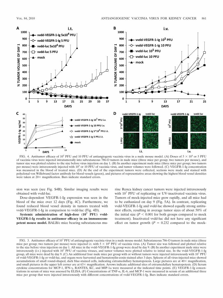
sion was seen (see Fig. S4B). Similar imaging results wereobtained with vvdd-luc.
Dose-dependent VEGFR-1-Ig expression was seen in theblood of the mice over 12 days (Fig. 4C). Furthermore, wefound reduced blood vessel density in tumors treated withvvdd-VEGFR-1-Ig in comparison to vvdd-luc (Fig. 4D).
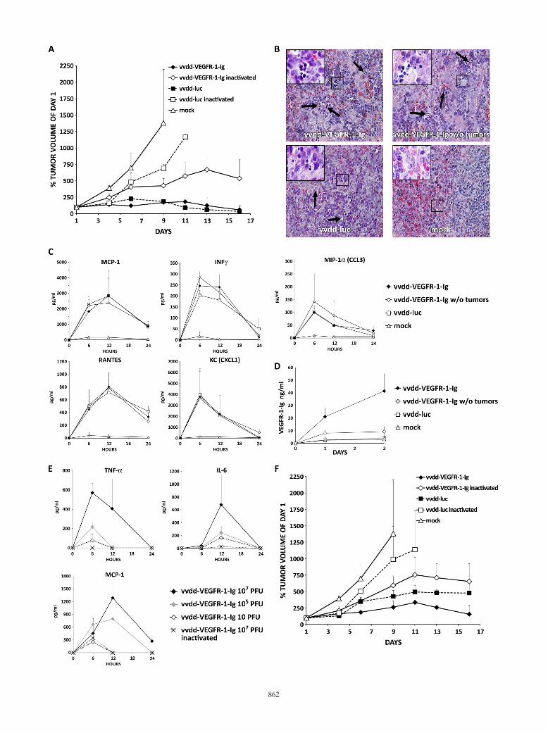
Systemic administration of high-dose (107 PFU) vvdd-VEGFR-1-Ig results in antitumor efficacy in an immunocom-petent mouse model. BALB/c mice bearing subcutaneous mu-
rine Renca kidney cancer tumors were injected intravenouslywith 107 PFU of replicating or UV-inactivated vaccinia virus.Tumors of mock-injected mice grew rapidly, and all mice hadto be euthanized on day 9 (Fig. 5A). In contrast, replicatingvvdd-VEGFR-1-Ig and vvdd-luc showed equally strong antitu-mor effects, resulting in average tumor sizes of about 50% ofthe initial size (P � 0.001 for both groups compared to mocktreatment). Inactivated vvdd-luc did not have any significanteffect on tumor growth (P � 0.232 compared to the mock-
FIG. 3. Antitumor efficacy of 107 PFU of antiangiogenic vaccinia virus in a nude mouse model. Subcutaneous 786-O tumors in nude mice (threemice per group; two tumors per mouse) were injected i.t. with 5 � 106 PFU of vaccinia virus. (A) Tumor size was followed and plotted relativeto the size before virus injection on day 1. All mice in the vvdd-VEGFR-1-Ig group were dead by day 9. (B) In another experiment nude mice wereintravenously (i.v.) injected with 107 PFU of vaccinia viruses, and tumor volumes were plotted relative to initial size. In the vvdd-VEGFR-1-Iggroup, all mice were dead by day 9. (C) An additional four nude mice per group with or without tumors were injected intravenously with 107 PFUof vvdd-VEGFR-1-Ig or vvdd-luc, and organs were harvested and hematoxylin-eosin stained after 3 days. Spleens of all virus-injected mice showedaccumulations of small round-shaped, dark blue-stained cells, indicating extramedullary hematopoiesis. Large pictures are at 40� magnification,and small pictures in the upper left corner are at 63� magnification. Arrows indicate additional sites of extramedullary hematopoiesis. (D) Serumcytokine concentrations for MCP-1, IFN-�, RANTES, KC, and MIP-1� were measured at the indicated time points. (E) VEGFR-1-Ig concen-trations in serum of mice was assessed by ELISA. (F) Concentrations of TNF-�, IL-6, and MCP-1 were measured in serum of an additional threemice per group that were injected intravenously with different concentrations of vvdd-VEGFR-1-Ig. Bars indicate standard errors.
FIG. 4. Antitumor efficacy of 105 PFU and 10 PFU of antiangiogenic vaccinia virus in a nude mouse model. (A) Doses of 5 � 104 or 5 PFUof vaccinia virus were injected intratumorally into subcutaneous 786-O tumors in nude mice (three mice per group; two tumors per mouse), andtumor size was plotted relative to the size before virus injection on day 1. (B) In another experiment nude mice (three mice per group; two tumorsper mouse) were intravenously injected with 105 or 10 PFU of vaccinia virus, and tumor volumes were followed. (C) VEGFR-1-Ig concentrationwas measured in the blood of treated mice. (D) At the end of the experiment tumors were collected, sections were made and stained withpolyclonal von Willebrand factor antibody for blood vessels (green), and pictures of representative areas showing the highest blood vessel densitieswere taken at 20� magnification. Bars indicate standard errors.
VOL. 84, 2010 ANTIANGIOGENIC VACCINIA VIRUS FOR KIDNEY CANCER 861
862
treated group on day 9), whereas inactivated vvdd-VEGFR-1-Ig was able to inhibit tumor progression in this aggressivecancer model (P � 0.04 compared to the mock-treated groupon day 9).
Mice injected with replicating viruses showed luciferase ex-pression coming mainly from tumors but also from elsewhere(see Fig. S5A in the supplemental material). In mice injectedwith inactivated viruses, tumors did not express detectablelevels of luciferase, but strong signals were detected from theregion of the lungs and/or liver. To confirm this finding, imag-ing of inactivated viruses was also performed at an earlier timepoint (2 days), and again luciferase expression was seen inlungs and/or liver (see Fig. S6 in the supplemental material).
We repeated the experiment and also included a non-tumor-bearing group. Hematoxylin-eosin-stained sections of organscollected on day 3 revealed no abnormalities of liver, heart,kidney, and lung tissues. Confirming the nude mouse data,spleens of all vvdd-VEGFR-1-Ig-injected mice showed signsof extramedullary hematopoiesis, which was seen to a lowerextent also in vvdd-luc-injected mice but not in the mock treat-ment group (Fig. 5B). Analysis of the cytokine markersMCP-1, IFN-�, RANTES, KC, and MIP-1� revealed tempo-rary elevations after injection of vvdd-VEGFR-1-Ig and vvdd-luc (Fig. 5C). An increasing concentration of VEGFR-1-Ig wasdetected in tumor-bearing mice injected with vvdd-VEGFR-1-Ig, while tumor-free mice showed low expression levels (Fig. 5D).
To find a safe dose with regard to the acute inflammatoryresponse, we analyzed the cytokine concentrations after intra-venous administration of different concentrations of replicat-ing or inactivated vvdd-VEFGR-1-Ig. Serum concentrations ofTNF-�, IL-6, and MCP-1 peaked between 6 and 12 h in miceinjected with 107 PFU of replicating virus (Fig. 5E), while miceinjected with 105 and 10 PFU had a less pronounced cytokineresponse. Inactivated virus resulted in almost no elevation ofTNF-� or IL-6, while MCP-1 was increased at 6 h.
Systemic administration of a medium dose (105 PFU) ofvvdd-VEGFR-1-Ig results in antitumor efficacy with a reducedcytokine response in an immunocompetent mouse model. Micewith syngeneic Renca tumors were injected intravenously with105 PFU of replicating or inactivated virus. Replicating vvdd-luc exhibited a strong antitumor effect, but vvdd-VEGFR-1-Igwas significantly more effective (P � 0.001 compared to mocktreatment; P � 0.002 compared to vvdd-luc) (Fig. 5F). Tumorsof mice injected with inactivated vvdd-luc grew almost as rap-idly as tumors in the mock treatment group (P � 0.184 com-pared to mock treatment on day 9), whereas inactivated vvdd-
VEGFR-1-Ig significantly inhibited tumor progression (P �0.043 compared to mock treatment on day 9). Localized lucif-erase expression from the tumors was only seen in one of thevvdd-VEGFR-1-Ig-injected mice, while vvdd-luc-injected miceshowed stronger and more disseminated expression (see Fig.S5B in the supplemental material). Mice injected with inacti-vated viruses did not show detectable luciferase expression.
DISCUSSION
Engineered oncolytic vaccinia viruses have demonstratedpromising results in the treatment of cancer in preclinical mod-els and early clinical trials (22, 26, 32, 40, 42, 45). However,most patients have not been cured, and thus the efficacy of theapproach would benefit from further improvement. We gen-erated vvdd-VEGFR-1-Ig, a targeted and armed oncolytic vac-cinia virus designed to enhance oncolysis and reduce tumorangiogenesis for improved anticancer efficacy. To our knowl-edge, this is the first time that an antiangiogenic oncolyticvaccinia virus has been developed for and evaluated in kidneycancer models.
vvdd-VEGFR-1-Ig showed efficient transduction (Fig. 1; seealso Fig. S1 in the supplemental material) and a strong onco-lytic effect in vitro (Fig. 2; see also Fig. S2). In an immunocom-promised mouse model, high-dose (107 PFU) application ofthe unarmed control virus vvdd-luc resulted in tumor eradica-tion after intratumoral injection (Fig. 3A) and in tumor growtharrest after intravenous application (Fig. 3B). We hypothesizedthat a possible reason for mice dying in the vvdd-VEGFR-1-Iggroup might have been high levels of the VEGF-inhibitingtransgene product, as VEGF inhibition has been shown to harborthe potential for damage to the liver, kidneys, and vascularsystem (24, 41). However, histopathologic analysis of organsdid not reveal tissue damage in livers or kidneys in mice in-jected with vvdd-VEGFR-1-Ig (Fig. 3C). Instead, extramedul-lary hematopoiesis in the spleen was found in vvdd-VEGFR-1-Ig-injected tumor-bearing and tumor-free animals. Also,mice injected with vvdd-luc showed splenic extramedullaryhematopoiesis, although to a lesser extent, while it was notfound in mock-injected mice. Thus, it seems that vaccinia virusitself can cause splenic extramedullary hematopoiesis, and thisis further increased by VEGFR-1-Ig. Another possibility isdecreased oxygenation due to infection of vascular elementsand/or increased VEGFR-1-Ig levels. Splenic extramedullaryhematopoiesis has been described after infection with otherviruses, such as cytomegalovirus, herpes simplex virus, dengue
FIG. 5. Antitumor efficacy of 107 and 105 PFU of an antiangiogenic vaccinia virus in an immunocompetent mouse model. (A) BALB/c micebearing subcutaneous tumors (three mice per group; two tumors per mouse) induced with Renca cells were injected intravenously with 107 PFUof replicating or UV-inactivated vaccinia virus. Tumor size was followed and plotted relative to the size before virus injection on day 1. Note thatall mice in the mock and vvdd-luc-inactivated groups had to be killed because of large tumors that formed by days 9 and 11, respectively. (B) Anadditional four tumor-bearing and tumor-free BALB/c mice per group were injected intravenously with 107 PFU of the indicated virus, and organswere harvested and hematoxylin-eosin stained 3 days later. Spleens of all virus-injected mice showed accumulations of small round-shaped, darkblue-stained cells, indicating extramedullary hematopoiesis. Large pictures are at 40� magnification, and small pictures in the upper left cornerare at 63� magnification. Arrows indicate additional sites of extramedullary hematopoiesis. (C) Serum cytokine concentrations for MCP-1, IFN-�,RANTES, KC, and MIP-1� were measured at the indicated time points. (D) VEGFR-1-Ig concentrations in serum of the mice were assessed byELISA. (E) An additional three BALB/c mice per group were intravenously injected with different concentrations of replicating or inactivatedvvdd-VEGFR-1-Ig, and TNF-�, IL-6, and MCP-1 serum levels were measured. (F) In another experiment, tumor-bearing BALB/c mice (threemice per group; two tumors per mouse) were intravenously injected with 105 PFU of replicating or inactivated vaccinia virus. Rapid tumor growthrequired killing of all mice in the mock-treated and vvdd-luc-inactivated groups by day 9 and 11, respectively. Bars indicate standard errors.
VOL. 84, 2010 ANTIANGIOGENIC VACCINIA VIRUS FOR KIDNEY CANCER 863
virus, and human immunodeficiency virus (1, 23), and hepaticextramedullary hematopoiesis has been seen with adenovirus(37), but neither has previously been reported for vacciniavirus. Formal toxicity studies would be needed to understandthe safety/toxicity profile of the viruses used here.
Widely disseminated virus-mediated luciferase expressionwas seen (see Fig. S3 in the supplemental material), possiblyoriginating from the skin, since vaccinia virus is known to havedermal tropism (4, 28). However, for the vaccinia viruses usedhere, luciferase expression is not linked to replication. There-fore, luciferase expression is indicative of biodistribution of thevirus rather than of off-target replication. Tumor selective rep-lication of TK- and VGF-deleted viruses (vvdd backbone) innude mice has been confirmed before (27). In fact, whentumor-free mice were injected with vvdd-VEGFR-1-Ig, lowVEGFR-1-Ig serum concentrations were observed, suggestingtumor selectivity of the virus (Fig. 3E). Since the transgene isnot linked to replication, input virus is expected to produceVEGFR-1-Ig, but in the absence of virus replication only lowlevels result.
Because strong activation of innate immune sensors afterhigh-dose application of vaccinia virus (36, 46) can be toxic, weassessed the levels of key cytokines following intravenous ad-ministration of 107 PFU. Temporary increases in MCP-1,IFN-�, RANTES, KC, and MIP-1� were seen for all viruses,mostly returning to baseline after 24 h (Fig. 3D). To find a safedose, as estimated by the cytokine response, additional micewere injected with high (107 PFU), medium (105 PFU), andlow (10 PFU) doses. With the high dose, elevations of TNF-�,IL-6, and MCP-1 levels were observed, and values were stillincreasing at the last time point (24 h) analyzed (Fig. 3F).Although medium and low doses also resulted in an inflam-matory response, cytokine levels were markedly lower, peakingalready at 12 h and decreasing earlier than with the high dose.Because cytokine induction was dose dependent, and high cy-tokine levels may correlate with severe or even lethal toxicity inanimals and humans treated with viral gene therapy (1, 33), itwould be key to maintain low cytokine concentrations whileretaining antitumor efficacy. Therefore, we performed anotheranimal experiment with lower amounts of virus (105 and 10PFU). In this experiment, none of the mice died or showedobvious signs of toxicity. Regardless of the route of adminis-tration, vvdd-VEGFR-1-Ig was more effective than vvdd-luc(Fig. 4A and B), which was probably caused by VEGFR-1-Ig-mediated inhibition of the vasculature (Fig. 3C and D).
Since the immune system may play an important role indetermining the safety and efficacy of oncolytic virotherapy,immunocompetent models might be superior to immunodefi-cient systems in predicting patient outcomes. Therefore, weused an aggressive syngeneic mouse model of renal cell cancer,where we injected replicating and inactivated viruses intrave-nously. While high doses (107 PFU) of replicating vvdd-VEGFR-1-Ig and vvdd-luc were equally efficient in tumor sizereduction, inactivated vvdd-VEGFR-1-Ig seemed to result inbetter tumor growth inhibition than inactivated vvdd-luc, sug-gesting antitumor efficacy of the transgene product VEGFR-1-Ig (Fig. 5A).
Luciferase expression mediated by the replicating virusesappeared to be more restricted to tumor tissue in the immu-nocompetent models than in the immunodeficient models
(compare Fig. S5A and S3B in the supplemental material).This might be due to more rapid virus clearance in normaltissues of hosts with an intact immune system versus thosefrom a partially immune-privileged tumor environment (30).
Surprisingly, and in contrast to data with replication-compe-tent viruses, luciferase expression mediated by UV-inactivated,and therefore replication-defective, viruses seemed to comeprimarily from the liver and/or lungs (see Fig. S5A and S6 inthe supplemental material). One possible reason for the dif-ference could be that uptake of the imaging substrate D-lucif-erin might be increased in areas of virus replication. However,we speculate that replication competence results in enhancedtumor selectivity, as the virus multiplies in the tumor but not innormal tissues, therefore increasing the signal from the tumor(43). This, however, does not explain the smaller liver/lungsignal seen in the mice injected with replication-competentvirus. Perhaps as a clue, we found that a replication-competentvaccinia virus induces a stronger and faster innate immuneresponse than a replication-deficient virus. Therefore, virus mightbe cleared more rapidly from normal organs in the presence ofvirus replication (in the tumor) than in the absence of virusreplication (in the tumor). Taken together, faster clearancefrom normal tissues and concurrent amplification in tumorsmight explain the differences in biodistribution.
We also analyzed possible organ damage and cytokine levelsin the immunocompetent Renca model. Extramedullary hema-topoiesis in the spleen was found in vvdd-VEGFR-1-Ig-in-jected mice and to a lower extent in vvdd-luc-injected mice(Fig. 5B), as was the case in the immunodeficient model. Liv-ers, kidneys, hearts, and lungs did not show any histopathologicchanges. Further studies could include bone marrow and otherorgans. Temporary induction of the cytokine markers MCP-1,IFN-�, RANTES, KC, and MIP-1� was seen, with levelsmostly returning to baseline after 24 h (Fig. 5C). Comparedto tumor-bearing mice, tumor-free animals injected with vvdd-VEGFR-1-Ig had only low serum levels of VEGFR-1-Ig, sug-gesting tumor-specific replication and transgene expression(Fig. 5D). In a dose range-finding experiment with vvdd-VEGFR-1-Ig, we observed less-pronounced cytokine responses than in theimmunocompromised model, with values returning to baseline at24 h (Fig. 5E). Lowering the dose from 107 PFU to 105 PFUresulted in a lower cytokine response. Further studies are neededto evaluate whether these viruses can cause toxicity.
To assess if antitumor efficacy could be retained with theless immunogenic dose, an additional three mice per groupwere injected with 105 PFU. We found that replicating vvdd-VEGFR-1-Ig performed significantly better than vvdd-luc(P � 0.002) (Fig. 5F). Also, inactivated vvdd-VEGFR-1-Igsignificantly inhibited tumor growth while inactivated vvdd-lucdid not have any effect.
In conclusion, we have described here the arming of anoncolytic vaccinia virus with an antiangiogenic molecule whichresulted in improved antitumor efficacy in immunodeficientand immunocompetent murine kidney cancer models. Cyto-kine analysis underscored the importance of using an optimalvirus dose to avoid innate immune reactions. Systemic deliveryof moderate (105 PFU) doses of vvdd-VEGFR-1-Ig inhibitedtumor growth significantly better than the unarmed controlvirus, confirming the rationale that the antiangiogenic arming
864 GUSE ET AL. J. VIROL.
approach allows for lowering the virus dose while retainingantitumor efficacy.
These preclinical data facilitate clinical development of armedoncolytic vaccinia viruses for clinical trials in renal cell cancerpatients. Since antiangiogenic therapies have recently beendemonstrated to be useful for many other common tumortypes, including colorectal, breast, lung, ovarian, pancreatic,and liver (11), it is unlikely that the utility of the approachdescribed here would be restricted to renal cell cancer. How-ever, because high concentrations of VEGF-inhibiting mole-cules have been shown to cause toxicity in mice (24, 41) andhumans (12, 15, 47), additional regulatory elements for theVEGFR-1-Ig expression might be needed. Also, our findingssuggest that extramedullary hematopoiesis might be a sensitiveindicator of vaccinia virus effects in mice.
ACKNOWLEDGMENTS
This work was supported by the Helsinki Graduate School in Bio-technology and Molecular Biology, Finnish Cancer Society, K. AlbinJohansson Foundation, Orion-Farmos Research Foundation, Euro-pean Research Council, EU FP6 APOTHERAPY and THERADPOX,HUCH Research Funds (EVO), Finnish Cancer Society, Sigrid Juse-lius Foundation, Academy of Finland, Biocentrum Helsinki, Universityof Helsinki, an industrial CIHR Fellowship, and a Terry Fox Programproject grant through the National Cancer Institute of Canada, On-tario Cancer Research Network, Canadian Institutes of Health Re-search. A. Hemminki is the K. Albin Johansson Research Professor ofThe Finnish Cancer Institute.
J. C. Bell is cofounder and sits on the board of Jennerex Biothera-peutics, a company involved in the development of oncolytic virustherapeutics.
We thank Eerika Karli, Aila Karioja-Kallio, Sirkka-Liisa Holm,Paivi Hannuksela, and Roxana Ola for their technical contribution tothis work. Further, we thank A. Scarzello, Bernie Moss, and K. Alitalofor providing materials for this study.
REFERENCES
1. Adler, S. P., and B. Marshall. 2007. Cytomegalovirus infections. Pediatr.Rev. 28:92–100.
2. Brunetti-Pierri, N., D. J. Palmer, A. L. Beaudet, K. D. Carey, M. Finegold,and P. Ng. 2004. Acute toxicity after high-dose systemic injection of helper-dependent adenoviral vectors into nonhuman primates. Hum. Gene Ther.15:35–46.
3. Buller, R. M., S. Chakrabarti, J. A. Cooper, D. R. Twardzik, and B. Moss.1988. Deletion of the vaccinia virus growth factor gene reduces virus viru-lence. J. Virol. 62:866–874.
4. Buller, R. M., and G. J. Palumbo. 1991. Poxvirus pathogenesis. Microbiol.Rev. 55:80–122.
5. Buller, R. M., G. L. Smith, K. Cremer, A. L. Notkins, and B. Moss. 1985.Decreased virulence of recombinant vaccinia virus expression vectors isassociated with a thymidine kinase-negative phenotype. Nature 317:813–815.
6. Chakrabarti, S., J. R. Sisler, and B. Moss. 1997. Compact, synthetic, vacciniavirus early/late promoter for protein expression. Biotechniques 23:1094–1097.
7. Chowdhury, S., J. M. Larkin, and M. E. Gore. 2008. Recent advances in thetreatment of renal cell carcinoma and the role of targeted therapies. Eur. J.Cancer 44:2152–2161.
8. Condit, R. C. 2007. Principles of virology, p. 38–40. In D. M. Knipe and P. M.Howley (ed.), Fields virology, 5th ed., vol. 1. Lippincott Williams & Wilkins,Philadelphia, PA.
9. Coppin, C., F. Porzsolt, A. Awa, J. Kumpf, A. Coldman, and T. Wilt. 2005.Immunotherapy for advanced renal cell cancer. Cochrane Database Syst.Rev. 2005(1):CD001425.
10. Earl, P. L., B. Moss, L. S. Wyatt, and M. W. Carroll. 1998. Preparation of cellcultures and vaccinia virus stocks, p. 16.17.1–16.17.19. Current protocols inmolecular biology, vol. 2. Greene Publishing Associates and Wiley Inter-science, New York, NY.
11. Ellis, L. M., and D. J. Hicklin. 2008. VEGF-targeted therapy: mechanisms ofanti-tumour activity. Nat. Rev. Cancer 8:579–591.
12. Eskens, F. A., and J. Verweij. 2006. The clinical toxicity profile of vascularendothelial growth factor (VEGF) and vascular endothelial growth factorreceptor (VEGFR) targeting angiogenesis inhibitors a review. Eur. J. Cancer42:3127–3139.
13. Ferrara, N. 1999. Role of vascular endothelial growth factor in the regulationof angiogenesis. Kidney Int. 56:794–814.
14. Foloppe, J., J. Kintz, N. Futin, A. Findeli, P. Cordier, Y. Schlesinger, C.Hoffmann, C. Tosch, J. M. Balloul, and P. Erbs. 2008. Targeted delivery ofa suicide gene to human colorectal tumors by a conditionally replicatingvaccinia virus. Gene Ther. 15:1361–1371.
15. George, B. A., X. J. Zhou, and R. Toto. 2007. Nephrotic syndrome after bevaci-zumab: case report and literature review. Am. J. Kidney Dis. 49:e23–e29.
16. Gnant, M. F., M. Puhlmann, H. R. Alexander, Jr., and D. L. Bartlett. 1999.Systemic administration of a recombinant vaccinia virus expressing the cy-tosine deaminase gene and subsequent treatment with 5-fluorocytosine leadsto tumor-specific gene expression and prolongation of survival in mice.Cancer Res. 59:3396–3403.
17. Godley, P., and S. W. Kim. 2002. Renal cell carcinoma. Curr. Opin. Oncol.14:280–285.
18. Goldman, C. K., R. L. Kendall, G. Cabrera, L. Soroceanu, Y. Heike, G. Y.Gillespie, G. P. Siegal, X. Mao, A. J. Bett, W. R. Huckle, K. A. Thomas, andD. T. Curiel. 1998. Paracrine expression of a native soluble vascular endo-thelial growth factor receptor inhibits tumor growth, metastasis, and mor-tality rate. Proc. Natl. Acad. Sci. U. S. A. 95:8795–8800.
19. Haviv, Y. S., W. J. van Houdt, B. Lu, D. T. Curiel, and Z. B. Zhu. 2004.Transcriptional targeting in renal cancer cell lines via the human CXCR4promoter. Mol. Cancer Ther. 3:687–691.
20. Kaelin, W. G., Jr. 2004. The von Hippel-Lindau tumor suppressor gene andkidney cancer. Clin. Cancer Res. 10:6290S–6295S.
21. Kim, K. J., B. Li, J. Winer, M. Armanini, N. Gillett, H. S. Phillips, and N.Ferrara. 1993. Inhibition of vascular endothelial growth factor-inducedangiogenesis suppresses tumour growth in vivo. Nature 362:841–844.
22. Kirn, D. H., and S. H. Thorne. 2009. Targeted and armed oncolytic poxvi-ruses: a novel multi-mechanistic therapeutic class for cancer. Nat. Rev.Cancer 9:64–71.
23. Kolb-Maurer, A., and W. Goebel. 2003. Susceptibility of hematopoietic stemcells to pathogens: role in virus/bacteria tropism and pathogenesis. FEMSMicrobiol. Lett. 226:203–207.
24. Mahasreshti, P. J., M. Kataram, M. H. Wang, C. R. Stockard, W. E. Grizzle,D. Carey, G. P. Siegal, H. J. Haisma, R. D. Alvarez, and D. T. Curiel. 2003.Intravenous delivery of adenovirus-mediated soluble FLT-1 results in livertoxicity. Clin. Cancer Res. 9:2701–2710.
25. Mahasreshti, P. J., J. G. Navarro, M. Kataram, M. H. Wang, D. Carey, G. P.Siegal, M. N. Barnes, D. M. Nettelbeck, R. D. Alvarez, A. Hemminki, andD. T. Curiel. 2001. Adenovirus-mediated soluble FLT-1 gene therapy forovarian carcinoma. Clin. Cancer Res. 7:2057–2066.
26. Mastrangelo, M. J., H. C. Maguire, Jr., L. C. Eisenlohr, C. E. Laughlin, C. E.Monken, P. A. McCue, A. J. Kovatich, and E. C. Lattime. 1999. Intratumoralrecombinant GM-CSF-encoding virus as gene therapy in patients with cuta-neous melanoma. Cancer Gene Ther. 6:409–422.
27. McCart, J. A., J. M. Ward, J. Lee, Y. Hu, H. R. Alexander, S. K. Libutti, B.Moss, and D. L. Bartlett. 2001. Systemic cancer therapy with a tumor-selective vaccinia virus mutant lacking thymidine kinase and vaccinia growthfactor genes. Cancer Res. 61:8751–8757.
28. Naik, A. M., S. Chalikonda, J. A. McCart, H. Xu, Z. S. Guo, G. Langham, D.Gardner, S. Mocellin, A. E. Lokshin, B. Moss, H. R. Alexander, and D. L.Bartlett. 2006. Intravenous and isolated limb perfusion delivery of wild typeand a tumor-selective replicating mutant vaccinia virus in nonhuman pri-mates. Hum. Gene Ther. 17:31–45.
29. Nicol, D., S. I. Hii, M. Walsh, B. Teh, L. Thompson, C. Kennett, and D.Gotley. 1997. Vascular endothelial growth factor expression is increased inrenal cell carcinoma. J. Urol. 157:1482–1486.
30. O’Connell, J., M. W. Bennett, G. C. O’Sullivan, J. K. Collins, and F. Shana-han. 1999. Fas counter-attack: the best form of tumor defense? Nat. Med.5:267–268.
31. Olofsson, B., E. Korpelainen, M. S. Pepper, S. J. Mandriota, K. Aase, V.Kumar, Y. Gunji, M. M. Jeltsch, M. Shibuya, K. Alitalo, and U. Eriksson.1998. Vascular endothelial growth factor B (VEGF-B) binds to VEGF re-ceptor-1 and regulates plasminogen activator activity in endothelial cells.Proc. Natl. Acad. Sci. U. S. A. 95:11709–11714.
32. Park, B. H., T. Hwang, T. C. Liu, D. Y. Sze, J. S. Kim, H. C. Kwon, S. Y. Oh,S. Y. Han, J. H. Yoon, S. H. Hong, A. Moon, K. Speth, C. Park, Y. J. Ahn, M.Daneshmand, B. G. Rhee, H. M. Pinedo, J. C. Bell, and D. H. Kirn. 2008.Use of a targeted oncolytic poxvirus, JX-594, in patients with refractoryprimary or metastatic liver cancer: a phase I trial. Lancet Oncol. 9:533–542.
33. Parkin, D. M., F. Bray, J. Ferlay, and P. Pisani. 2005. Global cancer statis-tics, 2002. CA Cancer J. Clin. 55:74–108.
34. Peplinski, G. R., A. K. Tsung, M. J. Casey, J. B. Meko, T. N. Fredrickson,R. M. Buller, and J. A. Norton. 1996. In vivo murine tumor gene delivery andexpression by systemic recombinant vaccinia virus encoding interleukin-1beta. Cancer J. Sci. Am. 2:21–27.
35. Puhlmann, M., M. Gnant, C. K. Brown, H. R. Alexander, and D. L. Bartlett.1999. Thymidine kinase-deleted vaccinia virus expressing purine nucleosidephosphorylase as a vector for tumor-directed gene therapy. Hum. GeneTher. 10:649–657.
36. Quigley, M., J. Martinez, X. Huang, and Y. Yang. 2009. A critical role for
VOL. 84, 2010 ANTIANGIOGENIC VACCINIA VIRUS FOR KIDNEY CANCER 865
direct TLR2-MyD88 signaling in CD8 T cell clonal expansion and memoryformation following vaccinia viral infection. Blood 113:2256–2264.
37. Raki, M., A. Kanerva, A. Ristimaki, R. A. Desmond, D. T. Chen, T. Ranki,M. Sarkioja, L. Kangasniemi, and A. Hemminki. 2005. Combination ofgemcitabine and Ad5/3-�24, a tropism modified conditionally replicatingadenovirus, for the treatment of ovarian cancer. Gene Ther. 12:1198–1205.
38. Raper, S. E., N. Chirmule, F. S. Lee, N. A. Wivel, A. Bagg, G. P. Gao, J. M.Wilson, and M. L. Batshaw. 2003. Fatal systemic inflammatory responsesyndrome in a ornithine transcarbamylase deficient patient following adeno-viral gene transfer. Mol. Genet. Metab. 80:148–158.
39. Sallinen, H., M. Anttila, J. Narvainen, J. Koponen, K. Hamalainen, I.Kholova, T. Heikura, P. Toivanen, V. M. Kosma, S. Heinonen, K. Alitalo,and S. Yla-Herttuala. 2009. Antiangiogenic gene therapy with solubleVEGFR-1, -2, and -3 reduces the growth of solid human ovarian carcinomain mice. Mol. Ther. 17:278–284.
40. Shen, Y., and J. Nemunaitis. 2005. Fighting cancer with vaccinia virus:teaching new tricks to an old dog. Mol. Ther. 11:180–195.
41. Sivanandam, V. G., S. L. Stephen, R. Hernandez-Alcoceba, P. Alzuguren, M.Zabala, N. van Rooijen, C. Qian, I. Berger, M. L. Gross, J. Prieto, and S.Kochanek. 2008. Lethality in an anti-angiogenic tumor gene therapy modelupon constitutive but not inducible expression of the soluble vascular endo-thelial growth factor receptor 1. J. Gene Med. 10:1083–1091.
42. Thorne, S. H., D. L. Bartlett, and D. H. Kirn. 2005. The use of oncolyticvaccinia viruses in the treatment of cancer: a new role for an old ally? Curr.Gene Ther. 5:429–443.
43. Thorne, S. H., T. H. Hwang, W. E. O’Gorman, D. L. Bartlett, S. Sei, F. Kanji,C. Brown, J. Werier, J. H. Cho, D. E. Lee, Y. Wang, J. Bell, and D. H. Kirn.2007. Rational strain selection and engineering creates a broad-spectrum,systemically effective oncolytic poxvirus, JX-963. J. Clin. Investig. 117:3350–3358.
44. Tsung, K., J. H. Yim, W. Marti, R. M. Buller, and J. A. Norton. 1996. Geneexpression and cytopathic effect of vaccinia virus inactivated by psoralen andlong-wave UV light. J. Virol. 70:165–171.
45. Zeh, H. J., and D. L. Bartlett. 2002. Development of a replication-selective,oncolytic poxvirus for the treatment of human cancers. Cancer Gene Ther.9:1001–1012.
46. Zhu, J., J. Martinez, X. Huang, and Y. Yang. 2007. Innate immunity againstvaccinia virus is mediated by TLR2 and requires TLR-independent produc-tion of IFN-beta. Blood 109:619–625.
47. Zhu, X., S. Wu, W. L. Dahut, and C. R. Parikh. 2007. Risks of proteinuriaand hypertension with bevacizumab, an antibody against vascular endothelialgrowth factor: systematic review and meta-analysis. Am. J. Kidney Dis.49:186–193.
866 GUSE ET AL. J. VIROL.
Related Documents