ARTICLES Anti-diabetic drugs inhibit obesity-linked phosphorylation of PPARc by Cdk5 Jang Hyun Choi 1 , Alexander S. Banks 1 *, Jennifer L. Estall 1 *, Shingo Kajimura 1 *, Pontus Bostro ¨m 1 , Dina Laznik 1 , Jorge L. Ruas 1 , Michael J. Chalmers 2 , Theodore M. Kamenecka 2 , Matthias Blu ¨her 3 , Patrick R. Griffin 2 & Bruce M. Spiegelman 1 Obesity induced in mice by high-fat feeding activates the protein kinase Cdk5 (cyclin-dependent kinase 5) in adipose tissues. This results in phosphorylation of the nuclear receptor PPARc (peroxisome proliferator-activated receptor c), a dominant regulator of adipogenesis and fat cell gene expression, at serine 273. This modification of PPARc does not alter its adipogenic capacity, but leads to dysregulation of a large number of genes whose expression is altered in obesity, including a reduction in the expression of the insulin-sensitizing adipokine, adiponectin. The phosphorylation of PPARc by Cdk5 is blocked by anti-diabetic PPARc ligands, such as rosiglitazone and MRL24. This inhibition works both in vivo and in vitro, and is completely independent of classical receptor transcriptional agonism. Similarly, inhibition of PPARc phosphorylation in obese patients by rosiglitazone is very tightly associated with the anti-diabetic effects of this drug. All these findings strongly suggest that Cdk5-mediated phosphorylation of PPARc may be involved in the pathogenesis of insulin-resistance, and present an opportunity for development of an improved generation of anti-diabetic drugs through PPARc. Adipose tissue is at the centre of metabolic syndrome. Excessive body fat defines obesity and leads to insulin resistance, dyslipidaemia, type 2 diabetes, certain cancers and cardiovascular disease. Understanding the molecular pathways that link adipose tissue biology to this array of pathologies is of paramount scientific and medical importance. Adipose cells and tissues secrete a variety of cytokines and cytokine- like molecules, called adipokines, which have positive or negative effects on insulin sensitivity, circulating lipid levels and appetite 1 . In obesity, TNF-a, IL-1 and resistin are secreted from adipose tissues and suppress insulin action on peripheral tissues 2–4 . Conversely, adipose tissues from lean individuals secrete higher levels of adipo- nectin, a circulating protein that has insulin-sensitizing effects on liver and other tissues 5–7 . Action of PPARc The nuclear receptor PPARc can be thought of as a ‘master’ gene of fat cell biology and differentiation, being both necessary and suf- ficient to drive conversion of fibroblastic precursors into fat cells 8–10 . Other key transcription factors, including C/EBPs and EBF proteins, regulate the expression of PPARc 11,12 . Thiazolidinedione drugs (such as rosiglitazone and pioglitazone) are synthetic ligands functioning as strong agonists on PPARc, and are potent insulin-sensitizing agents 13 . Although the structural basis for the insulin sensitizing effects of these drugs is unclear, their actions result in decreased expression of insulin-resistance-inducing adipokines including TNF-a, IL-1 and resistin, and increased production of the insulin- sensitizing hormone, adiponectin 14,15 . Unfortunately, PPARc ago- nists can have long-term adverse effects on the health of certain patients, such as increased body weight, fluid retention and increased risk of heart failure 16 . Several important aspects of PPARc action remain confusing and unresolved. First, there is no general deficiency in PPARc function in obesity or insulin-resistant states. Hence, it is not clear why synthetic activation of a receptor should give such dramatic anti-diabetic effects. Second, although anti-diabetic potency of the PPARc ligand drugs correlates very well with their binding affinities 17 , some ligands with full agonist action, like rosiglitazone, have powerful insulin- sensitizing actions, while other compounds with poor agonist activ- ities, such as the benzyl indole MRL24, retain very good anti-diabetic effects 18 . We show here that phosphorylation of PPARc by Cdk5 neither activates nor suppresses general receptor transcriptional activity, but changes the expression of specific genes, such as adiponectin. Cdk5- mediated phosphorylation of PPARc is linked to obesity induced in mice by high-fat feeding. Several anti-diabetic PPARc ligands, with or without classical agonist properties, directly inhibit this action of Cdk5 on PPARc, and restore a more normal, non-diabetic pattern of gene expression. In addition, inhibition of this PPARc phosphoryla- tion in humans by rosiglitazone is closely associated with its anti- diabetic effects. This unusual pharmacology suggests a surprising model for a Cdk5-PPARc link in the pathogenesis of obesity/diabetes and for the therapeutic action of PPARc ligands in these disorders. Cdk5 phosphorylates PPARc at Ser 273 in vitro and in vivo Pro-inflammatory cytokines are secreted from both fat cells and immune cells residing in adipose tissue, particularly when animals or humans become obese 2 . We noted with interest that the primary amino acid sequence of PPARc contained a consensus site for phos- phorylation by Cdk5 at Ser 273 of PPARc2 (Supplementary Fig. 1a). This protein kinase is not regulated by cyclins, but is instead activated by p35/25, which are targets of numerous cytokines and pro-inflam- matory signals 19 . When murine PPARc was incubated in vitro with Cdk5 and its cofactor p35 (Fig. 1a), the nuclear receptor was phos- phorylated as efficiently as histone H1, a known substrate of the *These authors contributed equally to this work. 1 Department of Cancer Biology and Division of Metabolism and Chronic Disease, Dana-Farber Cancer Institute and Department of Cell Biology, Harvard Medical School, Boston, Massachusetts 02115, USA. 2 Department of Molecular Therapeutics, The Scripps Research Institute, Scripps Florida, 130 Scripps Way, Jupiter, Florida 33458, USA. 3 Department of Medicine, University of Leipzig, Liebigstr. 20, Leipzig, Germany. Vol 466 | 22 July 2010 | doi:10.1038/nature09291 451 Macmillan Publishers Limited. All rights reserved ©2010

Welcome message from author
This document is posted to help you gain knowledge. Please leave a comment to let me know what you think about it! Share it to your friends and learn new things together.
Transcript
ARTICLES
Anti-diabetic drugs inhibit obesity-linkedphosphorylation of PPARc by Cdk5Jang Hyun Choi1, Alexander S. Banks1*, Jennifer L. Estall1*, Shingo Kajimura1*, Pontus Bostrom1, Dina Laznik1,Jorge L. Ruas1, Michael J. Chalmers2, Theodore M. Kamenecka2, Matthias Bluher3, Patrick R. Griffin2
& Bruce M. Spiegelman1
Obesity induced in mice by high-fat feeding activates the protein kinase Cdk5 (cyclin-dependent kinase 5) in adipose tissues.This results in phosphorylation of the nuclear receptor PPARc (peroxisome proliferator-activated receptor c), a dominantregulator of adipogenesis and fat cell gene expression, at serine 273. This modification of PPARc does not alter its adipogeniccapacity, but leads to dysregulation of a large number of genes whose expression is altered in obesity, including a reduction inthe expression of the insulin-sensitizing adipokine, adiponectin. The phosphorylation of PPARc by Cdk5 is blocked byanti-diabetic PPARc ligands, such as rosiglitazone and MRL24. This inhibition works both in vivo and in vitro, and iscompletely independent of classical receptor transcriptional agonism. Similarly, inhibition of PPARc phosphorylation inobese patients by rosiglitazone is very tightly associated with the anti-diabetic effects of this drug. All these findings stronglysuggest that Cdk5-mediated phosphorylation of PPARc may be involved in the pathogenesis of insulin-resistance, andpresent an opportunity for development of an improved generation of anti-diabetic drugs through PPARc.
Adipose tissue is at the centre of metabolic syndrome. Excessive bodyfat defines obesity and leads to insulin resistance, dyslipidaemia, type2 diabetes, certain cancers and cardiovascular disease. Understandingthe molecular pathways that link adipose tissue biology to this arrayof pathologies is of paramount scientific and medical importance.Adipose cells and tissues secrete a variety of cytokines and cytokine-like molecules, called adipokines, which have positive or negativeeffects on insulin sensitivity, circulating lipid levels and appetite1.In obesity, TNF-a, IL-1 and resistin are secreted from adipose tissuesand suppress insulin action on peripheral tissues2–4. Conversely,adipose tissues from lean individuals secrete higher levels of adipo-nectin, a circulating protein that has insulin-sensitizing effects onliver and other tissues5–7.
Action of PPARc
The nuclear receptor PPARc can be thought of as a ‘master’ gene offat cell biology and differentiation, being both necessary and suf-ficient to drive conversion of fibroblastic precursors into fat cells8–10.Other key transcription factors, including C/EBPs and EBF proteins,regulate the expression of PPARc11,12. Thiazolidinedione drugs (suchas rosiglitazone and pioglitazone) are synthetic ligands functioning asstrong agonists on PPARc, and are potent insulin-sensitizingagents13. Although the structural basis for the insulin sensitizingeffects of these drugs is unclear, their actions result in decreasedexpression of insulin-resistance-inducing adipokines includingTNF-a, IL-1 and resistin, and increased production of the insulin-sensitizing hormone, adiponectin14,15. Unfortunately, PPARc ago-nists can have long-term adverse effects on the health of certainpatients, such as increased body weight, fluid retention and increasedrisk of heart failure16.
Several important aspects of PPARc action remain confusing andunresolved. First, there is no general deficiency in PPARc function in
obesity or insulin-resistant states. Hence, it is not clear why syntheticactivation of a receptor should give such dramatic anti-diabeticeffects. Second, although anti-diabetic potency of the PPARc liganddrugs correlates very well with their binding affinities17, some ligandswith full agonist action, like rosiglitazone, have powerful insulin-sensitizing actions, while other compounds with poor agonist activ-ities, such as the benzyl indole MRL24, retain very good anti-diabeticeffects18.
We show here that phosphorylation of PPARc by Cdk5 neitheractivates nor suppresses general receptor transcriptional activity, butchanges the expression of specific genes, such as adiponectin. Cdk5-mediated phosphorylation of PPARc is linked to obesity induced inmice by high-fat feeding. Several anti-diabetic PPARc ligands, withor without classical agonist properties, directly inhibit this action ofCdk5 on PPARc, and restore a more normal, non-diabetic pattern ofgene expression. In addition, inhibition of this PPARc phosphoryla-tion in humans by rosiglitazone is closely associated with its anti-diabetic effects. This unusual pharmacology suggests a surprisingmodel for a Cdk5-PPARc link in the pathogenesis of obesity/diabetesand for the therapeutic action of PPARc ligands in these disorders.
Cdk5 phosphorylates PPARc at Ser 273 in vitro and in vivo
Pro-inflammatory cytokines are secreted from both fat cells andimmune cells residing in adipose tissue, particularly when animalsor humans become obese2. We noted with interest that the primaryamino acid sequence of PPARc contained a consensus site for phos-phorylation by Cdk5 at Ser 273 of PPARc2 (Supplementary Fig. 1a).This protein kinase is not regulated by cyclins, but is instead activatedby p35/25, which are targets of numerous cytokines and pro-inflam-matory signals19. When murine PPARc was incubated in vitro withCdk5 and its cofactor p35 (Fig. 1a), the nuclear receptor was phos-phorylated as efficiently as histone H1, a known substrate of the
*These authors contributed equally to this work.
1Department of Cancer Biology and Division of Metabolism and Chronic Disease, Dana-Farber Cancer Institute and Department of Cell Biology, Harvard Medical School, Boston,Massachusetts 02115, USA. 2Department of Molecular Therapeutics, The Scripps Research Institute, Scripps Florida, 130 Scripps Way, Jupiter, Florida 33458, USA. 3Department ofMedicine, University of Leipzig, Liebigstr. 20, Leipzig, Germany.
Vol 466 | 22 July 2010 | doi:10.1038/nature09291
451Macmillan Publishers Limited. All rights reserved©2010

Cdk5/p35 complex. Mutation of Ser 273 to alanine completelyblocked phosphorylation by Cdk5, indicating that there were noother Cdk5 sites in PPARc detected by an antibody that recognizesphosphorylated Cdk5 consensus sites (Fig. 1a). Other members of theCdk protein family did not phosphorylate PPARc (SupplementaryFig. 1b). Cdk5 also phosphorylated PPARc at Ser 273 in cells, asshown by co-transfection of the kinase with wild-type (WT) andmutant PPARc (Supplementary Fig. 1c). Finally, Cdk5 did not mod-ify murine PPARa or PPARd (Supplementary Fig. 1d).
Obesity is characterized by elevated circulating and local levels ofpro-inflammatory cytokines and free fatty acids, and Cdk5 is acti-vated by various cytokines19,20. Figure 1b illustrates that treatment of3T3-L1 adipocytes with TNF-a caused phosphorylation at Ser 273 ofPPARc. Furthermore, the phosphorylation of PPARc at Ser 273occurred on treatment of fat cells with IL-6 or free fatty acids(FFAs) (Supplementary Fig. 2a and b). These modifications weregreatly suppressed by a short hairpin RNA (shRNA) directed againstmurine Cdk5 (Fig. 1c and Supplementary Fig. 2c) or roscovitine, aselective Cdk5 inhibitor (Supplementary Fig. 2d). Although cyto-kines and FFA may affect other phosphorylation sites withinPPARc, these results strongly suggest that the phosphorylation atSer 273 of PPARc is occurring specifically through Cdk5.
We next asked how the Ser-273 phosphorylation of PPARc alteredthe ability of this receptor to affect adipogenesis and gene expressionwithin differentiated adipocytes. Wild-type and S273A mutant allelesof PPARc had the same transcriptional activity on a PPAR-responseelement (Supplementary Fig. 3) when expressed in fibroblasts prev-iously engineered to completely lack this receptor (Fig. 1d,Supplementary Fig. 4a)21. These initial experiments took advantageof the considerable serum-induced basal level of Cdk5 activity incultured cells22. Although treatment of cells with cytokines andFFAs induced a more robust activation of Cdk5, these treatments
also lead to de-differentiation of adipocytes23. The mutant and WTPPARc alleles both drove PPARc-mediated adipogenesis with equalefficiency (Fig. 1e). Although most classical adipocyte-selectivegenes, like aP2 and C/EBPa, were expressed to equal levels, certaintranscripts—including the key fatty acid transporter cd36, and theadipokines adiponectin, adipsin and leptin—were sensitive to muta-tion of the Cdk5 site in PPARc (Fig. 1e). Mutation of the Cdk5 sitealso increased the secretion of adiponectin into the culture medium(Supplementary Fig. 4b). To understand how regulation of Ser 273affects expression of adiponectin and other specific genes, we com-pared the chromatin association of phosphorylated and non-phos-phorylated PPARc (Supplementary Fig. 5). There were no differencesin DNA binding by the phosphorylation of PPARc, suggesting thatother factors, such as co-regulator recruitment to PPARc, may bedifferentially regulated in a phosphorylation-dependent manner. Wenext compared the ability of mutant and WT PPARc alleles to alterfat cell gene expression in vivo. Adiponectin and adipsin were mark-edly dysregulated, being elevated in transplanted fat pads expressingmutant versus WT PPARc (Fig. 1f). Although both cd36 and leptinwere expressed at slightly higher levels in cells expressing mutantreceptor, this did not reach statistical significance.
It is notable that adiponectin and adipsin are inappropriately regu-lated in obesity14,15. This led us to ask whether Cdk5 modification ofPPARc is activated in adipose tissues of obese mice. Adipose tissueswere harvested at several time points from mice placed on either astandard chow or a high-fat/high-sugar diet (in which 60% of thecalorific value was derived from fat). As shown in SupplementaryTable 1, overt hyperinsulinaemia was apparent at 13 weeks. Therewere detectable basal levels of Ser 273 phosphorylation of PPARcin chow fed animals, with no increase in this modification after3 weeks on the high-fat diet (Fig. 2a, Supplementary Fig. 6a). High-fat feeding for 7 weeks led to increased phosphorylation of PPARc
His
tone
H1
+Cdk5/p35: + – + – +
PPA
RγW
T
PPA
RγS
273A
pPPARγ
PPARγ
PPA
Rγ
PPA
Rγ
pPPARγ
PPARγ
pPPARγ
PPARγpHistone H1
Proteins:
2.5
3
2
1
0
2
1.5
1
0.5
0
BS
A
IB: anti-Cdk substrate
IB: anti-PPARγ
IB: anti-PPARγ
(min)TNF-α: 0 5 60 12015 30
IB: anti-pSer273
IB: anti-PPARγ
PPARγWT PPARγS273A
IB: anti-pSer273
Scr. shRNA
NT
FFA
s
TNF-
αIL
-6
IL-1
β
NT
FFA
s
TNF-
αIL
-6
IL-1
β
Cdk5 shRNA
Diff. day 7
Ad
ipsi
n
Ad
ipsi
n
Ad
ipon
ectin
Ad
ipon
ectin
Res
istin
Ap2
C/EBPα
PAI-1
IL-6
IL-6
TNF-α
TNF-α
LPL
Fasn
CD36
CD36
Glut4
Lep
tin
Lep
tin
Rel
ativ
e ge
ne e
xpre
ssio
n
Rel
ativ
e ge
ne e
xpre
ssio
n
Ang
iote
nsin
ogen
PPARγ–/– MEFd
e **
** ** ***
**
f
a b c
PPARγWT
PPARγS273APPARγWT
PPARγS273A
Figure 1 | Specific fat cell gene dysregulation by the Cdk5-mediated S273phosphorylation of PPARc. a, In vitro CDK assays performed using Cdk5/p35 with either WT or S273A mutated PPARc. IB, immunoblot. Prefix pindicates phosphorylated moiety. b, Phosphorylation of PPARc indifferentiated 3T3-L1 adipocytes stimulated with TNF-a for the indicatedtimes. c, Phosphorylation of PPARc in cells expressing scrambled (Scr.) or
CDK5 shRNA stimulated with indicated cytokines. NT, no treatment.d, Staining of PPARc-null fibroblasts expressing WT or S273A mutantPPARc with Oil Red O. Diff., differentiated. e, Gene expression in these cellswas analysed by real-time quantitative PCR (qPCR) for expression of variousgenes (n 5 3). f, mRNA expression in transplanted fat pads was analysed byqPCR (n 5 5). Error bars, s.e.m.; *P , 0.05, **P , 0.01.
ARTICLES NATURE | Vol 466 | 22 July 2010
452Macmillan Publishers Limited. All rights reserved©2010
compared to chow-fed controls, with the difference becoming morepronounced after 13 weeks. Similarly, there was no phosphorylationof Cdk5 after 3 weeks of high-fat feeding; however, activated Cdk5was easily observed after 7 and 13 weeks of the high-fat diet(Supplementary Fig. 6c) along with increased levels of the cleavedp25 protein, the more stable form of the activating subunit for Cdk5(ref. 19). We also compared the Cdk5-mediated modification ofPPARc in two different white fat depots: inguinal fat, a type of sub-cutaneous fat, and epididymal fat, a visceral depot24. Figure 2b andSupplementary Fig. 6b and d illustrate that the increase in phosphor-ylation by Cdk5 occurred in both white fat depots, with greaterintensity in the epididymal depot.
Anti-diabetic PPARc ligands block phosphorylation of PPARc
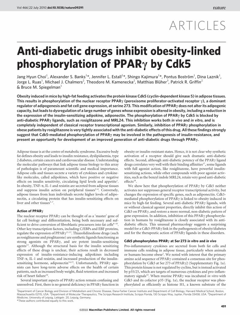
Anti-diabetic drugs of the thiazolidinedione (TZD) class, such asrosiglitazone, are agonist ligands for PPARc, improving insulin sens-itivity in both mice and humans10. To investigate whether anti-dia-betic PPARc ligands alter Cdk5-mediated phosphorylation of thisreceptor, we treated fat cells expressing WT PPARc with TNF-a,rosiglitazone, or a combination of these two agents. Figure 3a showsthat rosiglitazone inhibited Ser 273 phosphorylation at approxi-mately 1 mM, similar to the dose required for other PPARc-mediatedactivities in adipose cells13. GW9662, a PPARc antagonist25, comple-tely blocked this effect of rosiglitazone. We also examined the effect ofthese compounds on the Cdk5-mediated phosphorylation of a nat-urally occurring mutant form of PPARc (Q286P) that no longerdirectly binds known ligands26. Rosiglitazone could not interfere withCdk5 phosphorylation in this case, implying that direct binding ofthe ligand was required for inhibition. To address whether ligandswork directly to inhibit Cdk5-mediated phosphorylation, we mixedpurified PPARc, Cdk5/p35 and rosiglitazone under conditions toachieve modification in vitro. Rosiglitazone blocked Ser 273-phos-phorylation in vitro, with a half maximally effective dose of about30 nM (Fig. 3b), near the Kd of this compound for PPARc binding13.Importantly, this inhibition is not caused by a general inhibition ofCdk5 activity, as incubation with rosiglitazone did not inhibit theability of Cdk5 to phosphorylate Rb (Fig. 3c).
MRL24 is a non-TZD compound with high affinity for PPARc,with excellent anti-diabetic activity in mice18, but poor agonist prop-erties towards PPARc in transcription and adipogenesis assays. Asreported previously18 and shown in Fig. 3d, MRL24 showed weaktranscriptional activity compared to rosiglitazone. On the otherhand, MRL24 was very effective at blocking Cdk5-mediated phos-phorylation of PPARc (Fig. 3b); 30 nM of MRL24 inhibited themodification of PPARc as effectively as 300 nM of rosiglitazone, bothin vitro and in cells (Fig. 3b, Supplementary Fig. 7a and b). We alsoexamined the ability of several other published PPARc ligands thathave been shown to have poor agonist properties, but strong anti-diabetic activities in vivo27–29. As shown in Supplementary Fig. 8,Mbx-102, BVT.13 and nTZDpa were all effective at inhibiting theCdk5-mediated phosphorylation of PPARc.
To systematically compare changes in gene expression caused byboth the Ser 273 mutation in PPARc and specific PPARc ligands, we
performed Affymetrix analyses of gene expression with RNA fromcells expressing either WT or mutant PPARc, plus WT cells treatedwith rosiglitazone or MRL24. As shown in Fig. 3e, unbiased clusteringof these data resulted in several notable clusters of expression. Theoverlap in global regulation of gene expression between the mutantform of PPARc and the PPARc ligands is highly significant (P , 0.005for each drug versus mutation). Genes exhibiting differential express-ion between WT and mutant PPARc segregated into four majorgroups. One group, labelled ‘a’, were genes whose expression wasdecreased by Ser 273 mutation. This same group of genes was alsosuppressed by both rosiglitazone and MRL24, though neither ligandfunctioned as dramatically as did the non-phosphorylatable mutant ofPPARc. This is understandable, as it is unlikely that ligand treatmentcaused complete dephosphorylation of PPARc comparable to themutant protein. The very small cluster labelled ‘b’ contained Ahnaknucleoprotein and proteolipid protein (plp) 1, which are critical formyelination30,31, and sorting nexin 5 (Snx5), which is important forvesicle trafficking32. A larger cluster, labelled ‘c’, represented genesdecreased by Cdk5-mediated phosphorylation of PPARc. Several ofthe genes known to be dysregulated in obesity, including adiponectin
Ros
i
Ros
i (0.
1 μM
)
NT
NT
Ros
i (1
μM)
Ros
i (10
μM
)G
W96
62/R
osi (
10 μ
M)
Ros
i
Ros
i (0.
1 μM
)
NT
NT
Ros
i (1
μM)
Ros
i (10
μM
)G
W96
62/R
osi (
10 μ
M)
PPARγWT
pPPARγ
pPPARγ
PPARγ
PPARγ
pPPARγ
PPARγ
PPARγQ286P
PPARγ
a b
RosiMRL24
Conc. (μM)
Rel
ativ
e tr
ansc
riptio
nal a
ctiv
ity
No
ATP
NT
Ros
i (0.
0003
μM
)
Ros
i (0.
003
μM)
Ros
i (0.
03 μ
M)
Ros
i (0.
3 μM
)
Ros
i (3
μM)
MR
L24
(0.0
003
μM)
MR
L24
(0.0
03 μ
M)
MR
L24
(0.0
3 μM
)
MR
L24
(0.3
μM
)
MR
L24
(3 μ
M)
CDK5/p35 + PPARγ
CDK5
CDK5
No
ATP
NT
Ros
i (3
μM)
MR
L24
(3 μ
M)
No
ATP
NT
Ros
i (3
μM)
MR
L24
(3 μ
M)
Rb
pRb
IB: anti-CDK substrate
IB: anti-PPARγ
IB: anti-CDK5
IB: anti-CDK substrate
IB: anti-PPARγ
IB: anti-CDK5
cd e W
T
Mut
WT
/ M
RL2
4
WT
/ R
osi
IP: FLAG-PPARγIB: anti-CDK substrate
IP: FLAG-PPARγIB: anti-PPARγ
15
10
5
0
2.0
1.5
1.0
0.5
0.0
a
b
c
d
ChowHFD
Cyp2f2
Rarres2
Selenbp1
Car3
Peg10
Cidec
Cd24a
Acyl
Nr1d2
Ddx17
Aplp2
Nr3c1
Rybp
Txnip
Nr1d1
Ad
ipon
ectin
Ad
ipsi
n
Rel
ativ
e ge
ne e
xpre
ssio
nf
0 0.01 1
TNF-α TNF-α
******
*** ***
* *** ** **
** ****
–3.0 –1.3 2.1 3.00.4
Figure 3 | Anti-diabetic PPARc ligands block CDK5-mediatedphosphorylation of PPARc. a, TNF-a-induced phosphorylation of PPARcin 3T3-L1 adipocytes expressing either WT or Q286P mutant of PPARctreated with rosiglitazone and/or GW9662. b, c, In vitro CDK5 assay witheither rosiglitazone or MRL24. d, Transcriptional activity of a PPAR-derivedreporter gene in response to rosiglitazone or MRL24 (n 5 3). e, Microarrayanalysis of differentiated PPARc-null fibroblasts expressing WT (NT,rosiglitazone or MRL24 treated) or S273A mutant PPARc. f, mRNAexpression of genes regulated by the phosphorylation of PPARc inepididymal fat tissue of mice fed with either chow or HFD for 13 weeks(n 5 5). Error bars, s.e.m.; *P , 0.05, **P , 0.01, ***P , 0.001.
Chow
3 weeks 7 weeks 13 weeks13 weeks
Epi.13 weeks
Ing.
HFD Chow HFD Chow HFD Chow HFD Chow HFD
pPPARγ
PPARγ
pPPARγ
PPARγ
IB: anti-pSer273
IB: anti-PPARγ
IB: anti-pSer273
IB: anti-PPARγ
a b
Figure 2 | Cdk5-mediated phosphorylation of PPARc is increased in fattissues of mice fed a high-fat diet. a, White adipose tissue (epididymal)from mice on high-fat diet (HFD) for the indicated time was analysed withpSer 273 PPARc and PPARc antibodies. b, Epididymal (Epi.) or inguinal(Ing.) fat tissue from 13 weeks HFD mice was analysed with pSer 273antibody.
NATURE | Vol 466 | 22 July 2010 ARTICLES
453Macmillan Publishers Limited. All rights reserved©2010
and adipsin, were present in this cluster. Rosiglitazone increasedessentially all of these genes, but Fig. 3e indicates that this occurredas part of a very large gene set increased by rosiglitzone action. Indeed,genes induced most dramatically by rosiglitazone (labelled ‘d’) did notcorrespond to the Cdk5 mutation-induced gene set and were largelythe classic genes of adipogenesis, like aP2 and lipoprotein lipase (Lpl).In sharp contrast, the gene cluster strongly increased by MRL24 wasmuch smaller than that induced by rosiglitazone, and correspondedremarkably well to the gene set induced by the mutation in the Cdk5site of PPARc.
Taken together, the data presented in Figs 1–3 suggest that Cdk5modification of PPARc could be a major source of gene dysregula-tion and pathology of adipose tissues in obesity. To directly addressthis, we created refined gene sets regulated by the Cdk5 modificationof PPARc, using the concept of principal component analysis of geneexpression data (Supplementary Fig. 9). Figure 3f shows that thegreat majority of these genes had dysregulated expression in an obes-ity-dependent manner. In total, of the 17 genes most significantlyreduced by Cdk5 phosphorylation of PPARc, at least 12 were alsodecreased in obese mice. Interestingly, most of these genes weremore significantly affected in epididymal fat tissue compared to theinguinal depot (Supplementary Fig. 10), consistent with differentialphosphorylation of PPARc in the two depots (Fig. 2b). In addition,shRNA-mediated knock-down of Cdk5 in cells resulted in markeddysregulation of the gene sets, particularly adiponectin and adipsin(Supplementary Fig. 11), further confirming that Cdk5-mediatedphosphorylation of PPARc plays a significant role in the regulationof specific fat cell genes.
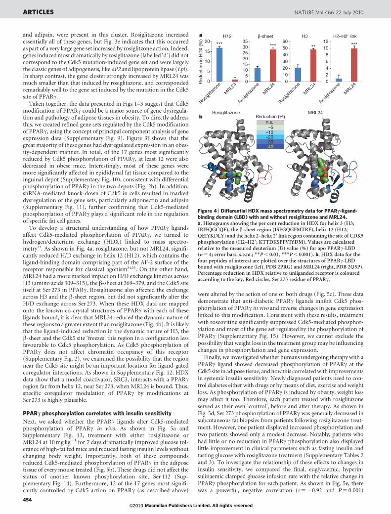
To develop a structural understanding of how PPARc ligandsaffect Cdk5-mediated phosphorylation of PPARc, we turned tohydrogen/deuterium exchange (HDX) linked to mass spectro-metry33. As shown in Fig. 4a, rosiglitazone, but not MRL24, signifi-cantly reduced H/D exchange in helix 12 (H12), which contains theligand-binding domain comprising part of the AF-2 surface of thereceptor responsible for classical agonism34,35. On the other hand,MRL24 had a more marked impact on H/D exchange kinetics acrossH3 (amino acids 309–315), the b-sheet at 369–379, and the Cdk5 siteitself at Ser 273 in PPARc. Rosiglitazone also affected the exchangeacross H3 and the b-sheet region, but did not significantly alter theH/D exchange across Ser 273. When these HDX data are mappedonto the known co-crystal structures of PPARc with each of theseligands bound, it is clear that MRL24 reduced the dynamic nature ofthese regions to a greater extent than rosiglitazone (Fig. 4b). It is likelythat the ligand-induced reduction in the dynamic nature of H3, theb-sheet and the Cdk5 site ‘freezes’ this region in a configuration lessfavourable to Cdk5 phosphorylation. As Cdk5 phosphorylation ofPPARc does not affect chromatin occupancy of this receptor(Supplementary Fig. 2), we examined the possibility that the regionnear the Cdk5 site might be an important location for ligand-gatedcoregulator interactions. As shown in Supplementary Fig. 12, HDXdata show that a model coactivator, SRC3, interacts with a PPARcregion far from helix 12, near Ser 273, when MRL24 is bound. Thus,specific coregulator modulation of PPARc by modifications atSer 273 is highly plausible.
PPARc phosphorylation correlates with insulin sensitivity
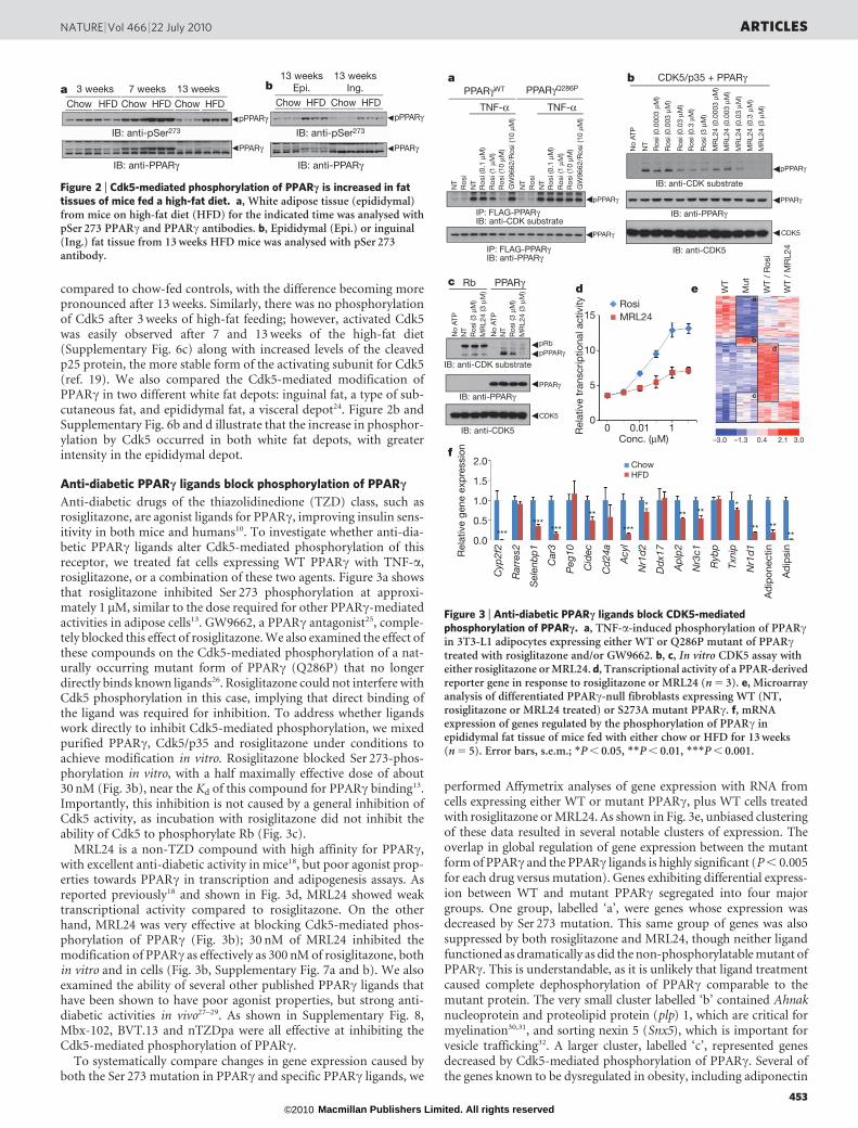
Next, we asked whether the PPARc ligands alter Cdk5-mediatedphosphorylation of PPARc in vivo. As shown in Fig. 5a andSupplementary Fig. 13, treatment with either rosiglitazone orMRL24 at 10 mg kg21 for 7 days dramatically improved glucose tol-erance of high-fat fed mice and reduced fasting insulin levels withoutchanging body weight. Importantly, both of these compoundsreduced Cdk5-mediated phosphorylation of PPARc in the adiposetissue of every mouse treated (Fig. 5b). These drugs did not affect thestatus of another known phosphorylation site, Ser 112 (Sup-plementary Fig. 14). Furthermore, 12 of the 17 genes most signifi-cantly controlled by Cdk5 action on PPARc (as described above)
were altered by the action of one or both drugs (Fig. 5c). These datademonstrate that anti-diabetic PPARc ligands inhibit Cdk5 phos-phorylation of PPARc in vivo and reverse changes in gene expressionlinked to this modification. Consistent with these results, treatmentwith roscovitine significantly suppressed Cdk5-mediated phosphor-ylation and most of the gene set regulated by the phosphorylation ofPPARc (Supplementary Fig. 15). However, we cannot exclude thepossibility that weight loss in the treatment group may be influencingchanges in phosphorylation and gene expression.
Finally, we investigated whether humans undergoing therapy with aPPARc ligand showed decreased phosphorylation of PPARc at theCdk5 site in adipose tissue, and how this correlated with improvementsin systemic insulin sensitivity. Newly diagnosed patients need to con-trol diabetes either with drugs or by means of diet, exercise and weightloss. As phosphorylation of PPARc is induced by obesity, weight lossmay affect it too. Therefore, each patient treated with rosiglitazoneserved as their own ‘control’, before and after therapy. As shown inFig. 5d, Ser 273 phosphorylation of PPARc was generally decreased insubcutaneous fat biopsies from patients following rosiglitazone treat-ment. However, one patient displayed increased phosphorylation andtwo patients showed only a modest decrease. Notably, patients whohad little or no reduction in PPARc phosphorylation also displayedlittle improvement in clinical parameters such as fasting insulin andfasting glucose with rosiglitazone treatment (Supplementary Tables 2and 3). To investigate the relationship of these effects to changes ininsulin sensitivity, we compared the final, euglycaemic, hyperin-sulinaemic clamped glucose infusion rate with the relative change inPPARc phosphorylation for each patient. As shown in Fig. 5e, therewas a powerful, negative correlation (r 5 20.92 and P 5 0.001)
a
MRL2
4
Rosigl
itazo
ne
MRL2
4
Rosigl
itazo
ne
MRL2
4
Rosigl
itazo
ne
MRL2
4
Rosigl
itazo
ne
H1220 35 60 12
1086420
50403020100
302520151050
15
10
5
0
β-sheet H3 H2–H2′ link
Red
uctio
n in
HD
X (%
)
*** *** *****
RosiglitazoneReduction (%)
MRL24
n.s.–5
–10–20–30–40
b
Figure 4 | Differential HDX mass spectrometry data for PPARc-ligand-binding domain (LBD) with and without rosiglitazone and MRL24.a, Histograms showing the per cent reduction in HDX for helix 3 (H3;IRIFQGCQF), the b-sheet region (ISEGQGFMTRE), helix 12 (H12;QEIYKDLY) and the helix 2–helix 29 link region containing the site of CDK5phosphorylation (H2–H29; KTTDKSPFVIYDM). Values are calculatedrelative to the measured deuterium (D) value (%) for apo PPARc-LBD(n 5 4; error bars, s.e.m.; **P , 0.01, ***P , 0.001). b, HDX data for thefour peptides of interest are plotted over the structures of PPARc-LBDbound with rosiglitazone (left, PDB 2PRG) and MRL24 (right, PDB 2Q5P).Percentage reduction in HDX relative to unliganded receptor is colouredaccording to the key. Red circles, Ser 273 residue of PPARc.
ARTICLES NATURE | Vol 466 | 22 July 2010
454Macmillan Publishers Limited. All rights reserved©2010

between changes in glucose infusion rate and changes in PPARc phos-phorylation. Thus, in this patient cohort, improvement of insulinsensitivity using rosiglitazone was tightly coupled to decreased phos-phorylation of PPARc at the Cdk5 site.
Discussion
The data presented here suggest a unified framework for understand-ing the relationship between fat cell dysfunction in obesity and anti-diabetic therapies through PPARc (Supplementary Fig. 16). High-fatdiets in vivo activate the protein kinase Cdk5. Cdk5-mediated phos-phorylation of PPARc at Ser 273 dysregulated expression of a subsetof genes; these included a number of key metabolic regulators,including adipsin, the first fat cell-selective gene whose expressionis altered in obesity36 and adiponectin, a central regulator of insulinsensitivity in vivo. Ser 273 phosphorylation did not alter the chro-matin occupancy of PPARc, suggesting that other mechanisms, suchas differential recruitment of co-regulators, may cause these differ-ences in target gene expression. Anti-diabetic PPARc ligands inhib-ited Ser 273 phosphorylation and reversed associated changes in geneexpression. Crucially, inhibition of Cdk5-mediated phosphorylationof PPARc by ligands altered the conformation/dynamic nature of theprotein, such that Ser 273 was no longer modified by the proteinkinase. In fact, a PPARc ligand with minimal agonist activity, suchas MRL24, did this as well or better than a full agonist, such asrosiglitazone. Given that both have potent anti-diabetic activities;it is likely that a substantial portion of the therapeutic benefits ofPPARc ligands in metabolic disease is through inhibition of Cdk5-mediated phosphorylation. The studies presented in Fig. 5d and e
also indicate that this specific phosphorylation was closely coupled tothe therapeutic effects of rosiglitazone in humans. It is worth notingthat the therapeutic doses used in animals did not lead to a completeinhibition of the Cdk5 phosphorylation of PPARc, so more robusttherapy might be obtained with other, more narrowly targetedligands.
On the basis of these results, the role of agonism in the actions ofPPARc ligand drugs is quite unclear. It is possible that a small degreeof agonism further enhances the beneficial effects of blocking Cdk5-mediated phosphorylation of PPARc. On the other hand, it seemslikely that at least some of the problematic side-effects of these drugs,including weight gain and fluid retention16, may occur through clas-sical agonist actions. This can be better assessed as ligands with vari-ous degrees of agonism are evaluated in both preclinical models andhuman subjects. More broadly, the ability of ‘partial agonists’ to alterprotein phosphorylation may be true for other members of the nuc-lear receptor family, possibly allowing for identification of new drugs.
How is Cdk5 activated in adipose tissues of obese mice? Bothcytokines and fatty acids are elevated in adipose tissues in most obesestates, and both may contribute to Cdk5 activation in the experi-ments preformed here. Cytokines and other stimulators of Cdk5typically work via cleavage of the Cdk5 partner p35 into a more activeand more stable p25 subunit19. Although the extracellular stimulatorthat functions on fat cells as a consequence of high-fat feeding is notestablished, it is clear from Supplementary Fig. 6 that the activation ofCdk5 in obesity is accompanied by enhanced formation of p25.Whether high-fat feeding and obesity lead to activation of Cdk5 inother non-adipose tissues is a very important question, as the nega-tive effects of obesity extend far beyond the metabolic syndrome tocancer and neurodegeneration37,38. Neurodegeneration has beenassociated with the abnormal activation of Cdk5 in the CNS39, sofurther studies are required to determine if Cdk5 is activated in thebrain and other tissues in obesity.
METHODS SUMMARYCell culture. PPARc-null mouse embryonic fibroblasts (MEFs)21 were cultured
in Dulbecco’s modified Eagle’s medium and 10% fetal bovine serum. FLAG-
PPARc and FLAG-PPARc S273A were subcloned into pMSCV-puro retrovial
vector (Stratagene). Following infection of the cells with retrovirus, cells expres-
sing ectopic protein were selected by incubation with 2mg ml21 puromycin.
Adipocyte differentiation in 3T3-L1 or MEFs was induced by treating cells with
1 mM dexamethasone, 0.5 mM isobutylmethylxanthine, and 850 nM insulin for
48 h and cells were switched to the maintenance medium containing 850 nM
insulin.
In vitro kinase assay. Immuno-purified WT or S273A mutant of PPARc was
incubated with active Cdk kinases in kinase assay buffer containing ATP for
15 min at 30 uC. Positive controls, either purified histone H1 (Millipore) or
Rb (Cell Signaling Technology) were used. PPARc ligands were pre-incubated
with substrates for 30 min prior to the assay.
Gene expression analysis. Total RNA was isolated from cells or tissues using
Trizol reagents (Invitrogen). The RNA was reverse-transcribed using ABI reverse
transcription kit. Quantitative PCR reactions were performed with SYBR green
fluorescent dye using an ABI 9300 PCR machine. Relative mRNA expression was
determined by the DD-Ct method using TATA-binding protein (TBP) levels.
Animals. All animal experiments were performed according to procedures
approved by Beth Israel Deaconess Medical Center’s Institutional Animal Care
and Use Committee. 4 to 5 week-old male C57BL/6J mice were obtained from
the Jackson Laboratory. Mice were fed a regular diet (10% kcal fat, D12450B,
Research Diets) or a high-fat diet (60% kcal fat, D12492, Research Diets) as
indicated. For glucose tolerance tests, mice were intraperitoneally (i.p.) injected
with 10 mg kg21 rosiglitazone or MRL24 for 6 days, and fasted overnight before
i.p. injection of 2 g kg21D-glucose.
Full Methods and any associated references are available in the online version ofthe paper at www.nature.com/nature.
Received 5 March; accepted 24 June 2010.
1. Kershaw, E. E. & Flier, J. S. Adipose tissue as an endocrine organ. J. Clin. Endocrinol.Metab. 89, 2548–2556 (2004).
Glu
cose
(mg
dl–1
)
Time (min)
2.5
2
1.5
1
0.5
0
160
120
80
Before After
40
0
0 50 100 150
Vehicle400
1.5
1
0.5
0
300
200
100
0
RosiglitazoneMRL24
Vehicle MRL24
pPPARγ
PPARγIB: anti-pSer273
IB: anti-PPARγ
a b
c
d
Vehicle MRL24
****
**** **
**
** *
*** **
**
*
**
Cyp2f2
Rarres2
Selenbp1Car3
Peg10Cidec
Cd24aAcyl
Nr1d2
Ddx17Aplp2
Nr3c1RybpTxnip
Nr1d1
Adiponec
tin
Adipsin
e
Rel
ativ
e ge
ne e
xpre
ssio
n
***
*
****
***
******
**
**
Rosiglitazone
Vehicle MRL24 Rosiglitazone
Rosiglitazone
pP
PARγ/
PPA
Rγ
pP
PARγ/
PPA
Rγ
(%)
* *
R = –0.92P = 0.001
P = 0.046 200
150
100
50
00 50 100 150C
hang
e in
glu
cose
infu
sion
rat
e (%
)
Change in pPPARγ (%)
Figure 5 | Correlation between the inhibition of phosphorylation andimprovement of insulin sensitivity by anti-diabetic PPARc ligands.a, Glucose-tolerance tests in 16-week HFD mice treated with vehicle,rosiglitazone or MRL24 (n 5 10). b, Phosphorylation of PPARc in whiteadipose tissue (WAT). c, The expression of gene sets regulated by PPARcphosphorylation in WAT (error bars, s.e.m.; *P , 0.05, **P , 0.01,***P , 0.001). d, Changes of phosphorylation of PPARc in human fatbiopsies from patients that were treated with rosiglitazone for 6 months.‘Before’, non-treated; ‘after’, 6 months treatment. e, Correlation between thechanges of PPARc phosphorylation normalized to total PPARc protein andthe changes of glucose infusion rate measured by clamp. The data arepresented as percentage change after 6 months of rosiglitazone treatment.
NATURE | Vol 466 | 22 July 2010 ARTICLES
455Macmillan Publishers Limited. All rights reserved©2010
2. Hotamisligil, G. S., Shargill, N. S. & Spiegelman, B. M. Adipose expression of tumornecrosis factor-a: direct role in obesity-linked insulin resistance. Science 259,87–91 (1993).
3. Lagathu, C. et al. Long-term treatment with interleukin-1b induces insulinresistance in murine and human adipocytes. Diabetologia 49, 2162–2173 (2006).
4. Steppan, C. M. et al. The hormone resistin links obesity to diabetes. Nature 409,307–312 (2001).
5. Berg, A. H. et al. The adipocyte-secreted protein Acrp30 enhances hepatic insulinaction. Nature Med. 7, 947–953 (2001).
6. Hu, E., Liang, P. & Spiegelman, B. M. AdipoQ is a novel adipose-specific genedysregulated in obesity. J. Biol. Chem. 271, 10697–10703 (1996).
7. Yamauchi, T. et al. The fat-derived hormone adiponectin reverses insulinresistance associated with both lipoatrophy and obesity. Nature Med. 7, 941–946(2001).
8. Morrison, R. F. & Farmer, S. R. Hormonal signaling and transcriptional control ofadipocyte differentiation. J. Nutr. 130, 3116S–3121S (2000).
9. Tontonoz, P., Hu, E. & Spiegelman, B. M. Stimulation of adipogenesis in fibroblastsby PPAR c 2, a lipid-activated transcription factor. Cell 79, 1147–1156 (1994).
10. Willson, T. M., Lambert, M. H. & Kliewer, S. A. Peroxisome proliferator-activatedreceptor c and metabolic disease. Annu. Rev. Biochem. 70, 341–367 (2001).
11. Jimenez, M. A. et al. Critical role for Ebf1 and Ebf2 in the adipogenic transcriptionalcascade. Mol. Cell. Biol. 27, 743–757 (2007).
12. Wu, Z., Bucher, N. L. & Farmer, S. R. Induction of peroxisome proliferator-activatedreceptor c during the conversion of 3T3 fibroblasts into adipocytes is mediated byC/EBPb, C/EBPd, and glucocorticoids. Mol. Cell. Biol. 16, 4128–4136 (1996).
13. Lehmann, J. M. et al. An antidiabetic thiazolidinedione is a high affinity ligand forperoxisome proliferator-activated receptor c (PPAR c). J. Biol. Chem. 270,12953–12956 (1995).
14. Trujillo, M. E. & Scherer, P. E. Adipose tissue-derived factors: impact on health anddisease. Endocr. Rev. 27, 762–778 (2006).
15. Sharma, A. M. & Staels, B. Peroxisome proliferator-activated receptor c andadipose tissue — understanding obesity-related changes in regulation of lipid andglucose metabolism. J. Clin. Endocrinol. Metab. 92, 386–395 (2007).
16. Lipscombe, L. L. et al. Thiazolidinediones and cardiovascular outcomes in olderpatients with diabetes. J. Am. Med. Assoc. 298, 2634–2643 (2007).
17. Willson, T. M. et al. The structure-activity relationship between peroxisomeproliferator-activated receptor gamma agonism and the antihyperglycemicactivity of thiazolidinediones. J. Med. Chem. 39, 665–668 (1996).
18. Acton, J. J. III et al. Benzoyl 2-methyl indoles as selective PPARc modulators.Bioorg. Med. Chem. Lett. 15, 357–362 (2005).
19. Dhavan, R. & Tsai, L. H. A decade of CDK5. Nature Rev. Mol. Cell Biol. 2, 749–759(2001).
20. Utreras, E. et al. Tumor necrosis factor-a regulates cyclin-dependent kinase 5activity during pain signaling through transcriptional activation of p35. J. Biol.Chem. 284, 2275–2284 (2009).
21. Rosen, E. D. et al. C/EBPa induces adipogenesis through PPARc: a unifiedpathway. Genes Dev. 16, 22–26 (2002).
22. Musa, F. R. et al. Effects of luteinizing hormone, follicle-stimulating hormone, andepidermal growth factor on expression and kinase activity of cyclin-dependentkinase 5 in Leydig TM3 and Sertoli TM4 cell lines. J. Androl. 21, 392–402 (2000).
23. Torti, F. M. et al. A macrophage factor inhibits adipocyte gene expression: an invitro model of cachexia. Science 229, 867–869 (1985).
24. Cinti, S. The adipose organ. Prostaglandins Leukot. Essent. Fatty Acids 73, 9–15(2005).
25. Leesnitzer, L. M. et al. Functional consequences of cysteine modification in theligand binding sites of peroxisome proliferator activated receptors by GW9662.Biochemistry 41, 6640–6650 (2002).
26. Sarraf, P. et al. Loss-of-function mutations in PPARc associated with human coloncancer. Mol. Cell 3, 799–804 (1999).
27. Berger, J. P. et al. Distinct properties and advantages of a novel peroxisomeproliferator-activated protein c selective modulator. Mol. Endocrinol. 17, 662–676(2003).
28. Gregoire, F. M. et al. MBX-102/JNJ39659100, a novel peroxisome proliferator-activated receptor-ligand with weak transactivation activity retains antidiabeticproperties in the absence of weight gain and edema. Mol. Endocrinol. 23, 975–988(2009).
29. Ostberg, T. et al. A new class of peroxisome proliferator-activated receptoragonists with a novel binding epitope shows antidiabetic effects. J. Biol. Chem.279, 41124–41130 (2004).
30. Hakak, Y. et al. Genome-wide expression analysis reveals dysregulation ofmyelination-related genes in chronic schizophrenia. Proc. Natl Acad. Sci. USA 98,4746–4751 (2001).
31. Salim, C. et al. The giant protein AHNAK involved in morphogenesis and lamininsubstrate adhesion of myelinating Schwann cells. Glia 57, 535–549 (2009).
32. Merino-Trigo, A. et al. Sorting nexin 5 is localized to a subdomain of the earlyendosomes and is recruited to the plasma membrane following EGF stimulation.J. Cell Sci. 117, 6413–6424 (2004).
33. Maier, C. S. & Deinzer, M. L. Protein conformations, interactions, and H/Dexchange. Methods Enzymol. 402, 312–360 (2005).
34. Nolte, R. T. et al. Ligand binding and co-activator assembly of the peroxisomeproliferator-activated receptor-c. Nature 395, 137–143 (1998).
35. Bruning, J. B. et al. Partial agonists activate PPARc using a helix 12 independentmechanism. Structure 15, 1258–1271 (2007).
36. Flier, J. S. et al. Severely impaired adipsin expression in genetic and acquiredobesity. Science 237, 405–408 (1987).
37. Calle, E. E. et al. Overweight, obesity, and mortality from cancer in a prospectivelystudied cohort of U.S. adults. N. Engl. J. Med. 348, 1625–1638 (2003).
38. Whitmer, R. A. et al. Obesity in middle age and future risk of dementia: a 27 yearlongitudinal population based study. Br. Med. J. 330, 1360–1362 (2005).
39. Patrick, G. N. et al. Conversion of p35 to p25 deregulates Cdk5 activity andpromotes neurodegeneration. Nature 402, 615–622 (1999).
Supplementary Information is linked to the online version of the paper atwww.nature.com/nature.
Acknowledgements We thank V. Mootha for help with the analysis of microarraydata and for critical comments. We thank M. Kirschner for a critical reading of themanuscript and for comments. We are grateful to R. Gupta and P. Cohen for criticalcomments on the manuscript. J.H.C., A.S.B., J.L.E., P.B., D.L., J.L.R. and B.M.S aresupported by NIH DK31405. S.K. is supported by NIH grant DK087853. M.B. issupported by a grant from Deutsche Forschungsgemeinschaft (DFG) and theClinical Research group ‘Atherobesity’ KFO 152 (project BL 833/1-1). P.R.G., M.J.C.and T.M.K. are supported in part by the by the Intramural Research Program of theN National Institute of Mental Health (P.R.G., M.J.C., T.M.K., U54-MH084512;H. Rosen Principal Investigator) and by the NIH National Institute of GeneralMedical Sciences (P.R.G. and M.J.C., R01-GM084041).
Author Contributions J.H.C. and B.M.S. conceived and designed the experiments.J.H.C., A.S.B., J.L.E., S.K., P.B., D.L., J.L.R., M.J.C., T.M.K, M.B. and P.R.G performedthe experiments. All authors analysed the data. J.H.C., A.S.B., J.L.E. and B.M.S.wrote the manuscript.
Author Information Microarray data have been deposited in Gene ExpressionOmnibus: GSE22033. Reprints and permissions information is available atwww.nature.com/reprints. The authors declare no competing financial interests.Readers are welcome to comment on the online version of this article atwww.nature.com/nature. Correspondence and requests for materials should beaddressed to B.M.S. ([email protected]).
ARTICLES NATURE | Vol 466 | 22 July 2010
456Macmillan Publishers Limited. All rights reserved©2010
METHODSCell culture. 3T3-L1 and HEK-293 cells were obtained from ATCC. PPARc-null
mouse embryonic fibroblasts (MEFs)21 were cultured in Dulbecco’s modified
Eagle’s medium and 10% fetal bovine serum. FLAG-PPARc and FLAG-PPARcS273A were subcloned into pMSCV-puro retroviral vector (Stratagene). For
retrovirus production, Phoenix packaging cells were transfected with 10 mg ret-
roviral vectors40. After 48 h, the viral supernatant was collected and filtered.
Following infection of the cells with the retrovirus, cells expressing the ectopic
protein were selected by incubation with 2 mg ml21 puromycin. Adipocyte dif-
ferentiation on 3T3-L1 or MEFs was induced by treating cells with 1mM dex-amethasone, 0.5 mM isobutylmethylxanthine and 850 nM insulin for 48 h and
cells were switched to the maintenance medium containing 850 nM insulin.
Lipid accumulation in the cells was detected by Oil Red O staining. The amount
of secreted adiponectin into cell medium was analysed by ELISA (Millipore). All
chemicals for cell culture were obtained from Sigma unless otherwise indicated.
DNA constructs and shRNA of CDK5. HA-WT CDK5, HA-KD CDK5 and
Myc-p35 were obtained from Addgene. Murine PPARa or PPARd were sub-
cloned into FLAG-pcDNA3.1 (Invitrogen). The sequence used for lentiviral
shRNA expression vector (pLKO.1; Open Biosystems) targeting CDK5 was 59-
CGGGAGATCTGTCTACTCAAA-39. For lentivirus production, HEK-293T
cells (ATCC) were transfected with 10 mg lentiviral vectors40. Following infection
of the cells with the lentivirus, cells were selected by incubation with 2mg ml21
puromycin.
In vitro kinase assay. Active Cdk5/p35, Cdk1/cdc2, Cdk2/cyclin A, Cdk2/cyclin
E or Cdk4/cyclin D1 kinases were purchased from Millipore or Cell Signaling
Technology. In vitro CDK kinase assay was performed using the HTScan CDK/
Cyc kinase assay kit according to the manufacturer’s instructions (Cell Signaling
Technology). Briefly, 1mg of immuno-purified WT or S273A mutant of PPARcwere incubated with active CDK kinase in kinase assay buffer (25 mM Tris-HCl
pH 7.5, 5 mM b-glycerophosphate, 2 mM dithiothreitol (DTT), 0.1 mM
Na3VO4, 10 mM MgCl2) containing 20 mM ATP for 15 min at 30 uC. As positive
control for assay, purified histone H1 (Millipore) or Rb (Cell Signaling
Technology) were used. Several PPARc ligands were pre-incubated with sub-
strates for 30 min, and the assay was performed. Phosphorylation of substrates
after SDS–PAGE was analysed with anti-CDK substrate antibody to detect phos-
pho-Ser in a K/R-S-P-K/R motif, which is the consensus motif for Cdk substrate
proteins (Cell Signaling Technology).
Preparation of cell or tissue lysates, immunoprecipitation and immunoblot-
ting. HEK-293 cells expressing CDK5 or PPARc were collected after transfec-
tion. Total cell lysates were incubated with FLAG M2 agarose (Sigma) at 4 uC.
Immunoprecipitates or total cell lysates were analysed with anti-CDK substrate,
FLAG or HA antibodies (Roche). Differentiated 3T3-L1 adipocytes were
treated with TNF-a (50 ng ml21), IL-6 (50 ng ml21), IL-1b (50 ng ml21) or
FFAs (400 mM palmitic acid and oleic acid mixtures) for the indicated times,
and cell lysates were analysed with phospho-specific or PPARc antibodies. 3T3-
L1 adipocytes were pre-treated with various PPARc ligands, and incubated withTNF-a. For tissue lysates, WAT from mice was homogenized in RIPA buffer
(50 mM Tris pH 7.5, 150 mM NaCl, 1% NP-40, 0.5% sodium deoxycholate,
0.1% SDS with protease and phosphatase inhibitors). For western blotting, a
rabbit polyclonal phospho-specific antibody against PPARc Ser 273 was pro-
duced by New England Peptides with a synthetic phosphopeptide corresponding
to residues surrounding Ser 273 of PPARc (Ac-KTTDKpSPFVIYDC-amide).
Total tissue lysates were analysed with anti-PPARc, phospho-CDK5 (Y15),
CDK5 and p35 antibodies (Santa Cruz).
Reporter gene assay. HEK-293 cells were transfected with pDR-1 luciferase
reporter plasmid, PPARc, RXRa and pRL-renillin using Lipofectamine 2000
(Invitrogen). Following an overnight transfection, the cells were treated with
rosiglitazone or MRL24 for 24 h. The cells were harvested and reporter gene
assays were carried out using the Dual-Luciferase kit (Promega). Luciferase
activity was normalized to renillia activity.
Gene expression analysis. Total RNA was isolated from cells or tissues using Trizol
reagents (Invitrogen). The RNA was reverse-transcribed using ABI reverse tran-
scription kit. Quantitative PCR reactions were performed with SYBR green fluor-
escent dye using an ABI 9300 PCR machine. Relative mRNA expression wasdetermined by theDD-Ct method normalized to tata-binding protein (TBP) levels.
The sequences of primers used in this study are found in Supplementary Table 4.
Generation of fat pads in nude mice. PPARc-null fibroblasts (1 3 107) stably
expressing WT or the S273A mutant of PPARc were implanted subcutaneously
into 7–8-week-old male NCR nude mice (Taconic) according to the previous
methods (n 5 5 mice per group)41. Six weeks after injection, the fat pads were
isolated for the analysis of gene expression.
Microarray analysis. Total RNA was isolated from PPARc-null fibroblasts
expressing WT or the S273A mutant of PPARc or WT cells treated with 1mM
rosiglitazone or MRL24 for 24 h. Array hybridization and scanning were per-
formed by the Dana-Farber Cancer Institute Microarray Core Facility using
Affymetrix GeneChip Mouse Genome 430 2.0 arrays according to established
methods42. The array data were analysed using the DNA-Chip Analyser (dChip)
sofware43. The statistical significance of differences in gene expression was
assessed by an unpaired t-test (P , 0.05). Global overlap of genes was statistically
assessed using x2 statistics. To create refined gene sets regulated by Cdk5 phos-
phorylation of PPARc, we first calculated the P-value as well as fold-change of
gene expression in WT versus S273A mutant cells, and we plotted 2log(p-value)
versus log2 fold-change. From this list of genes, we selected 53 genes which were
changed in magnitude ($1.4 fold difference) and statistical significance
(P , 0.05). The selected genes were validated in cells or transplanted fat pads
using by qPCR; the resulting gene set, consisting of 17 genes, was analysed in
WAT of mice using qPCR.
Animals. All animal experiments were performed according to procedures
approved by Beth Israel Deaconess Medical Center’s Institutional Animal Care
and Use Committee. 4–5-week-old male C57BL/6J mice were obtained from the
Jackson Laboratory. Mice were fed a regular diet (10% kcal fat, D12450B,
Research Diets) or a high-fat diet (60% kcal fat, D12492, Research Diets) as
indicated time periods. All mice initiated the HFD at the same age (4–5 week
old), and were killed at the indicated time after starting the HFD. For glucose
tolerant tests, mice were intraperitoneally (i.p.) injected daily with 10 mg kg21
rosiglitazone or MRL24 for 6 days, and fasted overnight before i.p. injection of
2 g kg21D-glucose. Glucose was measured in tail vein blood at intervals after
glucose injection using a Truetrack glucometer. Serum insulin concentrations
were determined by ELISA (Crystal Chem).
Human study. We used fat biopsies from subjects enrolled in a parallel-group
randomized and controlled trial to evaluate the effects of rosiglitazone and an
exercise programme on insulin sensitivity and endothelial function in patients
with impaired glucose tolerance over a 6-month period. Consecutive patients
referred to the University of Leipzig Heart Center, Germany, were invited to
participate if they displayed impaired glucose tolerance (2 h OGTT plasma glu-
cose .7.8 and ,11.1 mmol l21)44. Exclusion criteria included diabetes type 1 or
2, concomitant medication, unstable angina, coronary artery disease, myocardial
infarction within preceding 3 months, ejection fraction ,40%, significant heart
valve disease, severe metabolic disorders, severe disorders in lipoprotein meta-
bolism, thyroid disorders, alcohol or drug abuse, pregnancy, and participation in
another trial. Included patients received 4 mg rosiglitazone daily (Avandia;
GlaxoSmithKline) for 6 months. At day 1 and after 6 months, patients were
clinically examined, euglycaemic-hyperinsulinaemic clamps were performed,
and blood samples were obtained. All baseline blood samples were collected
between 8 and 10 AM after an 12-h overnight fast. Plasma insulin was measured
with an enzyme immunometric assay for the IMMULITE automated analyser
(Diagnostic Products). Serum total HDL- or LDL-cholesterol, triglycerides, free
fatty acids, adiponectin, were measured as previously described45. Insulin sens-
itivity was assessed with the euglycaemic-hyperinsulinaemic clamp method
using a previously described protocol46. Fat biopsies were analysed with anti-
phospho-Ser 273 and PPARc antibody. Statistical analyses were performed using
SPSS statistics 17.0 and Pearson’s two-sided correlation was used for statistical
tests. All correlations with P , 0.05 yielded similar results using Spearman’s rank
correlation.
40. Kinsella, T. M. & Nolan, G. P. Episomal vectors rapidly and stably produce high-titer recombinant retrovirus. Hum. Gene Ther. 7, 1405–1413 (1996).
41. Walkey, C. J. & Spiegelman, B. M. A functional peroxisome proliferator-activatedreceptor-c ligand-binding domain is not required for adipogenesis. J. Biol. Chem.283, 24290–24294 (2008).
42. Lockhart, D. J. et al. Expression monitoring by hybridization to high-densityoligonucleotide arrays. Nature Biotechnol. 14, 1675–1680 (1996).
43. Li, C. & Wong, W. H. Model-based analysis of oligonucleotide arrays: expressionindex computation and outlier detection. Proc. Natl Acad. Sci. USA 98, 31–36(2001).
44. Moreno-Navarrete, J. M. et al. Complement factor H is expressed in adiposetissue in association with insulin resistance. Diabetes 59, 200–209 (2010).
45. Kloting, N. et al. Serum retinol-binding protein is more highly expressed in visceralthan in subcutaneous adipose tissue and is a marker of intra-abdominal fat mass.Cell Metab. 6, 79–87 (2007).
46. Bluher, M. et al. Relation between glycaemic control, hyperinsulinaemia andplasma concentrations of soluble adhesion molecules in patients with impairedglucose tolerance or Type II diabetes. Diabetologia 45, 210–216 (2002).
doi:10.1038/nature09291
Macmillan Publishers Limited. All rights reserved©2010
Related Documents