Molecules 2014, 19, 5191-5204; doi:10.3390/molecules19045191 molecules ISSN 1420-3049 www.mdpi.com/journal/molecules Article Anti-Amoebic Properties of Carbonyl Thiourea Derivatives Maizatul Akma Ibrahim, Mohd Sukeri Mohd Yusof and Nakisah Mat Amin * School of Fundamental Science, Universiti Malaysia Terengganu, Kuala Terengganu, Terengganu 21030, Malaysia; E-Mails: [email protected] (M.A.I.); [email protected] (M.S.M.Y.) * Author to whom correspondence should be addressed; E-Mail: [email protected]; Tel.: +609-668-3245; Fax: +609-668-3608. Received: 26 November 2013; in revised form: 25 March 2014 / Accepted: 9 April 2014 / Published: 22 April 2014 Abstract: Thiourea derivatives display a broad spectrum of applications in chemistry, various industries, medicines and various other fields. Recently, different thiourea derivatives have been synthesized and explored for their anti-microbial properties. In this study, four carbonyl thiourea derivatives were synthesized and characterized, and then further tested for their anti-amoebic properties on two potential pathogenic species of Acanthamoeba, namely A. castellanii (CCAP 1501/2A) and A. polyphaga (CCAP 1501/3A). The results indicate that these newly-synthesized thiourea derivatives are active against both Acanthamoeba species. The IC 50 values obtained were in the range of 2.39–8.77 μg·mL -1 (9.47–30.46 μM) for A. castellanii and 3.74–9.30 μg·mL -1 (14.84–31.91 μM) for A. polyphaga. Observations on the amoeba morphology indicated that the compounds caused the reduction of the amoeba size, shortening of their acanthopodia structures, and gave no distinct vacuolar and nuclear structures in the amoeba cells. Meanwhile, fluorescence microscopic observation using acridine orange and propidium iodide (AOPI) staining revealed that the synthesized compounds induced compromised-membrane in the amoeba cells. The results of this study proved that these new carbonyl thiourea derivatives, especially compounds M1 and M2 provide potent cytotoxic properties toward pathogenic Acanthamoeba to suggest that they can be developed as new anti-amoebic agents for the treatment of Acanthamoeba keratitis. Keywords: thiourea derivatives; anti-amoebic agent; Acanthamoeba; Acanthamoeba keratitis; morphology; membrane integrity OPEN ACCESS

Welcome message from author
This document is posted to help you gain knowledge. Please leave a comment to let me know what you think about it! Share it to your friends and learn new things together.
Transcript
Molecules 2014, 19, 5191-5204; doi:10.3390/molecules19045191
molecules ISSN 1420-3049
www.mdpi.com/journal/molecules
Article
Anti-Amoebic Properties of Carbonyl Thiourea Derivatives
Maizatul Akma Ibrahim, Mohd Sukeri Mohd Yusof and Nakisah Mat Amin *
School of Fundamental Science, Universiti Malaysia Terengganu, Kuala Terengganu,
Terengganu 21030, Malaysia; E-Mails: [email protected] (M.A.I.);
[email protected] (M.S.M.Y.)
* Author to whom correspondence should be addressed; E-Mail: [email protected];
Tel.: +609-668-3245; Fax: +609-668-3608.
Received: 26 November 2013; in revised form: 25 March 2014 / Accepted: 9 April 2014 /
Published: 22 April 2014
Abstract: Thiourea derivatives display a broad spectrum of applications in chemistry,
various industries, medicines and various other fields. Recently, different thiourea
derivatives have been synthesized and explored for their anti-microbial properties.
In this study, four carbonyl thiourea derivatives were synthesized and characterized,
and then further tested for their anti-amoebic properties on two potential pathogenic
species of Acanthamoeba, namely A. castellanii (CCAP 1501/2A) and A. polyphaga
(CCAP 1501/3A). The results indicate that these newly-synthesized thiourea derivatives
are active against both Acanthamoeba species. The IC50 values obtained were in the range
of 2.39–8.77 µg·mL-1 (9.47–30.46 µM) for A. castellanii and 3.74–9.30 µg·mL-1
(14.84–31.91 µM) for A. polyphaga. Observations on the amoeba morphology indicated
that the compounds caused the reduction of the amoeba size, shortening of their
acanthopodia structures, and gave no distinct vacuolar and nuclear structures in the amoeba
cells. Meanwhile, fluorescence microscopic observation using acridine orange and
propidium iodide (AOPI) staining revealed that the synthesized compounds induced
compromised-membrane in the amoeba cells. The results of this study proved that these
new carbonyl thiourea derivatives, especially compounds M1 and M2 provide potent
cytotoxic properties toward pathogenic Acanthamoeba to suggest that they can be
developed as new anti-amoebic agents for the treatment of Acanthamoeba keratitis.
Keywords: thiourea derivatives; anti-amoebic agent; Acanthamoeba; Acanthamoeba
keratitis; morphology; membrane integrity
OPEN ACCESS
Molecules 2014, 19 5192
1. Introduction
Acanthamoeba is one of the free-living amoebae that are widely distributed in the environment [1].
This amoeba genus is among the most common protozoa to be found in soil and water samples [2].
Acanthamoeba is known as the causative agent for a sight-threatening disease, Acanthamoeba
keratitis. This eye infection is recognized as one of the most challenging and severe ocular parasitic
diseases [3]. The Acanthamoeba species which have been reported to cause Acanthamoeba keratitis
are A. castellanii, A. polyphaga, A. hatchetti, A. culbertsoni, A. rhysodes, A. griffini, A. quina, and
A. lugdunensis [4]. An effective medical therapy for treating the infection is currently not available.
Several antiseptics such as chlorhexidine gluconate and polyhexamethylene biguanide have been used
to lessen the symptoms [5,6], but they are not specifically designed to treat the ocular disease, thus side
effects are frequently reported [7,8]. Some surveys showed that Acanthamoeba are resistant to these
agents, which make them less effective [9,10] especially at later stages of infection. Therefore, new
potential agents are in high demand to assist the current treatment of Acanthamoeba keratitis.
Since synthetic organic compounds are being widely designed nowadays in parallel with the
development of combinatorial chemistry and compound libraries, they could be exploited for the
development of new drugs. Some synthetic compounds such as quinoxaline derivatives and
thiosemicarbazone analogs were investigated on the cells of Entamoeba histolytica and found to
display beneficial properties which can be developed as anti-amoebic agents [11,12]. Thiourea, which
is one of the earliest synthetic organic compounds, has been globally used directly and indirectly due
to its ready availability. This factor has attracted researchers to evaluate thiourea-based compounds
from their safety point of view [13] and potential medical properties [14–16].
Previous studies have shown the potential of certain thiourea derivatives as anti-microbial
agents [17,18]. Drugs which are based on thiourea have also been used clinically to treat patients of
tuberculosis [19] and thyroid conditions [20]. Therefore, in the present study, four new carbonyl
thiourea derivatives were synthesized and characterized, and could possibly be developed as new agent
to treat Acanthamoeba keratitis after their anti-amoebic properties were examined. Cytotoxicity tests
which involved investigation of the inhibition of amoeba population and disruption of the amoeba
membrane integrity caused by the compounds were conducted. Microscopic observation was
also carried out to examine the morphological alterations in the amoeba cells caused by these
newly-synthesized compounds.
2. Results and Discussion
2.1. Preparation of Carbonyl Thiourea Derivatives
The preparation of compounds M1–M4 is shown in Scheme 1 [21,22], while the compounds
obtained and their molecular weights are listed in Table 1.
2.2. Anti-Amoebic Properties: IC50 Values
Experiments were carried out to analyze the in vitro anti-amoebic activity of the four newly-
synthesized carbonyl thiourea derivatives on two pathogenic species of Acanthamoeba, namely
Molecules 2014, 19 5193
A. castellanii (CCAP 1501/2A) and A. polyphaga (CCAP 1501/3A). The amoebae were obtained from
the UK Culture Collection of Algae and Protozoa (CCAP, Argyll, UK). The IC50 values which were
obtained from the absorbance readings and represented in non-linear sigmoidal dose-response curve
derived from GraphPrism software are presented in Table 2.
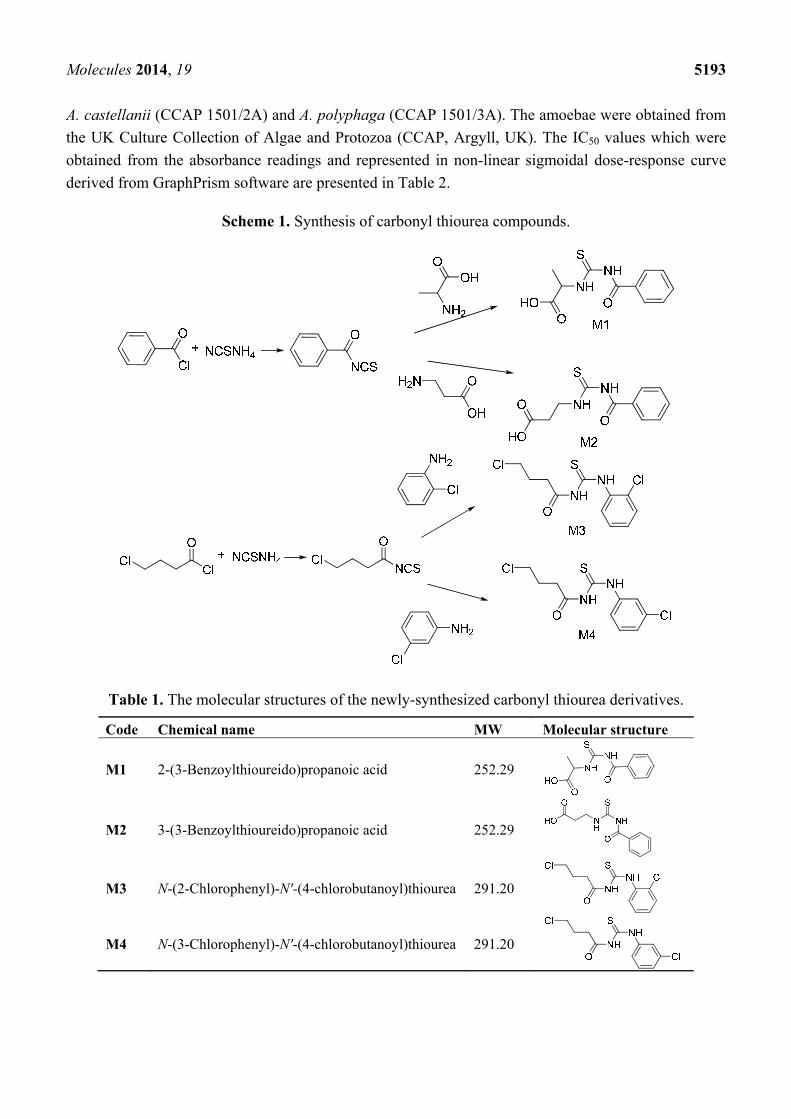
Scheme 1. Synthesis of carbonyl thiourea compounds.
Table 1. The molecular structures of the newly-synthesized carbonyl thiourea derivatives.
Code Chemical name MW Molecular structure
M1 2-(3-Benzoylthioureido)propanoic acid 252.29
M2 3-(3-Benzoylthioureido)propanoic acid 252.29
M3 N-(2-Chlorophenyl)-N'-(4-chlorobutanoyl)thiourea 291.20
M4 N-(3-Chlorophenyl)-N'-(4-chlorobutanoyl)thiourea 291.20
Molecules 2014, 19 5194
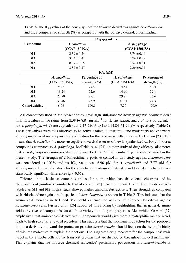
Table 2. The IC50 values of the newly-synthesized thiourea derivatives against Acanthamoeba
and their comparative strength (%) as compared with the positive control, chlorhexidine.
Compound
IC50 (µg·mL−1)
A. castellanii (CCAP 1501/2A)
A. polyphaga (CCAP 1501/3A)
M1 2.39 ± 0.24 3.74 ± 0.44 M2 3.34 ± 0.41 3.76 ± 0.27 M3 8.07 ± 0.65 8.52 ± 0.81 M4 8.87 ± 0.27 9.30 ± 0.55
IC50 (µM)
A. castellanii
(CCAP 1501/2A) Percentage of strength (%)
A. polyphaga (CCAP 1501/3A)
Percentage of strength (%)
M1 9.47 73.5 14.84 52.4 M2 13.24 52.6 14.90 52.1 M3 27.70 25.1 29.25 26.6 M4 30.46 22.9 31.91 24.3
Chlorhexidine 6.96 100.0 7.77 100.0
All compounds used in the present study have high anti-amoebic activity against Acanthamoeba
with IC50 values in the range from 2.39 to 8.87 µg·mL−1 for A. castellanii, and 3.74 to 9.30 µg·mL−1
for A. polyphaga, which are equivalent to 9.47–30.46 μM and 14.84–31.91 μM respectively (Table 2).
These derivatives were thus observed to be active against A. castellanii and moderately active toward
A. polyphaga based on compounds classification for the protozoan cells proposed by Deharo [23]. This
means that A. castellanii is more susceptible towards the series of newly-synthesized carbonyl thiourea
compounds compared to A. polyphaga. McBride et al. [24], in their study of drug efficacy, also noted
that A. polyphaga was more resistant compared to A. castellanii, confirming the data obtained in the
present study. The strength of chlorhexidine, a positive control in this study against Acanthamoeba
was considered as 100% and its IC50 value was 6.96 μM for A. castellanii and 7.77 μM for
A. polyphaga. The t-test analysis for the absorbance readings of untreated and treated amoebae showed
statistically significant differences (p < 0.05).
Thiourea in its basic structure has one sulfur atom, which has six valence electrons and its
electronic configuration is similar to that of oxygen [25]. The amino acid type of thiourea derivatives
labeled as M1 and M2 in this study showed higher anti-amoebic activity. Their strength as compared
with chlorhexidine against both species of Acanthamoeba is shown in Table 2. This indicates that the
amino acid moieties in M1 and M2 could enhance the activity of thiourea derivatives against
Acanthamoeba cells. Fustero et al. [26] supported this finding by highlighting that in general, amino
acid derivatives of compounds can exhibit a variety of biological properties. Meanwhile, Ye et al. [27]
emphasized that amino acids derivatives in compounds would give them a hydrophilic moiety which
leads to high selectivity toward receptors. This suggests that the mechanism of action for the proposed
thiourea derivatives toward the protozoan parasite Acanthamoeba should focus on the hydrophobicity
of thiourea molecules to explain their actions. The suggested drug-receptors for the compounds’ main
target in the amoeba cells are the transport proteins that are distributed throughout the cell membrane.
This explains that the thiourea chemical molecules’ preliminary penetration into Acanthamoeba is
Molecules 2014, 19 5195
through its membrane. However, the detail of the mechanism of action of the amino acid group toward
the amoeba cells is poorly understood.
Compounds M3 and M4 contain one chloride halogen atom in their benzene rings. The presence of
these halogens contributes to the compounds’ activity against Acanthamoeba. Patel and Shaikh [28]
reported that several compounds containing chlorine atom had better anti-microbial activity compared
to compounds without the halogen atom. Furthermore, the presence of chlorine in chlorhexidine was
proven to contribute in its anti-amoebic activity. However, the anti-amoebic activity of compounds M3
and M4 in this study were non-comparable to M1 and M2 that contain amino acid groups which gave
stronger in actions against the tested amoeba cells.
2.3. Morphological Changes in Acanthamoeba
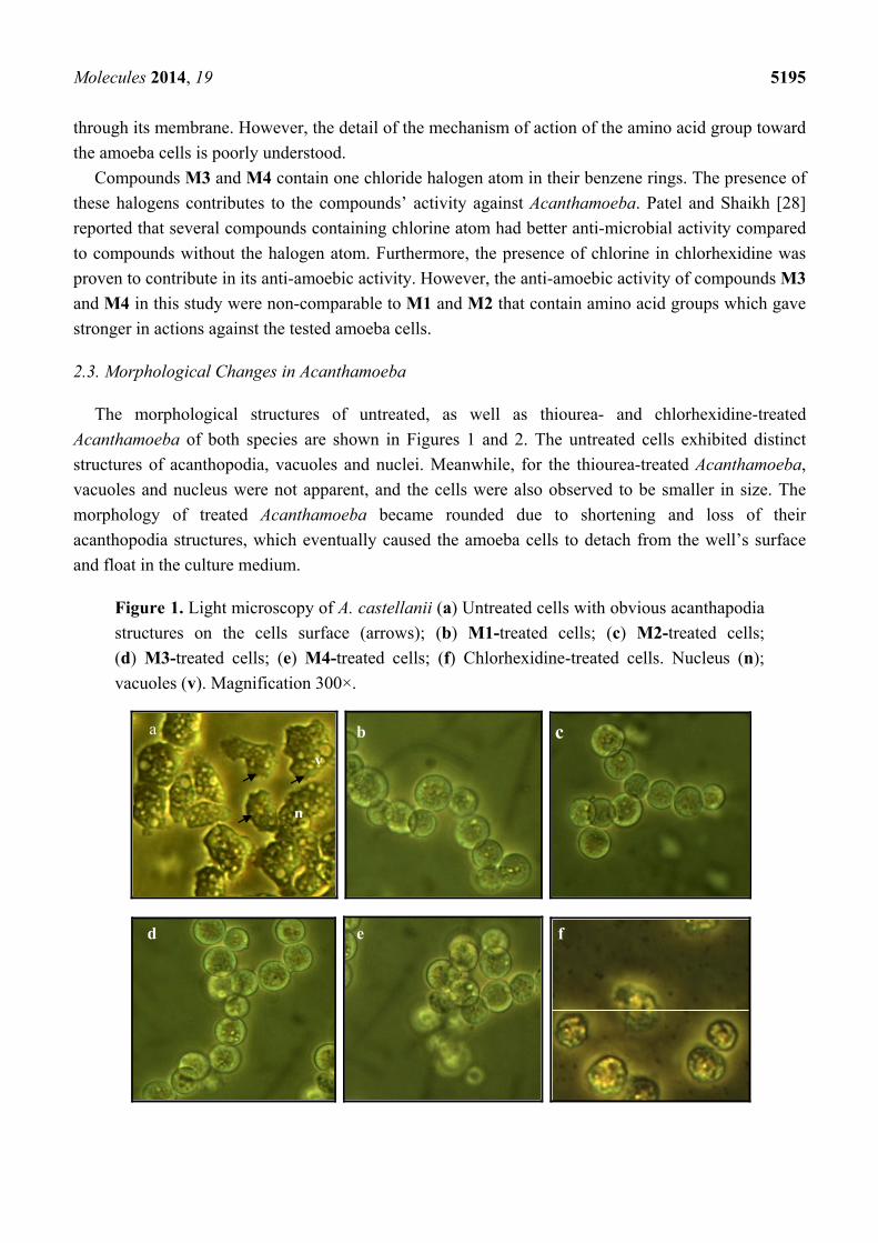
The morphological structures of untreated, as well as thiourea- and chlorhexidine-treated
Acanthamoeba of both species are shown in Figures 1 and 2. The untreated cells exhibited distinct
structures of acanthopodia, vacuoles and nuclei. Meanwhile, for the thiourea-treated Acanthamoeba,
vacuoles and nucleus were not apparent, and the cells were also observed to be smaller in size. The
morphology of treated Acanthamoeba became rounded due to shortening and loss of their
acanthopodia structures, which eventually caused the amoeba cells to detach from the well’s surface
and float in the culture medium.
Figure 1. Light microscopy of A. castellanii (a) Untreated cells with obvious acanthapodia
structures on the cells surface (arrows); (b) M1-treated cells; (c) M2-treated cells;
(d) M3-treated cells; (e) M4-treated cells; (f) Chlorhexidine-treated cells. Nucleus (n);
vacuoles (v). Magnification 300×.
a b c
d
n
v
e f
Molecules 2014, 19 5196
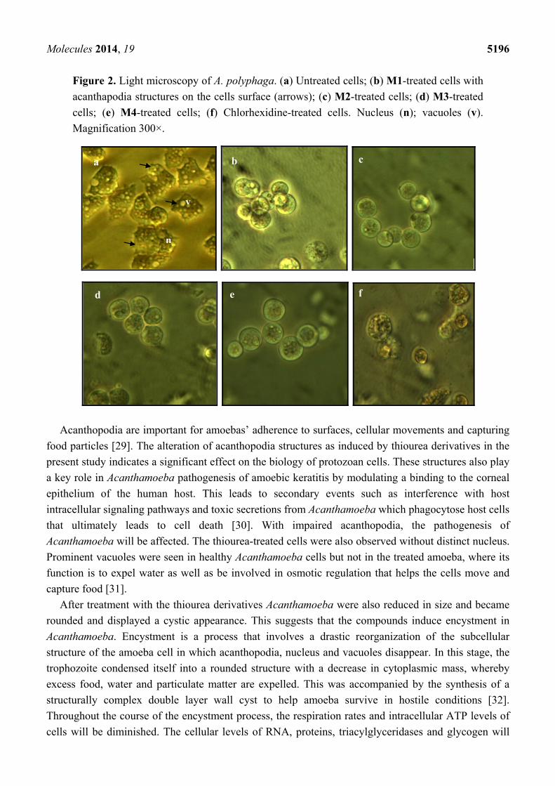
Figure 2. Light microscopy of A. polyphaga. (a) Untreated cells; (b) M1-treated cells with
acanthapodia structures on the cells surface (arrows); (c) M2-treated cells; (d) M3-treated
cells; (e) M4-treated cells; (f) Chlorhexidine-treated cells. Nucleus (n); vacuoles (v).
Magnification 300×.
Acanthopodia are important for amoebas’ adherence to surfaces, cellular movements and capturing
food particles [29]. The alteration of acanthopodia structures as induced by thiourea derivatives in the
present study indicates a significant effect on the biology of protozoan cells. These structures also play
a key role in Acanthamoeba pathogenesis of amoebic keratitis by modulating a binding to the corneal
epithelium of the human host. This leads to secondary events such as interference with host
intracellular signaling pathways and toxic secretions from Acanthamoeba which phagocytose host cells
that ultimately leads to cell death [30]. With impaired acanthopodia, the pathogenesis of
Acanthamoeba will be affected. The thiourea-treated cells were also observed without distinct nucleus.
Prominent vacuoles were seen in healthy Acanthamoeba cells but not in the treated amoeba, where its
function is to expel water as well as be involved in osmotic regulation that helps the cells move and
capture food [31].
After treatment with the thiourea derivatives Acanthamoeba were also reduced in size and became
rounded and displayed a cystic appearance. This suggests that the compounds induce encystment in
Acanthamoeba. Encystment is a process that involves a drastic reorganization of the subcellular
structure of the amoeba cell in which acanthopodia, nucleus and vacuoles disappear. In this stage, the
trophozoite condensed itself into a rounded structure with a decrease in cytoplasmic mass, whereby
excess food, water and particulate matter are expelled. This was accompanied by the synthesis of a
structurally complex double layer wall cyst to help amoeba survive in hostile conditions [32].
Throughout the course of the encystment process, the respiration rates and intracellular ATP levels of
cells will be diminished. The cellular levels of RNA, proteins, triacylglyceridases and glycogen will
a b c
d e f
v
n
Molecules 2014, 19 5197
also decline substantially. This would result in a decreased cellular volume and dry weight [33]. As a
conclusion, with the treatment of the carbonyl thiourea, Acanthamoeba became inactivated, making
them unable to affect the host cells during pathogenesis. Chlorhexidine gave comparable effects on the
morphology of Acanthamoeba as shown by the thiourea derivatives.
2.4. Integrity of Acanthamoeba Membrane
Acanthamoeba trophozoites consist of a plasma membrane which is a thin layer that
surrounds the cells and is comprised of phospholipids (25%), proteins (33%), sterols (13%), and
lipophosphonoglycans (29%) [31], while the cytoplasm of Acanthamoeba possesses large numbers of
fibrils, glycogen, lipid droplets, and a variety of lysosomal enzymes such as α- and β-glycosidases,
amylase, β-galactosidase, β-N-acetylglucosaminidase, β-glucuronidase, protease, phosphatase,
hydrolase acid, RNAse, and DNAse [33]. In all living cells, membrane integrity is essential in
maintaining their internal part in order to keep them viable. Compounds with cytotoxic effects would
often lead to compromised membrane integrity [34]. Disturbed membrane integrity would disrupt the
physiology of the cells’ inner state as well as organelles normal functions. In this study, fluorescence
microscopic observation based on a dual staining technique was conducted to evaluate the integrity of
the amoeba membrane with the given treatment. Acridine orange/propidium iodide (AO/PI)
simultaneous staining was applied to distinguish between cells of intact membrane with compromised-
membrane integrity as shown in Figures 3 and 4.
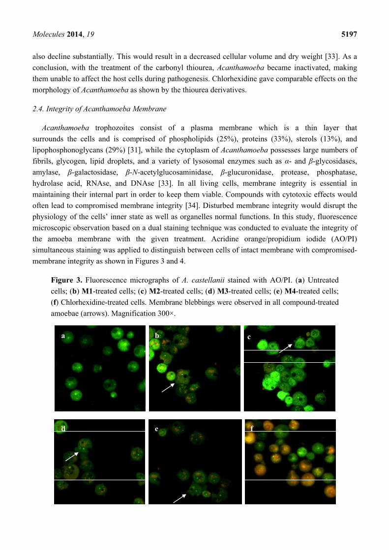
Figure 3. Fluorescence micrographs of A. castellanii stained with AO/PI. (a) Untreated
cells; (b) M1-treated cells; (c) M2-treated cells; (d) M3-treated cells; (e) M4-treated cells;
(f) Chlorhexidine-treated cells. Membrane blebbings were observed in all compound-treated
amoebae (arrows). Magnification 300×.
a b c
d e f
Molecules 2014, 19 5198
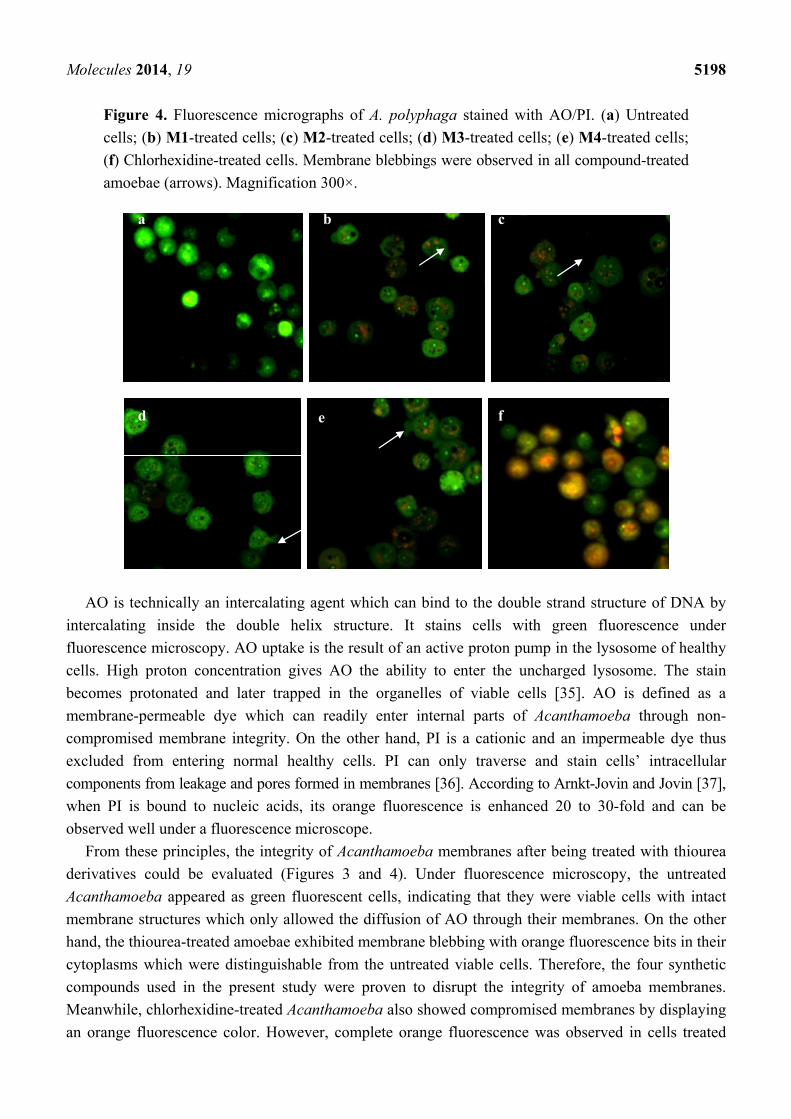
Figure 4. Fluorescence micrographs of A. polyphaga stained with AO/PI. (a) Untreated
cells; (b) M1-treated cells; (c) M2-treated cells; (d) M3-treated cells; (e) M4-treated cells;
(f) Chlorhexidine-treated cells. Membrane blebbings were observed in all compound-treated
amoebae (arrows). Magnification 300×.
AO is technically an intercalating agent which can bind to the double strand structure of DNA by
intercalating inside the double helix structure. It stains cells with green fluorescence under
fluorescence microscopy. AO uptake is the result of an active proton pump in the lysosome of healthy
cells. High proton concentration gives AO the ability to enter the uncharged lysosome. The stain
becomes protonated and later trapped in the organelles of viable cells [35]. AO is defined as a
membrane-permeable dye which can readily enter internal parts of Acanthamoeba through non-
compromised membrane integrity. On the other hand, PI is a cationic and an impermeable dye thus
excluded from entering normal healthy cells. PI can only traverse and stain cells’ intracellular
components from leakage and pores formed in membranes [36]. According to Arnkt-Jovin and Jovin [37],
when PI is bound to nucleic acids, its orange fluorescence is enhanced 20 to 30-fold and can be
observed well under a fluorescence microscope.
From these principles, the integrity of Acanthamoeba membranes after being treated with thiourea
derivatives could be evaluated (Figures 3 and 4). Under fluorescence microscopy, the untreated
Acanthamoeba appeared as green fluorescent cells, indicating that they were viable cells with intact
membrane structures which only allowed the diffusion of AO through their membranes. On the other
hand, the thiourea-treated amoebae exhibited membrane blebbing with orange fluorescence bits in their
cytoplasms which were distinguishable from the untreated viable cells. Therefore, the four synthetic
compounds used in the present study were proven to disrupt the integrity of amoeba membranes.
Meanwhile, chlorhexidine-treated Acanthamoeba also showed compromised membranes by displaying
an orange fluorescence color. However, complete orange fluorescence was observed in cells treated
a b c
d e f
Molecules 2014, 19 5199
with chlorhexidine, suggesting that the agent caused total leakage of Acanthamoeba membranes.
Under fluorescence microscopy, when both dyes are used simultaneously on compromised cell
membranes, an orange color fluorescence will be emitted from the cells due to stronger action of PI
compared to AO [38].
Perrine et al. [39] studied the lethal effects of amidine compounds toward Acanthamoeba and
showed that protonated substituents attached to compounds interact with the amphipathic lipids of
amoeba’s plasma membrane bilayer. This could induce the membrane’s structural changes which lead
to the modifications of the cell membrane permeability. From this study, it is suggested that the
penetration across the Acanthamoeba membrane by the compounds reflects the lipophilic properties of
the newly-synthesized thiourea derivative compounds. Nakisah et al. [40] used the same AO/PI
staining technique to explain the mode of cell death promoted by crude extracts from Malaysian
marine sponges on A. castellanii.
3. Experimental
3.1. General Information
All the compounds utilized in this work were commercially available Merck, Darmstadt, Germany
and use as supplied with no further purification. The infrared spectrum (IR) of the product (KBr
pellets) was recorded using a Perkin Elmer Spectrum GX spectrophotometer (Perkin Elmer, Waltham,
MA, USA) in the range of 400–4000 cm−1. NMR spectra were recorded on a Bruker Ultrashield
400 MHz NMR spectrometer using CDCl3 as the solvent.
3.2. Synthesis of Carbonyl Thiourea Derivatives
The method to prepare M1–M2 was based on Yusof and Yamin [21], while compounds M3 and
M4 followed the method of Yusof et al. [22] according to the routes shown at Scheme 1. Generally,
the carbonyl chloride reacted with ammonium isothiocyanate in acetone resulting carbonylisothiocyanate.
The carbonylisothiocyanate then will be reacted with amine derivate and the mixture was put at reflux
for 2.5 h then filtered off and left to evaporate at room temperature. For compound M1 (benzoyl
chloride, 2.03 g (14.44 mmol), α-alanin, 1.29 g (14.44 mmol), ammonium thiocyanate, 1.10 g
(14.44 mmol); compound M2, (benzoyl chloride, 1.9 5 g (13.87 mmol), β-alanin, 1.24 g (13.87 mmol),
ammonium thiocyanate, 1.06 g (13.87 mmol); compound M3, (4-chlorobutyryl chloride, 2.12 g
(15.04 mmol), 2-chloroaniline, 1.92 g (15.04 mmol), ammonium thiocyanate, 1.14 g (15.04 mmol);
compound M4, (4-chlorobutanoyl chloride, 2.05 g (14.54 mmol), 3-chloroaniline, 1.85 g
(14.54 mmol), ammonium thiocyanate, 1.11 g (14.54 mmol).
3.3. Characterization of the Newly-Synthesized Carbonyl Thiourea Derivatives
2-(3-Benzoylthioureido)propanoic acid (M1). The title compound was obtained as colourless crystals
in 38% yield after recrystallization from ethanol; IR (KBr pellets, υ/cm−1): 3389.22 (O-H), 3234.82
(N-H), 1772.31 (C=O), 1355.82 (C-N), 782.93 (C=S); 1H-NMR (400.130 MHz, DMSO-d6, ppm): 1.42
(3H, d, CH3), 3.52 (1H, dd, CH), 7.27 (1H, dd, C6H4), 7.65 (2H, m, C6H4), 7.88 (2H, d, C6H4), 11.44
(1H, s, NH), 12.01 (1H, s, OH), 12.20 (1H, s, NH); 13C-NMR (100.613 MHz, DMSO-d6; ppm): 17.23
Molecules 2014, 19 5200
(CH3), 62.32 (NHCH), 126.82 (CHAr), 129.09 (CHAr), 130.24 (NHCAr), 172.02 (C=O), 175.52
(C=OOH), 180.43 (C=S).
3-(3-Benzoylthioureido)propanoic acid (M2). The title compound was obtained as colourless crystals
in 52% yield after recrystallization from ethanol; IR (KBr pellets, υ/cm−1): 3324.61 (O-H), 3203.79
(N-H), 1794.05 (C=O), 1365.13 (C-N), 774.02 (C=S); 1H-NMR (400.130 MHz, DMSO-d6, ppm): 2.63
(2H, dd, NHCH2CH2), 3.67 (2H, dd, NHCH2CH2), 7.29 (1H, dd, C6H4), 7.64 (2H, m, C6H4), 7.87 (2H,
d, C6H4), 11.54 (1H, s, NH), 12.03 (1H, s, OH), 12.23 (1H, s, NH); 13C-NMR (100.613 MHz, DMSO-
d6, ppm): 34.25 (NHCH2CH2), 43.18 (NHCH2), 127.64 (CHAr), 130.29 (CHAr), 133.71 (NHCAr),
172.84 (C=O), 175.61 (C=OOH), 181.32 (C=S).
N-(2-Chlorophenyl)-N'-(4-chlorobutanoyl)thiourea (M3). The title compound was obtained as
colorless crystal in 73% yield after recrystallization from dimethylformamide; IR (KBr pellets,
υ/cm−1): 3164.31 (N-H), 1697.18(C=O), 1337.40(C-N), 723.53 (C=S); 1H-NMR (400.130 MHz,
DMSO-d6, ppm): 2.02 (2H, m, COCH2CH2CH2Cl), 2.65 (2H, t, COCH2CH2CH2Cl), 3.66 (2H, t,
COCH2CH2CH2Cl), 7.25 (1H, d, C6H4), 7.56 (1H, t, C6H4), 7.59 (1H, t, C6H4), 8.01 (1H, d, C6H4),
11.51 (1H, s, NH), 12.45 (1H, s, NH); 13C-NMR (100.613 MHz, DMSO-d6, ppm): 27.28
(COCH2CH2CH2Cl), 33.53 (COCH2CH2CH2Cl), 45.01 (COCH2CH2CH2Cl), 115.94 (CHAr), 116.10
(CHAr), 127.41 (NHCAr), 134.69 (ClCAr), 175.92 (C=O), 180.12 (C=S).
N-(3-Chlorophenyl)-N'-(4-chlorobutanoyl)thiourea, M4. The title compound was obtained as
colourless crystal in 75% yield after recrystallization from dimethylformamide; IR (KBr pellets,
υ/cm−1): 3165.88 (N-H), 1694.05 (C=O), 1325.09 (C-N), 780.65 (C=S); 1H-NMR (400.130 MHz,
DMSO-d6, ppm): 2.03 (2H, m, COCH2CH2CH2Cl), 2.64 (2H, t, COCH2CH2CH2Cl), 3.69 (2H, t,
COCH2CH2CH2Cl), 7.24 (1H, d, C6H4), 7.29 (1H, t, C6H4), 7.62 (1H, d, C6H4), 7.96 (1H, s, C6H4),
11.47 (1H, s, NH), 12.42 (1H, s, NH). 13C-NMR (100.613 MHz, DMSO-d6, ppm): 27.25
(COCH2CH2CH2Cl), 45.04 (COCH2CH2CH2Cl), 33.54 (COCH2CH2CH2Cl), 115.70 (CHAr), 115.92
(CHAr), 127.31 (NHCAr), 134.67 (ClCAr), 175.81 (C=O), 179.89 (C=S).
3.4. Determination of IC50 Values
Thiourea derivatives were prepared by dissolving 1 mg of compound in 10 µL absolute DMSO
(Fisher Scientific, Schwerte, UK) and added with 990 µL sterile culture media, to make a 1 mg·mL−1
solution. Dissolution was facilitated by mild sonication in a sonicator bath (Branson, CT, USA) for
two minutes. Then, 100 µL of the 1 mg·mL−1 samples were further diluted with 900 µL of culture
media to produce compound stocks of 100 µg·mL−1 with 0.1% DMSO. These thiourea compounds
solutions were freshly prepared before conducting every experiment. The experiment was conducted in
96-well plates (Nunc, Schwerte, Germany). Nine different concentrations of compounds were prepared
to give final concentrations of compounds as follows: 100, 50, 25, 12.5, 6.25, 3.13, 1.56, 0.78 and
0.39 µg·mL−1. Each concentration was prepared in three replicates. Chlorhexidine gluconate (Raza
Manufacturing, Kuala Lumpur, Malaysia) which is a common agent used for treatment of amoebic
keratitis infections was used as the positive control. The nine final concentrations of chlorhexidine
used for the assays were as follows: 200, 100, 50, 25, 12.5, 6.25, 3.13, 1.56 and 0.78 µM.
Molecules 2014, 19 5201
The number of viable Acanthamoeba for treatment was calculated by using a hemocytometer with
trypan blue. A calculated amount of ~104 viable cells·mL−1 was used as the number or concentration of
Acanthamoeba of which the cells would reach their confluence stage after 72 h of incubation without
excessive growth [41]. Negative control was 104 cells·mL−1 of healthy Acanthamoeba without any
treatment. The plates were later incubated at 30 °C for 72 h. After incubation, the staining process was
done following Wright’s technique [42]. The final solutions from all wells were read for their
absorbance at 490 nm by ELISA microplate reader (Tecan, Victoria, Australia). The readings were
plotted in GraphPad Prism software version 5.03 (GraphPad Inc., San Diego, CA, USA) to give a non-
linear sigmoidal dose-response curve. The cytotoxicity was expressed as the IC50 value that represents
the concentration of a compound that is required for inhibition of 50% of an Acanthamoeba population
in vitro. A t-test (SPSS, version 11.5., SSPS Inc., Armonk, NY, USA) was done to compare the mean
values between untreated and treated cultures with p < 0.05 considered as statistically significant.
3.5. Observation of Changes in Acanthamoeba Morphology
Acanthamoeba both untreated and treated with the compounds were observed for their
morphological changes. Acanthamoeba (104 cells·mL−1) were treated with the thiourea compounds and
the positive control (chlorhexidine) at their IC50 concentration in 6-well-plates, which were then
incubated at 30 °C for 72 h. After the incubation, the morphology of Acanthamoeba was observed
directly from the well plates under an inverted microscope (Leica Leitz, Wetzlar, Germany). Images
were captured by using Image Master Video Test Package (Trioptics, Wetzlar, Germany) software.
3.6. Evaluation of Acanthamoeba Membrane Integrity
Acanthamoeba were adjusted to 104 cells in 1 mL culture media prior to the treatment with thiourea
compounds and chlorhexidine, at their IC50 concentration in 25-cm2 tissue culture flasks and later
incubated at 30 °C for 72 h. After the incubation, the cell suspension was resuspended, harvested and
transferred into Eppendorf tubes for AO/PI staining. Stock solution for AO/PI staining was prepared
by adding AO (2 µL, 1 mg·mL−1, Sigma, St. Louis, MO, USA) and PI (2 µL, 1 mg·mL−1, Sigma) to
give a mixture of 1:1 (v/v) ratio in 996 µL phosphate buffered saline (PBS, Sigma). The AO/PI
staining protocol followed the technique by Mascotti et al. [43]. Both dyes are light sensitive therefore
they were handled in a dark room. The harvested Acanthamoeba cells were centrifuged at 1,000 rpm
for 5 min at 4 °C. The supernatant were discarded and pellets were washed with PBS and
re-centrifuged at 1,000 rpm for 5 min. The fresh pellets were mixed with 20 µL of AO/PI staining from
the stock and transferred onto microscope slides and viewed under a fluorescence microscope (Leica
Dmire, Wetzlar, Germany) in dark condition. Images were captured by Image Master Video Test
Package software (Trioptics).
4. Conclusions
The results of this study indicate that the newly-synthesized carbonyl thiourea derivatives
provide promising anti-Acanthamoeba properties against pathogenic A. castellanii and A. polyphaga.
Based on their low IC50 values the compounds 2-(3-benzoylthioureido)propanoic acid (M1) and
Molecules 2014, 19 5202
3-(3-benzoylthioureido)propanoic acid (M2) exhibited stronger anti-amoebic activity compared to the
other tested compounds used, and this finding correlates with the presence of amino acids groups in
their molecular structures. All thiourea derivatives used in this study were proven to cause
Acanthamoeba to become inactive, and can disrupt the integrity of the amoeba cell membrane.
Therefore, these new carbonyl thiourea derivatives can be suggested as future anti-amoebic agents.
Acknowledgments
The authors are greatly appreciative to Ministry of Science, Technology and Innovation, Malaysia
(MOSTI) for the research financial support through E-Science Fund (52022) and The Institute of
Oceanography, Universiti Malaysia Terengganu for providing the space and facilities to conduct
this work.
Conflicts of Interest
The authors declare no conflict of interest.
References
1. De Jonckheere, J.F. Ecology of Acanthamoeba. Rev. Infect. Dis. 1991, 13, S385–S387.
2. Page, F.C. A New Key to Freshwater and Soil Gymnamoebae. In Freshwater Biological
Association; Culture Collection of Algae and Protozoa: Ambleside, Cumbria, UK, 1988; p. 122.
3. Narasimhan, S.; Madhavan, H.; Therese, L. Development and application of an in vitro
susceptibility test for Acanthamoeba species isolated from keratitis to polyhexamethylene
biguanide and chlorhexidine. Cornea 2002, 21, 203–205.
4. Marciano-Cabral, F.; Cabral, G. Acanthamoeba spp. as agents of disease in humans. Clin. Microbiol.
Rev. 2003, 16, 273–307.
5. Elder, M.J.; Dart, J.K.G. Chemotherapy for Acanthamoeba keratitis. Lancet 1995, 345, 791–792.
6. Larkin, D.F.P.; Kilvington, S.; Dart, J.K.G. Treatment of Acanthamoeba keratitis with
polyhexamethylene biguanide. Ophthalmology 1992, 99, 185–191.
7. Seal, D.V. Acanthamoeba keratitis update—Incidence, molecular epidemiology and new drugs for
treatment. Eye 2003, 17, 893–905.
8. Murdoch, D.; Gray, T.B.; Cursons, R.; Parr, D. Acanthamoebakeratitis in New Zealand, including
two cases with in vivo resistance to polyhexamethylene biguanide. Aust. New Zeal. J. Ophthalmol.
1998, 26, 231–236.
9. Turner, N.A.; Russell, A.D.; Furr, J.R.; Lloyd, D. Emergence of resistance to biocides during
differentiation of Acanthamoeba castellanii. J. Antimicrob. Chemother. 2000, 46, 27–34.
10. Ficker, L.; Seal, D.; Warhurst, D.; Wright, P. Acanthamoeba keratitis: Resistance to medical
therapy. Eye 1990, 4, 835–838.
11. Abid, M.; Agarwal, S.M.; Azam, A. Synthesis and anti-amoebic activity of metronidazole
thiosemicarbazone analogues. Eur. J. Med. Chem. 2008, 43, 2035–2039.
Molecules 2014, 19 5203
12. Budakoti, A.; Bhat, A.R.; Athar, F.; Azam, A. Syntheses and evaluation of 3-(3-bromophenyl)-5-
phenyl-1-(thiazolo[4,5-b]quinoxaline-2-yl)-2pyrazoline derivatives. Eur. J. Med. Chem. 2008, 43,
1749–1757.
13. Ziegler-Skylakakis, K.; Nill, S.; Pan, J.F.; Andrae, U. S-Oxygenation of thiourea results in the
formation of genotoxic products. Environ. Mol. Mutagen. 1998, 31, 362–373.
14. Khan, S.A.; Singh, N.; Saleem, K. Synthesis, characterization and in vitro antibacterial activity of
thiourea and urea derivatives of steroids. Eur. J. Med. Chem. 2008, 43, 2272–2277.
15. Zhong, Z.; Xing, R.; Liu, S.; Wang, L.; Chai, S.; Li, P. Synthesis of acyl thiourea derivatives of
chitosan and their anti-microbial activities in vitro. Carbohydr. Res. 2008, 343, 566–570.
16. Eweis, M.; Elkholy, S.S.; Elsabee, M.Z. Antifungal efficacy of chitosan and its thiourea
derivatives upon the growth of some sugar-beet pathogens. Int. J. Biol. Macromol. 2006, 38, 1–8.
17. Chen, S.; Wu, G.; Zeng, H. Preparation of high anti-microbial activity chitosan-Ag+ complex.
Carbohydr. Polym. 2005, 60, 33–38.
18. Turan-Zitouni, G.; Sıvacı, D.M.; Kaplancıklı, Z.A.; Özdemir, A. Synthesis and anti-microbial
activity of some pyridinyliminothiazoline derivatives. Il Farmaco 2002, 57, 569–572.
19. Phetsuksiri, B.; Jackson, M.; Scherman, H.; McNeil, M.; Besra, G.S.; Baulard, A.R.; Slayden, R.A.;
DeBarber, A.E.; Barry, C.E., III; Baird, M.S.; et al. Unique mechanism of action of the thiourea
drug isoxyl on Mycobacterium tuberculosis. J. Biol. Chem. 2003, 278, 53123–53130.
20. Paynter, O.E.; Burin, G.J.; Jaeger, R.B.; Gregorio, C.A. Goitrogens and thyroid follicular cell
neoplasia. Evidence for a threshold process. Regul. Toxicol. Pharmacol. 1988, 8, 102–119.
21. Yusof, M.S.M.; Yamin, B.M. 3-(3-Benzoylthioureido) propionic acid. Acta Crystallogr. 2003,
E59, o828–o829.
22. Yusof, M.S.M.; Embong, N.F.; Yamin, B.M.; Ngah, N. 1-(4-Chlorobutanoyl)-3-(2-chloro
phenyl)thiourea. Acta Crystallogr. 2012, E68, o1536.
23. Deharo, E.; Bourdy, G.; Quenevo, C.; Munoz, V.; Ruiz, G.; Sauvain, M. A search for natural
bioactive compounds in Bolivia through a multi disciplinary sciences approach. Part V.
Evaluation of the antimalarial activity of plants used by the Tacana Indians. J. Ethnopharmacol.
2001, 77, 91–98.
24. McBride, J.; Ingram, R.P.; Henriquez, F.L.; Roberts, C.W. Development of colorimetric
microtiter plate assay for assessment of anti-microbials against Acanthamoeba. J. Clin. Microbiol.
2005, 43, 629–634.
25. Patnaik, P. A Comprehensive Guide to the Hazardous Properties of Chemical Substances;
Wiley-Interscience: Hoboken, NJ, USA, 2007; p. 904.
26. Fustero, S.; Salavert, E.; Pina, B.; de Arellano, C.R.; Asensio, R. Novel strategy for the synthesis
of fluorinated β-amino acid derivatives from Δ2-oxazolines. Tetrahedron 2001, 57, 6475–6486.
27. Ye, Y.H.; Huang, Y.S.; Wang, Z.Q.; Chen, S.M.; Tian, Y. Synthesis of new amino acid and
peptide derivatives of estradiol and their binding affinities for the estrogen receptor. Steroids
1993, 58, 35–39.
28. Patel, N.B.; Shaikh, F.M. Synthesis and anti-microbial activity of new 4-thiazolidinone
derivatives containing 2-amino-6-methoxybenzothiazole. Saudi Pharm. J. 2010, 18, 129–136.
29. Bowers, B.; Korn, E.D. The fine structure of Acanthamoeba castellanii, kinetics and morphology.
I. The Trophozoite. J. Cell Biol. 1968, 39, 95–111.
Molecules 2014, 19 5204
30. Khan, N.A. Pathogenicity, morphology, and differentiation of Acanthamoeba. Curr. Microbiol.
2001, 43, 391–395.
31. Bowers, B.; Korn, E.D. Localization of lipophosphonoglycan on both sides of Acanthamoeba
plasma membrane. J. Cell Biol. 1974, 62, 533–540.
32. Khan, N.A. Emerging Protozoan Pathogens; Taylor & Francis Group: Oxford, UK, 2008;
pp. 5–24.
33. Weisman, R.A. Differentiation in Acanthamoeba castellanii. Annu. Rev. Microbiol. 1976, 30,
189–219.
34. Coder, D.M. Assessment of cell viability. In Current Protocols in Cytometry, 2nd ed.; Wiley:
New York, NY, USA, 1997; pp. 8–11.
35. Darzynkiewicz, Z.; Juan, G.; Li, X.; Gorczyka, W.; Murakami, T.; Traganos, F. Cytometry in cell
necrobiology: Analysis of apoptosis and accidental cell death (necrosis). Cytometry 1997, 27, 1–20.
36. Riss, T.L.; Moravec, R.A. Use of multiple assay endpoints to investigate the effects of incubation
time, dose of toxin, and plating density in cell-based cytotoxicity assays. Assay Drug Dev. Technol.
2004, 2, 51–62.
37. Arnkt-Jovin, D.J.; Jovin, T.M. Fluorescence labeling and microscopy of DNA. Methods Cell Biol.
1989, 30, 417–448.
38. Puranam, K.L.; Boustany, R.M. Assessment of cell viability and histochemical methods in
apoptosis. In Apoptosis in Neurobiology; Hannun, Y.A., Boustany, R.M., Eds.; CRC Press:
Washington, DC, USA, 1999; p. 78.
39. Perrine, D.; Chenu, J.P.; Georges, P.; Lancelot, J.C.; Saturnino, C.; Robba, M. Amoebicidal
efficiencies of various diamidines against two strains of Acanthamoeba polyphaga. Antimicrob.
Agents Chemother. 1995, 39, 339–342.
40. Nakisah, M.A.; Ida Muryany, M.Y.; Fatimah, H.; Nor Fadilah, R.; Zalilawati, M.R.; Khamsah, S.;
Habsah, M. Anti-amoebic properties of a Malaysian marine sponge Aaptos sp. on Acanthamoeba
castellanii. World J. Microbiol. Biotechnol. 2012, 28, 1237–1244.
41. Asiri, S.; Ogbunade, P.O.J.; Warhust, D.C. In vitro assessment of susceptibility of Acanthamoeba
polyphaga to drugs using combined methods of dye-binding assay and uptake of radiolabeled
adenosine. Int. J. Parasitol. 1994, 24, 975–980.
42. Wright, C.W.; O’Neill, M.J.; Phillipson, J.D.; Warhurst, D.C. Use of microdilution to assess
in vitro anti-amoebic activities of Bruceajavanica fruits, Simaroubaamara Stem, and a number of
Quassinoids. Antimicrob. Agents Chemother. 1988, 32, 1725–1729.
43. Mascotti, K.; McCullough, J.; Burger, S.R. HPC viability measurement: Trypan blue versus
acridine orange and propidium iodide. Transfusion 2000, 40, 693–696.
Sample Availability: Samples of the compoundsare available from the authors.
© 2014 by the authors; licensee MDPI, Basel, Switzerland. This article is an open access article
distributed under the terms and conditions of the Creative Commons Attribution license
(http://creativecommons.org/licenses/by/3.0/).
Related Documents