RANDOMIZED TRIALS, REGISTER STUDIES AND EXPERIMENTAL ANALYSIS OF KNEE LAXITY ANTERIOR CRUCIATE LIGAMENT RECONSTRUCTION Mattias Ahldén, MD Department of Orthopaedics Institute of Clinical Sciences Sahlgrenska Academy at University of Gothenburg Gothenburg 2013

Welcome message from author
This document is posted to help you gain knowledge. Please leave a comment to let me know what you think about it! Share it to your friends and learn new things together.
Transcript
RANDOMIZED TRIALS, REGISTER STUDIES AND EXPERIMENTAL ANALYSIS OF KNEE LAXITY
ANTERIOR CRUCIATE LIGAMENT RECONSTRUCTION
Mattias Ahldén, MD
Department of OrthopaedicsInstitute of Clinical SciencesSahlgrenska Academy at University of GothenburgGothenburg 2013

Anterior Cruciate Ligament Reconstruction© Mattias Ahldén 2013http://hdl.handle.net/2077/32383
ISBN 978-91-628-8640-0
Printed in Gothenburg, Sweden 2013 by Ineko ABCover illustration by Pontus Andersson / Pontus Art ProductionDesign by Annika Enderlein Samuelsson / A little company AB

“Attitude is a little thing that makes a big difference.”
Winston Churchill 1874-1965

1 ABSTRACT 6
2 SAMMANFATTNING PÅ SVENSKA 7
3 LIST OF PAPERS 8
4 ABBREVIATIONS 10
5 INTRODUCTION 12
5.1 Anatomy 125.2 Epidemiology 165.3 Etiology 165.4 Osteoarthritis after ACL injury 175.5 The Swedish National ACL Register 185.6 Prevention 185.7 Knee laxity 195.7.1 Antero-posterior knee laxity 195.7.2 Rotatory knee laxity and the pivot-shift test 195.8 Non-operative versus operative treatment for ACL injury 205.9 Surgical techniques of arthroscopic ACL reconstruction 215.9.1 The early era of arthroscopic ACL reconstruction 215.9.2 Non-anatomic arthroscopic ACL reconstruction 225.9.3 Anatomic ACL reconstruction 225.9.4 Double-bundle ACL reconstruction 235.10 Why is this thesis needed? 24
6 AIMS 25
7 PATIENTS 26
8 METHODS 32
8.1 Blinded examiners 32
8.2 Surgical technique 32
8.3 Rehabilitation 38
8.4 Standard radiography 38
8.5 Clinical examinations 38
8.5.1 Manual Lachman test 39
8.5.2 Instrumented laxity KT-1000 arthrometer 39
CONTENTS

8.5.3 The pivot-shift test 40
8.5.4 Range of motion 418.5.5 Loss of skin sensitivity 418.5.6 Anterior knee pain 418.5.7 Patients’ subjective evaluation 428.6 Functional tests 428.6.1 One-leg-hop test 428.6.2 Square-hop test 438.6.3 Knee-walking test 438.7 Functional scores 448.7.1 KOOS 448.7.2 Lysholm knee scoring scale 458.7.3 Tegner activity scale 458.8 Quantitative evaluation of the pivot-shift test 458.8.1 Electromagnetic tracking system 468.8.2 Accelerometer 468.8.3 Simple image analysis 47
9 STATISTICAL METHODS 48
10 SUMMARY OF THE PAPERS 50
11 STRENGTHS AND LIMITATIONS 61
12 DISCUSSION 63
12.1 Outcome measurements 6312.2 Antero-posterior knee laxity 6312.3 Graft selection 6512.4 Gender differences 6612.5 Register studies 6712.6 Rotatory laxity and the pivot-shift test 6912.7 Double-bundle versus single-bundle reconstruction 7012.8 General discussion 74
13 CONCLUSIONS 75
14 FINAL CONSIDERATIONS AND THE FUTURE 76
15 ACKNOWLEDGEMENTS 78
16 REFERENCES 80

6 mattias ahldén / anterior cruciate ligament reconstruction
The aim of this thesis was to evaluate the short- and mid-term outcome of Anterior Cruciate Ligament (ACL) reconstruction, with special emphasis on surgical techniques, type of autograft and the influence of gender, using data from registers and randomized, controlled trials. A further aim was to evaluate and quantitate the pivot-shift test and correlate it to the clinical grading. In Study I, 17,794 registrations in the Swedish National ACL Register were included and analyzed. Primary ACL reconstruction sig-nificantly improves all subscales of the KOOS, while patients undergoing revision ACL reconstructions do less well than those undergoing primary reconstructions. Moreover, young female soccer players re-injure their ACL or the contralateral ACL within 5 years more frequently than young males. In Study II, a randomized, controlled trial (RCT) with a 7-year follow-up, the change in knee laxity over time after ACL reconstruction, using either bone-patellar-tendon-bone (BPTB) or hamstring tendon (HS) autografts was analyzed and knee laxity was compared between the study groups on multiple follow-up occasions. Furthermore, the radiographic findings in terms of degenerative changes were compared. There were no significant differences in the mean side-to-side antero-posterior (AP) knee laxity or radiographic assessment between the BPTB and the HS group, preoperatively or at follow-up. There was a tendency towards a decrease in side-to-side knee laxity over time in both groups, as measured with the KT-1000 arthrometer. In Study III, a retrospective study of 244 patients, the results after ACL reconstruction using HS autografts were compared in male versus female patients. At the 2-year follow-up, there were no significant differences between male and female patients in terms of clinical outcome or functional scores. In Study IV, an RCT with 105 patients, the results after ACL reconstruction using either the double-bundle (DB) or single-bundle (SB) technique with HS autografts were compared. At the 2-year follow-up, the subjective and objective outcomes revealed no statistically significant differences between the DB group and the SB group. In Study V, an experimental cadaver study, objective quantitative measurements of the pivot-shift test using three different meas-urement devices were performed. The pivot-shift tests were performed by twelve blinded expert surgeons on a cadaver knee prepared to display a high-grade pivot-shift test. The best correlation to the clinical grading was found using tibial acceleration parameters.
Keywords: Anterior Cruciate Ligament, Reconstruction, Double-Bundle, Knee Laxity, Pivot-Shift, Register, Outcome
ISBN: 978-91-628-8640-0
01 ABSTRACT

7
Syftet med avhandlingen var att utvärdera resultatet efter främre korsbandsrekonstruk-tion med avseende på olika kirurgiska tekniker, graftval och kön, utifrån registerstudier och prospektiva randomiserade studier. Ytterligare ett syfte var att i en experimentell studie utvärdera och kvantifiera pivot-shift testet och utvärdera dess korrelation till klinisk gradering. I Studie I inkluderades och analyserades 17,794 registreringar från Svenska Korsbandsregistret. Slutsatsen blev att efter primär främre korsbandsrekon-struktion förbättrades samtliga delskalor av KOOS signifikant. Patienter som opererats med korsbandsrevision uppvisade generellt sämre resultat jämfört med patienter som opererats med primär rekonstruktion. Unga kvinnliga fotbollsspelare ådrar sig ny främre korsbandskada under en femårsperiod antingen på den opererade sidan eller i motsatt knä i större utsträckning jämfört med motsvarande manliga grupp och jämfört med främre korsbandsopererade patienter generellt. Studie II var en randomiserad kon-trollerad studie (RCT) med 7 års uppföljning där förändring av knälaxitet över tid efter främre korsbandsrekonstruktion med antingen patellarsenegraft (BPTB) eller ham-stringssenegraft (HS) studerades och knälaxitet jämfördes vid upprepade uppföljning-stillfällen. Det förelåg ingen signifikant sidoskillnad i antero-posterior (AP) knälaxitet mätt med KT-1000 arthrometer mellan grupperna preoperativt eller vid de olika upp-följningstillfällena. Det förelåg en tendens till att sidoskillnaden minskade över tid i båda grupperna. Graden av degenerativa förändringar undersöktes radiologiskt och visade ingen skillnad mellan studiegrupperna efter 7 år. Studie III var en retrospektiv studie av 244 patienter där resultatet mellan män och kvinnor efter främre korsbandsrekon-struktion med HS-graft jämfördes. Vid 2-års-uppföljningen förelåg inga signifikanta skillnader varken avseende kliniska utfallsmått eller funktionell utvärdering. Studie IV var en RCT med 105 patienter där resultatet jämfördes mellan “double-bundle” (DB) och “single-bundle” (SB) teknik vid främre korsbandsrekonstruktion med HS-graft. Efter 2 år förelåg inga signifikanta skillnader mellan grupperna avseende subjektiva eller objektiva utfallsmått. Studie V var en experimentell kadaverstudie där tre olika mätmetoder användes för att objektivt kvantifiera pivot-shift testet samt undersöka korrelationen mellan mätresultaten och den kliniska graderingen av pivot-shift testet. Pivot-shift testet utfördes av 12 blindade erfarna ortopedkirurger på ett kadaverknä vilket var preparerat med en främre korsbandsskada. Tibiala accelerationsparametrar uppvisade bäst korrelation med den kliniska graderingen.
02 SAMMANFATTNING PÅ SVENSKA
8 mattias ahldén / anterior cruciate ligament reconstruction
03 LIST OF PAPERS
This thesis is based on the following papers, referred to in the text by their Roman numerals.
I. The Swedish National Anterior Cruciate Ligament Register, a report on baseline variables and outcomes of surgery for almost 18,000 patients. Ahldén M, Samuelsson K, Sernert N, Forssblad M, Karlsson J, Kartus J. Am J Sports Med 2012; 40:2230-2235.
II. Knee laxity measurements after anterior cruciate ligament recon-struction, using either bone-patellar-tendon-bone or hamstring tendon autograft, with special emphasis on comparison over time. Ahldén M, Kartus J, Ejerhed L, Karlsson J, Sernert N. Knee Surg Sports Traumatol Arthrosc 2009; 17:1117-1124.
III. Outcome of anterior cruciate ligament reconstruction, with emphasis on sex-related differences. Ahldén M, Sernert N, Karlsson J, Kartus J. Scand J Med Sci Sports 2012; 22:618-626.
IV. A prospective randomized study comparing double- and single-bundle techniques for ACL reconstruction. Ahldén M, Sernert N, Karlsson J, Kartus J. Submitted to Am J Sports Med
V. Clinical grading of the pivot shift test correlates best with tibial acceleration. Ahldén M, Araujo P, Hoshino Y, Samuelsson K, Middleton K, Nagamune K, Karlsson J, Musahl V. Knee Surg Sports Traumatol Arthrosc 2012; 20:708-712.
9
Additional relevant papers by the author not included in this thesis:
Rotatory knee laxity. Ahldén M, Samuelsson K, Fu F, Karlsson J, Musahl V. Clin Sports Med 2013; 32:37-46.
Dynamic knee laxity measurement devices. Ahldén M, Hoshino Y, Samuelsson K, Araujo P, Musahl V, Karlsson J. Knee Surg Sports Traumatol Arthrosc 2012; 20:621-632.
Comparison of three non-invasive quantative measurements systems for the pivot-shift test. Araujo P, Ahldén M, Hoshino Y, Muller B, Moloney G, Fu F, Musahl V. Knee Surg Sports Traumatol Arthrosc 2012; 20:692-697.
Standardized pivot shift test improves measurement accuracy. Hoshino Y, Araujo P, Ahldén M, Moore C, Kuroda R, Zaffagnini S, Karlsson J, Fu F, Musahl V. Knee Surg Sports Traumatol Arthrosc 2012; 20:732-736.
The pivot shift: a global user guide. Musahl V, Hosino Y, Ahldén M, Araujo P, Irrgang J, Zaffagnini S, Karlsson J, Fu F. Knee Surg Sports Traumatol Arthrosc 2012; 20:724-731.
Quantitative evaluation of the pivot shift by image analysis using the iPad. Hoshino Y, Araujo P, Ahldén M, Samuelsson K, Muller B, Hofbauer M, Wolf M, Irrgang J, Fu F, Musahl V. Knee Surg Sports Traumatol Arthrosc 2013; 21:975-980
Trends in surgeons preferences on anterior cruciate ligament reconstruc-tive techniques. Samuelsson K, Andersson D, Ahldén M, Fu F, Musahl V, Karlsson J. Clin Sports Med 2013; 32:111-126.

10 mattias ahldén / anterior cruciate ligament reconstruction
04 ABBREVIATIONS
AARSS
ACL
AL
AM
AP
BMI
BPTB
CAS
CT
DB
DSX
EQ-5D
EBM
G
HS
KOOS
MMT
MOON
MRI
N
PCL
Anatomic ACL Reconstruction Scoring System
Anterior Cruciate Ligament
Antero-Lateral
Antero-Medial
Antero-Posterior
Body Mass Index
Bone-Patellar-Tendon-Bone
Computer-Assisted Surgery
Computed Tomography
Double-Bundle
Dynamic Stereo Radiography
European Quality of Life-5 Dimensions
Evidence-Based Medicine
Gracilis tendon
Hamstring tendons
Knee Osteoarthritis and Outcome Score
Manual Maximum Test
Multicenter Orthopedic Outcomes Network
Magnetic Resonance Imaging
Newton
Posterior Cruciate Ligament

11

12 mattias ahldén / anterior cruciate ligament reconstruction
5.1 Anatomy
Anatomy is one of the bases for orthopedic surgery and descriptions of the anatomy of the cruciate ligaments were made long before the modern era. In 460-370 BC, Hippocrates of Greece described cruciate pathology, as he suggested that instability of the knee could be attributed to torn internal ligaments of the knee. Galen of Greece (201-131 BC) was the first to name the ligaments based on their appearance of cross-ing over as “ligament genu cruciate”. The first detailed anatomic description of the anterior cruciate ligament (ACL) was given by the Weber brothers in the early 19th century166. They defined the two bundles of the ACL and showed different tension patterns in the separate bundles at different knee flexion angles. They also reported the basis of the anterior drawer sign and showed that sectioning the ACL resulted in abnormal antero-posterior (AP) movement. Ivar Palmer from Sweden, a pioneer of ACL surgery, published his thesis: “On the injuries to the ligaments of the knee joint” in 1938. He described the ACL as consisting of two bundles and stated that anatomic reconstruction with the repair of both bundles separately is advantageous. Furthermore, early surgery benefits the visualization of the anatomic conditions of the knee. In this area, he was well before his time, as little attention was paid by the orthopedic society to Palmer’s description until, in 1975, Girgis described more precisely the two bundles of the ACL; the antero-medial (AM) and postero-lateral (PL) bundles60. Progressive imaging techniques for visualizing knee anatomy and its landmarks have advanced the concept of modern anatomic ACL reconstruction.
The ACL is composed of mainly type I-collagen fibers, covered by a synovial mem-brane. ACL vascularization arises from the middle genicular artery and vessels of the infrapatellar fat pad and adjacent synovium. Nerve fibers run together with the vessels and the ACL has also been shown to have mechanoreceptors providing proprioceptive feedback37. This knowledge has been actualized in discussions regarding the value of ACL remnant preservation during ACL reconstruction in order to recover the proprio-ceptive properties after reconstruction. The two functional bundles of the ACL, the AM and PL bundles, can already be identified at the fetal stage43 (Figure 1).
There are substantial individual variations in terms of the orientation and size of the ACL footprint which forms the foundation of individualized surgery in order to perform anatomic ACL reconstruction38,39. The challenge when characterizing and measuring the ACL footprint is illustrated in the review by Kopf et al.103, showing that different studies report different mean sizes of the ACL footprint. Possible explanations include
05 INTRODUCTION

13
the method of measurement and ways of delineating the footprint; for example, if the synovial sheet has been removed before measurement. Other possible explanations in-clude age, gender, ethnic origin and degree of degeneration103. Based on the individual situation, guidelines for footprint restoration have been reported173,174.
The intra-articular length of the ACL has been reported to range from 22 to 41 mm, with a mean of 32 mm8. In an MRI study by Cohen et al., the AM bundle averaged 36.9+/-2.8 mm in length and 5.1+/-0.7 mm in width. The PL bundle averaged 20.5+/-2.4 mm in length and 4.4+/-0.8 mm in width33. Females have been reported to have a smaller ACL, even when normalized for body mass index (BMI)154. The size of the femo-ral footprint is somewhat smaller than that of the tibial footprint, but it is more than three times the area of the midsubstance of the ACL67. The size of the femoral footprint is between 83mm2 and 197mm2 and the length varies between 14 and 23mm103,176. The femoral insertion site can also be defined by two bony ridges (Figure 2). The intercon-
FIGURE 1Anatomy of the ACL with the knee flexed to around 90 degrees. AM = antero-medial bundle, PL = postero-lateral bundle, PCL = posterior cruciate ligament (reprinted with kind permission from Springer Inc).
FIGURE 2Arthroscopic antero-medial portal view of the right knee in 90° of flexion. Both the lateral intercondylar ridge and lateral bifurcate ridge are present. LFC = lateral femoral condyle (reprinted with kind permission from Springer Inc).
14 mattias ahldén / anterior cruciate ligament reconstruction
dylar ridge forms the anterior border of the femoral footprint and there are no fibers of the ACL anterior to this ridge. The lateral bifurcate ridge, which runs perpendicular to the lateral intercondylar ridge, separates the AM and PL bundles. These anatomic bony ridges, together with ACL remnants, are essential in order to determine the ac-curate position of the femoral bone tunnel(s) in ACL reconstruction. The appearance of the ridges probably diminishes with time after an ACL injury, but they can often be visualized long after an ACL injury190.
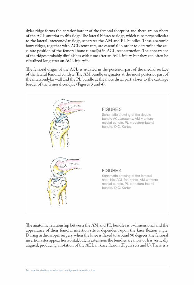
The femoral origin of the ACL is situated in the posterior part of the medial surface of the lateral femoral condyle. The AM bundle originates at the most posterior part of the intercondylar wall and the PL bundle at the more distal part, closer to the cartilage border of the femoral condyle (Figures 3 and 4).
The anatomic relationship between the AM and PL bundles is 3-dimensional and the appearance of their femoral insertion site is dependent upon the knee flexion angle. During arthroscopic surgery, when the knee is flexed to around 90 degrees, the femoral insertion sites appear horizontal, but, in extension, the bundles are more or less vertically aligned, producing a rotation of the ACL in knee flexion (Figures 5a and b). There is a
FIGURE 3Schematic drawing of the double-bundle ACL anatomy. AM = antero-medial bundle, PL = postero-lateral bundle. © C. Kartus.
FIGURE 4Schematic drawing of the femoral and tibial ACL footprints. AM = antero-medial bundle, PL = postero-lateral bundle. © C. Kartus.

15
change in the slope of the bony surface in the attachments of the AM and PL bundles, producing an angle between the footprints of the more concave bony surface of the AM bundle compared with the more planar bony surface of the PL bundle43.
The tibial insertion of the two bundles has given them their name as the AM-bundle inserts antero-medial on the tibia and the PL-bundle inserts postero-lateral on the tibia (Figures 6a and b). The tibial insertion is slightly larger than the femoral insertion and is delineated by the medial eminentia spine medially and the extension of the posterior horn of the lateral meniscus posteriorly. Anteriorly, the tibial insertion passes just under the inter-meniscal ligament. The area of the tibial insertion ranges from 114mm2 to 229mm2. The tibial AM insertion is only slightly larger (56mm2-136mm2) than the PL insertion (52-93mm2) and they represent around 56% and 44% of the total tibial footprint respectively103,177. In the anterior part of the tibial insertion, there is an area in which the fibers change direction; this is the area bending around the anterior part of the intracondylar roof in what is called physiological impingement18.
FIGURE 5a. Extended knee with vertical femoral ACL origin and parallel AM and PL bundles; b. a 90° flexed knee with an almost horizontal ACL origin and twisted AM and PL bundles (reprinted with kind permission from Springer Inc).
FIGURE 6a. and b. Tibial insertion patterns of the ACL (reprinted with kind permission from Springer Inc).

16 mattias ahldén / anterior cruciate ligament reconstruction
The angle between the tibial axis and the posterior inclination of the tibial plateau is called the posterior-inferior tibial slope (PITS). A possible correlation between the slope and ACL insufficiency and the need for reconstruction has also been reported105.
The AM and PL bundles work synergistically throughout the full range of motion (ROM) and both bundles are responsible for controlling AP and rotatory laxity but with different tension patterns. During ROM, the AM bundle is elongated with full extension and is more isometric than the PL bundle, which is elongated in full extension and gradually relaxes with increasing knee flexion203.
During anterior loading from an external force, the PL bundle carries higher forces near extension compared with the AM bundle, but, with a higher knee flexion angle, the op-posite is observed54. During combined rotatory loads (valgus and internal rotation), the AM bundle is subjected to higher forces overall, but significant force is also observed in the PL bundle, revealing the importance of the PL bundle for controlling laxity54,203. Yamamoto et al. and Sakane et al. also showed that the AM bundle takes most of the force in the AP direction at high flexion angles and the PL bundle takes a large part of the force in the AP direction and rotation at low flexion angles161,201.
5.2 Epidemiology
5.3 Etiology
A recent study revealed an annual incidence of 81 ACL injuries per 100,000 inhabit-ants aged 10-64 years in Sweden46. In soccer, the annual prevalence of ACL injury is reported to be 0.5-8.5% of players193. According to the Swedish National ACL Register, the number of performed ACL reconstructions was 3,311 in 2011. This number ap-pears to be constant over time. The Swedish National ACL Register has a high national coverage, where 90% of all performed reconstructions are registered. This indicates that about half the ACL injuries in Sweden are reconstructed. In the United States, prob-ably 100-200,000 ACL reconstructions are performed every year28,147. Female gender, younger age and contact sports are associated with a higher incidence of ACL injuries152.
ACL injury most commonly presents as a non-contact injury in association with sports. In Sweden, the most common sports associated with ACL injury are soccer and floorball for men and soccer and skiing for women. Two common scenarios causing ACL injury in sports are either when the foot is planted and the player changes direction or when landing from a jump. The mechanism usually includes valgus collapse in slight flexion in combination with rotation or hyperextension and rotation144. The injury mechanism can also be categorized as either the compressive-force type (as in weight-bearing) or the torsional-force type. The compressive-force type probably results in more meniscal injuries and osteochondral fractures48.

17
The risk of rupturing the ACL is known to be at least two to five times higher in females. Risk factors that differ between the sexes are anatomic factors, such as the width of the notch and the size of the ACL, hormonal factors, as well as neuromuscular properties126,154. Moreover, females have greater general joint laxity, including greater knee laxity, com-pared with men, which has been proposed to affect the incidence of ACL rupture158,187.
ACL injuries are traditionally divided into total or partial ruptures. A partial rupture can be an elongation of the entire ACL or an isolated AM- or PL-bundle tear. An isolated AM-bundle tear is probably more common in flexion, when the PL-bundle is slack, whereas an isolated PL-bundle rupture is probably more common in hyperextension or close to full extension.
Injury to the ACL is rarely an isolated injury. Associated bone marrow edema, cartilage and meniscus lesions are common. One example of a combination injury is the “un-happy triad”, where the ACL, the medial meniscus and the medial collateral ligament are conjointly injured from a valgus rotation force.
5.4 Osteoarthritis after ACL injury
ACL injury is often associated with the subsequent development of post-traumatic osteoarthritis (OA), with a reported prevalence ranging from 10 to 90%121,142. The highly variable prevalence is probably caused by heterogeneity in patient populations, associated injuries, treatments and activity levels, together with differences in the way OA is clas-sified and reported in different studies. A systematic review by Oiestad et al. suggested that the prevalence of OA after ACL reconstruction reported by previous reviews has been too high142. Their review included 31 studies and reported a low prevalence of knee osteoarthritis for individuals with an isolated ACL injury (0%-13%) and a higher preva-lence of knee OA for subjects with combined injuries (21%-48%). The strongest risk factor reported for the development of OA is meniscal injury and meniscectomy96,121,139,142. Additional proposed risk factors in the literature are chondral damage, age, high BMI, graft choice and the time between injury and surgical intervention45. The mechanisms responsible for the development of OA are not fully understood, but probably injuries sustained at the initial time, secondary injuries in the ACL-deficient knee, together with changes in the static and dynamic loading of the knee, are all relevant.
No studies have been able to show that ACL reconstruction is able to prevent post-traumatic OA. However, the preservation of the menisci appears to be important for preserving long-term knee health, especially in terms of OA. Several studies have shown that knee kinematics are not restored to normal after traditional ACL reconstruc-tion25,56,182. Factors proposed to affect the restoration of kinematics are anatomic ACL reconstruction and early ACL reconstruction83,91.
18 mattias ahldén / anterior cruciate ligament reconstruction
5.5 The Swedish National ACL Register
5.6 Prevention
It is not always possible to answer questions using an RCT. Today, national quality registers are being used in several medical specialties. In Scandinavia, the Hip and Knee Arthroplasty Registers are two early examples124. The purpose of the hip and knee replacement registers is primarily to detect inferior implants at an early stage. This is best accomplished through register studies involving a large number of patients. Large cohorts can also be used to identify prognostic factors, which can be correlated to good and poor outcome. Until January 2005, there were no national registers for monitoring the functional outcome of knee ligament surgery, especially ACL reconstructions. The Swedish National ACL Register (www.aclregister.nu) comprises patients undergoing ACL reconstruction, ACL revisions or re-operations for other reasons. The register covers more than 90% of all ACL reconstructions performed annually in Sweden. The register is a general database utilizing a web-based protocol. Age and gender are regis-tered automatically based on the Swedish social security number. The protocol comprises two parts, one section is surgeon based, where factors, such as activity at injury, time from injury to reconstruction, graft selection and fixation techniques, are registered. Previous surgery on the reconstructed knee, the contralateral knee and all concomitant injuries are also registered. All surgical procedures performed on the injured knee, including meniscal surgery (resection or repair) and treatment for chondral lesions, are reported. Revisions and re-operations for other reasons are registered as separate entries in the database and correlated with the primary ACL reconstruction procedure. The second section is patient based, including the Knee Injury and Osteoarthritis Outcome Score (KOOS)155,156, Lysholm knee scoring scale183, Tegner activity scale183 and European Qual-ity of Life-5 Dimensions (EQ-5D)32. Demographic questions, such as height, weight and smoking habits, are also asked. The KOOS is knee specific and covers knee-related quality of life (QoL) and function in sport and recreation (Sport/Rec), for example. The EQ-5D is a generic (disease non-specific) quality of life instrument27. The patient section is reported using a web-based protocol, before the reconstruction, as well as 1, 2 and 5 years after surgery. The database complies with the Swedish legislation relating to data security, which means that a non-authorized person can never gain access to the data.
Further possible development of the ACL register includes the registration of non-operatively treated ACL ruptures.
The majority of ACL ruptures are non-contact injuries, suggesting that the prevention of ACL injury is possible. A recent systematic review by Sadoghi et al. indicated strong evidence in support of significant effects of ACL injury-prevention programs, with an estimated risk reduction of 62% in athletes159. However, the authors concluded that the eight included studies were of poor quality. A recent Swedish study cluster-randomized
19
5.7 Knee laxity
The ACL is the primary restraint against anterior translation. The evaluation of AP knee laxity is regarded as the basis of diagnosis, treatment selection and follow-up in the ACL-injured knee. The manual Lachman test is the most sensitive and common manual laxity test when it comes to detecting an ACL injury151. However, manual tests display a large variability between examiners. Instrumented manual systems, such as the KT-1000 arthrometer (MEDmetric corp, San Diego, CA, USA), are commonly utilized in order to quantify AP knee laxity in a standardized way. Daniel et al. developed the KT-1000 arthrometer in order to diagnose an ACL injury36. Daniel et al. used a force of 89 Newtons (N), but a force of 134 N or maximum manual tension (MMT) is normally recommended nowadays189. The reproducibility of the KT-1000 arthrometer has been questioned and it has been suggested for use at group level and not at individual level168,169. Moreover, it is recommended that one examiner should perform all the measurements in order to improve reliability. The KT-1000 is included in most follow-up studies in order to measure the objective clinical benefit of an ACL reconstruction in terms of AP laxity. However, AP laxity does not correlate well with the functional outcome99,113.
Rotatory knee laxity measurements are more challenging to perform compared with measurements of AP laxity and are less common than reports on the use of AP laxity measurements. One problem is that there is a large inter-individual difference in rotatory knee laxity30. Devices for measuring rotatory laxity are still under development and differ considerably in terms of methodology5. Rotatory laxity can be measured as static laxity in one degree of freedom or be assessed as dynamic rotatory laxity using the pivot-shift test or during functional activities such as squatting or running5. Bignozzi et al. evaluated the relevance of static and dynamic tests after anatomic DB ACL reconstruction using computer-assisted surgery (CAS) and concluded that static rotatory laxity measurements were inferior when evaluating knee laxity after DB ACL reconstructions19. In terms of dynamic knee laxity tests, the pivot-shift test may be preferable in a large clinical setting or as part of the routine in clinical follow-up, because functional dynamic knee laxity tests, such as RSA or DSX, are more complicated to perform, expensive and labor intensive5.
The pivot-shift test is the most common way to assess rotatory knee laxity. It can be re-garded as a link between the static testing of rotatory laxity in one degree of freedom and
4,564 soccer players 12-17 years of age to undergo neuromuscular training for 15 min-utes twice a week throughout the season. The prevention program resulted in a 64% reduction in the number of ACL injuries192.
5.7.1 Antero-posterior knee laxity
5.7.2 Rotatory knee laxity and the pivot-shift test

20 mattias ahldén / anterior cruciate ligament reconstruction
functional dynamic laxity testing in multiple degrees of freedom. The pivot-shift test entails a complex motion which can be described as a two-component rotation in the axis of knee flexion and around the axis of tibial rotation31. The pivot-shift test is the most specific test for diagnosing an ACL injury and has been shown to correlate to subjective function and the development of OA90,99,114,151. The pivot-shift test represents motions in the extremes of the rotatory laxity envelope and simulates the patient’s giving-way symptom. However, the pivot-shift test is subjective in nature, in terms of both conduct and interpretation107,137,141.
The contribution of different factors to controlling rotatory laxity in the knee is still poorly understood. The envelope of laxity was described by Bull et al. and describes primary restraints, such as ACL, and secondary restraints, such as collateral ligaments, menisci and joint capsule30,31. The pivot-shift grade and rotatory laxity are therefore not only dependent on the integrity of the ACL. Musahl et al. reported that, in the event of a grade-1 pivot shift, the ACL injury was more often isolated compared with a grade-2 pivot shift136. Furthermore, they showed that the lateral meniscus is more important than the medial meniscus in controlling the pivot-shift test.
The antero-lateral (AL) capsule and iliotibial band perform a similar role in controlling rotatory laxity31. An AL capsular injury can be represented by a Segond’s fracture, a bony avulsion of the insertion site of the AL capsule on the proximal AL tibia. Furthermore, injuries to the collateral ligaments can influence the pivot-shift test125.
The influence of bony morphology on the pivot-shift test is a growing area of interest. Factors reported to influence the rotatory kinematics of the knee and the pivot-shift test are the size and convexity of the lateral tibial plateau, the tibial slope and the distal femur geometry23,77,125,135.
In terms of the ACL as a primary restraint against rotatory laxity, both the AM and PL bundles are probably important. It has been suggested that the more horizontal orien-tation of the PL bundle makes it more capable of controlling rotatory loads than the AM bundle203. However, the two bundles display reciprocal behavior and the individual significance in controlling rotatory laxity varies with knee flexion angle6,54.
There is a lack of validated measurement devices that can be used to assess the pivot-shift test and rotatory laxity of the knee. Further studies of non-invasive devices are warranted.
5.8 Non-operative versus operative treatment for ACL injury
A Cochrane review in 2009 aimed to evaluate the effect of the surgical treatment com-pared with the non-surgical treatment of ACL rupture by including only randomized, controlled trials. It resulted in only two randomized trials from 1987 and 1991, both of limited quality, being included. Moreover, the two studies did not evaluate the re-construction of the ACL but instead compared suturing the ACL with or without augmentation with non-surgical treatment. Consequently, the Cochrane review was

21
5.9 Surgical techniques of arthroscopic ACL reconstruction
The first reported arthroscopic ACL reconstruction was performed by Dandy in 1980, using a carbon-fiber ligament prosthesis, complemented by an extra-articular lateral tenodesis34. Dandy reported deep infections in 2 of his 23 reconstructed patients. A two-incision technique was used and it became popular in the orthopedic community. The first incision was used for graft harvest and tibial drilling. The second incision was placed over the lateral femur condyle for the outside-in drilling of the femoral tunnel utilizing a rear-entry guide. In the 1990s, with the development of intra-articular drills and drill guides, the one-incision transtibial technique became more and more popular. Even though it was argued that the new technique utilizing transtibial drilling was less anatomic, several studies reported no difference in outcome between the two techniques24,57,68. Moreover, in terms of surgical time, scar morbidity and cosmesis, the new transtibial technique was in favor and became the gold standard for more than 10 years, until the concept of anatomic reconstruction advanced at the beginning of the 21st century.
inconclusive. In 2011, an RCT by Frobell et al. reported the 2-year results after early ACL reconstruction in 61 patients compared with non-surgical treatment and optional delayed reconstruction in 59 patients47. The study consisted of early inclusion within 4 weeks after injury and structured rehabilitation for both groups. In the non-surgical group, 23 patients had undergone reconstruction at the 2-year follow-up. There were no differences in terms of the KOOS, which was the primary outcome, between the two groups at follow-up. Recently, the 5-year results revealed that an additional seven patients had undergone delayed ACL reconstruction between the 2- and 5-year follow-ups49. In all, 51% of the patients assigned to optional delayed ACL reconstruction were reconstructed within the 5-year follow-up period. No statistically significant differences in terms of the KOOS, Tegner activity scale or meniscal injuries were found between the groups. Their conclusion was that structured rehabilitation alone as primary treatment in young, active individuals with an acute ACL tear should be encouraged49. However, this conclusion has recently been seriously questioned by Petersen and Levy115,148.
Important parts of the care of ACL-injured patients are an early, correct diagnosis, together with individualized treatment selection. Because the results of prospective studies of ACL reconstruction reveal strong evidence that the patients improve in terms of laxity, Patient-Reported Outcome Measures (PROM) and activity level, randomized trials of operative versus non-operative treatment for ACL injuries will still be very difficult to perform.
5.9.1 The early era of arthroscopic ACL reconstruction

22 mattias ahldén / anterior cruciate ligament reconstruction
During the 1990s, the concept of isometry in ACL reconstruction was developed. Iso-metric means that the distance between the femoral and tibial attachment sites does not change as the knee flexes. It was believed that the exact isometric placement of the graft was critical for the success of an ACL reconstruction and that non-isometric placement would produce irreversible slackening of the graft or limited ROM206. However, the isometric point is close to the posterior end of Blumensaat’s line, which is far higher in the notch than the native ACL footprint138. The native ACL is not isometric but is instead organized in multiple fibers with different tension patterns throughout ROM, mainly divided into the two functional AM and PL bundles. The concept of isometry is therefore usually regarded as part of the era of non-anatomic reconstruction.
The use of the transtibial technique has been criticized for placing the ACL graft in a non-anatomic, vertical position outside the native ACL footprints13,101 and, with the transtibial technique, notchplasty was commonly performed as a matter of routine. Notchplasty involves removing bone from the medial wall of the lateral femoral condyle to avoid graft impingement and enhance visualization. Today, notchplasty is only performed in the event of anatomic diversities such as the formation of osteophytes or notch stenosis. To improve visualization in ACL reconstruction today, a three-portal technique is frequently used, with improved visualization of the femoral footprint using the accessory AM portal.
The concept of anatomic ACL reconstruction advanced at the beginning of the 21st century. Studies reported that the lower femoral tunnel position in the notch, closer to the femoral footprint, provided greater control of rotatory laxity120,167. Biomechanical studies suggested more normal biomechanics and graft tension patterns when placing the tunnels in the footprint of the native ACL197,200,201. The aim of anatomic ACL recon-struction is therefore to reproduce the native anatomy of the ACL by restoring native insertion site anatomy, the two functional bundles, the tension pattern of the ACL and individualizing the surgery for each patient91. This can be achieved using either the SB or DB technique, depending on the patients’ individual prerequisites. With the aim of restoring biomechanics as much as possible, the goal of anatomic reconstruction is to benefit clinical outcome in the short term and possibly reduce the prevalence of osteo-arthritis (OA) in the long term.
The transtibial technique often fails to place the tunnels in the native ACL footprint13,101. Using the transtibial technique, the placement of the femoral tunnel is dependent on the position of the tibial tunnel, which may lead to a posteriorly placed tibial tunnel and difficulty placing the femoral tunnel in the native ACL footprint, especially in large knees. The result is a non-anatomic vertical graft, which can be efficient in control-ling AP laxity but less effective when it comes to rotatory laxity. In order to drill the
5.9.2 Non-anatomic arthroscopic ACL reconstruction
5.9.3 Anatomic ACL reconstruction

23
tunnels independently, the use of an AM portal for femoral drilling is recommended. Another option is to drill the femoral tunnel outside-in, a technique which has been re-popularized since it was first introduced in the 1990s.
In order to perform an anatomic ACL reconstruction, it is necessary to identify the ana-tomic insertion sites of the ACL. This can be accomplished through the identification and preservation of the ACL remnants and the lateral intercondylar and bifurcate ridges. Especially in chronic cases, native ACL anatomy can be difficult to identify and arthro-scopic measurements based on anatomic landmarks have been presented176,177. When using peroperative radiographic measurements, Bernard and Hertel’s grid method has been used, especially for the femoral side17. A grid coordinate system is placed along the Blumensaat line and tunnel locations can be categorized within the coordinate system. For the tibial side, the AM is located at 30% and the PL at 40% of the AP distance of the tibia on a lateral radiographic projection149.
However, in a recent report from the Danish knee ligament reconstruction register, there was a significantly increased risk of revision ACL surgery and a positive pivot-shift test when using the AM-portal technique for femoral drill-hole placement compared with the transtibial technique153. The findings could possibly be explained by technical failures resulting from the introduction of a new and more complex procedure. Another hypothesis that has been mentioned is that a greater force is carried by the anatomic ACL reconstruction compared with a non-anatomic graft placement and, as a result, there is a concomitant higher risk of re-rupture198.
The standard technique for ACL reconstruction has been the arthroscopic SB recon-struction. The SB reconstruction technique has mainly focused on the restoration of the AM bundle, while paying limited attention to the PL bundle. Traditional SB ACL reconstructions are therefore successful at restoring anterior stability to the knee, but the restoration of rotatory laxity has been questioned197. The development of surgical techniques led to the introduction of the DB technique, in which both bundles of the ACL are reconstructed separately. The hypothesis is that DB reconstruction has the ability more closely to mimic the native ACL with its two functional bundles, the AM and PL bundle. However, a DB reconstruction can still be performed non-anatomically if the tunnels are placed outside the native ACL footprint. The techniques and number of tunnels used to perform a DB reconstruction differ greatly between different studies. DB reconstruction is therefore a highly heterogeneous group and comparisons between different studies are difficult. DB reconstructions are more technically demanding, inva-sive and costly, which underlines the importance of thoroughly evaluating the possible benefits and indications of the new technique.
The first reported arthroscopic DB ACL reconstruction with two tunnels in both the femur and tibia was by Mott et al. in 1983131. Mott et al. used a semitendinosus (ST) graft
5.9.4 Double-bundle ACL reconstruction

24 mattias ahldén / anterior cruciate ligament reconstruction
with an open technique. The report was merely a description of the surgical technique and no outcome was reported. During the 1980s and 1990s, there were several reports on the DB technique, but it was not until the 21st century that the anatomic DB procedure was further developed with early clinical studies from Muneta and Yasuda from Japan133,134,202. Dr Fu in Pittsburgh further developed the anatomic DB concept into its current form and he has provided guidelines for reproducing the native anatomy of the ACL52,91,132.
5.10 Why is this thesis needed?
ACL reconstruction is one of the most commonly performed procedures worldwide in orthopedics. Treatment selection, surgical technique and timing of the procedure are still the subject of debate. The implementation of new surgical techniques calls for a thorough, critical evaluation, preferably utilizing RCTs. The development of national registers plays an important part in the quality surveillance and identification of prog-nostic factors. Moreover, when it comes to the evaluation of the ACL-injured and reconstructed knee, we need valid, reliable outcome measures, in terms of both PROM and objective examinations such as the pivot-shift test. The present thesis will have an important impact on all these factors, with the goal of providing the best available treatment for the patients.

25
In Study I, the aim was to evaluate ACL reconstruction in Sweden today in terms of baseline variables and PROM from the Swedish National ACL Register.
In Study II, the primary aims were to analyze the changes in knee laxity over time, after an index ACL reconstruction using either BPTB or HS autografts, and to compare the knee laxity measurements between the two study groups during 7 years of follow-up. The secondary aim was to compare radiographic findings between the two groups at the 7-year follow-up.
In Study III, the aim was to compare the clinical results and PROM after arthroscopic ACL reconstruction using the HS autograft in male versus female patients.
In Study IV, the aim was to compare the DB technique with the SB technique for ACL reconstruction in terms of clinical outcome and PROM.
In Study V, the aim was to make objective measurements of the pivot-shift test using three different measurement devices and to examine the correlation of the measurements to clinical grading of the pivot-shift test.
06 AIMS

26 mattias ahldén / anterior cruciate ligament reconstruction
Study IFrom 2005-2010, 17,794 unique ACL registrations had been included in the register. 1,443 registrations were excluded because they were multi-ligament reconstructions or re-operations other than revision ACL reconstructions. Primary or revision ACL reconstruction was performed on 16,351 patients for whom pre- and peroperative demographic data were available. KOOS evaluations were available for 10,473 patients (64% of possible patients) preoperatively, 7,493 patients (58% of possible patients) at 1 year, 5,580 patients (49% of possible patients) at 2 years and 1,452 patients (40% of possible patients) at 5 years. Information on smoking was included in the register from 2009 and was available for 4,466 patients (4,173 non-smokers; 293 smokers). DB reconstructions were performed in 493 patients.
The male:female ratio was 57.5:42.5 in both primary (n=15,387) and revision (n=964) reconstructions. The mean age at primary reconstruction was 25.3 (±10.4) and 27.8 (±9.2) years for females and males respectively; the corresponding age at revision recon-struction was 26.2 (±9.0) and 29.0 (±8.4) years respectively. The age distribution of pri-mary ACL reconstruction in the Swedish National ACL Register is shown in Figure 7.
07 PATIENTS
FIGURE 7Age distribution for primary ACL reconstruction in 2012 in the Swedish National ACL Register.

27
Study IIBetween April 1995 and May 1998, 71 patients who underwent an ACL reconstruction at the NU Hospital Group, Trollhättan/Uddevalla, Sweden, using either an ipsilateral BPTB autograft or an ipsilateral triple or quadruple ST autograft, were included in a randomized series. Patients who fulfilled the inclusion criteria were consecutively asked to participate in the study. The inclusion criterion was unilateral ACL injury, verified by clinical examination or arthroscopy. The exclusion criteria were posterior cruciate liga-ment (PCL) injury, more than + 1 medial or lateral collateral ligament injury, re-injury during the follow-up period and radiographically visible OA at the time of inclusion. Of the patients, 47/71 (66%) attended a clinical examination preoperatively and on all four scheduled postoperative occasions; 6 months (Fp I), 1 year (Fp II), 2 years (Fp III) and 7 years (Fp IV) postoperatively. The BPTB group consisted of 22 patients, while there were 25 patients in the HS group. The groups were comparable in terms of preoperative demographics and time to follow-up (Table 1). Forty-four patients (HS 23, BPTB 21, missing 3) underwent the radiographic assessment.
Study IIIBetween November 1996 and November 2005, 567 eligible primary ACL reconstruc-tions were performed at the NU Hospital Group, Trollhättan/Uddevalla, Sweden. Pa-tients who fulfilled the inclusion criteria were consecutively asked to participate in the
TABLE 1Demographics of patients in Study II
Group BPTB Group ST Significance
Number of patients 22 25 n.s.
Age (years)) median (range) 26 (14-48) 29 (15-40) n.s.
Gender Male/female 14/8 18/7 n.s.
Injured side Right/left 9/13 11/14 n.s.
Time between the injury and index operation (months) median (range) 11 (2-252) 17 (3-240) n.s.
Follow-up I (months)median (range) 6 (5-8) 6 (5-7) n.s.
Follow-up II (months)median (range) 12 (11-28) 12 (11-24) n.s.
Follow-up III (months)median (range) 25 (23-30) 24 (23-30) n.s.
Follow-up IV (months)median (range) 89 (77-110) 86 (69-109) n.s.
28 mattias ahldén / anterior cruciate ligament reconstruction
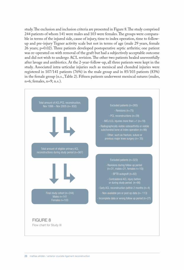
study. The exclusion and inclusion criteria are presented in Figure 8. The study comprised 244 patients of whom 141 were males and 103 were females. The groups were compara-ble in terms of the injured side, cause of injury, time to index operation, time to follow-up and pre-injury Tegner activity scale but not in terms of age (male 29 years, female 26 years; p=0.02). Three patients developed postoperative septic arthritis; one patient was re-operated on with removal of the graft but had a subjectively acceptable outcome and did not wish to undergo ACL revision. The other two patients healed uneventfully after lavage and antibiotics. At the 2-year-follow-up, all three patients were kept in the study. Associated intra-articular injuries such as meniscal and chondral injuries were registered in 107/141 patients (76%) in the male group and in 85/103 patients (83%) in the female group (n.s., Table 2). Fifteen patients underwent meniscal sutures (males, n=6; females, n=9; n.s.).
FIGURE 8Flow chart for Study III
Total amount of ACL/PCL reconstruction,Nov 1996 – Nov 2005 (n= 832) Excluded patients (n=265)
- Revisions (n=75)
- PCL reconstructions (n=39)
- MCL/LCL injuries more than +1 (n=18)
- Radiographically visible osteoarthritis or visible subchondral bone at index operation (n=98)
- Other; such as fracture, suture or previous major knee surgery (n= 35)
Excluded patients (n=323)
- Revisions during follow up period (n=31, males=21, females n=10))
- BPTB autograft (n=82)
- Contralateral ACL injury before or during study period (n=66)
- Early ACL reconstruction (within 2 months (n=4)
- Non-available pre or post op data (n= 113)
- Incomplete data or wrong follow up period (n=27)
Total amount of eligible primary ACL reconstructions during study period (n=567)
Final study cohort (n=244) Males n=141
Females n=103

29
TABLE 2Demographics of patients in Study III
Male Female Significance
Number of patients 141 103
Age (years) median (range) 29 (15-61) 26 (13-53) 0.02
Injured side Right/left 73/68 57/46 n.s.
Pre-injury Tegner activity scalemedian (range) missing values
8 (3-10) 1
8 (2-10)2
n.s.
Time between the injury and index operation (months) median (range) missing values
13 (2-360) 15 (2-276)2
n.s.
Follow-up period (months) median (range) missing values
25 (23-34)1
25 (23-36)1
n.s.
Associated injuries addressed at the time of the index operation or during the follow-up periodMeniscal (medial and/or lateral)Meniscal and chondralChondral
107 (76%)514511
85 (83%)47308
n.s.
Study IVBetween March 2008 and September 2009, 105 patients were randomized to either the SB group (n=52) or the DB group (n=53) (Figure 9). Participants were recruited from Sahlgrenska University Hospital and NU Hospital Group; n=31 and n=74 respec-tively. The inclusion criterion was a unilateral ACL injury on patients over 18 years of age. The exclusion criteria were PCL injury, more than +1 medial or lateral collateral ligament injury or previous major knee surgery. Patients who fulfilled the inclusion criterion were consecutively asked to participate in the study. The participants were randomized to either the SB or DB technique using closed envelopes administered by the study coordinator. Two patients did not receive allocated intervention. One patient discontinued the intervention due to a contralateral femur fracture and four patients were lost to follow-up. A two-year-follow-up was performed on 98 patients (93%) and was concluded in December 2011 (SB n=48, DB n=50).
The patients in the groups that received allocated intervention (n=103) were comparable in terms of age, gender, injured side, time between the injury and index operation and length of follow-up period but not in terms of pre-injury Tegner activity scale (Table 3). However, the preoperative Tegner activity scale revealed no statistically significant difference between the groups. Contact sport was the major cause of injury (SB=68%, DB=68%).

30 mattias ahldén / anterior cruciate ligament reconstruction
FIGURE 9Flow chart for Study IV
ENROLLMENT
Allocated to single-bundle (n=52)
• Received allocated intervention (n=50)
• Did not receive allocated intervention (n=2) (wrongly included; contra-lateral ACL
injury n=1, declined to participate n=1)
Allocated to double-bundle (n=53)
• Received allocated intervention (n=53)
Lost to follow-up (n= 2)
Lost to follow-up (n=2)
Discontinued intervention (sustained contra-lateral femur fracture n=1)
Analyzed (n=48) Analyzed (n=50)
ALLOCATION
FOLLOW-UP
ANALYSIS
Randomized (n=105)
All patients in the clinics of the participating surgeons were assessed for eligibility

31
TABLE 3Demographics of patients in Study IV
SB DB Significance
Number of patients 50 53
Age (years)) median (range) 25 (18-52) 29 (18-52) n.s.
Gender Male/female 35/15 35/18
Injured side (right/left) 27/22 32/21
Pre-injury Tegner activity scalemedian (range) mean (SD) missing values
8 (3-9)7.8 (1.3)
8 (5-9)7.3 (1.5)
2
0.03
Time between the injury and index operation (months)median(range) mean (SD)
10 (3-240)23 (37)
9 (2-240)24 (42)
n.s.
Follow-up period (months) median (range)missing values
26 (22-32)2
26 (22-42)3
n.s.
Associated injuries at the time of the index operation or during the follow-up period Meniscal (medial and/or lateral)Meniscal and chondralChondralNone
18 (36%)13 (26%)7 (14%)12 (24%)
15 (28%)14 (26%)6 (11%)18 (34%)
n.s.
Study VThe right knee of a fresh whole lower body specimen was used (70-year-old male). The specimen was evaluated by clinical examination, radiographs and diagnostic arthros-copy to rule out previous injuries and OA. During arthroscopy, the knee was prepared to display a high-grade pivot-shift test by means of resection of the ACL and anterior horn of the lateral meniscus. Twelve expert surgeons, authorities in the field of ACL reconstruction, comprised the study group performing their preferred technique of the pivot-shift test.

32 mattias ahldén / anterior cruciate ligament reconstruction
08 METHODS
8.1 Blinded examiners
8.2 Surgical technique
In Studies II and III, two physiotherapists, and in Study IV one, who were not involved in the rehabilitation, performed all the pre- and postoperative clinical assessments. The physiotherapist(s) were blinded to the aim of the studies and, in Study IV, also to the type of surgical technique that had been used. In Study V, the surgeons were blinded to how the cadaver knee was prepared and to the grading performed by the other surgeons.
Study I
The Swedish National ACL Register includes information on the method of fixation that has been used, together with the size and type of graft. In the surgeon-based part, no infor-mation on the location of tunnels or type of drilling is included (transtibial, anteromedial or outside-in). However, information on the treatment that is performed with regard to associated injuries is included (meniscus and/or cartilage). In the event of a revision re-construction, information regarding the primary ACL reconstruction is also included. In the case of re-operation, the cause of surgery is registered (infection, screw removal etc.).
Study II
One senior surgeon performed all the reconstructions. Associated intra-articular inju-ries, such as meniscal ruptures and chondral lesions, were addressed at the time of the index operation.
BPTB technique: the arthroscopic transtibial technique and interference screw fixation were used during the index procedure108. The mid-third of the patellar tendon was har-vested through two 25-mm long vertical incisions, one over the apex of the patella and the other just above the tibial tubercle. The graft was tunneled subcutaneously under the paratenon with the aim of protecting the infrapatellar nerve and its branches and leaving the major part of the paratenon intact, as described previously by Kartus et al92. The proximal bone block was sized to 9 mm and the distal bone block to 10 mm. The femoral tunnel was drilled transtibially and placed in approximately the 10.30 o’clock po-sition in the right knee and the tibial tunnel was placed just anterior to the normal PCL.

33
A 7 mm and a 9 mm Acufex® (Acufex, Microsurgical Inc., Mansfield, MA, USA) “silk” interference screw were used on the femoral and tibial sides respectively (Figure 10).
ST technique: the graft was harvested through an approximately 3-cm long incision over the pes anserinus. The tendons were palpated and the sartorius fascia was incised parallel to the fibers of the fascia, just above the thicker and more distally inserted ST tendon. After the vinculae had been cut under visual control, the ST tendon was harvested with a semi-blunt, semi-circular open tendon stripper (Acufex, Microsurgical Inc., Mans-field, MA, USA). Depending on its length, the ST tendon was prepared as a triple or quadruple graft. The femoral tunnel was drilled transtibially and the tibial tunnel was drilled in a standard fashion. Both the femoral and tibial tunnels were placed in the same locations as in the BPTB group. A 7 mm soft-threaded RCI® (Smith and Nephew, Inc, Andover, MA 01810, USA) metal interference screw was used on both the femoral and tibial sides. After the femoral screw had been inserted, firm traction was applied to the graft during the insertion of the tibial screw with the knee in full extension.
FIGURE 10SB ACL reconstruction using the transtibial technique and BPTB autograft. © C. Kartus.

34 mattias ahldén / anterior cruciate ligament reconstruction
Study III
One senior surgeon performed all the reconstructions. The surgical technique was similar to that used Study II, but the femoral tunnel was drilled through the AM portal and placed approximately at the 10.30 o’clock position in the right knee. Furthermore, both the ST and gracilis (G) tendons were harvested and prepared for a quadruple graft. On the tibial side, a 7-9 mm soft-threaded RCI® (Smith and Nephew, Inc, Andover, MA 01810, USA) metal interference screw was used (Figure 11).
Study IV
Four senior surgeons performed all the reconstructions. Standard AL and AM portals were established. Associated intra-articular injuries, such as meniscal ruptures and chondral lesions, were addressed at the time of the index operation. The footprints of the ACL on both the femur and tibia were identified, together with the intercondylar and bifurcate ridges on the femur, after which the ACL remnants were resected. The ST and G tendons were harvested with an open tendon stripper through a standard longitudinal incision at the pes anserinus, on the AM aspect of the proximal tibia. The femoral drilling was performed using a fluted reamer through the AM portal. All tibial drilling was performed using a tibial elbow aimer and a fluted reamer. All bone tunnels were drilled to approximately 0.5 mm above the diameters of the respective grafts. On the femoral side, the fixation was performed using metal interference screws (RCI®,
FIGURE 11SB ACL reconstruction using the AM transportal technique and HS autograft. © C. Kartus.
35
Smith and Nephew, Inc, Andover, MA 01810, USA). On the tibial side, bioresorbable screws were used (Matryx®, ConMedLinvatec, Largo, FL, USA).
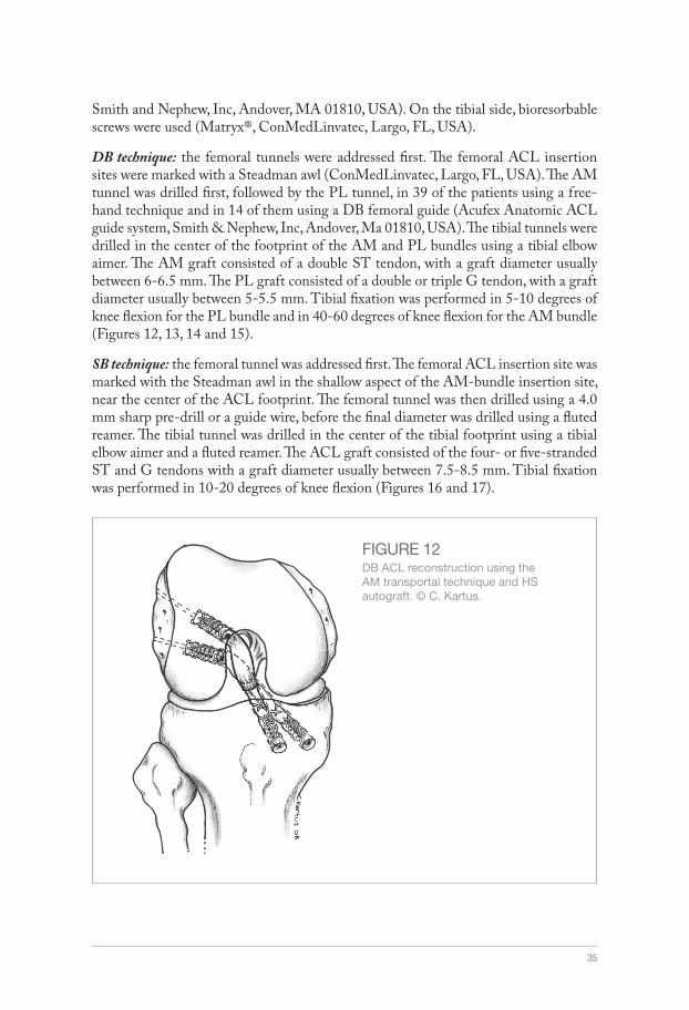
DB technique: the femoral tunnels were addressed first. The femoral ACL insertion sites were marked with a Steadman awl (ConMedLinvatec, Largo, FL, USA). The AM tunnel was drilled first, followed by the PL tunnel, in 39 of the patients using a free-hand technique and in 14 of them using a DB femoral guide (Acufex Anatomic ACL guide system, Smith & Nephew, Inc, Andover, Ma 01810, USA). The tibial tunnels were drilled in the center of the footprint of the AM and PL bundles using a tibial elbow aimer. The AM graft consisted of a double ST tendon, with a graft diameter usually between 6-6.5 mm. The PL graft consisted of a double or triple G tendon, with a graft diameter usually between 5-5.5 mm. Tibial fixation was performed in 5-10 degrees of knee flexion for the PL bundle and in 40-60 degrees of knee flexion for the AM bundle (Figures 12, 13, 14 and 15).
SB technique: the femoral tunnel was addressed first. The femoral ACL insertion site was marked with the Steadman awl in the shallow aspect of the AM-bundle insertion site, near the center of the ACL footprint. The femoral tunnel was then drilled using a 4.0 mm sharp pre-drill or a guide wire, before the final diameter was drilled using a fluted reamer. The tibial tunnel was drilled in the center of the tibial footprint using a tibial elbow aimer and a fluted reamer. The ACL graft consisted of the four- or five-stranded ST and G tendons with a graft diameter usually between 7.5-8.5 mm. Tibial fixation was performed in 10-20 degrees of knee flexion (Figures 16 and 17).
FIGURE 12DB ACL reconstruction using the AM transportal technique and HS autograft. © C. Kartus.

36 mattias ahldén / anterior cruciate ligament reconstruction
FIGURE 13DB ACL reconstruction at second look 6 months after index reconstruc-tion. © J. Kartus
FIGURE 143D CT reconstruction of femoral tunnels for DB ACL reconstruction in Study IV. © M.Ahldén
FIGURE 153D CT reconstruction of tibial tunnels for DB ACL reconstruction in Study IV. © M.Ahldén

37
FIGURE 163D CT reconstruction of femoral tunnel for SB ACL reconstruction in Study IV. © M.Ahldén
FIGURE 173D CT reconstruction of tibial tunnel for SB ACL reconstruction in Study IV. © M.Ahldén

38 mattias ahldén / anterior cruciate ligament reconstruction
8.3 Rehabilitation
8.4 Standard radiography
8.5 Clinical examinations
Study I
The Swedish ACL register does not include information on completed rehabilitation. Accordingly, all patients are rehabilitated using the surgeons’ preferred regimen.
Studies II-IV
The local physiotherapist used an individual-based training program according to our rehabilitation guidelines, permitting immediate full weight-bearing and full ROM in-cluding full hyperextension. However, no external load in open kinetic chain exercises apart from the weight of the operated leg was used during the first six postoperative weeks from 30 degrees to full extension172. No rehabilitation brace was used66. Closed-chain exercises were started immediately postoperatively. Running was permitted at three months and contact sports at six months at the earliest, provided that the patient had regained full functional stability in terms of strength, coordination and balance as compared with the contralateral leg.
Study II
Radiographic evaluation and classification were performed by an independent experi-enced radiologist, blinded to the type of graft. Standard weight-bearing radiographic examinations using AP and lateral views, with 30° of knee flexion, were classified ac-cording to the Fairbank and the Ahlbäck rating systems4,40. Fairbank’s classification relates primarily to mild changes, ranging from the flattening of the condyles to joint space narrowing. In 1968, Ahlbäck presented his grading system for OA in the knee from mild stages with joint narrowing to severe remodeling of the bone. In long-term follow-up studies after ACL reconstruction, most radiographic changes can be described using Fairbank’s and Ahlbäck’s classification systems59.
Study II
For Study I, the web-based protocol of the Swedish National ACL Register was used. For Studies II-III, a special protocol was used for the preoperative and postoperative clinical examinations. For Study IV, both the web-based protocol of the Swedish Na-tional ACL register and the special protocol were used.
39
Studies II-IV
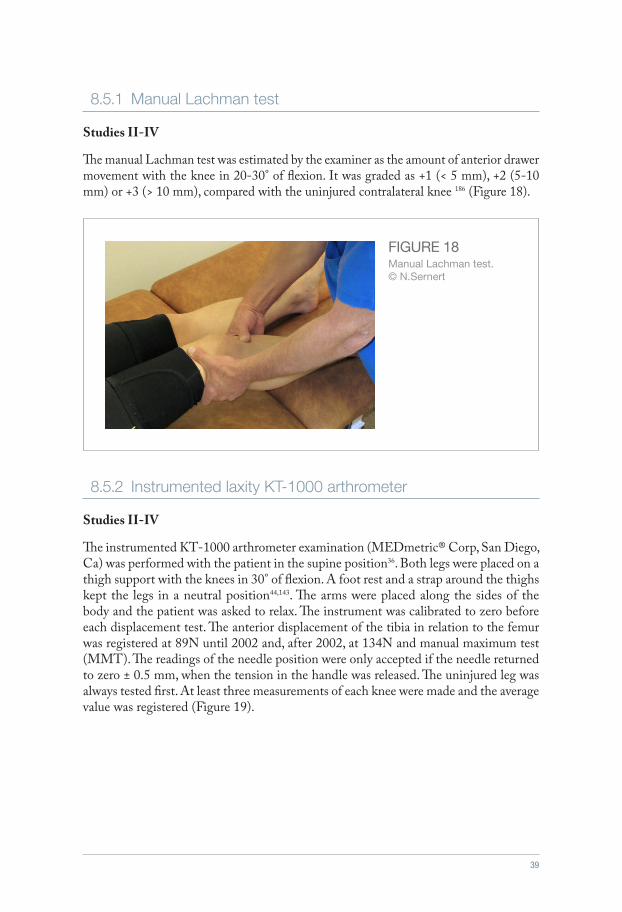
The manual Lachman test was estimated by the examiner as the amount of anterior drawer movement with the knee in 20-30° of flexion. It was graded as +1 (< 5 mm), +2 (5-10 mm) or +3 (> 10 mm), compared with the uninjured contralateral knee 186 (Figure 18).
Studies II-IV
The instrumented KT-1000 arthrometer examination (MEDmetric® Corp, San Diego, Ca) was performed with the patient in the supine position36. Both legs were placed on a thigh support with the knees in 30° of flexion. A foot rest and a strap around the thighs kept the legs in a neutral position44,143. The arms were placed along the sides of the body and the patient was asked to relax. The instrument was calibrated to zero before each displacement test. The anterior displacement of the tibia in relation to the femur was registered at 89N until 2002 and, after 2002, at 134N and manual maximum test (MMT). The readings of the needle position were only accepted if the needle returned to zero ± 0.5 mm, when the tension in the handle was released. The uninjured leg was always tested first. At least three measurements of each knee were made and the average value was registered (Figure 19).
8.5.1 Manual Lachman test
8.5.2 Instrumented laxity KT-1000 arthrometer
FIGURE 18Manual Lachman test. © N.Sernert

40 mattias ahldén / anterior cruciate ligament reconstruction
FIGURE 19KT-1000 arthrometer. © N.Sernert
FIGURE 20The pivot-shift test used in Study IV. © N.Sernert
Studies IV-V
The pivot-shift test is a clinical dynamic knee laxity test which evaluates a combination of translational and rotatory laxity, which represents the patients’ typical giving-way phenomena31. The subjectivity, in terms of both conduct and interpretation, makes the results difficult to compare between various studies and justifies the same observer per-forming all the tests in a clinical study in order to increase the reliability of the test141. The pivot-shift test was graded from I-III according to IKDC guidelines14,69,82 (Figure 20).
8.5.3 The pivot-shift test

41
FIGURE 21ROM measurement. © N.Sernert
Studies II-IV
The ROM measurement was performed in the supine position using a hand-held goniometer graded in one-degree increments29. The patient first made an active full extension, followed by an active full flexion. The uninjured leg was always measured first and the side-to-side differ-ence including hyperextension was calculated. If the measurements displayed a side-to-side difference of ≥ 5˚ in either extension or flexion, the patients were dichotomously classified as having or not having an extension or flexion deficit. The examiner always made a visual check to ensure that the measured side-to-side difference appeared reasonable (Figure 21).
Study III
The loss of or disturbance in skin sensitivity was measured by the examiner palpating the anterior knee region. The length multiplied by the width was registered and the result is shown in cm2 93,94.
Study III
The patients were classified dichotomously as having or not having subjective anterior knee pain if they registered pain while climbing stairs, sitting with the knee in 90° of flexion and during or after activity.
8.5.4 Range of motion (ROM)
8.5.5 Loss of skin sensitivity
8.5.6 Anterior knee pain
42 mattias ahldén / anterior cruciate ligament reconstruction
Study III
The patients graded their knee function as excellent, good, fair or poor.
8.5.7 Patients’ subjective evaluation
8.6 Functional tests
Studies III and IV
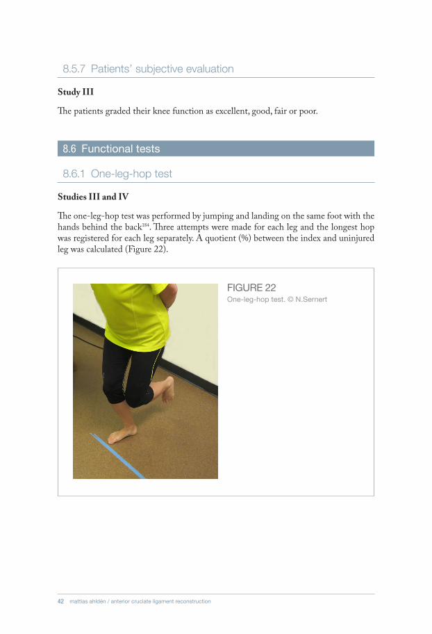
The one-leg-hop test was performed by jumping and landing on the same foot with the hands behind the back184. Three attempts were made for each leg and the longest hop was registered for each leg separately. A quotient (%) between the index and uninjured leg was calculated (Figure 22).
8.6.1 One-leg-hop test
FIGURE 22One-leg-hop test. © N.Sernert

43
Study IV
The square-hop test was performed by standing on the leg to be tested, outside a 40x40 cm square marked with a tape marked square on the floor. For the right leg, the subjects were instructed to jump clockwise in and out of the square as many times as possible during a period of 30 s. For the left leg, the subject performed the test in a counter-clockwise mode. The test was videotaped and both total jumps and the number of suc-cessful jumps performed, without touching the taped frame, were recorded. A quotient (%) between the index and non-injured leg was calculated. This test was modified from Östenberg et al.145 (Figure 23).
Study III
The classification of kneeling discomfort compared with the contralateral knee was based on the knee-walking test involving direct loading of the anterior knee region. The knee-walking test was performed on the floor of the examination room. The patients were not allowed to use any protection or clothing during the test while walking six steps forward on their knees. The test was subjectively classified by the patient as OK, unpleasant, dif-ficult, or impossible to perform, as described by Kartus et al.93,94 (Figure 24).
8.6.2 Square-hop test
8.6.3 Knee-walking test
FIGURE 23Square-hop test. © N.Sernert

44 mattias ahldén / anterior cruciate ligament reconstruction
FIGURE 24Knee-walking test. © N.Sernert
8.7 Functional scores
Studies I and IV
The KOOS (www.koos.nu) is a knee-specific, self-administered PROM validated for both the short-term and long-term follow-up of ACL reconstructions, meniscectomies and post-traumatic OA155,156. The KOOS consists of five subscales; Pain, Other symp-toms (Symptoms), Function in daily living (ADL), Function in sports and recreation (Sports/Rec) and Knee-related quality of life (QoL). The patient answers nine questions to assess Pain, seven questions to assess Symptoms, 17 questions regarding ADL, five questions regarding Sports/Rec and four questions regarding QoL. All questions are graded from zero to four points. A normalized score for each subscale is then calculated, with a maximum of 100 points indicating no symptoms and zero points indicating extreme symptoms.
8.7.1 KOOS

45
Studies II, III and IV
The modified Lysholm knee scoring scale was assessed by the patient using a self-administered questionnaire26,183. The questionnaire did not show the scores for the alter-native answers, as described by Höher et al.70. It consists of eight items, where pain and instability each account for 25 of the total score of 100 points.
Studies II-IV
The Tegner activity scale was assessed by the examiner during the course of the patient interview/examination26,183. The score is graded between 0-10, where grades 0-4 cover activities of daily living and work and grades 5-10 indicate whether the patient is able to participate in recreational or competitive sports.
8.7.2 Lysholm knee scoring scale
8.7.3 Tegner activity scale
8.8 Quantitative evaluation of the pivot-shift test
Study II
In Study V, the pivot-shift tests were performed three times by 12 blinded expert sur-geons using their preferred technique. The surgeons clinically graded the pivot-shift test using grades I-III according to the IKDC guidelines69,82,84. When the surgeons performed the pivot-shift tests, simultaneous data samplings were made by three dif-ferent measurement devices as follows (Figure 25).
FIGURE 25Quantitative evalua-tion of the pivot-shift test (reprinted with kind permission from Springer Inc)

46 mattias ahldén / anterior cruciate ligament reconstruction
Study V
The system consists of a transmitter that produces an electromagnetic field, which is used together with two receivers attached to the lower extremity. An additional third receiver is used to identify seven anatomic landmarks (major trochanter, medial epicondyle, lateral epicondyle, intersection of the MCL and joint line, fibular head, medial malleolus, lateral malleolus). The acquired position data for each landmark were converted to the relative position of the electromagnetic receivers and were used to create a coordinate system63. The method has been utilized in several previous studies9,53,75,76,106,107.
An electromagnetic tracking system (LIBERTY, Polhemus, VT) with bone and skin sensors respectively was used (Figure 25). The bone sensors were fixed using two half pins (4.0mm diameter) placed in the femur and tibia respectively. The two skin sensors were attached to a plastic brace fixed by a circumferential Velcro strap 10 cm above the patella on the thigh and 7 cm below the tibial tubercle on the lower leg. A third sensor was used to digitize the anatomic landmarks before the six degrees of freedom measure-ment of kinematics was made.
Study V
The system consists of a triaxial accelerometer (KiRA, Orthokey LLC, Lewes, DE, USA), wirelessly connected to a standard laptop. The sensor is non-invasively mounted between the lateral aspect of the anterior tuberosity and Gerdy’s tubercle (Figure 25). The chosen position of the sensor is derived from the fact that the anterior translation and the accel-eration reached during a pivot-shift test have been reported to display the largest values when monitoring the lateral compartment16,122. A second sensor can be mounted on the femur for reference102. The system generates various acceleration parameters, i.e. maximum, minimum, range and “slope” of acceleration, which is an average value for the first deriva-tive of acceleration as a suggestion of the smoothness of the pivot-shift phenomenon122.
Lopomo et al. reported on a previous study with non-invasive acceleration measurements for the pivot-shift test122. The sensor was placed on the skin between the lateral aspect of the anterior tibial tuberosity and Gerdy’s tubercle. They reported an acceptable level of intra-examiner repeatability (intra-class correlation coefficient/ICC 0.69~0.93). The probability of a correct diagnosis of ACL deficiency was 70% using the slope of accelera-tion and 80% using the range of acceleration.
The accelerometer was utilized in Study V. The assessed parameters were maximum acceleration (amax=“acceleration of reduction”), minimal acceleration (amin), range of ac-celeration (amax-amin) and “slope”.
8.8.1 Electromagnetic tracking system
8.8.2 Accelerometer

47
Study V
The simple image analysis system is a novel method for evaluating lateral compartment translation using a digital camera and was first reported by Hoshino et al.16,72. Small target stickers (white portable reinforcements, item #636156, Staples, Inc., Framingham, MA, USA) are placed on bony landmarks of the lateral compartment, i.e. lateral epicondyle, Gerdy’s tubercle and fibular head (Figure 25). The distance between the centers of the markers located over Gerdy’s tubercle and the fibular head is measured by a ruler. A video recording of the lateral aspect of the knee captures movement of the stickers during the manually performed pivot-shift test using a digital camera (Nikon COOLPIX S8100, Nikon Corp., Tokyo, Japan). Movies are analyzed frame by frame for lateral compartment translation using Image J Software (NIH, Bethesda, MD, USA), (Figure 26).
8.8.3 Simple image analysis
FIGURE 26Simple image analysis; the position of the distal femur is calculated as [(a actual measurement) x (b/a from image analysis)]). (reprinted with kind permission from Springer Inc).
48 mattias ahldén / anterior cruciate ligament reconstruction
Study IMean (SD) values are reported for all the KOOS subscales. The Mann-Whitney U-test is used to compare the KOOS values between subgroups, the Wilcoxon signed-rank test is used for the within-group comparisons of the preoperative and postoperative data and the chi-square test is used for dichotomous comparisons. A p-value of < 0.05 was considered statistically significant. All p-values are two-tailed.
Study IIMean (SD) and median (range) values are presented when applicable. For comparisons of dichotomous variables between the groups, Fisher’s exact test and the chi-square test were used. For both continuous and non-continuous variables, the Mann-Whitney U test was used. Friedman’s and Wilcoxon’s signed rank test were used for within-group comparisons of the preoperative and postoperative data. A p-value of < 0.05 was con-sidered statistically significant. All p-values are two-tailed.
Study IIIMean (SD) and median (range) values are presented when applicable. For compari-sons of dichotomous variables between the groups, the chi-square test was used. For both continuous and non-continuous variables, the Mann-Whitney U test was used. Wilcoxon’s signed rank test was used for within-group comparisons of the preoperative and postoperative data. A p-value of < 0.05 was considered statistically significant. All p-values are two-tailed.
Study IVMean (SD) and median (range) values are presented when applicable. For comparisons of dichotomous variables between the groups, the chi-square test was used. For comparisons of both continuous and non-continuous variables, the Mann-Whitney U test was used. Wilcoxon’s signed rank test was used for within-group comparisons of the preoperative and postoperative data. A p-value of <0.05 was considered statistically significant.
The primary variable in Study IV was the pivot-shift test. Study IV was powered to reveal a difference of less than one unit in the pivot-shift test between the study groups, with a power of more than 80%.
09 STATISTICAL METHODS

49
Study VCorrelations were calculated using Spearman’s rank correlation coefficient. Statistical significance was set at a p-value of < 0.05. Correlation coefficients were interpreted as poor (rho<0.3), moderate (0.3<rho<0.6) and good (0.6<rho<0.8).

50 mattias ahldén / anterior cruciate ligament reconstruction
Study I: The Swedish National Anterior Cruciate Ligament Register, a report on baseline variables and outcomes of surgery for almost 18,000 patients.
Aim: To evaluate ACL reconstruction in terms of baseline variables and PROM from the Swedish National ACL Register.
Patients: 17,794 registrations in the Swedish National ACL Register were included and analyzed.
Methods: The register utilizes a web-based protocol. The protocol consists of two parts, one patient section, which includes the KOOS. The second section is surgeon based, where factors relating to the initial injury, previous surgery and surgical procedures performed on the injured knee are registered.
Results: The most common causes of injury were soccer (48.7%) and floorball (9.5%) in the male patients and soccer (35.8%) and alpine skiing (16.1%) in the female patients. The use of HS autografts increased from 81.8% of the primary ACL reconstructions in 2005 to 96.1% in 2010. In 2005, transfixation pins (59.8%) were the most common femoral fixation and, in 2010, cortical button fixation (58.9%) was the most common femoral fixation. On the tibial side, the most common fixation method was a metal or bioresorbable interference screw.
All subscales of the KOOS score had improved significantly 1, 2 and 5 years postopera-tively for primary reconstructions (p<0.001), (Figure 27). For revision reconstructions, all subscales of the KOOS score had improved significantly at 1 and 2 years (p<0.05), apart from Symptoms at 2 years (p=0.07) (Figure 28). At 5 years, only the Sport/Rec subscale and knee-related QOL had improved significantly compared with the preoperative values for the revisions (p<0.002). On all follow-up occasions, revisions obtained significantly lower scores in terms of all the KOOS subscales compared with primary reconstructions (p<0.001). At the time of primary reconstruction, 53.9% of the patients had one or more concomitant intra-articular injuries. The corresponding number for revision reconstruc-tions was 59.4% (p<0.001). Preoperatively, DB reconstructions had significantly lower values than SB reconstructions in terms of all the KOOS subscales (p<0.04), except for knee-related QoL. At 1 and 2 years, no significant differences were found, except for the Sport/Rec subscale at 1 year, which was significantly better for the SB reconstructions (p=0.031). Smokers fared significantly less well than non-smokers in terms of all the KOOS subscales preoperatively (p<0.001) and at 2 years (p<0.01). During a 5-year period, 9.1% (contralateral 5.0% (n=106); revision 4.1% (n=88)) of the patients included in 2005 underwent a contralateral ACL reconstruction or revision surgery of the index knee. The
10 SUMMARY OF THE PAPERS

51
corresponding number for the group of 15- to 18-year-old female soccer players was 22.0% (contralateral 10.2% (n=12); revision 11.8% (n=14)), (p<0.001 vs all patients). For the group of 15- to 18-year-old male soccer players, the number was 9.8% (contralateral 4.4% (n=4); revision 5.4% (n=5)), (p=0.02 vs corresponding female subgroup).
Conclusion: Primary ACL reconstruction significantly improves all the subscales of the KOOS score. Young female soccer players run a major risk of re-injuring their ACL or injuring the contralateral ACL. Revision ACL reconstructions do less well than primary reconstructions and smokers do less well than non-smokers.
FIGURE 27KOOS pre-operati-vely and at 1, 2 and 5 years post-opera-tively after primary ACL reconstruc-tions. © M.Ahldén
FIGURE 28KOOS pre-operati-vely and at 1, 2 and 5 years post-operatively after revision ACL reconstruction. © M.Ahldén

52 mattias ahldén / anterior cruciate ligament reconstruction
Study II: Knee laxity measurements after anterior cruciate ligament reconstruction, using either bone-patellar-tendon-bone or hamstring tendon autografts, with special emphasis on comparison over time.
Aim: The primary aims were to analyze the changes in knee laxity over time, after an index ACL reconstruction using either BPTB or HS autografts, and to compare the knee laxity measurements between the two study groups during 7 years of follow-up. The secondary aim was to compare radiographic findings between the two groups at the 7-year follow-up.
Patients: Seventy-one patients who underwent an ACL reconstruction using either an ipsilateral BPTB autograft or an ipsilateral triple or quadruple ST autograft were included in a randomized series.
Methods: The KT-1000 arthrometer was used to measure knee laxity preoperatively and on four postoperative occasions; 6 months, 1 year, 2 years and 7 years after the recon-struction. Radiographic and clinical assessments were performed at the 7-year follow-up.
Results: There were no significant differences in knee laxity between the BPTB group and HS group preoperatively or at the follow-ups. There was a tendency towards reduced knee laxity over time in both groups in terms of the side-to-side difference (Figure 29).
This decrease was significant when analyzing the injured and non-injured knees sepa-rately (injured side p<0.001 and p=0.005 BPTB and HS groups respectively; non-injured side p=0.008 and p=0.042 respectively), (Figures 30 and 31).
Both groups improved significantly between the preoperative assessment and the 7-year follow-up in terms of the Tegner activity scale (BPTB group p=0.003 and HS group p=0.007), Lysholm knee scoring scale (BPTB group p=0.008 and HS group p=0.005) and the manual Lachman test (BPTB group and HS group p<0.001 respectively).
The radiographic assessment revealed no significant differences between the study groups at the 7-year follow-up in terms of degenerative changes classified according to the Fairbank and Ahlbäck rating systems. In overall terms, degenerative changes were identified in 16% (BPTB 19%; HS 13%; n.s.), according to the Ahlbäck rating system, and 68% (BPTB 67%; HS 70%; n.s), according to the Fairbank rating system. No sig-nificant differences in the prevalence of patello-femoral osteophytes (BPTB 38%; HS 44%) were found between the study groups.
Conclusion: There were no significant differences in knee laxity measurements between the two study groups preoperatively or at the 7-year follow-up. A decrease in knee laxity over time was seen in both groups. There were no significant differences between the BPTB and HS groups in terms of degenerative changes at 7 years.
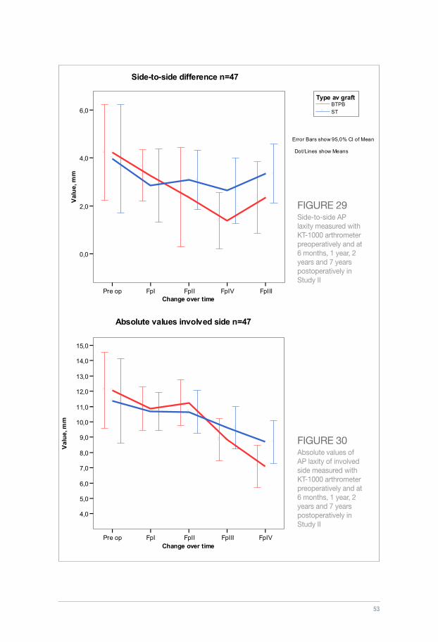
53
BTPB ST
Type av graft
Error Bars show 95,0% Cl of Mean
Dot/Lines show Means
Side-to-side difference n=47
Pre op FpI FpII FpIV FpIIIChange over time
0,0
2,0
4,0
6,0
Val
ue,
mm
BTPB ST
Type of graft
Absolute values involved side n=47
Error Bars show 95,0% Cl of Mean
Dot/Lines show Means
Pre op FpI FpII FpIII FpIVChange over time
4,0
5,0
6,0
7,0
8,0
9,0
10,0
11,0
12,0
13,0
14,0
15,0
Val
ue,
mm
FIGURE 29Side-to-side AP laxity measured with KT-1000 arthrometer preoperatively and at 6 months, 1 year, 2 years and 7 years postoperatively in Study II
FIGURE 30Absolute values of AP laxity of involved side measured with KT-1000 arthrometer preoperatively and at 6 months, 1 year, 2 years and 7 years postoperatively in Study II
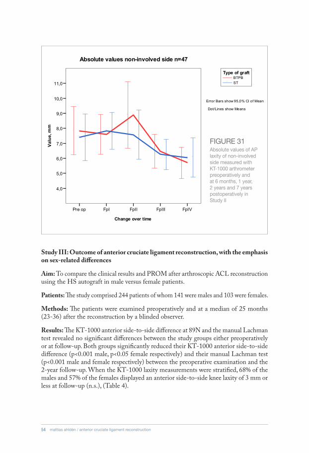
54 mattias ahldén / anterior cruciate ligament reconstruction
BTPB ST
Type of graft
Absolute values non-involved side n=47
Error Bars show 95,0% Cl of Mean
Dot/Lines show Means
Pre op FpI FpII FpIII FpIV
Change over time
4,0
5,0
6,0
7,0
8,0
9,0
10,0
11,0
Val
ue,
mm
FIGURE 31Absolute values of AP laxity of non-involved side measured with KT-1000 arthrometer preoperatively and at 6 months, 1 year, 2 years and 7 years postoperatively in Study II
Study III: Outcome of anterior cruciate ligament reconstruction, with the emphasis on sex-related differences
Aim: To compare the clinical results and PROM after arthroscopic ACL reconstruction using the HS autograft in male versus female patients.
Patients: The study comprised 244 patients of whom 141 were males and 103 were females.
Methods: The patients were examined preoperatively and at a median of 25 months (23-36) after the reconstruction by a blinded observer.
Results: The KT-1000 anterior side-to-side difference at 89N and the manual Lachman test revealed no significant differences between the study groups either preoperatively or at follow-up. Both groups significantly reduced their KT-1000 anterior side-to-side difference (p<0.001 male, p<0.05 female respectively) and their manual Lachman test (p<0.001 male and female respectively) between the preoperative examination and the 2-year follow-up. When the KT-1000 laxity measurements were stratified, 68% of the males and 57% of the females displayed an anterior side-to-side knee laxity of 3 mm or less at follow-up (n.s.), (Table 4).

55
There were no significant differences at follow-up between the study groups in terms of the Lysholm knee scoring scale, Tegner activity scale and extension or flexion deficits (Table 5). At follow-up, 19% of the males and 22% of the females had returned to their previous Tegner level of activity (n.s.).
There were no significant differences in terms of knee-walking ability, disturbance in anterior knee sensitivity or subjective anterior knee pain between the groups either pre-operatively or at follow-up. Both groups improved significantly between the preoperative and follow-up assessment in terms of the Lysholm knee scoring scale, Tegner activity scale and the one-leg-hop test (p<0.001 respectively), (Table 5).
Conclusion: Two years after ACL reconstruction using HS autografts, there were no significant differences between male and female patients in terms of clinical outcome or functional scores.
TABLE 4Knee laxity assessments according to the KT-1000 arthrometer and manual Lachman tests pre-operatively and at the 2-year follow-up in males and females in Study III
Male (n=141) Female (n=103)
Pre-operativeTwo-year follow-up
Pre-operativeTwo-year follow-up
Pre-operativeMale vs Female
Two-year follow-upMale vs Female
KT-1000 anterior 89Nside-to-side difference ≤ 3 mm> 3mmmissing values
70 (50%)70 (50%)
1
94 (68%)45 (32%)***
2
47 (46%)56 (54%)
58 (57%)44 (43%)
1
n.s. n.s.
KT-1000 anterior 89Nside-to-side difference median (range)mean (SD)missing values
3.25 (-6-12.5)3.3 (3.3)
1
2.0 (-5-10)***2.2 (2.7)
2
4.0 (-2.5-13)3.6 (2.8)
3.0 (-3-10) *2.9 (2.8)
1
n.s. n.s.
Manual Lachman test
0 1 (1%) 36 (25%)*** 1 (1%) 33 (32%)***
+1 36 (26%) 98 (70%) 20 (19%) 65 (63%)
+2 97 (69%) 7 (5%) 75 (73%) 5 (5%)
+3 6 (4%) 7 (7%)
missing value 1
* p< 0.05, **p<0.01, ***p<0.001, comparison between pre-operative and 2-year follow-up values within the group

56 mattias ahldén / anterior cruciate ligament reconstruction
TABLE 5The functional, objective and subjective results preoperatively and at the 2-year follow-up for males and females in Study III
Male (n=141) Female (n=103) Significance
Pre-operativeTwo-year follow-up
Pre-operativeTwo-year follow-up
Pre-operativeMale vs Female
Two-year follow-upMale vs Female
Tegner activity level median (range)mean (SD)missing values
4 (1-9)3.9 (1.5)
6 (1-10)***
1
4 (0-9)3.5 (1.5)
1
5 (1-9)***
1
0.02 n.s.
Lysholm knee score median (range)missing values
73 (24-100)1
89 (23-100)*** 66 (22-99)1
85 (28-100)*** 0.004 n.s.
One-leg-hop test (%) median (range)missing values
82 (0-117)1
95 (0-162)***1
73 (0-118)1
88 (0-113)*** 0.009 <0.001
Extension deficit 3 (2%) 20 (14%)*** 5 (5%) 21 (20%)*** n.s. n.s.
Flexion deficit 71 (50%) 87 (62%)* 58 (56%) 64 (62%) n.s. n.s.
Patients’ subjective evaluation
Excellent 48 (34%) 26 (26%)
n.s.Good 58 (42%) 47 (46%)
Fair 27 (19%) 22 (22%)
Poor 7 (5%) 6 (6%)
missing value 1 2
*p<0.05, ** p<0.01, *** p<0.001, comparison between pre-operative and 2-year follow-up values within the group
Study IV: A prospective randomized study comparing double- and single-bundle techniques for ACL reconstruction
Aim: To compare the DB technique with the SB technique for ACL reconstruction in terms of clinical outcome and PROM.
Patients: The study comprised 105 patients who were randomized to either the SB technique (n=52) or the DB technique (n=53).
Methods: The patients were examined preoperatively and at a median of 26 months (22-42) after the reconstruction by a blinded observer.
Results: During the follow-up period, no patient sustained septic arthritis or underwent revision ACL reconstruction. Ten patients underwent second-look arthroscopy; six patients in the SB group (meniscus/cartilage n=3, loose bodies n=1, notchplasty n=2), and four patients in the DB group (meniscus/cartilage n=3, notchplasty n=1). Menis-cal and/or cartilage injuries that were addressed at the index reconstruction or during

57
the follow-up period were registered in 38 of 50 patients in the SB group (76%) and in 35 of 53 patients in the DB group (66%; n.s.). At the 2-year follow-up, there were no significant differences in terms of the KOOS, Tegner activity scale or Lysholm knee scoring scale between the groups. However, both groups improved significantly between the preoperative assessments and follow-up (p<0.001), (Tables 6 and 7). The return to pre-injury Tegner activity level was 26% for the SB group and 20% for the DB group at the 2-year follow-up (n.s.).
The KT-1000 anterior 134N, MMT side-to-side difference and manual Lachman test revealed no significant differences in knee laxity between the study groups, either preoperatively or at follow-up. There was a significant decrease in knee laxity in both study groups, as measured with the KT-1000 arthrometer and the manual Lachman test, between the preoperative assessments and follow-up (p<0.001), (Table 8).
The pivot-shift test revealed no significant difference between the groups either preop-eratively or at follow-up. Sixty-seven percent of the SB group and 79% of the DB group had a negative (0) pivot-shift test at follow-up (n.s.), (Table 8).
In terms of the one-leg-hop test and square-hop test, there were no significant dif-ferences between the groups at follow-up. Loss of motion in extension or flexion was present in more than 50% of the patients at follow-up; however, there were no significant differences between the groups either preoperatively or at follow-up.
Conclusion: There were no significant differences in terms of the primary variable, the pivot-shift test, between the DB and SB techniques. The subjective and objective outcomes revealed no significant differences between the DB and SB techniques 2 years after ACL reconstruction in an unselected group of patients. A significant improve-ment was seen in both groups compared with the preoperative values in terms of most clinical assessments.
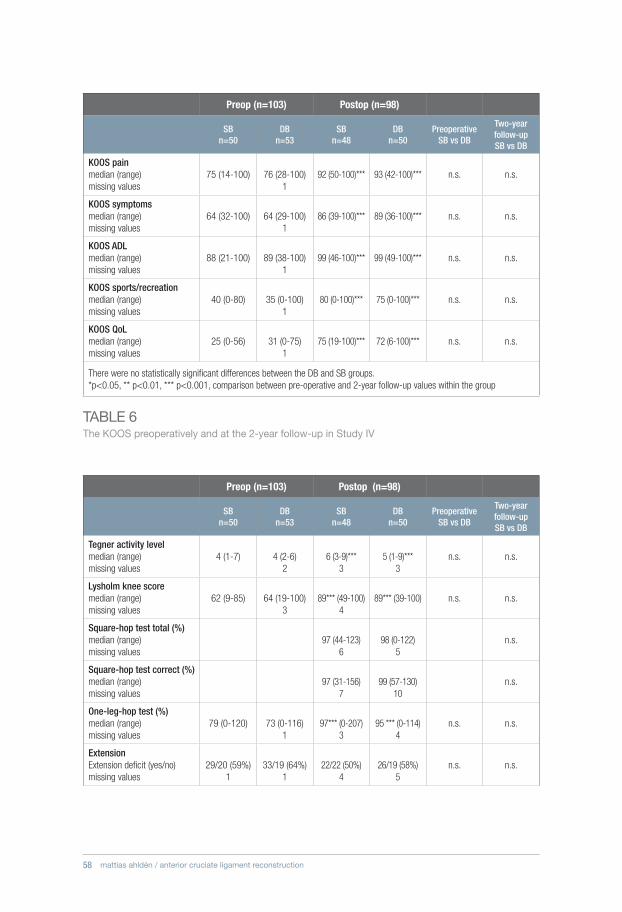
58 mattias ahldén / anterior cruciate ligament reconstruction
TABLE 6The KOOS preoperatively and at the 2-year follow-up in Study IV
Preop (n=103) Postop (n=98)
SBn=50
DBn=53
SBn=48
DBn=50
PreoperativeSB vs DB
Two-year follow-up SB vs DB
KOOS painmedian (range)missing values
75 (14-100) 76 (28-100)1
92 (50-100)*** 93 (42-100)*** n.s. n.s.
KOOS symptomsmedian (range)missing values
64 (32-100) 64 (29-100)1
86 (39-100)*** 89 (36-100)*** n.s. n.s.
KOOS ADLmedian (range)missing values
88 (21-100) 89 (38-100)1
99 (46-100)*** 99 (49-100)*** n.s. n.s.
KOOS sports/recreationmedian (range)missing values
40 (0-80) 35 (0-100)1
80 (0-100)*** 75 (0-100)*** n.s. n.s.
KOOS QoLmedian (range)missing values
25 (0-56) 31 (0-75)1
75 (19-100)*** 72 (6-100)*** n.s. n.s.
There were no statistically significant differences between the DB and SB groups.*p<0.05, ** p<0.01, *** p<0.001, comparison between pre-operative and 2-year follow-up values within the group
Preop (n=103) Postop (n=98)
SBn=50
DBn=53
SBn=48
DBn=50
PreoperativeSB vs DB
Two-year follow-up SB vs DB
Tegner activity levelmedian (range)missing values
4 (1-7) 4 (2-6)2
6 (3-9)***3
5 (1-9)***3
n.s. n.s.
Lysholm knee score median (range)missing values
62 (9-85) 64 (19-100)3
89*** (49-100)4
89*** (39-100) n.s. n.s.
Square-hop test total (%)median (range)missing values
97 (44-123)6
98 (0-122)5
n.s.
Square-hop test correct (%)median (range)missing values
97 (31-156)7
99 (57-130)10
n.s.
One-leg-hop test (%)median (range)missing values
79 (0-120) 73 (0-116)1
97*** (0-207) 3
95 *** (0-114)4
n.s. n.s.
ExtensionExtension deficit (yes/no)missing values
29/20 (59%)1
33/19 (64%)1
22/22 (50%)4
26/19 (58%)5
n.s. n.s.
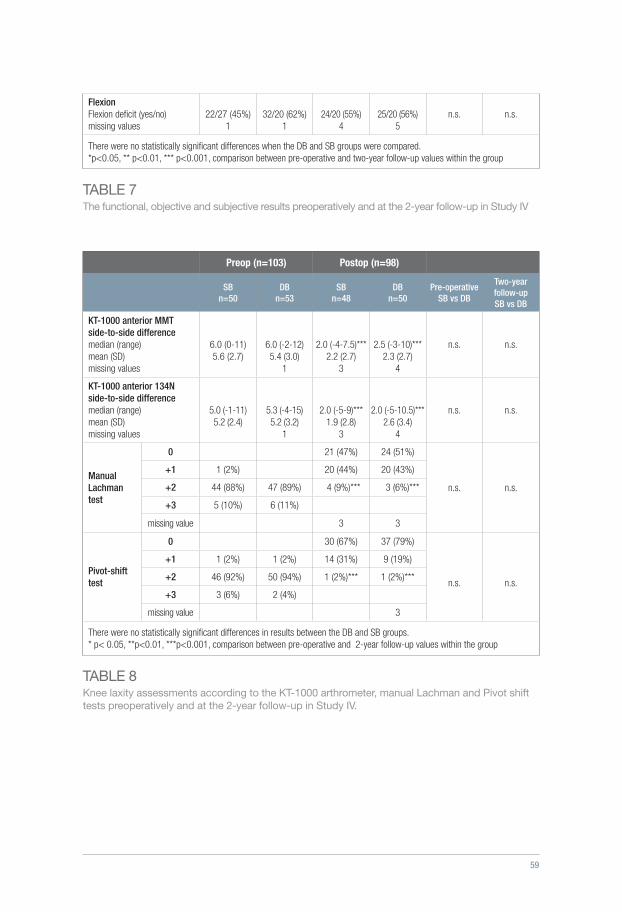
59
TABLE 7The functional, objective and subjective results preoperatively and at the 2-year follow-up in Study IV
FlexionFlexion deficit (yes/no)missing values
22/27 (45%)1
32/20 (62%)1
24/20 (55%)4
25/20 (56%)5
n.s. n.s.
There were no statistically significant differences when the DB and SB groups were compared.*p<0.05, ** p<0.01, *** p<0.001, comparison between pre-operative and two-year follow-up values within the group
TABLE 8Knee laxity assessments according to the KT-1000 arthrometer, manual Lachman and Pivot shift tests preoperatively and at the 2-year follow-up in Study IV.
Preop (n=103) Postop (n=98)
SBn=50
DB n=53
SBn=48
DBn=50
Pre-operativeSB vs DB
Two-year follow-upSB vs DB
KT-1000 anterior MMTside-to-side difference median (range)mean (SD)missing values
6.0 (0-11) 5.6 (2.7)
6.0 (-2-12)5.4 (3.0)
1
2.0 (-4-7.5)***2.2 (2.7)
3
2.5 (-3-10)***2.3 (2.7)
4
n.s. n.s.
KT-1000 anterior 134Nside-to-side difference median (range)mean (SD)missing values
5.0 (-1-11)5.2 (2.4)
5.3 (-4-15)5.2 (3.2)
1
2.0 (-5-9)***1.9 (2.8)
3
2.0 (-5-10.5)***2.6 (3.4)
4
n.s. n.s.
Manual Lachman test
0 21 (47%) 24 (51%)
n.s. n.s.
+1 1 (2%) 20 (44%) 20 (43%)
+2 44 (88%) 47 (89%) 4 (9%)*** 3 (6%)***
+3 5 (10%) 6 (11%)
missing value 3 3
Pivot-shift test
0 30 (67%) 37 (79%)
n.s. n.s.
+1 1 (2%) 1 (2%) 14 (31%) 9 (19%)
+2 46 (92%) 50 (94%) 1 (2%)*** 1 (2%)***
+3 3 (6%) 2 (4%)
missing value 3
There were no statistically significant differences in results between the DB and SB groups.* p< 0.05, **p<0.01, ***p<0.001, comparison between pre-operative and 2-year follow-up values within the group
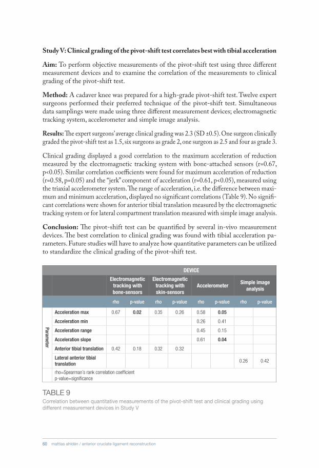
60 mattias ahldén / anterior cruciate ligament reconstruction
Study V: Clinical grading of the pivot-shift test correlates best with tibial acceleration
Aim: To perform objective measurements of the pivot-shift test using three different measurement devices and to examine the correlation of the measurements to clinical grading of the pivot-shift test.
Method: A cadaver knee was prepared for a high-grade pivot-shift test. Twelve expert surgeons performed their preferred technique of the pivot-shift test. Simultaneous data samplings were made using three different measurement devices; electromagnetic tracking system, accelerometer and simple image analysis.
Results: The expert surgeons’ average clinical grading was 2.3 (SD ±0.5). One surgeon clinically graded the pivot-shift test as 1.5, six surgeons as grade 2, one surgeon as 2.5 and four as grade 3.
Clinical grading displayed a good correlation to the maximum acceleration of reduction measured by the electromagnetic tracking system with bone-attached sensors (r=0.67, p<0.05). Similar correlation coefficients were found for maximum acceleration of reduction (r=0.58, p=0.05) and the “jerk” component of acceleration (r=0.61, p<0.05), measured using the triaxial accelerometer system. The range of acceleration, i.e. the difference between maxi-mum and minimum acceleration, displayed no significant correlations (Table 9). No signifi-cant correlations were shown for anterior tibial translation measured by the electromagnetic tracking system or for lateral compartment translation measured with simple image analysis.
Conclusion: The pivot-shift test can be quantified by several in-vivo measurement devices. The best correlation to clinical grading was found with tibial acceleration pa-rameters. Future studies will have to analyze how quantitative parameters can be utilized to standardize the clinical grading of the pivot-shift test.
TABLE 9Correlation between quantitative measurements of the pivot-shift test and clinical grading using different measurement devices in Study V
DEVICE
Electromagnetic tracking with bone-sensors
Electromagnetic tracking with skin-sensors
AccelerometerSimple image
analysis
rho p-value rho p-value rho p-value rho p-value
Acceleration max 0.67 0.02 0.35 0.26 0.58 0.05
Acceleration min 0.26 0.41
Acceleration range 0.45 0.15
Acceleration slope 0.61 0.04
Anterior tibial translation 0.42 0.18 0.32 0.32
Lateral anterior tibial translation
0.26 0.42
rho=Spearman´s rank correlation coefficient p-value=significance
Parameter

61
Study I
The strengths of Study I are the large number of patients that were included and the fact that the evaluation of the results was performed by the patients (PROM). The major limi-tation is that no clinical or radiological follow-up was performed and that several patients were lost to follow-up. In terms of the KOOS, the rate of non-responders ranged from 36% (preoperatively) to 60% (5-year follow-up), which is a major limitation. However, no selection bias in terms of gender or age when comparing responders with non-responders has been found in the register (unpublished data from the register).
Study II
The strengths of Study II are its randomized design and the long and continuous 7-year follow-up. Further strengths include the fact that the same surgeon performed all the reconstructions and the assessments were made by two observers blinded to the aim of the study. Both study groups were operated on using the same surgical technique and the same fixation method. The limitation is the small number of patients. Only patients that attended each and every one of the four postoperative follow-ups at 6 months, 1 year, 2 years and 7 years were included. The follow-up rate was 66%.
Study III
The major strength of Study III is the large number of patients. Further strengths include the fact that the same surgeon performed all the reconstructions and the assessments were made by two observers blinded to the aim of the study. Both study groups were operated on using the same surgical technique and the same fixation method. The limitation is the retrospective design and the fact that ACL revision surgery was an exclusion criterion.
Study IV
The major strength of Study IV is its randomized design, with a follow-up rate of 93%. Further strengths include the AM transportal approach to the femoral tunnels in both groups and the use of a blinded examiner.
There are some limitations. Firstly, the long time period between the injury and recon-struction might have resulted in a poorer outcome. The effect of associated injuries and psychosocial factors might have a larger impact on outcome and return to activity than different surgical techniques. It is possible that more strict inclusion criteria in terms
11 STRENGTHS AND LIMITATIONS

62 mattias ahldén / anterior cruciate ligament reconstruction
of associated injuries and time to reconstruction would have rendered different results. Secondly, an individualized approach based on factors, such as the size of the knee, might have rendered different results, but, due to the randomized design of Study IV, such an approach was not possible.
Study V
The main strength of Study V is that three different devices were used for the quantifica-tion of the pivot-shift test. Another strength is the group of 12 experts in the field of ACL reconstruction from different parts of the world performing their preferred technique.
The main limitation is that only one cadaver knee was evaluated.
63
12 DISCUSSION
12.1 Outcome measurements
12.2 AP knee laxity
When performing a clinical study, one of the most important aspects is to select a valid and reliable outcome measurement. Traditionally, clinical orthopedic studies have fo-cused on measuring outcome using objective measurements such as knee laxity, ROM and muscle strength. Impairments such as pain or swelling are also used as outcome measurements for a better evaluation of the injured or surgically treated knee. In the last two decades, PROM has been regarded as a valuable outcome tool, especially because of its focus on disability and impairment of function. PROM adds more information with regard to what is important to the patient and his/her perception of the pathology. Moreover, objective measurements have evolved to include more complex evaluations of dynamic knee laxity and enhanced visualization of the graft and tunnel position, using MRI and 3D CT reconstruction, for example. Today, a combination of objective evaluations including functional tests and PROM is usually used to evaluate the results after ACL reconstruction.
This development of outcome measurements is mirrored in the present thesis. Study II focused primarily on the evaluation of AP laxity measured with the KT-1000 arthrom-eter. In Study IV, the primary outcome measurement was the pivot-shift test, which is a dynamic test of rotatory knee laxity. Moreover, in Study V, the aim was to decompose and quantitate the pivot-shift test and correlate the findings to clinical grading in order to validate the pivot-shift test and develop a better understanding of the complicated pivot-shift phenomenon. In Study IV, the square-hop test was added as a complement to the one-leg-hop test, as it includes quick changes in direction. Moreover, in terms of PROM, the KOOS, which is a PROM validated for ACL reconstruction and a complement to the commonly used Lysholm knee scoring scale, has been included in recent studies.
One of the primary goals of ACL reconstruction is to restore knee laxity and provide the patient with a stable knee without giving-way episodes to promote long-term knee health. The manual Lachman test has been regarded as the gold standard in terms of evaluations of AP knee laxity. Because of the subjective nature of the manual Lachman test, instrumented measurement systems, such as the KT-1000 arthrometer, were devel-oped. The KT-1000 arthrometer provides a measurement of AP laxity with a standard-

64 mattias ahldén / anterior cruciate ligament reconstruction
ized force and is non-invasive and applicable in a large clinical setting. Even though its validity and reliability have been questioned, it has been widely used for evaluation after ACL reconstruction since it was introduced by Daniel et al.36. Daniel et al. stated that a side-to-side difference in AP laxity is a sensitive measurement of the integrity of the ACL and reported a difference of 2mm or less in 92% of healthy subjects and more than 2mm in 96% of ACL-injured subjects36. They also listed factors responsible for influenc-ing the measurements apart from knee laxity, such as the position of the joint, external constraints on motion (for example, different rotations), applied force and muscle tone.
In Study II, the KT-1000 arthrometer was used to evaluate AP laxity over time with comparisons between the BPTB and HS autografts. The results from Study II indicated that there were no statistically significant changes in side-to-side AP laxity over time measured with the KT-1000 arthrometer at the 7-year follow-up. However, there was a tendency for side-to-side knee laxity to decrease over time and, when the absolute values for each side were compared, there was a significant decrease in AP knee laxity over time. The reason for a possible decrease in AP knee laxity over time is unclear. Increased laxity, as a result of graft elongation or graft failure, is regarded as multifactorial and is influenced by mechanical factors, such as tunnel positioning, graft impingement, graft fixation and tensioning, graft strength, integrity of secondary stabilizers, such as combined ligament injuries or meniscal and cartilage damage. Furthermore, biological and immunological factors can also play a role in changes in knee laxity over time127. Shelbourne et al. sug-gested that a reduction in ROM, especially a lack of hyperextension, might result in lower KT-1000 values171. It is possible that the development of early degenerative changes and secondary changes in the integrity of ligamentous tissue, as well as the soft-tissue envelope, may influence laxity over time at mid- to long-term follow-ups.
The most important conclusion of Study II is that ACL reconstruction displayed no increase in AP laxity over time at the 7-year follow-up, which indicates the ability of ACL reconstruction to maintain control of AP knee laxity not only in the short term. In the literature, long-term follow-ups of AP laxity over time after ACL reconstruction are rare. Recently, Bourke et al. and Hui et al. reported the outcome after ACL reconstruc-tion using HS autografts and BPTB autografts at a 15-year follow-up21,78. Neither graft type was associated with increased AP laxity between 2 and 15 years postoperatively.
One important question is the role of measurements of AP laxity after ACL recon-struction in the future. The manual Lachman test and the KT-1000 arthrometer will definitely be used as a part of clinical assessment in the future in order to provide an evaluation of the integrity of the ACL. However, interest in the importance of rotational laxity and the pivot-shift test will certainly affect the use of AP laxity as an outcome measurement. An automated, robotic measurement of AP laxity and static rotatory laxity has been reported with the potential for more standardized and informative measure-ments of knee laxity22. On the other hand, AP laxity has been reported not to correlate with the patients’ symptoms or function99. New techniques for evaluating rotatory laxity and the pivot-shift test will probably provide a comprehensive assessment of the envelope of laxity, as described by Bull et al.30,31. Dynamic evaluations utilizing RSA or

65
DSX in high-speed functional movements such as running are able to evaluate knee kinematics and joint contact dynamics with high accuracy73,182.
12.3 Graft selection
The choice of graft is a combination of the surgeon’s preference and the individual patient’s needs and wishes. Several studies comparing BPTB and HS autografts have spotlighted the characteristics of the two grafts and will help surgeons to decide which graft to use.
In Study II, there were no differences in AP laxity when comparing BPTB and HS autografts on any follow-up occasion. This finding is in line with the systematic review by Samuelsson et al. who concluded that there are no convincing differences in AP laxity when comparing the two grafts164. However, the Cochrane report by Mohtadi et al. used data from 19 studies comprising 1,597 patients and it found less AP knee laxity when the BPTB graft was used130. Moreover, anterior knee pain and extension deficit were correlated to the use of BPTB grafts in this report. This suggests that a large number of patients are needed to reach statistically significant differences in AP laxity when BPTB and HS autografts are compared.
One important strength of Study II is its long follow-up of 7 years. Leys et al. compared HS autografts and BPTB autografts at a 15-year follow-up and reported no differences in AP laxity between the two groups in terms of KT-1000 measurements or the manual Lachman test116.
In overall terms, there appear to be no major difference in AP knee laxity when compar-ing the two grafts at short-, mid- or long-term follow-ups, which is in line with Study II.
Study II revealed no significant differences between BPTB and HS autografts in terms of OA findings classified according to the Fairbank and Ahlbäck rating systems at the 7-year follow-up, which is in line with other studies comparing the results after 5 years or more65,118. In some studies, OA has been shown to be correlated to the use of BPTB grafts116,150,160. The overall prevalence of OA in Study II was 16%, according to the Ahlbäck rating system, and 68%, according to the Fairbank rating system, which clearly demonstrates how different grading systems affect the result. The Fairbank rating system has been criticized for overestimating OA and for poor reliability142. In the review by Oiestad et al., the authors stated that previous reviews have reported too high a prevalence of OA after ACL reconstruction and suggested a prevalence of OA of 0-13% after isolated ACL injury, compared with 21-48% after combined ACL and meniscal injuries142. They also reported large variation in terms of the prevalence of OA in the included studies. One important contributory factor was the fact that different rating systems were used, which is in line with Study II.
With the development of individualized surgery, the choice of graft is even more ver-satile. Anatomic reconstruction includes the restoration of as large an area as possible
66 mattias ahldén / anterior cruciate ligament reconstruction
of the native ACL footprint. In knees with large footprints, the size of the graft is one factor to consider in order to choose the right graft.
Moreover, in revision cases with enlarged bone tunnels, the size of the graft may be important. Recently, the use of the quadriceps tendon autograft with its larger cross-sectional area has been re-popularized to ensure adequate graft material.
In contrast, the results from the Swedish National ACL register reveal an annual in-crease in the use of the HS autograft. In 2010, more than 95% of the primary ACL reconstructions in Sweden were performed using the HS autograft. Some explanations could be that there have been no major differences between BPTB and HS autografts at short- and mid-term follow-ups, the HS autograft is easier to harvest and donor-site morbidity is more common after using the BPTB autograft112,130. With the implementa-tion of individualized surgery, the use of BPTB and quadriceps tendon autografts will possibly increase in the future.
12.4 Gender differences
In terms of ACL reconstruction using the BPTB graft, most studies reveal no differences in outcome between males and females15,42,196. The effect of gender on outcome after ACL reconstruction using HS autografts has been the subject of debate61,140,162. In the National Swedish ACL Register, Ageberg et al. reported that a significantly poorer outcome was associated with female gender in terms of the KOOS and, in some age groups, these differences were clinically relevant2. Salmon et al. reported their 7-year follow-up of 143 patients who had undergone ACL reconstruction using HS autografts and interference screw fixation. The females had significantly greater knee laxity, as measured with the KT-1000 arthrometer, manual Lachman test and the pivot-shift test, but no gender-related differences were found in terms of subjective function or activity level162.
In conclusion, the current literature on the impact of gender on outcome after ACL reconstruction using HS autografts has revealed conflicting results. A recent review by Paterno et al. found indications that female patients have greater AP knee laxity follow-ing an ACL reconstruction using HS autografts compared with males146.
In Study III, a large sample size was used to examine differences in outcome after ACL reconstruction using HS autografts based on gender. The results in Study III revealed no significant differences in outcome between the genders in terms of laxity or PROM, implying that gender alone cannot play a decisive part in graft selection. One interesting question is whether there is a subgroup of females with a poorer outcome after ACL reconstruction using HS grafts compared with males. In Study I, 22% of the 15- to 18-year-old female soccer players reported a revision or contralateral ACL reconstruc-tion during a 5-year period, which was significantly more than the 9.8% reported by an age-matched male subgroup. With individualized surgery, predisposing anatomic or biological factors may affect the choice of graft, especially in females. Kim et al. reported

67
a poorer outcome after using HS autografts compared with BPTB autografts in patients with hyperlaxity, which is more frequent in females97. Hyperextension of the knee was reported as a predictor of poorer outcome in terms of both laxity and function after ACL reconstruction and the BPTB autograft was found to be a superior graft choice compared with the HS autograft98. On the other hand, long-term results with a 15-year follow-up indicate that the BPTB graft may be an inferior graft choice in terms of the development of OA, loss of ROM and presence of donor-site morbidity116. Further-more, the use of a triple ST graft with or without a double or triple gracilis graft may be an advantageous alternative, especially in patients with inherently small HS tendons. Consequently, graft choice is still the subject of debate and the future use of quadriceps tendon autografts will be interesting to follow.
12.5 Register studies
RCTs are used to study the effect of an intervention and the randomization process mini-mizes confounding factors. On the other hand, the advantage of prospective observational studies from registers is the large number of patients reported, which is usually necessary in order to identify prognostic factors, poor implants or poor procedures at an early stage. Another advantage of register studies is that several clinics and surgeons, together with wider inclusion criteria, improve the general applicability of the results. Furthermore, when studying events with low prevalence, an RCT may not be adequately powered.
Study I reports 17,794 unique registrations included in the Swedish National ACL register. Patients’ subjective evaluations in terms of the KOOS were entered in 64% of possible patients preoperatively, which is higher compared with the Danish registry but lower compared with the Norwegian registry62. At the 5-year follow-up, only 40% had completed the KOOS questionnaire in the Swedish register. One future goal should be to accomplish a better follow-up to provide reliable outcome reports. In terms of the KOOS at baseline, as well as changes over time, they are similar in overall terms to what has been reported from other Scandinavian register studies62,119.
The patients in Study I improved significantly over time in all the KOOS subscales after primary ACL reconstruction and the improvements appear to persist over time. The largest improvements were found on the subscales of QoL and Sports/Rec, which is in line with earlier studies reporting that these two subscales are the most important and the most sensitive for patients undergoing ACL reconstruction64. In comparison with reference data obtained from 118 football players with healthy knees, the patients do not achieve normal function in terms of the KOOS 1, 2 or 5 years after ACL recon-struction51. The greatest differences between the patients and the reference group were found in the subscales of Sports/Rec and QoL.
The reason why patients do not return to the same function is probably multifactorial. Associated meniscal injury is known to increase the risk of OA in the long term and
68 mattias ahldén / anterior cruciate ligament reconstruction
it has also been associated with inferior outcome in the short and mid term89, 95, 96, 111, 129,
142, 170. Cartilage injuries at the time of the primary ACL reconstruction have indicated an increased risk of developing future OA96. Moreover, in a study from the Norwegian Knee Ligament Registry, Rotterud et al. reported less clinical improvement after ACL reconstruction in patients with a full-thickness cartilage lesion compared with patients without such injuries157. At the 2-year follow-up in the study by Rotterud et al., most KOOS subscales were significantly lower in the group with cartilage injuries and the largest differences were seen in the subscales of Sports/Rec and QoL. This is in contrast to previous studies showing that cartilage lesions have no clinically relevant impact on PROM after ACL reconstruction178,195. In Study I, associated injuries to menisci and/or cartilage were analyzed together and were reported in 53.9% of the patients undergo-ing primary ACL reconstruction. The patients with associated injuries had significantly lower scores preoperatively and at 1 year in terms of most KOOS subscales compared with patients with no meniscal and/or cartilage injuries, but the differences were small. Moreover, at 5 years, a significant difference was only found in terms of the Sports/Rec subscale. One explanation for these conflicting results between the present study and previous publications may be differences in the classification and grouping of associated injuries, outcome measurements and time to follow-up. In the Swedish register, the response rate is inversely associated with a longer follow-up time, which may increase the risk of bias. Furthermore, in terms of OA, a period longer than 5 years of follow-up is probably needed.
A cross-cultural comparison between the Multicenter Orthopedic Outcomes Network (MOON) in the United States and the Norwegian National Knee Ligament Registry (NKLR) found significant differences in demographic and treatment data between the two cohorts123. Differences in age, gender, type of activity at injury, rate of surgically treated patients compared with non-surgically treated ones, time from injury to recon-struction, intra-articular injuries and graft choice were found between the two cohorts. The authors conclude that these characteristics should be taken into consideration when applying knowledge from one population to another. Moreover, this emphasizes that the general applicability of register studies is relative.
Another example of the value of register studies is the documentation of revision ACL reconstructions. Previous studies have usually comprised retrospective case series with small sample sizes of usually fewer than 50 patients55. Registers make it possible to evalu-ate and analyze large sample sizes of revisions. In the United States, MOON has created cohorts of ACL reconstructions and ACL revisions with a high rate of follow-up20. Similarly, the Swedish National ACL Register gives an excellent opportunity to study revisions. Study I comprises 964 revisions, which corresponds to 6% of the registered reconstructions between 2005 and 2010. Study I reported that revisions were associ-ated with a significantly poorer outcome in terms of all the KOOS subscales compared with primary reconstructions on all follow-up occasions. This difference was especially evident in terms of the subscales of Sports/Rec and QoL where the difference between primary and revision reconstructions was approximately 15 points at both the 1- and 5-year follow-ups. Moreover, patients undergoing revisions displayed no significant

69
improvements in terms of the subscales of Symptoms, Pain and ADL at the 5-year follow-up compared with the preoperative values.
These results emphasize the importance of providing preoperative information to pa-tients undergoing revision surgery and that revisions are associated with inferior results58.
12.6 Rotatory laxity and the pivot-shift test
Today, the clinical grading of the pivot-shift test is used in many studies to grade and report the results after an ACL reconstruction, especially when comparing DB with SB techniques, in Study IV, for example.
New technology has produced improved measurement devices for quantifying the com-plicated pivot-shift phenomenon. The question of how to decompose and characterize the complicated translational-rotational movement that comprises the pivot shift has been thoroughly investigated5,16,76,122,137. Furthermore, in order to use the devices in large clinical settings and as part of clinical follow-up, the devices need to be non-invasive. Consequently, current non-invasive devices need to be evaluated, validated and cor-related to clinical grading, which is addressed in Study V.
Another concern, in addition to quantification, is the performance of the pivot-shift test which is highly examiner dependent35,107,137,141. In Study V, twelve expert surgeons within the field of ACL reconstruction used their preferred technique. Musahl et al. character-ized the pivot-shift test performed by the surgeons in Study V137. Nine of the twelve expert surgeons used the reduction-type or flexion-type of the pivot-shift test, which is when the pivot-shift test starts with the knee extended and subluxated in internal rota-tion and progresses with flexion and grading the reduction phase from I-III, as defined by the IKDC82. The technique of these nine surgeons was then further categorized as the fixed-internal rotation type (n=5), fixed-external rotation type (n=1) or motion-allowing type (n=3), illustrating the variation in performance of the pivot-shift test. The research-ers reported variable quantitative parameters such as acceleration between different examiners. However, the variation in acceleration between examiners was significantly smaller when a standardized technique was introduced71,137. This further illustrates that the pivot-shift test poses challenges not only in terms of quantification but also in terms of performance. Moreover, the idea of standardizing the pivot-shift test is beneficial.
In Study V, three different non-invasive devices were used to quantify the pivot-shift test using the surgeons’ own preferred technique. Of the three devices; the electromagnetic tracking system is so far the most frequently reported for quantification of the pivot-shift test, whereas the accelerometer and simple-image analysis are more novel systems.
Pivot-shift test measurements in vivo using electromagnetic sensors were first reported by Bull et al. in 200230. They made measurements of knee kinematics on anesthetized patients before and after an ACL reconstruction. The most important finding was that the move-

70 mattias ahldén / anterior cruciate ligament reconstruction
ments during pivot-shift were greatly reduced by an ACL reconstruction. The authors also reported large individual variations in knee kinematics, especially in terms of internal and external rotation, during the pivot-shift test. The same research group later reported on the development of skin-mounted sensors using clamps for non-invasive measure-ment with minimal motion between bone and skin. An accuracy of approximately 1mm and 1° during a static knee laxity test was achieved. However, when these were used for more dynamic pivot-shift tests, the actual motion of the bones could be underestimated, which was a limitation of this method. A sampling rate of 30Hz was used7. Kubo et al. developed a non-invasive measurement system using a similar electromagnetic tracking device (Fastrak®, Pohlemus, Colchester, VT, USA)106. Their skin-mounted sensors were able to provide good correlation with the direct bone-fixed measurement with a correlation coefficient of 0.995 and maximum error in translation of 0.85mm during the pivot-shift test. The authors also reported a positive correlation between the clinical assessment and velocity of the pivot-shift movement. Hoshino et al. utilized a non-invasive electromag-netic tracking system with a higher sampling rate from 60 Hz up to 240Hz and further defined the kinematic characteristics of the pivot-shift test74, 76.
The main finding in Study V was that parameters of acceleration are best correlated to the clinical grading of the pivot-shift test. Previous studies have often shown correlations of anterior tibial translation to clinical grading, especially lateral compartment anterior translation. Bedi et al. reported an average of a 10mm difference in anterior translation in the lateral compartment between each of the clinical grades of the pivot-shift test16. How-ever, more dynamic parameters such as acceleration and velocity have also been reported to correlate with the clinical grading of the pivot-shift test, which is in line with Study V76,110. Hoshino et al.76 reported that both anterior translation and acceleration were cor-related with the pivot-shift test using electromagnetic tracking. Labbe et al. reported that acceleration and velocity accounted for greater differences in kinematic recordings of the pivot-shift test than the actual magnitude of displacement between the femur and tibia110.
The question remains whether several different components of the pivot-shift test have to be evaluated to quantify the test. Study V indicates that devices to measure parameters of acceleration require further evaluation. Recently, the use of the accelerometer in the quantification of the pivot-shift test has been reported by other researchers102,122. Lopomo et al. used an accelerometer and reported a probability of more than 70% of identifying an ACL injury based on acceleration parameters and good reliability122. Study V confirms that the acceleration of tibial reduction, in addition to the more traditional parameters of anterior or lateral compartment translation, should be utilized to standardize the grading of the pivot-shift test.
12.7 Double-bundle versus single-bundle reconstruction
Study IV, with a sample size of 105 patients, compared DB with SB reconstruction and did not reveal any statistically significant differences in outcome between the two techniques.
71
The comparison of DB versus SB reconstructions has been a very important topic in ACL research over the last couple of years. More than 15 RCTs have compared the DB with the SB technique and several meta-analyses have been performed 1,3,9,79,80,85-87,133,165,175,179,181,191,194,199,204,205,100,117,128,185,188.
In their recent RCT comprising 238 patients, Hussein et al. reported that the anatomic DB technique was superior to both the anatomic SB and conventional SB techniques79. However, the differences were more evident when comparing anatomic DB with the conventional SB technique and significant differences were reported in terms of AP laxity, the pivot-shift test, Lysholm knee scoring scale and subjective IKDC. When comparing anatomic DB with anatomic SB, significant differences were found in terms of AP laxity, the pivot-shift test and ROM. The pivot-shift test was negative in 66.7% of the cases in the anatomic SB group and in 93.1% in the DB group. Their conclusion was that it is important to perform an ACL reconstruction anatomically and that the DB technique can improve the control of rotatory laxity. In Study IV, 67% of the SB group and 79% of the DB group had a negative pivot-shift test at follow-up, but this difference was not statistically significant. Moreover, in Study IV, there were no differ-ences in PROM, which is in line with Hussein et al.79.
The first meta-analysis comparing DB with SB reconstruction was performed by Meredick et al. Their meta-analysis included 4 RCTs and revealed a difference in AP laxity of 0.52 mm in favor of DB but no difference in terms of the pivot-shift test128. However, this meta-analysis has been criticized for placing normal and nearly normal results of the pivot-shift test in the same group, which may affect the results81.
The systematic review by Samuelsson et al. from 2009 including 8 RCTs reported that DB reconstruction yields less rotatory laxity, but it raised the question of whether the surgical technique for the SB reconstructions in some studies was performed in a less anatomic manner than the DB reconstructions. No differences in clinical outcome were found between the two techniques and, in terms of AP laxity, the results were inconclusive164.
The systematic review by Kongtharvonskul et al. from 2012 reported findings similar to those of Samuelsson et al.100. The authors included 9 RCTs with pivot-shift test data and reported that DB reconstructions more frequently had a normal pivot-shift test. There were no significant differences in terms of AP laxity, IKDC score, Lysholm knee scoring scale and Tegner activity scale.
Moreover, in 2012, Li et al. reported in their meta-analysis less rotatory and AP laxity in DB reconstruction compared with SB reconstruction117. Thirteen studies with a total of 985 patients analyzing the pivot-shift test were included and the authors reported that DB reconstruction had a normal pivot-shift test more frequently. Eleven studies with a total of 794 patients were included to analyze AP laxity using the KT-1000 ar-thrometer, which revealed that the DB reconstructions displayed less laxity. Moreover, the study reported a higher subjective IKDC score and less knee extension deficit in DB reconstruction but no significant differences in terms of objective IKDC score or Lysholm knee scoring scale.
72 mattias ahldén / anterior cruciate ligament reconstruction
Recently, a Cochrane review of DB versus SB reconstruction included 17 studies involv-ing 1,433 patients185. Twelve of the 17 studies were RCTs and 5 were quasi-randomized RCTs. Data for individual outcomes were available for a maximum of nine trials and 54% of the participants. They found no statistically or clinically significant differences between the two groups in terms of PROM. Knee laxity, in terms of both AP laxity measured with the KT-1000 arthrometer and rotatory laxity measured using the pivot-shift test, revealed significant differences between the two techniques at two to five years of follow-up in favor of the DB technique. The Cochrane review concluded that there is limited evidence that DB reconstruction renders superior results for knee stability and protection from repeat ACL rupture and subsequent meniscal injury. Interestingly, the Cochrane review was also able to identify 9 ongoing RCTs comparing the DB with the SB technique but with no results as yet.
The Cochrane review did not consider placement of the tunnels in the different stud-ies, i.e. whether or not the reconstruction can be classified as anatomic. Obviously, the classification into anatomic or non-anatomic is challenging. In their meta-analysis, van Eck et al. included twelve studies with a total of 1,127 patients188. Five of the included studies were RCTs. Van Eck et al. also sub-classified the studies as anatomic or not using the researchers’ description of their surgical technique. In line with the Cochrane review, van Eck et al. reported that knee laxity measured with KT-1000 arthrometer and rotatory laxity measured with the pivot-shift test were superior in the DB group. This difference was also significant in the subgroup of anatomic reconstructions but not in the subgroup of non-anatomic reconstructions. However, the non-anatomic subgroup consisted of five studies of which only one was an RCT.
One problem when assessing studies of different surgical techniques, such as DB versus SB reconstructions, is the difficulty involved in assessing the extent to which the recon-structions are anatomic or not. Recently, improved reporting of the surgical technique in ACL reconstruction studies has been suggested, such as providing intraoperative pictures or performing postoperative CT scans to document tunnel locations. The Ana-tomic ACL Reconstruction Scoring System (AARSS) has been developed by van Eck et al. to help characterize reconstructions as anatomic or not163. Furthermore, the aim of the AARSS is also to improve the overall reporting of surgical data in studies of ACL reconstruction. One of the items in the AARS is documentation of tunnel position, with the highest score for 3D visualization, by CT, for example, and a lower score for 2D documentation with arthroscopic pictures or standard radiographic evaluation. In Study IV, the ACL reconstructions were performed in 2008 and 2009 and, as a result, documentation on tunnel location utilizing CT was not routinely kept.
In conclusion, DB in clinical studies appears to yield less knee laxity, especially in terms of rotatory laxity measured with the pivot-shift test. In Study IV, the pivot-shift test was normal in 79% of the DB group and 67% of the SB group at the 2-year follow-up, which was a non-significant difference. Considering the results from earlier meta-analyses and systematic reviews, however, the question of whether this difference is relevant is an intriguing thought, even if it did not reach statistical significance.
73
In Study IV, there were no significant differences between DB and SB reconstruction in terms of the Lysholm knee scoring scale and the KOOS, which is in line with most previous meta-analyses and systematic reviews100,164,185. Moreover, no patients in Study IV underwent revision reconstruction during the follow-up period. However, the true number of possible graft failures is not known as this is a parameter that is difficult to define and definitions differ between studies. In Study IV, MRI was not performed as part of the follow-up; it is probably needed to define graft failure in addition to the laxity testing. Suomalainen et al. reported 5-year results of DB and SB reconstructions in 90 patients in three groups and found that the DB technique rendered fewer graft failures and subsequently fewer revision reconstructions180. In line with Suomalainen et al., the Cochrane review also found some limited evidence that DB reconstruction is associated with fewer re-ruptures.
On the other hand, a study from the Danish Knee Ligament Registry indicated that the change in Denmark from transtibial to transportal drilling was associated with a higher re-rupture rate153. One current theory is that anatomical reconstruction is associated with a higher re-rupture rate due to the fact that an anatomically placed graft has to withstand greater forces compared with a non-anatomically placed graft198. However, to date, there are insufficient data to draw any conclusion from the current literature regarding differ-ences in re-rupture rate between anatomic and non-anatomic reconstructions and DB and SB reconstructions respectively.
In Study IV, the return to pre-injury Tegner activity level was 26% for SB and 20% for DB at the 2-year follow-up. One possible explanation is the long time between injury and reconstruction, in combination with a high pre-injury Tegner activity level of a median of 8 in both groups. Jerre et al. reported that the Tegner activity level at follow-up compared with the pre-injury Tegner level had decreased less in recreational athletes compared with competitive athletes88. In a meta-analysis, Ardern et al. reported that 63% had returned to their pre-injury level of activity and 44% had returned to competitive sports11. In two studies, Ardern et al. reported that, at the 1-year follow-up, 33% of the patients had returned to competitive sports and, 2 to 7 years after reconstruction, 45% were playing sport at their pre-injury level and 29% were playing competitive sport10, 12. Kvist et al. reported reasons for not returning to sports after ACL reconstruction109. In their review, only 36% of the patients did not return to sports due to their knee func-tion alone. It is reasonable to suppose that psychosocial factors matter greatly when a competitive athlete undergoes ACL reconstruction a long time after the index injury. This, however, reflects the health-care system in Sweden, where many patients usually undergo a conservative regimen before ACL reconstruction is considered and there is usually a delay in time before contact with an orthopedic surgeon who performs ACL reconstructions is established. A recent review by Feller et al. also put forward the need for precise and consistent terminology for reporting return to previous level of activity41. For example, there will probably be different results when reporting the level of activity at a certain time to follow-up compared with the highest level of activity at any time during a certain time interval.
74 mattias ahldén / anterior cruciate ligament reconstruction
These facts raise the question of whether, in Study IV, factors such as time to reconstruc-tion, associated injuries and psychosocial factors may have an equally large impact on the outcome as different surgical techniques, such as DB or SB reconstruction. Furthermore, in the randomized setting of Study IV, an individualized approach was not possible. As a result, the patients in Study IV will take part in additional mid-and long-term follow-ups.
12.8 General discussion
In the care of the ACL-injured patient, optimizing the surgical procedure is essential, but factors such as the timing of intervention and adequate follow-up may be equally important. In Sweden, factors to improve the care of the ACL-injured patient should include early diagnosis and early treatment selection in order to minimize patient or doctor delay. Early treatment selection should include information, structured rehabili-tation and a decision to proceed to early reconstruction or non-surgical treatment. If a non-surgical regimen is chosen, adequate information and follow-up are mandatory in order to ensure that activity modification is applied. Moreover, the patient should not suffer from recurrent giving-way episodes that warrant a delayed reconstruction50.
Delayed diagnosis, delayed treatment selection and inadequate follow-up will undoubt-edly result in more patients with recurrent episodes of giving-way with additional as-sociated injuries and increased laxity in secondary restraints. Ultimately, these factors may have a negative impact on the outcome in both the short and long term, including an increased risk of OA. If an ACL reconstruction is indicated, an early reconstruction is most probably beneficial. The greatest challenge is to use an individualized approach to identify copers and non-copers as early as possible. The clinically performed pivot-shift test has been reported to correlate with coping104. The improved identification of copers may include objective and standardized quantification of the pivot-shift test.
Furthermore, an individualized approach includes different surgical techniques for ACL reconstruction. An anatomic DB concept should be used, which does not necessarily include DB reconstruction but nonetheless a thorough understanding of the anatomy and biomechanics of the ACL. The DB concept may include a subgroup of patients who would benefit from DB reconstruction.
Improved outcome measurements in terms of evaluating the important pivot-shift test, rotatory laxity and functional kinematics, together with more sensitive PROM, may provide prognostic factors for a good or poor outcome. Ultimately, these outcome meas-urements may provide indications for surgical techniques such as DB reconstruction. However, it is clear that the anatomic DB concept has made a major contribution to the overall knowledge in the field of ACL reconstruction and has raised important ques-tions with regard to surgical techniques in SB reconstruction. Moreover, the improved understanding and evaluation of footprint anatomy has had an impact on the com-prehension of partial ACL ruptures. The DB concept provides knowledge to perform isolated AM- or PL-bundle reconstructions and use remnant-preservative techniques.

75
13 CONCLUSIONS
Patients undergoing primary ACL reconstruction improve significantly on all the subscales of the KOOS.
Patients undergoing revision ACL reconstruction do less well in terms of the KOOS compared with patients undergoing primary ACL reconstruction.
After an ACL reconstruction, young female soccer players re-injure their ACL or the contralateral ACL within 5 years to a higher extent than corresponding young males or ACL-reconstructed patients overall.
Smokers do less well than non-smokers after ACL reconstruction.
There are no significant differences in terms of AP laxity measurements or radiographic OA findings between patients operated on using BPTB or HS autografts at 7 years after ACL reconstruction.
A decrease in AP laxity over time was seen at the 7-year follow-up for both BPTB and HS autografts, but, when measuring side-to-side laxity, this difference was not significant.
There were no significant differences between male and female patients in terms of clini-cal outcome or functional scores 2 years after ACL reconstruction using HS autografts.
Subjective and objective outcomes revealed no significant differences between the DB and SB techniques at 2 years after ACL reconstruction in an unselected group of patients using HS autografts.
The best correlation to clinical grading of the pivot-shift test was found with tibial acceleration parameters.
76 mattias ahldén / anterior cruciate ligament reconstruction
The present thesis has enlarged our insight into the outcome after ACL reconstruction at short- and mid-term follow-ups using register data and RCTs. Moreover, the important evaluation of rotatory laxity, i.e. the pivot-shift test, has been studied in an experimental cadaver study. However, despite more than 10,000 scientific publications on ACL-related subjects, we still do not have a definite answer to many important questions with regard to the development of OA, outcome and a return to the previous level of activity. There is still a long way to go before the treatment of the ACL-injured patient is optimized on the backbone of good evidence. We know from many cohort studies that, in overall terms, ACL reconstruction displays good results in terms of reducing AP and rotatory laxity and improving PROM and activity level. The concept of Evidence-Based Medicine (EBM) has for ever changed modern orthopedics both in the clinical setting and in research. EBM was chosen as one of the top 15 medical breakthroughs in the last 160 years in an international survey performed by the British Medical Journal. The impact of EBM on how to perform studies and critically evaluate results has improved our awareness of possible bias in overall terms. As often proposed, the goal should be studies with a high level of evidence. However, one equally important issue is the outcome measurements that are used. The standardization and quantification of the pivot-shift test, kinematic evaluation during functional activity, development of more valid PROM and quantitative and qualitative assessments of cartilage are among the great challenges when it comes to improving the assessment after ACL reconstruction. Furthermore, with the standardiza-tion of outcome measurements, the quality of meta-analyses and systematic reviews will be improved and multi-center studies can be performed.
The concept of individualized treatment and anatomic reconstruction is a major step forward and makes the field of ACL reconstruction even more complex and versatile in terms of both research and clinical work. In the future, more knowledge will be acquired in terms of individual predictors of control of laxity, coping and the development of OA. Ultimately, these predictors will guide the individualized treatment to improve the care of the patient. Individualized footprint surgery is advancing and will be evaluated in more studies in terms of laxity, kinematics, graft rupture and OA. Moreover, individual-ized treatment should include the postoperative rehabilitation of the patient. A return to activity may be guided individually, based not only on time after reconstruction and performance tests. Already today, MRI is able to provide information on the maturation of the graft and qualitative assessments of the cartilage, which could have implications for a return to activity.
Furthermore, interesting advances in surgical techniques for ACL reconstruction include remnant-preservative techniques and the use of biological enhancements such as an
14 FINAL CONSIDERATIONS AND THE FUTURE

77
autologous fibrin clot or platelet-rich plasma (PRP). Even the healing and primary repair of the ACL is discussed as a result of these biological advances.
However, when discussing these general advances in ACL reconstruction, we must be aware of the differences between different populations. In the USA, the expanding cost of the health-care system is one major issue. In Sweden, the health-care system has to deal with problems of productivity and the fact that delays for patients may result in an inferior outcome. These differences have major implications when comparing studies and optimizing treatment in different countries. However, most important in the search for the best available care for the ACL-injured patient is to remain open-minded and to critically evaluate our own results.

78 mattias ahldén / anterior cruciate ligament reconstruction
This thesis and my research in general have benefitted from assistance and encouragement of several people to whom I would like to express my deepest gratitude.
Ninni Sernert, Associate Professor, RPT, PhD, my main supervisor during the work on this thesis. Thank you for your never-ending support, help and immense knowledge of all the important details.
Jüri Kartus, Professor, MD, PhD, my co-supervisor. For sharing all your knowledge of research and life with never-failing enthusiasm and for providing the backbone of this thesis.
Jón Karlsson, Professor, MD, PhD, my co-supervisor. For endless support, encour-agement and professional guidance in both research and clinic. You always help people around you to evolve, including myself.
Leif Swärd, Associate Professor, MD, PhD, my clinical mentor, who educated me with never-ending faith in my abilities and introduced me at an early stage to surgery of the knee as well as hip arthroscopy.
Lars Ejerhed, Associate Professor, MD, PhD, my co-supervisor. For help and guidance.
Freddie H. Fu, Professor, MD, DPs, DSc. For opening the door to the Department of Orthopaedic Surgery in Pittsburgh in terms of both research and clinic. Thank you for sharing your immense knowledge and inspiration in the field of ACL research.
Volker Musahl, Associate Professor, MD. For your friendly hospitality and for including me in your research projects. Also, thanks for the hard but good training sessions at 5 am.
James J. Irrgang, Professor, PT, PhD, ATC, FAPTA. For hospitality and help in research and preparing manuscripts.
Yuichi Hoshino, MD, PhD. For including me in your research and teaching me about the pivot-shift test.
Paulo Araujo, MD. For fantastic hospitality and collaboration and letting me stay at your house.
Carola F. van Eck, MD, PhD. For hospitality and all your help in getting introduced in Pittsburgh.
Zbigniew K. Wszolek, Professor, MD. For unmatched support and help in all matters, from orthopedic trauma to visa issues.
Mary O´Connor, Professor, MD. For opening the door to the Department of Or-thopedic surgery, Mayo Clinic, Jacksonville, Florida.
15 ACKNOWLEDGEMENTS

79
Magnus Forssblad, MD, PhD, coauthor. For help with the study of the Swedish National ACL Register.
Mikael Sansone, MD, colleague and friend. For good times and friendship. Thank you for great collaboration, which I hope we can expand even more.
Nicklas Olsson, MD, colleague and friend. For fun days at work and good friend-ship. I look forward to future collaboration.
Kristian Samuelsson, MD, PhD, colleague and friend. For being a good friend and providing sharp insights into work and life. I look forward to enriching our col-laboration in the future.
Klas Halldin, MD, PhD, colleague and my oldest and dearest friend, for many laughs since the second day of med school.
All colleagues and friends at the Department of Orthopedics, Sahlgrenska Uni-versity Hospital/Mölndal and Orthocenter/IFK-kliniken.
The Gran Canaria Research Group, for productive collaboration in friendly environments.
Helena Brisby, Professor, MD, PhD, chairman of the Department of Orthopedics, Sahlgrenska Academy, for giving me the opportunity to do research.
Magnus Karlsson, MD, head of the Department of Orthopedics, Sahlgrenska Uni-versity Hospital, for giving me the opportunity to combine research with clinical work.
Annika Samuelsson, for excellent work on the layout of this thesis.
Catarina Kartus, for superb illustrations.
Pontus Andersson, for an excellent cover illustration.
Linda Johansson, administrator, for academic guidance and knowledge in all matters relating to the Sahlgrenska Academy.
Jeanette Kliger, for excellent editing of the English language in this thesis.
My parents, Gunnar and Birgitta, and sister, Frida. For love. Where and who would I be without your support?
Jonas, for encouragement and support.
Adam, my beloved son, who opened my eyes with mutual and unconditional love and everyday brings a meaning to my life and this thesis.
Christina, my beloved life partner, who with endless passion, support and care always stands out as an inspiration to me.

80 mattias ahldén / anterior cruciate ligament reconstruction
1. Adachi N, Ochi M, Uchio Y, Iwasa J, Kuriwaka M, Ito Y. Reconstruction of the anterior cruciate ligament. Single- versus double-bundle multistranded hamstring tendons. J Bone Joint Surg Br. 2004;86(4):515-520.
2. Ageberg E, Forssblad M, Herbertsson P, Roos EM. Sex differences in patient-reported outcomes after anterior cruciate ligament reconstruction: data from the Swedish knee ligament register. Am J Sports Med. 2010;38(7):1334-1342.
3. Aglietti P, Giron F, Losco M, Cuomo P, Ciardullo A, Mondanelli N. Comparison between single-and double-bundle anterior cruciate ligament reconstruction: a prospective, randomized, single-blinded clinical trial. Am J Sports Med. 2010;38(1):25-34.
4. Ahlback S. Osteoarthrosis of the knee. A radiographic investigation. Acta Radiol Diagn (Stockh). 1968:Suppl 277:7-72.
5. Ahlden M, Hoshino Y, Samuelsson K, Araujo P, Musahl V, Karlsson J. Dynamic knee laxity measurement devices. Knee Surg Sports Traumatol Arthrosc. 2012;20(4):621-632.
6. Amis AA. The functions of the fibre bundles of the anterior cruciate ligament in anterior drawer, rotational laxity and the pivot shift. Knee Surg Sports Traumatol Arthrosc. 2012;20(4):613-620.
7. Amis AA, Cuomo P, Rama RBS, et al. Measurement of knee laxity and pivot-shift kinematics with magnetic sensors. Oper Tech Orthop. 2008;18(3):196-203.
8. Amis AA, Dawkins GP. Functional anatomy of the anterior cruciate ligament. Fibre bundle actions related to ligament replacements and injuries. J Bone Joint Surg Br. 1991;73(2):260-267.
9. Araki D, Kuroda R, Kubo S, et al. A prospective randomised study of anatomical single-bundle versus double-bundle anterior cruciate ligament reconstruction: quantitative evaluation using an electromagnetic measurement system. Int Orthop. 2011;35(3):439-446.
10. Ardern CL, Taylor NF, Feller JA, Webster KE. Return-to-sport outcomes at 2 to 7 years after anterior cruciate ligament reconstruction surgery. Am J Sports Med. 2012 ;40(1):41-8.
11. Ardern CL, Webster KE, Taylor NF, Feller JA. Return to sport following anterior cruciate ligament reconstruction surgery: a systematic review and meta-analysis of the state of play. Br J Sports Med. 2011;45(7):596-606.
12. Ardern CL, Webster KE, Taylor NF, Feller JA. Return to the preinjury level of competitive sport after anterior cruciate ligament reconstruction surgery: two-thirds of patients have not returned by 12 months after surgery. Am J Sports Med. 2011;39(3):538-543.
13. Arnold MP, Kooloos J, van Kampen A. Single-incision technique misses the anatomical femoral anterior cruciate ligament insertion: a cadaver study. Knee Surg Sports Traumatol Arthrosc. 2001;9(4):194-199.
14. Ayeni OR, Chahal M, Tran MN, Sprague S. Pivot shift as an outcome measure for ACL reconstruction: a systematic review. Knee Surg Sports Traumatol Arthrosc. 2012;20(4):767-777.
15. Barber-Westin SD, Noyes FR, Andrews M. A rigorous comparison between the sexes of results and complications after anterior cruciate ligament reconstruction. Am J Sports Med. 1997;25(4):514-526.
16. Bedi A, Musahl V, Lane C, Citak M, Warren RF, Pearle AD. Lateral compartment translation predicts the grade of pivot shift: a cadaveric and clinical analysis. Knee Surg Sports Traumatol Arthrosc. 2010;18(9):1269-1276.
17. Bernard M, Hertel P. [Intraoperative and postoperative insertion control of anterior cruciate ligament-plasty.
16 REFERENCES

81
A radiologic measuring method (quadrant method)]. Der Unfallchirurg. 1996;99(5):332-340.
18. Bicer EK, Lustig S, Servien E, Selmi TA, Neyret P. Current knowledge in the anatomy of the human anterior cruciate ligament. Knee Surg Sports Traumatol Arthrosc. 2010;18(8):1075-1084.
19. Bignozzi S, Zaffagnini S, Lopomo N, Fu FH, Irrgang JJ, Marcacci M. Clinical relevance of static and dynamic tests after anatomical double-bundle ACL reconstruction. Knee Surg Sports Traumatol Arthrosc. 2010;18(1):37-42.
20. Borchers JR, Kaeding CC, Pedroza AD, Huston LJ, Spindler KP, Wright RW. Intra-articular findings in primary and revision anterior cruciate ligament reconstruction surgery: a comparison of the MOON and MARS study groups. Am J Sports Med. 2011;39(9):1889-1893.
21. Bourke HE, Gordon DJ, Salmon LJ, Waller A, Linklater J, Pinczewski LA. The outcome at 15 years of endoscopic anterior cruciate ligament reconstruction using hamstring tendon autograft for ‘isolated’ anterior cruciate ligament rupture. J Bone Joint Surg Br 2012;94(5):630-637.
22. Branch TP, Mayr HO, Browne JE, Campbell JC, Stoehr A, Jacobs CA. Instrumented examination of anterior cruciate ligament injuries: minimizing flaws of the manual clinical examination. Arthroscopy. 2010;26(7):997-1004.
23. Brandon ML, Haynes PT, Bonamo JR, Flynn MI, Barrett GR, Sherman MF. The association between posterior-inferior tibial slope and anterior cruciate ligament insufficiency. Arthroscopy. 2006;22(8):894-899.
24. Brandsson S, Faxen E, Eriksson BI, Sward L, Lundin O, Karlsson J. Reconstruction of the anterior cruciate liga-ment: comparison of outside-in and all-inside techniques. Br J Sports Med. 1999;33(1):42-45.
25. Brandsson S, Karlsson J, Sward L, Kartus J, Eriksson BI, Karrholm J. Kinematics and laxity of the knee joint after anterior cruciate ligament reconstruction: pre- and postoperative radiostereometric studies. Am J Sports Med. 2002;30(3):361-367.
26. Briggs KK, Lysholm J, Tegner Y, Rodkey WG, Kocher MS, Steadman JR. The reliability, validity, and responsiveness of the Lysholm score and Tegner activity scale for anterior cruciate ligament injuries of the knee: 25 years later. Am J Sports Med. 2009;37(5):890-897.
27. Brooks R. EuroQol: the current state of play. Health Policy. 1996;37(1):53-72.
28. Brophy RH, Wright RW, Matava MJ. Cost analysis of converting from single-bundle to double-bundle anterior cruciate ligament reconstruction. Am J Sports Med. 2009;37(4):683-687.
29. Brosseau L, Balmer S, Tousignant M, et al. Intra- and intertester reliability and criterion validity of the parallelo-gram and universal goniometers for measuring maximum active knee flexion and extension of patients with knee restrictions. Arch Phys Med Rehabil. 2001;82(3):396-402.
30. Bull AM, Earnshaw PH, Smith A, Katchburian MV, Hassan AN, Amis AA. Intraoperative measurement of knee kinematics in reconstruction of the anterior cruciate ligament. J Bone Joint Surg Br. 2002;84(7):1075-1081.
31. Bull AMJ, Amis AA. The pivot-shift phenomenon: A clinical and biomechanical perspective. The Knee. 1998;5(3):141-158.
32. Burstrom K, Johannesson M, Diderichsen F. Swedish population health-related quality of life results using the EQ-5D. Qual Life Res. 2001;10(7):621-635.
33. Cohen SB, VanBeek C, Starman JS, Armfield D, Irrgang JJ, Fu FH. MRI measurement of the 2 bundles of the normal anterior cruciate ligament. Orthopedics. 2009;32(9).
34. Dandy DJ. Arthroscopic surgery of the knee. Br J Hosp Med. 1982;27(4):360, 362, 365.
35. Daniel DM. Assessing the limits of knee motion. Am J Sports Med. 1991;19(2):139-147.
36. Daniel DM, Stone ML, Sachs R, Malcom L. Instrumented measurement of anterior knee laxity in patients with acute anterior cruciate ligament disruption. Am J Sports Med. 1985;13(6):401-407.

82 mattias ahldén / anterior cruciate ligament reconstruction
37. Dhillon MS, Bali K, Vasistha RK. Immunohistological evaluation of proprioceptive potential of the residual stump of injured anterior cruciate ligaments (ACL). Int Orthop. 2010;34(5):737-741.
38. Edwards A, Bull AM, Amis AA. The attachments of the anteromedial and posterolateral fibre bundles of the an-terior cruciate ligament: Part 1: tibial attachment. Knee Surg Sports Traumatol Arthrosc. 2007;15(12):1414-1421.
39. Edwards A, Bull AM, Amis AA. The attachments of the anteromedial and posterolateral fibre bundles of the anterior cruciate ligament. Part 2: femoral attachment. Knee Surg Sports Traumatol Arthrosc. 2008;16(1):29-36.
40. Fairbank. Knee joint changes after meniscectomy. J Bone Joint 1948;30B(4):7.
41. Feller J, Webster KE. Return to sport following anterior cruciate ligament reconstruction. Int Orthop. 2013;37(2):285-290.
42. Ferrari JD, Bach BR, Jr., Bush-Joseph CA, Wang T, Bojchuk J. Anterior cruciate ligament reconstruction in men and women: An outcome analysis comparing gender. Arthroscopy. 2001;17(6):588-596.
43. Ferretti M, Ekdahl M, Shen W, Fu FH. Osseous landmarks of the femoral attachment of the anterior cruciate ligament: an anatomic study. Arthroscopy. 2007;23(11):1218-1225.
44. Fiebert I, Gresley J, Hoffman S, Kunkel K. Comparative measurements of anterior tibial translation using the KT-1000 knee arthrometer with the leg in neutral, internal rotation, and external rotation. J Orthop Sports Phys Ther. 1994;19(6):331-334.
45. Friel NA, Chu CR. The role of ACL injury in the development of posttraumatic knee osteoarthritis. Clin Sport Med. 2013;32(1):1-12.
46. Frobell RB, Lohmander LS, Roos HP. Acute rotational trauma to the knee: poor agreement between clinical as-sessment and magnetic resonance imaging findings. Scand J Med Sci Sports. 2007;17(2):109-114.
47. Frobell RB, Roos EM, Roos HP, Ranstam J, Lohmander LS. A randomized trial of treatment for acute anterior cruciate ligament tears. N Engl J Med. 2010;363(4):331-342.
48. Frobell RB, Roos HP, Roos EM, et al. The acutely ACL injured knee assessed by MRI: are large volume traumatic bone marrow lesions a sign of severe compression injury? Osteoarthritis Cartilage. 2008;16(7):829-836.
49. Frobell RB, Roos HP, Roos EM, Roemer FW, Ranstam J, Lohmander LS. Treatment for acute anterior cruciate ligament tear: five year outcome of randomised trial. BMJ. 2013;346:f232.
50. Frobell RB, Roos HP, Roos EM, Roemer FW, Ranstam J, Lohmander LS. Treatment for acute anterior cruciate ligament tear: five year outcome of randomised trial. BMJ. 2013;346:f232.
51. Frobell RB, Svensson E, Gothrick M, Roos EM. Self-reported activity level and knee function in amateur football players: the influence of age, gender, history of knee injury and level of competition. Knee Surg Sports Traumatol Arthrosc. 2008;16(7):713-719.
52. Fu FH, Shen W, Starman JS, Okeke N, Irrgang JJ. Primary anatomic double-bundle anterior cruciate ligament reconstruction: a preliminary 2-year prospective study. Am J Sports Med. 2008;36(7):1263-1274.
53. Fujita N, Kuroda R, Matsumoto T, et al. Comparison of the clinical outcome of double-bundle, anteromedial single-bundle, and posterolateral single-bundle anterior cruciate ligament reconstruction using hamstring tendon graft with minimum 2-year follow-up. Arthroscopy. 2011;27(7):906-913.
54. Gabriel MT, Wong EK, Woo SLY, Yagi M, Debski RE. Distribution of in situ forces in the anterior cruciate liga-ment in response to rotatory loads. J Orthop Res. 2004;22(1):85-89.
55. George MS, Dunn WR, Spindler KP. Current concepts review: revision anterior cruciate ligament reconstruction. Am J Sports Med. 2006;34(12):2026-2037.
83
56. Georgoulis A, Ristanis S, Moraiti C, Mitsou A, Bernard M, Stergiou N. Three-dimensional kinematics of the tibiofemoral joint in ACL-deficient and reconstructed patients shows increased tibial rotation. Oper Tech Orthop. 2005;15(1):49-56.
57. Gerich TG, Lattermann C, Fremerey RW, Zeichen J, Lobenhoffer HP. One- versus two-incision technique for anterior cruciate ligament reconstruction with patellar tendon graft. Results on early rehabilitation and stability. Knee Surg Sports Traumatol Arthrosc. 1997;5(4):213-216.
58. Gifstad T, Drogset JO, Viset A, Grontvedt T, Hortemo GS. Inferior results after revision ACL reconstructions: a comparison with primary ACL reconstructions. Knee Surg Sports Traumatol Arthrosc. 2012 [Epub ahead of print] doi:23238924
59. Gillquist J, Messner K. Anterior cruciate ligament reconstruction and the long-term incidence of gonarthrosis. Sports Med. 1999;27(3):143-156.
60. Girgis FG, Marshall JL, Monajem A. The cruciate ligaments of the knee joint. Anatomical, functional and experi-mental analysis. Clin Orthop Rel Res. 1975(106):216-231.
61. Gobbi A, Domzalski M, Pascual J. Comparison of anterior cruciate ligament reconstruction in male and female athletes using the patellar tendon and hamstring autografts. Knee Surg Sports Traumatol Arthrosc. 2004;12(6):534-539.
62. Granan LP, Forssblad M, Lind M, Engebretsen L. The Scandinavian ACL registries 2004-2007: baseline epide-miology. Acta Orthop. 2009;80(5):563-567.
63. Grood ES, Suntay WJ. A joint coordinate system for the clinical description of three-dimensional motions: application to the knee. J Biomech Eng. 1983;105(2):136-144.
64. Hambly K, Griva K. IKDC or KOOS: which one captures symptoms and disabilities most important to patients who have undergone initial anterior cruciate ligament reconstruction? Am J Sports Med. 2010;38(7):1395-1404.
65. Harilainen A, Linko E, Sandelin J. Randomized prospective study of ACL reconstruction with interference screw fixation in patellar tendon autografts versus femoral metal plate suspension and tibial post fixation in hamstring tendon autografts: 5-year clinical and radiological follow-up results. Knee Surg Sports Traumatol Arthrosc. 2006;14(6):517-528.
66. Harilainen A, Sandelin J. Post-operative use of knee brace in bone-tendon-bone patellar tendon anterior cruciate ligament reconstruction: 5-year follow-up results of a randomized prospective study. Scand J Med Sci Sports. 2006;16(1):14-18.
67. Harner CD, Baek GH, Vogrin TM, Carlin GJ, Kashiwaguchi S, Woo SL. Quantitative analysis of human cruciate ligament insertions. Arthroscopy. 1999;15(7):741-749.
68. Harner CD, Marks PH, Fu FH, Irrgang JJ, Silby MB, Mengato R. Anterior cruciate ligament reconstruction: endoscopic versus two-incision technique. Arthroscopy. 1994;10(5):502-512.
69. Hefti F, Müller W, Jakob RP, Stäubli HU. Evaluation of knee ligament injuries with the IKDC form. Knee Surg Sports Traumatol Arthrosc. 1993;1(3-4):226-234.
70. Hoher J, Bach T, Munster A, Bouillon B, Tiling T. Does the mode of data collection change results in a subjective knee score? Self-administration versus interview. Am J Sports Med. 1997;25(5):642-647.
71. Hoshino Y, Araujo P, Ahlden M, et al. Standardized pivot shift test improves measurement accuracy. Knee Surg Sports Traumatol Arthrosc. 2012;20(4):732-736.
72. Hoshino Y, Araujo P, Irrgang JJ, Fu FH, Musahl V. An image analysis method to quantify the lateral pivot shift test. Knee Surg Sports Traumatol Arthrosc. 2012;20(4):703-707.
73. Hoshino Y, Fu FH, Irrgang JJ, Tashman S. Can Joint Contact Dynamics Be Restored by Anterior Cruciate Liga-ment Reconstruction? Clin Orthop Relat Res. 2013 [Epub ahead of print] doi:23283673.
84 mattias ahldén / anterior cruciate ligament reconstruction
74. Hoshino Y, Kuroda R, Nagamune K, et al. Optimal measurement of clinical rotational test for evaluating anterior cruciate ligament insufficiency. Knee Surg Sports Traumatol Arthrosc. 2012;20(7):1323-30.
75. Hoshino Y, Kuroda R, Nagamune K, et al. The effect of graft tensioning in anatomic 2-bundle ACL reconstruction on knee joint kinematics. Knee Surg Sports Traumatol Arthrosc. 2007;15(5):508-514.
76. Hoshino Y, Kuroda R, Nagamune K, et al. In vivo measurement of the pivot-shift test in the anterior cruciate ligament-deficient knee using an electromagnetic device. Am J Sports Med. 2007;35(7):1098-1104.
77. Hoshino Y, Wang JH, Lorenz S, Fu FH, Tashman S. The effect of distal femur bony morphology on in vivo knee translational and rotational kinematics. Knee Surg Sports Traumatol Arthrosc. 2012 ;20(7):1331-8.
78. Hui C, Salmon LJ, Kok A, Maeno S, Linklater J, Pinczewski LA. Fifteen-year outcome of endoscopic anterior cruciate ligament reconstruction with patellar tendon autograft for “isolated” anterior cruciate ligament tear. Am J Sports Med. 2011;39(1):89-98.
79. Hussein M, van Eck CF, Cretnik A, Dinevski D, Fu FH. Prospective randomized clinical evaluation of conventional single-bundle, anatomic single-bundle, and anatomic double-bundle anterior cruciate ligament reconstruction: 281 cases with 3- to 5-year follow-up. Am J Sports Med. 2012 ;40(3):512-20.
80. Ibrahim SA, Hamido F, Al Misfer AK, Mahgoob A, Ghafar SA, Alhran H. Anterior cruciate ligament reconstruc-tion using autologous hamstring double bundle graft compared with single bundle procedures. J Bone Joint Surg Br. 2009;91(10):1310-1315.
81. Irrgang JJ, Bost JE, Fu FH. Letter to the editor. Am J Sports Med. 2009;37(2):421-422.
82. Irrgang JJ, Ho H, Harner CD, Fu FH. Use of the International Knee Documentation Committee guidelines to assess outcome following anterior cruciate ligament reconstruction. Knee Surg Sports Traumatol Arthrosc. 1998;6(2):107-114.
83. Isberg J, Faxen E, Laxdal G, Eriksson BI, Karrholm J, Karlsson J. Will early reconstruction prevent abnormal kinematics after ACL injury? Two-year follow-up using dynamic radiostereometry in 14 patients operated with hamstring autografts. Knee Surg Sports Traumatol Arthrosc. 2011 ;19(10):1634-42.
84. Jakob RP, Staubli HU, Deland JT. Grading the pivot shift. Objective tests with implications for treatment. J Bone Joint Surg Br. 1987;69(2):294-299.
85. Jarvela T. Double-bundle versus single-bundle anterior cruciate ligament reconstruction: a prospective, randomize clinical study. Knee Surg Sports Traumatol Arthrosc. 2007;15(5):500-507.
86. Jarvela T, Moisala AS, Paakkala T, Paakkala A. Tunnel enlargement after double-bundle anterior cruciate ligament reconstruction: a prospective, randomized study. Arthroscopy. 2008;24(12):1349-1357.
87. Jarvela T, Moisala AS, Sihvonen R, Jarvela S, Kannus P, Jarvinen M. Double-bundle anterior cruciate ligament reconstruction using hamstring autografts and bioabsorbable interference screw fixation: prospective, randomized, clinical study with 2-year results. Am J Sports Med. 2008;36(2):290-297.
88. Jerre R, Ejerhed L, Wallmon A, Kartus J, Brandsson S, Karlsson J. Functional outcome of anterior cruciate ligament reconstruction in recreational and competitive athletes. Scand J Med Sci Sports. 2001;11(6):342-346.
89. Jomha NM, Borton DC, Clingeleffer AJ, Pinczewski LA. Long-term osteoarthritic changes in anterior cruciate ligament reconstructed knees. Clin Orthop Relat Res. 1999;358:188-193.
90. Jonsson H, Riklund-Ahlström K, Lind J. Positive pivot shift after ACL reconstruction predicts later osteoarthrosis: 63 patients followed 5-9 years after surgery. Acta Orthop Scand. 2004;75(5):594-599.
91. Karlsson J, Irrgang JJ, van Eck CF, Samuelsson K, Mejia HA, Fu FH. Anatomic single- and double-bundle anterior cruciate ligament reconstruction, part 2: clinical application of surgical technique. Am J Sports Med. 2011;39(9):2016-26.

85
92. Kartus J, Lindahl S, Stener S, Eriksson BI, Karlsson J. Magnetic resonance imaging of the patellar tendon after harvesting its central third: a comparison between traditional and subcutaneous harvesting techniques. Arthroscopy. 1999;15(6):587-593.
93. Kartus J, Stener S, Köhler K, Sernert N, Eriksson BI, Karlsson J. Is bracing after anterior cruciate ligament recon-struction necessary? A 2-year follow-up of 78 consecutive patients rehabilitated with or without a brace. Knee Surg Sports Traumatol Arthrosc. 1997;5(3):157-161.
94. Kartus J, Stener S, Lindahl S, Engström B, Eriksson BI, Karlsson J. Factors affecting donor-site morbidity after anterior cruciate ligament reconstruction using bone-patellar tendon-bone autografts. Knee Surg Sports Traumatol Arthrosc. 1997;5(4):222-228.
95. Kartus JT, Russell VJ, Salmon LJ, et al. Concomitant partial meniscectomy worsens outcome after arthroscopic anterior cruciate ligament reconstruction. Acta Orthop Scand. 2002;73(2):179-185.
96. Keays SL, Newcombe PA, Bullock-Saxton JE, Bullock MI, Keays AC. Factors involved in the development of osteoarthritis after anterior cruciate ligament surgery. Am J Sports Med. 2010;38(3):455-463.
97. Kim SJ, Kim TE, Lee DH, Oh KS. Anterior cruciate ligament reconstruction in patients who have excessive joint laxity. J Bone Joint Surg Am. 2008;90(4):735-741.
98. Kim SJ, Moon HK, Kim SG, Chun YM, Oh KS. Does severity or specific joint laxity influence clinical outcomes of anterior cruciate ligament reconstruction? Clin Orthop Relat Res. 2010;468(4):1136-1141.
99. Kocher MS, Steadman JR, Briggs KK, Sterett WI, Hawkins RJ. Relationships between objective assessment of ligament stability and subjective assessment of symptoms and function after anterior cruciate ligament reconstruc-tion. Am J Sports Med. 2004;32(3):629-634.
100. Kongtharvonskul J, Attia J, Thamakaison S, Kijkunasathian C, Woratanarat P, Thakkinstian A. Clinical outcomes of double- vs single-bundle anterior cruciate ligament reconstruction: A systematic review of randomized control trials. Scand J Med Sci Sports. 2013 ;23(1):1-14.
101. Kopf S, Forsythe B, Wong AK, et al. Nonanatomic tunnel position in traditional transtibial single-bundle anterior cruciate ligament reconstruction evaluated by three-dimensional computed tomography. J Bone Joint Surg Am. 2010;92(6):1427-1431.
102. Kopf S, Kauert R, Halfpaap J, Jung T, Becker R. A new quantitative method for pivot shift grading. Knee Surg Sports Traumatol Arthrosc. 2012;20(4):718-723.
103. Kopf S, Musahl V, Tashman S, Szczodry M, Shen W, Fu FH. A systematic review of the femoral origin and tibial insertion morphology of the ACL. Knee Surg Sports Traumatol Arthrosc. 2009;17(3):213-219.
104. Kostogiannis I, Ageberg E, Neuman P, Dahlberg LE, Friden T, Roos H. Clinically assessed knee joint laxity as a predictor for reconstruction after an anterior cruciate ligament injury: a prospective study of 100 patients treated with activity modification and rehabilitation. Am J Sports Med. 2008;36(8):1528-1533.
105. Kostogiannis I, Sward P, Neuman P, Friden T, Roos H. The influence of posterior-inferior tibial slope in ACL injury. Knee Surg Sports Traumatol Arthrosc. 2011;19(4):592-597.
106. Kubo S, Muratsu H, Yoshiya S, Mizuno K, Kurosaka M. Reliability and usefulness of a new in vivo measurement system of the pivot shift. Clin Orthop Relat Res. 2007;454:54-58.
107. Kuroda R, Hoshino Y, Kubo S, et al. Similarities and differences of diagnostic manual tests for anterior cruciate ligament insufficiency: a global survey and kinematics assessment. Am J Sports Med. 2012;40(1):91-9.
108. Kurosaka M, Yoshiya S, Andrish JT. A biomechanical comparison of different surgical techniques of graft fixation in anterior cruciate ligament reconstruction. Am J Sports Med. 1987;15(3):225-229.
109. Kvist J. Rehabilitation following anterior cruciate ligament injury: current recommendations for sports participation. Sports Med. 2004;34(4):269-280.

86 mattias ahldén / anterior cruciate ligament reconstruction
110. Labbe DR, de Guise JA, Mezghani N, et al. Feature selection using a principal component analysis of the kinematics of the pivot shift phenomenon. J Biomech. 2010;43(16):3080-3084.
111. Laxdal G, Kartus J, Ejerhed L, et al. Outcome and risk factors after anterior cruciate ligament reconstruction: a follow-up study of 948 patients. Arthroscopy. 2005;21(8):958-964.
112. Laxdal G, Kartus J, Hansson L, Heidvall M, Ejerhed L, Karlsson J. A prospective randomized comparison of bone-patellar tendon-bone and hamstring grafts for anterior cruciate ligament reconstruction. Arthroscopy. 2005;21(1):34-42.
113. Leitze Z, Losee RE, Jokl P, Johnson TR, Feagin JA. Implications of the pivot shift in the ACL-deficient knee. Clin Orthop Relat Res. 2005;(436):229-36.
114. Leitze Z, Losee RE, Jokl P, Johnson TR, Feagin JA. Implications of the pivot shift in the ACL-deficient knee. Clin Orthop Relat Res. 2005;(436):229-236.
115. Levy BA, Krych AJ, Dahm DL, Stuart MJ. Treating ACL injuries in young moderately active adults. BMJ. 2013;346:f963.
116. Leys T, Salmon L, Waller A, Linklater J, Pinczewski L. Clinical results and risk factors for reinjury 15 years after anterior cruciate ligament reconstruction: a prospective study of hamstring and patellar tendon grafts. Am J Sports Med. 2012;40(3):595-605.
117. Li X, Xu CP, Song JQ, Jiang N, Yu B. Single-bundle versus double-bundle anterior cruciate ligament reconstruc-tion: an up-to-date meta-analysis. Int Orthop. 2013;37(2):213-26.
118. Liden M, Sernert N, Rostgard-Christensen L, Kartus C, Ejerhed L. Osteoarthritic changes after anterior cruciate ligament reconstruction using bone-patellar tendon-bone or hamstring tendon autografts: a retrospective, 7-year radiographic and clinical follow-up study. Arthroscopy. 2008;24(8):899-908.
119. Lind M, Menhert F, Pedersen AB. The first results from the Danish ACL reconstruction registry: epidemiologic and 2 year follow-up results from 5,818 knee ligament reconstructions. Knee Surg Sports Traumatol Arthrosc. 2009;17(2):117-124.
120. Loh JC, Fukuda Y, Tsuda E, Steadman RJ, Fu FH, Woo SL. Knee stability and graft function following anterior cruciate ligament reconstruction: Comparison between 11 o’clock and 10 o’clock femoral tunnel placement. Ar-throscopy. 2003;19(3):297-304.
121. Lohmander LS, Englund PM, Dahl LL, Roos EM. The long-term consequence of anterior cruciate ligament and meniscus injuries: osteoarthritis. Am J Sports Med. 2007;35(10):1756-1769.
122. Lopomo N, Zaffagnini S, Signorelli C, et al. An original clinical methodology for non-invasive assessment of pivot-shift test. Comput Methods Biomech Biomed Engin. 2012;15(12):1323-8.
123. Magnussen RA, Granan LP, Dunn WR, et al. Cross-cultural comparison of patients undergoing ACL reconstruc-tion in the United States and Norway. Knee Surg Sports Traumatol Arthrosc. 2010;18(1):98-105.
124. Malchau H, Herberts P, Eisler T, Garellick G, Söderman P. The Swedish total hip replacement register. J Bone Joint Surg. Am. 2002;84-A Suppl 2:2-20.
125. Matsumoto H. Mechanism of the pivot shift. J Bone Joint Surg Br. 1990;72(5):816-821.
126. McClay Davis I, Ireland ML. ACL injuries--the gender bias. J Orthop Sports Phys Ther. 2003;33(8):A2-8.
127. Menetrey J, Duthon VB, Laumonier T, Fritschy D. “Biological failure” of the anterior cruciate ligament graft. Knee Surg Sports Traumatol Arthrosc. 2008;16(3):224-31.
128. Meredick RB, Vance KJ, Appleby D, Lubowitz JH. Outcome of single-bundle versus double-bundle reconstruction of the anterior cruciate ligament: a meta-analysis. Am J Sports Med. 2008;36(7):1414-1421.
87
129. Meunier A, Odensten M, Good L. Long-term results after primary repair or non-surgical treatment of anterior cruciate ligament rupture: a randomized study with a 15-year follow-up. Scand J Med Sci Sports. 2007;17(3):230-237.
130. Mohtadi NG, Chan DS, Dainty KN, Whelan DB. Patellar tendon versus hamstring tendon autograft for anterior cruciate ligament rupture in adults. Cochrane database of syst rev. 2011;7(9).
131. Mott HW. Semitendinosus anatomic reconstruction for cruciate ligament insufficiency. Clin Orthop Relat Res. 1983;(172):90-92.
132. Muller B, Hofbauer M, Wongcharoenwatana J, Fu FH. Indications and contraindications for double-bundle ACL reconstruction. Int Orthop. 2013;37(2):239-46.
133. Muneta T, Koga H, Mochizuki T, et al. A prospective randomized study of 4-strand semitendinosus tendon anterior cruciate ligament reconstruction comparing single-bundle and double-bundle techniques. Arthroscopy. 2007;23(6):618-628.
134. Muneta T, Sekiya I, Yagishita K, Ogiuchi T, Yamamoto H, Shinomiya K. Two-bundle reconstruction of the anterior cruciate ligament using semitendinosus tendon with endobuttons: operative technique and preliminary results. Arthroscopy. 1999;15(6):618-624.
135. Musahl V, Ayeni OR, Citak M, Irrgang JJ, Pearle AD, Wickiewicz TL. The influence of bony morphology on the magnitude of the pivot shift. Knee Surg Sports Traumatol Arthrosc. 2010;18(9):1232-1238.
136. Musahl V, Citak M, O’Loughlin PF, Choi D, Bedi A, Pearle AD. The effect of medial versus lateral meniscectomy on the stability of the anterior cruciate ligament-deficient knee. Am J Sports Med. 2010;38(8):1591-1597.
137. Musahl V, Hoshino Y, Ahlden M, et al. The pivot shift: a global user guide. Knee Surg Sports Traumatol Arthrosc. 2012;20(4):724-731.
138. Musahl V, Plakseychuk A, VanScyoc A, et al. Varying femoral tunnels between the anatomical footprint and isometric positions: effect on kinematics of the anterior cruciate ligament-reconstructed knee. Am J Sports Med. 2005;33(5):712-718.
139. Neuman P, Englund M, Kostogiannis I, Friden T, Roos H, Dahlberg LE. Prevalence of tibiofemoral osteoarthritis 15 years after nonoperative treatment of anterior cruciate ligament injury: a prospective cohort study. Am J Sports Med. 2008;36(9):1717-1725.
140. Noojin FK, Barrett GR, Hartzog CW, Nash CR. Clinical comparison of intraarticular anterior cruciate ligament reconstruction using autogenous semitendinosus and gracilis tendons in men versus women. Am J Sports Med. 2000;28(6):783-789.
141. Noyes FR, Grood ES, Cummings JF, Wroble RR. An analysis of the pivot shift phenomenon. The knee motions and subluxations induced by different examiners. Am J Sports Med. 1991;19(2):148-155.
142. Oiestad BE, Engebretsen L, Storheim K, Risberg MA. Knee osteoarthritis after anterior cruciate ligament injury: a systematic review. Am J Sports Med. 2009;37(7):1434-1443.
143. Oliver JH, Coughlin LP. Objective knee evaluation using the Genucom Knee Analysis System. Clinical implica-tions. Am J Sports Med. 1987;15(6):571-578.
144. Olsen OE, Myklebust G, Engebretsen L, Bahr R. Injury mechanisms for anterior cruciate ligament injuries in team handball: a systematic video analysis. Am J Sports Med. 2004;32(4):1002-1012.
145. Östenberg A, Roos E, Ekdahl C, Roos H. Isokinetic knee extensor strength and functional performance in healthy female soccer players. Scand J Med Sci Sports. 1998;8(5 Pt 1):257-264.
146. Paterno MV, Weed AM, Hewett TE. A between sex comparison of anterior-posterior knee laxity after anterior cruciate ligament reconstruction with patellar tendon or hamstrings autograft: a systematic review. Sports Med. 2012;42(2):135-152.

88 mattias ahldén / anterior cruciate ligament reconstruction
147. Paxton ES, Kymes SM, Brophy RH. Cost-effectiveness of anterior cruciate ligament reconstruction: a preliminary comparison of single-bundle and double-bundle techniques. Am J Sports Med. 2010;38(12):2417-2425.
148. Petersen W. Does ACL reconstruction lead to degenerative joint disease or does it prevent osteoarthritis? How to read science. Arthroscopy. 2012;28(4):448-450.
149. Petersen W, Zantop T. Anatomy of the anterior cruciate ligament with regard to its two bundles. Clin Orthop Relat Res. 2007;454:35-47.
150. Pinczewski LA, Lyman J, Salmon LJ, Russell VJ, Roe J, Linklater J. A 10-year comparison of anterior cruciate ligament reconstructions with hamstring tendon and patellar tendon autograft: A controlled, prospective trial. Am J Sports Med. 2007 Apr;35(4):564-74.
151. Prins M. The Lachman test is the most sensitive and the pivot shift the most specific test for the diagnosis of ACL rupture. Aust J Physiother. 2006;52(1):66.
152. Prodromos CC, Han Y, Rogowski J, Joyce B, Shi K. A meta-analysis of the incidence of anterior cruciate ligament tears as a function of gender, sport, and a knee injury-reduction regimen. Arthroscopy. 2007;23(12):1320-1325.
153. Rahr-Wagner L, Thillemann TM, Pedersen AB, Lind MC. Increased risk of revision after anteromedial compared with transtibial drilling of the femoral tunnel during primary anterior cruciate ligament reconstruction: results from the danish knee ligament reconstruction register. Arthroscopy. 2013;29(1):98-105.
.154. Renström P, Ljungqvist A, Arendt E, et al. Non-contact ACL injuries in female athletes: an International Olympic
Committee current concepts statement. Br J Sports Med. 2008;42(6):394-412.
155. Roos EM, Roos HP, Ekdahl C, Lohmander LS. Knee injury and Osteoarthritis Outcome Score (KOOS)--vali-dation of a Swedish version. Scand J Med Sci Sports. 1998;8(6):439-448.
156. Roos EM, Roos HP, Lohmander LS, Ekdahl C, Beynnon BD. Knee Injury and Osteoarthritis Outcome Score (KOOS)--development of a self-administered outcome measure. J Orthop Sports Phys Ther. 1998;28(2):88-96.
157. Rotterud JH, Risberg MA, Engebretsen L, Aroen A. Patients with focal full-thickness cartilage lesions benefit less from ACL reconstruction at 2-5 years follow-up. Knee Surg Sports Traumatol Arthrosc. 2012;20(8):1533-1539.
158. Rozzi SL, Lephart SM, Gear WS, Fu FH. Knee joint laxity and neuromuscular characteristics of male and female soccer and basketball players. Am J Sports Med. 1999;27(3):312-319.
159. Sadoghi P, von Keudell A, Vavken P. Effectiveness of anterior cruciate ligament injury prevention training programs. J Bone Joint Surg Am. 2012;94(9):769-776.
160. Sajovic M, Vengust V, Komadina R, Tavcar R, Skaza K. A prospective, randomized comparison of semitendinosus and gracilis tendon versus patellar tendon autografts for anterior cruciate ligament reconstruction: five-year follow-up. Am J Sports Med. 2006;34(12):1933-1940.
161. Sakane M, Fox RJ, Woo SL, Livesay GA, Li G, Fu FH. In situ forces in the anterior cruciate ligament and its bundles in response to anterior tibial loads. J Orthop Res. 1997;15(2):285-293.
162. Salmon LJ, Refshauge KM, Russell VJ, Roe JP, Linklater J, Pinczewski LA. Gender differences in outcome after anterior cruciate ligament reconstruction with hamstring tendon autograft. Am J Sports Med. 2006;34(4):621-629.
163. Samuelsson K. Anatomic anterior cruciate ligament reconstruction-current evidence and future directions. Thesis. 2012.
164. Samuelsson K, Andersson D, Karlsson J. Treatment of anterior cruciate ligament injuries with special reference to graft type and surgical technique: an assessment of randomized controlled trials. Arthroscopy. 2009;25(10):1139-1174.
165. Sastre S, Popescu D, Nunez M, Pomes J, Tomas X, Peidro L. Double-bundle versus single-bundle ACL reconstruc-tion using the horizontal femoral position: a prospective, randomized study. Knee Surg Sports Traumatol Arthrosc. 2010;18(1):32-36.

89
166. Schindler OS. Surgery for anterior cruciate ligament deficiency: a historical perspective. Knee Surg Sports Trau-matol Arthrosc. 2012;20(1):5-47.
167. Scopp JM, Jasper LE, Belkoff SM, Moorman CT, 3rd. The effect of oblique femoral tunnel placement on rotational constraint of the knee reconstructed using patellar tendon autografts. Arthroscopy. 2004;20(3):294-299.
168. Sernert N, Helmers J, Kartus C, Ejerhed L, Kartus J. Knee-laxity measurements examined by a left-hand- and a right-hand-dominant physiotherapist, in patients with anterior cruciate ligament injuries and healthy controls. Knee Surg Sports Traumatol Arthrosc. 2007 ;15(10):1181-6.
169. Sernert N, Kartus J, Köhler K, Ejerhed L, Karlsson J. Evaluation of the reproducibility of the KT-1000 arthrometer. Scand J Med Sci Sports. 2001;11(2):120-125.
170. Shelbourne KD, Gray T. Results of anterior cruciate ligament reconstruction based on meniscus and articular cartilage status at the time of surgery. Five- to fifteen-year evaluations. Am J Sports Med. 2000;28(4):446-452.
171. Shelbourne KD, Klootwyk TE, Wilckens JH, De Carlo MS. Ligament stability two to six years after anterior cruci-ate ligament reconstruction with autogenous patellar tendon graft and participation in accelerated rehabilitation program. Am J Sports Med. 1995;23(5):575-579.
172. Shelbourne KD, Nitz P. Accelerated rehabilitation after anterior cruciate ligament reconstruction. Am J Sports Med. 1990;18(3):292-299.
173. Siebold R. The concept of complete footprint restoration with guidelines for single- and double-bundle ACL reconstruction. Knee Surg Sports Traumatol Arthrosc. 2011;19(5):699-706.
174. Siebold R. The concept of complete footprint restoration with guidelines for single- and double-bundle ACL reconstruction. Knee Surg Sports Traumatol Arthrosc. 2011;19(5):699-706.
175. Siebold R, Dehler C, Ellert T. Prospective randomized comparison of double-bundle versus single-bundle anterior cruciate ligament reconstruction. Arthroscopy. 2008;24(2):137-145.
176. Siebold R, Ellert T, Metz S, Metz J. Femoral insertions of the anteromedial and posterolateral bundles of the anterior cruciate ligament: morphometry and arthroscopic orientation models for double-bundle bone tunnel placement--a cadaver study. Arthroscopy. 2008;24(5):585-592.
177. Siebold R, Ellert T, Metz S, Metz J. Tibial insertions of the anteromedial and posterolateral bundles of the ante-rior cruciate ligament: morphometry, arthroscopic landmarks, and orientation model for bone tunnel placement. Arthroscopy. 2008;24(2):154-161.
178. Spindler KP, Warren TA, Callison JC, Jr., Secic M, Fleisch SB, Wright RW. Clinical outcome at a minimum of five years after reconstruction of the anterior cruciate ligament. J Bone Joint Surg Am. 2005;87(8):1673-1679.
179. Streich NA, Friedrich K, Gotterbarm T, Schmitt H. Reconstruction of the ACL with a semitendinosus tendon graft: a prospective randomized single blinded comparison of double-bundle versus single-bundle technique in male athletes. Knee Surg Sports Traumatol Arthrosc. 2008;16(3):232-238.
180. Suomalainen P, Jarvela T, Paakkala A, Kannus P, Jarvinen M. Double-bundle versus single-bundle anterior cruciate ligament reconstruction: A prospective randomized study with 5-year results. Am J Sports Med. 2012;40(7):1511-8.
181. Suomalainen P, Moisala AS, Paakkala A, Kannus P, Jarvela T. Double-bundle versus single-bundle anterior cruciate ligament reconstruction: randomized clinical and magnetic resonance imaging study with 2-year follow-up. Am J Sports Med. 2011;39(8):1615-1622.
182. Tashman S, Collon D, Anderson K, Kolowich P, Anderst W. Abnormal rotational knee motion during running after anterior cruciate ligament reconstruction. Am J Sports Med. 2004;32(4):975-983.
183. Tegner Y, Lysholm J, Lysholm M, Gillquist J. Rating systems in evaluation of knee ligament injuries. Clin Orthop. 1985;198:43-49.
90 mattias ahldén / anterior cruciate ligament reconstruction
184. Tegner Y, Lysholm J, Lysholm M, Gillquist J. A performance test to monitor rehabilitation and evaluate anterior cruciate ligament injuries. Am J Sports Med. 1986;14(2):156-159.
185. Tiamklang T, Sumanont S, Foocharoen T, Laopaiboon M. Double-bundle versus single-bundle reconstruction for anterior cruciate ligament rupture in adults. Cochrane database syst rev. 2012;11:CD008413.
186. Torg JS, Conrad W, Kalen V. Clinical diagnosis of anterior cruciate ligament instability in the athlete. Am J Sports Med. 1976;4(2):84-93.
187. Uhorchak JM, Scoville CR, Williams GN, Arciero RA, St Pierre P, Taylor DC. Risk factors associated with non-contact injury of the anterior cruciate ligament: a prospective four-year evaluation of 859 West Point cadets. Am J Sports Med. 2003;31(6):831-842.
188. van Eck CF, Kopf S, Irrgang JJ, et al. Single-bundle versus double-bundle reconstruction for anterior cruciate ligament rupture: a meta-analysis—does anatomy matter? Arthroscopy. 2012;28(3):405-424.
189. van Eck CF, Loopik M, van den Bekerom MP, Fu FH, Kerkhoffs GM. Methods to diagnose acute anterior cruciate ligament rupture: a meta-analysis of instrumented knee laxity tests. Knee Surg Sports Traumatol Arthrosc. 2012. [Epub ahead of print] doi:23073818.
190. van Eck CF, Morse KR, Lesniak BP, et al. Does the lateral intercondylar ridge disappear in ACL deficient patients? Knee Surg Sports Traumatol Arthrosc. 2010;18(9):1184-1188.
191. Volpi P, Cervellin M, Denti M, et al. ACL reconstruction in sports active people: transtibial DB technique with ST/G vs. transtibial SB technique with BPTB: preliminary results. Injury. 2010;41(11):1168-1171.
192. Walden M, Atroshi I, Magnusson H, Wagner P, Hagglund M. Prevention of acute knee injuries in adolescent female football players: cluster randomised controlled trial. BMJ. 2012;344:e3042.
193. Walden M, Hagglund M, Werner J, Ekstrand J. The epidemiology of anterior cruciate ligament injury in football (soccer): a review of the literature from a gender-related perspective. Knee Surg Sports Traumatol Arthrosc. 2011;19(1):3-10.
194. Wang JQ, Ao YF, Yu CL, Liu P, Xu Y, Chen LX. Clinical evaluation of double-bundle anterior cruciate ligament reconstruction procedure using hamstring tendon grafts: A prospective, randomized and controlled study. Chin Med J. 2009;122(6):706-711.
195. Widuchowski W, Widuchowski J, Koczy B, Szyluk K. Untreated asymptomatic deep cartilage lesions associated with anterior cruciate ligament injury: results at 10- and 15-year follow-up. Am J Sports Med. 2009;37(4):688-692.
196. Wiger P, Brandsson S, Kartus J, Eriksson BI, Karlsson J. A comparison of results after arthroscopic anterior cruciate ligament reconstruction in female and male competitive athletes. A two- to five-year follow-up of 429 patients. Scand J Med Sci Sports. 1999;9(5):290-295.
197. Woo SL, Kanamori A, Zeminski J, Yagi M, Papageorgiou C, Fu FH. The effectiveness of reconstruction of the anterior cruciate ligament with hamstrings and patellar tendon . A cadaveric study comparing anterior tibial and rotational loads. J Bone Joint Surg Am. 2002;84-A(6):907-914.
198. Xu Y, Liu J, Kramer S, et al. Comparison of in situ forces and knee kinematics in anteromedial and high anteromedial bundle augmentation for partially ruptured anterior cruciate ligament. Am J Sports Med. 2011;39(2):272-278.
199. Yagi M, Kuroda R, Nagamune K, Yoshiya S, Kurosaka M. Double-bundle ACL reconstruction can improve rota-tional stability. Clin Orthop Relat Res. 2007;454:100-107.
200. Yagi M, Wong EK, Kanamori A, Debski RE, Fu FH, Woo SL. Biomechanical analysis of an anatomic anterior cruciate ligament reconstruction. Am J Sports Med. 2002;30(5):660-666.
201. Yamamoto Y, Hsu WH, Woo SL, Van Scyoc AH, Takakura Y, Debski RE. Knee stability and graft function after anterior cruciate ligament reconstruction: a comparison of a lateral and an anatomical femoral tunnel placement. Am J Sports Med. 2004;32(8):1825-1832.

91
202. Yasuda K, Kondo E, Ichiyama H, Tanabe Y, Tohyama H. Clinical evaluation of anatomic double-bundle anterior cruciate ligament reconstruction procedure using hamstring tendon grafts: comparisons among 3 different proce-dures. Arthroscopy. 2006;22(3):240-251.
203. Yasuda K, van Eck CF, Hoshino Y, Fu FH, Tashman S. Anatomic single- and double-bundle anterior cruciate ligament reconstruction, part 1: basic science. Am J Sports Med. 2011;39(8):1789-1799.
204. Zaffagnini S, Bruni D, Marcheggiani Muccioli GM, et al. Single-bundle patellar tendon versus non-anatomical double-bundle hamstrings ACL reconstruction: a prospective randomized study at 8-year minimum follow-up. Knee Surg Sports Traumatol Arthrosc. 2011;19(3):390-397.
205. Zaffagnini S, Bruni D, Russo A, et al. ST/G ACL reconstruction: double strand plus extra-articular sling vs double bundle, randomized study at 3-year follow-up. Scand J Med Sci Sports. 2008;18(5):573-581.
206. Zavras TD, Race A, Bull AMJ, Amis AA. A comparative study of ‘isometric’ points for anterior cruciate ligament graft attachment. Knee Surg Sports Traumatol Arthrosc. 2001;9(1):28-33.
Related Documents