Antenatal Hydronephrosis

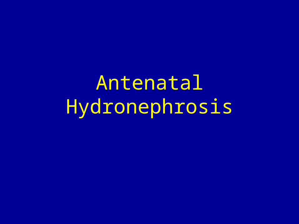
Antenatal Hydronephrosis. Definition: AP diameter renal pelvis > 4mm @ 20 wk EGA AP diameter renal pelvis > 7mm @ 30 wk EGA Incidence: 5% of pregnancies.
Dec 17, 2015
Welcome message from author
This document is posted to help you gain knowledge. Please leave a comment to let me know what you think about it! Share it to your friends and learn new things together.
Transcript
Antenatal Hydronephrosis
Antenatal Hydronephrosis
• Definition:AP diameter renal pelvis > 4mm @ 20 wk EGA
AP diameter renal pelvis > 7mm @ 30 wk EGA
• Incidence: 5% of pregnancies
Antenatal Hydronephrosis
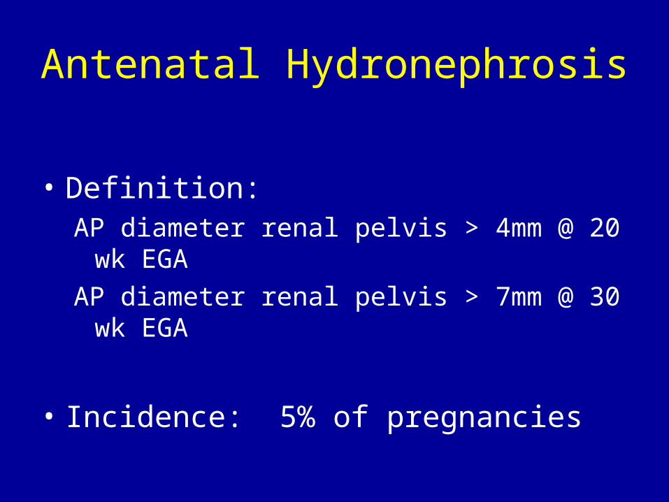
• Standard work-up:– Postnatal ultrasound
• Look for– AP diameter
– Calyceal/ureteral dilation
– Renal size
– Corticomedullary differentiation
– Thinned/hyperechoic cortex
– Cortical cysts
– Ureterocele
– Ectopic ureteral insertion
• Best after first 24 hours of life/when not volume depleted
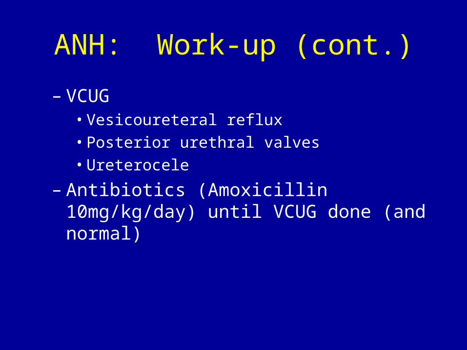
ANH: Work-up (cont.)
– VCUG• Vesicoureteral reflux• Posterior urethral valves• Ureterocele
– Antibiotics (Amoxicillin 10mg/kg/day) until VCUG done (and normal)
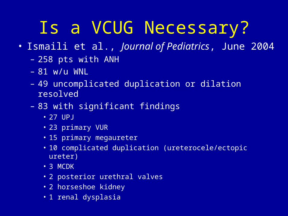
Is a VCUG Necessary?• Ismaili et al., Journal of Pediatrics, June 2004
– 258 pts with ANH– 81 w/u WNL– 49 uncomplicated duplication or dilation resolved– 83 with significant findings
• 27 UPJ• 23 primary VUR• 15 primary megaureter• 10 complicated duplication (ureterocele/ectopic ureter)• 3 MCDK• 2 posterior urethral valves• 2 horseshoe kidney• 1 renal dysplasia
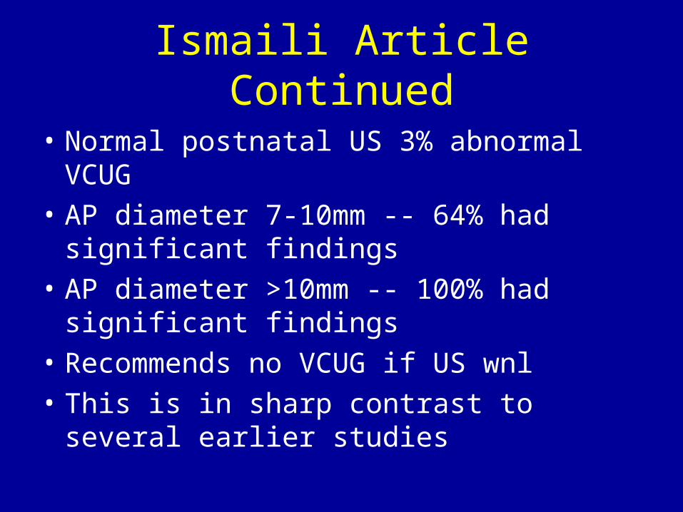
Ismaili Article Continued
• Normal postnatal US 3% abnormal VCUG
• AP diameter 7-10mm -- 64% had significant findings
• AP diameter >10mm -- 100% had significant findings
• Recommends no VCUG if US wnl
• This is in sharp contrast to several earlier studies
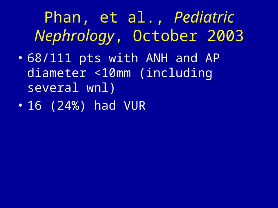
Phan, et al., Pediatric Nephrology, October 2003
• 68/111 pts with ANH and AP diameter <10mm (including several wnl)
• 16 (24%) had VUR
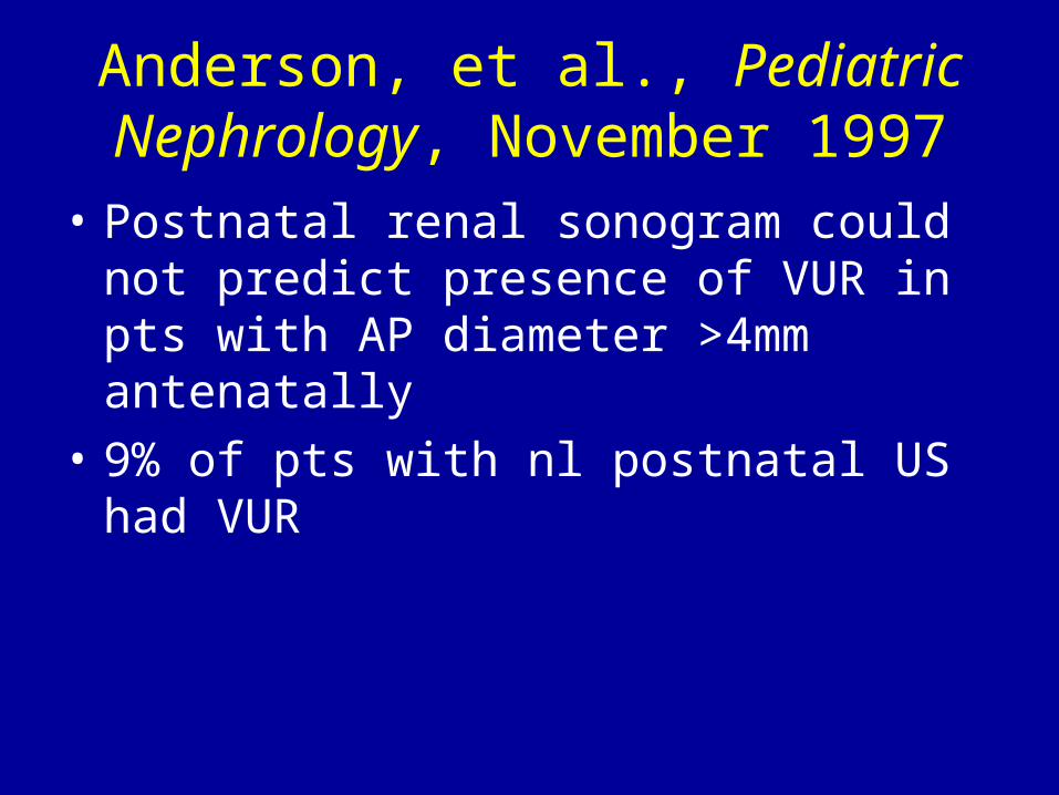
Anderson, et al., Pediatric Nephrology, November 1997
• Postnatal renal sonogram could not predict presence of VUR in pts with AP diameter >4mm antenatally
• 9% of pts with nl postnatal US had VUR

Farhat, et al., Journal of Urology, September 2000
• 27 % of pts with VUR (w/u prompted by ANH) had a normal postnatal RBUS
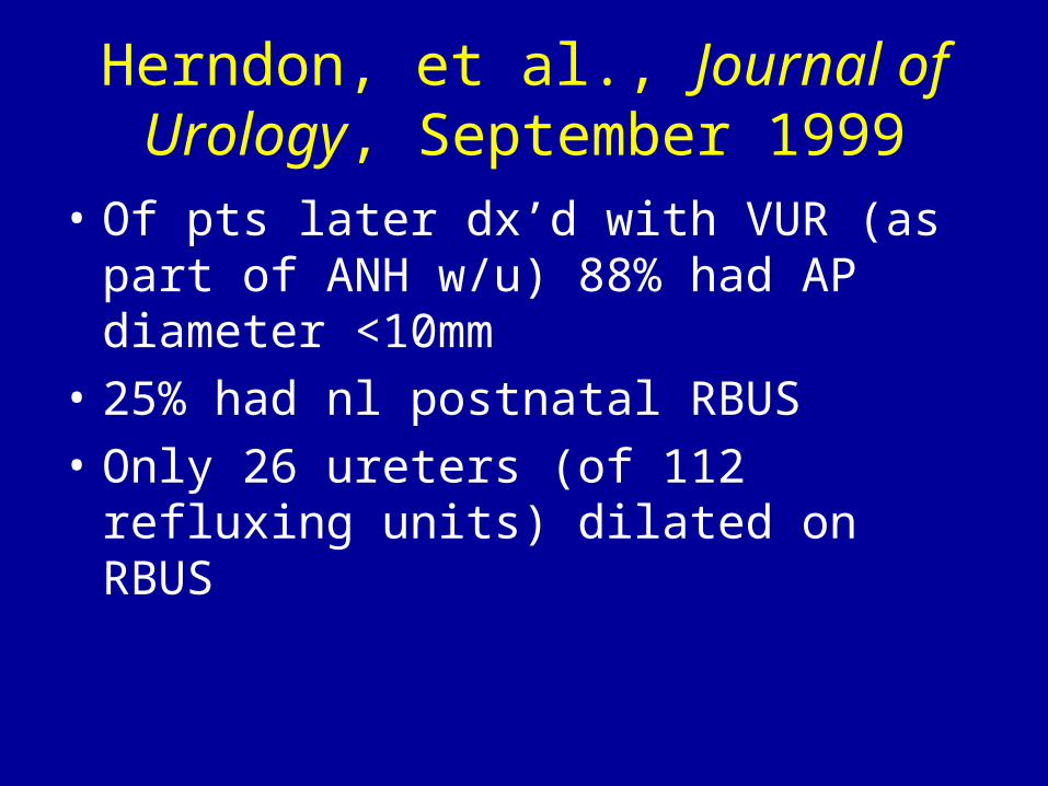
Herndon, et al., Journal of Urology, September 1999
• Of pts later dx’d with VUR (as part of ANH w/u) 88% had AP diameter <10mm
• 25% had nl postnatal RBUS
• Only 26 ureters (of 112 refluxing units) dilated on RBUS
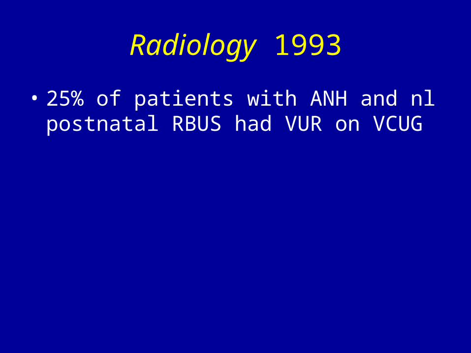
Radiology 1993
• 25% of patients with ANH and nl postnatal RBUS had VUR on VCUG
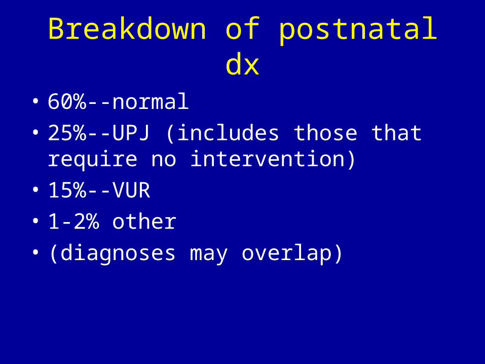
Breakdown of postnatal dx
• 60%--normal
• 25%--UPJ (includes those that require no intervention)
• 15%--VUR
• 1-2% other
• (diagnoses may overlap)
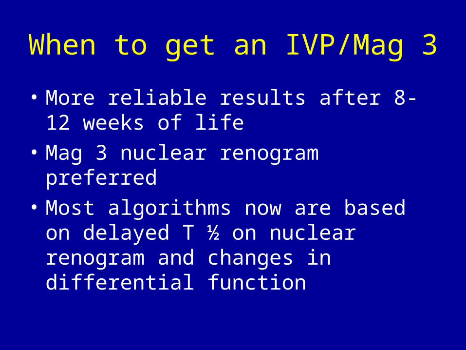
When to get an IVP/Mag 3
• More reliable results after 8-12 weeks of life
• Mag 3 nuclear renogram preferred
• Most algorithms now are based on delayed T ½ on nuclear renogram and changes in differential function
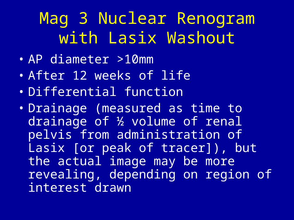
Mag 3 Nuclear Renogram with Lasix Washout
• AP diameter >10mm• After 12 weeks of life• Differential function• Drainage (measured as time to drainage
of ½ volume of renal pelvis from administration of Lasix [or peak of tracer]), but the actual image may be more revealing, depending on region of interest drawn
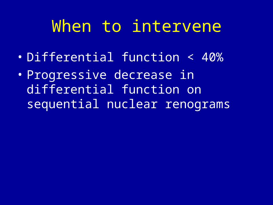
When to intervene
• Differential function < 40%
• Progressive decrease in differential function on sequential nuclear renograms
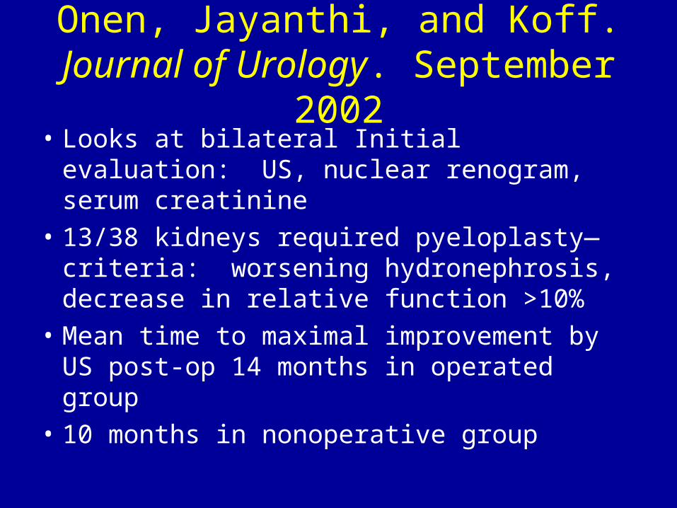
Onen, Jayanthi, and Koff. Journal of Urology. September 2002
• Looks at bilateral Initial evaluation: US, nuclear renogram, serum creatinine
• 13/38 kidneys required pyeloplasty—criteria: worsening hydronephrosis, decrease in relative function >10%
• Mean time to maximal improvement by US post-op 14 months in operated group
• 10 months in nonoperative group
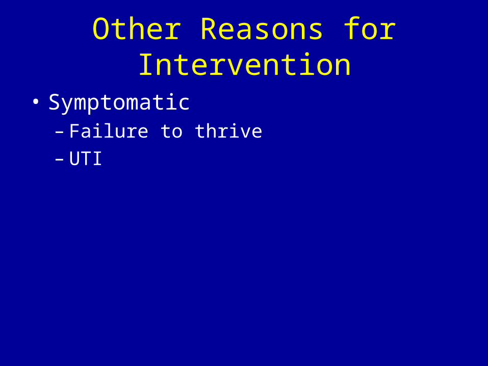
Other Reasons for Intervention
• Symptomatic– Failure to thrive– UTI
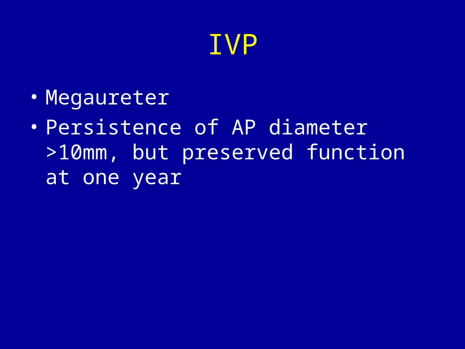
IVP
• Megaureter
• Persistence of AP diameter >10mm, but preserved function at one year
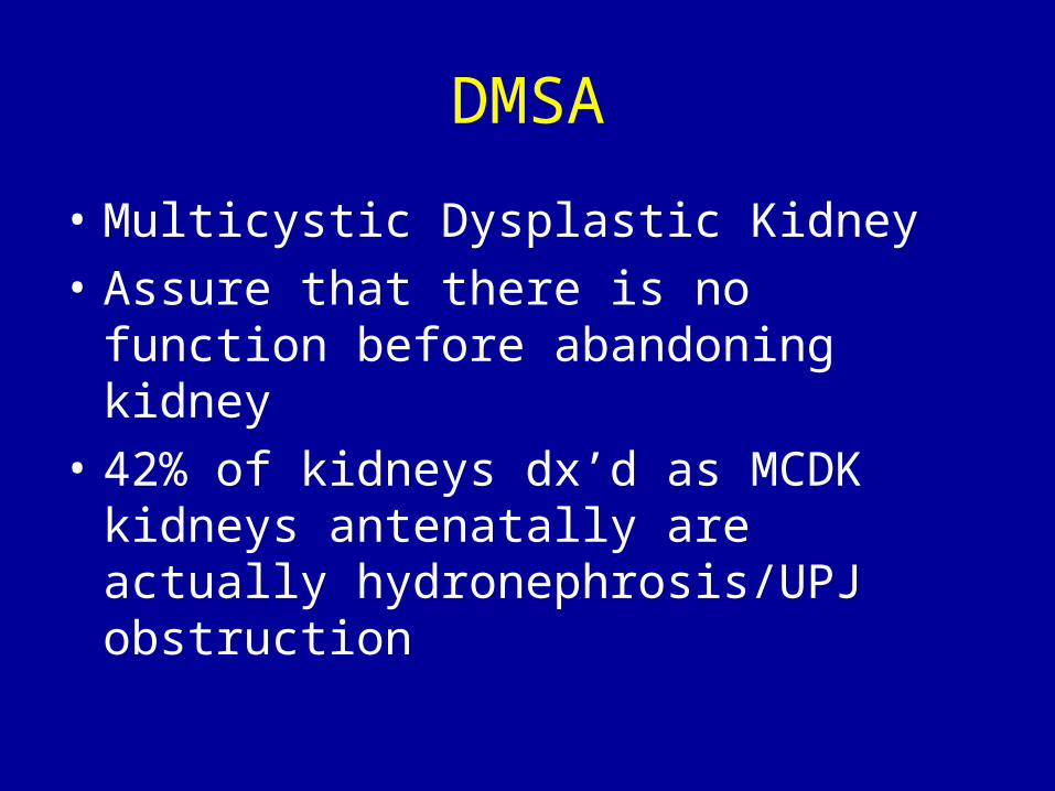
DMSA
• Multicystic Dysplastic Kidney
• Assure that there is no function before abandoning kidney
• 42% of kidneys dx’d as MCDK kidneys antenatally are actually hydronephrosis/UPJ obstruction
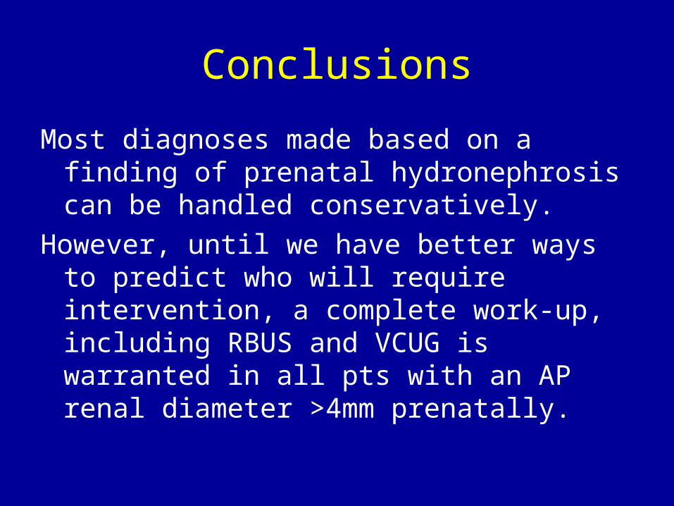
Conclusions
Most diagnoses made based on a finding of prenatal hydronephrosis can be handled conservatively.
However, until we have better ways to predict who will require intervention, a complete work-up, including RBUS and VCUG is warranted in all pts with an AP renal diameter >4mm prenatally.
Urinary Tract Infections in Children
Incidence– Neonates: M > F– Thereafter: F > M

Organisms
• Enterobacteriaciae– Escherichia (80%)– Klebsiella– Enterobacter– Citrobacter– Proteus– Providencia– Morganella– Serratia– Salmonella
Other Organisms
• Pseudamonas
• Staphylococcus
• Enterobacter
Risk Factors
• Perineal colonization• Family hx• Presence of a prepuce
– 10x risk– Periurethral colonization—circ eliminates this– Adherence of P fimbriated E. coli to prepuce
• Urethral length• Urine pH (6-7 favors growth)• Urine concentration—dilute has less nutrients• Dysfunctional elimination
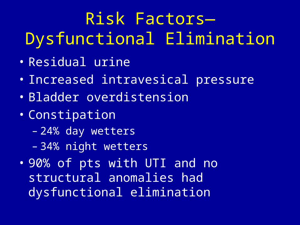
Risk Factors—Dysfunctional Elimination
• Residual urine
• Increased intravesical pressure
• Bladder overdistension
• Constipation – 24% day wetters– 34% night wetters
• 90% of pts with UTI and no structural anomalies had dysfunctional elimination
Not Risk Factors
• Bubble baths
• Improper wiping
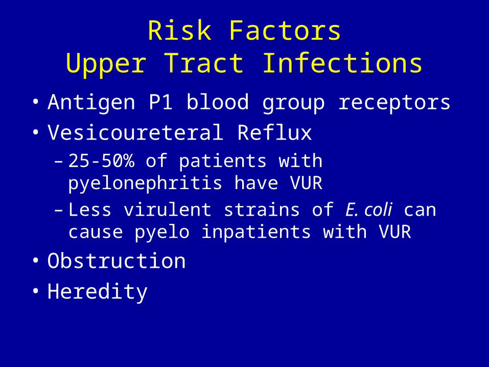
Risk FactorsUpper Tract Infections
• Antigen P1 blood group receptors
• Vesicoureteral Reflux– 25-50% of patients with pyelonephritis have
VUR– Less virulent strains of E. coli can cause pyelo
inpatients with VUR
• Obstruction
• Heredity
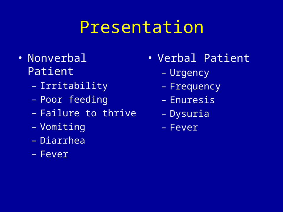
Presentation
• Nonverbal Patient– Irritability– Poor feeding– Failure to thrive– Vomiting– Diarrhea– Fever
• Verbal Patient– Urgency– Frequency– Enuresis– Dysuria– Fever
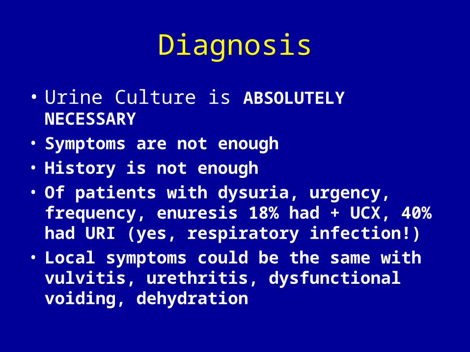
Diagnosis
• Urine Culture is ABSOLUTELY NECESSARY
• Symptoms are not enough• History is not enough• Of patients with dysuria, urgency, frequency,
enuresis 18% had + UCX, 40% had URI (yes, respiratory infection!)
• Local symptoms could be the same with vulvitis, urethritis, dysfunctional voiding, dehydration
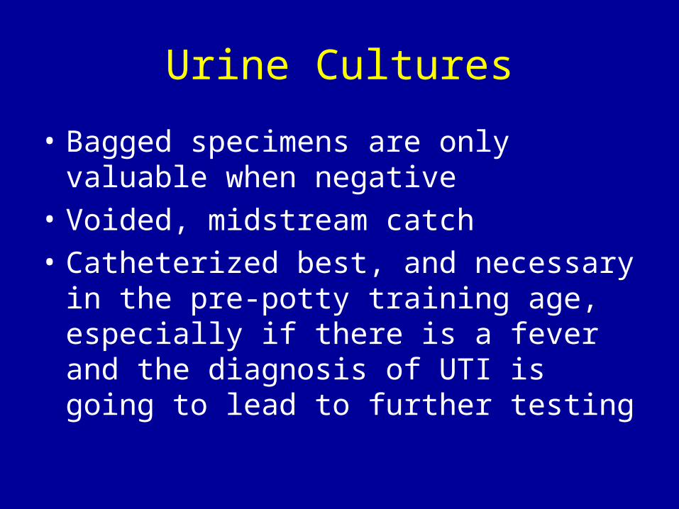
Urine Cultures
• Bagged specimens are only valuable when negative
• Voided, midstream catch
• Catheterized best, and necessary in the pre-potty training age, especially if there is a fever and the diagnosis of UTI is going to lead to further testing
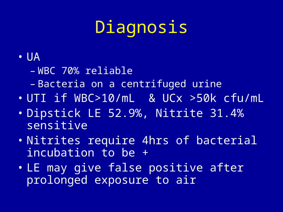
Diagnosis
• UA– WBC 70% reliable– Bacteria on a centrifuged urine
• UTI if WBC>10/mL & UCx >50k cfu/mL• Dipstick LE 52.9%, Nitrite 31.4% sensitive• Nitrites require 4hrs of bacterial incubation
to be +• LE may give false positive after prolonged
exposure to air
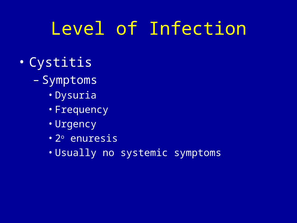
Level of Infection
• Cystitis– Symptoms
• Dysuria• Frequency• Urgency• 2o enuresis• Usually no systemic symptoms
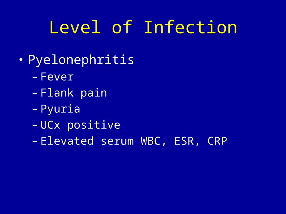
Level of Infection
• Pyelonephritis– Fever– Flank pain– Pyuria– UCx positive– Elevated serum WBC, ESR, CRP
Asymptomatic Bacteruria
• Positive urine culture• No urinary symptoms• Only 4% later progress to symptomatic
infection• The organism may be commensal and
protective to prevent infection with a more virulent organism
• In the absence of VUR, no treatment necessary, but look for voiding dysfunction

Pyelonephritis (continued
• Diagnosis: UCx and pyuria, but DMSA to be absolutely certain (in the first several days of symptoms)
• Risks from episodes of pyelo– Focal ischemia– Inflammatory changes– Renal scarring– Hypertension– Renal insufficiency
Treatment
• Lower Tract (no fever)– Treat 3-5 days– Start with TMP-SMX, nitrofurantoin or
cephalosporin– Amoxil may change gut flora and lead to
future infections with resistant organisms – FQ ok if there is no other oral agent to use
Treatment
• Pyelonephritis– Treat 10-14 days– Start with Bactrim of Cephalosporin until
culture is back– Hospitalization in severe cases
• Abscess– UCx may be negative– Parenteral abx x 10 days then 14d oral
therapy
Work-up after a UTI
• Who?– Fever or documented pyelonephritis– <5yo
• What– RBUS (prior to discharge & yes, kidneys &
bladder)– VCUG once afebrile– DMSA
• Prophylactic antibiotics until work-up
Prophylaxis
• Vesicoureteral reflux
• No Reflux, but <1yo– 30-75% recurrence in the first year
• Frequent symptomatic UTIs
Related Documents