Answer Keys To Lab Notebook Lab 1 Part A Diagram (p. 1) 1. Frontal 17. Inguinal 2. Orbital 18. Pelvic 3. Nasal 19. Coxa. 4. Buccal 20. Carpal 5. Oral 21. Palmar 6. Mental 22. Digital 7. Cervical 23. Pollex 8. Thoracic (pectoral) 24. Pubic 9. Acromial 25. Femoral 10. Axillary 26. Patellar 11. Sternal 27. Crural 12. Brachial 28. Fibular (peroneal) 13. Antecubital 29. Tarsal 14. Abdominal 30. Digital (phalangeal) 15. Antebrachial 31. Hallux 16. Umbilical 32. Pedal Part A Diagram (p. 2) 33. Cephalic 41. Sacral 34. Otic 42. Gluteal 35. Occipital 43. Perineal 36. Scapular 44. Manus 37. Vertebral 45. Popliteal 38. Dorsal 46. Sural 39. Lumbar 47. Calcaneal 40. Olecranal 48. Plantar Part B Table (p.3) Cavity Major Organs 1. Thoracic a. Pleural cavities Lungs 120

Welcome message from author
This document is posted to help you gain knowledge. Please leave a comment to let me know what you think about it! Share it to your friends and learn new things together.
Transcript
Answer Keys To Lab NotebookLab 1
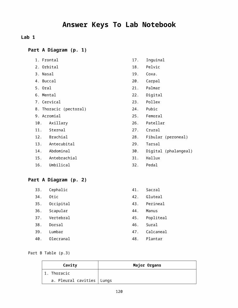
Part A Diagram (p. 1)
1. Frontal 17. Inguinal
2. Orbital 18. Pelvic
3. Nasal 19. Coxa.
4. Buccal 20. Carpal
5. Oral 21. Palmar
6. Mental 22. Digital
7. Cervical 23. Pollex
8. Thoracic (pectoral) 24. Pubic
9. Acromial 25. Femoral
10. Axillary 26. Patellar
11. Sternal 27. Crural
12. Brachial 28. Fibular (peroneal)
13. Antecubital 29. Tarsal
14. Abdominal 30. Digital (phalangeal)
15. Antebrachial 31. Hallux
16. Umbilical 32. Pedal
Part A Diagram (p. 2)
33. Cephalic 41. Sacral
34. Otic 42. Gluteal
35. Occipital 43. Perineal
36. Scapular 44. Manus
37. Vertebral 45. Popliteal
38. Dorsal 46. Sural
39. Lumbar 47. Calcaneal
40. Olecranal 48. Plantar
Part B Table (p.3)
Cavity Major Organs
1. Thoracic
a. Pleural cavities Lungs
b. Mediastinum Pericardial cavity, heart, aorta, esophagus, and trachea
120
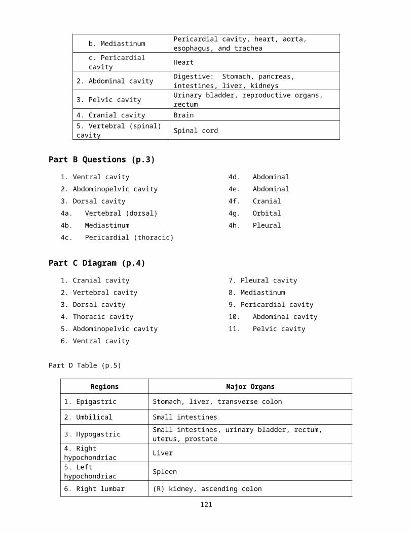
c. Pericardial cavity Heart
2. Abdominal cavity Digestive: Stomach, pancreas, intestines, liver, kidneys
3. Pelvic cavity Urinary bladder, reproductive organs, rectum
4. Cranial cavity Brain
5. Vertebral (spinal) cavity Spinal cord
Part B Questions (p.3)
1. Ventral cavity 4d. Abdominal
2. Abdominopelvic cavity 4e. Abdominal
3. Dorsal cavity 4f. Cranial
4a. Vertebral (dorsal) 4g. Orbital
4b. Mediastinum 4h. Pleural
4c. Pericardial (thoracic)
Part C Diagram (p.4)
1. Cranial cavity 7. Pleural cavity
2. Vertebral cavity 8. Mediastinum
3. Dorsal cavity 9. Pericardial cavity
4. Thoracic cavity 10. Abdominal cavity
5. Abdominopelvic cavity 11. Pelvic cavity
6. Ventral cavity
Part D Table (p.5)
Regions Major Organs
1. Epigastric Stomach, liver, transverse colon
2. Umbilical Small intestines
3. Hypogastric Small intestines, urinary bladder, rectum, uterus, prostate
4. Right hypochondriac Liver
5. Left hypochondriac Spleen
6. Right lumbar (R) kidney, ascending colon
7. Left lumbar (L) kidney, descending colon
8. Right inguinal (iliac) Cecum (beginning colon), appendix
9. Left inguinal (iliac) Sigmoid colon, small intestines
121
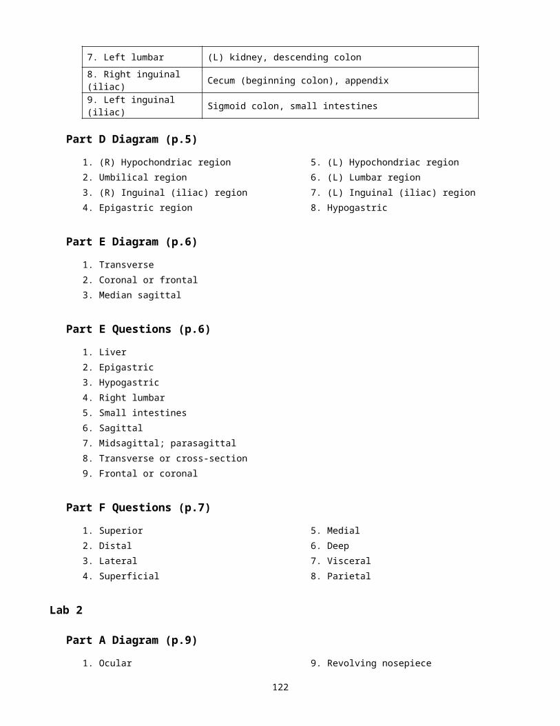
Part D Diagram (p.5)
1. (R) Hypochondriac region 5. (L) Hypochondriac region
2. Umbilical region 6. (L) Lumbar region
3. (R) Inguinal (iliac) region 7. (L) Inguinal (iliac) region
4. Epigastric region 8. Hypogastric
Part E Diagram (p.6)
1. Transverse
2. Coronal or frontal
3. Median sagittal
Part E Questions (p.6)
1. Liver
2. Epigastric
3. Hypogastric
4. Right lumbar
5. Small intestines
6. Sagittal
7. Midsagittal; parasagittal
8. Transverse or cross-section
9. Frontal or coronal
Part F Questions (p.7)
1. Superior 5. Medial
2. Distal 6. Deep
3. Lateral 7. Visceral
4. Superficial 8. Parietal
Lab 2
Part A Diagram (p.9)
1. Ocular 9. Revolving nosepiece
2. Eye tube 10. Objective
3. Arm 11. Mechanical stage
4. Stage clip 12. Condenser
5. On/Off switch 13. Iris diaphragm lever
6. Illuminator knob 14. X-Y stage control knob
7. Coarse adjustment knob 15. Illuminator
8. Fine adjustment knob
122
Part B Questions (p.10-11)
1. Multiply ocular magnification, times objective magnification
2. Parfocal microscopes remain in focus at high power once they’ve been focused on low power
3. Lower the light intensity by closing the iris diaphragm
4. The clarity of the image being viewed
5. The higher power of the objective, the more light is necessary
6. In order not to break the slide or more importantly, to avoid damaging the objective
Part C Questions (p.11)
1a. To the left
1b. Turns the image upside down and reverses it
3a. Varies with the slide
Part D Questions (p.12)
6a. Nucleus, vacuole, cell membrane
Part E Questions (p.13)
2a. Onion cells are rectangular shaped and onion cells have a cell wall.
2b. Cell wall
3a. Nucleus, cell wall
Part F Questions (p.14)
1a. Chloroplasts
1b. Human or onion cells do not possess chloroplasts. Onions do not photosynthesize because they are the roots of the plant.
1c. Plants: cell wall, chloroplasts, large vacuoles, no centrioles; Animal: no cell wall, no chloroplasts, smaller vacuoles, centrioles
Lab 3
Part A Conversions (p.16) Part B Conversions (p.16) Part C Conversions (p.16)
1. 3520 mm 1. 1000 mL 1. 154.35 lbs
2. 150,000 m 2. 150 mL 2. 300,000 mg
3. 2 mm 3. 900 mL 3. 4 mg
4. 120,000,000 nm 4. 200 mL 4. 99.792 mg
5. 0.001 m 5. 520,000 mg
6. 0.0001 mm 6. 0.0043 g
7. 0.1 nm
123
Part D Questions (p.17) Part E Questions (p.17) Part E Questions (p.18)
1. 0.039 in 2a. 4 mm 3b. 0.5 mm
2. 1000 2b. 2 mm 3c.
3. Graduated cylinder 2c. 0.5 mm 4. Depends upon individual slides
4. mL or ml
5. kL
Part F Questions (p.19)
1. 25.4 mm
2. Use 10X objective, then convert mm to m
3.
Lab 4
Part A Diagram (p.21)
1. Lysosome 9. Nuclear membrane
2. Smooth ER 10. Nucleolus
3. Cytoplasm or Cytosol 11. Golgi apparatus
4. Vacuole 12. Chromatin (DNA)
5. Centriole 13. Nuclear pore
6. Microvilli 14. Vesicle (phagocytic or pinocytic)
7. Mitochondrion 15. Rough ER
8. Nucleus 16. Plasma membrane
Lab 5
Part A Diagram (p.25)
1. S = Synthesis 5. 3rd phase = Anaphase
2. G2 = Gap 2 6. 4th phase = Telophase
3. 1st phase = Prophase 7. G1 = Gap 1
4. 2nd phase = Metaphase
124
Lab 6
Part A Diagram (p.27)
1. Phosphate 4. Hydrogen bonds
2. Deoxyribose sugar 5. Nitrogenous base
3. Nucleotide
Part A Questions (p.28)
1. Adenine; guanine; cytosine; thymine; uracil
2. Sugar; phosphate
3. Nitrogenous base; hydrogen bond
4. Double helix
5. Adenine; cytosine; uracil
6. Nucleus; mitochondria
7. Determines the genetic code for protein construction; makes up genes on chromosomes
8. Gene
9. Replicate
Part B Questions (p.28-29)
1. RNA - ribose sugar, single-stranded, and uracil base instead of thymine; DNA - deoxyribose sugar, double-stranded, and thymine base
2. Cytoplasm (mRNA, tRNA), ribosomes (rRNA) and nucleolus. Some in nucleus (mRNA)
3. Carries out orders of DNA
4a. Formed in nucleolus. Used to make ribosomes
4b. Carries DNA code out of nucleus to ribosomes in cytoplasm
4c. Carries amino acids to ribosomes for construction of proteins
Lab 7
Part A Questions (p.30)
1. Uncoil or unwind
2. Separate; bases
3. New DNA strand
4. DNA polymerase
5. DNA ligase
6. Two double-stranded DNA molecules
Lab 8125
Part A Diagram Questions (p.31)
1a. RNA polymerase; unwind 1d. Introns; exons
1b. Separate; sense; mRNA 1e. Cytoplasm; ribosome
1c. Codon
Part A Questions (p.32)
1. DNA cannot leave the nucleus
2. A template for construction of a protein
3. RNA has uracil as a base instead of thymine
Part B Diagram Questions (p.32)
1a. Ribosome 1d. Hydrogen; mRNA
1b. tRNA 1e. Peptide; polypeptide
1c. 20; anticodon
Part B Questions (p.33)
1. On the ribosomes (in cytoplasm)
2. mRNA (originally from DNA)
3. Carries amino acids to site where they attach to mRNA and form a polypeptide
Lab 10
Part D Questions (p.44)
1. Mucus producing, single-celled glands
2. Simple columnar and pseudostratified ciliated columnar
3. Tiny, hair-like projections on cell surfaces that move substances; found in upper respiratory tract
4. Tiny projections on the free surfaces of some epithelial cells, such as cells lining digestive system; increase surface are for absorption
5. Answers vary
6. Protein fibers formed from underlying connective tissue to reinforce the epithelial tissue. Helps to keep E.T. from overstretching or tearing.
7. Simple – one layer of cells above the basement membrane; stratified – many cell layers above the membrane
8. Stratified squamous tissue cells are flat at the apical surface, whereas transitional tissue cells are cuboidal at the apical surface
9. Simple columnar – nucleus near the basement membrane; simple cuboidal – centrally located nucleus; pseudostratified ciliated columnar – scattered nuclei giving the appearance of stratification
10. Stratified cuboidal
11. Mesothelium (simple squamous epithelium) on a connective tissue base
126
12. Diffusion, filtration, protection
Lab 11
Part A-D Diagrams (p.45)
1. Fibroblast on a collagen fiber 4. Reticular fiber
2. Mast cell 5. Fibroblast
3. Collagen fiber 6. Elastic fiber
Part G Questions (p.47-48)
1. Ground substance – composed of interstitial fluid and proteoglycans and fibers (collagen, elastic, reticular);
2a. Fibroblasts – produce proteoglycans and all three type of fibers (collagen, elastic, reticular)
2b. Mast cells – produce histamines and heparin for the inflammatory response
2c. Plasma cells – produce antibodies
2d. Macrophages – phagocytize foreign particles
2e. Leukocytes – white blood cells acting as body’s defense
2f. Chondroblasts (cytes) – produce matrix in cartilage
2g. Osteoblasts (cytes) – produce organic matrix in bone
3a Protein fibers that provide support for connective tissue
3b Three (collagen, elastic and reticular)
3c. Synthesized by blast cell types
3d. In the matrix
3e(i). Collagen – thick protein fibers constructed primarily of the fibrous protein collagen
3e(ii). Elastic – long, thin fibers containing elastin protein, that allows them to stretch and recoil
3e(iii). Reticular – fine collagenous fibers forming delicate networks that support soft tissue of organs
4a. Vascular or avascular
4b. Extracellular matrix composed of ground substance and fibers
4c. Loosely scattered cells
4d. Binds, supports, protects, insulates, transports
4e. Blast cell types produce organic matrix
4f. Many cells types
Lab 12
Part C Questions (p.49)
1. Small spaces surrounding chrondrocytes and osteocytes
2. Polysaccharides attached to proteoglycans; produced by blast cells to thicken the matrix
3. Yes
4. No. The fibers are not visible in hyaline cartilage.
127
Part F Questions (p.51-52)
1. Fibrous, vascular connective tissue on the surface of cartilage
2. Vascular connective tissue on the surface of bone
3. Elastic cartilage, although some collagen fibers are visible in fibrocartilage
4. Hyaline cartilage
5. No because it does not have its own blood supply. Bone on the other hand is vascular so it heals well.
6. Hyaline cartilage located on the end of bones that articulate at a joint
7. A growth plate where the growth in long bones occurs
8. Gelatinous substance filling the space between cells and contains the fibers. Composed of interstitial fluid and proteoglycans
9. Mineral salts such as calcium hydroxyapatite
10. Organic matter contains fibers and ground substance produced by blast cell types; inorganic matter contains mineral salts deposited from the blood
11. Disorders such as rickets/osteomalacia
Lab 13
Part A Diagram (p.53)
1. Stratum corneum 4. Stratum basale (germinativum)
2. Stratum granulosum 5. Papillary layer of dermis
3. Stratum spinosum
Part B Diagram (p.54)
1. Hair shaft 9. Sebaceous gland
2. Epidermis 10. Arrector pili muscle
3. Dermis 11. Sudoriferous (sweat) gland
4. Hypodermis 12. Pacinian corpuscle
5. Epidermal peg 13. Adipose tissue
6. Dermal papilla 14. Hair follicle
7. Meissner’s corpuscle 15. Artery
8. Free nerve endings 16. Vein
Part C Questions (p.55-56)
1. Epidermis, dermis, and hypodermis
2. Epidermis (ectoderm); dermis (mesoderm)
3. Stratum basale, stratum spinosum, stratum granulosum, stratum lucidum, and stratum corneum
4a. Stratum basale - mitotic; contains melanocytes
4b. Stratum spinosum - largest, living layer containing spiny-shaped cells
4c. Stratum granulosum - numerous keratohyaline granules128
4d. Stratum lucidum - translucent, dead cell layer located only in thick skin
4e. Stratum corneum - thick layer of dead, flaking cells
5. Dermal and epidermal ridges
6. Produced by melanocytes in the stratum basale. Melanosomes (melanin granules) may be present in the stratum spinosum
7. Melanocytes
8. Melanin is transferred from the melanocytes processes to nearby keratinocytes
9. UV protection
10. UV light stimulates melanin production therefore, the skin becomes darker
11. Localized patches of melanin
12. Both have same number of melanocytes but dark skin contains more numerous and darker colored melanosomes
13. Contraction of arrector pili muscles
14. Secretes sebum to soften and lubricate skin and hair
15. Stratum basale (germinativum)
16. Detects light pressure or discriminative touch
17. Detects deep pressure or crude touch
Lab 14
Part A Questions (p.57)
1. Intramembranous and endochondral
2. Intramembranous (skull); endochondral (long bones)
3. Yes; veins, arteries and lymph
4. Interstitial lamella – incomplete lamella lying between intact Haversian systems; concetric lamella – rings of lamella making up each osteon; circumferential lamella – lamella extentending around the entire circumference of the bone shaft
Part B Diagram (p.58)
1. Epiphysis 6. Medullary cavity (lined with endosteum)
2. Diaphysis 7. Yellow marrow
3. Epiphysis 8. Compact bone
4. Cancellous bone 9. Periosteum
5. Epiphyseal plate
Part C Questions (p.60-61)
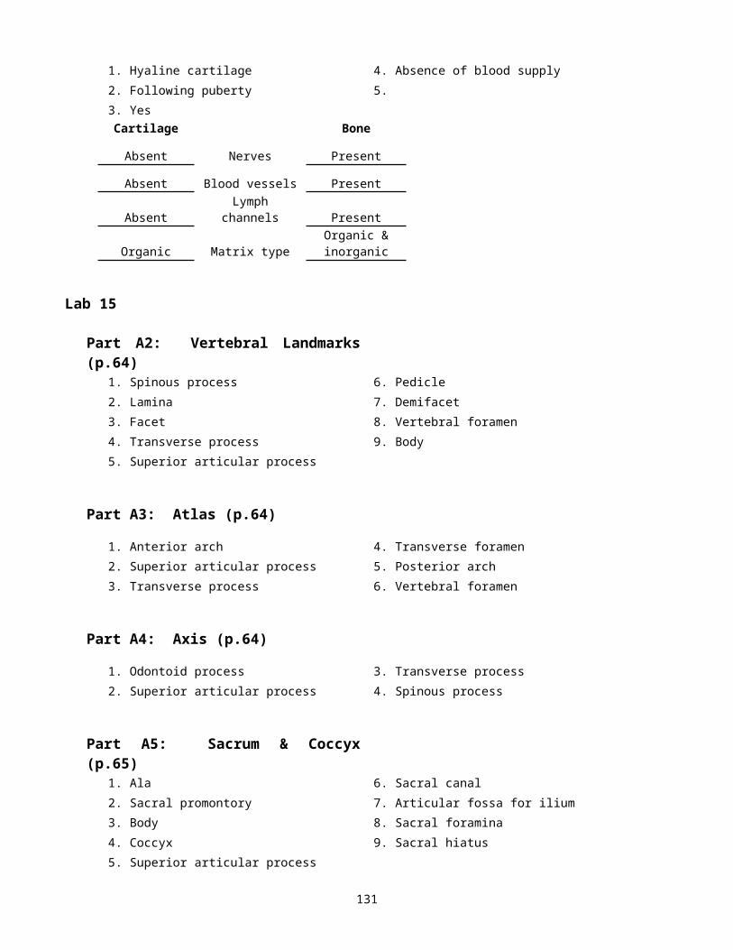
1. Hyaline cartilage 4. Absence of blood supply
2. Following puberty 5.
129
3. YesCartilage Bone
Absent Nerves Present
Absent Blood vessels Present
Absent Lymph channels Present
Organic Matrix typeOrganic & inorganic
Lab 15
Part A2: Vertebral Landmarks (p.64)
1. Spinous process 6. Pedicle
2. Lamina 7. Demifacet
3. Facet 8. Vertebral foramen
4. Transverse process 9. Body
5. Superior articular process
Part A3: Atlas (p.64)
1. Anterior arch 4. Transverse foramen
2. Superior articular process 5. Posterior arch
3. Transverse process 6. Vertebral foramen
Part A4: Axis (p.64)
1. Odontoid process 3. Transverse process
2. Superior articular process 4. Spinous process
Part A5: Sacrum & Coccyx (p.65)
1. Ala 6. Sacral canal
2. Sacral promontory 7. Articular fossa for ilium
3. Body 8. Sacral foramina
4. Coccyx 9. Sacral hiatus
5. Superior articular process
Part A Questions (p.66)
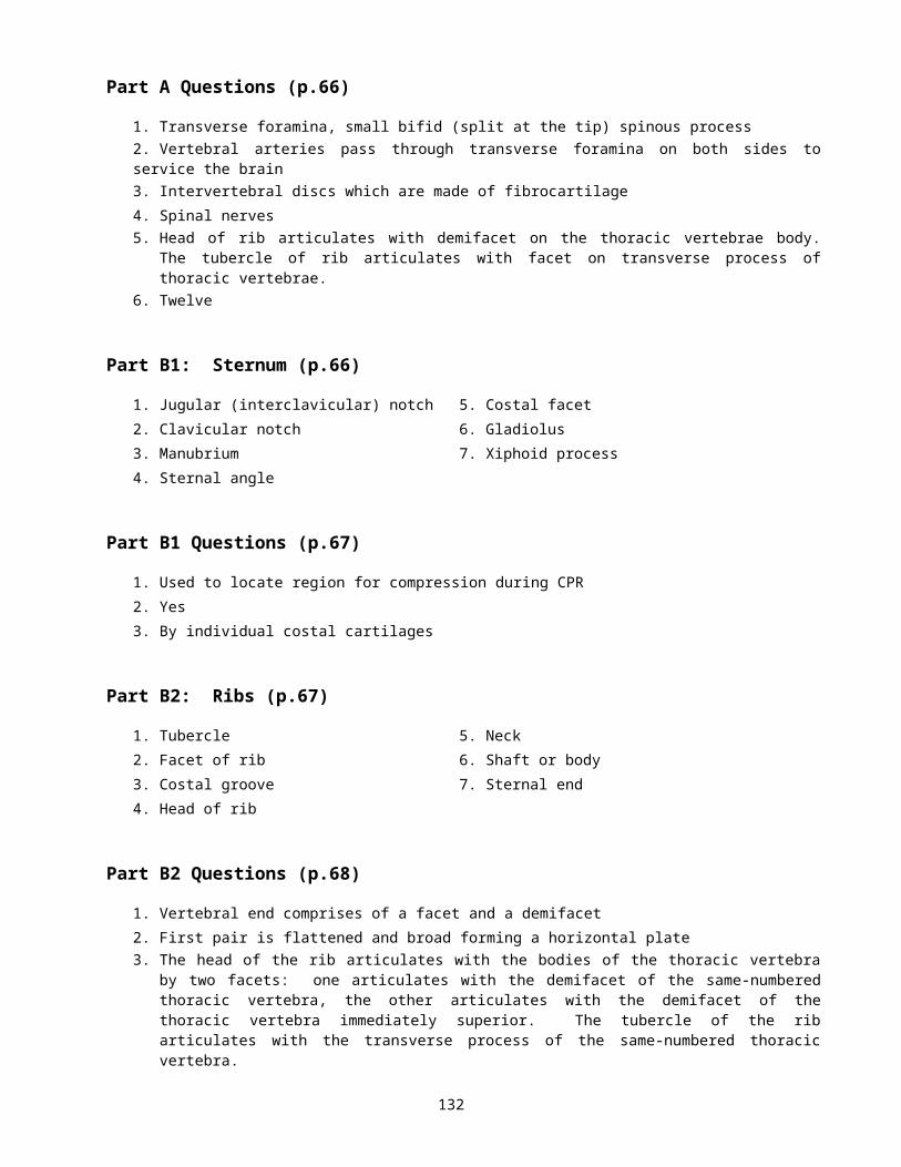
1. Transverse foramina, small bifid (split at the tip) spinous process
2. Vertebral arteries pass through transverse foramina on both sides to service the brain
130
3. Intervertebral discs which are made of fibrocartilage
4. Spinal nerves
5. Head of rib articulates with demifacet on the thoracic vertebrae body. The tubercle of rib articulates with facet on transverse process of thoracic vertebrae.
6. Twelve
Part B1: Sternum (p.66)
1. Jugular (interclavicular) notch 5. Costal facet
2. Clavicular notch 6. Gladiolus
3. Manubrium 7. Xiphoid process
4. Sternal angle
Part B1 Questions (p.67)
1. Used to locate region for compression during CPR
2. Yes
3. By individual costal cartilages
Part B2: Ribs (p.67)
1. Tubercle 5. Neck
2. Facet of rib 6. Shaft or body
3. Costal groove 7. Sternal end
4. Head of rib
Part B2 Questions (p.68)
1. Vertebral end comprises of a facet and a demifacet
2. First pair is flattened and broad forming a horizontal plate
3. The head of the rib articulates with the bodies of the thoracic vertebra by two facets: one articulates with the demifacet of the same-numbered thoracic vertebra, the other articulates with the demifacet of the thoracic vertebra immediately superior. The tubercle of the rib articulates with the transverse process of the same-numbered thoracic vertebra.
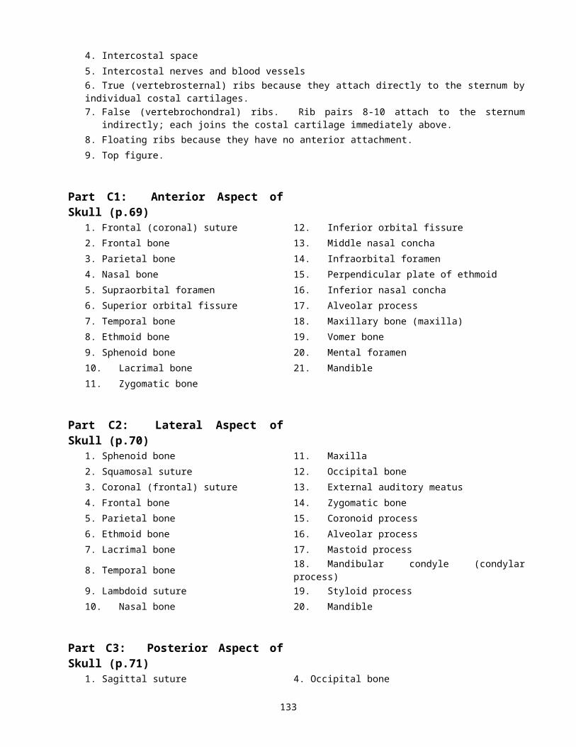
4. Intercostal space
5. Intercostal nerves and blood vessels
6. True (vertebrosternal) ribs because they attach directly to the sternum by individual costal cartilages.
7. False (vertebrochondral) ribs. Rib pairs 8-10 attach to the sternum indirectly; each joins the costal cartilage immediately above.
8. Floating ribs because they have no anterior attachment.
9. Top figure.
131
Part C1: Anterior Aspect of Skull (p.69)
1. Frontal (coronal) suture 12. Inferior orbital fissure
2. Frontal bone 13. Middle nasal concha
3. Parietal bone 14. Infraorbital foramen
4. Nasal bone 15. Perpendicular plate of ethmoid
5. Supraorbital foramen 16. Inferior nasal concha
6. Superior orbital fissure 17. Alveolar process
7. Temporal bone 18. Maxillary bone (maxilla)
8. Ethmoid bone 19. Vomer bone
9. Sphenoid bone 20. Mental foramen
10. Lacrimal bone 21. Mandible
11. Zygomatic bone
Part C2: Lateral Aspect of Skull (p.70)
1. Sphenoid bone 11. Maxilla
2. Squamosal suture 12. Occipital bone
3. Coronal (frontal) suture 13. External auditory meatus
4. Frontal bone 14. Zygomatic bone
5. Parietal bone 15. Coronoid process
6. Ethmoid bone 16. Alveolar process
7. Lacrimal bone 17. Mastoid process
8. Temporal bone 18. Mandibular condyle (condylar process)
9. Lambdoid suture 19. Styloid process
10. Nasal bone 20. Mandible
Part C3: Posterior Aspect of Skull (p.71)
1. Sagittal suture 4. Occipital bone
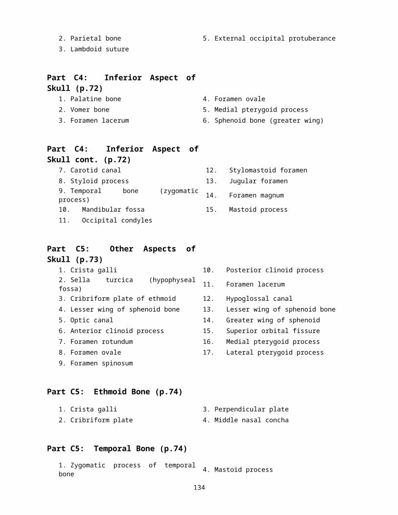
2. Parietal bone 5. External occipital protuberance
3. Lambdoid suture
Part C4: Inferior Aspect of Skull (p.72)
1. Palatine bone 4. Foramen ovale
2. Vomer bone 5. Medial pterygoid process
3. Foramen lacerum 6. Sphenoid bone (greater wing)
Part C4: Inferior Aspect of Skull cont. (p.72)
7. Carotid canal 12. Stylomastoid foramen
132
8. Styloid process 13. Jugular foramen
9. Temporal bone (zygomatic process) 14. Foramen magnum
10. Mandibular fossa 15. Mastoid process
11. Occipital condyles
Part C5: Other Aspects of Skull (p.73)
1. Crista galli 10. Posterior clinoid process
2. Sella turcica (hypophyseal fossa) 11. Foramen lacerum
3. Cribriform plate of ethmoid 12. Hypoglossal canal
4. Lesser wing of sphenoid bone 13. Lesser wing of sphenoid bone
5. Optic canal 14. Greater wing of sphenoid
6. Anterior clinoid process 15. Superior orbital fissure
7. Foramen rotundum 16. Medial pterygoid process
8. Foramen ovale 17. Lateral pterygoid process
9. Foramen spinosum
Part C5: Ethmoid Bone (p.74)
1. Crista galli 3. Perpendicular plate
2. Cribriform plate 4. Middle nasal concha
Part C5: Temporal Bone (p.74)
1. Zygomatic process of temporal bone 4. Mastoid process
2. Mandibular fossa 5. External auditory (acoustic) meatus
3. Syloid process
Part C5: Additional Features (p.75)
1. Parietal bone 7. Frontal bone
2. Occipital bone 8. Supraorbital foramen
3. Zygomatic bone 9. Nasal bone
4. Occipital condyle 10. Lacrimal bone
5. Foramen magnum 11. Vomer bone
6. External occipital protuberance 12. Inferior nasal concha
Part C5: Maxilla & Palatine Bones (p.76)
1. Infraorbital foramen 3. Horizontal plate
2. Alveolar process133
Part C5: Mandible (p.76)
1. Coronoid process 5. Body
2. Mandibular foramen 6. Ramus
3. Alveolar process 7. Mandibular notch
4. Mental foramen
Part C5: Ossicles (p.77)
1. Incus 3. Stapes
2. Malleus
Part C Questions (p.77-78)
1. Vomer and the perpendicular plate of ethmoid
2. Temporal bone and zygomatic bone
3. Palatine bone and maxilla
4. Ethmoid bone (cribriform plate and crista galli)
5. Temporal bone
6. Parietal bones
7. Occipital bone and the parietal bones
8. Frontal bone and the parietal bones
9. Pituitary gland (hypophysis)
10. Foramen rotundum, foramen ovale, foramen spinosum, and foramen lacerum
11. Air-filled cavity in certain cranial bones
12. Frontal bone, sphenoid bone, ethmoid bone, and maxilla
13. Temporal bone
Part A1: Clavicle (p.79)
1. Acromial end 2. Sternal end
Part A1 Questions (p.79)
1. Figure A
Part A2: Scapulae (p.80)
1. Coracoid process 10. Vertebral (medial) border
134
2. Subscapular fossa 11. Spine
3. Vertebral (medial) border 12. Supraspinous fossa
4. Axillary (lateral) border 13. Superior angle
5. Glenoid cavity 14. Superior border
6. Acromion process 15. Suprascapular notch
7. Infraspinous fossa 16. Supraglenoid tubercle
8. Axillary (lateral) border 17. Infraglenoid tubercle
9. Inferior angle
Part A2 Questions (p.81)
1. Left scapula
Part B1: Humerus (p.81)
1. Greater tubercle 9. Lateral supracondylar ridge
2. Head 10. Coronoid fossa
3. Surgical neck 11. Capitulum
4. Anatomical neck 12. Medial epicondyle
5. Lesser tubercle 13. Lateral epicondyle
6. Deltoid tuberosity 14. Olecranon fossa
7. Radial groove 15. Trochlea
8. Medial supracondylar ridge
Part B1 Questions (p.82)
1. Right humerus
Part B2: Radius & Ulna (p.82)
1. Trochlear notch 7. Radius
2. Radial notch 8. Ulna
3. Head of radius 9. Ulnar notch
4. Olecranon process 10. Styloid process of ulna
5. Coronoid process 11. Head of ulna
6. Radial tuberosity 12. Styloid process of radius
Part B2 Questions (p.83)
1. Left forearm
Part B3: Hand (p.83)
135
1. Middle phalanx #4 7. Hamate
2. Proximal phalanx #2 8. Trapezium
3. Distal phalanx #1 9. Scaphoid
4. Metacarpal #3 10. Pisiform
5. Trapezoid 11. Triquetral
6. Capitate 12. Lunate
Part B3 Questions (p.83)
1. Left hand. Thumb is located on the left
Part C1: Os Coxa (p.84)
1. Iliac crest 10. Ischial spine
2. Ilium 11. Acetabular notch
3. Anterior superior iliac spine 12. Pubis
4. Anterior inferior iliac spine 13. Lesser sciatic notch
5. Posterior superior iliac spine 14. Ischial tuberosity
6. Posterior inferior iliac spine 15. Inferior ramus of pubis
7. Acetabulum 16. Obturator foramen
8. Acetabular fossa 17. Ischium
9. Greater sciatic notch 18. Inferior ramus of ischium (ischial ramus)
Part C1 Questions (p.84)
1. Right os coxa. Acetabulum is always lateral.
Part D1: Femur (p.85)
1. Head 9. Linea aspera
2. Intertrochanteric line 10. Intercondylar notch
3. Neck 11. Lateral epicondyle
4. Fovea capitis 12. Medial epicondyle
5. Greater trochanter 13. Adductor tubercle
6. Intertrochanteric crest 14. Medial condyle
7. Lesser trochanter 15. Lateral condyle
8. Gluteal tuberosity
Part D1 Questions (p.85)
1. Right femur. Head is located medially
136

Part D2: Tibia & Fibula (p.86)
1. Intercondylar eminence 5. Anterior crest
2. Lateral condyle 6. Medial malleolus
3. Head of fibula 7. Lateral malleolus
4. Tibial tuberosity
Part D2 Questions (p.86)
1. Right leg
Part D3: Foot (p.87)
1. Middle phalanx #2 7. Middle cuneiform
2. Distal phalanx #4 8. Cuboid
3. Proximal phalanx #1 9. Navicular
4. Metatarsals 10. Talus
5. Lateral cuneiform 11. Calcaneus
6. Medial cuneiform
Part D3 Questions (p.87)
1. Left foot
Part D4: Patella (p.88)
1. Anterior view 2. Posterior view
Part D4 Questions (p.88)
1. Left patella
Part E1: Hyoid Bone (p.88)
1. Greater cornu
Part D4 Questions (p.88)
1. It does not articulate directly with any other bone. It is anchored to the styloid process of the temporal bones by ligaments.
137

Lab 16
Part B: Diagrams (p.90-91)
1. Muscle 10. T-tubule 19. Sarcomere
2. Epimysium 11. Muscle fiber 20. Troponin complex
3. Fascicle 12. Sarcoplasmic reticulum 21. G actin
4. Perimysium 13. Terminal cisternae 22. Tropomyosin
5. Muscle fiber (cell) 14. I band 23. Thin (actin) filament
6. Endomysium 15. A band 24. Myosin head
7. Nucleus 16. Z disc 25. Thick (myosin) filament
8. Myofibril 17. H band
9. Myofibril 18. M line
Part B Questions (p.92)
1. Calcium (Ca2+) ions
2. Sarcolemma
3. Muscle contraction is controlled by action potentials travelling along sarcolemma. Since t-tubules are continuations of the sarcolemma, they conduct impulses (action potential) deep into the muscle fiber.
4. Binding of Ca2+
5. Tropomyosin strand moves away from actin’s binding sites
6. As myosin heads bind to the active sites on the actin myofilament, it changes from its high-energy, “cocked” position to its low-energy shape, which pulls on the thin filament, sliding it toward the center of the sarcomere.
7. As a new ATP molecule binds to the myosin heads, the myosin heads detach from actin
8. Hydrolysis of ATP into ADP + P i provides the energy needed to return the myosin head to its high-energy, or “cocked,” position.
Part C: Diagram (p.93)
1. Sternocleidomastoid 10. Brachioradialis 19. Gracilis
2. Pectoralis minor 11. Internal oblique 20. Rectus femoris
3. Serratus anterior 12. Flexors 21. Vastus lateralis
4. Deltoid 13. Transversus abdominis 22. Vastus medialis
5. Pectoralis major 14. Iliopsoas 23. Gastrocnemius
6. Biceps brachii 15. Tensor fasciae latae 24. Extensor digitorum
7. Rectus abdominis 16. Pectineus 25. Tibialis anterior
8. Brachialis 17. Sartorius 26. Soleus
9. External oblique 18. Adductor longus
138
Part C: Diagram (p.94)
27. Sternocleidomastoid 33. Triceps brachii 39. Biceps femoris
28. Trapezius 34. Extensors 40. Semitendinosus
29. Deltoid 35. Gluteus medius 41. Semimembranosus
30. Infraspinatus 36. Gluteus maximus 42. Gastrocnemius
31. Teres major 37. Adductor magnus 43. Soleus
32. Latissimus dorsi 38. Iliotibial tract 44. Calcaneal tendon
Lab 17
Part A: Diagram (p.112)
1. Superior oblique 4. Lateral rectus
2. Superior rectus 5. Inferior rectus
3. Medial rectus 6. Inferior oblique
Part B1: Diagram (p.113)
1. Sclera 6. Optic disc 11. Cornea
2. Choroid 7. Posterior segment (vitreous humor)
12. Anterior segment (aqueous humor)
3. Retina 8. Canal of Schlemm 13. Iris
4. Macula lutea & fovea centralis 9. Suspensory ligaments 14. Ciliary body
5. Optic nerve 10. Lens 15. Ora serrata
Part A: Diagram (p.114)
1. Ganglion cell layer 5. Sclera
2. Bipolar layer 6. Photoreceptor layer
3. Retina 7. Cone
4. Choroid 8. Rod
Part E Questions (p.116)
1. Process of ciliary muscles contracting and releasing tension on the suspensory ligaments of the lens. As a resut, the lens thickens to focus on a near object.
2. A condition resulting in the loss of near focusing ability due to decreased elasticity in the lens as one ages
3. At twilight, it is not dark enough to fully activate the rods for night vision and there is not enough light to fully activate the cones for vision in bright light
4. Carrots contain vitamin A which is necessary to form the visual pigment retinal
5. Because rods stop functioning in low-intensity light and rods pigments have been bleached out by the bright light, and the rods are still initially inhibited
6. Optic chiasm is superior and anterior to the sella turcica where the pituitary gland (hypophysis) sits. Any 139
tumors or enlargements of the pituitary gland can compress the optic chiasm causing visual impairments or blindness
7. The lacrimal canals (sacs) drain the eye to the nasal cavity. Infections from the throat can spread to the nasal cavity and reach the lacrimal sac to the eye
8. Condition in which intraocular pressure (due to blocked drainage of the aqueous humor) increases to levels that cause compression of the retina and optic nerve, resulting in blindness
9. Inability to supply mitochondria with nutrients to the eye, resulting in blindness
Lab 18
Part A: Diagram (p.117)
1. Outer Ear 4. Pinna
2. Middle Ear 5. External auditory canal
3. Inner Ear
Part A: Diagrams (p.118)
6. Malleus 16. Saccule
7. Tensor tympani muscle 17. Utricle
8. Incus 18. Ampulla
9. Stapedius muscle 19. Vestibule
10. Stapes 20. Oval window
11. Tympanic membrane 21. Vestibular nerve
12. Eustachian or auditory tube 22. Cochlear nerve
13. Bony labyrinth 23. Cochlea
14. Membranous labyrinth 24. Stapes in oval window
15. Semicircular canal 25. Cupula of crista ampullaris
Part B: Diagram (p.119)
1. Cochlea 5. Tectorial membrane
2. Scala vestibuli 6. Organ of Corti
3. Scala media or cochlear duct 7. Basilar membrane
4. Scala tympani
140
Related Documents