Ankle Sprains and Instability in Dancers Padhraig F. O’Loughlin, MD a, * , Christopher W. Hodgkins, MD b , John G. Kennedy, MD, MMSc, MCh, FRCSI, FRCS (Orth) a a Foot and Ankle Department, Hospital for Special Surgery, 523 East 72nd Street, Suite 514, New York, NY 10021, USA b Department of Orthopaedic Surgery, University of Miami, Miller School of Medicine, Jackson Memorial Hospital, Ryder Trauma Center, P.O. Box 016960 (D-27), Miami, FL 33101, USA It takes an athlete to dance, but an artist to be a dancer. —Shanna LaFleur. Ankle inversion injuries are the most common traumatic injuries in dancers [1]. Ankle stability is integral to normal mobilization and to minimizing the risk for ankle sprain. The ability of the dynamic and static stabilizers of the ankle joint to maintain their structural integrity is a major component of the normal gait cycle. In the world of dance, this quality assumes even greater importance given the range of movement and stresses imposed on the ankle during various dance routines. Dancing is a unique combination of artistry and athleticism. INCIDENCE In the general population, the incidence of ankle sprain is high. Ligament injuries to the lateral ankle ligaments are shown, in several studies, to be the most common sports-related injuries, accounting for approximately 25% of all sports-related injuries [2–8]. Other studies cite their incidence as approxi- mately 5000 injuries per day in the United Kingdom and 23,000 per day in the United States [3,4,9]. Foot and ankle injuries in dancers are common, with rates of 17% to 24% per 100 in modern dancers and 67% to 95% per 100% among professional ballet dancers [1,10–14]. The feet and ankles of dancers are particularly vulnerable to injury and represent 34% to 62% per 100 of all injuries reported [1,10–14]. This may be in part because dancers have a greater incidence of cavus feet, predisposing to lateral ankle stress. Female ballet dancers have a higher incidence of foot and ankle injuries than male, most likely due to *Corresponding author. E-mail address: [email protected] (P.F. O’Loughlin). 0278-5919/08/$ – see front matter ª 2008 Elsevier Inc. All rights reserved. doi:10.1016/j.csm.2007.12.006 sportsmed.theclinics.com Clin Sports Med 27 (2008) 247–262 CLINICS IN SPORTS MEDICINE

Welcome message from author
This document is posted to help you gain knowledge. Please leave a comment to let me know what you think about it! Share it to your friends and learn new things together.
Transcript

Clin Sports Med 27 (2008) 247–262
CLINICS IN SPORTS MEDICINE
Ankle Sprains and Instabilityin Dancers
Padhraig F. O’Loughlin, MDa,*, Christopher W. Hodgkins, MDb,John G. Kennedy, MD, MMSc, MCh, FRCSI, FRCS (Orth)aaFoot and Ankle Department, Hospital for Special Surgery, 523 East 72nd Street,Suite 514, New York, NY 10021, USAbDepartment of Orthopaedic Surgery, University of Miami, Miller School of Medicine, JacksonMemorial Hospital, Ryder Trauma Center, P.O. Box 016960 (D-27), Miami, FL 33101, USA
*Co
0278doi:1
It takes an athlete to dance, but an artist to be a dancer.—Shanna LaFleur.
Ankle inversion injuries are the most common traumatic injuries in dancers[1]. Ankle stability is integral to normal mobilization and to minimizing the riskfor ankle sprain. The ability of the dynamic and static stabilizers of the anklejoint to maintain their structural integrity is a major component of the normalgait cycle. In the world of dance, this quality assumes even greater importancegiven the range of movement and stresses imposed on the ankle during variousdance routines. Dancing is a unique combination of artistry and athleticism.
INCIDENCEIn the general population, the incidence of ankle sprain is high. Ligamentinjuries to the lateral ankle ligaments are shown, in several studies, to be themost common sports-related injuries, accounting for approximately 25% ofall sports-related injuries [2–8]. Other studies cite their incidence as approxi-mately 5000 injuries per day in the United Kingdom and 23,000 per day inthe United States [3,4,9].
Foot and ankle injuries in dancers are common, with rates of 17% to 24% per100 in modern dancers and 67% to 95% per 100% among professional balletdancers [1,10–14]. The feet and ankles of dancers are particularly vulnerableto injury and represent 34% to 62% per 100 of all injuries reported[1,10–14]. This may be in part because dancers have a greater incidence ofcavus feet, predisposing to lateral ankle stress. Female ballet dancers havea higher incidence of foot and ankle injuries than male, most likely due to
rresponding author. E-mail address: [email protected] (P.F. O’Loughlin).
-5919/08/$ – see front matter ª 2008 Elsevier Inc. All rights reserved.0.1016/j.csm.2007.12.006 sportsmed.theclinics.com
248 O’LOUGHLIN, HODGKINS, & KENNEDY
the amount of time spent in the pointe position [1]. In professional dancers inmusical theater, foot and ankle injuries are reported as comprising 23% to 45%per 100 of all injuries [15–18].
In a study by Wiesler and colleagues [11] of 101 ballet and 47 modern dancestudents, 56% reported lower-limb injuries, most common being ankle sprains(28% of all dancers). The dancers were studied for a year, and during this timeankle sprain represented 13.8% of all the injuries reported, second only to ten-donitis in terms of frequency. Within the dancing population, this is an issuewith significant implications for performance, progression, and career. Thisconsequently has an impact on the figures reported given that dancers oftencontinue to dance through injury and seek treatment only when they are phys-ically unable to perform or compete. This is an important consideration fortreatment so as to prevent a cycle of recurrent injury that has a detrimentalimpact on a dancer’s career.
ETIOLOGY AND BIOMECHANICSImproper jump landings and rolling over the lateral aspect of the foot while ondemi-pointe are typical mechanisms of injury. In these positions, the foot isloaded in plantarflexion and inversion, typically stressing the anterior talofibu-lar ligament (ATFL).
Ankle stability is related to the bony architecture and the soft tissue constraints.The primary dynamic constraints are the peroneal tendons and flexor tendons.The primary passive constraints are the lateral ligament complex, extensor retinac-ulum, peroneal retinaculum, talocalcaneal ligaments, and interosseous ligaments.
Biomechanics should be an integral consideration when assessing dancers, asmalalignment elsewhere in the body could contribute to susceptibility to anklesprain. Thus, an ankle injury should not be looked at as a single entity but asa component of the greater biokinetic chain.
Kinetic chain dysfunctions are common in ballet dancers, in particular withoveruse injuries, which commonly follow ankle sprains. They may representa secondary phenomenon that developed in response to the compensatorymovement changes caused by the initial injury. This may be more likely ifdancers continue to train and perform despite injury. These dysfunctions,however, may have been long-standing and a causative factor in the injury.
Accordingly, it is essential to examine the entire chain thoroughly for func-tional movements when dealing with an injury, because identification and treat-ment of kinetic chain dysfunction is important in the rehabilitation of dancingathletes and in prevention of reinjury or chronic damage.
ANKLE INSTABILITYRisk FactorsIn recurrent ankle sprains of the ankle or chronic ankle instability, there arecertain factors that most be considered to plan appropriate management. Thosegeneral considerations that affect athletes and their propensity to ankle sprain

249ANKLE SPRAINS AND INSTABILITY IN DANCERS
apply as readily to dancers. Anatomic malalignment, inadequate conditioning,and improper technique all are important factors.
Dance-Specific Risk Factors
Environmental issues, such as dancing surface, ambient temperature, and sizeof the performance are important considerations [1]. Frequency and duration ofperformance, with a consequent increase in training or rehearsal time, alsohave an impact on frequency of injury [19].Inversion ankle sprains are most common when dancers lose balance whilelanding from a jump with an ankle in plantarflexion [19]. The specific ligamentthat is injured depends on the position of the ankle at the time of landing.
Dancing en pointe
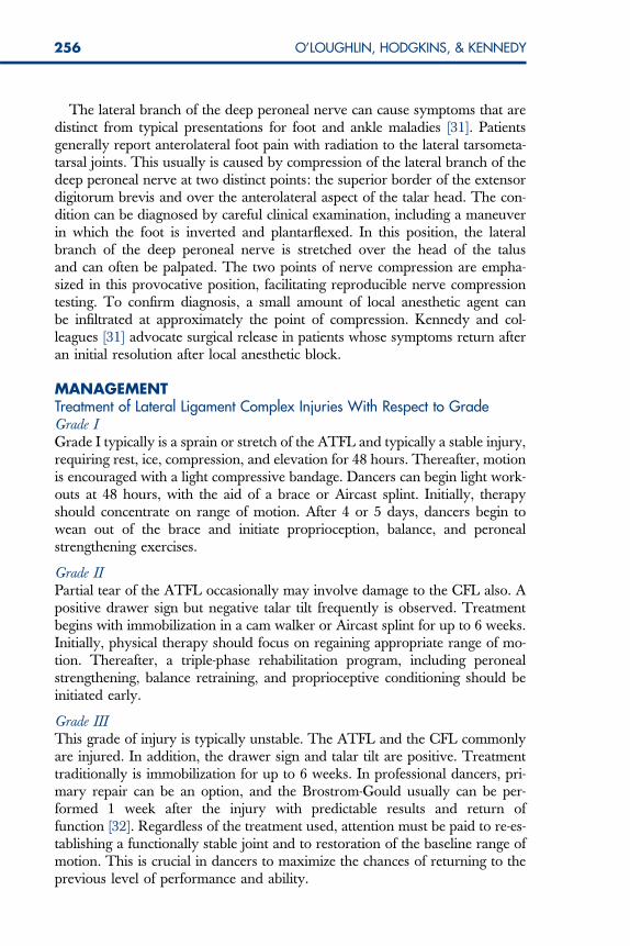
Female ballet dancers frequently adopt the full-pointe position, which requiresmarked plantarflexion with the toes in a neutral position relative to the longi-tudinal axis of the foot (Fig. 1). In full pointe, the ankle is relatively stable, asthe posterior lip of the tibia rests and locks on the calcaneus, and the subtalarjoint is locked with the heel and forefoot in varus. Thus, a midfoot sprain ismore likely in this position than an ankle sprain [19]. With slight dorsiflexion,however, this complex releases, placing greater pressure on the lateral ligamentcomplex and rendering the ankle more vulnerable to an inversion injury. Asthe ankle inverts progressively, the strain on the other ankle ligament increases.In plantarflexion, the ATFL assumes a vertical position while the narrowportion of the talus is in position within the ankle mortise. In this position, thereFig. 1. En pointe.

250 O’LOUGHLIN, HODGKINS, & KENNEDY
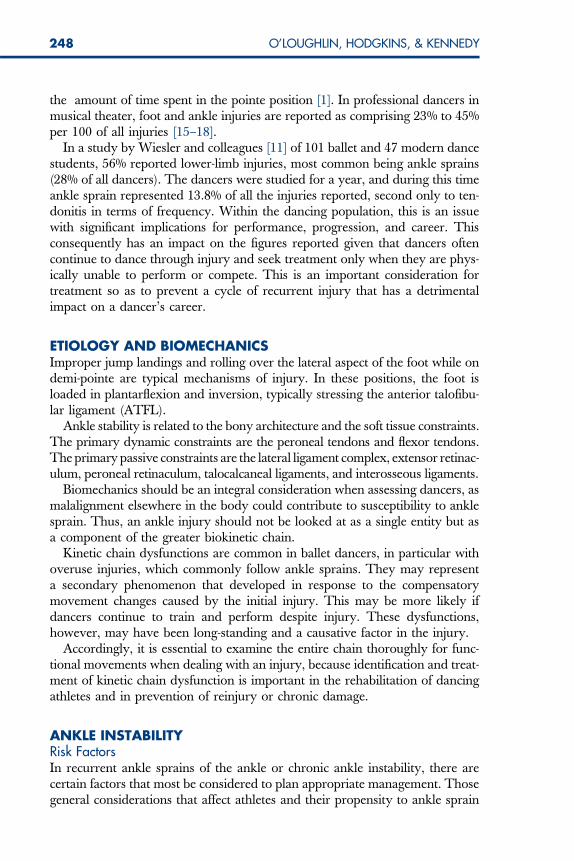
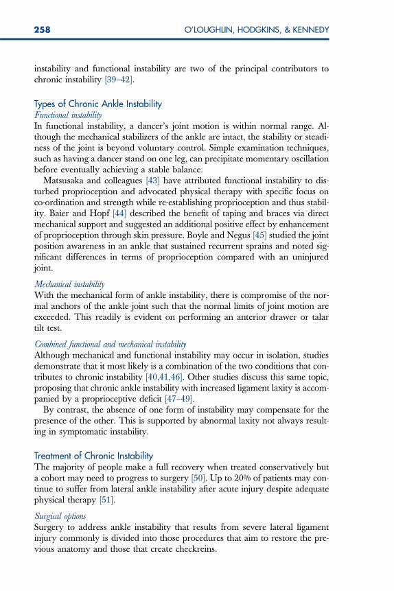
is minimal support offered by the calcaneofibular ligament (CFL) and syndes-motic ligaments. Demi-pointe is a variation of this position (Figs. 2 and 3).
Turnout
In ballet, turnout is an external rotation of the leg, causing the knee and foot toturn outward, away from the center of the body. This rotation allows forgreater extension of the leg, especially when raising it to the side and rear.Turnout is deemed essential to classical ballet technique and is the basis onwhich all ballet movement follows. Thus, ballet dancers are encouraged todevelop this posture.It may cause stress, however, on dancers’ lower back, hip, and lower extrem-ities and put them at risk for injury, including ankle injury. Coplan [20] estab-lished that, based on self-reported history of low back and lower extremityinjuries, ballet dancers have a greater risk for injury if they reach a turnoutposition that is greater than their available bilateral passive hip external rotationrange of motion. Alternatively, Negus and colleagues [21] found, in a study of29 ballet dancers, that 93.1% reported a history of nontraumatic injuries and41.4% a history of traumatic injuries. Number and severity of nontraumaticinjuries were associated with a reduced functional turnout but not with therange of external rotation at the hip. This study was based on the theorythat overuse or nontraumatic dance injuries often are attributed to faults intechnique, with poor turnout and inappropriate compensatory strategies majorfactors. In summary, it is evident that turnout has an important role in dancers’injuries, including insults to the ankle joint.
Fig. 2. En demi-pointe.
Fig. 3. Radiograph of dancer en demi-pointe.
251ANKLE SPRAINS AND INSTABILITY IN DANCERS
Footwear
There is a specific pointe shoe that acts a major additional stabilizer of the footen pointe. This is demonstrated in a cadaveric study [22]. The pointe shoes tra-ditionally have been constructed with layers of paper, glue, and materials, suchas satin, canvas, or leather. This design has not changed dramatically over theyears. Although the shoes are hard to begin with, they are broken in quicklyand become soft and pliable. A principal dancer may use two to three pairsof these shoes per performance [1]. These softened shoes with compromisedsupportive properties themselves may contribute to ankle injury.Thus, dancers’ foot wear should be examined and fitting of dance shoes,especially pointe shoes, should be performed only by an expert who is suitablytrained and who understands the nature and proper fit of the shoes.
LATERAL ANKLE INJURYLateral Ligament ComplexThis is comprised of three major ligaments: the ATFL, CFL, and posteriortalofibular ligament (PTFL). As with other athletes, in dancers, it is theATFL that is injured most frequently.
Anterior talofibular ligament
The ATFL is an intra-articular thickening of the anterolateral portion of thejoint capsule, which is on average 10 mm in length, 8 mm in width, and2 mm in thickness. It is the weakest of the ankle ligaments and, given its orien-tation, the most commonly injured of the ligaments of the lateral ligamentcomplex.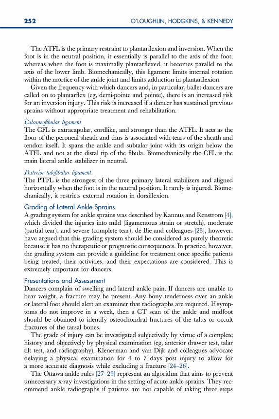
252 O’LOUGHLIN, HODGKINS, & KENNEDY
The ATFL is the primary restraint to plantarflexion and inversion. When thefoot is in the neutral position, it essentially is parallel to the axis of the foot,whereas when the foot is maximally plantarflexed, it becomes parallel to theaxis of the lower limb. Biomechanically, this ligament limits internal rotationwithin the mortice of the ankle joint and limits adduction in plantarflexion.
Given the frequency with which dancers and, in particular, ballet dancers arecalled on to plantarflex (eg, demi-pointe and pointe), there is an increased riskfor an inversion injury. This risk is increased if a dancer has sustained previoussprains without appropriate treatment and rehabilitation.
Calcaneofibular ligament
The CFL is extracapular, cordlike, and stronger than the ATFL. It acts as thefloor of the peroneal sheath and thus is associated with tears of the sheath andtendon itself. It spans the ankle and subtalar joint with its origin below theATFL and not at the distal tip of the fibula. Biomechanically the CFL is themain lateral ankle stabilizer in neutral.Posterior talofibular ligament
The PTFL is the strongest of the three primary lateral stabilizers and alignedhorizontally when the foot is in the neutral position. It rarely is injured. Biome-chanically, it restricts external rotation in dorsiflexion.Grading of Lateral Ankle Sprains
A grading system for ankle sprains was described by Kannus and Renstrom [4],which divided the injuries into mild (ligamentous strain or stretch), moderate(partial tear), and severe (complete tear). de Bie and colleagues [23], however,have argued that this grading system should be considered as purely theoreticbecause it has no therapeutic or prognostic consequences. In practice, however,the grading system can provide a guideline for treatment once specific patientsbeing treated, their activities, and their expectations are considered. This isextremely important for dancers.Presentations and Assessment
Dancers complain of swelling and lateral ankle pain. If dancers are unable tobear weight, a fracture may be present. Any bony tenderness over an ankleor lateral foot should alert an examiner that radiographs are required. If symp-toms do not improve in a week, then a CT scan of the ankle and midfootshould be obtained to identify osteochondral fractures of the talus or occultfractures of the tarsal bones.The grade of injury can be investigated subjectively by virtue of a completehistory and objectively by physical examination (eg, anterior drawer test, talartilt test, and radiography). Klenerman and van Dijk and colleagues advocatedelaying a physical examination for 4 to 7 days post injury to allow fora more accurate diagnosis while excluding a fracture [24–26].
The Ottawa ankle rules [27–29] represent an algorithm that aims to preventunnecessary x-ray investigations in the setting of acute ankle sprains. They rec-ommend ankle radiographs if patients are not capable of taking three steps

253ANKLE SPRAINS AND INSTABILITY IN DANCERS
(limping is permitted) after an injury or if there is tenderness over the midpor-tion or crest of the medial or lateral malleoluls from the tip to 6 cm proximally.Young dancers who have open physes may require radiographs more fre-quently. Radiographic findings include fractures, widening of the ankle mor-tise, avulsion fragments from the malleoli, and osteochondral injuries to thetalar dome or tibial plafond. Attention also should be paid to the lateral processof the talus, the os trigonum, the anterior process of the calcaneus, and theproximal portion of the fifth metatarsal. In dancers, the os is at particularrisk and an MRI often is helpful in delineating an os trigonum from a fracturedos or Stieda lesion, which can present with lateral ankle pain.
MEDIAL ANKLE INJURYAlthough posterior tibial tendon pathology is common in other athletes, it israre in dancers for many reasons. Typically, a dancer’s foot is cavus, whichtends to protect him or her from tibialis posterior pathology as opposed toa more planus foot. Also, when a dancer is in equinus, the posterior tibial ten-don is relatively shortened as the subtalar joint is inverted.
Although these medial injuries occur infrequently in dancers, they typicallyare associated with a pronated foot landing off balance. If a foot is in plantar-flexion, the anterior deltoid ligament is maximally affected, with the tensiongreatest in the deltoid in this position. Similarly, when a foot is flat on theground and hyperpronated, a tear occurs in the midportion of the deltoid.
An accessory bone, the os subtibiale, can be found in the substance of thedeltoid ligament. When injured, it may manifest as a trigger point of painwhen ligamentous healing should be complete. A local injection of a steroidcan be successful in treating this complaint.
Chronic strain of the deltoid ligament from poor form in the rolling in (pro-nation) of the foot is a common overuse injury in dancers. Chronic strain of theanterior aspect of the deltoid ligament, anchored to the capsule of the talonavic-ular joint, may predispose an ankle to chronic rotatory instability.
Recalcitrant medial ankle pain may be caused by an osteochondral lesion ofthe talus after a sprain (Fig. 4). Clinical suspicion warrants further investigationwith CT or MRI to demonstrate the extent of the lesion. The size of the osteo-chondral lesion determines the most appropriate treatment. Microfracturetreatment, chondrocyte transplant, allograft implants, and osteochondral graft-ing are available techniques.
Osteochondral autologous transplant surgery is indicated for large lesions(typically greater than 8 mm) with cartilage collapse or deficit and extensiveunderlying bone necrosis. The lesion is cored out of the talus and filled withosteochondral autograft, commonly from a non–weight-bearing location inthe lateral femoral condyle (Fig. 5).
ASSOCIATED INJURIESInversion injury (Table 1) can cause tearing of the syndesmosis, diastasis of thedistal tibiofibular joint, and a fracture of the proximal fibula (a Maisonneuve
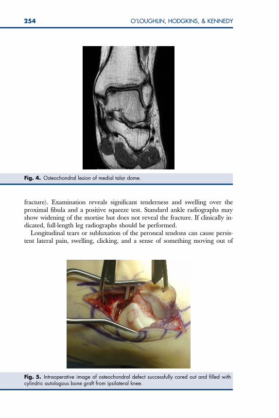
Fig. 4. Osteochondral lesion of medial talar dome.
254 O’LOUGHLIN, HODGKINS, & KENNEDY
fracture). Examination reveals significant tenderness and swelling over theproximal fibula and a positive squeeze test. Standard ankle radiographs mayshow widening of the mortise but does not reveal the fracture. If clinically in-dicated, full-length leg radiographs should be performed.
Longitudinal tears or subluxation of the peroneal tendons can cause persis-tent lateral pain, swelling, clicking, and a sense of something moving out of
Fig. 5. Intraoperative image of osteochondral defect successfully cored out and filled withcylindric autologous bone graft from ipsilateral knee.
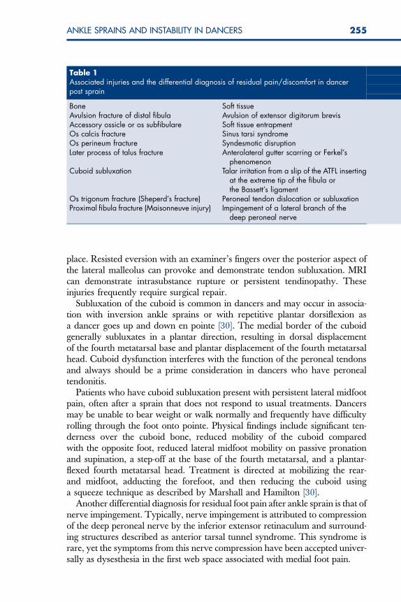
Table 1Associated injuries and the differential diagnosis of residual pain/discomfort in dancerpost sprain
Bone Soft tissueAvulsion fracture of distal fibula Avulsion of extensor digitorum brevisAccessory ossicle or os subfibulare Soft tissue entrapmentOs calcis fracture Sinus tarsi syndromeOs perineum fracture Syndesmotic disruptionLater process of talus fracture Anterolateral gutter scarring or Ferkel’s
phenomenonCuboid subluxation Talar irritation from a slip of the ATFL inserting
at the extreme tip of the fibula orthe Bassett’s ligament
Os trigonum fracture (Sheperd’s fracture) Peroneal tendon dislocation or subluxationProximal fibula fracture (Maisonneuve injury) Impingement of a lateral branch of the
deep peroneal nerve
255ANKLE SPRAINS AND INSTABILITY IN DANCERS
place. Resisted eversion with an examiner’s fingers over the posterior aspect ofthe lateral malleolus can provoke and demonstrate tendon subluxation. MRIcan demonstrate intrasubstance rupture or persistent tendinopathy. Theseinjuries frequently require surgical repair.
Subluxation of the cuboid is common in dancers and may occur in associa-tion with inversion ankle sprains or with repetitive plantar dorsiflexion asa dancer goes up and down en pointe [30]. The medial border of the cuboidgenerally subluxates in a plantar direction, resulting in dorsal displacementof the fourth metatarsal base and plantar displacement of the fourth metatarsalhead. Cuboid dysfunction interferes with the function of the peroneal tendonsand always should be a prime consideration in dancers who have peronealtendonitis.
Patients who have cuboid subluxation present with persistent lateral midfootpain, often after a sprain that does not respond to usual treatments. Dancersmay be unable to bear weight or walk normally and frequently have difficultyrolling through the foot onto pointe. Physical findings include significant ten-derness over the cuboid bone, reduced mobility of the cuboid comparedwith the opposite foot, reduced lateral midfoot mobility on passive pronationand supination, a step-off at the base of the fourth metatarsal, and a plantar-flexed fourth metatarsal head. Treatment is directed at mobilizing the rear-and midfoot, adducting the forefoot, and then reducing the cuboid usinga squeeze technique as described by Marshall and Hamilton [30].
Another differential diagnosis for residual foot pain after ankle sprain is that ofnerve impingement. Typically, nerve impingement is attributed to compressionof the deep peroneal nerve by the inferior extensor retinaculum and surround-ing structures described as anterior tarsal tunnel syndrome. This syndrome israre, yet the symptoms from this nerve compression have been accepted univer-sally as dysesthesia in the first web space associated with medial foot pain.
256 O’LOUGHLIN, HODGKINS, & KENNEDY
The lateral branch of the deep peroneal nerve can cause symptoms that aredistinct from typical presentations for foot and ankle maladies [31]. Patientsgenerally report anterolateral foot pain with radiation to the lateral tarsometa-tarsal joints. This usually is caused by compression of the lateral branch of thedeep peroneal nerve at two distinct points: the superior border of the extensordigitorum brevis and over the anterolateral aspect of the talar head. The con-dition can be diagnosed by careful clinical examination, including a maneuverin which the foot is inverted and plantarflexed. In this position, the lateralbranch of the deep peroneal nerve is stretched over the head of the talusand can often be palpated. The two points of nerve compression are empha-sized in this provocative position, facilitating reproducible nerve compressiontesting. To confirm diagnosis, a small amount of local anesthetic agent canbe infiltrated at approximately the point of compression. Kennedy and col-leagues [31] advocate surgical release in patients whose symptoms return afteran initial resolution after local anesthetic block.
MANAGEMENT
Treatment of Lateral Ligament Complex Injuries With Respect to GradeGrade I Grade I typically is a sprain or stretch of the ATFL and typically a stable injury,requiring rest, ice, compression, and elevation for 48 hours. Thereafter, motionis encouraged with a light compressive bandage. Dancers can begin light work-outs at 48 hours, with the aid of a brace or Aircast splint. Initially, therapyshould concentrate on range of motion. After 4 or 5 days, dancers begin towean out of the brace and initiate proprioception, balance, and peronealstrengthening exercises.Grade II
Partial tear of the ATFL occasionally may involve damage to the CFL also. Apositive drawer sign but negative talar tilt frequently is observed. Treatmentbegins with immobilization in a cam walker or Aircast splint for up to 6 weeks.Initially, physical therapy should focus on regaining appropriate range of mo-tion. Thereafter, a triple-phase rehabilitation program, including peronealstrengthening, balance retraining, and proprioceptive conditioning should beinitiated early.Grade III
This grade of injury is typically unstable. The ATFL and the CFL commonlyare injured. In addition, the drawer sign and talar tilt are positive. Treatmenttraditionally is immobilization for up to 6 weeks. In professional dancers, pri-mary repair can be an option, and the Brostrom-Gould usually can be per-formed 1 week after the injury with predictable results and return offunction [32]. Regardless of the treatment used, attention must be paid to re-es-tablishing a functionally stable joint and to restoration of the baseline range ofmotion. This is crucial in dancers to maximize the chances of returning to theprevious level of performance and ability.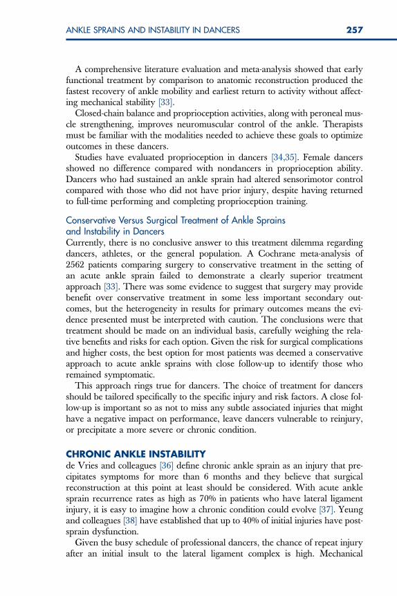
257ANKLE SPRAINS AND INSTABILITY IN DANCERS
A comprehensive literature evaluation and meta-analysis showed that earlyfunctional treatment by comparison to anatomic reconstruction produced thefastest recovery of ankle mobility and earliest return to activity without affect-ing mechanical stability [33].
Closed-chain balance and proprioception activities, along with peroneal mus-cle strengthening, improves neuromuscular control of the ankle. Therapistsmust be familiar with the modalities needed to achieve these goals to optimizeoutcomes in these dancers.
Studies have evaluated proprioception in dancers [34,35]. Female dancersshowed no difference compared with nondancers in proprioception ability.Dancers who had sustained an ankle sprain had altered sensorimotor controlcompared with those who did not have prior injury, despite having returnedto full-time performing and completing proprioception training.
Conservative Versus Surgical Treatment of Ankle Sprainsand Instability in Dancers
Currently, there is no conclusive answer to this treatment dilemma regardingdancers, athletes, or the general population. A Cochrane meta-analysis of2562 patients comparing surgery to conservative treatment in the setting ofan acute ankle sprain failed to demonstrate a clearly superior treatmentapproach [33]. There was some evidence to suggest that surgery may providebenefit over conservative treatment in some less important secondary out-comes, but the heterogeneity in results for primary outcomes means the evi-dence presented must be interpreted with caution. The conclusions were thattreatment should be made on an individual basis, carefully weighing the rela-tive benefits and risks for each option. Given the risk for surgical complicationsand higher costs, the best option for most patients was deemed a conservativeapproach to acute ankle sprains with close follow-up to identify those whoremained symptomatic.This approach rings true for dancers. The choice of treatment for dancersshould be tailored specifically to the specific injury and risk factors. A close fol-low-up is important so as not to miss any subtle associated injuries that mighthave a negative impact on performance, leave dancers vulnerable to reinjury,or precipitate a more severe or chronic condition.
CHRONIC ANKLE INSTABILITYde Vries and colleagues [36] define chronic ankle sprain as an injury that pre-cipitates symptoms for more than 6 months and they believe that surgicalreconstruction at this point at least should be considered. With acute anklesprain recurrence rates as high as 70% in patients who have lateral ligamentinjury, it is easy to imagine how a chronic condition could evolve [37]. Yeungand colleagues [38] have established that up to 40% of initial injuries have post-sprain dysfunction.
Given the busy schedule of professional dancers, the chance of repeat injuryafter an initial insult to the lateral ligament complex is high. Mechanical
258 O’LOUGHLIN, HODGKINS, & KENNEDY
instability and functional instability are two of the principal contributors tochronic instability [39–42].
Types of Chronic Ankle InstabilityFunctional instability
In functional instability, a dancer’s joint motion is within normal range. Al-though the mechanical stabilizers of the ankle are intact, the stability or steadi-ness of the joint is beyond voluntary control. Simple examination techniques,such as having a dancer stand on one leg, can precipitate momentary oscillationbefore eventually achieving a stable balance.Matsusaka and colleagues [43] have attributed functional instability to dis-turbed proprioception and advocated physical therapy with specific focus onco-ordination and strength while re-establishing proprioception and thus stabil-ity. Baier and Hopf [44] described the benefit of taping and braces via directmechanical support and suggested an additional positive effect by enhancementof proprioception through skin pressure. Boyle and Negus [45] studied the jointposition awareness in an ankle that sustained recurrent sprains and noted sig-nificant differences in terms of proprioception compared with an uninjuredjoint.
Mechanical instability
With the mechanical form of ankle instability, there is compromise of the nor-mal anchors of the ankle joint such that the normal limits of joint motion areexceeded. This readily is evident on performing an anterior drawer or talartilt test.Combined functional and mechanical instability
Although mechanical and functional instability may occur in isolation, studiesdemonstrate that it most likely is a combination of the two conditions that con-tributes to chronic instability [40,41,46]. Other studies discuss this same topic,proposing that chronic ankle instability with increased ligament laxity is accom-panied by a proprioceptive deficit [47–49].By contrast, the absence of one form of instability may compensate for thepresence of the other. This is supported by abnormal laxity not always result-ing in symptomatic instability.
Treatment of Chronic Instability
The majority of people make a full recovery when treated conservatively buta cohort may need to progress to surgery [50]. Up to 20% of patients may con-tinue to suffer from lateral ankle instability after acute injury despite adequatephysical therapy [51].Surgical options
Surgery to address ankle instability that results from severe lateral ligamentinjury commonly is divided into those procedures that aim to restore the pre-vious anatomy and those that create checkreins.
259ANKLE SPRAINS AND INSTABILITY IN DANCERS
Those that seek to restore correct anatomy involve direct ligament repairand include the Brostrom and modified Brostrom procedures [8,32]. Thecheckrein procedures involve tendon transfers and include the Elmslie (fascialata) [52], Watson-Jones (peroneus brevis) [53,54], Evans (peroneus brevis)[55], Chrisman-Snook [56], and Larsen (peroneus brevis) procedures [57].
Hamilton and colleagues [32] have described the efficacy of the modifiedBrostrom technique in restoring function and stability in dancers. In addition,retrospective comparative studies have suggested that anatomic restorationshows superior results in the long term [58,59]. Hennrikus and colleagues[46] have shown a significantly higher rate of nerve damage in the Chris-man-Snook procedure.
Specific treatment considerations with respect to dancers
Early and aggressive treatment of ankle sprains is essential in dancers. Dancingen pointe necessitates maximal mobility of all joints of the lower leg, ankle, andfoot, with restricted motion leading to difficulty with technique, further injury,and, ultimately, implications for a dancer’s career.Swelling contributes to loss of motion and should be minimized. Simple tech-niques may be used, such as use of icing and compression taping with horse-shoe pads. The subtalar joint and the talocalcaneal ligament frequently areinjured in inversion sprains. Subtalar sprains are linked to chronic givingway and limitations of ankle motion [60], which can result in significant disabil-ity for dancers. Range-of-motion exercises, therefore, are initiated as early aspossible to maintain and restore mobility of the ankle and subtalar joints.
Strength training with specific emphasis on the everters should be performedin the neutral position and, crucially, with the foot in the plantarflexed, pointeposition. Flexibility of the gastrocnemius and soleus muscles is critical. Func-tional mobility drills should be used first at the barre and subsequently onthe center of the dance floor before returning to full dance.
SUMMARYAggressive treatment of the sprained ankle is essential to maintaining foot andankle mobility and preventing prolonged disability and subsequent overuseinjuries.
References
[1] Kadel NJ. Foot and ankle injuries in dance. Phys Med Rehabil Clin N Am 2006;17:813–26, vii.[2] Balduini FC, Vegso JJ, Torg JS, et al. Management and rehabilitation of ligamentous injuries
to the ankle. Sports Med 1987;4:364–80.[3] Karlsson J, Sancone M. Management of acute ligament injuries of the ankle. Foot Ankle Clin
2006;11:521–30.[4] Kannus P, Renstrom P. Treatment for acute tears of the lateral ligaments of the ankle. Oper-
ation, cast, or early controlled mobilization. J Bone Joint Surg Am 1991;73:305–12.[5] Brostrom L. Sprained ankles. 3. Clinical observations in recent ligament ruptures. Acta Chir
Scand 1965;130:560–9.

260 O’LOUGHLIN, HODGKINS, & KENNEDY
[6] Brostrom L, Sundelin P. Sprained ankles. IV. Histologic changes in recent and ‘‘chronic’’ligament ruptures. Acta Chir Scand 1966;132:248–53.
[7] Brostrom L. Sprained ankles. V. Treatment and prognosis in recent ligament ruptures. ActaChir Scand 1966;132:537–50.
[8] Brostrom L. Sprained ankles. VI. Surgical treatment of ‘‘chronic’’ ligament ruptures. ActaChir Scand 1966;132:551–65.
[9] Lynch SA, Renstrom PA. Treatment of acute lateral ankle ligament rupture in the athlete.Conservative versus surgical treatment. Sports Med 1999;27:61–71.
[10] Nilsson C, Leanderson J, Wykman A, et al. The injury panorama in a Swedish professionalballet company. Knee Surg Sports Traumatol Arthrosc 2001;9:242–6.
[11] Wiesler ER, Hunter DM, Martin DF, et al. Ankle flexibility and injury patterns in dancers. Am JSports Med 1996;24:754–7.
[12] Garrick JG, Requa RK. Ballet injuries. An analysis of epidemiology and financial outcome.Am J Sports Med 1993;21:586–90.
[13] Byhring S, Bo K. Musculoskeletal injuries in the Norwegian National Ballet: a prospectivecohort study. Scand J Med Sci Sports 2002;12:365–70.
[14] Bronner S, Ojofeitimi S, Rose D. Injuries in a modern dance company: effect of comprehen-sive management on injury incidence and time loss. Am J Sports Med 2003;31:365–73.
[15] Washington EL. Musculoskeletal injuries in theatrical dancers: site frequency, and severity.Am J Sports Med 1978;6:75–98.
[16] Rovere GD, Webb LX, Gristina AG, et al. Musculoskeletal injuries in theatrical dancestudents. Am J Sports Med 1983;11:195–8.
[17] Evans RW, Evans RI, Carvajal S. Survey of injuries among West End performers. OccupEnviron Med 1998;55:585–93.
[18] Evans RW, Evans RI, Carvajal S, et al. A survey of injuries among Broadway performers.Am J Public Health 1996;86:77–80.
[19] Macintyre J, Joy E. Foot and ankle injuries in dance. Clin Sports Med 2000;19:351–68.[20] Coplan JA. Ballet dancer’s turnout and its relationship to self-reported injury. J Orthop Sports
Phys Ther 2002;32:579–84.[21] Negus V, Hopper D, Briffa NK. Associations between turnout and lower extremity injuries in
classical ballet dancers. J Orthop Sports Phys Ther 2005;35:307–18.[22] Kadel N, Boenisch M, Teitz C, et al. Stability of Lisfranc joints in ballet pointe position. Foot
Ankle Int 2005;26:394–400.[23] de Bie RA, de Vet HC, van den Wildenberg FA, et al. The prognosis of ankle sprains. Int J
Sports Med 1997;18:285–9.[24] Klenerman L. The management of sprained ankle. J Bone Joint Surg Br 1998;80:11–2.[25] van Dijk CN, Lim LS, Bossuyt PM, et al. Physical examination is sufficient for the diagnosis of
sprained ankles. J Bone Joint Surg Br 1996;78:958–62.[26] van Dijk CN, Mol BW, Lim LS, et al. Diagnosis of ligament rupture of the ankle joint. Physical
examination, arthrography, stress radiography and sonography compared in 160 patientsafter inversion trauma. Acta Orthop Scand 1996;67:566–70.
[27] Pigman EC, Klug RK, Sanford S, et al. Evaluation of the Ottawa clinical decision rules for theuse of radiography in acute ankle and midfoot injuries in the emergency department: anindependent site assessment. Ann Emerg Med 1994;24:41–5.
[28] Stiell IG, Greenberg GH, McKnight RD, et al. Decision rules for the use of radiography inacute ankle injuries. Refinement and prospective validation. JAMA 1993;269:1127–32.
[29] Stiell IG, McKnight RD, Greenberg GH, et al. Implementation of the Ottawa ankle rules.JAMA 1994;271:827–32.
[30] Marshall P, Hamilton WG. Cuboid subluxation in ballet dancers. Am J Sports Med1992;20:169–75.
[31] Kennedy JG, Brunner JB, Bohne WH, et al. Clinical importance of the lateral branch of thedeep peroneal nerve. Clin Orthop Relat Res 2007;459:222–8.
261ANKLE SPRAINS AND INSTABILITY IN DANCERS
[32] Hamilton WG, Thompson FM, Snow SW. The modified Brostrom procedure for lateral ankleinstability. Foot Ankle 1993;14:1–7.
[33] Kerkhoffs GM, Handoll HH, de Bie R, et al. Surgical versus conservative treatment for acuteinjuries of the lateral ligament complex of the ankle in adults. Cochrane Database Syst Rev2007;(2):CD000380.
[34] Leanderson J, Eriksson E, Nilsson C, et al. Proprioception in classical ballet dancers. A pro-spective study of the influence of an ankle sprain on proprioception in the ankle joint. Am JSports Med 1996;24:370–4.
[35] Schmitt H, Kuni B, Sabo D. Influence of professional dance training on peak torque andproprioception at the ankle. Clin J Sport Med 2005;15:331–9.
[36] de Vries JS, Krips R, Sierevelt IN, et al. Interventions for treating chronic ankle instability.Cochrane Database Syst Rev 2006;(4):CD004124.
[37] McKay GD, Goldie PA, Payne WR, et al. Ankle injuries in basketball: injury rate and riskfactors. Br J Sports Med 2001;35:103–8.
[38] Yeung MS, Chan KM, So CH, et al. An epidemiological survey on ankle sprain. Br J SportsMed 1994;28:112–6.
[39] Freeman MA. Instability of the foot after injuries to the lateral ligament of the ankle. J BoneJoint Surg Br 1965;47:669–77.
[40] Hertel J. Functional anatomy, pathomechanics, and pathophysiology of lateral ankle insta-bility. J Athl Train 2002;37:364–75.
[41] Tropp H, Odenrick P, Gillquist J. Stabilometry recordings in functional and mechanical insta-bility of the ankle joint. Int J Sports Med 1985;6:180–2.
[42] Hubbard TJ, Kramer LC, Denegar CR, et al. Contributing factors to chronic ankle instability.Foot Ankle Int 2007;28:343–54.
[43] Matsusaka N, Yokoyama S, Tsurusaki T, et al. Effect of ankle disk training combined withtactile stimulation to the leg and foot on functional instability of the ankle. Am J SportsMed 2001;29:25–30.
[44] Baier M, Hopf T. Ankle orthoses effect on single-limb standing balance in athletes withfunctional ankle instability. Arch Phys Med Rehabil 1998;79:939–44.
[45] Boyle J, Negus V. Joint position sense in the recurrently sprained ankle. Aust J Physiother1998;44:159–63.
[46] Hennrikus WL, Mapes RC, Lyons PM, et al. Outcomes of the Chrisman-Snook and modified-Brostrom procedures for chronic lateral ankle instability. A prospective, randomizedcomparison. Am J Sports Med 1996;24:400–4.
[47] Halasi T, Kynsburg A, Tallay A, et al. Changes in joint position sense after surgically treatedchronic lateral ankle instability. Br J Sports Med 2005;39:818–24.
[48] LarsenE.Static ordynamic repairof chronic lateral ankle instability.Aprospective randomizedstudy. Clin Orthop Relat Res 1990;257:184–92.
[49] Rosenbaum D, Engelhardt M, Becker HP, et al. Clinical and functional outcome after ana-tomic and nonanatomic ankle ligament reconstruction: Evans tenodesis versus periostealflap. Foot Ankle Int 1999;20:636–9.
[50] Kerkhoffs GM, Handoll HH, de Bie R, et al. Surgical versus conservative treatment for acuteinjuries of the lateral ligament complex of the ankle in adults. Cochrane Database Syst Rev2002;(3):CD000380.
[51] Karlsson J, Eriksson BI, Sward L. Early functional treatment for acute ligament injuries of theankle joint. Scand J Med Sci Sports 1996;6:341–5.
[52] Elmslie RC. Recurrent subluxation of the ankle-joint. Ann Surg 1934;100:364–7.[53] Gillespie HS, Boucher P. Watson-Jones repair of lateral instability of the ankle. J Bone Joint
Surg Am 1971;53:920–4.[54] Brattstrom H. Tenodesis employing the method of Watson-Jones for the treatment of recurrent
subluxation of the ankle. Acta Orthop Scand 1953;23:132–6.[55] Evans DL. Recurrent instability of the ankle; a method of surgical treatment. Proc R Soc Med
1953;46:343–4.

262 O’LOUGHLIN, HODGKINS, & KENNEDY
[56] Chrisman OD, Snook GA. Reconstruction of lateral ligament tears of the ankle. An experi-mental study and clinical evaluation of seven patients treated by a new modification of theElmslie procedure. J Bone Joint Surg Am 1969;51:904–12.
[57] Larsen E. Tendon transfer for lateral ankle and subtalar joint instability. Acta Orthop Scand1988;59:168–72.
[58] Krips R, Brandsson S, Swensson C, et al. Anatomical reconstruction and Evans tenodesis ofthe lateral ligaments of the ankle. Clinical and radiological findings after follow-up for 15 to30 years. J Bone Joint Surg Br 2002;84:232–6.
[59] Krips R, van Dijk CN, Halasi T, et al. Anatomical reconstruction versus tenodesis for thetreatment of chronic anterolateral instability of the ankle joint: a 2- to 10-year follow-up,multicenter study. Knee Surg Sports Traumatol Arthrosc 2000;8:173–9.
[60] Tochigi Y, Yoshinaga K, Wada Y, et al. Acute inversion injury of the ankle: magneticresonance imaging and clinical outcomes. Foot Ankle Int 1998;19:730–4.
Related Documents